EP3390672B1 - Detektion und quantifizierung einer zielnukleinsäuresequenz eines mikroorganismus - Google Patents
Detektion und quantifizierung einer zielnukleinsäuresequenz eines mikroorganismus Download PDFInfo
- Publication number
- EP3390672B1 EP3390672B1 EP16876172.4A EP16876172A EP3390672B1 EP 3390672 B1 EP3390672 B1 EP 3390672B1 EP 16876172 A EP16876172 A EP 16876172A EP 3390672 B1 EP3390672 B1 EP 3390672B1
- Authority
- EP
- European Patent Office
- Prior art keywords
- sequence
- nucleotides
- seq
- ebv
- microorganism
- Prior art date
- Legal status (The legal status is an assumption and is not a legal conclusion. Google has not performed a legal analysis and makes no representation as to the accuracy of the status listed.)
- Active
Links
Images


Classifications
-
- C—CHEMISTRY; METALLURGY
- C12—BIOCHEMISTRY; BEER; SPIRITS; WINE; VINEGAR; MICROBIOLOGY; ENZYMOLOGY; MUTATION OR GENETIC ENGINEERING
- C12Q—MEASURING OR TESTING PROCESSES INVOLVING ENZYMES, NUCLEIC ACIDS OR MICROORGANISMS; COMPOSITIONS OR TEST PAPERS THEREFOR; PROCESSES OF PREPARING SUCH COMPOSITIONS; CONDITION-RESPONSIVE CONTROL IN MICROBIOLOGICAL OR ENZYMOLOGICAL PROCESSES
- C12Q1/00—Measuring or testing processes involving enzymes, nucleic acids or microorganisms; Compositions therefor; Processes of preparing such compositions
- C12Q1/70—Measuring or testing processes involving enzymes, nucleic acids or microorganisms; Compositions therefor; Processes of preparing such compositions involving virus or bacteriophage
- C12Q1/701—Specific hybridization probes
-
- C—CHEMISTRY; METALLURGY
- C12—BIOCHEMISTRY; BEER; SPIRITS; WINE; VINEGAR; MICROBIOLOGY; ENZYMOLOGY; MUTATION OR GENETIC ENGINEERING
- C12Q—MEASURING OR TESTING PROCESSES INVOLVING ENZYMES, NUCLEIC ACIDS OR MICROORGANISMS; COMPOSITIONS OR TEST PAPERS THEREFOR; PROCESSES OF PREPARING SUCH COMPOSITIONS; CONDITION-RESPONSIVE CONTROL IN MICROBIOLOGICAL OR ENZYMOLOGICAL PROCESSES
- C12Q1/00—Measuring or testing processes involving enzymes, nucleic acids or microorganisms; Compositions therefor; Processes of preparing such compositions
- C12Q1/68—Measuring or testing processes involving enzymes, nucleic acids or microorganisms; Compositions therefor; Processes of preparing such compositions involving nucleic acids
- C12Q1/6876—Nucleic acid products used in the analysis of nucleic acids, e.g. primers or probes
- C12Q1/6888—Nucleic acid products used in the analysis of nucleic acids, e.g. primers or probes for detection or identification of organisms
- C12Q1/689—Nucleic acid products used in the analysis of nucleic acids, e.g. primers or probes for detection or identification of organisms for bacteria
-
- C—CHEMISTRY; METALLURGY
- C12—BIOCHEMISTRY; BEER; SPIRITS; WINE; VINEGAR; MICROBIOLOGY; ENZYMOLOGY; MUTATION OR GENETIC ENGINEERING
- C12Q—MEASURING OR TESTING PROCESSES INVOLVING ENZYMES, NUCLEIC ACIDS OR MICROORGANISMS; COMPOSITIONS OR TEST PAPERS THEREFOR; PROCESSES OF PREPARING SUCH COMPOSITIONS; CONDITION-RESPONSIVE CONTROL IN MICROBIOLOGICAL OR ENZYMOLOGICAL PROCESSES
- C12Q1/00—Measuring or testing processes involving enzymes, nucleic acids or microorganisms; Compositions therefor; Processes of preparing such compositions
- C12Q1/68—Measuring or testing processes involving enzymes, nucleic acids or microorganisms; Compositions therefor; Processes of preparing such compositions involving nucleic acids
- C12Q1/6876—Nucleic acid products used in the analysis of nucleic acids, e.g. primers or probes
- C12Q1/6888—Nucleic acid products used in the analysis of nucleic acids, e.g. primers or probes for detection or identification of organisms
- C12Q1/6895—Nucleic acid products used in the analysis of nucleic acids, e.g. primers or probes for detection or identification of organisms for plants, fungi or algae
-
- C—CHEMISTRY; METALLURGY
- C12—BIOCHEMISTRY; BEER; SPIRITS; WINE; VINEGAR; MICROBIOLOGY; ENZYMOLOGY; MUTATION OR GENETIC ENGINEERING
- C12Q—MEASURING OR TESTING PROCESSES INVOLVING ENZYMES, NUCLEIC ACIDS OR MICROORGANISMS; COMPOSITIONS OR TEST PAPERS THEREFOR; PROCESSES OF PREPARING SUCH COMPOSITIONS; CONDITION-RESPONSIVE CONTROL IN MICROBIOLOGICAL OR ENZYMOLOGICAL PROCESSES
- C12Q2600/00—Oligonucleotides characterized by their use
- C12Q2600/112—Disease subtyping, staging or classification
-
- C—CHEMISTRY; METALLURGY
- C12—BIOCHEMISTRY; BEER; SPIRITS; WINE; VINEGAR; MICROBIOLOGY; ENZYMOLOGY; MUTATION OR GENETIC ENGINEERING
- C12Q—MEASURING OR TESTING PROCESSES INVOLVING ENZYMES, NUCLEIC ACIDS OR MICROORGANISMS; COMPOSITIONS OR TEST PAPERS THEREFOR; PROCESSES OF PREPARING SUCH COMPOSITIONS; CONDITION-RESPONSIVE CONTROL IN MICROBIOLOGICAL OR ENZYMOLOGICAL PROCESSES
- C12Q2600/00—Oligonucleotides characterized by their use
- C12Q2600/118—Prognosis of disease development
-
- C—CHEMISTRY; METALLURGY
- C12—BIOCHEMISTRY; BEER; SPIRITS; WINE; VINEGAR; MICROBIOLOGY; ENZYMOLOGY; MUTATION OR GENETIC ENGINEERING
- C12Q—MEASURING OR TESTING PROCESSES INVOLVING ENZYMES, NUCLEIC ACIDS OR MICROORGANISMS; COMPOSITIONS OR TEST PAPERS THEREFOR; PROCESSES OF PREPARING SUCH COMPOSITIONS; CONDITION-RESPONSIVE CONTROL IN MICROBIOLOGICAL OR ENZYMOLOGICAL PROCESSES
- C12Q2600/00—Oligonucleotides characterized by their use
- C12Q2600/158—Expression markers
Definitions
- the present disclosure relates to molecular biology in particular biomarkers.
- the present disclosure relates to the detection and quantification of biomarkers associated with microorganisms such as bacteria, viruses, fungi and parasites, and methods of determining the likelihood that a patient suffers or is likely to suffer from diseases associated with microorganisms, and to predict the treatment outcome of a patient having diseases associated with microorganisms, by detecting and quantifying the biomarkers associated with the microorganisms.
- Infections with microorganisms have been recognized as risk factors for a wide range of diseases in humans, such as cancers, autoimmune diseases and cardiovascular diseases, which are associated with high mortality rates.
- diseases in humans such as cancers, autoimmune diseases and cardiovascular diseases, which are associated with high mortality rates.
- Early detection of the causative microorganisms is crucial for the prevention, detection, diagnosis and treatment of these diseases associated with infections with microorganisms.
- Examples of conventional methods used for the detection of microorganisms include culture-based procedures, serologic tests and microscopy. Each of these methods is associated with its own limitations. For example, culture-based procedures, which are dependent on the growth of the microorganisms, often takes days for results to become available, and such procedures often reveal false negative results due to the administration of an empiric antibiotic therapy. Serologic tests often face high false negative rate due to the absence or weakness of antibody production in the patient's body, or high false positive rate due to the presence of cross-reacting antibodies. Microscopy techniques may be limited by sample staining and fixing and the use of strong illumination, which may destroy or distort cellular features of the microorganisms to be detected.
- a method for detecting and/or quantifying the presence of a target nucleic acid sequence of a microorganism in a sample obtained from a subject comprising amplifying the target sequence in a CpG island of the nucleic acid of the microorganism, irrespective of the methylation status of the CpG island.
- a method for detecting and/or quantifying the presence of a target nucleic acid sequence of Epstein-Barr virus (EBV) in a sample obtained from a subject comprising amplifying a target sequence in the BamHI-W region of EBV, wherein the target sequence comprises the sequence of AGATCTAAGGCCGGGAGAGGCAGCCCCAAAGCGGGTGCAGTAACAGGTAATCTC TGGTAGTGATTTGGACCCGAAATCTGACACTTTAGAGCTCTGGAGGACTTTAAAA CTCTAAAAATCAAAACTTTAGAGGCGAATGGGCG (SEQ ID NO: 1), wherein amplifying the target sequence comprises the use of a pair of oligonucleotide primers and a probe, wherein the first oligonucleotide primer comprises the sequence of 5'-AGATCTAAGGCCGGGAGAGG-3' (SEQ ID NO:2), and the second oligonucleotide primer comprises the sequence of 5'-CGCCCATTCG
- a method of detecting a disease associated with microorganism infection, or risk of developing a disease associated with microorganism infection in a subject comprising detecting and/or quantifying the presence of a nucleic acid sequence of the microorganism using the method of the present invention in a sample obtained from the subject, wherein the presence of the nucleic acid sequence of the microorganism in the sample indicates that the subject has a disease associated with microorganism infection or is at risk of developing a disease associated with microorganism infection.
- a method of detecting and treating a disease associated with microorganism infection comprising: (i) detecting and/or quantifying the presence of a nucleic acid sequence of the microorganism using the method of the present invention in a sample obtained from the subject, wherein the presence of the nucleic acid sequence of the microorganism in the sample indicates that the subject has a disease associated with microorganism; and (ii) administering to the subject a medicament suitable for the treatment of the disease associated with the microorganism.
- a method of predicting the treatment outcome of a disease associated with microorganism infection in a patient comprising:(i) quantifying the nucleic acid sequence of the microorganism in a sample collected from the patient before treatment or before a treatment step, and quantifying the nucleic acid sequence of the microorganism in a sample collected from the same patient after treatment or after a treatment step; (ii) comparing the amount of the nucleic acid sequence of the microorganism in the sample before and after treatment or a treatment step, wherein a decrease in the amount of the nucleic acid sequence of the microorganism in the sample after treatment or a treatment step indicates that treatment outcome of the disease associated with microorganism infection in the patient is positive, wherein the quantifying of the nucleic acid sequence of the microorganism in the sample is performed according to the method of the present invention.
- kits for detecting and/or quantifying the nucleic acid sequence of a microorganism in a sample obtained from a subject comprising a pair of oligonucleotide primers specific for the amplification of a target sequence in a CpG island of the nucleic acid of the microorganism.
- Fig. 1 depicts the overall survival curves of patients with nasopharyngeal carcinoma (NPC) with NPC levels measured using different circulating biomarkers. Results are shown using Kaplan-Meier plots for dichotomized biomarker variables.
- Fig. 1A shows the overall survival curves of NPC patients with EBV measured by the number of copies of Bam HI-W per ml using qPCR.
- Fig. 1B shows the overall survival curves of NPC patients with EBV measured by the number of copies of EBNA1 per ml using qPCR.
- Fig. 1A shows the overall survival curves of NPC patients with EBV measured by the number of copies of Bam HI-W per ml using qPCR.
- Fig. 1B shows the overall survival curves of NPC patients with EBV measured by the number of copies of EBNA1 per ml using qPCR.
- Fig. 1A shows the overall survival curves of NPC patients with EBV measured by the number of copies
- FIG. 1C shows the overall survival curves of NPC patients with EBV measured by the number of copies of EBNA1 per ml using dPCR.
- Fig. ID and IE show the overall survival curves of NPC patients with NPC level measured in terms of the counts of circulating tumor cells (CTCs). The results show that the levels of Bam HI-W (detected using dPCR) and EBNA1 (detected using dPCR or qPCR) serve as better indicators of the overall survival rate as compared to CTCs.
- Fig. 2 is a flow chart depicting the overall results of detecting NPC using different EBV biomarkers. A total of 46 NPC patients, all of Asian ethnicity, took part in the study.
- BamHI-W qPCR assay detected 41 patients as NPC positive and 5 patients as NPC negative
- EBNA1 qPCR assay detected 31 patients as NPC positive and 15 patients as NPC negative
- EBNA1 dPCR assay detected 39 patients as NPC positive and 7 patients as NPC negative.
- the results indicate that the Bam HI-W qPCR assay has better sensitivity for the detection of NPC as compared to EBNA1 qPCR and EBNA1 dPCR assays.
- 3 is a flow chart depicting the process of designing and selecting the synthetic internal control.
- 50 random sequences with the length of 150 bases were generated using R program. Sequences with multiple base repeats and runs (i.e. ATATAT, CCCCCC, etc.) were eliminated. 15 sequences passed this criterion. Primers and probes were designed for these 15 sequences such that the target sequences are about 100bp and located in the central region of the 150-base sequences. Primer-dimer and primer-probe dimer formation were checked by NetPrimer program. The ⁇ G values between all the primers and probe (in both Internal Control (IC) and Bam HI-W reaction) must be more positive than -9 kcal/mole (see Table 1).
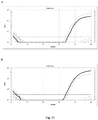
- FIG. 4 depicts the detection of EBV using Bam HI-W qPCR assay, in plasma samples with and without the internal control (IC) plasmid.
- X-axis represents the copy number of Bam HI-W detected
- Y-axis represents the threshold cycle (Ct) which is the intersection between an amplification curve and a threshold line.
- Ct threshold cycle
- Fig. 5 illustrates the different experimental settings used to test the impact of the internal control (IC) plasmid on the detection of EBV using Bam HI-W qPCR assay.
- FIG. 6A and 6B indicate positive results for EBV detection using Bam HI-W qPCR assay.
- Fig. 6C indicates true negative result for EBV detection using Bam HI-W qPCR assay, as the IC plasmid was amplified by the qPCR assay.
- Fig. 6D indicates false negative result for EBV detection using Bam HI-W qPCR assay, as the IC plasmid was not amplified by the qPCR assay. Over all, it can be concluded that IC can serve as a control to identify a false negative detection of Bam HI-W.
- Fig. 7 depicts the construction of a Bam HI-W standard plasmid and an internal control (IC) plasmid.
- FIG. 8 shows the confirmation of the Bam HI-W sequence in the Bam HI-W standard plasmid and the internal control (IC) sequence in the IC plasmid constructed as depicted in Fig. 7 .
- Fig. 8A shows the sequence of the BamHI-W standard forward strand.
- Fig. 8B shows the sequence of the IC forward strand.
- Fig. 8C shows the sequence of the Bam HI-W standard reverse strand.
- Fig. 8D shows the sequence of the IC reverse strand.
- Fig. 9 shows the results of plasmid validation in clinical samples using Bam HI-W qPCR assay.
- Fig. 9A shows the results where the internal control (IC) plasmids were added in the plasma sample from the patient prior to DNA extraction.
- Fig. 9B shows the results of amplification of the constructed Bam HI-W standard plasmids added directly to PCR plates.
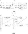
- Fig. 9A shows the derivation of conversion factor between Bam HI-W standard plasmids constructed and the International Standard (IU) of EBV defined by the National Institute for Biological Standards and Control (NIBSC).
- Fig. 10A shows the plot of the number of copies of Bam HI-W plasmids added in the sample (y-axis) against the number of IUs defined by NIBSC (x-axis), both in log scale.
- FIG. 10B shows the plot of the number of copies of Bam HI-W plasmids added in the sample (y-axis) against the number of IUs defined by NIBSC (x-axis).
- Fig. 11 shows the PCR amplification curves of the target sequences of Mycobacterium tuberculosis.
- the target sequence is nucleotide sequence from nucleotide position 1542511 to nucleotide position 1542349 of the genomic sequence of Mycobacterium tuberculosis.
- Fig. 11A and 11C the target sequence is nucleotide sequence from nucleotide position 1542511 to nucleotide position 1542349 of the genomic sequence of Mycobacterium tuberculosis.
- Fig. 11A and 11C the target sequence is nucleot
- the target sequence is nucleotide sequence from nucleotide position 1542328 to nucleotide position 1542215 of the genomic sequence of Mycobacteriaum tuberculosis.
- Fig. 11A and 11B represent the amplification curve using a sample from a patient known to have tuberculosis
- 11C and 11D represent the amplification curve using a sample from a healthy subject control. The results indicate that both target sequences of Mycobacterium tuberculosis are present in the sample from the patient with tuberculocis, but absent in the sample from the healthy subject control.
- FIG. 12 shows a gel electrophoresis photo of using different sets of Bam HI-W primers directed to different target sequences for the amplification of Bam HI-W.
- primer pair 2 The sequences of primer pair 2 are: forward primer 5'-GGAATAAGCCCCCAGACAGG-3' (SEQ ID NO:12), reverse primer 5'- TTACGTAAACGCGCTGGACT-3' (SEQ ID NO.: 14); the sequences of primer pair 7 are: forward primer 5'-AGATCTAAGGCCGGGAGAGG-3' (SEQ ID NO:2), reverse primer 5'-CGCCCATTCGCCTCTAAAGT-3' (SEQ ID NO: 3); the sequences of primer pair 10 are: forward primer 5'- AGGAAGCGGGTCTATGGTTG-3' (SEQ ID NO:13), reverse primer 5'-GACTGAGAAGGTGGCCTAGC-3' (SEQ ID NO:15). The results indicate that all three sets of primers can be used for the successful detection of Bam HI-W.
- the present disclosure provides a method of detecting and/or quantifying a microorganism that has better sensitivity and/or specificity compared to the currently available methods.
- a method for detecting and/or quantifying the presence of a target nucleic acid sequence of a microorganism in a sample obtained from a subject comprising amplifying the target sequence in a CpG island of the nucleic acid of the microorganism, irrespective of the methylation status of the CpG island.
- amplifying or “amplification” as used herein refers to the production of additional copies of the target sequence.
- CpG island refers to nucleic acid regions with a high frequency of CpG sites.
- a CpG site (or CG site) is a region of DNA where a cytosine nucleotide is followed by a guanine nucleotide in the linear sequence of nucleotides along its 5' to 3' direction.
- CpG is shorthand for 5'-C-phosphate-G-3'.
- a CpG island can have at least about 100 nucleotides, or at least about 500 nucleotides, or at least about 1000 nucleotides, or at least about 2000 nucleotides, or at least about 3000 nucleotides, or at least about 4000 nucleotides, or at least about 5000 nucleotides, or at least about 6000 nucleotides, or at least about 7000 nucleotides, or at least about 8000 nucleotides, or at least about 9000 nucleotides, or at least about 10000 nucleotides, or at least about 15000 nucleotides, or at least about 20000 nucleotides, or at least about 25000 nucleotides, or at least about 30000 nucleotides, or at least about 35000 nucleotides, or at least about 40000 nucleotides, or at least about 45000 nucleotides, or at least about 50000 nucleotides, or at least about 60000 nucleotides,
- a CpG island usually has a C, G percentage of greater than about 50%, or greater than about 51%, or greater than about 52%, or greater than about 53%, or greater than about 54%, or greater than about 55%, or greater than about 56%, or greater than about 57%, or greater than about 58%, or greater than about 59%, or greater than about 60%, or greater than about 61%, or greater than about 62%, or greater than about 63%, or greater than about 64%, or greater than about 65%, or greater than about 66%, or greater than about 67%, or greater than about 68%, or greater than about 69%, or greater than about 70%, or greater than about 71%, or greater than about 72%, or greater than about 73%, or greater than about 74%, or greater than about 75%, or greater than about 76%, or greater than about 77%, or greater than about 78%, or greater than about 79%, or greater than about 80%.
- the observed-to-expected CpG ratio in CpG island is usually greater than about 55%, or greater than about 56%, or greater than about 57%, or greater than about 58%, or greater than about 59%, or greater than about 60%, or greater than about 61%, or greater than about 62%, or greater than about 63%, or greater than about 64%, or greater than about 65%, or greater than about 66%, or greater than about 67%, or greater than about 68%, or greater than about 69%, or greater than about 70%, or greater than about 71%, or greater than about 72%, or greater than about 73%, or greater than about 74%, or greater than about 75%, or greater than about 76%, or greater than about 77%, or greater than about 78%, or greater than about 79%, or greater than about 80%, or greater than about 81%, or greater than about 82%, or greater than about 83%, or greater than about 84%, or greater than about 85%, or greater than about 86%, or greater than about 87%, or greater than about 88%, or greater
- the "observed-to-expected CpG ratio" can be derived where the "observed” is the actual number of CpGs in the sequence, and where the “expected” is calculated as: actual number of C ⁇ actual number of G / length of sequence or as : or as: actual number of C + actual number of G / 2 2 / length of sequence .
- a number of softwares or analytical tools can be used for the prediction of CpG island in a nucleic acid sequence.
- analytical tools available online include but are not limited to: Sequence Manipulation Suite available at http://www.bioinformatics.org/sms2/cpg_islands.html; and Emboss Cpgplot available at http://www.ebi.ac.uk/Tools/seqstats/emboss_cpgplot/.
- target sequence or “target nucleic acid sequence” or their grammatical variants as used herein refer to a region of the nucleic acid sequence of the microorganism of interest to be amplified.
- the presence of the target sequence in a sample obtained from a subject indicates the presence of the nucleic acid sequence associated with the microorganism of interest, and/or the presence of the microorganism in the sample or in the subject.
- the target sequence to be amplified for the detection and/or quantification of the microorganism is located within the 5'end of the CpG island.
- the 5' end location would allow for preferential preservation during exonuclease III degradation of the DNA.
- the target sequence is located within the first 50%, or the first 49%, or the first 48%, or the first 47%, or the first 46%, or the first 45%, or the first 44%, or the first 43%, or the first 42%, or the first 41%, or the first 40%, or the first 39%, or the first 38%, or the first 37%, or the first 36%, or the first 35%, or the first 34%, or the first 33%, or the first 32%, or the first 31%, or the first 30%, or the first 29%, or the first 28%, or the first 27%, or the first 26%, or the first 25%, or the first 24%, or the first 23%, or the first 22%, or the first 21%, or the first 20%, or the first 19%, or the first 18%, or the first 17%, or the first 16%, or the first 15%, or the first 14%, or the first 13%, or the first 12%, or the first 11%, or the first 10%, or the first 9%
- methylation refers to DNA methylation which typically occurs at a CpG site. Such methylation results in the conversion of the cytosine to 5-methylcytosine, and can by catalysed by the enzyme DNA methyltransferase.
- a CpG cite can be either methylated or unmethylated.
- the method of for detecting and/or quantifying the presence of a target nucleic acid sequence of a microorganism as provided in the present disclosure comprises amplifying the target sequence in a CpG island of the nucleic acid of the microorganism, irrespective of the methylation status of the CpG island. This means that the methylation status of the CpG island for which the target sequence lies within is not important.
- One of the advantages provided by such a method is that no methylation-status-specific sequencing will be required to select the target sequence to be amplified.
- microorganism refers to a microscopic living organism, which may be single-celled or multicellular.
- the microorganism contains DNAs, thus microorganisms that do not contain DNAs (e.g. RNA viruses) are excluded.
- microorganisms include but are not limited to bacteria, DNA viruses, fungi and parasites.
- the microorganisms are bacteria and DNA viruses.
- DNA viruses include but are not limited to DNA viruses with double stranded DNAs and DNA viruses with single stranded DNAs.
- the microorganisms are pathogenic.
- pathogenic refers to the ability of the microorganisms to cause diseases.
- diseases include but are not limited to acinetobacter infections, Actinomycosis, African sleeping sickness (African trypanosomiasis), AIDS (Acquired immunodeficiency syndrome), Amebiasis, Anaplasmosis, Angiostrongyliasis, Anisakiasis, Anthrax, Arcanobacterium haemolyticum infection, Argentine hemorrhagic fever, Ascariasis, Aspergillosis, Astrovirus infection, Alzheimer's disease, Amyotrophic lateral sclerosis, Anorexia nervosa, Anxiety disorder, Asthma, Atherosclerosis, Autoimmune diseases, Babesiosis, Bacillus cereus infection, Bacterial pneumonia, Bacterial vaginosis, Bacteroides infection, Balantidiasis, Bart
- Epstein-Barr virus also called human herpesvirus 4 (HHV-4).
- EBV Epstein-Barr virus
- HHV-4 human herpesvirus 4
- EBV is one of the most common viruses in humans, and is commonly transmitted by saliva and established latent infection in B lymphocytes where it persists for the lifetime of the host.
- EBV infection is associated with particular forms of cancer, such as nasopharyngeal carcinoma, gastric cancer, Hodgkin's lymphoma and Burkitt's lymphoma.
- the target sequence to be amplified for the detection of EBV comprises a sequence within a CpG island of the Bam HI-W region of EBV.
- Bam HI-W region refers to a repeating Bam HI-W restriction fragment of the EBV genome.
- Bam HI-W is a 3-kb long sequence and the genome of an EBV typically contains six to twenty copies of the Bam HI-W sequence.
- the Bam HI-W has the following sequence: >J02072.1 Epstein Barr Virus large internal repeat ( Bam HI-W fragment)
- EBV Bam HI-W fragment is important to its expression as a W promoter that drives expression of the EB viral nuclear antigens (EBNAs) at the initiation of virus-induced B-cell transformation.
- EBNAs EB viral nuclear antigens
- the role of methylation in EBV activity has also been recognized in human tissue. Pharmacologic reversal of dense CpG methylation in tumor tissue can be achieved in patients undergoing treatment with DNA methyltransferase inhibitor.
- the CpG island of EBV Bam HI-W has been shown to be unmethylated in the latent cycle of EBV.
- the method as disclosed herein can be used for the detection and/or quantification of such a target sequence.
- the CpG island of the Bam HI-W region of EBV is selected from the group consisting of: CTCCTCTCCAACCTTCGCTCCACCCTAGACCCCAGCTTCTGGCCTCCCCGGGTCCACCAGGCCAGCCGGAGGGACCCCGGCAGCCCGGGCGAGTCGCCTTCCCTCTCCCCTGGCCTCTCCTTCCCGCCTCCCACCCGAGCCCCCTCAGCTTGCCTCCCCACCGGGTCCATCAGGCCGGCCGGAGGGACCCCGGCGGCCCGGTGTCA (SEQ ID NO.: 6)(nucleotides at position number 36 to 241 of Bam HI-W region), AGGCCATGCGCCCTGTCACCAGGCCTGCCAAAGAGCCAGATCTAAGGCCGGGAGAGGCAGCCCCAAAGCGGGTGCAGTAACAGGTAATCTCTGGTAGTGATTTGGACCCGAAATCTGACACTTTAGAGCTCTGGAGGACTTTAAAACTCTAAAAATCAAAACTTTAGAGGCGAATGGGCGCCATTTTG
- the target sequence within the CpG island comprises the sequence selected from the group consisting of: AGATCTAAGGCCGGGAGAGGCAGCCCCAAAGCGGGTGCAGTAACAGGTAATCTCTGGTAGTGATTTGGACCCGAAATCTGACACTTTAGAGCTCTGGAGGACTTTAAAACTCTAAAAATCAAAACTTTAGAGGCGAATGGGCG (SEQ ID NO.: 1) (nucleotides at position number 730 to 872 of Bam HI-W region), GGAATAAGCCCCCAGACAGGGGAGTGGGCTTGTTTGTGACTTCACCAAAGGTCAGGGCCCAAGGGGGTTCGCGTTGCTAGGCCACCTTCTCAGTCCAGCGCGTTTACGTAA (SEQ ID NO.: 10) (nucleotides at position number 976 to 1086 of Bam HI-W region), AGGAAGCGGGTCTATGGTTGGCTGCGCTGCTGCTATCTTTAGAGGGGAAAAGAGGAATAAGCCCCCAGACAGGGGAGTGGG
- fragment refers to a nucleic acid sequence that is a constituent of the reference, or a constituent of the complementary sequence of the reference sequence.
- a fragment comprises about 9 to about 300 nucleotides, or about 10 to about 290 nucleotides, or about 20 to about 280 nucleotides, or about 30 to about 270 nucleotides, or about 40 to about 260 nucleotides, or about 50 to about 250 nucleotides, or about 60 to about 240 nucleotides, or about 70 to about 230 nucleotides, or about 80 to about 220 nucleotides, or about 90 to about 210 nucleotides, or about 100 to about 200 nucleotides, or about 110 to about 190 nucleotides, or about 120 to about 180 nucleotides, or about 130 to about 170 nucleotides, or about 140 to about 160 nucleotides, or 10 nucleotides, 15 nucleotides, 25
- a fragment can also comprise about 5% to about 95%, about 10% to about 90%, about 15% to about 85%, about 20% to about 80%, about 25% to about 75%, about 30% to about 70%, about 35% to about 65%, about 40% to about 60%, about 45% to about 55%, or about 50% of the reference sequence or the complementary sequence of the reference sequence.
- variant refers to sequences that are substantially similar to the reference sequence. These nucleotide sequence variants may have at least 25%, 30%, 35%, 40%, 45%, 50%, 55%, 60%, 65%, 70%, 75%, 80%, 85%, 90%, 95%, 96%, 97%, 98% or 99% sequence identity to the "non-variant" reference sequence. Variants may be a result of substitution, deletion or additional of any number of nucleotides in the refernce sequence as a result of the mutation of the wild type DNA .
- the amplification of the target sequence comprises the use of at least one oligonucleotide primer capable of binding to the target sequence.
- the at least one oligonucleotide primer binds within a region from about 9 to about 300 nucleotides, or about 10 to about 290 nucleotides, or about 20 to about 280 nucleotides, or about 30 to about 270 nucleotides, or about 40 to about 260 nucleotides, or about 50 to about 250 nucleotides, or about 60 to about 240 nucleotides, or about 70 to about 230 nucleotides, or about 80 to about 220 nucleotides, or about 90 to about 210 nucleotides, or about 100 to about 200 nucleotides, or about 110 to about 190 nucleotides, or about 120 to about 180 nucleotides, or about 130 to about 170 nucleotides, or about 140 to about 160 nucleotides, from the first nucleotide of
- the length of the at least one primer is between 5 to 40 nucleotides, or between 10 to 35 nucleotides, or between 15 to 30 nucleotides, or between 20 to 25 nucleotides, or 8 nucleotides, or 9 nucleotides, or 10 nucleotides, or 12 nucleotides, or 14 nucleotides, or 16 nucleotides, or 18 nucleotides, or 20 nucleotides, or 22 nucleotides, or 24 nucleotides, or 26 nucleotides, or 28 nucleotides, or 30 nucleotides, or 32 nucleotides, or 34 nucleotides, or 36 nucleotides, or 38 nucleotides, or 40 nucleotides.
- the length of the at least one primer is 20 nucleotides.
- the at least one primer has a sequence identity of at least 70%, at least 75%, at least 80%, at least 85%, at least 90%, at least 95%, at least 96%, at least 97%, at least 98% or at least 99% to at least one sequence selected from the group consisting of AGATCTAAGGCCGGGAGAGG (SEQ ID NO.: 2), GGAATAAGCCCCCAGACAGG (SEQ ID NO.: 12), AGGAAGCGGGTCTATGGTTG (SEQ ID NO.: 13), CGCCCATTCGCCTCTAAAGT (SEQ ID NO.: 3), TTACGTAAACGCGCTGGACT (SEQ ID NO.: 14), and GACTGAGAAGGTGGCCTAGC (SEQ ID NO.: 15), or a complementary sequence thereof.
- AGATCTAAGGCCGGGAGAGG SEQ ID NO.: 2
- GGAATAAGCCCCCAGACAGG SEQ ID NO.: 12
- AGGAAGCGGGTCTATGGTTG SEQ ID NO.: 13
- the primer comprises one sequence selected from the group consisting of AGATCTAAGGCCGGGAGAGG (SEQ ID NO.: 2), GGAATAAGCCCCCAGACAGG (SEQ ID NO.: 12), AGGAAGCGGGTCTATGGTTG (SEQ ID NO.: 13), CGCCCATTCGCCTCTAAAGT (SEQ ID NO.: 3), TTACGTAAACGCGCTGGACT (SEQ ID NO.: 14), and GACTGAGAAGGTGGCCTAGC (SEQ ID NO.: 15), or a complementary sequence thereof.
- the amplification of the target sequence comprises the use of at least one pair of oligonucleotide primers.
- the at least one pair of oligonucleotide primers can comprise one forward primer and one reverse primer.
- the at least one pair of oligonucleotide primers can be selected from the group consisting of: forward primer AGATCTAAGGCCGGGAGAGG (SEQ ID NO.: 2) and reverse primer CGCCCATTCGCCTCTAAAGT (SEQ ID NO.: 3); forward primer GGAATAAGCCCCCAGACAGG (SEQ ID NO.: 12) and reverse primer TTACGTAAACGCGCTGGACT (SEQ ID NO.: 14); forward primer AGGAAGCGGGTCTATGGTTG (SEQ ID NO.: 13) and reverse primer GACTGAGAAGGTGGCCTAGC (SEQ ID NO.: 15).
- the at least one pair of oligonucleotide primers are forward primer AGATCTAAGGCCGGGAGAGG (SEQ ID NO.: 2) and reverse primer CGCCCATTCGCCTCTAAAGT (SEQ ID NO.: 3).
- the amplification of the target sequence comprises the use of a probe capable of binding to the target sequence.
- the probe binds within a region from between 9 to 500 nucleotides, or from between 10 to 490 nucleotides, or from between 20 to 480 nucleotides, or from between 30 to 470 nucleotides, or from between 40 to 460 nucleotides, or from between 50 to 450 nucleotides, or from between 60 to 440 nucleotides, or from between 70 to 430 nucleotides, or from between 80 to 420 nucleotides, or from between 90 to 410 nucleotides, or from between 100 to 400 nucleotides, or from between 110 to 390 nucleotides, or from between 120 to 380 nucleotides, or from between 130 to 370 nucleotides, or from between 140 to 360 nucleotides, or from between 150 to 350 nucleotides, or from between 160 to 340 nucleotides, or from between between
- the specific nucleotide can be nucleotide at position number 1, 5, 10, 15, 20, 25, 30, 35, 40, 45, 50, 52, 54, 56, 58, 60, 62, 64, 66, 68, 70, 72, 74, 76, 78, 80, 82, 84, 86, 88, 90, 92, 94, 96, 98, 100, 105, 110, 115, 120, 125, 130, 135, 140, 145, 150, 155, 160, 165, 170, 175, 180, 185, 190, 195, 200, 210, 220, 230, 240, 250, 260, 270, 280, 290, 300, 310, 320, 330, 340, 350, 360, 370, 380, 390, 400, 410, 420, 430, 440, 450, 460, 470, 480, 490 or 500 of the target sequence.
- the length of a probe can be between 5 to 40 nucleotides, or between 10 to 35 nucleotides, or between 15 to 30 nucleotides, or between 20 to 25 nucleotides, or 8 nucleotides, or 9 nucleotides, or 10 nucleotides, or 12 nucleotides, or 14 nucleotides, or 16 nucleotides, or 18 nucleotides, or 20 nucleotides, or 22 nucleotides, or 24 nucleotides, or 26 nucleotides, or 28 nucleotides, or 30 nucleotides, or 32 nucleotides, or 34 nucleotides, or 36 nucleotides, or 38 nucleotides, or 40 nucleotides.
- the probe has a sequence identity of at least 70%, at least 75%, at least 80%, at least 85%, at least 90%, at least 95%, at least 96%, at least 97%, at least 98% or at least 99% to a sequence selected from the group consisting of CTCTGGTAGTGATTTGGACCCGAAATCTG (SEQ ID NO.: 16), CCACCTTCTCAGTCCAGCGCGTTT (SEQ ID NO.: 17), GTGACTTCACCAAAGGTCAGGGCCC (SEQ ID NO.: 18), GGTGGTAAGCGGTTCACCTTCAGGG (SEQ ID NO.: 19), and complementary sequences thereof.
- the probe comprises the sequence selected from the group consisting of CTCTGGTAGTGATTTGGACCCGAAATCTG (SEQ ID NO.: 16), CCACCTTCTCAGTCCAGCGCGTTT (SEQ ID NO.: 17), GTGACTTCACCAAAGGTCAGGGCCC (SEQ ID NO.: 18), GGTGGTAAGCGGTTCACCTTCAGGG (SEQ ID NO.: 19) and complementary sequences thereof.
- the probe comprises the sequence of CTCTGGTAGTGATTTGGACCCGAAATCTG (SEQ ID NO.: 16).
- the amplification of the target sequence comprises the use of at least one oligonucleotide primer and one probe as defined above. In another example, the amplification of the target sequence comprises the use of at least one pair of oligonucleotide primers and one probe as defined above.
- a method for detecting and/or quantifying the presence of a target nucleic acid sequence of Epstein-Barr virus (EBV) in a sample obtained from a subject comprising amplifying a target sequence in the Bam HI-W region of EBV, wherein the target sequence comprises the sequence of AGATCTAAGGCCGGGAGAGGCAGCCCCAAAGCGGGTGCAGTAACAGGTAATCTC TGGTAGTGATTTGGACCCGAAATCTGACACTTTAGAGCTCTGGAGGACTTTAAAA CTCTAAAAATCAAAACTTTAGAGGCGAATGGGCG (SEQ ID NO.: 1), wherein amplifying the target sequence comprises the use of a pair of oligonucleotide primers and a probe, wherein the first oligonucleotide primer comprises the sequence of 5'-AGATCTAAGGCCGGGAGAGG-3' (SEQ ID NO.: 2), and the second oligonucleotide primer comprises the sequence of 5'-CGCC
- the method of detecting and/or quantifying the presence of a target nucleic acid sequence of Epstein-Barr virus (EBV) in a sample obtained from a subject further comprises amplifying a control.
- EBV Epstein-Barr virus
- control or "internal control” as used herein refers to a reference sequence which can be used to indicate whether the amplification system is functioning.
- control includes a target sequence of TTAGCAGCGACGAAGATCATGCGCTCACGCTCGGTGTCCTCATTCATCAGTTA TTCACAACGCTATGCTGTAACTCGACCTGACAAGACTGTACCTATGAGAAGGCA CTTGCTACCTTATGCAAGCGTCAGCCCGCGGTATCGCTTGG (SEQ ID NO.: 80), a complementary sequence, a fragment or a variant thereof.
- amplifying the control includes the use of a pair of oligonucleotide primers.
- a forward primer sequence is 5'-CGCTCTCGGTGTCCTCATTC-3' (SEQ ID NO.: 81), a complementary sequence, a fragment or a variant thereof.
- a reverse primer sequence is 5'-GGCTGACGCTTGCATAAGGT-3' (SEQ ID NO.: 82), a complementary sequence, a fragment or a variant thereof.
- amplifying the control further includes the use of a probe capable of binding to the target sequence of the control.
- the probe comprises the sequence of CACAACGCTATGCTGTAACTCGACCTGAC (SEQ ID NO.: 89), a complementary sequence, a fragment or a variant thereof.
- a probe is 5'-VIC-CACAACGCTATGCTGTAACTCGACCTGAC-TAMRA-3' (SEQ ID NO.: 83).
- the microorganism of which a target nucleic acid sequence is to be detected and/or quantified using the method as disclosed herein is a bacterium.
- a bacterium is Mycobacterium tuberculosis.
- CpG island serial no Start position (nucleotide number) End position (nucleotide number) 1 250 485,607 2 486,395 598,721 3 598,929 889,723 4 890,230 1,103,130 5 1,103,439 1,691,493 6 1,691,745 1,697,185 7 1,697,464 1,779,283 8 1,779,789 2,268,374 9 2,268,597 2,367,062 10 2,367,073 2,680,742 11 2,681,249 2,807,592 12 2,807,742 3,587,732 13 3,588,238 3,791,614 14 3,791,647 3,792,550 15 3,793,135 3,798,679 16 3,798,717 3,964,564 17 3,965,008 4,411,
- target sequences to be used for the detection and/or quantification of Mycobacterium tuberculosis are:
- the above identified target sequences in the genomic nucleic acid sequence of Mycobacterium tuberculosis are derived from whole genome sequencing from the plasma sample of a patient who has pulmonary tuberculosis. It is worth noting that it is generally unexpected to detect tuberculosis DNA signal from circulating plasma through whole genome sequencing. It is also to be noted that all of these target sequences fall within the above 17 CpG islands identified for Mycobacterium tuberculosis.
- primers that can be used for the amplification of a target sequence in the CpG island of the genomic nucleic acid sequence of Mycobacterium tuberculosis include but are not limited to: forward primer GGCTGTGGGTAGCAGACC (SEQ ID NO.: 74) and ACCTGAAAGACGTTATCCACCAT (SEQ ID NO.: 75), reverse primer CGGGTCCAGATGGCTTGC (SEQ ID NO.: 76) and CGGCTAGTGCATTGTCATAGGA (SEQ ID NO.: 77).
- the amplification of a target sequence in the CpG island of the genomic nucleic acid sequence of Mycobacterium tuberculosis may also comprise the use of a probe.
- the probe used comprises the sequence of TGTCGACCTGGGCAGGGTTCG (SEQ ID NO.: 78) and TCCGACCGCGCTCCGACCGACG (SEQ ID NO.: 79).
- the probe used comprises a component comprises at least one detectable label.
- the detectable label is capable of producing an optical signal.
- the detectable label comprises a fluorophore.
- fluorophores include but are not limited to fluorescent proteins, for example GFP (green fluorescent protein), YFP (yellow fluorescent protein), RFP (red fluorescent protein); non-protein fluorophores selected from the group consisting of xanthene derivatives (for example, fluorescein, rhodamine, Oregon green, eosin, 6-carboxyfluorescein and Texas red); cyanine derivatives (for example, cyanine, indocarbocyanine, oxacarbocyanine, thiacarbocyanine, and merocyanine), squaraine derivatives and ring-substituted squaraines, including Seta, SeTau, and Square dyes, naphthalene derivatives (dansyl and prodan derivatives), cousine, and red
- the fluorophore is selected from the group consisting of FAM (carboxyfluorescein), TET (carboxy-2',4,7,7' - tetrachlorofluorescein succinimidyl ester), HEX (carboxy-2,4,4,5,7,7 -hexachlorofluorescein succinimidyl ester), ROX (carboxy-X-rhodamine) and NED.
- the fluorophore is FAM (carboxyfluorescein), or 6-FAM (6-carboxyfluorescein).
- the at least one detectable label is capable of producing a changeable signal.
- the changeable signal may be produced upon the hybridization of the probe to the target sequence.
- the signal may be detectable before the probe binds to the target sequence, and upon the hybridization of the probe to the target sequence, the signal is reduced in strength or becomes completely undetectable.
- the detectable signal may be produced only upon the hybridization of the probe to the target sequence, or the strength of the detectable signal may be increased upon the hybridization of the probe to the target sequence.
- the component comprises two detectable labels.
- the two detectable labels function independently, while in another example, the two detectable labels are an interactive pair of labels.
- the interactive pair of labels are capable of generating a changeable signal.
- the signal may be detectable before the probe binds to the target sequence, and upon the hybridization of the probe to the target sequence, the signal is reduced in strength or becomes completely undetectable.
- the detectable signal may be produced only upon the hybridization of the probe to the target sequence, or the strength of the detectable signal may be increased upon the hybridization of the probe to the target sequence.
- the detectable signal is not generated when both detectable labels are linked together by the probe sequence. Once at least one detectable label is cleaved from the probe, the detectable signal is generated.
- the interactive pair of labels may comprise a fluorophore and a quencher pair.
- the fluorophore is located at the 5' end of the probe, and the quencher is located at the 3'end of the probe.
- quenchers include but are not limited to TAMRA (tetramethylrhodamine), TaqMan® MGB (minor groove binder) and BHQTM (Black Hole QuencherTM).
- the fluorophore is FAM (carboxyfluorescein), more particularly 6-FAM (6-carboxyfluorescein), and the quencher is TAMRA (tetramethylrhodamine).
- PCR polymerase chain reaction
- examples of PCRs include but are not limited to real-time polymerase chain reaction, digital polymerase chain reaction, quantitative polymerase chain reaction, qualitative polymerase chain reaction, quantitative real-time polymerase chain reaction, or quantitative reverse transcription polymerase chain reaction.
- Real-time PCR monitors the amplification of a targeted DNA molecule during the PCR, i.e. in real-time, and not at its end, as in conventional PCR.
- Real-time PCR can be used quantitatively (Quantitative real-time PCR), and semi-quantitatively, i.e. above/below a certain amount of DNA molecules (Semi quantitative real-time PCR).
- Quantitative Real-Time PCR (qrt-PCR) methods use fluorescent dyes or fluorophore-containing DNA probes to measure the amount of amplified product as the amplification progresses.
- Digital PCR simultaneously amplifies thousands of samples, each in a separate droplet within an emulsion.
- Quantitative PCR is used to measure the specific amount of target DNA (or RNA) in a sample. By measuring amplification only within the phase of true exponential increase, the amount of measured product more accurately reflects the initial amount of target. Special thermal cyclers are used that monitor the amount of product during the amplification.
- Qualitative PCR refers to a PCR method used to detect the present or absence of target DNA (RNA) in a sample without quantifying the amount present.
- Reverse Transcription PCR is used to reverse-transcribe and amplify RNA to cDNA. PCR is preceded by a reaction using reverse transcriptase, an enzyme that converts RNA into cDNA. The two reactions may be combined in a tube, with the initial heating step of PCR being used to inactivate the transcriptase.
- RT-PCR is widely used in expression profiling, which detects the expression of a gene. It can also be used to obtain sequence of an RNA transcript, which may aid the determination of the transcription start and termination sites and facilitate mapping of the location of exons and introns in a gene sequence.
- the amplified product obtained using the methods described above can be purified and the resulting purified product can be quantified using conventional nucleic acid purification methods and quantification methods.
- nucleic acid purification methods include but are not limited to gel electrophoresis followed by gel extraction, and silica based membrane technologies.
- methods to quantify the purified nucleic acid include but are not limited to spectrophotometric analysis and analysis using fluorescent dye tagging.
- kits and systems are commercially available for the purification and quantification of amplified, nucleic acid products.
- the methods described herein have better sensitivity and specificity compared to the currently known methods of detecting microorganisms.
- the lowest concentration of EBVs in a sample that can be detected using the methods described herein is 100 International Unity (IU)/ml or sample, or 90 IU/ml of sample, or 80 IU/ml of sample, or 70 IU/ml of sample, or 60 IU/ml of sample, or 50 IU/ml of sample, or 40 IU/ml of sample, or 30 IU/ml of sample, or 20 IU/ml of sample, or 10 IU/ml of sample, or 5 IU/ml of sample, or 1 IU/ml of sample.
- IU International Unity
- IU International Unit
- NIBSC National Institute for Biological Standards and Control
- the genome of an EBV typically contains six to twenty copies of the Bam HI-W sequence. Therefore higher sensitivity can be achieved when the detection of EBV is based on the detection of the Bam HI-W region.
- the variability of Bam HI-W copy numbers in different EBV strains has been considered as a challenged in assay comparison and standardization between laboratories. Therefore, a new method of standardizing the number of copies of Bam HI-W to the amount of EBV in a sample has been developed in the present disclosure to solve this problem.
- the sequence of Bam HI-W standard was incorporated as an insert in plasmid which was propagated in competent bacteria cells, such as E.coli. Single colony was picked and grown for scale-up production of the plasmids. Bacteria were harvested and plasmids were extracted and quantified.
- the Bam HI-W standard plasmid obtained carries only Bam HI-W sequence of EBV whereas NIBSC standard is the whole genome sequence of EBV.
- the presence of other genes in EBV genome might interfere with the amplification of Bam HI-W or generate higher background signals as compared to the Bam HI-W standard plasmid.
- the Bam HI-W standard plasmid obtained carries one Bam HI-W copy, allowing absolute quantification of Bam HI-W. This is not feasible in the case of NIBSC standards because number of Bam HI-W copies is unknown in EBV genome.
- the method of detecting and/or quantifying a target nucleic acid sequence of the EBV can be used alone or in combination with other available methods of detecting and/or quantifying EBV.
- a method of detecting a disease associated with microorganism infection, or risk of developing a disease associated with microorganism infection in a subject comprising detecting and/or quantifying the presence of a nucleic acid sequence of the microorganism using the method of the present invention in a sample obtained from the subject, wherein the presence of the nucleic acid sequence of the microorganism in the sample indicates that the subject has a disease associated with microorganism infection or is at risk of developing a disease associated with microorganism infection.
- a method of detecting and treating a disease associated with microorganism infection comprising: (i) detecting and/or quantifying the presence of a nucleic acid sequence of the microorganism using the method of the present invention in a sample obtained from the subject, wherein the presence of the nucleic acid sequence of the microorganism in the sample indicates that the subject has a disease associated with microorganism; (ii) administering to the subject a medicament suitable for the treatment of the disease associated with the microorganism.
- a method of predicting the treatment outcome of a disease associated with microorganism infection in a patient comprising: (i) quantifying the nucleic acid sequence of the microorganism in a sample collected from the patient before treatment or before a treatment step, and quantifying the nucleic acid sequence of the microorganism in a sample collected from the same patient after treatment or after a treatment step; (ii) comparing the amount of the nucleic acid sequence of the microorganism in the sample before and after treatment or a treatment step, wherein a decrease in the amount of the nucleic acid sequence of the microorganism in the sample after treatment or a treatment step indicates that treatment outcome of the disease associated with microorganism infection in the patient is positive, wherein the quantifying of the nucleic acid sequence of the microorganism in the sample is performed according to the method of the present invention.
- disease associated with microorganism infection refers to any disease that can be caused by a microorganism, in particular a pathogenic microorganism as described herein.
- the disease is EBV-associated disease, in particular EBV-associated cancers.
- EBV-associated cancers include but are not limited to nasopharyngeal carcinoma (NPC), gastric cancer, Hodgkin's lymphoma and Burkitt's lymphoma.
- NPC nasopharyngeal carcinoma
- gastric cancer Hodgkin's lymphoma
- Burkitt's lymphoma nasopharyngeal carcinoma
- the EBV-associated cancer is NPC.
- sample refers to a biological sample, or a sample that comprises at least some biological materials such as nucleic acid molecules, more particularly cell free DNAs (cfDNAs).
- the biological samples may include liquid samples, such as whole blood, blood serum, blood plasma, buffy coat, peripheral blood mononuclear cells (PBMCs), cerebrospinal fluid, central spinal fluid, lymph fluid, cystic fluid, sputum, stool, pleural effusion, mucus, pleural fluid, ascitic fluid, amniotic fluid, peritoneal fluid, saliva, bronchial washes and urine.
- PBMCs peripheral blood mononuclear cells
- cerebrospinal fluid cerebrospinal fluid
- central spinal fluid lymph fluid
- cystic fluid sputum
- stool pleural effusion
- mucus mucus
- pleural fluid ascitic fluid, amniotic fluid, peritoneal fluid, saliva, bronchial washes and urine.
- the biological sample is a blood sample
- the subject from which the sample is obtained is of Asian ethnicity.
- cfDNA cell free DNA
- cfDNAs can be microorganism cfDNAs, or cfDNAs directly released from mammalian cells, in particular abnormal cells such as cancer cells.
- Microorganism cfDNAs may be released from the circulating cell free microorganisms, or from microorganisms present in mammalian cells.
- cfDNAs directly released from mammalian cells could have been incorporated into the DNAs of the mammalian cells as a result of mammalian cells infection caused by the microorganisms.
- cfDNAs which are residing within the CpG island is more stable and less susceptible to degradation and hence more likely to be detected. Therefore when a sample contains both microorganism cfDNAs and human cfDNAs, targeting microorganism cfDNAs residing within the CpG island makes the microorganism cfDNAs more likely to be detected amongst the presence of human cfDNAs.
- target sequences within a nucleic acid sequence that occurs in multiple repeats in a microorganism in order to make detection and/or quantification more feasible, especially in sample with low content of the microorganism.
- the presence of the DNA sequence of a microorganism in a sample from a subject can also be used as an indication of the stage of the disease associated with the microorganism.
- the presence of an EBV DNA sequence in a sample from a subject can be used as an indication of the stage of the EBV-associated cancers.
- treatment refers to any methods or substances or combination thereof, which remedy a disease state or symptoms, prevent the establishment of disease, or otherwise prevent, hinder, retard, or reverse the progression of disease or other undesirable symptoms.
- the disease is an EBV-associated disease, such as EBV-associated cancers, which include but are not limited to nasopharyngeal carcinoma, gastric cancer, Hodgkin's lymphoma and Burkitt's lymphoma.
- EBV-associated cancers include but are not limited to nasopharyngeal carcinoma, gastric cancer, Hodgkin's lymphoma and Burkitt's lymphoma.
- Types of cancer treatment generally include chemotherapy, radiation therapy, immunotherapy, and targeted therapy.
- the term “decrease” or “reduce” and their grammatically variance refers to a decrease in the level of EBV in a sample collected from the patient after treatment or a treatment step as compared to the level of EBV in a sample collected from the same patient before treatment or before a treatment step.
- the level of EBVs in the sample collected after treatment or a treatment step is reduced by at least 10%, at least 20%, at least 30%, at least 40%, at least 50%, at least 60%, at least 70%, at least 80%, at least 90% or at least 95% as compared to the level of EBVs in a sample collected from the same patient before treatment or before a treatment step.
- the present disclosure also provides a kit for detecting and/or quantifying the presence of a target nucleic acid of a microorganism in a sample, where the kit can be used according to the methods of the present invention.
- kits for detecting and/or quantifying the nucleic acid sequence of a microorganism in a sample obtained from a subject comprising a pair of oligonucleotide primers specific for the amplification of a target sequence in a CpG island of the nucleic acid of the microorganism.
- the kit further comprises a probe capable of binding to the target sequence.
- the probe is any probe as described above.
- oligonucleotide primers for the amplification of a target sequence in the CpG island of the nucleic acid of a microorganism.
- the only available WHO-approved international EBV standard was used to benchmark the sensitivity and specificity of the three EBV cfDNA assays.
- the Bam HI-W qPCR assay demonstrated the highest reproducible sensitivity.
- the lowest EBV concentration detected in triplicates was 100 IU/mL for Bam HI-W qPCR assay and 1,000 IU/mL for both EBNA1 assays (Table 2).
- the Bam HI-W qPCR assay was also able to detect positive signal in one replicate of the standard containing 1 IU/mL, whereas EBNA1 assays were not able to.
- all assays produced no false-positive detection in five EBV-free standards, indicating their high specificity against EBV cfDNA.
- Table 3 The Bam HI-W qPCR assay demonstrated the highest reproducible sensitivity.
- the lowest EBV concentration detected in triplicates was 100 IU/mL for Bam HI-W qPCR assay and 1,000 IU/mL
- the IU of NIBSC standards is derived from a mean value of highly variable EBV copy number measured by various qPCR assays of 28 laboratories in the world. These assays employ different DNA extraction methods, and target a wide range of genes, including a single-copy gene, EBNA1, and a multiple-repeat gene, Bam HI-W.
- EBNA1 single-copy gene
- Bam HI-W multiple-repeat gene
- stage I stage II-III
- stage IV stage IV
- NPC circulating biomarkers Mean Values LR Chi-Square Values a Degree of Freedom P -Values a Stage I Stage II-III Stage IV BamHI-W qPCR Assay (copies/mL) 98 12,140 225,847 14.15 1 0.0002 b EBNA1- qPCR Assay (copies/mL) 13 1,146 5,658 10.84 1 0.0010 b EBNA1 -dPCR Assay (copies/mL) 10 1,699 27,885 14.52 1 0.0001 b Canonical CTC Enumeration (cells/mL) 8 8 7 0.05 1 0.8250 Potential CTC Enumeration (cells/mL) 25 39 49 1.07 1 0.3000 Abbreviations: NPC, nasopharyngeal carcinoma; CTCs, circulating tumour cells a Likelihood ratio Chi-square and P -values were determined using logistic ordinal regression for the
- EBV cfDNA levels were observed in all EBV-positive patients following treatment, strongly correlating with the local radiological response (Table 5).
- the nucleic acid sequence of the Internal Control (IC) selected is TTAGCAGCGACGAAGATCATGCGCTCACGCTCGGTGTCCTCATTCATCAGTTAT TCACAACGCTATGCTGTAACTCGACCTGACAAGACTGTACCTATGAGAAGGCACTT GCTACCTTATGCAAGCGTCAGCCCGCGGTATCGCTTGG (SEQ ID NO.: 80).
- sequences of the primers and probes used to amplify the IC are: forward Primer: 5'-CGCTCTCGGTGTCCTCATTC-3' (SEQ ID NO.: 81), Reverse Primer: 5'-GGCTGACGCTTGCATAAGGT-3' (SEQ ID NO.: 82), and Probe: 5'-VIC-CACAACGCTATGCTGTAACTCGACCTGAC-TAMRA-3' (SEQ ID NO.: 83).
- IC primers and probe Concentration of IC primers and probe is 400nM and 100nM respectively (same as Bam HI-W primers and probe).
- Master mix 1 contains IC and all the components of the duplex assay. Therefore, both FAM and VIC signals should be seen in C666-1, an EBV-positive cell line. RKO, buffy coat of healthy donor and no-template control are EBV-negative, thus, will only emit VIC signal.
- Master mix 2, 3, 4 contain EBV primers and probe without IC Primers and probes or IC sequence or both. So VIC signal should not be present in all samples. This is to test if any component of the IC assay interfere the EBV assay. The last master mix only contains the IC sequence, primers and probe.
- VIC signal should be positive whereas FAM signal should be negative for all samples. And the results are as expected.
- the mean Ct values of both FAM and VIC signals in different setups are about the same.
- the duplex assay is then applied on a serial dilution of EBV sample with or without IC. As shown in Fig. 4 , the two straight lines are almost overlapping, indicating no interference between EBV and IC assays.
- the duplex assay is tested in clinical samples, including 5 EBV-positive NPC samples, and 5 healthy donor's samples. As illustrated in Fig. 5 and shown in Table 9, in setup 1, no IC is spiked, thus, VIC signal should be negative in all samples. In setup 2 and 3, IC is spiked to extracted DNA or samples respectively, so VIC signal will be seen in both setups. And once again, the results are as expected.
- the small SD of mean Ct in each NPC sample indicates consistent measurement of EBV regardless whether IC is spiked or which step it is spiked. If can be noticed that the mean Ct of IC in setup 3 is smaller than the one in setup 2. This is because the quantity of IC spiked in setup 3 was 10 times higher than the one in setup 2.
- IC plasmids were spiked to plasma samples and underwent DNA extraction whereas EBV standard plasmids were added directly to the PCR reactions. As shown in Fig. 6 , successful amplification of both EBV standard and IC indicates plasmids can be amplified as whole or fragments. As shown in Table 10, in 6 clinical samples, IC were detected, confirming true negative detection of EBV in sample 2 and 6. Sample 6 was indeed negative control, from healthy donor whereas sample 5 was positive control, from an NPC sample with known EBV positive result in plasma. Samples 1 to 4 were also derived from NPC patients but EBV levels in plasma were unknown.
- Example 7 Derivation of a new standard for quantifying EBV based on the number of copies of BamHI-W
- EBV cfDNA Despite being a powerful tool in NPC prognosis, the quantification of EBV cfDNA faces challenges of standardization.
- the NIBSC standards which are derived from whole EBV produced by B95-8 cells provide a consensus estimate of EBV IU, but are not ideal for standardization of Bam HI-W copy number.
- the NIBSC spike-in standards do not truly represent the NPC plasma samples.
- Naturally occurring cfDNA has a size of less than 181 bp in NPC plasma whereas DNA obtained from NIBSC was genomic DNA with a size of 170 kb. The differences in DNA size influence the choice of DNA extraction kit, which in turn has meaningful impact on DNA recovery, and subsequently DNA quantification.
- Bam HI-W The sequence of Bam HI-W standard was incorporated as an insert in plasmid which was propagated in competent bacteria cells (such as E.coli). Single colony was picked and further grown in Lysogeny Broth (LB) for scale-up production of the plasmids. Bacteria were harvested and plasmids were extracted using the QIAprep Spin Miniprep Kit (Qiagen) and quantified using Quantus Fluorometer (Promega). Sequence of Bam HI-W in the newly produced plasmids was confirmed by Sanger Sequencing.
- Bam HI-W standard plasmids were prepared with known copy number (preferably 10-time serial dilution) then added directly to the PCR well. Standard curve was plotted based on the Ct values obtained from the Bam HI-W standard plasmids, which were used to calculate sample's Bam HI-W copy number. In the conversion factor experiment, NIBSC standards were set at standard curve to which Bam HI-W standard plasmids were calibrated.
- targets sequences were tested for the amplification of nucleic acid sequence of Mycobacterium tuberculosis in samples collected from a patient known to have tuberculosis and a healthy subject as control.
- the target sequences are: (1) nucleotide sequences from nucleotide position 1542511 to nucleotide position 1542349; and (2) nucleotide sequences from nucleotide position 1542328 to nucleotide position 1542215 of the genomic sequence of Mycobacteriaum tuberculosis.
- the primers and probes used for the amplification of target sequence (1) are: forward primer 5'-GGCTGTGGGTAGCAGACC-3' (SEQ ID NO.: 74), reverse primer 5'-CGGGTCCAGATGGCTTGC-3' (SEQ ID NO.: 76) and probe 5'-FAM-TGTCGACCTGGGCAGGGTTCG-TAMRA-3' (SEQ ID NO.: 84),.
- the primers and probes used for the amplification of target sequence (2) are: forward primer 5'-ACCTGAAAGACGTTATCCACCAT-3'(SEQ ID NO.: 75), reverse primer 5'-CGGCTAGTGCATTGTCATAGGA- 3' (SEQ ID NO.: 77), and probe 5'-FAM-TCCGACCGCGCTCCGACCGACG-TAMRA-3' (SEQ ID NO.: 85). From the PCR amplification results shown in Fig. 11 , it is concluded that both target sequences of Mycobacterium tuberculosis are present in the sample from the patient with tuberculocis, but absent in the sample from the healthy subject control.
- Non-invasive approaches of NPC diagnosis have been available for the past decade via the detection of immunoglobulin A antibody against EBV antigens in patients' serum.
- these techniques are inefficient in NPC prognosis and relapse prediction.
- EBV cfDNA in NPC patients for prediction of post-treatment outcomes, and its role in selecting patients for additional adjuvant treatment following definitive therapy.
- EBV cfDNA showed far stronger correlation with tumor stage, short-term radiological response as well as overall survival, in comparison with CTC counts.
- the detection rate of the Bam HI-W qPCR assay in the present study was 89%. In comparison with clinically validated assays, the Bam HI-W qPCR assay demonstrated better performance.
- the detection rate of the CE-IVD EBNA1- qPCR assay reported in this study was 67%, despite its claimed clinical sensitivity of 100%, based on 80 EBV-positive samples.
- EBV positive cases reported by the Bam HI-W qPCR assay were matched with the ones reported by the SGH assay, which had clinical sensitivity of 79%.
- the Bam HI-W qPCR assay yielded the highest detection rate in NPC pre-treatment samples. It also yielded the highest sensitivity in measurement of NIBSC spike-in standards despite the possible DNA losses due to the DNA extraction method potentially not optimized to genomic DNA.
- Bam HI-W qPCR and EBNA1- dPCR assays were strongly correlated in the measurement of EBV levels in pre-treatment samples. This correlation could possibly be aided by the same extraction process from which the cfDNA used in Bam HI-W qPCR and EBNA1- dPCR assays was extracted. Altogether, in our interpretation, the Bam HI-W qPCR and EBNA1- dPCR assays are more likely to quantify the true values of EBV cfDNA level in pre-treatment samples of NPC patients.
- plasma sample of NPC patients contains both human cfDNA and EBV cfDNA.
- the two major challenges in detection of EBV cfDNA, and in general, microorganism cfDNA in clinical samples are the degradation of EBV cfDNA and the abundant presence of human genomic DNA which hinders the signals from EBV cfDNA.
- a target region ( Bam HI-W region) in EBV that is preserved and present in high copy number was selected, in particular the region within the CpG island and near to the 5' end of CpG island was selected for the following reasons: 1.
- Bam HI-W region occurs in multiple repeats per EBV genome, making detection and quantification more feasible, especially in sample with low EBV copy number and/or limited input volume; 2.
- the region is near to the 5' location which would allow for preferential preservation during exonuclease III degradation of the EBV dsDNA; and 3. cfDNA residing within CpG islands is more stable and thus less susceptible to degradation and more likely to be detected amongst the presence of human cfDNA.
- Mycobacterium tuberculosis plasma sample was selected from a tuberculosis patient because there were bacterium and human cfDNA present in the plasma and the genome of Mycobacterium is rich in GC content.
- Our analysis showed CpG islands cover 99.9% of the Mycobacterium tuberculosis genome.
- cfDNA was extracted from the tuberculosis plasma sample and undergone whole genome sequencing (WGS). All reads were mapped to Mycobacterium tuberculosis (Reference genome M.TB H37Rv) and aligned to the CpG islands data mentioned earlier. The results showed all reads belong to CpG islands on the Mycobacterium tuberculosis genome.
- IBN Institute of Bioengineering and Nanotechnology
- Blood samples were collected from consenting NPC patients at National Cancer Centre Singapore, and sent to IBN within the same day of their visits within 4 hours.
- whole blood was used for immediate CTC enumeration, and plasma was obtained, assigned blinded IDs and stored at -80°C until further use.
- Each plasma assay had its individually optimized volumes. 250 ⁇ L of frozen plasma was distributed to Singapore General Hospital (SGH) where cfDNA extraction and quantification was performed using the Sentosa ® SA EBV Quantitative PCR Test (Vela Diagnostics) following manufacturer's requirements.
- cfDNA 50 ⁇ L was extracted from 1 mL of thawed plasma using the QIAamp Circulating Nucleic Acid Kit (Qiagen).
- the Bam HI-W7 primers (Sigma Aldrich) and dual-labelled Bam HI-W7 hydrolysis probe (Life Technologies) were designed for the amplification of a 143-bp region of Bam HI-W.
- Each 20- ⁇ L reaction consisted of 1x Taqman® Fast Advanced Master Mix (Life Technologies), 400 nM Bam HI-W7 primers (sense 5'- AGATCTAAGGCCGGGAGAGG-3' (SEQ ID NO.: 2) and antisense 5'- CGCCCATTCGCCTCTAAAGT- 3') (SEQ ID NO.: 3), 100 nM Bam HI-W7 probe (5'-(6-FAM) CTCTGGTAGTGATTTGGACCCGAAATCTG (TAMRA)-3') (SEQ ID NO.:4) and 2 ⁇ L of DNA template, which was equivalent to 40 ⁇ l of plasma.
- Standard calibrators for Bam HI-W were generated with 8 dilutions of DNA derived from EBV-immortalised cell lines ranging from 1 to 10 7 Bam HI-W copies per reaction.
- qPCR was performed using the ViiATM7 Real-time PCR System (Life Technologies). Each run included patients' cfDNA, standard calibrators, EBV-positive, -negative and no-template controls (NTCs). The reactions were run at 50°C for 2 min, followed by 95°C for 20 sec to activate Uracil N-Glycosylase (UNG) and AmpliTaq® Fast DNA Polymerase, respectively.
- UNG Uracil N-Glycosylase
- the reactions underwent 40 two-step cycles of denaturation and annealing at 95°C for 1 sec, and 60°C for 20 sec, respectively.
- Initial optimization of the Bam HI-W assay was conducted by conventional PCR using EBV-positive C666-1 DNA (see Fig. 12 ). Bam HI-W specificity for healthy controls has been previously determined to be high and testing of 30 healthy donors also showed no signal.
- the Sentosa ® SA EBV Quantitative PCR Test (Vela Diagnostics) was applied for quantification of EBV cfDNA with the aid of the integrated Sentosa ® SX101 (Vela Diagnostics) and Rotor-Gene® Q MDx 5-plex HRM (Qiagen) instruments.
- 60 ⁇ L of DNA was automatically extracted from 200 ⁇ L of plasma using the Sentosa ® SX Virus Total Nucleic Acid Kit v2.0 (Vela Diagnostics). 10 ⁇ L of purified DNA, equivalent to 33 ⁇ L of plasma was used for each reaction.
- the PCR master mix contained reagents and enzymes for the amplification of a 79-bp fragment of EBNA1, as well as a second set of primers/probes designed to detect EC3, a control for PCR inhibition and cfDNA extraction.
- the ClarityTM Digital PCR System (JN Medsys) was used. The assay was designed to amplify a 118-bp fragment of EBNA1. Each 15- ⁇ L reaction consisted of IX FastStart Essential DNA Probes Master (Roche), 200 nM EBNA1 primers (sense 5'- TCATCATCATCCGGGTCTCC-3' (SEQ ID NO.: 86) and antisense 5'- GCTCACCATCTGGGCCAC-3') (SEQ ID NO.: 87), 200 nM probe (5'-(6-FAM) CCTCCAGGTAGAAGGCCATTTTTCCACCCTGTAG (IABKFQ)-3') (SEQ ID NO.: 88) (Integrated DNA Technologies), IX ClarityTM JN Solution (JN Medsys), 0.15 U UNG (Roche) and 3 ⁇ L of plasma DNA or controls.
- the equivalent plasma volume per reaction was 60 ⁇ L.
- Each reaction mix was incubated at 40°C for 10 min to allow UNG to degrade carry-over PCR products, followed by 95°C for 10 min for UNG inactivation.
- the reaction mix was partitioned into approximately 10,000 individual reactions in the ClarityTM Digital PCR tube-strip (JN Medsys). Thereafter, the tube-strips were stabilised for 2 min, sealed with 230 ⁇ L sealing fluid and subjected to thermal cycling using the following parameters: 1 cycle at 95°C for 5 min, 40 cycles at 95°C for 50 sec and 58°C for 1.5 min. Afterward, the tube-strips were transferred to the ClarityTM Reader (JN Medsys), which detected and quantified fluorescence signals from all partitions. Absolute copy number of EBNA1 in each reaction was determined by the ClarityTM Software (JN Medsys) after analysis of the ratio of positive partitions (i.e. those that contained amplified products) over the total number of partitions, using
- EBV cfDNA assays All three EBV cfDNA assays were benchmarked against the EBV qPCR assay routinely performed by the College of American Pathologists (CAP)-certified laboratory in SGH. The clinical sensitivity and clinical specificity of the SGH assay was reported as 79% and 100% respectively, based on 66 untreated nasopharyngeal carcinoma patients and 30 normal volunteers. In addition, sensitivity and specificity of EBV cfDNA assays were benchmarked against the 1 st World Health Organization (WHO) International Standards for EBV, code 09/260; from National Institute for Biological Standards and Control (NIBSC).
- WHO World Health Organization
- the NIBSC standards and nuclease-free water were spiked into EBV-free plasma to obtain 18 standards of 6 known EBV concentrations, ranging from 0 to 1,000,000 IU/mL. In addition, two aliquots of EBV-free plasma served as blank standards.
- the protocol of DNA extraction, sample distribution and EBV cfDNA assays of spike-in standards was identical to the one for clinical plasma samples.
- CTCs from 1 mL of whole blood were captured using the microsieve technology and enumerated with the aid of biomarker characterization.
- the microsieve technology is a size-based method capable of isolating both epithelial and mesenchymal CTCs, unlike the affinity system, which only captures EpCAM-expressed CTCs.
- Cell counting, and image analysis were performed subject to sample availability, using the MetaMorph software (Molecular Devices) and manually verified by trained laboratory technicians. Cytokeratin-positive and CD45-negative nucleated cells were classified as canonical CTCs. Other nucleated cells that were negative for both cytokeratin and CD45 biomarkers were defined as potential CTCs. All nucleated cells with CD45-positive were classified as white blood cells.
Landscapes
- Chemical & Material Sciences (AREA)
- Life Sciences & Earth Sciences (AREA)
- Organic Chemistry (AREA)
- Health & Medical Sciences (AREA)
- Proteomics, Peptides & Aminoacids (AREA)
- Engineering & Computer Science (AREA)
- Zoology (AREA)
- Wood Science & Technology (AREA)
- Analytical Chemistry (AREA)
- Immunology (AREA)
- Biotechnology (AREA)
- Microbiology (AREA)
- Biophysics (AREA)
- Molecular Biology (AREA)
- Physics & Mathematics (AREA)
- Biochemistry (AREA)
- Bioinformatics & Cheminformatics (AREA)
- General Engineering & Computer Science (AREA)
- General Health & Medical Sciences (AREA)
- Genetics & Genomics (AREA)
- Virology (AREA)
- Mycology (AREA)
- Botany (AREA)
- Measuring Or Testing Involving Enzymes Or Micro-Organisms (AREA)
Claims (17)
- Verfahren zum Nachweisen und/oder Quantifizieren der Gegenwart einer Zielnucleinsäuresequenz eines Mikroorganismus in einer von einem Individuum erhaltenen Probe, wobei die Probe eine Blutplasma- oder Blutserumprobe ist; wobei die Probe zellfreie DNA (cfDNA) umfasst; und wobei das Verfahren das Amplifizieren einer Zielsequenz in einer CpG-Insel der Nucleinsäure des Mikroorganismus, unabhängig von dem Methylierungszustand der CpG-Insel, umfasst und wobei die Zielnucleinsäuresequenz des Mikroorganismus cfDNA ist.
- Verfahren nach Anspruch 1, wobei die Zielsequenz innerhalb des 5'-Endes der Nucleinsäure der CpG-Insel der Nucleinsäure des Mikroorganismus liegt.
- Verfahren nach Anspruch 1 oder 2, wobei das Verfahren eine Polymerase-Kettenreaktion (PCR) ist, wobei die Polymerase-Kettenreaktion gegebenenfalls aus der aus quantitativer Polymerase-Kettenreaktion, digitaler Polymerase-Kettenreaktion, Echtzeit-Polymerase-Kettenreaktion und traditioneller Polymerase-Kettenreaktion bestehenden Gruppe ausgewählt ist.
- Verfahren nach einem der Ansprüche 1 bis 3, wobei der Mikroorganismus ein Virus, ein Bakterium, ein Pilz oder ein Parasit ist;
wobei das Virus gegebenenfalls das Epstein-Barr-Virus (EBV) ist;
wobei das Bakterium gegebenenfalls ein Mykobakterium ist; und
wobei das Mykobakterium gegebenenfalls Mycobacterium tuberculosis ist. - Verfahren nach Anspruch 4, wobei die Zielsequenz in einer CpG-Insel der BamHI-W-Region von EBV liegt;
wobei die CpG-Insel der BamHI-W-Region von EBV gegebenenfalls aus folgender Gruppe ausgewählt ist: wobei die Zielsequenz gegebenenfalls die Sequenz umfasst, die aus folgender Gruppe ausgewählt ist:
 AGGAAGCGGGTCTATGGTTGGCTGCGCTGCTGCTATCTTTAGAGGGGAAAAGAG GAATAAGCCCCCAGACAGGGGAGTGGGCTTGTTTGTGACTTCACCAAAGGTCAG GGCCCAAGGGGGTTCGCGTTGCTAGGCCACCTTCTCAGTC (SEQ ID NO: 11) oder eine komplementäre Sequenz, ein Fragment oder eine Variante davon;
wobei das Amplifizieren der Zielsequenz gegebenenfalls die Verwendung eines Paars Oligonucleotid-Primer umfasst, wobei der erste Oligonucleotid-Primer eine Sequenz umfasst, die aus folgender Gruppe ausgewählt ist: 5'-AGATCTAAGGCCGGGAGAGG-3' (SEQ ID NO: 2), 5'-GGAATAAGCCCCCAGACAGG-3' (SEQ ID NO:12), 5'-AGGAAGCGGGTCTATGGTTG-3' (SEQ ID NO: 13), ein Fragment oder eine Variante davon und der zweite Oligonucleotid-Primer eine Sequenz umfasst, die aus folgender Gruppe ausgewählt ist: 5'-CGCCCATTCGCCTCTAAAGT-3' (SEQ ID NO: 3), 5'-TTACGTAAACGCGCTGGACT-3' (SEQ ID NO: 14), 5'-GACTGAGAAGGTGGCCTAGC-3' (SEQ ID NO: 15), ein Fragment oder eine Variante davon. - Verfahren nach Anspruch 4 oder 5, wobei das Amplifizieren der Zielsequenz außerdem die Verwendung einer Sonde umfasst, die zur Bindung an die Zielsequenz in der Lage ist;
wobei die Sonde gegebenenfalls eine Sequenz umfasst, die aus folgender Gruppe ausgewählt ist: 5'-CTCTGGTAGTGATTTGGACCCGAAATCTG-3' (SEQ ID NO: 16), 5'-CCACCTTCTCAGTCCAGCGCGTTT-3' (SEQ ID NO: 17), 5'-GTGACTTCACCAAAGGTCAGGGCCC-3' (SEQ ID NO: 18), 5'-GGTGGTAAGCGGTTCACCTTCAGGG-3' (SEQ ID NO: 19), ein Fragment oder eine Variante davon. - Verfahren nach einem der Ansprüche 4 bis 6, das außerdem das Berechnen der Kopienanzahl der BamHI-W-Region in dem amplifizierten Produkt umfasst, wobei die Kopienanzahl unter Verwendung der folgenden Formel berechnet wird:

- Verfahren nach einem der Ansprüche 4 bis 7, wobei das Verfahren in der Lage ist, die geringste Konzentration von EBV von 100 IU (Internationale Einheit)/ml Probe oder 10 IU/ml Probe oder 1 IU/ml Probe nachzuweisen.
- Verfahren zum Nachweisen und/oder Quantifizieren der Gegenwart einer Zielnucleinsäuresequenz von Epstein-Barr-Virus (EBV) in einer von einem Individuum erhaltenen Probe, wobei die Probe eine Blutplasma- oder Blutserumprobe ist; wobei die Probe zellfreie DNA (cfDNA) umfasst; und wobei das Verfahren das Amplifizieren einer Zielsequenz in der BamHI-W-Region von EBV umfasst, wobei die Zielnucleinsäuresequenz des Epstein-Barr-Virus cfDNA ist, wobei die Zielsequenz die Sequenz von AGATCTAAGGCCGGGAGAGGCAGCCCCAAAGCGGGTGCAGTAACAGGTAATCTC TGGTAGTGATTTGGACCCGAAATCTGACACTTTAGAGCTCTGGAGGACTTTAAAA CTCTAAAAATCAAAACTTTAGAGGCGAATGGGCG (SEQ ID NO: 1) umfasst,
wobei das Amplifizieren der Zielsequenz die Verwendung eines Paars Oligonucleotid-Primer und einer Sonde umfasst, wobei der erste Oligonucleotid-Primer die Sequenz von 5'-AGATCTAAGGCCGGGAGAGG-3' (SEQ ID NO: 2) umfasst und der zweite Oligonucleotid-Primer die Sequenz von 5'-CGCCCATTCGCCTCTAAAGT-3' (SEQ ID NO: 3) umfasst und wobei die Sonde die Sequenz von 5'-(6-FAM)CTCTGGTAGTGATTTGGACCCGAAATCTG(TAMRA)-3' (SEQ ID NO: 4) umfasst und wobei das Verfahren eine quantitative Polymerase-Kettenreaktion (qPCR) ist. - Verfahren nach einem der Ansprüche 4 bis 9, wobei das Verfahren außerdem das Amplifizieren einer Kontrolle umfasst, die eine Zielsequenz von TTAGCAGCGACGAAGATCATGCGCTCACGCTCTCGGTGTCCTCATTCATCAGTTAT TCACAACGCTATGCTGTAACTCGACCTGACAAGACTGTACCTATGAGAAGGCACTT GCTACCTTATGCAAGCGTCAGCCCGCGGTATCGCTTGG (SEQ ID NO: 80), eine komplementäre Sequenz, ein Fragment oder eine Variante davon umfasst;
wobei das Amplifizieren der Kontrolle gegebenenfalls die Verwendung eines Paars Oligonucleotid-Primer umfasst, wobei der erste Oligonucleotid-Primer die Sequenz von 5'-CGCTCTCGGTGTCCTCATTC-3' (SEQ ID NO: 81), eine komplementäre Sequenz, ein Fragment oder eine Variante davon umfasst und der zweite Oligonucleotid-Primer die Sequenz von 5'-GGCTGACGCTTGCATAAGGT-3' (SEQ ID NO: 82), eine komplementäre Sequenz, ein Fragment oder eine Variante davon umfasst;
wobei das Amplifizieren der Kontrolle gegebenenfalls außerdem die Verwendung einer Sonde umfasst, die zur Bindung an die Zielsequenz der Kontrolle in der Lage ist, wobei die Sonde die Sequenz von 5'-VIC-CACAACGCTATGCTGTAACTCGACCTGAC-TAMRA-3' (SEQ ID NO: 83) umfasst. - Verfahren zum Nachweisen einer Erkrankung, die mit einer Mikroorganismus-Infektion in Zusammenhang steht, oder einer Gefahr der Entwicklung einer Erkrankung, die mit einer Mikroorganismus-Infektion in Zusammenhang steht, in einem Individuum, das das Nachweisen und/oder Quantifizieren der Gegenwart einer Nucleinsäuresequenz des Mikroorganismus unter Verwendung eines Verfahrens nach einem der Ansprüche 1 bis 10 in einer von dem Individuum erhaltenen Probe umfasst, wobei die Gegenwart der Nucleinsäuresequenz des Mikroorganismus in der Probe anzeigt, dass das Individuum eine Erkrankung aufweist, die mit einer Mikroorganismus-Infektion in Zusammenhang steht, oder bei dem die Gefahr einer Entwicklung einer Erkrankung besteht, die mit einer Mikroorganismus-Infektion in Zusammenhang steht;
wobei die mit einer Mikroorganismus-Infektion in Zusammenhang stehende Erkrankung aus der aus Alzheimer-Krankheit, amyotropher Lateralsklerose, Anorexia nervosa, Angststörungen, Asthma, Atherosklerose, Autoimmunerkrankungen, Krebserkrankungen, chronisch obstruktiver Lungenerkrankung, Morbus Crohn, koronarer Herzkrankheit, Demenz, Diabetes mellitus Typ 1, Diabetes mellitus Typ 2, dilatativer Kardiomyopathie, Epilepsie, Guillain-Barre-Syndrom, Reizdarmsyndrom, Lupus, multipler Sklerose, Myokardinfarkt, Morbus Parkinson, Psoriasis, rheumatoider Arthritis, Sarkoidose, Schizophrenie, Schlaganfall, Thromboangiitis obliterans, Tourette-Syndrom und Vaskulitis bestehenden Gruppe ausgewählt ist;
wobei die mit einer Mikroorganismus-Infektion in Zusammenhang stehende Erkrankung gegebenenfalls eine Krebserkrankung ist;
wobei die Krebserkrankung gegebenenfalls eine Krebserkrankung ist, die mit EBV in Zusammenhang steht;
wobei die mit EBV in Zusammenhang stehende Krebserkrankung gegebenenfalls aus der aus Nasopharynxkarzinom (NPC), Magenkrebs, Hodgkin-Lymphom und Burkitt-Lymphom bestehenden Gruppe ausgewählt ist; und
wobei die mit EBV in Zusammenhang stehende Krebserkrankung gegebenenfalls NPC ist. - Verfahren zur Prognose des Behandlungsergebnisses einer mit einer Mikroorganismus-Infektion in Zusammenhang stehenden Erkrankung bei einem Patienten, das Folgendes umfasst:(i) das Quantifizieren der Nucleinsäuresequenz des Mikroorganismus in einer Probe, die dem Patienten vor der Behandlung oder vor einem Behandlungsschritt entnommen wurde, und das Quantifizieren der Nucleinsäuresequenz des Mikroorganismus in einer Probe, die dem gleichen Patienten nach der Behandlung oder nach einem Behandlungsschritt entnommen wurde;(ii) das Vergleichen der Menge der Nucleinsäuresequenz des Mikroorganismus in der Probe vor und nach der Behandlung oder einem Behandlungsschritt, wobei eine Abnahme der Menge der Nucleinsäuresequenz des Mikroorganismus in der Probe nach der Behandlung oder einem Behandlungsschritt anzeigt, dass das Behandlungsergebnis der mit einer Mikroorganismus-Infektion in Zusammenhang stehenden Erkrankung bei dem Patienten positiv ist,wobei das Quantifizieren der Nucleinsäuresequenz des Mikroorganismus in der Probe gemäß einem Verfahren nach einem der Ansprüche 1 bis 10 durchgeführt wird,
wobei die mit einer Mikroorganismus-Infektion in Zusammenhang stehende Erkrankung gegebenenfalls aus der aus Alzheimer-Krankheit, amyotropher Lateralsklerose, Anorexia nervosa, Angststörungen, Asthma, Atherosklerose, Autoimmunerkrankungen, Krebserkrankungen, chronisch obstruktiver Lungenerkrankung, Morbus Crohn, koronarer Herzkrankheit, Demenz, Diabetes mellitus Typ 1, Diabetes mellitus Typ 2, dilatativer Kardiomyopathie, Epilepsie, Guillain-Barre-Syndrom, Reizdarmsyndrom, Lupus, multipler Sklerose, Myokardinfarkt, Morbus Parkinson, Psoriasis, rheumatoider Arthritis, Sarkoidose, Schizophrenie, Schlaganfall, Thromboangiitis obliterans, Tourette-Syndrom und Vaskulitis bestehenden Gruppe ausgewählt ist;
wobei die mit einer Mikroorganismus-Infektion in Zusammenhang stehenden Erkrankung gegebenenfalls eine Krebserkrankung ist;
wobei die Krebserkrankung gegebenenfalls eine Krebserkrankung ist, die mit EBV in Zusammenhang steht;
wobei die mit EBV in Zusammenhang stehende Krebserkrankung gegebenenfalls aus der aus Nasopharynxkarzinom (NPC), Magenkrebs, Hodgkin-Lymphom und Burkitt-Lymphom bestehenden Gruppe ausgewählt ist; und
wobei die mit EBV in Zusammenhang stehende Krebserkrankung gegebenenfalls NPC ist. - Set zum Nachweisen und/oder Quantifizieren der Nucleinsäuresequenz eines Mikroorganismus in einer von einem Individuum erhaltenen Probe, wobei die Probe eine Blutplasma- oder Blutserumprobe ist; wobei die Probe zellfreie DNA (cfDNA) umfasst; und wobei das Set ein Paar Oligonucleotid-Primer umfasst, die für die Amplifikation einer Zielsequenz in einer CpG-Insel der Nucleinsäure des Mikroorganismus spezifisch sind.
- Set nach Anspruch 13, wobei die Zielsequenz innerhalb des 5'-Endes der CpG-Insel der Nucleinsäure des Mikroorganismus liegt.
- Set nach Anspruch 13 oder 14, wobei der Mikroorganismus ein Virus, ein Bakterium, ein Pilz oder ein Parasit ist;
wobei das Virus gegebenenfalls das Epstein-Barr-Virus (EBV) ist. - Set nach Anspruch 15, wobei die Zielsequenz in einer CpG-Insel der BamHI-W-Region von EBV liegt;
wobei die Zielsequenz gegebenenfalls die Sequenz umfasst, die aus folgender Gruppe ausgewählt ist: AGGAAGCGGGTCTATGGTTGGCTGCGCTGCTGCTATCTTTAGAGGGGAAAAGAG GAATAAGCCCCCAGACAGGGGAGTGGGCTTGTTTGTGACTTCACCAAAGGTCAG GGCCCAAGGGGGTTCGCGTTGCTAGGCCACCTTCTCAGTC (SEQ ID NO: 11), eine komplementäre Sequenz, ein Fragment oder eine Variante davon;
wobei der erste Oligonucleotid-Primer gegebenenfalls eine Sequenz umfasst, die aus folgender Gruppe ausgewählt ist: 5'-AGATCTAAGGCCGGGAGAGG-3' (SEQ ID NO: 2), 5'-GGAATAAGCCCCCAGACAGG-3' (SEQ ID NO: 12), 5'-AGGAAGCGGGTCTATGGTTG-3' (SEQ ID NO: 13), ein Fragment oder eine Variante davon und der zweite Oligonucleotid-Primer eine Sequenz umfasst, die aus folgender Gruppe ausgewählt ist: 5'-CGCCCATTCGCCTCTAAAGT-3' (SEQ ID NO: 3), 5'-TTACGTAAACGCGCTGGACT-3' (SEQ ID NO: 14), 5'-GACTGAGAAGGTGGCCTAGC-3' (SEQ ID NO: 15), ein Fragment oder eine Variante davon. - Set nach einem der Ansprüche 13 bis 16, das außerdem eine Sonde umfasst, die zur Bindung an die Zielsequenz in der Lage ist:
wobei die Sonde gegebenenfalls eine Sequenz umfasst, die aus folgender Gruppe ausgewählt ist: 5'-CTCTGGTAGTGATTTGGACCCGAAATCTG-3' (SEQ ID NO: 16), 5'-CCACCTTCTCAGTCCAGCGCGTTT-3' (SEQ ID NO: 17), 5'-GTGACTTCACCAAAGGTCAGGGCCC-3' (SEQ ID NO: 18), 5'-GGTGGTAAGCGGTTCACCTTCAGGG-3' (SEQ ID NO: 19), ein Fragment oder eine Variante davon.
Applications Claiming Priority (2)
| Application Number | Priority Date | Filing Date | Title |
|---|---|---|---|
| SG10201510448Q | 2015-12-18 | ||
| PCT/SG2016/050611 WO2017105353A1 (en) | 2015-12-18 | 2016-12-19 | Detection and quantification of target nucleic acid sequence of a microorganism |
Publications (3)
| Publication Number | Publication Date |
|---|---|
| EP3390672A1 EP3390672A1 (de) | 2018-10-24 |
| EP3390672A4 EP3390672A4 (de) | 2019-05-15 |
| EP3390672B1 true EP3390672B1 (de) | 2021-05-26 |
Family
ID=59057100
Family Applications (1)
| Application Number | Title | Priority Date | Filing Date |
|---|---|---|---|
| EP16876172.4A Active EP3390672B1 (de) | 2015-12-18 | 2016-12-19 | Detektion und quantifizierung einer zielnukleinsäuresequenz eines mikroorganismus |
Country Status (5)
| Country | Link |
|---|---|
| US (1) | US20190085417A1 (de) |
| EP (1) | EP3390672B1 (de) |
| CN (1) | CN108603236A (de) |
| SG (1) | SG11201805115PA (de) |
| WO (1) | WO2017105353A1 (de) |
Families Citing this family (14)
| Publication number | Priority date | Publication date | Assignee | Title |
|---|---|---|---|---|
| WO2018081130A1 (en) | 2016-10-24 | 2018-05-03 | The Chinese University Of Hong Kong | Methods and systems for tumor detection |
| EP4421489B1 (de) | 2017-01-25 | 2026-03-11 | The Chinese University of Hong Kong | Diagnostische anwendungen unter verwendung von nukleinsäurefragmenten |
| KR20250171389A (ko) * | 2017-07-26 | 2025-12-08 | 더 차이니즈 유니버시티 오브 홍콩 | 세포-무함유 바이러스 핵산을 사용하는 암 스크리닝의 증강 |
| CN111801426B (zh) * | 2018-01-02 | 2023-12-22 | 加州理工学院 | 用于检测微生物的基于环介导等温扩增(lamp)的测定 |
| CN110295166A (zh) * | 2018-03-21 | 2019-10-01 | 北京智德医学检验所有限公司 | 一组用于检测眼内液中eb病毒的lamp引物及应用 |
| CN110387434A (zh) * | 2018-04-16 | 2019-10-29 | 马东礼 | 用于检测疱疹类病毒ebv的引物组及试剂盒 |
| CN110684863A (zh) * | 2018-07-06 | 2020-01-14 | 苏州云泰生物医药科技有限公司 | 用于定量检测eb病毒核酸的数字pcr试剂盒及检测方法 |
| WO2021126997A1 (en) * | 2019-12-18 | 2021-06-24 | Petdx, Inc. | Methods and compositions for cancer detection, characterization or management in companion animals |
| CN111020063A (zh) * | 2020-01-10 | 2020-04-17 | 上海润达榕嘉生物科技有限公司 | 一种eb病毒的检测引物及其试剂盒 |
| US20230183811A1 (en) * | 2020-05-14 | 2023-06-15 | Georgia Tech Research Corporation | Methods of detecting the efficacy of anticancer agents |
| CN111647665B (zh) * | 2020-06-28 | 2023-09-05 | 中国农业科学院上海兽医研究所(中国动物卫生与流行病学中心上海分中心) | 一种日本血吸虫cfDNA及其应用 |
| WO2022271860A2 (en) * | 2021-06-22 | 2022-12-29 | Janus-I Science Inc. | Method for broad analysis of microorganisms and drug-resistance determinants in samples from a subject and devices therefor |
| CN115521996A (zh) * | 2021-10-25 | 2022-12-27 | 杭州诺辉健康科技有限公司 | 用于鼻咽癌诊断的试剂和方法 |
| CN116814748B (zh) * | 2023-06-15 | 2025-09-26 | 安徽智飞龙科马生物制药有限公司 | 一种免疫佐剂质量差异的检测方法 |
Citations (1)
| Publication number | Priority date | Publication date | Assignee | Title |
|---|---|---|---|---|
| US20150361505A1 (en) * | 2014-06-16 | 2015-12-17 | JBS Science Inc. | Detection of hepatitis B virus (HBV) DNA and methylated HBV DNA in urine of patients with HBV-associated hepatocellular carcinoma |
Family Cites Families (2)
| Publication number | Priority date | Publication date | Assignee | Title |
|---|---|---|---|---|
| US20030228575A1 (en) * | 2001-01-31 | 2003-12-11 | Yuk Ming Dennis Lo | Combination of circulating epstein-barr virus (EBV) DNA in the serum or plasma of patients and a method to assess EBV subtypes for the prediction and detection of epstein-barr virus associated cancers |
| CN102140498B (zh) * | 2010-02-03 | 2014-12-10 | 北京市肿瘤防治研究所 | 体外预测肿瘤转移和侵袭能力的方法及核苷酸片段 |
-
2016
- 2016-12-19 EP EP16876172.4A patent/EP3390672B1/de active Active
- 2016-12-19 US US16/062,244 patent/US20190085417A1/en not_active Abandoned
- 2016-12-19 WO PCT/SG2016/050611 patent/WO2017105353A1/en not_active Ceased
- 2016-12-19 SG SG11201805115PA patent/SG11201805115PA/en unknown
- 2016-12-19 CN CN201680080427.4A patent/CN108603236A/zh active Pending
Patent Citations (1)
| Publication number | Priority date | Publication date | Assignee | Title |
|---|---|---|---|---|
| US20150361505A1 (en) * | 2014-06-16 | 2015-12-17 | JBS Science Inc. | Detection of hepatitis B virus (HBV) DNA and methylated HBV DNA in urine of patients with HBV-associated hepatocellular carcinoma |
Also Published As
| Publication number | Publication date |
|---|---|
| WO2017105353A1 (en) | 2017-06-22 |
| US20190085417A1 (en) | 2019-03-21 |
| EP3390672A4 (de) | 2019-05-15 |
| EP3390672A1 (de) | 2018-10-24 |
| SG11201805115PA (en) | 2018-07-30 |
| CN108603236A (zh) | 2018-09-28 |
Similar Documents
| Publication | Publication Date | Title |
|---|---|---|
| EP3390672B1 (de) | Detektion und quantifizierung einer zielnukleinsäuresequenz eines mikroorganismus | |
| Song et al. | Limitations and opportunities of technologies for the analysis of cell-free DNA in cancer diagnostics | |
| US20250215495A1 (en) | Noninvasive diagnosis of fetal aneuploidy by sequencing | |
| US20240368711A1 (en) | Spatial detection of sars-cov-2 using templated ligation | |
| Jirawannaporn et al. | Rapid and sensitive point-of-care detection of Leptospira by RPA-CRISPR/Cas12a targeting lipL32 | |
| Gong et al. | An enhanced method for nucleic acid detection with CRISPR-Cas12a using phosphorothioate modified primers and optimized gold-nanopaticle strip | |
| US11414704B2 (en) | Compositions and methods for detecting predisposition to cardiovascular disease | |
| TR201807917T4 (tr) | Maternal numunelerde fetal nükleik asitlerin fraksiyonunu belirleme yöntemleri. | |
| Choy et al. | Diagnostic accuracy of the BAC s‐on‐Beads™ assay versus karyotyping for prenatal detection of chromosomal abnormalities: a retrospective consecutive case series | |
| US20110098192A1 (en) | Methods for assessing liver pathologies | |
| Afrin et al. | Targeted next-generation sequencing for detecting MLL gene fusions in leukemia | |
| Gao et al. | Applying the pathogen-targeted next-generation sequencing method to pathogen identification in cerebrospinal fluid | |
| CA3153071A1 (en) | Systems, methods, and compositions for the rapid early-detection of host rna biomarkers of infection and early identification of covid-19 coronavirus infection in humans | |
| CN105400904A (zh) | 用于检测埃博拉病毒的rpa试剂盒、其专用引物和探针及其用途 | |
| Doan et al. | MIPP-Seq: ultra-sensitive rapid detection and validation of low-frequency mosaic mutations | |
| Butler et al. | Host, viral, and environmental transcriptome profiles of the severe acute respiratory syndrome coronavirus 2 (SARS-CoV-2) | |
| Chen et al. | A copy number variation genotyping method for aneuploidy detection in spontaneous abortion specimens | |
| Van Nguyen et al. | Developing pharmacogenetic screening methods for an emergent country: Vietnam | |
| Xiao et al. | Comparison of three TaqMan real-time reverse transcription-PCR assays in detecting SARS-CoV-2 | |
| Paxton et al. | Rapid aneusomy detection in products of conception using the KaryoLite™ BACs‐on‐Beads™ assay | |
| Zhang et al. | Hairpin structure facilitates multiplex high-fidelity DNA amplification in real-time polymerase chain reaction | |
| Mao et al. | Genetic analysis of products of conception using a HLPA/SNP-array strategy | |
| Chen et al. | Copy number variation sequencing for the products of conception: what is the optimal testing strategy | |
| Alhamlan et al. | Development and validation of an in-house, low-cost SARS-CoV-2 detection assay | |
| CN106591442A (zh) | 用于y染色体微缺失检测的引物组合及试剂盒 |
Legal Events
| Date | Code | Title | Description |
|---|---|---|---|
| STAA | Information on the status of an ep patent application or granted ep patent |
Free format text: STATUS: THE INTERNATIONAL PUBLICATION HAS BEEN MADE |
|
| PUAI | Public reference made under article 153(3) epc to a published international application that has entered the european phase |
Free format text: ORIGINAL CODE: 0009012 |
|
| STAA | Information on the status of an ep patent application or granted ep patent |
Free format text: STATUS: REQUEST FOR EXAMINATION WAS MADE |
|
| 17P | Request for examination filed |
Effective date: 20180705 |
|
| AK | Designated contracting states |
Kind code of ref document: A1 Designated state(s): AL AT BE BG CH CY CZ DE DK EE ES FI FR GB GR HR HU IE IS IT LI LT LU LV MC MK MT NL NO PL PT RO RS SE SI SK SM TR |
|
| AX | Request for extension of the european patent |
Extension state: BA ME |
|
| DAV | Request for validation of the european patent (deleted) | ||
| DAX | Request for extension of the european patent (deleted) | ||
| A4 | Supplementary search report drawn up and despatched |
Effective date: 20190416 |
|
| RIC1 | Information provided on ipc code assigned before grant |
Ipc: C12Q 1/70 20060101AFI20190405BHEP Ipc: C12Q 1/68 20180101ALI20190405BHEP |
|
| RAP1 | Party data changed (applicant data changed or rights of an application transferred) |
Owner name: LUCENCE LIFE SCIENCES PTE LTD. |
|
| STAA | Information on the status of an ep patent application or granted ep patent |
Free format text: STATUS: EXAMINATION IS IN PROGRESS |
|
| 17Q | First examination report despatched |
Effective date: 20200323 |
|
| GRAP | Despatch of communication of intention to grant a patent |
Free format text: ORIGINAL CODE: EPIDOSNIGR1 |
|
| STAA | Information on the status of an ep patent application or granted ep patent |
Free format text: STATUS: GRANT OF PATENT IS INTENDED |
|
| INTG | Intention to grant announced |
Effective date: 20201209 |
|
| GRAS | Grant fee paid |
Free format text: ORIGINAL CODE: EPIDOSNIGR3 |
|
| GRAA | (expected) grant |
Free format text: ORIGINAL CODE: 0009210 |
|
| STAA | Information on the status of an ep patent application or granted ep patent |
Free format text: STATUS: THE PATENT HAS BEEN GRANTED |
|
| RAP3 | Party data changed (applicant data changed or rights of an application transferred) |
Owner name: LUCENCE LIFE SCIENCES PTE. LTD. |
|
| AK | Designated contracting states |
Kind code of ref document: B1 Designated state(s): AL AT BE BG CH CY CZ DE DK EE ES FI FR GB GR HR HU IE IS IT LI LT LU LV MC MK MT NL NO PL PT RO RS SE SI SK SM TR |
|
| REG | Reference to a national code |
Ref country code: GB Ref legal event code: FG4D |
|
| REG | Reference to a national code |
Ref country code: CH Ref legal event code: EP |
|
| REG | Reference to a national code |
Ref country code: AT Ref legal event code: REF Ref document number: 1396276 Country of ref document: AT Kind code of ref document: T Effective date: 20210615 |
|
| REG | Reference to a national code |
Ref country code: DE Ref legal event code: R096 Ref document number: 602016058601 Country of ref document: DE |
|
| REG | Reference to a national code |
Ref country code: IE Ref legal event code: FG4D |
|
| REG | Reference to a national code |
Ref country code: LT Ref legal event code: MG9D |
|
| REG | Reference to a national code |
Ref country code: AT Ref legal event code: MK05 Ref document number: 1396276 Country of ref document: AT Kind code of ref document: T Effective date: 20210526 |
|
| PG25 | Lapsed in a contracting state [announced via postgrant information from national office to epo] |
Ref country code: HR Free format text: LAPSE BECAUSE OF FAILURE TO SUBMIT A TRANSLATION OF THE DESCRIPTION OR TO PAY THE FEE WITHIN THE PRESCRIBED TIME-LIMIT Effective date: 20210526 Ref country code: AT Free format text: LAPSE BECAUSE OF FAILURE TO SUBMIT A TRANSLATION OF THE DESCRIPTION OR TO PAY THE FEE WITHIN THE PRESCRIBED TIME-LIMIT Effective date: 20210526 Ref country code: BG Free format text: LAPSE BECAUSE OF FAILURE TO SUBMIT A TRANSLATION OF THE DESCRIPTION OR TO PAY THE FEE WITHIN THE PRESCRIBED TIME-LIMIT Effective date: 20210826 Ref country code: FI Free format text: LAPSE BECAUSE OF FAILURE TO SUBMIT A TRANSLATION OF THE DESCRIPTION OR TO PAY THE FEE WITHIN THE PRESCRIBED TIME-LIMIT Effective date: 20210526 Ref country code: LT Free format text: LAPSE BECAUSE OF FAILURE TO SUBMIT A TRANSLATION OF THE DESCRIPTION OR TO PAY THE FEE WITHIN THE PRESCRIBED TIME-LIMIT Effective date: 20210526 |
|
| REG | Reference to a national code |
Ref country code: NL Ref legal event code: MP Effective date: 20210526 |
|
| PG25 | Lapsed in a contracting state [announced via postgrant information from national office to epo] |
Ref country code: RS Free format text: LAPSE BECAUSE OF FAILURE TO SUBMIT A TRANSLATION OF THE DESCRIPTION OR TO PAY THE FEE WITHIN THE PRESCRIBED TIME-LIMIT Effective date: 20210526 Ref country code: SE Free format text: LAPSE BECAUSE OF FAILURE TO SUBMIT A TRANSLATION OF THE DESCRIPTION OR TO PAY THE FEE WITHIN THE PRESCRIBED TIME-LIMIT Effective date: 20210526 Ref country code: PT Free format text: LAPSE BECAUSE OF FAILURE TO SUBMIT A TRANSLATION OF THE DESCRIPTION OR TO PAY THE FEE WITHIN THE PRESCRIBED TIME-LIMIT Effective date: 20210927 Ref country code: NO Free format text: LAPSE BECAUSE OF FAILURE TO SUBMIT A TRANSLATION OF THE DESCRIPTION OR TO PAY THE FEE WITHIN THE PRESCRIBED TIME-LIMIT Effective date: 20210826 Ref country code: PL Free format text: LAPSE BECAUSE OF FAILURE TO SUBMIT A TRANSLATION OF THE DESCRIPTION OR TO PAY THE FEE WITHIN THE PRESCRIBED TIME-LIMIT Effective date: 20210526 Ref country code: GR Free format text: LAPSE BECAUSE OF FAILURE TO SUBMIT A TRANSLATION OF THE DESCRIPTION OR TO PAY THE FEE WITHIN THE PRESCRIBED TIME-LIMIT Effective date: 20210827 Ref country code: IS Free format text: LAPSE BECAUSE OF FAILURE TO SUBMIT A TRANSLATION OF THE DESCRIPTION OR TO PAY THE FEE WITHIN THE PRESCRIBED TIME-LIMIT Effective date: 20210926 Ref country code: LV Free format text: LAPSE BECAUSE OF FAILURE TO SUBMIT A TRANSLATION OF THE DESCRIPTION OR TO PAY THE FEE WITHIN THE PRESCRIBED TIME-LIMIT Effective date: 20210526 |
|
| PG25 | Lapsed in a contracting state [announced via postgrant information from national office to epo] |
Ref country code: NL Free format text: LAPSE BECAUSE OF FAILURE TO SUBMIT A TRANSLATION OF THE DESCRIPTION OR TO PAY THE FEE WITHIN THE PRESCRIBED TIME-LIMIT Effective date: 20210526 |
|
| PG25 | Lapsed in a contracting state [announced via postgrant information from national office to epo] |
Ref country code: SM Free format text: LAPSE BECAUSE OF FAILURE TO SUBMIT A TRANSLATION OF THE DESCRIPTION OR TO PAY THE FEE WITHIN THE PRESCRIBED TIME-LIMIT Effective date: 20210526 Ref country code: SK Free format text: LAPSE BECAUSE OF FAILURE TO SUBMIT A TRANSLATION OF THE DESCRIPTION OR TO PAY THE FEE WITHIN THE PRESCRIBED TIME-LIMIT Effective date: 20210526 Ref country code: RO Free format text: LAPSE BECAUSE OF FAILURE TO SUBMIT A TRANSLATION OF THE DESCRIPTION OR TO PAY THE FEE WITHIN THE PRESCRIBED TIME-LIMIT Effective date: 20210526 Ref country code: ES Free format text: LAPSE BECAUSE OF FAILURE TO SUBMIT A TRANSLATION OF THE DESCRIPTION OR TO PAY THE FEE WITHIN THE PRESCRIBED TIME-LIMIT Effective date: 20210526 Ref country code: DK Free format text: LAPSE BECAUSE OF FAILURE TO SUBMIT A TRANSLATION OF THE DESCRIPTION OR TO PAY THE FEE WITHIN THE PRESCRIBED TIME-LIMIT Effective date: 20210526 Ref country code: EE Free format text: LAPSE BECAUSE OF FAILURE TO SUBMIT A TRANSLATION OF THE DESCRIPTION OR TO PAY THE FEE WITHIN THE PRESCRIBED TIME-LIMIT Effective date: 20210526 Ref country code: CZ Free format text: LAPSE BECAUSE OF FAILURE TO SUBMIT A TRANSLATION OF THE DESCRIPTION OR TO PAY THE FEE WITHIN THE PRESCRIBED TIME-LIMIT Effective date: 20210526 |
|
| REG | Reference to a national code |
Ref country code: DE Ref legal event code: R097 Ref document number: 602016058601 Country of ref document: DE |
|
| PLBE | No opposition filed within time limit |
Free format text: ORIGINAL CODE: 0009261 |
|
| STAA | Information on the status of an ep patent application or granted ep patent |
Free format text: STATUS: NO OPPOSITION FILED WITHIN TIME LIMIT |
|
| 26N | No opposition filed |
Effective date: 20220301 |
|
| PG25 | Lapsed in a contracting state [announced via postgrant information from national office to epo] |
Ref country code: IS Free format text: LAPSE BECAUSE OF FAILURE TO SUBMIT A TRANSLATION OF THE DESCRIPTION OR TO PAY THE FEE WITHIN THE PRESCRIBED TIME-LIMIT Effective date: 20210926 Ref country code: AL Free format text: LAPSE BECAUSE OF FAILURE TO SUBMIT A TRANSLATION OF THE DESCRIPTION OR TO PAY THE FEE WITHIN THE PRESCRIBED TIME-LIMIT Effective date: 20210526 |
|
| PG25 | Lapsed in a contracting state [announced via postgrant information from national office to epo] |
Ref country code: MC Free format text: LAPSE BECAUSE OF FAILURE TO SUBMIT A TRANSLATION OF THE DESCRIPTION OR TO PAY THE FEE WITHIN THE PRESCRIBED TIME-LIMIT Effective date: 20210526 Ref country code: IT Free format text: LAPSE BECAUSE OF FAILURE TO SUBMIT A TRANSLATION OF THE DESCRIPTION OR TO PAY THE FEE WITHIN THE PRESCRIBED TIME-LIMIT Effective date: 20210526 |
|
| REG | Reference to a national code |
Ref country code: BE Ref legal event code: MM Effective date: 20211231 |
|
| PG25 | Lapsed in a contracting state [announced via postgrant information from national office to epo] |
Ref country code: LU Free format text: LAPSE BECAUSE OF NON-PAYMENT OF DUE FEES Effective date: 20211219 Ref country code: IE Free format text: LAPSE BECAUSE OF NON-PAYMENT OF DUE FEES Effective date: 20211219 |
|
| PG25 | Lapsed in a contracting state [announced via postgrant information from national office to epo] |
Ref country code: BE Free format text: LAPSE BECAUSE OF NON-PAYMENT OF DUE FEES Effective date: 20211231 |
|
| PG25 | Lapsed in a contracting state [announced via postgrant information from national office to epo] |
Ref country code: HU Free format text: LAPSE BECAUSE OF FAILURE TO SUBMIT A TRANSLATION OF THE DESCRIPTION OR TO PAY THE FEE WITHIN THE PRESCRIBED TIME-LIMIT; INVALID AB INITIO Effective date: 20161219 |
|
| PG25 | Lapsed in a contracting state [announced via postgrant information from national office to epo] |
Ref country code: CY Free format text: LAPSE BECAUSE OF FAILURE TO SUBMIT A TRANSLATION OF THE DESCRIPTION OR TO PAY THE FEE WITHIN THE PRESCRIBED TIME-LIMIT Effective date: 20210526 |
|
| PGFP | Annual fee paid to national office [announced via postgrant information from national office to epo] |
Ref country code: GB Payment date: 20231107 Year of fee payment: 8 |
|
| PGFP | Annual fee paid to national office [announced via postgrant information from national office to epo] |
Ref country code: FR Payment date: 20231107 Year of fee payment: 8 Ref country code: DE Payment date: 20231108 Year of fee payment: 8 |
|
| PG25 | Lapsed in a contracting state [announced via postgrant information from national office to epo] |
Ref country code: MK Free format text: LAPSE BECAUSE OF FAILURE TO SUBMIT A TRANSLATION OF THE DESCRIPTION OR TO PAY THE FEE WITHIN THE PRESCRIBED TIME-LIMIT Effective date: 20210526 |
|
| PGFP | Annual fee paid to national office [announced via postgrant information from national office to epo] |
Ref country code: CH Payment date: 20240101 Year of fee payment: 8 |
|
| PG25 | Lapsed in a contracting state [announced via postgrant information from national office to epo] |
Ref country code: TR Free format text: LAPSE BECAUSE OF FAILURE TO SUBMIT A TRANSLATION OF THE DESCRIPTION OR TO PAY THE FEE WITHIN THE PRESCRIBED TIME-LIMIT Effective date: 20210526 |
|
| PG25 | Lapsed in a contracting state [announced via postgrant information from national office to epo] |
Ref country code: MT Free format text: LAPSE BECAUSE OF FAILURE TO SUBMIT A TRANSLATION OF THE DESCRIPTION OR TO PAY THE FEE WITHIN THE PRESCRIBED TIME-LIMIT Effective date: 20210526 |
|
| REG | Reference to a national code |
Ref country code: DE Ref legal event code: R119 Ref document number: 602016058601 Country of ref document: DE |
|
| REG | Reference to a national code |
Ref country code: CH Ref legal event code: PL |
|
| GBPC | Gb: european patent ceased through non-payment of renewal fee |
Effective date: 20241219 |
|
| PG25 | Lapsed in a contracting state [announced via postgrant information from national office to epo] |
Ref country code: DE Free format text: LAPSE BECAUSE OF NON-PAYMENT OF DUE FEES Effective date: 20250701 |
|
| PG25 | Lapsed in a contracting state [announced via postgrant information from national office to epo] |
Ref country code: GB Free format text: LAPSE BECAUSE OF NON-PAYMENT OF DUE FEES Effective date: 20241219 |
|
| PG25 | Lapsed in a contracting state [announced via postgrant information from national office to epo] |
Ref country code: FR Free format text: LAPSE BECAUSE OF NON-PAYMENT OF DUE FEES Effective date: 20241231 |
|
| PG25 | Lapsed in a contracting state [announced via postgrant information from national office to epo] |
Ref country code: CH Free format text: LAPSE BECAUSE OF NON-PAYMENT OF DUE FEES Effective date: 20241231 |