EP3406731A1 - Metabolische markierung von bakteriellen teichonsäurezellwänden - Google Patents
Metabolische markierung von bakteriellen teichonsäurezellwänden Download PDFInfo
- Publication number
- EP3406731A1 EP3406731A1 EP17305596.3A EP17305596A EP3406731A1 EP 3406731 A1 EP3406731 A1 EP 3406731A1 EP 17305596 A EP17305596 A EP 17305596A EP 3406731 A1 EP3406731 A1 EP 3406731A1
- Authority
- EP
- European Patent Office
- Prior art keywords
- bacterium
- choline
- group
- labeling
- modified
- Prior art date
- Legal status (The legal status is an assumption and is not a legal conclusion. Google has not performed a legal analysis and makes no representation as to the accuracy of the status listed.)
- Withdrawn
Links
- 210000002421 cell wall Anatomy 0.000 title claims abstract description 22
- 239000002253 acid Substances 0.000 title claims abstract description 14
- 230000001580 bacterial effect Effects 0.000 title claims abstract description 10
- 238000001466 metabolic labeling Methods 0.000 title abstract description 9
- 238000000034 method Methods 0.000 claims abstract description 95
- 150000003248 quinolines Chemical class 0.000 claims abstract description 63
- 239000012620 biological material Substances 0.000 claims abstract description 12
- 241000894006 Bacteria Species 0.000 claims description 150
- 238000002372 labelling Methods 0.000 claims description 93
- OEYIOHPDSNJKLS-UHFFFAOYSA-N choline Chemical compound C[N+](C)(C)CCO OEYIOHPDSNJKLS-UHFFFAOYSA-N 0.000 claims description 40
- 238000006243 chemical reaction Methods 0.000 claims description 37
- 229960001231 choline Drugs 0.000 claims description 36
- 238000009739 binding Methods 0.000 claims description 15
- 238000001514 detection method Methods 0.000 claims description 14
- 230000012010 growth Effects 0.000 claims description 10
- 239000001963 growth medium Substances 0.000 claims description 10
- 238000000338 in vitro Methods 0.000 claims description 10
- 239000000203 mixture Substances 0.000 claims description 10
- 239000000427 antigen Substances 0.000 claims description 9
- 102000036639 antigens Human genes 0.000 claims description 9
- 108091007433 antigens Proteins 0.000 claims description 9
- 239000003795 chemical substances by application Substances 0.000 claims description 9
- 241000588653 Neisseria Species 0.000 claims description 8
- IVRMZWNICZWHMI-UHFFFAOYSA-N azide group Chemical group [N-]=[N+]=[N-] IVRMZWNICZWHMI-UHFFFAOYSA-N 0.000 claims description 8
- 238000002360 preparation method Methods 0.000 claims description 8
- URYYVOIYTNXXBN-OWOJBTEDSA-N trans-cyclooctene Chemical compound C1CCC\C=C\CC1 URYYVOIYTNXXBN-OWOJBTEDSA-N 0.000 claims description 8
- DPOPAJRDYZGTIR-UHFFFAOYSA-N Tetrazine Chemical group C1=CN=NN=N1 DPOPAJRDYZGTIR-UHFFFAOYSA-N 0.000 claims description 7
- 125000002009 alkene group Chemical group 0.000 claims description 7
- 125000002355 alkine group Chemical group 0.000 claims description 7
- 125000000298 cyclopropenyl group Chemical group [H]C1=C([H])C1([H])* 0.000 claims description 7
- 239000012634 fragment Substances 0.000 claims description 6
- 238000011002 quantification Methods 0.000 claims description 6
- 229960005486 vaccine Drugs 0.000 claims description 6
- 241000606768 Haemophilus influenzae Species 0.000 claims description 5
- 230000004260 plant-type cell wall biogenesis Effects 0.000 claims description 5
- 230000002285 radioactive effect Effects 0.000 claims description 5
- 238000012360 testing method Methods 0.000 claims description 5
- YBJHBAHKTGYVGT-ZKWXMUAHSA-N (+)-Biotin Chemical compound N1C(=O)N[C@@H]2[C@H](CCCCC(=O)O)SC[C@@H]21 YBJHBAHKTGYVGT-ZKWXMUAHSA-N 0.000 claims description 4
- 239000012472 biological sample Substances 0.000 claims description 4
- 208000015181 infectious disease Diseases 0.000 claims description 4
- 241000606790 Haemophilus Species 0.000 claims description 3
- 241000194017 Streptococcus Species 0.000 claims description 3
- 238000003745 diagnosis Methods 0.000 claims description 3
- 208000035143 Bacterial infection Diseases 0.000 claims description 2
- 208000022362 bacterial infectious disease Diseases 0.000 claims description 2
- 235000020958 biotin Nutrition 0.000 claims description 2
- 239000011616 biotin Substances 0.000 claims description 2
- 229960002685 biotin Drugs 0.000 claims description 2
- 210000000805 cytoplasm Anatomy 0.000 claims description 2
- 230000002401 inhibitory effect Effects 0.000 claims description 2
- 150000004845 diazirines Chemical group 0.000 claims 1
- 238000003384 imaging method Methods 0.000 abstract description 4
- 238000002255 vaccination Methods 0.000 abstract description 4
- 238000007734 materials engineering Methods 0.000 abstract description 3
- 210000004027 cell Anatomy 0.000 description 60
- CDMUCZDUIQLBJB-UHFFFAOYSA-M 2-hydroxyethyl-dimethyl-prop-2-ynylazanium;bromide Chemical compound [Br-].OCC[N+](C)(C)CC#C CDMUCZDUIQLBJB-UHFFFAOYSA-M 0.000 description 29
- 239000002609 medium Substances 0.000 description 20
- 238000013167 light transmission aggregometry Methods 0.000 description 15
- 125000000217 alkyl group Chemical group 0.000 description 11
- ZYGHJZDHTFUPRJ-UHFFFAOYSA-N benzo-alpha-pyrone Natural products C1=CC=C2OC(=O)C=CC2=C1 ZYGHJZDHTFUPRJ-UHFFFAOYSA-N 0.000 description 11
- -1 Cu(I) ions Chemical class 0.000 description 10
- 239000010949 copper Substances 0.000 description 10
- RYGMFSIKBFXOCR-UHFFFAOYSA-N Copper Chemical compound [Cu] RYGMFSIKBFXOCR-UHFFFAOYSA-N 0.000 description 9
- MSFSPUZXLOGKHJ-UHFFFAOYSA-N Muraminsaeure Natural products OC(=O)C(C)OC1C(N)C(O)OC(CO)C1O MSFSPUZXLOGKHJ-UHFFFAOYSA-N 0.000 description 9
- 108010013639 Peptidoglycan Proteins 0.000 description 9
- 230000015572 biosynthetic process Effects 0.000 description 9
- 238000005119 centrifugation Methods 0.000 description 9
- 229910052802 copper Inorganic materials 0.000 description 9
- 239000008188 pellet Substances 0.000 description 9
- 238000011534 incubation Methods 0.000 description 8
- VAKXPQHQQNOUEZ-UHFFFAOYSA-N 3-[4-[[bis[[1-(3-hydroxypropyl)triazol-4-yl]methyl]amino]methyl]triazol-1-yl]propan-1-ol Chemical compound N1=NN(CCCO)C=C1CN(CC=1N=NN(CCCO)C=1)CC1=CN(CCCO)N=N1 VAKXPQHQQNOUEZ-UHFFFAOYSA-N 0.000 description 7
- 125000003118 aryl group Chemical group 0.000 description 7
- 239000012528 membrane Substances 0.000 description 7
- 125000002496 methyl group Chemical group [H]C([H])([H])* 0.000 description 7
- CIWBSHSKHKDKBQ-JLAZNSOCSA-N Ascorbic acid Chemical compound OC[C@H](O)[C@H]1OC(=O)C(O)=C1O CIWBSHSKHKDKBQ-JLAZNSOCSA-N 0.000 description 6
- 239000000872 buffer Substances 0.000 description 6
- 239000003153 chemical reaction reagent Substances 0.000 description 6
- 229960000956 coumarin Drugs 0.000 description 6
- 235000001671 coumarin Nutrition 0.000 description 6
- QAOWNCQODCNURD-UHFFFAOYSA-M hydrogensulfate Chemical compound OS([O-])(=O)=O QAOWNCQODCNURD-UHFFFAOYSA-M 0.000 description 6
- 238000010348 incorporation Methods 0.000 description 6
- YHHSONZFOIEMCP-UHFFFAOYSA-O phosphocholine Chemical compound C[N+](C)(C)CCOP(O)(O)=O YHHSONZFOIEMCP-UHFFFAOYSA-O 0.000 description 6
- 238000003786 synthesis reaction Methods 0.000 description 6
- 125000002088 tosyl group Chemical group [H]C1=C([H])C(=C([H])C([H])=C1C([H])([H])[H])S(*)(=O)=O 0.000 description 6
- 125000002827 triflate group Chemical group FC(S(=O)(=O)O*)(F)F 0.000 description 6
- VMQMZMRVKUZKQL-UHFFFAOYSA-N Cu+ Chemical compound [Cu+] VMQMZMRVKUZKQL-UHFFFAOYSA-N 0.000 description 5
- 238000002474 experimental method Methods 0.000 description 5
- 230000014509 gene expression Effects 0.000 description 5
- 239000003446 ligand Substances 0.000 description 5
- 239000012071 phase Substances 0.000 description 5
- QKNYBSVHEMOAJP-UHFFFAOYSA-N 2-amino-2-(hydroxymethyl)propane-1,3-diol;hydron;chloride Chemical compound Cl.OCC(N)(CO)CO QKNYBSVHEMOAJP-UHFFFAOYSA-N 0.000 description 4
- USFZMSVCRYTOJT-UHFFFAOYSA-N Ammonium acetate Chemical compound N.CC(O)=O USFZMSVCRYTOJT-UHFFFAOYSA-N 0.000 description 4
- 239000005695 Ammonium acetate Substances 0.000 description 4
- 150000008574 D-amino acids Chemical class 0.000 description 4
- 102000004190 Enzymes Human genes 0.000 description 4
- 108090000790 Enzymes Proteins 0.000 description 4
- 235000013290 Sagittaria latifolia Nutrition 0.000 description 4
- 150000007513 acids Chemical class 0.000 description 4
- 150000001345 alkine derivatives Chemical group 0.000 description 4
- 235000019257 ammonium acetate Nutrition 0.000 description 4
- 229940043376 ammonium acetate Drugs 0.000 description 4
- 235000015246 common arrowhead Nutrition 0.000 description 4
- 238000004191 hydrophobic interaction chromatography Methods 0.000 description 4
- 238000002955 isolation Methods 0.000 description 4
- BDERNNFJNOPAEC-UHFFFAOYSA-N propan-1-ol Chemical compound CCCO BDERNNFJNOPAEC-UHFFFAOYSA-N 0.000 description 4
- 239000000243 solution Substances 0.000 description 4
- 239000006228 supernatant Substances 0.000 description 4
- 235000008979 vitamin B4 Nutrition 0.000 description 4
- XLYOFNOQVPJJNP-UHFFFAOYSA-N water Substances O XLYOFNOQVPJJNP-UHFFFAOYSA-N 0.000 description 4
- WYAFUFFXGFUTJE-UHFFFAOYSA-N 2-(1-azidoethoxy)ethyl-trimethylazanium Chemical compound N(=[N+]=[N-])C(C)OCC[N+](C)(C)C WYAFUFFXGFUTJE-UHFFFAOYSA-N 0.000 description 3
- 235000014469 Bacillus subtilis Nutrition 0.000 description 3
- YXHKONLOYHBTNS-UHFFFAOYSA-N Diazomethane Chemical group C=[N+]=[N-] YXHKONLOYHBTNS-UHFFFAOYSA-N 0.000 description 3
- 241000588724 Escherichia coli Species 0.000 description 3
- PEDCQBHIVMGVHV-UHFFFAOYSA-N Glycerine Chemical compound OCC(O)CO PEDCQBHIVMGVHV-UHFFFAOYSA-N 0.000 description 3
- 235000010323 ascorbic acid Nutrition 0.000 description 3
- 239000011668 ascorbic acid Substances 0.000 description 3
- 150000001540 azides Chemical class 0.000 description 3
- 239000003054 catalyst Substances 0.000 description 3
- 108091016312 choline binding proteins Proteins 0.000 description 3
- REFPDDNXOSYQOB-UHFFFAOYSA-N chromen-2-one trimethyl(2-prop-2-ynoxyethyl)azanium Chemical compound C[N+](C)(C)CCOCC#C.O=c1ccc2ccccc2o1 REFPDDNXOSYQOB-UHFFFAOYSA-N 0.000 description 3
- 239000007979 citrate buffer Substances 0.000 description 3
- 238000012650 click reaction Methods 0.000 description 3
- 150000001875 compounds Chemical class 0.000 description 3
- ARUVKPQLZAKDPS-UHFFFAOYSA-L copper(II) sulfate Chemical compound [Cu+2].[O-][S+2]([O-])([O-])[O-] ARUVKPQLZAKDPS-UHFFFAOYSA-L 0.000 description 3
- 229910000366 copper(II) sulfate Inorganic materials 0.000 description 3
- 239000002158 endotoxin Substances 0.000 description 3
- 238000000605 extraction Methods 0.000 description 3
- 238000002073 fluorescence micrograph Methods 0.000 description 3
- 150000004676 glycans Chemical group 0.000 description 3
- 229920006008 lipopolysaccharide Polymers 0.000 description 3
- 230000004807 localization Effects 0.000 description 3
- 239000000463 material Substances 0.000 description 3
- 230000002503 metabolic effect Effects 0.000 description 3
- 230000004048 modification Effects 0.000 description 3
- 238000012986 modification Methods 0.000 description 3
- 231100000252 nontoxic Toxicity 0.000 description 3
- 230000003000 nontoxic effect Effects 0.000 description 3
- 230000037361 pathway Effects 0.000 description 3
- 230000009467 reduction Effects 0.000 description 3
- 239000000126 substance Substances 0.000 description 3
- FYPNRYCXRKIXOE-UHFFFAOYSA-N 2-hydroxyethyl(trimethyl)azanium;azide Chemical compound [N-]=[N+]=[N-].C[N+](C)(C)CCO FYPNRYCXRKIXOE-UHFFFAOYSA-N 0.000 description 2
- YXHRHSDKMLARBD-UHFFFAOYSA-N 3-azido-7-(diethylamino)chromen-2-one Chemical compound C1=C(N=[N+]=[N-])C(=O)OC2=CC(N(CC)CC)=CC=C21 YXHRHSDKMLARBD-UHFFFAOYSA-N 0.000 description 2
- 108700023418 Amidases Proteins 0.000 description 2
- 244000063299 Bacillus subtilis Species 0.000 description 2
- KCXVZYZYPLLWCC-UHFFFAOYSA-N EDTA Chemical compound OC(=O)CN(CC(O)=O)CCN(CC(O)=O)CC(O)=O KCXVZYZYPLLWCC-UHFFFAOYSA-N 0.000 description 2
- LFQSCWFLJHTTHZ-UHFFFAOYSA-N Ethanol Chemical compound CCO LFQSCWFLJHTTHZ-UHFFFAOYSA-N 0.000 description 2
- 241000192125 Firmicutes Species 0.000 description 2
- 101000753769 Homo sapiens Thiamine-triphosphatase Proteins 0.000 description 2
- CSNNHWWHGAXBCP-UHFFFAOYSA-L Magnesium sulfate Chemical compound [Mg+2].[O-][S+2]([O-])([O-])[O-] CSNNHWWHGAXBCP-UHFFFAOYSA-L 0.000 description 2
- LRHPLDYGYMQRHN-UHFFFAOYSA-N N-Butanol Chemical compound CCCCO LRHPLDYGYMQRHN-UHFFFAOYSA-N 0.000 description 2
- 238000005481 NMR spectroscopy Methods 0.000 description 2
- 229910019142 PO4 Inorganic materials 0.000 description 2
- 102100021911 Thiamine-triphosphatase Human genes 0.000 description 2
- 238000013019 agitation Methods 0.000 description 2
- 150000001336 alkenes Chemical class 0.000 description 2
- 102000005922 amidase Human genes 0.000 description 2
- 238000004458 analytical method Methods 0.000 description 2
- 239000008346 aqueous phase Substances 0.000 description 2
- 229960005070 ascorbic acid Drugs 0.000 description 2
- 230000008827 biological function Effects 0.000 description 2
- 238000007385 chemical modification Methods 0.000 description 2
- 230000002596 correlated effect Effects 0.000 description 2
- 230000003247 decreasing effect Effects 0.000 description 2
- 230000001419 dependent effect Effects 0.000 description 2
- 238000000635 electron micrograph Methods 0.000 description 2
- GNBHRKFJIUUOQI-UHFFFAOYSA-N fluorescein Chemical compound O1C(=O)C2=CC=CC=C2C21C1=CC=C(O)C=C1OC1=CC(O)=CC=C21 GNBHRKFJIUUOQI-UHFFFAOYSA-N 0.000 description 2
- 238000000799 fluorescence microscopy Methods 0.000 description 2
- 230000006870 function Effects 0.000 description 2
- 238000010191 image analysis Methods 0.000 description 2
- 230000008676 import Effects 0.000 description 2
- 239000003112 inhibitor Substances 0.000 description 2
- 101150071648 licB gene Proteins 0.000 description 2
- KWGKDLIKAYFUFQ-UHFFFAOYSA-M lithium chloride Chemical compound [Li+].[Cl-] KWGKDLIKAYFUFQ-UHFFFAOYSA-M 0.000 description 2
- 239000002207 metabolite Substances 0.000 description 2
- 238000000386 microscopy Methods 0.000 description 2
- 230000003647 oxidation Effects 0.000 description 2
- 238000007254 oxidation reaction Methods 0.000 description 2
- NBIIXXVUZAFLBC-UHFFFAOYSA-K phosphate Chemical compound [O-]P([O-])([O-])=O NBIIXXVUZAFLBC-UHFFFAOYSA-K 0.000 description 2
- 239000010452 phosphate Substances 0.000 description 2
- 229920000642 polymer Polymers 0.000 description 2
- 230000008569 process Effects 0.000 description 2
- 108090000623 proteins and genes Proteins 0.000 description 2
- 238000000163 radioactive labelling Methods 0.000 description 2
- PYWVYCXTNDRMGF-UHFFFAOYSA-N rhodamine B Chemical compound [Cl-].C=12C=CC(=[N+](CC)CC)C=C2OC2=CC(N(CC)CC)=CC=C2C=1C1=CC=CC=C1C(O)=O PYWVYCXTNDRMGF-UHFFFAOYSA-N 0.000 description 2
- 238000003756 stirring Methods 0.000 description 2
- 239000000725 suspension Substances 0.000 description 2
- 210000001519 tissue Anatomy 0.000 description 2
- QYMGRIFMUQCAJW-UHFFFAOYSA-N 1,2-dihydropyrazine Chemical group C1NC=CN=C1 QYMGRIFMUQCAJW-UHFFFAOYSA-N 0.000 description 1
- SHDPRTQPPWIEJG-UHFFFAOYSA-N 1-methylcyclopropene Chemical compound CC1=CC1 SHDPRTQPPWIEJG-UHFFFAOYSA-N 0.000 description 1
- 238000005160 1H NMR spectroscopy Methods 0.000 description 1
- SCRKMAWJYVNZNZ-UHFFFAOYSA-N 2-cyclopropyloxyethyl(trimethyl)azanium Chemical compound C[N+](C)(C)CCOC1CC1 SCRKMAWJYVNZNZ-UHFFFAOYSA-N 0.000 description 1
- 239000001763 2-hydroxyethyl(trimethyl)azanium Substances 0.000 description 1
- GUPXYSSGJWIURR-UHFFFAOYSA-N 3-octoxypropane-1,2-diol Chemical compound CCCCCCCCOCC(O)CO GUPXYSSGJWIURR-UHFFFAOYSA-N 0.000 description 1
- 238000004679 31P NMR spectroscopy Methods 0.000 description 1
- PWABGHWBAXRPAK-UHFFFAOYSA-N 4-nitro-n-prop-2-ynyl-2,1,3-benzoxadiazol-7-amine Chemical compound [O-][N+](=O)C1=CC=C(NCC#C)C2=NON=C12 PWABGHWBAXRPAK-UHFFFAOYSA-N 0.000 description 1
- XWFUOIKKJWHUTQ-UHFFFAOYSA-N 5-methyltetrazine Chemical compound CC1=CN=NN=N1 XWFUOIKKJWHUTQ-UHFFFAOYSA-N 0.000 description 1
- 102000009027 Albumins Human genes 0.000 description 1
- 108010088751 Albumins Proteins 0.000 description 1
- UXVMQQNJUSDDNG-UHFFFAOYSA-L Calcium chloride Chemical compound [Cl-].[Cl-].[Ca+2] UXVMQQNJUSDDNG-UHFFFAOYSA-L 0.000 description 1
- 235000019743 Choline chloride Nutrition 0.000 description 1
- JPVYNHNXODAKFH-UHFFFAOYSA-N Cu2+ Chemical class [Cu+2] JPVYNHNXODAKFH-UHFFFAOYSA-N 0.000 description 1
- QNAYBMKLOCPYGJ-UWTATZPHSA-N D-alanine Chemical compound C[C@@H](N)C(O)=O QNAYBMKLOCPYGJ-UWTATZPHSA-N 0.000 description 1
- 235000000638 D-biotin Nutrition 0.000 description 1
- 239000011665 D-biotin Substances 0.000 description 1
- SHZGCJCMOBCMKK-UHFFFAOYSA-N D-mannomethylose Natural products CC1OC(O)C(O)C(O)C1O SHZGCJCMOBCMKK-UHFFFAOYSA-N 0.000 description 1
- PGJBQBDNXAZHBP-UHFFFAOYSA-N Dimefox Chemical compound CN(C)P(F)(=O)N(C)C PGJBQBDNXAZHBP-UHFFFAOYSA-N 0.000 description 1
- 229930186217 Glycolipid Natural products 0.000 description 1
- SHZGCJCMOBCMKK-PQMKYFCFSA-N L-Fucose Natural products C[C@H]1O[C@H](O)[C@@H](O)[C@@H](O)[C@@H]1O SHZGCJCMOBCMKK-PQMKYFCFSA-N 0.000 description 1
- SHZGCJCMOBCMKK-DHVFOXMCSA-N L-fucopyranose Chemical compound C[C@@H]1OC(O)[C@@H](O)[C@H](O)[C@@H]1O SHZGCJCMOBCMKK-DHVFOXMCSA-N 0.000 description 1
- PNNNRSAQSRJVSB-UHFFFAOYSA-N L-rhamnose Natural products CC(O)C(O)C(O)C(O)C=O PNNNRSAQSRJVSB-UHFFFAOYSA-N 0.000 description 1
- OVRNDRQMDRJTHS-UHFFFAOYSA-N N-acelyl-D-glucosamine Natural products CC(=O)NC1C(O)OC(CO)C(O)C1O OVRNDRQMDRJTHS-UHFFFAOYSA-N 0.000 description 1
- OVRNDRQMDRJTHS-RTRLPJTCSA-N N-acetyl-D-glucosamine Chemical compound CC(=O)N[C@H]1C(O)O[C@H](CO)[C@@H](O)[C@@H]1O OVRNDRQMDRJTHS-RTRLPJTCSA-N 0.000 description 1
- MNLRQHMNZILYPY-MKFCKLDKSA-N N-acetyl-D-muramic acid Chemical group OC(=O)[C@@H](C)O[C@H]1[C@H](O)[C@@H](CO)OC(O)[C@@H]1NC(C)=O MNLRQHMNZILYPY-MKFCKLDKSA-N 0.000 description 1
- MNLRQHMNZILYPY-MDMHTWEWSA-N N-acetyl-alpha-D-muramic acid Chemical compound OC(=O)[C@@H](C)O[C@H]1[C@H](O)[C@@H](CO)O[C@H](O)[C@@H]1NC(C)=O MNLRQHMNZILYPY-MDMHTWEWSA-N 0.000 description 1
- OVRNDRQMDRJTHS-FMDGEEDCSA-N N-acetyl-beta-D-glucosamine Chemical group CC(=O)N[C@H]1[C@H](O)O[C@H](CO)[C@@H](O)[C@@H]1O OVRNDRQMDRJTHS-FMDGEEDCSA-N 0.000 description 1
- MBLBDJOUHNCFQT-LXGUWJNJSA-N N-acetylglucosamine Natural products CC(=O)N[C@@H](C=O)[C@@H](O)[C@H](O)[C@H](O)CO MBLBDJOUHNCFQT-LXGUWJNJSA-N 0.000 description 1
- UEEJHVSXFDXPFK-UHFFFAOYSA-N N-dimethylaminoethanol Chemical compound CN(C)CCO UEEJHVSXFDXPFK-UHFFFAOYSA-N 0.000 description 1
- 241000588649 Neisseria lactamica Species 0.000 description 1
- 108020002230 Pancreatic Ribonuclease Proteins 0.000 description 1
- 102000005891 Pancreatic ribonuclease Human genes 0.000 description 1
- 229930040373 Paraformaldehyde Natural products 0.000 description 1
- 241000589517 Pseudomonas aeruginosa Species 0.000 description 1
- 108020004511 Recombinant DNA Proteins 0.000 description 1
- 241001134658 Streptococcus mitis Species 0.000 description 1
- 241000193998 Streptococcus pneumoniae Species 0.000 description 1
- 210000001744 T-lymphocyte Anatomy 0.000 description 1
- 102000004142 Trypsin Human genes 0.000 description 1
- 108090000631 Trypsin Proteins 0.000 description 1
- 238000002835 absorbance Methods 0.000 description 1
- 230000001133 acceleration Effects 0.000 description 1
- 230000004913 activation Effects 0.000 description 1
- 150000001413 amino acids Chemical class 0.000 description 1
- 230000003698 anagen phase Effects 0.000 description 1
- 239000003242 anti bacterial agent Substances 0.000 description 1
- 229940088710 antibiotic agent Drugs 0.000 description 1
- 239000007864 aqueous solution Substances 0.000 description 1
- 229940072107 ascorbate Drugs 0.000 description 1
- 238000010461 azide-alkyne cycloaddition reaction Methods 0.000 description 1
- 210000003719 b-lymphocyte Anatomy 0.000 description 1
- 230000008901 benefit Effects 0.000 description 1
- 230000003851 biochemical process Effects 0.000 description 1
- HQMRIBYCTLBDAK-UHFFFAOYSA-M bis(2-methylpropyl)alumanylium;chloride Chemical compound CC(C)C[Al](Cl)CC(C)C HQMRIBYCTLBDAK-UHFFFAOYSA-M 0.000 description 1
- 230000000903 blocking effect Effects 0.000 description 1
- 238000009835 boiling Methods 0.000 description 1
- 239000001110 calcium chloride Substances 0.000 description 1
- 229910001628 calcium chloride Inorganic materials 0.000 description 1
- 229940041514 candida albicans extract Drugs 0.000 description 1
- 230000003197 catalytic effect Effects 0.000 description 1
- 230000022131 cell cycle Effects 0.000 description 1
- 230000032823 cell division Effects 0.000 description 1
- 210000000170 cell membrane Anatomy 0.000 description 1
- 230000009134 cell regulation Effects 0.000 description 1
- 239000006285 cell suspension Substances 0.000 description 1
- 230000003833 cell viability Effects 0.000 description 1
- 230000001413 cellular effect Effects 0.000 description 1
- 210000003850 cellular structure Anatomy 0.000 description 1
- 238000012512 characterization method Methods 0.000 description 1
- SGMZJAMFUVOLNK-UHFFFAOYSA-M choline chloride Chemical compound [Cl-].C[N+](C)(C)CCO SGMZJAMFUVOLNK-UHFFFAOYSA-M 0.000 description 1
- 229960003178 choline chloride Drugs 0.000 description 1
- 230000001143 conditioned effect Effects 0.000 description 1
- 230000021615 conjugation Effects 0.000 description 1
- 230000000875 corresponding effect Effects 0.000 description 1
- 230000008878 coupling Effects 0.000 description 1
- 238000010168 coupling process Methods 0.000 description 1
- 238000005859 coupling reaction Methods 0.000 description 1
- ZPWOOKQUDFIEIX-UHFFFAOYSA-N cyclooctyne Chemical class C1CCCC#CCC1 ZPWOOKQUDFIEIX-UHFFFAOYSA-N 0.000 description 1
- 238000005034 decoration Methods 0.000 description 1
- 230000007547 defect Effects 0.000 description 1
- 108010004031 deoxyribonuclease A Proteins 0.000 description 1
- 238000002405 diagnostic procedure Methods 0.000 description 1
- 229940079593 drug Drugs 0.000 description 1
- 239000003814 drug Substances 0.000 description 1
- 230000009977 dual effect Effects 0.000 description 1
- 238000004108 freeze drying Methods 0.000 description 1
- 125000000524 functional group Chemical group 0.000 description 1
- 125000002541 furyl group Chemical group 0.000 description 1
- 229920000550 glycopolymer Polymers 0.000 description 1
- 238000000227 grinding Methods 0.000 description 1
- 238000003306 harvesting Methods 0.000 description 1
- 238000010438 heat treatment Methods 0.000 description 1
- 238000004128 high performance liquid chromatography Methods 0.000 description 1
- 238000013537 high throughput screening Methods 0.000 description 1
- 230000005745 host immune response Effects 0.000 description 1
- 125000002887 hydroxy group Chemical group [H]O* 0.000 description 1
- 230000028993 immune response Effects 0.000 description 1
- 238000001727 in vivo Methods 0.000 description 1
- 238000011065 in-situ storage Methods 0.000 description 1
- 230000010354 integration Effects 0.000 description 1
- 230000003993 interaction Effects 0.000 description 1
- 230000002452 interceptive effect Effects 0.000 description 1
- 230000016507 interphase Effects 0.000 description 1
- 230000001788 irregular Effects 0.000 description 1
- 150000002576 ketones Chemical group 0.000 description 1
- 230000002147 killing effect Effects 0.000 description 1
- 101150014131 licA gene Proteins 0.000 description 1
- 101150008361 licC gene Proteins 0.000 description 1
- 238000009630 liquid culture Methods 0.000 description 1
- 238000010859 live-cell imaging Methods 0.000 description 1
- 229910052943 magnesium sulfate Inorganic materials 0.000 description 1
- 239000011159 matrix material Substances 0.000 description 1
- 239000002184 metal Substances 0.000 description 1
- 229910052751 metal Inorganic materials 0.000 description 1
- 210000000822 natural killer cell Anatomy 0.000 description 1
- 230000007935 neutral effect Effects 0.000 description 1
- 230000003287 optical effect Effects 0.000 description 1
- 239000012074 organic phase Substances 0.000 description 1
- 230000004792 oxidative damage Effects 0.000 description 1
- 229920002866 paraformaldehyde Polymers 0.000 description 1
- 125000002525 phosphocholine group Chemical group OP(=O)(OCC[N+](C)(C)C)O* 0.000 description 1
- 230000004962 physiological condition Effects 0.000 description 1
- 229920001282 polysaccharide Polymers 0.000 description 1
- 239000005017 polysaccharide Substances 0.000 description 1
- 239000002243 precursor Substances 0.000 description 1
- 102000004196 processed proteins & peptides Human genes 0.000 description 1
- 108090000765 processed proteins & peptides Proteins 0.000 description 1
- 102000004169 proteins and genes Human genes 0.000 description 1
- 125000004309 pyranyl group Chemical group O1C(C=CC=C1)* 0.000 description 1
- 239000000700 radioactive tracer Substances 0.000 description 1
- 239000000376 reactant Substances 0.000 description 1
- 230000002829 reductive effect Effects 0.000 description 1
- 238000011160 research Methods 0.000 description 1
- 238000012552 review Methods 0.000 description 1
- 239000000523 sample Substances 0.000 description 1
- 238000012216 screening Methods 0.000 description 1
- 239000013049 sediment Substances 0.000 description 1
- 150000003384 small molecules Chemical class 0.000 description 1
- 239000007787 solid Substances 0.000 description 1
- 241000894007 species Species 0.000 description 1
- 238000011895 specific detection Methods 0.000 description 1
- 238000001228 spectrum Methods 0.000 description 1
- 229940031000 streptococcus pneumoniae Drugs 0.000 description 1
- 150000004905 tetrazines Chemical class 0.000 description 1
- 238000003325 tomography Methods 0.000 description 1
- 231100000331 toxic Toxicity 0.000 description 1
- 230000002588 toxic effect Effects 0.000 description 1
- 231100000419 toxicity Toxicity 0.000 description 1
- 230000001988 toxicity Effects 0.000 description 1
- 150000003852 triazoles Chemical group 0.000 description 1
- 239000012588 trypsin Substances 0.000 description 1
- 125000000391 vinyl group Chemical group [H]C([*])=C([H])[H] 0.000 description 1
- 229920002554 vinyl polymer Polymers 0.000 description 1
- 239000012138 yeast extract Substances 0.000 description 1
- 150000003952 β-lactams Chemical class 0.000 description 1
Images


Classifications
-
- C—CHEMISTRY; METALLURGY
- C12—BIOCHEMISTRY; BEER; SPIRITS; WINE; VINEGAR; MICROBIOLOGY; ENZYMOLOGY; MUTATION OR GENETIC ENGINEERING
- C12Q—MEASURING OR TESTING PROCESSES INVOLVING ENZYMES, NUCLEIC ACIDS OR MICROORGANISMS; COMPOSITIONS OR TEST PAPERS THEREFOR; PROCESSES OF PREPARING SUCH COMPOSITIONS; CONDITION-RESPONSIVE CONTROL IN MICROBIOLOGICAL OR ENZYMOLOGICAL PROCESSES
- C12Q1/00—Measuring or testing processes involving enzymes, nucleic acids or microorganisms; Compositions therefor; Processes of preparing such compositions
- C12Q1/02—Measuring or testing processes involving enzymes, nucleic acids or microorganisms; Compositions therefor; Processes of preparing such compositions involving viable microorganisms
- C12Q1/04—Determining presence or kind of microorganism; Use of selective media for testing antibiotics or bacteriocides; Compositions containing a chemical indicator therefor
- C12Q1/16—Determining presence or kind of microorganism; Use of selective media for testing antibiotics or bacteriocides; Compositions containing a chemical indicator therefor using radioactive material
-
- C—CHEMISTRY; METALLURGY
- C12—BIOCHEMISTRY; BEER; SPIRITS; WINE; VINEGAR; MICROBIOLOGY; ENZYMOLOGY; MUTATION OR GENETIC ENGINEERING
- C12Q—MEASURING OR TESTING PROCESSES INVOLVING ENZYMES, NUCLEIC ACIDS OR MICROORGANISMS; COMPOSITIONS OR TEST PAPERS THEREFOR; PROCESSES OF PREPARING SUCH COMPOSITIONS; CONDITION-RESPONSIVE CONTROL IN MICROBIOLOGICAL OR ENZYMOLOGICAL PROCESSES
- C12Q1/00—Measuring or testing processes involving enzymes, nucleic acids or microorganisms; Compositions therefor; Processes of preparing such compositions
- C12Q1/02—Measuring or testing processes involving enzymes, nucleic acids or microorganisms; Compositions therefor; Processes of preparing such compositions involving viable microorganisms
- C12Q1/04—Determining presence or kind of microorganism; Use of selective media for testing antibiotics or bacteriocides; Compositions containing a chemical indicator therefor
-
- G—PHYSICS
- G01—MEASURING; TESTING
- G01N—INVESTIGATING OR ANALYSING MATERIALS BY DETERMINING THEIR CHEMICAL OR PHYSICAL PROPERTIES
- G01N33/00—Investigating or analysing materials by specific methods not covered by groups G01N1/00 - G01N31/00
- G01N33/48—Biological material, e.g. blood, urine; Haemocytometers
- G01N33/50—Chemical analysis of biological material, e.g. blood, urine; Testing involving biospecific ligand binding methods; Immunological testing
- G01N33/58—Chemical analysis of biological material, e.g. blood, urine; Testing involving biospecific ligand binding methods; Immunological testing involving labelled substances
- G01N33/582—Chemical analysis of biological material, e.g. blood, urine; Testing involving biospecific ligand binding methods; Immunological testing involving labelled substances with fluorescent label
Definitions
- the invention pertains to the field of bacterial labeling.
- the present invention provides a new method for the specific metabolic labeling of bacterial teichoic acids cell wall by modified choline and click chemistry, and its use in various applications such as bio-imaging, diagnostic, vaccination or bio-materials engineering.
- the bacterial cell wall is composed by peptidoglycan (PG), i.e. a matrix of linear glycan chains of N-acetylmuramic acid and N-acetylglucosamine residues cross-linked via peptides strands made of Land D-amino acids.
- PG peptidoglycan
- Labeling of the cell wall of bacteria is a very challenging task because this cell component displays essential functions (such as mechanical resistance, shape or attachment of other molecules) and, in particular, its assembly is the main Achilles' heel of bacteria targeted by beta-lactams, which encompass over 60% of the total antibiotics used today.
- the glycan polymers of the cell wall are not genetically encoded, they cannot be labelled by classic recombinant DNA techniques.
- immunolabeling is the main technique used to detect and localize cell surface components. This technique is convenient because it allows the labeling of both genetically and non-genetically encoded molecules but requires specific antibodies against each target molecule.
- the major disadvantage of this technique is that the cells have to be chemically fixed and permeabilized, a procedure that alters to a great extend the cell surface structure and prevents all kind of live cell imaging, a mandatory condition to decipher biological functions of molecules in a physiological environment.
- the cell wall also contains Teichoic Acids (TAs). These glycopolymers are either attached to the peptidoglycan (wall teichoic acids, WTA) or anchored to the cytoplasmic membrane (lipoteichoic acid, LTA).
- TAs are complex polysaccharides made of a succession of 4-8 repeating units (as illustrated in Figure 1 ). In pneumococci, one repeating unit contains AATGal p , Glc p , Rib-ol-5-P, and two Gal p NAc, both substituted in position O-6 with PhosphoCholine (P-Cho).
- All repeating units are ⁇ -1-linked, only the first is ⁇ -1-linked to the cell anchor.
- the hydroxyl groups of Ribol-5-P can be substituted in non-stoichiometric amounts by D-Ala.
- the cell anchor is a Glc p -diacylglycerol.
- WTAs the chain is attached to the MurNAc of the PG by way of a Gro p -ManNAc-GlcNAc p linkage unit.
- TAs play important roles in host infection and participate to the regulation of cell morphology.
- bacteria depleted in TAs display shape defects and irregular wall thickness, indicating an intimate interplay between PG and TAs ( Kawai et al., EMBO J., 30:4931, 2001 ; Santa Maria et al., Proc. Natl. Acad. Sci. USA, 111:12510, 2014 ).
- knowledge on TA biosynthesis is hampered by the lack of appropriate methods to trace TA in live cells. Therefore, there is a need for new tools allowing the specific labeling and tracking of TA on live bacteria.
- the Inventors provide a new method for the specific labeling of pneumococcal TA by modified choline and click chemistry. Since this method is rapid, cheap and easy-to-use, it can be used for numerous applications, in particular for bio-imaging, diagnostic, vaccination and bio-materials engineering.
- This invention results from the unexpected observation made by the Inventors that bacteria can metabolize a modified choline and incorporate it in the teichoic acid of the cell wall, allowing a bioorthogonal labeling reaction with a large variety of tag molecules.
- the invention relates to a method of labeling a bacterium that is able to metabolize choline, said method comprising a step (i) of incubating the bacterium in a culture medium containing a modified choline which is metabolized by the bacterium, covalently associated to the TA into the cytoplasm, before being exported and integrated into the cell wall of the bacterium.
- a bacterium that is able to metabolize choline refers to a group of bacteria that possess the cell machinery required to import the choline present in the medium, to metabolize and to load the choline with teichoic acids.
- the bacterium is a Gram-positive bacterium, which can be selected from the Streptococcus genus, or a Gram-negative bacterium, which can be selected from Haemophilus or Neisseria genera, and in particular from S . pneumoniae, H. influenzae and Neisseria ssp..
- a bacterium able to metabolize choline can also be identified by screening the presence of specific enzymes, such as those encoded by the lic loci ( licA, licB, licC and licD1 , licD2 ), as for example the transporter encoded by the gene licB (Accession Number NP_358739.1), its homologous or orthologous genes in S. pneumoniae.
- specific enzymes such as those encoded by the lic loci ( licA, licB, licC and licD1 , licD2 ), as for example the transporter encoded by the gene licB (Accession Number NP_358739.1), its homologous or orthologous genes in S. pneumoniae.
- the invention relates to the method as defined above, wherein the bacterium is selected from the Streptococcus genus, in particular from the species S . pneumoniae and S . mitis, more particularly from S . pneumoniae.
- the choline dependency of pneumococcal growth is harnessed to enable the metabolic labeling with a modified choline.
- the invention relates to the method of labeling a bacterium as defined above, wherein the bacterium is selected from the Haemophilus genus, in particular from the H. influenzae species.
- the invention relates to the method of labeling a bacterium as defined above, wherein the bacterium is selected from the Neisseria genus, in particular from the Neisseria ssp., more particularly from N. lactamica species.
- modified choline refers to a choline that has been modified to integrate a chemical modification allowing the direct detection of the modified choline, i.e. via a direct labeling, or a chemical modification allowing a bioorthogonal reaction of the modified choline with a tag molecule, i.e. via an indirect labeling.
- the modified choline is chemically modified to incorporate a radioactive isotope.
- the invention relates to the method of labeling a bacterium as defined above, wherein the modified choline comprises a radioactive isotope, such as 3 H or 15 N.
- the detection of the modified choline requires the addition of a tag molecule via a bioorthogonal reaction.
- bioorthogonal reaction is a generic and well-known expression that refers to a chemical reaction that is achieved inside or at the surface of a living cell without interfering with native biochemical processes.
- the invention relates to the method of labeling a bacterium as defined above, that further comprises a step (ii) of contacting the bacterium with a tag molecule to generate a binding reaction between the modified choline bound to the TA present in the cell wall of the bacterium and the tag molecule.
- the invention relates to the method of labeling a bacterium as defined above, wherein, in step (ii), the binding reaction between the modified choline bound to the TA present in the cell wall of the bacterium and the tag molecule is made by a click chemistry reaction.
- click chemistry is a generic term that encompasses a wide variety of chemical reactions between pairs of functional groups (or “clickable” reagents) that rapidly and selectively react with each other in aqueous conditions to form a stable conjugate.
- the click chemistry offers convenient, versatile and reliable two-steps coupling procedures of two molecules ( e.g . "A” and "B") that are widely used in chemical biology, especially in the field of cell labeling, to generate bioorthogonal ligation reactions (for review, King et Wagner, Bioconjugate Chem., 25: 825, 2014 ).
- the cell labeling requires reaction procedures that can be performed under physiological conditions (neutral pH, aqueous solution, ambient temperature) with low reactant concentrations to ensure non-toxic and low background labeling at reasonable time scales while still preserving the biological functions.
- This group of reactions mainly comprises, but is not limited to, the Cu(I)-catalyzed Azide-Alkyne (Copper-Catalyzed Azide-Alkyne Cycloaddition, CuAAC).
- CuAAC reaction is the most prominent example of click chemistry.
- An azide-functionalized molecule A reacts with a terminal alkyne-functionalized molecule B thereby forming a stable conjugate A-B via a triazole moiety.
- the efficiency of a CuAAC reaction strongly depends on the presence of a metal catalyst such as copper (Cu) in the +1 oxidation state (Cu(I)).
- a metal catalyst such as copper (Cu) in the +1 oxidation state (Cu(I)).
- Cu(I) +1 oxidation state
- Cu(I) chelating ligands such as tris(3-hydroxypropyltriazolylmethyl)amine (THPTA) that serve a dual purpose: (1) acceleration of the CuAAC reaction by maintaining the Cu(I) oxidation state and (2) protection of the biomolecule from oxidative damage.
- THPTA tris(3-hydroxypropyltriazolylmethyl)amine
- This group of reactions mainly comprises, but is not limited to, the following reactions:
- SPAAC reaction is a non-toxic labeling method. It relies on the use of strained cyclooctynes that possess a remarkably decreased activation energy in contrast to terminal Alkynes and thus do not require an exogenous catalyst.
- a number of structurally varied cyclooctyne derivatives e.g. DIFO, BCN, DIBAC, DIBO, ADIBO have been developed and they differ in terms of reaction kinetics and hydrophility.
- the Alkene-Tetrazine reaction is also a non-toxic labeling method that is ideally suited for in vivo cell labeling with high-speed and low concentration applications.
- a terminal or strained Alkene-functionalized molecule reacts with a Tetrazine-functionalized molecule B forming a stable conjugate A-B via dihydropyrazine moiety.
- a number of structurally varied alkene (e.g . TCO, vinyl, methylcyclopropene) and tetrazine derivatives (e.g . tetrazine, 6-Methyl-Tetrazine) have been developed and they differ in terms of reaction kinetics and hydrophility.
- the invention relates to the method of labeling a bacterium as defined above, wherein the modified choline comprises at least one reactive group X allowing the binding of the modified choline to the tag molecule, said at least one reactive group X being selected from the reactive groups consisting of an alkene group, an alkyne group, an azide group, a cyclopropenyl group and a diazirine group.
- the invention relates to the method of labeling a bacterium as defined above, wherein the modified choline corresponds to the formula (I): wherein the groups X 1 , X 2 and X 3 are selected, independently from each other, from the reactive groups consisting of:
- the invention relates to the method of labeling a bacterium as defined above, wherein the modified choline is selected from the group consisting of:
- the invention relates to the method of labeling a bacterium as defined above, wherein the tag molecule is selected from the group consisting of a fluorescent molecule, a luminescent molecule, a radioactive molecule, a biotin molecule or a derivative thereof and an antigen molecule.
- the invention relates to the method of labeling a bacterium as defined above, wherein the tag molecule comprises at least one reactive group Y allowing its binding to the modified choline, said at least one reactive group Y being preferably selected from the group consisting of an alkene group, an alkyne group, an azide group, a cyclopropenyl group, a tetrazine group, a dibenzocyclooctyl (DBCO) group, a dibenzocyclooctine (DIBO) group, a bicyclononine (BCN) group, a Trans-Cyclooctene (TCO) group and a strained Trans-Cyclooctene (sTCO) group.
- the tag molecule comprises at least one reactive group Y allowing its binding to the modified choline, said at least one reactive group Y being preferably selected from the group consisting of an alkene group, an alkyne group, an azide group, a cyclopropen
- the invention relates to the method of labeling a bacterium as defined above, wherein the tag molecule is selected from the group consisting of:
- the invention relates to the method of labeling a bacterium as defined above, wherein the tag molecule is fluorescent (such as Fluorescein, Rhodamine, Bodipy, ...) and contains a clickable function (such as alkyne, azide, DIBO, tetrazine, ).
- fluorescent such as Fluorescein, Rhodamine, Bodipy, .
- clickable function such as alkyne, azide, DIBO, tetrazine, .
- the invention relates to the method of labeling a bacterium as defined above, wherein the tag molecule is selected from the group consisting of :
- At least one reactive group X of the modified choline bound to the TA in the cell wall of the bacterium will react with one reactive group Y of the tag molecule via a bioorthogonal reaction, in particular by a Click chemistry reaction, to form a conjugate and to allow the labeling of the bacterium.
- the invention relates to the method of labeling a bacterium as defined above, wherein the reactive group X of the modified choline and the reactive group Y of tag molecule are respectively selected from the clickable couples recited in Table 1.
- Table 1 Clickable couples of reactive groups X of the modified choline / reactive groups Y of the tag molecule.
- Reactive group X (modified choline) Reactive group Y (tag molecule) Alkene Tetrazine Alkyne Azide, Tetrazine Azide DIBO, DBCO, BCN Cyclopropenyl Tetrazine
- the medium used to incubate the bacterium is selected according to the bacterium to label.
- the appropriate medium can be selected and/or adapted by one skilled in the art from commercial media or from routinely used media.
- a C-medium is preferably used.
- the composition of the C-medium is given in Lacks S , Hotchkiss RD. 1960 (A study of the genetic material determining an enzyme in Pneumococcus. Biochem. Biophys. Acta, 39 :508-518 ).
- the modified choline can be present in the culture medium at various concentrations.
- the invention relates to the method of labelling a bacterium as defined above, wherein, at step (i), the modified choline is present at a concentration of 1 ⁇ g/ml to 1 mg/ml, preferably 1 to 100 ⁇ g/ml, more preferably 1 to 10 ⁇ g/ml, in the culture medium.
- the modified choline can be present at a concentration of 1, 2, 3, 4, 5, 6, 7, 8, 9, 10, 20, 50, 100, 200, 500 or 1000 ⁇ g/ml in the culture medium.
- the bacterium can be incubated in presence of a Copper ligand, such as the THTPA.
- the THPTA ligand binds Cu(I), blocking the bioavailability of Cu(I) and ameliorating the potential toxic effects while maintaining the catalytic effectiveness in Click conjugations.
- the THPTA ligand is used to label living cells with high efficiency while maintaining cell viability.
- the time of incubation of the bacterium with the modified choline can vary from few seconds to few hours.
- the invention relates to the method of labelling a bacterium as defined above, wherein, at step (i), the bacterium is incubated with the modified choline for 15 sec to 3 hours or more, preferably for 15 sec to 30 min, even more preferably for 15 to 60 sec.
- the time of incubation with the modified choline can be 15 sec, 30 sec, 1 min, 2 min, 10 min, 30 min, 1h, 2h or 3h.
- the invention relates to the method of labelling a bacterium as defined above, wherein, at step (ii), a Copper ligand, such as THTPA, is present in the culture medium.
- a Copper ligand such as THTPA
- the invention relates to the method of labelling a bacterium as defined above, wherein, at step (ii), the bacterium is incubated with the tag molecule for 1 min to 3 hours or more, preferably for 1 min to 30 min, even more preferably for 1 min to 5 min.
- the time of incubation with the tag molecule can be 1 min, 2 min, 10 min, 30 min, 1h, 2h or 3h.
- the invention relates to the method of labeling a bacterium as defined above, that further comprises a step (iii) of detection and/or quantification of the bacterium by detecting and/or quantifying the tag molecule bound to the bacterium.
- the step of detection and/or quantification of the bacterium can be achieved by various techniques depending on the Tag molecule that has been used for the labeling. These routine techniques (such as epifluorescence, tomography, ...) are well-known by one skilled in the relevant art.
- the labeling method of the invention facilitates the detection of the bacterium since it allows a high density labeling, with a grafting level of more than 70%, compared to other labeling techniques such as incorporation of D-amino acids ( ⁇ 2-3%).
- the grafting level can be determined by various methods well known to the one skilled in the art, such as HPLC analyses ( Kuru et al.. ,2012. In situ probing of newly synthesized peptidoglycan in live bacteria with fluorescent D-amino acids. Angew. Chem. Int. Ed. Engl. 51, 12519-12523 ).
- the invention relates to a method of labeling a bacterium that is able to metabolize choline, preferably S . pneumoniae, said method comprising:
- the invention relates to a method of labeling a bacterium that is able to metabolize choline, preferably S . pneumoniae, said method comprising:
- a bacterium S . pneumoniae is grown, in C-medium in presence of choline, preferably at 6 ⁇ g/ml up to an absorbance of 0.3, then the culture is centrifuged and the bacterium is washed with C-medium without choline and concentrated, preferably by a factor 100. Then, 50 ⁇ l of the bacterium in suspension is incubated for 15 to 60 sec in C-medium containing 6 ⁇ g/ml of azide-choline at 37°C, then 25 ⁇ M of DIBO-ATTOS 488 is added in the C-medium, then the culture is incubated at 37°C for 5 min. The reaction is stopped by three washes with 1 ml of cold PBS. The bacterium is then re-suspended in 20-40 ⁇ l of PBS and observed immediately by fluorescence microscopy.
- the invention relates to an in vitro method of tracking a bacterium by bio-imagery comprising a step whereby the bacterium is labeled by using the labeling method defined above with a tag molecule allowing the follow-up, preferably in real-time, of the bacterium.
- the metabolites to be incorporated are all quite expensive to produce (e.g . azidio-functionalized L-fucose, KDO sugar, ketone bearing derivatives MurNAc-pentapeptide, GlcNAc precursor, modified tripeptideL-alanyl-g-D-glutamyl-L-Lysine, fluorescent D-amino acids).
- the method of the invention has the advantage to be more affordable because the modified cholines can be produced in on-step from 2-(dimethylamino)ethan-1-ol.
- radiolabeled metabolites like 18 F or 14 C sugar are commonly used but are expensive and complicated to synthesize and to purify.
- the method of the invention is simpler because it can use Na 125 I as radiolabeling reagent.
- the invention relates to an in vitro method for the diagnosis of a bacterial infection from a biological sample of a patient comprising a step whereby the bacterium responsible of the infection is labeled by using the labeling method defined above with a tag molecule allowing the detection of the bacterium.
- the method of labeling of the invention can be used to detect the bacteria that are present in a biological sample or the bacteria that have been previously isolated from a biological sample.
- the detection of a tagged bacterium is indicative of an infection by said bacterium.
- FIG. 4 An illustration of the diagnostic method is shown in Figure 4 .
- the invention in another aspect, relates to a method for the preparation of a vaccine composition containing a bacterium or fragments thereof, comprising a step whereby the bacterium is in vitro labeled by using the labeling method defined above with an antigen, said antigen being bound to the bacterium or to the fragments thereof.
- the method of the invention allows to graft a high-number of antigens at the surface of the bacterium and, advantageously, onto the cell wall of the bacterium, which is generally used as a shield by the bacterium to escape or to protect itself from the host immune response. Since the method of labeling allows a high density labeling, this feature is used to turn the bacterium into a multivalent antigen-presenting reagent.
- the term "antigen" refers to any molecule or compound that induces or enhances an immune response in the host to whom it is administered.
- the term antigen covers epitopes that are specifically recognized by antibodies and receptors of the immune agents, such as T-cells, B-cells, NK-cells, ...
- the invention relates to a method for the preparation of a vaccine composition as defined above, which further comprises a step wherein, after labeling, the bacterium is killed, for example by heat treatment or grinding.
- the invention relates to a method for the preparation of a vaccine composition as defined above, which further comprises a step wherein, after labeling, the content of the bacterium is emptied to generate tagged-sacculi, i.e. the exoskeleton of the bacterium.
- the vaccine composition obtained by the method of the invention can contain live bacteria, dead bacteria, emptied bacteria or fragments of bacteria.
- the invention relates to an in vitro method for the preparation of a bio-material comprising:
- the bio-material can be assimilated to a prokaryote tissue composed by interconnected bacteria, or fragments of bacteria, linked to each other.
- the method of labeling of the invention can thus be used to create bonds between populations of bacteria that have metabolized cross-reacting modified cholines.
- the invention relates to a method for the preparation of a bio-material as defined above that further comprises a step of killing the bacteria, while preserving the cell structure, i.e. the exoskeleton, of the bacteria and the links operated between bacteria.
- the invention relates to an in vitro method of identifying an agent that inhibits the bacterial cell wall synthesis, said method comprising:
- this method can be transposed for a high-throughput screening method to screen inhibitors of the peptidoglycane synthesis.
- the decreased in intensity (e.g. fluorescence) of the signal is correlated to drug susceptibility.
- the invention relates to a kit to label a bacterium comprising:
- Liquid cultures of the unencapsulated pneumococcal strain R6 were grown at 37°C - 5%CO 2 in a chemically defined medium (C-medium) supplemented with 4 ⁇ g/ml choline (Cmed-choline). Contrary to the original composition, the C-medium used here did not contain neither yeast extract nor albumin. Cells were harvested by centrifugation at 3,320 g for 10 min, washed three times with C-medium without choline, concentrated to OD 600nm of 2 and stored at -80°C as aliquots containing 15% glycerol (v/v).
- the cells were pelleted by centrifugation at 3,320 g for 10 min and subsequently incubated with 500 ⁇ l of 2% choline chloride (w/v) for 10 min at room temperature (RT) to remove the Choline-Binding Proteins (CBPs) that bind to choline residues. In the case of choline-alkyl residues, the presence of CBPs possibly impair the labeling of those molecules by the fluorescent azide reporter.
- the cells were washed twice with PBS (1 min centrifugation at 4,500 g) and resuspended in 400 ⁇ l of PBS. A volume of 100 ⁇ l was used for each bioorthogonal reaction.
- Escherichia coli, Bacillus subtilis and P. aeruginosa growth conditions in C-medium supplemented with both forms of choline were tested before conducting the click reactions with the same protocol as the one developed for Streptococcus pneumoniae.
- Labeling was performed on cells grown in presence of choline and choline-alkyl.
- a volume of 100 ⁇ l of cell suspension prepared as described above was incubated with the following reagents, which final concentration is indicated: coumarin (1 mM), ascorbic acid (1 mM), Copper (II) sulfate (50 ⁇ M), THPTA (tris(3-hydroxypropyltriazolylmethyl)amine) (300 ⁇ M) for 30 min at RT, under mild agitation and protected from the light. Labeled cells were washed twice with PBS and resuspended in PBS for microscopy observation.
- Cell fixation was performed after culture harvest.
- Cells from 10 ml culture were washed twice with PBS, resuspended in 500 ⁇ l of 4% (w/v) paraformaldehyde for 30 min at RT followed by a 2 h-incubation at 4°C.
- After two washes with PBS cells were resuspended in 400 ⁇ l of PBS and aliquots of 100 ⁇ l were used for the click reaction by adding the coumarin (1 mM), ascorbic acid (1 mM) and Copper (II) sulfate (100 ⁇ M).
- the labeling proceeded for 16 h at RT under mild agitation and protected from the light. Labeled cells were washed twice with PBS and resuspended in PBS before microscopy observation.
- Pneumococcal cells were transferred to microscope slides and observed using an Olympus BX61 optical microscope equipped with a UPFLN 100x O-2PH/1.3 objective and a QImaging Retiga-SRV 1394 cooled charge-coupled device camera. Image acquisition and analysis were performed using the software packages Volocity and open-source Oufti, respectively and processed with Adobe Photoshop CS5. Cell population demographs were constructed by Oufti which integrates the signal values in each cell. The cells are then sorted by their length value and the fluorescence values are plotted as a heat map.
- Pneumococcal cells were resuspended in citrate buffer (50 mM, pH 4.7) and disrupted three times by French press (Constant Cell Disruption System, Serial No. 1020) at 10 °C at a pressure of 20 kPSI. SDS was added to a final concentration of 4% to the combined supernatants. The solution was incubated for 30 min at 100 °C and was stirred afterwards overnight at room temperature. The solution was centrifuged at 30,000 x g for 15 min at 4 °C. The pellet was washed four times with citrate buffer using the centrifugation conditions as above.
- pellet A was resuspended in citrate buffer and extracted with an equal volume of butan-1-ol (Merck) at room temperature under vigorous stirring. The phases were separated by centrifugation at 4,000 x g for 15 min at 4 °C.
- the aqueous phase (containing LTA) was collected, and the extraction procedure was repeated twice with the organic phase plus interphase.
- the combined aqueous phases were lyophilized and subsequently dialyzed for 5 days at 4 °C against 50 mM ammonium acetate buffer (pH 4.7; 3.5 kDa cut-off membrane); the buffer was changed every 24 h.
- the resulting crude LTA was purified further by hydrophobic interaction chromatography (HIC) performed on a HiPrep Octyl-Sepharose column (GE Healthcare; 16 x 100 mm, bed volume 20 ml).
- the crude LTA material was dissolved in as little starting buffer (15% propan-1-ol (Roth) in 0.1 M ammonium acetate (pH 4.7)) as possible and centrifuged at 13,000 x g for 5 min at room temperature and the resulting supernatant was lyophilized.
- the LTA-containing pellet was dissolved in the HIC start buffer at a concentration of 30 mg/ml and purified by HIC using a linear gradient from 15% to 60% propan-1-ol (Roth) in 0.1 M ammonium acetate (pH 4.7).
- LTA-containing fractions were identified by a photometric phosphate test. The phosphate-containing fractions were combined, lyophilized and washed with water upon freeze-drying to remove residual buffer.
- Pellet B (containing the crude PGN-WTA complex), which arose during LTA isolation, was resuspended at a concentration of 10 mg/ml in 100 mM Tris-HCl (pH 7.5) containing 20 mM MgSO 4 . DNase A and RNase I were added to final concentrations of 10 and 50 ⁇ g/ml, respectively. The suspension was stirred for 2 h at 37 °C. Subsequently, 10 mM CaCl 2 and trypsin (100 ⁇ g/ml) were added and the stirring was continued overnight at 37 °C. SDS at a final concentration of 1% was added, and the mixture was incubated for 15 min at 80 °C to inactivate the enzymes.
- the cell wall was recovered by centrifugation for 45 min at 130,000 x g at 37 °C.
- the resulting pellet was resuspended in 0.8 ml 8 M LiCl per 1 ml initially used Tris-HCl solution and incubated for 15 min at 37 °C.
- the pellet was resuspended in 1 ml 10 mM ethylenediaminetetraacetic acid (EDTA, pH 7.0) per ml of the Tris-HCl solution used initially and this sample was incubated at 37 °C for 15 min.
- the pellet was washed twice with water.
- the pellet was resuspended in 2 to 4 ml of water and lyophilized, yielding the purified PGN-WTA complex.
- the PGN-WTA complex was dissolved in 50 mM Tris-HCl (pH 7.0; 10 mg/ml) and treated with the pneumococcal LytA amidase.
- Recombinant His-tagged LytA amidase (1 mg / 10 ⁇ g LytA) was added in three aliquots after 0, 24 and 48 h for a total period of incubation of 72 h at 37 °C. Subsequently, the enzyme was inactivated by boiling for 5 min at 100 °C.
- the crude LytA-treated PGN-WTA complex was further purified by GPC on a Bio-Gel P-30 (45-90 ⁇ m, BioRad; column size: 1.5 x 120 cm; buffer: 150 mM ammonium acetate (pH 4.7)) column.
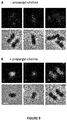
- a pulse of propargyl-choline was performed. Pneumococcal cells were grown in medium containing choline, washed, incubated in presence of propargyl-choline for 30 min and submitted to the bioorthogonal reaction ( Figure 11 ). In these conditions, only TA synthesized during the 30 min pulse are labeled. Reduced membrane labeling (when compared to the 3 h culture period shown in Figure 8 ) together with the septal site localization ( Figure 11A and 11B ) confirm that TA synthesis takes place in a relatively short time scale.
Landscapes
- Life Sciences & Earth Sciences (AREA)
- Health & Medical Sciences (AREA)
- Chemical & Material Sciences (AREA)
- Engineering & Computer Science (AREA)
- Organic Chemistry (AREA)
- Immunology (AREA)
- Molecular Biology (AREA)
- Zoology (AREA)
- Wood Science & Technology (AREA)
- Proteomics, Peptides & Aminoacids (AREA)
- Physics & Mathematics (AREA)
- General Health & Medical Sciences (AREA)
- Biotechnology (AREA)
- Biochemistry (AREA)
- Microbiology (AREA)
- Analytical Chemistry (AREA)
- Urology & Nephrology (AREA)
- Hematology (AREA)
- Biomedical Technology (AREA)
- General Engineering & Computer Science (AREA)
- Genetics & Genomics (AREA)
- Bioinformatics & Cheminformatics (AREA)
- Toxicology (AREA)
- Biophysics (AREA)
- Pathology (AREA)
- Cell Biology (AREA)
- General Physics & Mathematics (AREA)
- Medicinal Chemistry (AREA)
- Food Science & Technology (AREA)
- Measuring Or Testing Involving Enzymes Or Micro-Organisms (AREA)
Priority Applications (4)
| Application Number | Priority Date | Filing Date | Title |
|---|---|---|---|
| EP17305596.3A EP3406731A1 (de) | 2017-05-22 | 2017-05-22 | Metabolische markierung von bakteriellen teichonsäurezellwänden |
| US16/615,012 US20200087703A1 (en) | 2017-05-22 | 2018-05-22 | Metabolic labeling of bacterial teichoic acids cell wall |
| PCT/EP2018/063337 WO2018215432A1 (en) | 2017-05-22 | 2018-05-22 | Metabolic labeling of bacterial teichoic acids cell wall |
| EP18726151.6A EP3630995A1 (de) | 2017-05-22 | 2018-05-22 | Metabolische markierung von bakteriellen teichonsäurezellwänden |
Applications Claiming Priority (1)
| Application Number | Priority Date | Filing Date | Title |
|---|---|---|---|
| EP17305596.3A EP3406731A1 (de) | 2017-05-22 | 2017-05-22 | Metabolische markierung von bakteriellen teichonsäurezellwänden |
Publications (1)
| Publication Number | Publication Date |
|---|---|
| EP3406731A1 true EP3406731A1 (de) | 2018-11-28 |
Family
ID=59239869
Family Applications (2)
| Application Number | Title | Priority Date | Filing Date |
|---|---|---|---|
| EP17305596.3A Withdrawn EP3406731A1 (de) | 2017-05-22 | 2017-05-22 | Metabolische markierung von bakteriellen teichonsäurezellwänden |
| EP18726151.6A Withdrawn EP3630995A1 (de) | 2017-05-22 | 2018-05-22 | Metabolische markierung von bakteriellen teichonsäurezellwänden |
Family Applications After (1)
| Application Number | Title | Priority Date | Filing Date |
|---|---|---|---|
| EP18726151.6A Withdrawn EP3630995A1 (de) | 2017-05-22 | 2018-05-22 | Metabolische markierung von bakteriellen teichonsäurezellwänden |
Country Status (3)
| Country | Link |
|---|---|
| US (1) | US20200087703A1 (de) |
| EP (2) | EP3406731A1 (de) |
| WO (1) | WO2018215432A1 (de) |
Families Citing this family (7)
| Publication number | Priority date | Publication date | Assignee | Title |
|---|---|---|---|---|
| JP2024506954A (ja) | 2021-02-18 | 2024-02-15 | イェダ リサーチ アンド デベロップメント カンパニー リミテッド | ワクチン作製用の遺伝子改変細菌 |
| EP4294428A1 (de) | 2021-02-18 | 2023-12-27 | Yeda Research and Development Co. Ltd | Verfahren zur erzeugung von impfstoffen |
| CN114099555B (zh) * | 2021-11-11 | 2024-01-09 | 天津科技大学 | 一种植物乳杆菌脂磷壁酸及其在抑制淀粉样蛋白聚集中的应用 |
| CN115372341B (zh) * | 2022-05-14 | 2025-04-25 | 西北工业大学深圳研究院 | D-赖氨酸衍生物构筑的细菌细胞壁新生肽聚糖的生物发光检测体系及应用方法 |
| WO2024038462A1 (en) | 2022-08-17 | 2024-02-22 | Yeda Research And Development Co. Ltd. | Novel bacteria-based delivery system for tace (adam17) selective biological inhibitor |
| IL295726A (en) | 2022-08-17 | 2024-03-01 | Yeda res & development co ltd | Genetically modified bacteria for generating vaccines |
| CN116593721B (zh) * | 2023-05-19 | 2025-10-31 | 北京林业大学 | 一种标记植物固醇的试剂及其应用和标记植物固醇的方法 |
Citations (5)
| Publication number | Priority date | Publication date | Assignee | Title |
|---|---|---|---|---|
| WO1984003903A1 (en) * | 1983-03-31 | 1984-10-11 | Robert Edward Silman | Method and device for detecting microorganisms |
| WO1998012346A1 (en) * | 1996-09-23 | 1998-03-26 | The Children's Hospital Of Philadelphia | COMPOSITIONS AND METHODS FOR TREATMENT OF INFECTION CAUSED BY HAEMOPHILUS INFLUENZAE AND $i(STREPTOCOCCUS PNEUMONIAE) |
| WO2013048952A1 (en) * | 2011-09-28 | 2013-04-04 | Biomed Valley Discoveries, Inc. | Methods and compositions for detecting infections |
| CN104749369A (zh) * | 2013-12-31 | 2015-07-01 | 中国科学院深圳先进技术研究院 | 一种用于具有细胞膜结构的生物体的荧光标记方法 |
| WO2016177724A1 (en) * | 2015-05-04 | 2016-11-10 | Centre National De La Recherche Scientifique (Cnrs) | A method for labeling specifically living bacteria comprising the use of modified non endogenous monosaccharide compounds |
Family Cites Families (1)
| Publication number | Priority date | Publication date | Assignee | Title |
|---|---|---|---|---|
| WO2010091142A1 (en) * | 2009-02-04 | 2010-08-12 | President And Fellows Of Harvard College | Compositions and methods for labeling and imaging phospholipids |
-
2017
- 2017-05-22 EP EP17305596.3A patent/EP3406731A1/de not_active Withdrawn
-
2018
- 2018-05-22 US US16/615,012 patent/US20200087703A1/en not_active Abandoned
- 2018-05-22 EP EP18726151.6A patent/EP3630995A1/de not_active Withdrawn
- 2018-05-22 WO PCT/EP2018/063337 patent/WO2018215432A1/en not_active Ceased
Patent Citations (5)
| Publication number | Priority date | Publication date | Assignee | Title |
|---|---|---|---|---|
| WO1984003903A1 (en) * | 1983-03-31 | 1984-10-11 | Robert Edward Silman | Method and device for detecting microorganisms |
| WO1998012346A1 (en) * | 1996-09-23 | 1998-03-26 | The Children's Hospital Of Philadelphia | COMPOSITIONS AND METHODS FOR TREATMENT OF INFECTION CAUSED BY HAEMOPHILUS INFLUENZAE AND $i(STREPTOCOCCUS PNEUMONIAE) |
| WO2013048952A1 (en) * | 2011-09-28 | 2013-04-04 | Biomed Valley Discoveries, Inc. | Methods and compositions for detecting infections |
| CN104749369A (zh) * | 2013-12-31 | 2015-07-01 | 中国科学院深圳先进技术研究院 | 一种用于具有细胞膜结构的生物体的荧光标记方法 |
| WO2016177724A1 (en) * | 2015-05-04 | 2016-11-10 | Centre National De La Recherche Scientifique (Cnrs) | A method for labeling specifically living bacteria comprising the use of modified non endogenous monosaccharide compounds |
Non-Patent Citations (12)
| Title |
|---|
| BACKUS ET AL., NAT. CHEM. BIOL., vol. 7, 2011, pages 228 |
| FAN ET AL., MOL. MICROBIOL., vol. 50, 2003, pages 537 |
| GISCH ET AL., J. BIOL. CHEM., vol. 288, 2013, pages 15654 |
| KAWAI ET AL., EMBO J., vol. 30, 2001, pages 4931 |
| KING; WAGNER, BIOCONJUGATE CHEM., vol. 25, 2014, pages 825 |
| KURU ET AL.: "In situ probing of newly synthesized peptidoglycan in live bacteria with fluorescent D-amino acids", ANGEW. CHEM. INT. ED. ENGL., vol. 51, 2012, pages 12519 - 12523 |
| L. F. FITZSIMMONS ET AL: "Small-Molecule Inhibition of Choline Catabolism in Pseudomonas aeruginosa and Other Aerobic Choline-Catabolizing Bacteria", APPLIED AND ENVIRONMENTAL MICROBIOLOGY, vol. 77, no. 13, 20 May 2011 (2011-05-20), pages 4383 - 4389, XP055406791, ISSN: 0099-2240, DOI: 10.1128/AEM.00504-11 * |
| LACKS S; HOTCHKISS RD: "A study of the genetic material determining an enzyme in Pneumococcus", BIOCHEM. BIOPHYS. ACTA, vol. 39, 1960, pages 508 - 518, XP024556154, DOI: doi:10.1016/0006-3002(60)90205-5 |
| LIU ET AL., PROC. NATL. ACAD. SCI. USA, vol. 106, 2009, pages 4207 |
| SADAMOTO ET AL., J. AM. CHEM. SOC., vol. 124, 2002, pages 9018 - 9019 |
| SANTA MARIA ET AL., PROC. NATL. ACAD. SCI. USA, vol. 111, 2014, pages 12510 |
| SERINO ET AL., MOL. MICROBIOL., vol. 43, 2002, pages 437 |
Also Published As
| Publication number | Publication date |
|---|---|
| US20200087703A1 (en) | 2020-03-19 |
| EP3630995A1 (de) | 2020-04-08 |
| WO2018215432A1 (en) | 2018-11-29 |
Similar Documents
| Publication | Publication Date | Title |
|---|---|---|
| EP3406731A1 (de) | Metabolische markierung von bakteriellen teichonsäurezellwänden | |
| AU2014353835B2 (en) | Detection, isolation and identification of microorganisms | |
| Borrmann et al. | Bioorthogonal chemistry in living organisms | |
| JP6029688B2 (ja) | 生菌を特異的に標識する方法 | |
| US10544444B2 (en) | Compositions for in situ labeling of bacterial cell walls with fluorophores and methods of use thereof | |
| Marshall et al. | Enzyme-targeted fluorescent small-molecule probes for bacterial imaging | |
| CN105683389B (zh) | 包含使用修饰的单糖化合物的用于特异性标记活菌的方法 | |
| JP5139085B2 (ja) | 固相のオリゴ糖タグ付け:固定化糖質の操作技術 | |
| Di Guilmi et al. | Specific and spatial labeling of choline-containing teichoic acids in Streptococcus pneumoniae by click chemistry | |
| US20190024132A1 (en) | D-ala-d-ala-based dipeptides as tools for imaging peptidoglycan biosynthesis | |
| CN108865918A (zh) | 一种标记存活的革兰氏阳性细菌及其细胞壁的方法 | |
| Liu et al. | Acceptor specificity and inhibition of the bacterial cell‐wall glycosyltransferase MurG | |
| Rigolot et al. | A bioorthogonal chemistry approach to detect the K1 polysialic acid capsule in Escherichia coli | |
| Ziylan et al. | Evaluation of Kdo-8-N 3 incorporation into lipopolysaccharides of various Escherichia coli strains | |
| Zhang et al. | Transglycosylases in peptidoglycan biosynthesis: advances in structure, function, and antimicrobial development | |
| JPS58152496A (ja) | バリエナミンおよびバリダミンの製造法 | |
| Sadamoto et al. | Cell wall engineering of living bacteria through biosynthesis | |
| US20230375536A1 (en) | Methods for proximal molecular probe transfer | |
| Ziylan | Kdo derivatives as tools to metabolically label bacterial lipopolysaccharides | |
| Ratna | The Role of a Bacterial Kinase, AmgK, in the Regulation of Cell Wall Recycling and Pathogenesis | |
| Shipman et al. | Selective capture, isolation, and characterization of mucin foraging neu-raminidase-active bacteria from microbiomes using a non-inhibitory activ-ity-based probe | |
| JP3778603B2 (ja) | ソルビトールオキシダーゼ、その製造法およびその用途 | |
| Bahmed et al. | Use of dansyl N-acetyl glucosamine as substrate for chitin synthetase activities | |
| MATSUBARA et al. | Isolation and physiological activity of the chitosan from conidia and mycelia of Mycosphaerella pinodes | |
| Bykhovsky et al. | Coproporphyrins, Uroporphyrins, and Their Metal Comlexes: Biosynthesis and Application to Immune Analysis |
Legal Events
| Date | Code | Title | Description |
|---|---|---|---|
| PUAI | Public reference made under article 153(3) epc to a published international application that has entered the european phase |
Free format text: ORIGINAL CODE: 0009012 |
|
| STAA | Information on the status of an ep patent application or granted ep patent |
Free format text: STATUS: THE APPLICATION HAS BEEN PUBLISHED |
|
| AK | Designated contracting states |
Kind code of ref document: A1 Designated state(s): AL AT BE BG CH CY CZ DE DK EE ES FI FR GB GR HR HU IE IS IT LI LT LU LV MC MK MT NL NO PL PT RO RS SE SI SK SM TR |
|
| AX | Request for extension of the european patent |
Extension state: BA ME |
|
| STAA | Information on the status of an ep patent application or granted ep patent |
Free format text: STATUS: THE APPLICATION IS DEEMED TO BE WITHDRAWN |
|
| 18D | Application deemed to be withdrawn |
Effective date: 20190529 |