EP3407920B1 - Verfahren zur dosierung einer chromophoren substanz in einem hornhautgewebe und vorrichtung zur kontrolle der dosierung - Google Patents
Verfahren zur dosierung einer chromophoren substanz in einem hornhautgewebe und vorrichtung zur kontrolle der dosierung Download PDFInfo
- Publication number
- EP3407920B1 EP3407920B1 EP16834193.1A EP16834193A EP3407920B1 EP 3407920 B1 EP3407920 B1 EP 3407920B1 EP 16834193 A EP16834193 A EP 16834193A EP 3407920 B1 EP3407920 B1 EP 3407920B1
- Authority
- EP
- European Patent Office
- Prior art keywords
- corneal tissue
- agent
- electromagnetic radiation
- chromophoric agent
- measurement
- Prior art date
- Legal status (The legal status is an assumption and is not a legal conclusion. Google has not performed a legal analysis and makes no representation as to the accuracy of the status listed.)
- Active
Links
Images


Classifications
-
- A—HUMAN NECESSITIES
- A61—MEDICAL OR VETERINARY SCIENCE; HYGIENE
- A61F—FILTERS IMPLANTABLE INTO BLOOD VESSELS; PROSTHESES; DEVICES PROVIDING PATENCY TO, OR PREVENTING COLLAPSING OF, TUBULAR STRUCTURES OF THE BODY, e.g. STENTS; ORTHOPAEDIC, NURSING OR CONTRACEPTIVE DEVICES; FOMENTATION; TREATMENT OR PROTECTION OF EYES OR EARS; BANDAGES, DRESSINGS OR ABSORBENT PADS; FIRST-AID KITS
- A61F9/00—Methods or devices for treatment of the eyes; Devices for putting in contact-lenses; Devices to correct squinting; Apparatus to guide the blind; Protective devices for the eyes, carried on the body or in the hand
- A61F9/007—Methods or devices for eye surgery
- A61F9/008—Methods or devices for eye surgery using laser
-
- A—HUMAN NECESSITIES
- A61—MEDICAL OR VETERINARY SCIENCE; HYGIENE
- A61F—FILTERS IMPLANTABLE INTO BLOOD VESSELS; PROSTHESES; DEVICES PROVIDING PATENCY TO, OR PREVENTING COLLAPSING OF, TUBULAR STRUCTURES OF THE BODY, e.g. STENTS; ORTHOPAEDIC, NURSING OR CONTRACEPTIVE DEVICES; FOMENTATION; TREATMENT OR PROTECTION OF EYES OR EARS; BANDAGES, DRESSINGS OR ABSORBENT PADS; FIRST-AID KITS
- A61F9/00—Methods or devices for treatment of the eyes; Devices for putting in contact-lenses; Devices to correct squinting; Apparatus to guide the blind; Protective devices for the eyes, carried on the body or in the hand
- A61F9/007—Methods or devices for eye surgery
- A61F9/0079—Methods or devices for eye surgery using non-laser electromagnetic radiation, e.g. non-coherent light or microwaves
-
- A—HUMAN NECESSITIES
- A61—MEDICAL OR VETERINARY SCIENCE; HYGIENE
- A61F—FILTERS IMPLANTABLE INTO BLOOD VESSELS; PROSTHESES; DEVICES PROVIDING PATENCY TO, OR PREVENTING COLLAPSING OF, TUBULAR STRUCTURES OF THE BODY, e.g. STENTS; ORTHOPAEDIC, NURSING OR CONTRACEPTIVE DEVICES; FOMENTATION; TREATMENT OR PROTECTION OF EYES OR EARS; BANDAGES, DRESSINGS OR ABSORBENT PADS; FIRST-AID KITS
- A61F9/00—Methods or devices for treatment of the eyes; Devices for putting in contact-lenses; Devices to correct squinting; Apparatus to guide the blind; Protective devices for the eyes, carried on the body or in the hand
- A61F9/007—Methods or devices for eye surgery
- A61F9/008—Methods or devices for eye surgery using laser
- A61F2009/00861—Methods or devices for eye surgery using laser adapted for treatment at a particular location
- A61F2009/00872—Cornea
-
- A—HUMAN NECESSITIES
- A61—MEDICAL OR VETERINARY SCIENCE; HYGIENE
- A61F—FILTERS IMPLANTABLE INTO BLOOD VESSELS; PROSTHESES; DEVICES PROVIDING PATENCY TO, OR PREVENTING COLLAPSING OF, TUBULAR STRUCTURES OF THE BODY, e.g. STENTS; ORTHOPAEDIC, NURSING OR CONTRACEPTIVE DEVICES; FOMENTATION; TREATMENT OR PROTECTION OF EYES OR EARS; BANDAGES, DRESSINGS OR ABSORBENT PADS; FIRST-AID KITS
- A61F9/00—Methods or devices for treatment of the eyes; Devices for putting in contact-lenses; Devices to correct squinting; Apparatus to guide the blind; Protective devices for the eyes, carried on the body or in the hand
- A61F9/007—Methods or devices for eye surgery
- A61F9/008—Methods or devices for eye surgery using laser
- A61F2009/00878—Planning
- A61F2009/0088—Planning based on wavefront
-
- A—HUMAN NECESSITIES
- A61—MEDICAL OR VETERINARY SCIENCE; HYGIENE
- A61F—FILTERS IMPLANTABLE INTO BLOOD VESSELS; PROSTHESES; DEVICES PROVIDING PATENCY TO, OR PREVENTING COLLAPSING OF, TUBULAR STRUCTURES OF THE BODY, e.g. STENTS; ORTHOPAEDIC, NURSING OR CONTRACEPTIVE DEVICES; FOMENTATION; TREATMENT OR PROTECTION OF EYES OR EARS; BANDAGES, DRESSINGS OR ABSORBENT PADS; FIRST-AID KITS
- A61F9/00—Methods or devices for treatment of the eyes; Devices for putting in contact-lenses; Devices to correct squinting; Apparatus to guide the blind; Protective devices for the eyes, carried on the body or in the hand
- A61F9/007—Methods or devices for eye surgery
- A61F9/008—Methods or devices for eye surgery using laser
- A61F2009/00885—Methods or devices for eye surgery using laser for treating a particular disease
- A61F2009/00893—Keratoconus
-
- A—HUMAN NECESSITIES
- A61—MEDICAL OR VETERINARY SCIENCE; HYGIENE
- A61N—ELECTROTHERAPY; MAGNETOTHERAPY; RADIATION THERAPY; ULTRASOUND THERAPY
- A61N5/00—Radiation therapy
- A61N5/06—Radiation therapy using light
- A61N5/0613—Apparatus adapted for a specific treatment
- A61N5/062—Photodynamic therapy, i.e. excitation of an agent
Definitions
- the present invention relates to a control apparatus for controlling the dosing of a chromophoric agent in a corneal tissue and a process for dosing a chromophoric agent in a corneal tissue.
- the apparatus and the process proposed herewith are used to determine the efficacy of a corneal cross-linking treatment.
- keratoconus is a progressive degeneration of the cornea, which tends to thin and curve outwards (it is properly spoken of as "corneal ectasia").
- eyeglasses or contact lenses makes it possible to attenuate the symptoms of the pathology, that is, to correct the optical aberrations of the ectatic cornea, but does not halt its progression.
- cornea transplant surgery or keratoplasty, which consists in the replacement of the ecstatic cornea with a suitable human donor tissue from an eye bank.
- keratoplasty In order to slow down or halt the progression of keratoconus, in the past decade a para-surgical procedure known in the field as "corneal cross-linking" has been introduced.
- Corneal cross-linking has the aim of increasing corneal rigidity, which has been reduced as a result of evolutive keratoconus or other iatrogenic corneal ectasias.
- the list of clinical applications of corneal cross-linking is expanding with the development of knowledge on corneal biophysics.
- Corneal cross-linking entails the administration of a photopolymerising agent (referred to in the field as "cross-linking" agent) into the corneal stroma, followed by photo-activation by irradiation. Photo-activation brings about the creation of new covalent chemical bonds between stromal proteins (one speaks of photopolymerization), with a consequent stiffening of the corneal tissue.
- the most common cross-linking agent is riboflavin, which, when subjected to photo-activation, converts the oxygen dissolved in the corneal stroma into free radicals.
- the free radicals in turn, cause the generation of new covalent bonds between the molecules of the corneal stroma.
- UV-A ultraviolet
- green spectrum For photo-activation use is made, for example, of non-coherent luminous radiation in the ultraviolet (UV-A) or green spectrum.
- UV-A ultraviolet
- green spectrum For photo-activation use is made, for example, of non-coherent luminous radiation in the ultraviolet (UV-A) or green spectrum.
- UV-A ultraviolet
- green spectrum The use of a coherent laser source is also envisaged.
- the cross-linking agent is administered in ophthalmic solutions with a known concentration directly onto the corneal tissue.
- various protocols for administering the cross-linking agent whose variability resides, for example:
- corneal cross-linking must be performed by expert medical staff.
- corneal cross-linking treatment is tied to the safety of the treatment, where the power density of the radiation exceeds a certain threshold (e.g. 50 mW/cm 2 ) or the concentration of the cross-linking agent in the stroma is too low (e.g. the riboflavin is less than 0.001%) and the cornea is de-epithelialized.
- a certain threshold e.g. 50 mW/cm 2
- concentration of the cross-linking agent in the stroma e.g. the riboflavin is less than 0.001%
- the corneal cross-linking treatment besides not being effective, can damage the corneal endothelial layer, causing irreversible damage to the tissue.
- Document WO 2012/145853 discloses a compact apparatus, which is applied on a slit lamp and consists of a proximity sensor that detects the corneal tissue and a sensor for measuring the fluorescence emitted by the cornea saturated with the cross-linking agent (e.g. riboflavin).
- the cross-linking agent e.g. riboflavin
- corneal cross-linking treatment has been determined by an ophthalmologist who is an expert in the field of application by evaluating and comparing the corneal topography maps of the keratoconus acquired prior to treatment and in the twelve months following the treatment itself.
- the technical task at the basis of the present invention is to provide a control apparatus for controlling the dosing of a chromophoric agent in a corneal tissue and a process for dosing a chromophoric agent in a corneal tissue which overcome the drawbacks of the aforementioned prior art.
- Another object of the present invention is to provide a process for dosing a chromophoric agent in a corneal tissue that is more reliable and efficient than the known methods.
- a further object of the present invention is to provide a process for dosing a chromophoric agent in a corneal tissue that can also be implemented by less expert medical personnel.
- Another object of the present invention is to provide a control apparatus for controlling the dosing of a chromophoric agent in a corneal tissue which enables the efficacy and safety of the corneal cross-linking treatment to be evaluated in real time, i.e. while it is being performed.
- a further object of the present invention is to provide a control apparatus for controlling the dosing of a chromophoric agent in a corneal tissue which is reliable and efficient and can also be used by less expert medical personnel.
- a control apparatus for controlling the dosing of a chromophoric agent in a corneal tissue and a process for dosing a chromophoric agent in a corneal tissue comprising:
- the first irradiating means consist in a source configured to emit the first electromagnetic radiation with a wavelength selected so as to cause the fluorescence effect and the first measurement means are configured to measure the fluorescence intensity.
- the first irradiating means consist in a source configured to emit the first electromagnetic radiation with a wavelength selected so as to be absorbed by the chromophoric agent and the first measurement means are configured to measure the diffused intensity.
- the first measurement means comprise a video camera or a spectrometer or one or more photodiodes.
- the means used to transmit the first electromagnetic radiation to the corneal tissue is air or an optical fibre.
- the means used to receive the energy perturbation caused by the first electromagnetic radiation is air or a further optical fibre.
- the chromophoric agent is a fluorophore and the first electromagnetic radiation has a wavelength selected so as to cause the fluorescence effect of the fluorophoric agent.
- the first spectroscopic parameter is thus the fluorescence intensity, so that the measurement of the first spectroscopic parameter corresponds to the value of the fluorescence intensity of the corneal tissue without the fluorophoric agent and the further measurement of the first spectroscopic parameter corresponds to the value of the fluorescence intensity of the corneal tissue containing the fluorophoric agent.
- the first electromagnetic radiation has a wavelength selected so as to be absorbed by the chromophoric agent.
- the first spectroscopic parameter is thus the diffused intensity, so that the measurement of the first spectroscopic parameter corresponds to the value of the intensity diffused by the corneal tissue without the chromophoric agent and the further measurement of the first spectroscopic parameter corresponds to the value of the intensity diffused by the corneal tissue containing the chromophoric agent.
- the process can also comprise the following steps:
- the process comprises a step of photo-activating the chromophoric agent, after the factor representative of the concentration equals or exceeds the first pre-established threshold.
- It can also comprise a step of estimating the mechanical stiffening of the corneal tissue as a function of the values taken on by the factor representative of the concentration before and after the photo-activating step.
- the step of estimating the mechanical stiffening of the corneal tissue comprises a step of iteratively modifying the pattern of photo-activating intensity of the first electromagnetic radiation as long as the predicted value of the mechanical stiffening Y is below an efficacy threshold.
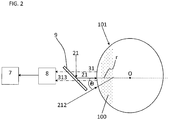
- the number 1 denotes a control apparatus for controlling the dosing of a chromophoric agent 100 in a corneal tissue 101.
- the corneal tissue 101 is assumed to be like a portion of a sphere having an optical axis r passing through the centre of the sphere O.
- the control apparatus 1 comprises:
- the first irradiating means 2 consist in a source configured to emit the first electromagnetic radiation 21 with a wavelength selected so as to cause the fluorescence effect in the fluorophoric agent 100.
- the first measurement means 3 are configured to measure the fluorescence intensity (which represents the first spectroscopic parameter 31).
- the first measurement means 3 consist in an RGB video camera configured to measure the average intensity value of the green pixels of the acquired image.
- the first irradiating means 2 consist in a source configured to emit the first electromagnetic radiation 21 with a wavelength selected so as to be absorbed by the chromophoric agent 100 (be it a fluorophore or not a fluorophore).
- the first measurement means 3 are configured to measure the diffused intensity (which represents the first spectroscopic parameter 31).
- the first measurement means 3 consist in an RGB video camera configured to measure the average intensity value of the blue pixels of the acquired image.
- control apparatus 1 comprises a processing unit 4 configured to calculate a factor C representative of the concentration of the chromophoric agent 100 inside the corneal tissue 101 in response to measurements of the first spectroscopic parameter 31 performed by the first measurement means 3.
- the processing unit 4 receives as input at least two measurements of the first spectroscopic parameter 31:
- control apparatus 1 further comprises:
- the first irradiating means 2 consist in a source (hereinafter called “first source”) configured to emit the first electromagnetic radiation 21 with a wavelength selected so as to cause the fluorescence effect in the chromophoric agent 100
- the second irradiating means 12 consist in a source (hereinafter indicated as “second source”) configured to emit the second electromagnetic radiation 212 with a wavelength selected so as to be absorbed by the chromophoric agent 100 (fluorophore or non-fluorophore).
- the first measurement means 3 are configured to measure the fluorescence intensity (which represents the first spectroscopic parameter 31), whilst the second measurement means 13 are configured to measure the diffused intensity (which represents the second spectroscopic parameter 313).
- the first measurement means 3 consist in an RGB video camera configured to measure the average intensity value of the green pixels of the acquired image.
- the second measurement means 13 consist in an RGB video camera configured to measure the average intensity value of the blue pixels of the acquired image.
- the second source 12 is disposed in such way that the second electromagnetic radiation 212 strikes the corneal tissue 101 forming an angle ⁇ comprised between 0 ° and 90 ° relative to the optical axis r.
- the control apparatus 1 comprises collimating means 5 for collimating the first electromagnetic radiation 21 on the corneal tissue 101.
- the collimating means 5 consist in an optical system capable of focusing and modifying the wave front of the first electromagnetic radiation 21.
- control apparatus 1 comprises a measurement unit for measuring the power density 6 of the first electromagnetic radiation 21 and of the second electromagnetic radiation 212.
- the first measurement means 3 and the second measurement means 13 consist in a single video camera 7 for acquiring images of the corneal tissue 101.
- the optical sensor is of the CMOS (acronym of the expression “Complementary Metal Oxide Semiconductor”) or CCD (acronym of the expression “Charge Coupled Device”) type.
- control apparatus 1 further comprises a lens or a system of lenses 8 for focusing the acquired images on the video camera 7.
- the processing unit 4 is thus configured to calculate the factor C representative of the concentration of the chromophoric agent 100 inside the corneal tissue 101 in response to the images acquired by the video camera 7.
- the processing unit 4 is further configured to receive signals from the power density measurement unit 6 which are representative of the power density S 21 , S 212 of the electromagnetic radiation 21, 212 issued and, in response to this:
- the processing unit 4 employs hyperspectral techniques of a known type, such as Wiener estimation, starting from the images acquired by the RGB video camera 7.
- the processing unit 4 can consist of an electronic device, suitably programmed to perform the described functions, which can have corresponding to it various hardware and/or routine software entities making up the programmed device.
- such functions can be performed by a plurality of electronic devices over which an equal number of functional modules can be distributed.
- the processing unit 4 can further avail itself of one or more processors for executing instructions contained in memory modules.
- the various functional modules can be distributed over local or remote computers based on the architecture of the network they reside in.
- the control apparatus 1 comprises a dichroic filter 9 placed downstream of the first source 2 so as to receive the first electromagnetic radiation 21 and deliver it to the corneal tissue 101.
- the dichroic filter 9 has a spectral response such as to transmit the fluorescence radiation and the intensity diffused by the corneal tissue 101 when saturated by the chromophoric agent 100.
- a spectrometer (of a known type) is used to measure the fluorescence intensity and the diffused intensity.
- the first measurement means 3 and the second measurement means 13 are distinct and consist respectively in a first photodiode 3 configured to measure the fluorescence intensity 31 and a second photodiode 13 configured to measure the intensity 313 diffused by the corneal tissue 101.
- the structure of the different components of the control apparatus 1 described for the third embodiment can also be used for the first and second embodiments, except for the duplication of the signals, due to the fact that in the third embodiment two sources are used and thus two distinct spectroscopic parameters are measured.
- the first irradiating means 2 can consist in a non-coherent light source or in a laser source configured to transmit photons in a continuous or pulsed mode with wavelengths comprised in an interval from ultraviolet (UV-A) to near or mid infrared (NIR or MIR).
- UV-A ultraviolet
- NIR near or mid infrared
- photo-activation of the fluorophoric agent 100 in the corneal tissue 101 can take place by absorption of non-coherent light or absorption of a single photon at a given wavelength (for example UV) or by absorption of two or more photons having longer wavelengths, for example NIR or MIR, in such a way that the striking photons penetrate more deeply into the corneal tissue 101 than photons with a shorter wavelength.
- a given wavelength for example UV
- two or more photons having longer wavelengths for example NIR or MIR
- air is used as the transmitting means in order to deliver electromagnetic radiation to the corneal tissue 101.
- an optical fibre (not illustrated) is envisaged as a transmitting means.
- another optical fibre (not illustrated) can be used as a means for measuring the energy perturbation caused by the electromagnetic radiation in the corneal tissue 101.
- the process envisages that the first electromagnetic radiation 21 has a wavelength selected so as to cause the fluorescence effect in the fluorophoric agent 100.
- the first spectroscopic parameter 31 is the fluorescence intensity.
- the process envisages that the first electromagnetic radiation 21 has a wavelength selected so as to be absorbed by the chromophoric agent 100.
- the first spectroscopic parameter 31 is the diffused intensity.
- the process then comprises cyclically performing at least the following steps:
- the first electromagnetic radiation 21 has a wavelength selected so as to cause the fluorescence effect in the fluorophoric agent 100 and the first spectroscopic parameter 31 is the fluorescence intensity.
- the second electromagnetic radiation 212 instead has a wavelength selected so as to be absorbed by the chromophoric agent 100 (fluorophore or non-fluorophore) and the second spectroscopic parameter 313 is the diffused intensity.
- the process comprises the following steps (performed only once):
- the first and second safety intervals are selected in such a way that the radiation of the sources 2, 12 does not photo-activate the chromophoric agent 100 and is safe for the corneal tissue 101.
- the first safety interval is comprised between 0.01 mW/cm 2 and 3 mW/cm 2
- the second safety interval is comprised between 0.01 mW/cm 2 and 10 mW/cm 2 .
- the factor C representative of the concentration of the chromophoric agent 100 in the corneal tissue 101 remains below the first pre-established threshold T h1 .
- the factor C representative of the concentration is obtained as a linear combination of:
- the factor C representative of the concentration based on the spectrometry measurements in general: fluorescence intensity and/or diffused intensity; in particular: average intensity value of the green and/or blue pixels
- known functions and algorithms are used.
- the chromophoric agent 100 and fluorophore use is made here of riboflavin, for which the excitation wavelength (i.e. the wavelength of the first electromagnetic radiation 21) is selected between 360 nm and 375 nm (to obtain the fluorescence effect), whilst the wavelength of the second electromagnetic radiation 212 is selected between 400 nm and 500 nm (absorbance interval of riboflavin).
- the excitation wavelength i.e. the wavelength of the first electromagnetic radiation 21
- the wavelength of the second electromagnetic radiation 212 is selected between 400 nm and 500 nm (absorbance interval of riboflavin).
- the first pre-established threshold T h1 of the factor C is equal to 0.010%.
- the fluorescence measurement is based on the exchange of energy that takes place between the first electromagnetic radiation 21 and the fluorophoric agent 100.
- the absorption of the energy carried by the first electromagnetic radiation 21 is capable of triggering in the fluorophoric agent 100 energy transitions of the outer electrons, whether or not they are engaged in a chemical bond.
- the fluorophoric agent 100 excited at a higher vibrational sublevel, can thus relax quickly by non-radiative decay to the lower vibrational level and from this it can decay radiatively to the fundamental state, emitting a photon with a lower energy than the absorbed one.
- fluorescence effect This physical phenomenon is known precisely as the "fluorescence effect".
- fluorescence measurement takes place at an emission wavelength comprised between 520 nm and 540 nm.
- I F is the stationary fluorescence intensity measured at a specific wavelength emitted by the fluorophoric agent 100, it will be proportional both to the emission spectrum F of the fluorophoric agent 100 and the intensity of absorbed light IA at the excitation wavelength , by virtue of the quenching function h(c, T), where c is the concentration and T is the temperature in Kelvin.
- the intensity of absorbed light is proportional to the optical absorption of the fluorophoric agent 100 and the intensity of incident light .
- the fluorescence intensity is proportional to the concentration c of the fluorophoric agent 100, by virtue of its optical absorption .
- the phenomena at the basis of the interaction between radiation and matter are diffusion and absorption.
- Diffusion (more commonly known by the term scattering) is a physical process whereby tissue diffuses radiation in all directions, maintaining the same wavelength of the incident radiation .
- the diffused electromagnetic field is not attenuated and a strong diffused signal will be detected.
- part of the intensity of the incident radiation will be absorbed by the chromophoric agent 100 and thus the diffused signal will have a lower intensity than the diffused signal in the absence of the chromophoric agent 100.
- the well-known Kubelka-Munk theory correlates the diffuse reflectance, i.e. the ratio between the intensity of the electromagnetic radiation diffused in the presence of the chromophoric agent 100 inside the corneal tissue 101 and the intensity of the radiation diffused by the corneal tissue 101 without the chromophoric agent 100, with an absorption coefficient and a characteristic diffusion (scattering) coefficient of the tissue :
- the coefficient directly depends on the absorption properties of the chromophoric agent 100 and thus its concentration c.
- the coefficient depends on the microstructure of the corneal tissue 101 and, as such, is specific to each eye.
- the processing unit 4 employs hyperspectral techniques of a known type, such as the Wiener estimation, starting from images acquired by an RGB 7 video camera.
- the Wiener estimation by taking account of a priori information obtained with spectral analysis on known standard samples, is able to establish a statistical correlation between the RGB values acquired by the video camera 7 and the reference optical spectrum ( P. Stigell, K. Miyata, M. Hauta-Kasari. Wiener Estimation Method in Estimating of Spectral Reflectance from RGB Images. Pattern Recognition and Image Analysis 2007 (17) 2: 233-242 ).
- the r estimate equation given above becomes a linear relationship of equality between the RGB pixel intensity value and the corresponding value of the reference spectral component.
- the first of the two equations associates the average intensity value of the green pixels, M FluoG , with the spectral value I Fluo
- the second equation associates the average intensity value of the blue pixels, M RiflB , with the diffused intensity value I Rifl .
- the Kubelka-Munk theory is valid when the material illuminated by radiation is sufficiently thick, more than 50% of the light is diffused and less than 20% is transmitted, relative to the incident light.
- this simplification is still valid, as demonstrated by experimental laboratory tests conducted on human corneal tissues from different donors cultured in an eye bank.
- the accuracy in the measurement of the concentration of the chromophoric agent 100 (fluorophore or non-fluorophore) with the two spectral methods specified above depends on the interval of the measured concentration. In the case of low concentrations, the Kubelka-Munk method yields more precise values than the fluorescence measurement method. Conversely, in the case of high concentrations, the fluorescence measurement method yields more precise values than the Kubelka-Munk method.
- a further distinction between the two spectral methods is that the Kubelka-Munk method can be used for chromophoric agents (fluorescent and non-fluorescent), whereas the fluorescence measurement method can be used exclusively for fluorescent agents.
- the method used to determine the concentration by measuring the fluorescence emitted both during the step of administering the fluorescent agent and during the photo-activating step enables real-time monitoring of the efficacy of the corneal cross-linking treatment. Monitoring the consumption of riboflavin in the corneal tissue 101 will prove advantageous, as described further below.
- the first pre-established threshold T h1 represents the threshold of concentration of the chromophoric agent 100 administered to the corneal tissue 101 prior to the photo-activating step.
- the photo-activating step comprises the following sub-steps, performed cyclically:
- the factor C' representative of the concentration of the chromophoric agent 100 in the corneal tissue 101 remains above a second pre-established threshold T h2 .
- the factor C' representative of the concentration is obtained here as a linear combination of:
- the first and second safety intervals are selected in such a way that the radiation of the first source 2 can photo-activate the chromophoric agent 100 in the corneal tissue 101 and the radiation from both sources 2, 12 is safe for the corneal tissue 101.
- the first safety interval is comprised between 3 mW/cm 2 and 45 mW/cm 2
- the second safety interval is comprised between 0.01 mW/cm 2 and 10 mW/cm 2 .
- the second pre-established threshold T h2 represents the threshold of concentration of the chromophoric agent 100 in the corneal tissue 101 immediately after the photo-activating step.
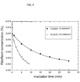
- Figure 4 shows the percent concentration of riboflavin (y-axis) as a function of the irradiation time in minutes (x-axis) recorded for different power densities of the UV-A radiation (in the figure the radiation at 370 ⁇ 5 nm is represented) in two different corneal tissues (indicated with different numbers and symbols) representative of the riboflavin concentrations during the photo-activating step at two different UV-A power densities.
- the solid circles correspond to the percent concentrations of riboflavin when the corneal tissue 101 is subjected to UV-A radiation of 3 mW/cm 2 .
- the empty circles correspond to the percent concentrations of riboflavin when the corneal tissue 101 is subjected to UV-A radiation of 10 mW/cm 2 .
- the inverse of the parameter t rate describes the velocity of riboflavin consumption in the corneal tissue 101 due to exposure to the UV-A radiation and y 0 is a fit parameter.
- Figure 5 shows the variations in the riboflavin concentration normalised to the initial value c 0 for both UV-A power densities (3 mW/cm 2 and 10 mW/cm 2 ) used for different corneal tissues (indicated with different numbers and symbols) with variations in the irradiated energy density (in mJ/cm 2 ).
- a corneal cross-linking treatment induces the generation of further chemical bonds between the amino acids of corneal tissue proteins (photopolymerization). This process induces a mechanical stiffening of the corneal tissue.
- a corneal cross-linking treatment must impart to the corneal tissue a biomechanical stability such as to withstand physiological ocular stresses (e.g. intraocular pressure).
- the efficacy of a cross-linking treatment can be estimated based on the value of the riboflavin concentration reached prior to the photo-activation thereof (indicated as C) and the concentration value after photo-activation (indicated as C').
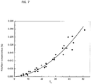
- Figure 6 represents, by way of example, the multiple linear regression model which correlates the predicted increase in mechanical stiffness in the corneal tissue 101 subjected to the above-described cross-linking treatment with the two regression variables: the concentration C and the consumption % of the chromophoric agent 100.
- the cross-linking treatment UV-A radiation with a power density of 3 mW/cm 2 was applied for 30 minutes.
- the experimental results (black circles) in figure 6 were obtained using the apparatus 1 to carry out the above-described process (including the cross-linking treatment) jointly with a biomechanical test apparatus or an atomic force microscope, which provides the values of the Young modulus for each tissue treated.
- the regression coefficient ⁇ 0 , ⁇ 1 and ⁇ 2 are the model parameters by means of which the mechanical stiffening Y of the corneal tissue 101 can be predicted as a function of the values C and consumption % measured during the corneal cross-linking treatment.
- the dashed regression line is obtained by means of known mathematical techniques.
- the cross-linking treatment cycle comprises:
- a two-dimensional map is created to assess the efficacy of the cross-linking treatment, whose values range between 0 (ineffectiveness of the treatment) and 1 (maximum efficacy of the treatment).
- the map illustrated in figure 9 , shows the operator the increase in the clinical efficacy of the cross-linking treatment in real time and teaches when the treatment reaches the maximum efficacy for the cornea undergoing treatment.
- the efficacy values on the map are shown with a probability colour scale that ranges linearly from 0 (not effective, black) to 1 (maximum efficacy, white).
- the efficacy map is automatically updated by means of a machine learning algorithm, on the basis of the values acquired during use of the control apparatus 1.
- the map is capable of instructing the operator in real time during the performance of the cross-linking treatment in order to assure the efficacy and safety thereof in a reliable and efficient manner, irrespective of the treatment protocol the operator is following.
- the process proposed herewith comprises modifying the pattern of photo-activating intensity of the first electromagnetic radiation 21 via the collimating means 5 during the corneal cross-linking treatment (comprising dosing and photo-activation of the chromophoric agent).
- This procedure envisages the possibility of iteratively modifying the spatial pattern of photo-activating intensity of the first source 2 as long as the value of clinical efficacy of the corneal cross-linking treatment exceeds the threshold of efficacy of the treatment.
- the dosing of the chromophoric agent in the corneal tissue is controlled in real time by monitoring at least one spectroscopic parameter (fluorescence or diffused intensity) representative of the interaction between the electromagnetic radiation and the corneal tissue.
- spectroscopic parameter fluorescence or diffused intensity
- the concentration of the chromophoric agent can be monitored both before and during the photo-activating step of the cross-linking treatment, a fact which enables the administration of the agent to be interrupted and the photo-activation of the agent be interrupted or continued in order to stiffen the corneal tissue in a personalised manner. This monitoring enables the cross-linking treatment to be personalised on the basis of the specific ocular tissue.
- the apparatus and process can also be used by ophthalmologists who are not experts in cross-linking treatments (or, in general, not experts in corneal surgery), as the dosing of the chromophoric agent is constantly monitored, so that the operator is guided to interrupt or continue the administration and interrupt or continue the photo-activation of the cross-linking agent in the corneal tissue.
- the proposed apparatus enables the efficacy and safety of the cross-linking treatment to be evaluated in real time immediately after the photo-activation based on the concentration of the chromophoric agent administered on the tissue and the consumption of the concentration of the chromophoric agent.
- measuring the concentration of the chromophoric agent (e.g. riboflavin) before photo-activation and during photo-activation enables the clinical efficacy of the corneal cross-linking treatment to be determined dynamically (i.e. during the performance of the treatment).
- the chromophoric agent e.g. riboflavin
Landscapes
- Health & Medical Sciences (AREA)
- Ophthalmology & Optometry (AREA)
- Biomedical Technology (AREA)
- Heart & Thoracic Surgery (AREA)
- Nuclear Medicine, Radiotherapy & Molecular Imaging (AREA)
- Veterinary Medicine (AREA)
- Surgery (AREA)
- Engineering & Computer Science (AREA)
- Physics & Mathematics (AREA)
- Optics & Photonics (AREA)
- Vascular Medicine (AREA)
- Life Sciences & Earth Sciences (AREA)
- Animal Behavior & Ethology (AREA)
- General Health & Medical Sciences (AREA)
- Public Health (AREA)
- Electromagnetism (AREA)
- Investigating, Analyzing Materials By Fluorescence Or Luminescence (AREA)
- Eye Examination Apparatus (AREA)
Claims (16)
- Verfahren, das nicht am lebenden Körper von Menschen oder Tieren durchgeführt wird, um ein Hornhautgewebe (101) eines Spenders zu vernetzen, umfassend die folgenden Schritte:das Hornhautgewebe (101) mindestens einer ersten elektromagnetischen Strahlung (21) aussetzen;eine Messung eines ersten spektroskopischen Parameters (31) durchführen, der die durch die erste elektromagnetische Strahlung (21) im Hornhautgewebe (101) verursachte Energiestörung anzeigt;solange ein Faktor (C), der für die Konzentration des chromophoren Mittels (100) im Hornhautgewebe (101) repräsentativ ist, unter einer vorgegebenen ersten Grenze (Tm) bleibt, zyklisch mindestens die folgenden Operationen in chronologischer Reihenfolge durchführen:- das chromophoren Mittel (100) an das Hornhautgewebe (101) verabreichen;- das Hornhautgewebe (101), das das crhomophore Mittel (100) enthält , der ersten elektromagnetischen Strahlung (21) aussetzen, deren Leistungsdichte (S21) in einem ersten Sicherheitsintervall für das Hornhautgewebe (101) enthalten und derart gewählt ist, dass die erste elektromagnetische Strahlung (21) das chromophore Mittel (100) nicht photoaktiviert;- eine weitere Messung des ersten spektroskopischen Parameters (31) durchführen, die auf die energetische Störung hinweist, die durch die erste elektromagnetische Strahlung (21) im Hornhautgewebe (101) verursacht wird, das das chromophore Mittel (100) enthält;- den Faktor (C), der für die Konzentration des chromophoren Mittels (100) im Hornhautgewebe (101) repräsentativ ist, mindestens anhand der Messung und der weiteren Messung des ersten spektroskopischen Parameters (31) nach dem Faktor (C) berechnen, der repräsentativ für die Konzentration des chromophoren Mittels (100) ist und gleich oder größer als die erste vorbestimmte Grenze (Tm) im Hornhautgewebe (101) ist, wobei eine Photoaktivierung des chromophoren Mittels (100) erfolgt, solange der Faktor (C'), der repräsentativ für die Konzentration des chromophoren Mittels (100) im Hornhautgewebe (101) ist, über einer vorher festgelegten zweiten Grenze (T112) verbleibt, während die Variation der Konzentration des chromophoren Mittels (100) im Hornhautgewebe (101) während der Photoaktivierung dem folgenden Exponentialgesetz entspricht:
Formel
wobei c0 ist die Konzentration des chromophoren Mittels (100), das zum Zeitpunkt t=0 im Hornhautgewebe (101) verabreicht wird und einen Wert hat, der höher als die erste voreingestellte Grenze (Th1) ist, wobei trate ist ein Parameter, dessen inverser Wert die Geschwindigkeit des Verbrauchs des chromophoren Mittels (100) im Hornhautgewebe (101), als Ergebnis einer Photoaktivierung beschreibt und yo ein entsprechender Parameter ist. - Verfahren nach Anspruch 1, wobei das chromophore Mittel (100) ein Fluorophore ist, wobei die erste elektromagnetische Strahlung (21) eine Wellenlänge (λ21) aufweist, die derart gewählt ist, dass sie den Fluoreszenzeffekt des fluorophoren Mittels (100) verursacht, wobei der erste spektroskopische Parameter (31) die Intensität der Fluoreszenz ist, so dass die Messung des ersten spektroskopischen Parameters (31) dem Wert der Fluoreszenzintensität des Hornhautgewebes (101) ohne das fluorophore Mittel (100) entspricht, und die weitere Messung des ersten spektroskopischen Parameters (31) dem Wert der Fluoreszenzintensität des Hornhautgewebes (101) entspricht, das das fluorophore Mittel (100) enthält.
- Verfahren nach Anspruch 1, wobei die erste elektromagnetische Strahlung (21) eine Wellenlänge (λ21) aufweist, die derart gewählt ist, dass sie vom chromophoren Mittel (100) absorbiert wird, wobei der erste spektroskopische Parameter (31) ist die diffuse Intensität, so dass die Messung des ersten spektroskopischen Parameters (31) dem Wert der Intensität entspricht, die vom Hornhautgewebe (101) ohne das chromophore Mittel (100) diffundiert wird, und die weitere Messung des ersten spektroskopischen Parameters (31) dem Wert der Intensität entspricht, die vom Hornhautgewebe (101) diffundiert wird, das das chromophore Mittel (100) enthält.
- Verfahren nach Anspruch 2, ferner umfassend die folgenden Schritte:das Hornhautgewebe (101) ohne das fluorophore Mittel (100) einer zweiten elektromagnetischen Strahlung (212) mit einer Wellenlänge (λ212 ) aussetzen, die derart gewählt ist, dass sie vom fluorophoren Mittel (100) absorbiert wird;eine Messung der diffusen Intensität (313) aus dem Hornhautgewebe (101) ohne das fluorophore Mittel (100) durchführen;solange der für die Konzentration repräsentative Faktor (C) unter dem festgelegten Grenzwert (Th1) liegt, auch die folgenden Schritte zyklisch ausführen:
nach Durchführung der weiteren Messung des ersten spektroskopischen Parameters (31) wird das Hornhautgewebe (101), das das chromophore Mittel (100) enthält, der zweiten elektromagnetischen Strahlung (212) ausgesetzt.eine weitere Messung der diffusen Intensität (313) durchführen, die aus dem Hornhautgewebe (101), das das fluorophore Mittel (100) enthält, diffundiert wird, und dabei auch die Berechnung des Konzentrationsfaktors (C) des fluorophoren Mittels (100) im Inneren des Hornhautgewebes (101), auch durch die Messung und die weitere Messung der diffusen Intensität (313), durchführen. - Verfahren nach Anspruch 1, ferner umfassend einen Schritt zum Schätzen der mechanischen Versteifung (Y) des Hornhautgewebes (101) als Funktion der Werte, die auf dem Faktor (C, C') berücksichtigt sind, der für die Konzentration vor und nach des Schrittes der Photoaktivierung repräsentativ ist.
- Verfahren nach Anspruch 5, wobei der Schritt des Schätzens der mechanischen Versteifung (Y) des Hornhautgewebes (101) eine iterative Modifikation des Intensitätsprofils der Photoaktivierung der ersten elektromagnetischen Strahlung (21) umfasst, solange der erwartete Wert der mechanischen Versteifung (Y) unter einer Wirksamkeitsgrenze (Yh) liegt.
- Kontrollvorrichtung (1) zur Kontrolle der Dosierung eines chromophoren Mittels (100) in einem Hornhautgewebe (101), das einer Vernetzung ausgesetzt ist, umfassend:erste Mittel (2) zum Bestrahlen des Hornhautgewebes (101) mit mindestens einer ersten elektromagnetischen Strahlung (21);erste Messungsmittel (3) zum Messen eines ersten spektroskopischen Parameters (31);eine Verarbeitungseinheit (4), die derart konfiguriert ist, um einen Faktor (C) zu berechnen, der für die Konzentration des chromophoren Mittels (100) innerhalb des Hornhautgewebes (101) als Reaktion auf mindestens zwei Messungen des ersten spektroskopischen Parameters (31) repräsentativ ist, von der eine Messung die energetische Störung anzeigt, die durch die erste elektromagnetische Strahlung (31) im Hornhautgewebe (101) ohne das chromophore Mittel (100) verursacht wird, und die andere Messung zeigt die energetische Störung an, die durch die erste elektromagnetische Strahlung (31) im Hornhautgewebe (101) verursacht wird (101), die das chromophore Mittel (100) enthält,dadurch gekennzeichnet, dass die Verarbeitungseinheit (4) auch derart konfiguriert ist, um die klinische Wirksamkeit der Vernetzung auf dem Hornhautgewebe (101) auf der Grundlage der vor der Photoaktivierung erreichten Konzentration (C) und der Konzentration (C') zu bewerten, die nach der Photoaktivierung mittels einer Berechnung des Verbrauchs% = (C-C')/C erreicht wird, wobei die Variation der Konzentration des chromophoren Mittels (100) im Hornhautgewebe (101) während der Photoaktivierung dem folgenden Exponentialgesetz entspricht:
Formel 2
wobei c0 die Konzentration des chromophoren Mittels (100) ist, das zum Zeitpunkt t=0 im Hornhautgewebe (101) verabreicht wird, und einen Wert aufweist, der größer als eine vorbestimmte erste Grenze (Th1) ist, die die Grenze der Konzentration des chromophoren Mittels (100) ist, die dem Hornhautgewebe (101) vor der Photoaktivierung verabreicht werden, wobei trate ist in Parameter, dessen inverser Wert die Verbrauchsrate des chromophoren Mittels (100) im Hornhautgewebe (101) aufgrund der Photoaktivierung beschreibt, wobei yo ist ein entsprechender Parameter. - Kontrollvorrichtung (1) nach Anspruch 7, wobei das erste Bestrahlungsmittel (2) aus einer Quelle besteht, die derart konfiguriert ist, um die erste elektromagnetische Strahlung (21) mit einer derart gewählten Wellenlänge (λ21) zu emittieren, um den Fluoreszenzeffekt zu verursachen, wobei die ersten Messungsmittel (3) derart konfiguriert sind, um die Intensität der Fluoreszenz zu messen.
- Kontrollvorrichtung (1) nach Anspruch 7, wobei das erste Bestrahlungsmittel (2) aus einer Quelle besteht, die derart konfiguriert ist, um die erste elektromagnetische Strahlung (21) mit einer so gewählten Wellenlänge (λ21) zu emittieren, um aus dem chromophoren Mittel (100) absorbiert zu werden, wobei die ersten Messungsmittel (3) derart konfiguriert sind, um die diffuse Intensität zu messen.
- Kontrollvorrichtung (1) nach Anspruch 7, auch umfassend:zweite Mittel (12) zum Bestrahlen des Hornhautgewebes (101) mit einer zweiten elektromagnetischen Strahlung (212);zweite Messungsmittel (13) zum Messen eines zweiten spektroskopischen Parameters (313),wobei das erste Bestrahlungsmittel (2) aus einer ersten Quelle besteht, die derart konfiguriert ist, um die erste elektromagnetische Strahlung (21) mit einer Wellenlänge (λ21) zu emittieren, die derart gewählt ist, um den Fluoreszenzeffekt zu verursachen, und die ersten Messungsmittel (3) derart konfiguriert sind, um die Intensität der Fluoreszenz zu messen, und das zweite Bestrahlungsmittel (12) besteht aus einer zweiten Quelle, die derart konfiguriert ist, um die zweite elektromagnetische Strahlung (212) mit einer ausgewählten Wellenlänge (λ212) zu emittieren, um vom chromophoren Mittel (100) absorbiert zu werden, und die zweiten Messungsmittel (13) derart konfiguriert sind, um die diffuse Intensität zu messen.
- Kontrollvorrichtung (1) nach den Ansprüchen 8 oder 9 oder 10, wobei das erste Messungsmittel (3) eine RGB-Kamera (7) oder ein Spektrometer oder eine oder mehrere Photodioden umfasst.
- Kontrollvorrichtung (1) nach einem der Ansprüche 7 bis 11, umfassend eine optische Faser zum Übertragen der ersten elektromagnetischen Strahlung (21) auf das Hornhautgewebe (101) .
- Kontrollvorrichtung (1) nach Anspruch 12, umfassend eine andere optische Faser zum Empfangen der energetischen Störung, die durch die erste elektromagnetische Strahlung (21) im Hornhautgewebe (101) verursacht wird.
- Kontrollvorrichtung (1) nach Anspruch 10, wobei die zweite Quelle (12) derart angeordnet ist, dass die zweite elektromagnetische Strahlung (212) auf das Hornhautgewebe (101) trifft und einen Winkel θ zwischen 0° und 90° in Bezug auf die optische Achse r des Hornhautgewebes (101), bildet.
- Kontrollvorrichtung (1) nach einem der Ansprüche 7 bis 14, wobei die Verarbeitungseinheit (4) derart konfiguriert ist, um die mechanische Versteifung (Y) des Hornhautgewebes (101) gemäß dem Faktor (C, C'), der für die Konzentration vor und nach der Photoaktivierung repräsentativ ist, zu schätzen.
- Kontrollvorrichtung (1) nach Anspruch 15, wobei die Verarbeitungseinheit (4) derart konfiguriert ist, um das Profil der Photoaktivierungsintensität der ersten elektromagnetischen Strahlung (21) iterativ zu modifizieren, solange der erwartete Wert der mechanischen Versteifung (Y) unterhalb einer Wirkungsgradgrenze (Yh) liegt.
Applications Claiming Priority (2)
| Application Number | Priority Date | Filing Date | Title |
|---|---|---|---|
| ITUB2016A000237A ITUB20160237A1 (it) | 2016-01-26 | 2016-01-26 | Apparato di controllo del dosaggio di un agente cromoforo in un tessuto corneale e procedimento per dosare un agente cromoforo in un tessuto corneale |
| PCT/IB2016/057885 WO2017130043A1 (en) | 2016-01-26 | 2016-12-21 | Process for dosing a chromophoric agent in a corneal tissue and apparatus for controlling the dosing |
Publications (2)
| Publication Number | Publication Date |
|---|---|
| EP3407920A1 EP3407920A1 (de) | 2018-12-05 |
| EP3407920B1 true EP3407920B1 (de) | 2020-05-06 |
Family
ID=55860962
Family Applications (1)
| Application Number | Title | Priority Date | Filing Date |
|---|---|---|---|
| EP16834193.1A Active EP3407920B1 (de) | 2016-01-26 | 2016-12-21 | Verfahren zur dosierung einer chromophoren substanz in einem hornhautgewebe und vorrichtung zur kontrolle der dosierung |
Country Status (5)
| Country | Link |
|---|---|
| US (1) | US11612518B2 (de) |
| EP (1) | EP3407920B1 (de) |
| CN (1) | CN108697814B (de) |
| IT (1) | ITUB20160237A1 (de) |
| WO (1) | WO2017130043A1 (de) |
Families Citing this family (4)
| Publication number | Priority date | Publication date | Assignee | Title |
|---|---|---|---|---|
| US9622911B2 (en) | 2010-09-30 | 2017-04-18 | Cxl Ophthalmics, Llc | Ophthalmic treatment device, system, and method of use |
| WO2013149075A1 (en) | 2012-03-29 | 2013-10-03 | Cxl Ophthalmics, Llc | Compositions and methods for treating or preventing diseases associated with oxidative stress |
| EP4420725A3 (de) | 2012-03-29 | 2025-04-16 | Epion Therapeutics, Inc. | Augenbehandlungslösungen, abgabevorrichtungen und abgabeverstärkungsverfahren |
| IT201900010341A1 (it) * | 2019-06-27 | 2020-12-27 | Vision Eng Italy Srl | Dispositivo, sistema e metodo di trattamento di un tessuto corneale |
Family Cites Families (16)
| Publication number | Priority date | Publication date | Assignee | Title |
|---|---|---|---|---|
| IT1393402B1 (it) * | 2008-08-28 | 2012-04-20 | Sooft Italia Spa | Uso di enhancer eventualmente con riboflavina, nonche' relative composizioni oftalmiche per cross-linking corneale del cheratocono o di altre patologie ectasiche corneali |
| WO2011050164A1 (en) | 2009-10-21 | 2011-04-28 | Avedro, Inc. | Eye therapy |
| US20110288466A1 (en) | 2010-04-13 | 2011-11-24 | Avedro, Inc. | Systems and methods for activating cross-linking in an eye |
| EP2582293B1 (de) * | 2010-06-18 | 2016-04-20 | DiagnOptics Holding B.V. | Verfahren und vorrichtung zur bestimmung eines autofluoreszenzwertes von hautgewebe |
| US9622911B2 (en) * | 2010-09-30 | 2017-04-18 | Cxl Ophthalmics, Llc | Ophthalmic treatment device, system, and method of use |
| US20120083772A1 (en) * | 2010-09-30 | 2012-04-05 | Curveright Llc | Corneal treatment system and method |
| AU2011355093B2 (en) | 2011-01-12 | 2015-11-26 | Sooft Italia Spa | Device and method for corneal delivery of riboflavin by iontophoresis for the treatment of keratoconus |
| KR101995083B1 (ko) * | 2011-04-29 | 2019-07-02 | 파하드 하페치 | 각막 질환의 치료 및/또는 예방을 위한 장치 |
| US20120310141A1 (en) * | 2011-05-06 | 2012-12-06 | Kornfield Julia A | Light delivery device and related compositions, methods and systems |
| WO2012167260A2 (en) * | 2011-06-02 | 2012-12-06 | Avedro, Inc. | Systems and methods for monitoring time based photo active agent delivery or photo active marker presence |
| EP2756303B1 (de) * | 2011-09-15 | 2018-08-22 | The Trustees of Columbia University in the City of New York | Messung eines fluoreszierenden analyten mittels gewebeerregung |
| US9555111B2 (en) * | 2012-03-29 | 2017-01-31 | Cxl Ophthalmics, Llc | Ocular cross-linking system and method for sealing corneal wounds |
| WO2013059837A2 (en) * | 2012-07-16 | 2013-04-25 | Avedro, Inc. | Systems and methods for corneal cross-linking with pulsed light |
| ES2574668T3 (es) * | 2012-12-20 | 2016-06-21 | Wavelight Gmbh | Aparato para monitorizar tejido corneal |
| EP3863576A4 (de) * | 2018-10-09 | 2022-07-06 | Avedro, Inc. | Photoaktivierungssysteme und -verfahren zur behandlung der hornhautvernetzung |
| US20200353279A1 (en) * | 2019-03-28 | 2020-11-12 | TECLens, LLC | Corneal Crosslinking With Catalyst Distribution Control |
-
2016
- 2016-01-26 IT ITUB2016A000237A patent/ITUB20160237A1/it unknown
- 2016-12-21 US US16/071,284 patent/US11612518B2/en active Active
- 2016-12-21 CN CN201680080266.9A patent/CN108697814B/zh active Active
- 2016-12-21 EP EP16834193.1A patent/EP3407920B1/de active Active
- 2016-12-21 WO PCT/IB2016/057885 patent/WO2017130043A1/en not_active Ceased
Non-Patent Citations (1)
| Title |
|---|
| None * |
Also Published As
| Publication number | Publication date |
|---|---|
| ITUB20160237A1 (it) | 2017-07-26 |
| CN108697814A (zh) | 2018-10-23 |
| CN108697814B (zh) | 2021-05-28 |
| EP3407920A1 (de) | 2018-12-05 |
| US11612518B2 (en) | 2023-03-28 |
| US20210196511A1 (en) | 2021-07-01 |
| WO2017130043A1 (en) | 2017-08-03 |
Similar Documents
| Publication | Publication Date | Title |
|---|---|---|
| AU2007317007B2 (en) | Apparatus for non or minimally disruptive photomanipulation of an eye | |
| EP3407920B1 (de) | Verfahren zur dosierung einer chromophoren substanz in einem hornhautgewebe und vorrichtung zur kontrolle der dosierung | |
| Boguslawski et al. | In vivo imaging of the human eye using a 2-photon-excited fluorescence scanning laser ophthalmoscope | |
| US10856734B2 (en) | Systems and methods of infrafred psychophysical measurement | |
| US20060063993A1 (en) | Method and apparatus for non-invasive measurement of blood analytes | |
| Lombardo et al. | All-optical method to assess stromal concentration of riboflavin in conventional and accelerated UV-A irradiation of the human cornea | |
| US20060063992A1 (en) | Method and apparatus for non-invasive measurement of blood analytes | |
| Steven et al. | Imaging corneal crosslinking by autofluorescence 2-photon microscopy, second harmonic generation, and fluorescence lifetime measurements | |
| WO2003085376A2 (en) | System and method for quantitative or qualitative measurement of exogenous substances in tissue and other materials using laser-induced fluorescence spectroscopy | |
| JPH08238221A (ja) | 非侵襲性の身体の化学的性質を決定する方法および装置 | |
| Marzejon et al. | Two-photon microperimetry with picosecond pulses | |
| Webb et al. | Biomechanical impact of localized corneal cross-linking beyond the irradiated treatment area | |
| Franke et al. | Corneal riboflavin gradients and UV-absorption characteristics after topical application of riboflavin in concentrations ranging from 0.1 to 0.5% | |
| US20130053699A1 (en) | Apparatus and method for performing photodynamic diagnosis and photodynamic therapy | |
| US20240173562A1 (en) | System and method for determining human skin attributes and treatments | |
| US20250205086A1 (en) | Method for determining irradiation parameters and apparatus for irradiation | |
| Zielińska et al. | Pupillary light reflex induced by two-photon vision | |
| KR102351785B1 (ko) | 조직의 기능적 영상 획득 장치 및 이의 생성 방법 | |
| Podlipec et al. | Method for controlled tissue theranostics using a single tunable laser source | |
| JP2008086412A (ja) | 網膜画像データ取得表示装置および網膜画像データ取得表示方法 | |
| Herzog et al. | Fluorescence imaging for the anterior segment of the eye | |
| Schweitzer | Autofluorescence diagnostics of ophthalmic diseases | |
| Barcikowski et al. | Contribution to the age determination of fingerprint constituents using laser fluorescence spectroscopy and confocal laser scanning microscopy | |
| Lora et al. | Dispersive Raman spectroscopy for the in vitro identification and quantification of injected vancomycin intra‐vitreous | |
| McAleer et al. | Deep learning–assisted multiphoton microscopy to reduce light exposure and expedite imaging in tissues with high and low light sensitivity. Transl Vis Sci Technol. 2021; 10 (12): 30 |
Legal Events
| Date | Code | Title | Description |
|---|---|---|---|
| STAA | Information on the status of an ep patent application or granted ep patent |
Free format text: STATUS: UNKNOWN |
|
| STAA | Information on the status of an ep patent application or granted ep patent |
Free format text: STATUS: THE INTERNATIONAL PUBLICATION HAS BEEN MADE |
|
| PUAI | Public reference made under article 153(3) epc to a published international application that has entered the european phase |
Free format text: ORIGINAL CODE: 0009012 |
|
| STAA | Information on the status of an ep patent application or granted ep patent |
Free format text: STATUS: REQUEST FOR EXAMINATION WAS MADE |
|
| 17P | Request for examination filed |
Effective date: 20180823 |
|
| AK | Designated contracting states |
Kind code of ref document: A1 Designated state(s): AL AT BE BG CH CY CZ DE DK EE ES FI FR GB GR HR HU IE IS IT LI LT LU LV MC MK MT NL NO PL PT RO RS SE SI SK SM TR |
|
| AX | Request for extension of the european patent |
Extension state: BA ME |
|
| DAV | Request for validation of the european patent (deleted) | ||
| DAX | Request for extension of the european patent (deleted) | ||
| RIN1 | Information on inventor provided before grant (corrected) |
Inventor name: VILLARI, VALENTINA Inventor name: MICALI, NORBERTO LIBORIO Inventor name: LOMBARDO, MARCO Inventor name: LOMBARDO, GIUSEPPE |
|
| STAA | Information on the status of an ep patent application or granted ep patent |
Free format text: STATUS: EXAMINATION IS IN PROGRESS |
|
| 17Q | First examination report despatched |
Effective date: 20190724 |
|
| GRAP | Despatch of communication of intention to grant a patent |
Free format text: ORIGINAL CODE: EPIDOSNIGR1 |
|
| STAA | Information on the status of an ep patent application or granted ep patent |
Free format text: STATUS: GRANT OF PATENT IS INTENDED |
|
| INTG | Intention to grant announced |
Effective date: 20200130 |
|
| GRAS | Grant fee paid |
Free format text: ORIGINAL CODE: EPIDOSNIGR3 |
|
| GRAA | (expected) grant |
Free format text: ORIGINAL CODE: 0009210 |
|
| STAA | Information on the status of an ep patent application or granted ep patent |
Free format text: STATUS: THE PATENT HAS BEEN GRANTED |
|
| AK | Designated contracting states |
Kind code of ref document: B1 Designated state(s): AL AT BE BG CH CY CZ DE DK EE ES FI FR GB GR HR HU IE IS IT LI LT LU LV MC MK MT NL NO PL PT RO RS SE SI SK SM TR |
|
| REG | Reference to a national code |
Ref country code: GB Ref legal event code: FG4D |
|
| REG | Reference to a national code |
Ref country code: AT Ref legal event code: REF Ref document number: 1265731 Country of ref document: AT Kind code of ref document: T Effective date: 20200515 Ref country code: CH Ref legal event code: EP |
|
| REG | Reference to a national code |
Ref country code: DE Ref legal event code: R096 Ref document number: 602016036093 Country of ref document: DE |
|
| REG | Reference to a national code |
Ref country code: IE Ref legal event code: FG4D |
|
| REG | Reference to a national code |
Ref country code: LT Ref legal event code: MG4D |
|
| REG | Reference to a national code |
Ref country code: NL Ref legal event code: MP Effective date: 20200506 |
|
| PG25 | Lapsed in a contracting state [announced via postgrant information from national office to epo] |
Ref country code: PT Free format text: LAPSE BECAUSE OF FAILURE TO SUBMIT A TRANSLATION OF THE DESCRIPTION OR TO PAY THE FEE WITHIN THE PRESCRIBED TIME-LIMIT Effective date: 20200907 Ref country code: SE Free format text: LAPSE BECAUSE OF FAILURE TO SUBMIT A TRANSLATION OF THE DESCRIPTION OR TO PAY THE FEE WITHIN THE PRESCRIBED TIME-LIMIT Effective date: 20200506 Ref country code: LT Free format text: LAPSE BECAUSE OF FAILURE TO SUBMIT A TRANSLATION OF THE DESCRIPTION OR TO PAY THE FEE WITHIN THE PRESCRIBED TIME-LIMIT Effective date: 20200506 Ref country code: IS Free format text: LAPSE BECAUSE OF FAILURE TO SUBMIT A TRANSLATION OF THE DESCRIPTION OR TO PAY THE FEE WITHIN THE PRESCRIBED TIME-LIMIT Effective date: 20200906 Ref country code: NO Free format text: LAPSE BECAUSE OF FAILURE TO SUBMIT A TRANSLATION OF THE DESCRIPTION OR TO PAY THE FEE WITHIN THE PRESCRIBED TIME-LIMIT Effective date: 20200806 Ref country code: GR Free format text: LAPSE BECAUSE OF FAILURE TO SUBMIT A TRANSLATION OF THE DESCRIPTION OR TO PAY THE FEE WITHIN THE PRESCRIBED TIME-LIMIT Effective date: 20200807 Ref country code: FI Free format text: LAPSE BECAUSE OF FAILURE TO SUBMIT A TRANSLATION OF THE DESCRIPTION OR TO PAY THE FEE WITHIN THE PRESCRIBED TIME-LIMIT Effective date: 20200506 |
|
| PG25 | Lapsed in a contracting state [announced via postgrant information from national office to epo] |
Ref country code: BG Free format text: LAPSE BECAUSE OF FAILURE TO SUBMIT A TRANSLATION OF THE DESCRIPTION OR TO PAY THE FEE WITHIN THE PRESCRIBED TIME-LIMIT Effective date: 20200806 Ref country code: RS Free format text: LAPSE BECAUSE OF FAILURE TO SUBMIT A TRANSLATION OF THE DESCRIPTION OR TO PAY THE FEE WITHIN THE PRESCRIBED TIME-LIMIT Effective date: 20200506 Ref country code: HR Free format text: LAPSE BECAUSE OF FAILURE TO SUBMIT A TRANSLATION OF THE DESCRIPTION OR TO PAY THE FEE WITHIN THE PRESCRIBED TIME-LIMIT Effective date: 20200506 Ref country code: LV Free format text: LAPSE BECAUSE OF FAILURE TO SUBMIT A TRANSLATION OF THE DESCRIPTION OR TO PAY THE FEE WITHIN THE PRESCRIBED TIME-LIMIT Effective date: 20200506 |
|
| REG | Reference to a national code |
Ref country code: AT Ref legal event code: MK05 Ref document number: 1265731 Country of ref document: AT Kind code of ref document: T Effective date: 20200506 |
|
| PG25 | Lapsed in a contracting state [announced via postgrant information from national office to epo] |
Ref country code: AL Free format text: LAPSE BECAUSE OF FAILURE TO SUBMIT A TRANSLATION OF THE DESCRIPTION OR TO PAY THE FEE WITHIN THE PRESCRIBED TIME-LIMIT Effective date: 20200506 Ref country code: NL Free format text: LAPSE BECAUSE OF FAILURE TO SUBMIT A TRANSLATION OF THE DESCRIPTION OR TO PAY THE FEE WITHIN THE PRESCRIBED TIME-LIMIT Effective date: 20200506 |
|
| PG25 | Lapsed in a contracting state [announced via postgrant information from national office to epo] |
Ref country code: EE Free format text: LAPSE BECAUSE OF FAILURE TO SUBMIT A TRANSLATION OF THE DESCRIPTION OR TO PAY THE FEE WITHIN THE PRESCRIBED TIME-LIMIT Effective date: 20200506 Ref country code: SM Free format text: LAPSE BECAUSE OF FAILURE TO SUBMIT A TRANSLATION OF THE DESCRIPTION OR TO PAY THE FEE WITHIN THE PRESCRIBED TIME-LIMIT Effective date: 20200506 Ref country code: AT Free format text: LAPSE BECAUSE OF FAILURE TO SUBMIT A TRANSLATION OF THE DESCRIPTION OR TO PAY THE FEE WITHIN THE PRESCRIBED TIME-LIMIT Effective date: 20200506 Ref country code: IT Free format text: LAPSE BECAUSE OF FAILURE TO SUBMIT A TRANSLATION OF THE DESCRIPTION OR TO PAY THE FEE WITHIN THE PRESCRIBED TIME-LIMIT Effective date: 20200506 Ref country code: DK Free format text: LAPSE BECAUSE OF FAILURE TO SUBMIT A TRANSLATION OF THE DESCRIPTION OR TO PAY THE FEE WITHIN THE PRESCRIBED TIME-LIMIT Effective date: 20200506 Ref country code: CZ Free format text: LAPSE BECAUSE OF FAILURE TO SUBMIT A TRANSLATION OF THE DESCRIPTION OR TO PAY THE FEE WITHIN THE PRESCRIBED TIME-LIMIT Effective date: 20200506 Ref country code: RO Free format text: LAPSE BECAUSE OF FAILURE TO SUBMIT A TRANSLATION OF THE DESCRIPTION OR TO PAY THE FEE WITHIN THE PRESCRIBED TIME-LIMIT Effective date: 20200506 Ref country code: ES Free format text: LAPSE BECAUSE OF FAILURE TO SUBMIT A TRANSLATION OF THE DESCRIPTION OR TO PAY THE FEE WITHIN THE PRESCRIBED TIME-LIMIT Effective date: 20200506 |
|
| REG | Reference to a national code |
Ref country code: DE Ref legal event code: R097 Ref document number: 602016036093 Country of ref document: DE |
|
| PG25 | Lapsed in a contracting state [announced via postgrant information from national office to epo] |
Ref country code: SK Free format text: LAPSE BECAUSE OF FAILURE TO SUBMIT A TRANSLATION OF THE DESCRIPTION OR TO PAY THE FEE WITHIN THE PRESCRIBED TIME-LIMIT Effective date: 20200506 Ref country code: PL Free format text: LAPSE BECAUSE OF FAILURE TO SUBMIT A TRANSLATION OF THE DESCRIPTION OR TO PAY THE FEE WITHIN THE PRESCRIBED TIME-LIMIT Effective date: 20200506 |
|
| PLBE | No opposition filed within time limit |
Free format text: ORIGINAL CODE: 0009261 |
|
| STAA | Information on the status of an ep patent application or granted ep patent |
Free format text: STATUS: NO OPPOSITION FILED WITHIN TIME LIMIT |
|
| 26N | No opposition filed |
Effective date: 20210209 |
|
| PG25 | Lapsed in a contracting state [announced via postgrant information from national office to epo] |
Ref country code: SI Free format text: LAPSE BECAUSE OF FAILURE TO SUBMIT A TRANSLATION OF THE DESCRIPTION OR TO PAY THE FEE WITHIN THE PRESCRIBED TIME-LIMIT Effective date: 20200506 |
|
| PG25 | Lapsed in a contracting state [announced via postgrant information from national office to epo] |
Ref country code: MC Free format text: LAPSE BECAUSE OF FAILURE TO SUBMIT A TRANSLATION OF THE DESCRIPTION OR TO PAY THE FEE WITHIN THE PRESCRIBED TIME-LIMIT Effective date: 20200506 |
|
| REG | Reference to a national code |
Ref country code: BE Ref legal event code: MM Effective date: 20201231 |
|
| PG25 | Lapsed in a contracting state [announced via postgrant information from national office to epo] |
Ref country code: LU Free format text: LAPSE BECAUSE OF NON-PAYMENT OF DUE FEES Effective date: 20201221 Ref country code: IE Free format text: LAPSE BECAUSE OF NON-PAYMENT OF DUE FEES Effective date: 20201221 |
|
| PG25 | Lapsed in a contracting state [announced via postgrant information from national office to epo] |
Ref country code: TR Free format text: LAPSE BECAUSE OF FAILURE TO SUBMIT A TRANSLATION OF THE DESCRIPTION OR TO PAY THE FEE WITHIN THE PRESCRIBED TIME-LIMIT Effective date: 20200506 Ref country code: MT Free format text: LAPSE BECAUSE OF FAILURE TO SUBMIT A TRANSLATION OF THE DESCRIPTION OR TO PAY THE FEE WITHIN THE PRESCRIBED TIME-LIMIT Effective date: 20200506 Ref country code: CY Free format text: LAPSE BECAUSE OF FAILURE TO SUBMIT A TRANSLATION OF THE DESCRIPTION OR TO PAY THE FEE WITHIN THE PRESCRIBED TIME-LIMIT Effective date: 20200506 |
|
| PG25 | Lapsed in a contracting state [announced via postgrant information from national office to epo] |
Ref country code: MK Free format text: LAPSE BECAUSE OF FAILURE TO SUBMIT A TRANSLATION OF THE DESCRIPTION OR TO PAY THE FEE WITHIN THE PRESCRIBED TIME-LIMIT Effective date: 20200506 |
|
| PG25 | Lapsed in a contracting state [announced via postgrant information from national office to epo] |
Ref country code: BE Free format text: LAPSE BECAUSE OF NON-PAYMENT OF DUE FEES Effective date: 20201231 |
|
| REG | Reference to a national code |
Ref country code: CH Ref legal event code: U11 Free format text: ST27 STATUS EVENT CODE: U-0-0-U10-U11 (AS PROVIDED BY THE NATIONAL OFFICE) Effective date: 20260101 |
|
| PGFP | Annual fee paid to national office [announced via postgrant information from national office to epo] |
Ref country code: GB Payment date: 20251229 Year of fee payment: 10 |
|
| PGFP | Annual fee paid to national office [announced via postgrant information from national office to epo] |
Ref country code: FR Payment date: 20251223 Year of fee payment: 10 |
|
| PGFP | Annual fee paid to national office [announced via postgrant information from national office to epo] |
Ref country code: DE Payment date: 20251230 Year of fee payment: 10 |
|
| PGFP | Annual fee paid to national office [announced via postgrant information from national office to epo] |
Ref country code: CH Payment date: 20260101 Year of fee payment: 10 |