EP3408655B1 - Procédé pour évaluer l'état d'une cellule vivante - Google Patents
Procédé pour évaluer l'état d'une cellule vivante Download PDFInfo
- Publication number
- EP3408655B1 EP3408655B1 EP17744653.1A EP17744653A EP3408655B1 EP 3408655 B1 EP3408655 B1 EP 3408655B1 EP 17744653 A EP17744653 A EP 17744653A EP 3408655 B1 EP3408655 B1 EP 3408655B1
- Authority
- EP
- European Patent Office
- Prior art keywords
- sers
- cells
- alkyne
- cell
- living
- Prior art date
- Legal status (The legal status is an assumption and is not a legal conclusion. Google has not performed a legal analysis and makes no representation as to the accuracy of the status listed.)
- Active
Links
Images





Classifications
-
- G—PHYSICS
- G01—MEASURING; TESTING
- G01N—INVESTIGATING OR ANALYSING MATERIALS BY DETERMINING THEIR CHEMICAL OR PHYSICAL PROPERTIES
- G01N21/00—Investigating or analysing materials by the use of optical means, i.e. using sub-millimetre waves, infrared, visible or ultraviolet light
- G01N21/62—Systems in which the material investigated is excited whereby it emits light or causes a change in wavelength of the incident light
- G01N21/63—Systems in which the material investigated is excited whereby it emits light or causes a change in wavelength of the incident light optically excited
- G01N21/65—Raman scattering
- G01N21/658—Raman scattering enhancement Raman, e.g. surface plasmons
-
- C—CHEMISTRY; METALLURGY
- C07—ORGANIC CHEMISTRY
- C07C—ACYCLIC OR CARBOCYCLIC COMPOUNDS
- C07C233/00—Carboxylic acid amides
- C07C233/01—Carboxylic acid amides having carbon atoms of carboxamide groups bound to hydrogen atoms or to acyclic carbon atoms
- C07C233/02—Carboxylic acid amides having carbon atoms of carboxamide groups bound to hydrogen atoms or to acyclic carbon atoms having nitrogen atoms of carboxamide groups bound to hydrogen atoms or to carbon atoms of unsubstituted hydrocarbon radicals
-
- G—PHYSICS
- G01—MEASURING; TESTING
- G01N—INVESTIGATING OR ANALYSING MATERIALS BY DETERMINING THEIR CHEMICAL OR PHYSICAL PROPERTIES
- G01N33/00—Investigating or analysing materials by specific methods not covered by groups G01N1/00 - G01N31/00
- G01N33/48—Biological material, e.g. blood, urine; Haemocytometers
- G01N33/50—Chemical analysis of biological material, e.g. blood, urine; Testing involving biospecific ligand binding methods; Immunological testing
- G01N33/53—Immunoassay; Biospecific binding assay; Materials therefor
- G01N33/5308—Immunoassay; Biospecific binding assay; Materials therefor for analytes not provided for elsewhere, e.g. nucleic acids, uric acid, worms, mites
-
- B—PERFORMING OPERATIONS; TRANSPORTING
- B82—NANOTECHNOLOGY
- B82Y—SPECIFIC USES OR APPLICATIONS OF NANOSTRUCTURES; MEASUREMENT OR ANALYSIS OF NANOSTRUCTURES; MANUFACTURE OR TREATMENT OF NANOSTRUCTURES
- B82Y20/00—Nanooptics, e.g. quantum optics or photonic crystals
-
- G—PHYSICS
- G01—MEASURING; TESTING
- G01N—INVESTIGATING OR ANALYSING MATERIALS BY DETERMINING THEIR CHEMICAL OR PHYSICAL PROPERTIES
- G01N21/00—Investigating or analysing materials by the use of optical means, i.e. using sub-millimetre waves, infrared, visible or ultraviolet light
- G01N21/62—Systems in which the material investigated is excited whereby it emits light or causes a change in wavelength of the incident light
- G01N21/63—Systems in which the material investigated is excited whereby it emits light or causes a change in wavelength of the incident light optically excited
- G01N21/65—Raman scattering
- G01N2021/653—Coherent methods [CARS]
- G01N2021/656—Raman microprobe
-
- G—PHYSICS
- G01—MEASURING; TESTING
- G01N—INVESTIGATING OR ANALYSING MATERIALS BY DETERMINING THEIR CHEMICAL OR PHYSICAL PROPERTIES
- G01N33/00—Investigating or analysing materials by specific methods not covered by groups G01N1/00 - G01N31/00
- G01N33/48—Biological material, e.g. blood, urine; Haemocytometers
-
- G—PHYSICS
- G02—OPTICS
- G02B—OPTICAL ELEMENTS, SYSTEMS OR APPARATUS
- G02B6/00—Light guides; Structural details of arrangements comprising light guides and other optical elements, e.g. couplings
- G02B6/02—Optical fibres with cladding with or without a coating
- G02B6/0229—Optical fibres with cladding with or without a coating characterised by nanostructures, i.e. structures of size less than 100 nm, e.g. quantum dots
-
- Y—GENERAL TAGGING OF NEW TECHNOLOGICAL DEVELOPMENTS; GENERAL TAGGING OF CROSS-SECTIONAL TECHNOLOGIES SPANNING OVER SEVERAL SECTIONS OF THE IPC; TECHNICAL SUBJECTS COVERED BY FORMER USPC CROSS-REFERENCE ART COLLECTIONS [XRACs] AND DIGESTS
- Y10—TECHNICAL SUBJECTS COVERED BY FORMER USPC
- Y10T—TECHNICAL SUBJECTS COVERED BY FORMER US CLASSIFICATION
- Y10T436/00—Chemistry: analytical and immunological testing
- Y10T436/24—Nuclear magnetic resonance, electron spin resonance or other spin effects or mass spectrometry
Definitions
- Various embodiments relate to a method for assessing a state of a living cell using surface enhanced Raman spectroscopy (SERS).
- SERS surface enhanced Raman spectroscopy
- Living cells are the building blocks of life, as they are the basic unit for all living organisms and must be present for life to exist. By assessing a state of one or more living cells from a living organism, such as detecting presence of certain biomolecules on the living cell and/or monitoring changes in the biomolecules, information such as health conditions about the living organism may be obtained.
- lipid peroxidation refers to the oxidation state of polyunsaturated lipids containing carbon-carbon double bonds.
- ROS reactive oxygen species
- HPLC high performance liquid chromatography
- mass spectrometry fluorescence and colorimetry
- glycans in the form of glycoproteins, glycolipids, glycosaminoglycans or other glycoconjugates may be found on an exterior surface of living cells and play important roles in a variety of cell activities, such as providing structural, modulation, and recognition functions of living cells.
- glycans may be correlated with the development and progression of many types of cancer, where changes in the gylcans affect malignant transformation and tumor progression.
- Sialic acid is an anionic monosaccharide found at N-glycans, O-glycans and glycosphingolipids, which forms an essential component of glycoprotein and glycolipid on the cellular membrane, and has been used as a tumor associated carbohydrate antigen to identify cancers.
- overexpression of sialic acid on the surface of cell may indicate malignant and metastatic phenotypes for various types of cancers, while decrease in sialic acid expression may imply erythrocytes of diabetic mellitus.
- Sialic acid-rich glycoproteins may bind selectin in human organisms such that when they are overexpressed, metastatic cancer cells experience negative charges on their membranes.
- the platform provides a method for the rapid monitoring of lipid peroxidation derived protein modification in cells.
- WO 2011/155901 A1 discloses a method for sensing comprising the steps of providing a photonic crystal fiber comprising a hollow core and a plurality of cladding holes around the hollow core; providing Surface Enhanced Raman Scattering (SERS) active nanoparticles; and adapting the SERS active nanoparticles and/or the fiber for SERS sensing.
- SERS Surface Enhanced Raman Scattering
- WO 2015/038077 A1 discloses a method of detecting one or more analytes comprising or consisting of hydrogen peroxide using surface enhanced Raman spectroscopy (SERS), the method including providing a SERS-active substrate having at least one metal carbonyl cluster compound attached thereon; contacting one or more analytes with the SERS-active substrate; and detecting changes in surface enhanced Raman signal from the at least one metal carbonyl cluster compound as an indication of the presence of one or more analytes comprising or consisting of hydrogen peroxide.
- SERS surface enhanced Raman spectroscopy
- the present invention is defined in claim 1, which is directed to a method for assessing a state of a living cell using surface enhanced Raman spectroscopy (SERS) is provided.
- SERS surface enhanced Raman spectroscopy
- a surface enhanced Raman spectroscopy (SERS) platform based on hollow core photonic crystal fiber is used to assess a state of a living cell. For example, presence and/or extent of lipid peroxidation derived protein modifications in cells, and presence and/or amount of sialic acid on membrane of cells may be assessed accurately.
- Alkyne-containing compounds are added to one or more living cells to modify the living cell(s), such that by mixing a SERS-active material such as noble metal nanoparticles with the modified cells, and loading the resultant mixture into the hollow core of the hollow core photonic crystal fiber, an interference-free alkyne Raman peak from the mixture may be measured using SERS. State of the living cell may be inferred from peak intensity of the alkyne Raman peak, which may in turn provide insight to a condition of the living organism from which the living cell is derived.
- various embodiments disclosed herein refer to a method for assessing a state of a living cell using surface enhanced Raman spectroscopy (SERS) according to claim 1.
- SERS surface enhanced Raman spectroscopy
- Raman spectroscopy is a form of Raman spectroscopy, which is based on an inelastic light scattering by molecules (the Raman effect).
- a photon interacts momentarily with a molecule and is then scattered into surroundings in all directions.
- photon loses or gains energy which is then detected and analyzed.
- An important aspect of the Raman scattering is the correlation between the amount of the frequency shifts and the vibrational modes of the molecules.
- vibrational modes refer to the "manner" in which the molecule vibrates. Since vibrational modes are sensitive to the chemical nature of the molecule, probing molecular vibrations may thus reveal information regarding its chemical geometry.
- high sensitivity may be achieved by intense enhancement of the local electromagnetic fields in the proximity of a SERS-active material such as a noble metal.
- a SERS-active material such as a noble metal.
- its low water background, production of narrower spectral line-widths and no signal bleaching renders its suitability for biological samples analysis.
- Living cell refers to a cell of a living organism, and may include a plant cell, an animal cell, or microorganisms such as bacteria, yeast, and fungi.
- state refers to a condition or status of a biological entity such as a living cell. State or condition of a living cell may be assessed or measured by determining a value of an indicator associated with the living cell. For example, assessing a state of the living cell may comprise determining presence and/or extent of lipid peroxidation of the living cell, which allows monitoring of lipid peroxidation derived protein modifications in the living cell.
- assessing a state of the living cell may comprise determining presence and/or amount of salic acid on the living cell, which allows monitoring of malignancy and metastasis of tumor, as well as other sialic acid related diseases.
- the assessment of the state of the living cell may be monitored, for example, by keeping track of a measurement over time, which may be carried out on a systematic, regular, continuous, and/or on-going basis.
- alkyne refers to an alkyl group as defined herein, having at least two carbon atoms and at least one carbon-carbon triple bond.
- alkyl refers to a saturated aliphatic hydrocarbon including straight chain, or branched chain groups.
- the alkyl group has 1 to 10 carbon atoms (whenever a numerical range; e.g.,”1-10", is stated herein, it means that the group, in this case the alkyl group, may contain 1 carbon atom, 2 carbon atoms, 3 carbon atoms, etc.
- alkyl group may be substituted or unsubstituted.
- the substituent group(s) is one or more, for example one or two groups, individually selected from the group consisting of C3-C 8 cycloalkyl, C 6 -C 14 aryl, 5-10 membered heteroaryl wherein 1 to 4 ring atoms are independently selected from nitrogen, oxygen or sulfur, 5-10 membered heteroalicyclic wherein 1 to 3 ring atoms are independently nitrogen, oxygen or sulfur, hydroxy, C 1 -C 10 alkoxy, C 3 -C 8 cycloalkoxy, aryloxy, mercapto, alkylthio, arylthio, cyano, halo, carbonyl, thiocarbonyl, C-carboxy, O-carboxy, nitro, silyl, sulfinyl, sulfonyl, amino, and -NR 3 R 4 wherein R 3 and R 4 are independently selected from the group consisting of hydrogen, C 1 -C 4 alkyl, C 3 -C 8 cycl
- alkyne group examples include, but are not limited to, acetylene, ethynyl, propynyl, butynyl, or pentynyl and their structural isomeric forms.
- the term "modifying" as used herein refers to replacing or adding one or more chemical groups to a compound, which may be carried out via physical incorporation or by chemical reaction of a substance containing the one or more chemical groups with the compound.
- the one or more living cells may be modified with an alkyne-containing compound by physical incorporation of the compound in the living cell(s), or may take place via chemical reaction of the compound with a biomolecule that is present on the living cell(s). In so doing, one or more modified living cells may be formed.
- the one or more living cells may be modified with an alkyne-containing compound by covalently attaching the alkyne-containing compound to the one or more living cells.
- the alkyne-containing compound may, for example, be selected from the group consisting of an alkyne-modified unsaturated fatty acid, an alkyne-functionalized boronic acid, and combinations thereof.
- a fatty acid otherwise termed herein as a lipid, may be formed of a hydrocarbon chain, usually between 18 to 24 carbons in length, which terminates in a carboxy acid group.
- the hydrocarbon chain may be saturated or unsaturated, and may be attached to functional groups containing oxygen, halogens, nitrogen or sulfur.
- the term "unsaturated fatty acid” refers to fatty acids having at least one double bond between carbon atoms of the chain. As a double bond exists, there is the possibility of either a cis or trans geometric isomerism. In embodiments wherein two or more double bonds exist, the term "polyunsaturated fatty acid" may be used.
- unsaturated fatty acid examples include, but are not limited to, linoleic acid, alpha-linolenic acid, arachidonic acids, eicosapentaenoic acid, docosahexaenoic acid, and combinations thereof.
- the unsaturated fatty acid is a naturally occurring fatty acid.
- the alkyne-containing compound is an alkyne-modified unsaturated fatty acid and/or an alkyne-functionalized boronic acid.
- the alkyne-containing compound may be selected from the group consisting of linoleamide alkyne, 4-(dihydroxyborophenyl) acetylene, and combinations thereof.
- Modifying the one or more living cells with the alkyne-containing compound may be carried out using any suitable method.
- modifying the one or more living cells with the alkyne-containing compound comprises incubating the one or more living cells with the alkyne-containing compound.
- the terms "contacting" or “incubating” as used interchangeably herein refer generally to providing access of one component, reagent, analyte or sample to another.
- the one or more living cells and the alkyne-containing compound may be incubated for a suitable time that allows interaction between them to take place.
- a suitable amount of time may be dependent on reaction conditions, such as type and amount of the living cells and the alkyne-containing compound, and temperature. A person skilled in the art would be able to determine the appropriate amount of time for any interaction that may take place to occur.
- incubating the one or more living cells with the alkyne-containing compound may take place for a period of time in the order of hours, and be carried out at ambient temperature, which generally refers to a temperature of between about 20 °C to about 40 °C.
- ambient temperature generally refers to a temperature of between about 20 °C to about 40 °C.
- incubating the one or more living cells with the alkyne-containing compound is carried out for a time period in the range of about 1 hour to about 5 hours, such as about 2 hour to about 5 hours, about 2 hour to about 4 hours, about 2 hours, or about 4 hours.
- Any unbound or free alkyne-containing compounds may be removed from the modified living cells by washing with a buffer solution such as phosphate-buffered saline.
- the method disclosed herein may comprise mixing the one or more modified living cells with a SERS-active material to form a mixture.
- a SERS-active material may be mixed with a SERS-active material to form a mixture.
- high sensitivity may be achieved by intense enhancement of the local electromagnetic fields in the proximity of a SERS-active material, where the Raman signal generated may be enhanced by several orders of magnitude due to the strong surface plasmon resonance.
- SERS-active material examples include, but are not limited to, noble metals such as silver, palladium, gold, platinum, iridium, osmium, rhodium, ruthenium; copper, aluminum, or alloys thereof.
- the SERS-active material is in the form of nanoparticles, which may be coated with or are formed entirely of a SERS-active material.
- the SERS-active nanoparticles may be formed from a non-SERS active material, such as plastic, ceramics, composites, glass or organic polymers, and coated with a SERS-active material such as that mentioned above.
- the SERS-active nanoparticles may alternatively be formed entirely from a SERS metal selected from the group consisting of a noble metal, copper, aluminum, and alloys thereof.
- the SERS-active nanoparticles are coated with or are formed entirely of gold, silver, or alloys thereof.
- the SERS-active material comprises gold nanoparticles.
- Size of the SERS-active nanoparticles may be characterized by their diameter.
- the term "diameter” as used herein refers to the maximal length of a straight line segment passing through the center of a figure and terminating at the periphery. In the context of a plurality of SERS-active nanoparticles, size of the SERS-active nanoparticles may be characterized by their mean diameter.
- the term "mean diameter” refers to an average diameter of the nanoparticles, and may be calculated by dividing sum of the diameter of each nanoparticle by the total number of nanoparticles.
- each of the SERS-active nanoparticles has a diameter in the range of about 5 nm to about 250 nm, such as about 40 nm to about 150 nm, about 60 nm to about 100 nm, about 50 nm to about 80 nm, about 60 nm to about 80 nm, about 30 nm to about 70 nm, about 30 nm to about 60 nm, about 50 nm to about 70 nm, or about 60 nm.
- each of the SERS-active nanoparticles has a diameter in the range of about 60 nm to about 100 nm.
- SERS-active nanoparticles having a diameter in the range of about 60 nm to about 100 nm provide the greatest enhancement for SERS signals.
- the SERS-active nanoparticles may be monodisperse.
- the term "monodisperse” refers to nanoparticles having a substantially uniform size and shape.
- the standard deviation of diameter distribution of the SERS-active nanoparticles is equal to or less than 20 % of the mean diameter value, such as equal to or less than 15 %, 10 %, 5 % or 3 % of the mean diameter value.
- the diameter of the SERS-active nanoparticles is essentially the same for each nanoparticle.
- a high concentration of the SERS-active nanoparticles may be present in the mixture.
- concentration of the SERS-active nanoparticles in the mixture may be in the range of about 1 x 10 10 particles/mL to about 1 x 10 12 particles/mL, such as about 0.5 x 10 11 particles/mL to about 1 x 10 12 particles/mL, about 1 x 10 11 particles/mL to about 1 x 10 12 particles/mL, about 1 x 10 10 particles/mL to about 1 x 10 11 particles/mL, or about 1 x 10 11 particles/mL.
- a high concentration of SERS-active nanoparticles may be used to ensure that most of the Raman signals may be enhanced, as such enhancement is effective only when Raman tags are located at very close range to the surface of a SERS-active substrate, given that effective enhancement distance is usually less than 100 nm. This is particularly advantageous when SERS analysis is carried out on mammalian cells which have a typical diameter of about 20 ⁇ m, since a nanostructured substrate is only able to enhance Raman signal from the bottom of the cell that is in contact with the substrate, resulting in low detection sensitivity. Furthermore, the SERS-active nanoparticles may surround each cell to provide a "three dimensional" enhancement of the Raman signals, as compared to analysis carried out on a conventional SERS-active substrate where only "two dimensional" enhancement is possible.
- Concentration of the one or more modified living cells in the mixture may be in the range of about 20,000 cells/mL to about 30,000 cells/mL, such as about 22,000 cells/mL to about 30,000 cells/mL, about 25,000 cells/mL to about 30,000 cells/mL, about 28,000 cells/mL to about 30,000 cells/mL, about 20,000 cells/mL to about 28,000 cells/mL, about 20,000 cells/mL to about 25,000 cells/mL, about 20,000 cells/mL to about 22,000 cells/mL, or about 22,000 cells/mL to about 28,000 cells/mL.
- the mixture containing the one or more modified living cells with the SERS-active material is injected into a conduit defined by an inner wall of a hollow core photonic crystal fiber. Injecting the mixture into the conduit may be carried by any suitable method, such as via a syringe.
- a hollow core photonic crystal fiber is a type of optical fiber, wherein the term "optical fiber” refers to wire that can transfer electromagnetic radiation such as light from one point to another point by internal reflection within the wire.
- An optical fiber may have a solid core or a hollow core.
- Solid core fibers are usually adopted as a fiber probe, where one end of the fiber may be tapered by, for example, heating the fiber and applying a tensile force to stretch and to reduce diameter of the fiber. Light from the tapered end may allow for interaction with a sample that is attached to or located near the surface of the tip. As the process to taper the fiber is usually complex, and the light loss from tapered tip is generally large, detection sensitivity using a solid core fiber is therefore limited.
- Hollow core photonic crystal fibers are more favorable as samples may be pumped into the hollow core or a conduit defined by an inner wall of the fiber through one end of the fiber.
- a radiation such as laser into the hollow core or conduit
- transmission of the radiation within the hollow core or conduit may interact with the samples through the fiber with improved confinement.
- the conduit of the hollow core photonic crystal fiber is dimensioned to accommodate one modified living cell at each location along its length.
- a cross-section of the conduit may have a dimension or area that is big enough to hold only one modified living cell at each position along its length.
- the conduit may have a circular cross-sectional area. Accordingly, the conduit that is in the hollow photonic crystal fiber may assume a cylindrical shape.
- the conduit has a cross-sectional width in the range of about 15 ⁇ m to about 25 ⁇ m, such as about 18 ⁇ m to about 25 ⁇ m, about 20 ⁇ m to about 25 ⁇ m, about 15 ⁇ m to about 22 ⁇ m, about 15 ⁇ m to about 20 ⁇ m, about 18 ⁇ m to about 22 ⁇ m, or about 20 ⁇ m.
- one, two, three, four, five, or more modified living cells may be arranged in a single file, and are spaced apart at various positions along the length of the conduit such as that depicted in FIG. 1C .
- the conduit is dimensioned to accommodate one modified living cell.
- the method disclosed herein comprises detecting a surface enhanced Raman signal from the mixture in the conduit.
- detecting refers to a method of verifying the presence of a given molecule, and includes in vitro as well as in vivo detection. The detection may also be quantitative, such as correlating the detected signal with amount of biomolecules present.
- detecting a surface enhanced Raman signal from the mixture in the conduit comprises directing a radiation such as laser into the conduit for a time period in the range of about 8 seconds to about 12 seconds, such as about 8 seconds to about 10 seconds, about 10 seconds to about 12 seconds, about 9 seconds to about 11 seconds, or about 10 seconds.
- the surface enhanced Raman signal may be collected in a backscattering geometry, meaning that the radiation source and detector(s) of the radiation do not have a straight line of sight, but rather rely on the material to be measured to scatter some of the radiation into the detector(s) in order to make a measurement.
- the hollow core photonic crystal fiber allows signal to be collected from a length of the fiber rather than from a single spot on the fiber. This may result in a much stronger signal intensity while reducing point-to-point variation in intensity at different spots to result in improved accuracy.
- each living cell in the conduit is surrounded by a plurality of the SERS-active nanoparticles to provide a "three dimensional" enhancement of the Raman signals.
- the method disclosed herein is highly sensitive due to the improved "three dimensional" enhancement of the Raman signals, and detection limits of as low as 50 nM have been demonstrated.
- detecting a surface enhanced Raman signal from the mixture in the conduit comprises detecting changes in pattern and/or intensity of surface enhanced Raman signal in the region of 1800 cm -1 to 2200 cm -1 , such as about 1900 cm -1 to about 2100 cm -1 , about 2000 cm -1 to about 2100 cm -1 , or about 2000 cm -1 to about 2200 cm -1 .
- alkyne group possesses a unique SERS signal at a mid-IR region of 1800 cm -1 to 2200 cm -1 which is isolated from the signature region of Raman peaks characteristic of cells, thus allowing specific identification of the alkyne group without the need to decouple signals.
- an alkyne-modified unsaturated fatty acid such as linoleamide alkyne may be used in a method disclosed herein to determine presence and/or extent of lipid peroxidation of the living cell.
- the alkyne-modified unsaturated fatty acid may be oxidized upon lipid peroxidation, and in so doing, modify the proteins in cells with an alkyne group.
- state of lipid peroxidation may be monitored.
- Peak intensity may reflect extent of lipid peroxidation, where a higher intensity may mean that there is a higher level of lipid peroxidation due to increased oxidation of the alkyne-modified unsaturated fatty acid, to result in a larger number of proteins in cells being modified with an alkyne group.
- an alkyne-functionalized boronic acid such as 4-(dihydroxyborophenyl) acetylene may be used in a method disclosed herein to determine presence and/or amount of salic acid on a living cell. Amount of the salic acid on the living cell may be correlated with surface enhanced Raman signal from the mixture in the conduit. For example, complexing between 4-(dihydroxyborophenyl) acetylene and salic acid molecule on cell membrane may be unity, such that by evaluating number of 4-(dihydroxyborophenyl) acetylene molecules, amount of sialic acid may be quantified.
- the number of 4-(dihydroxyborophenyl) acetylene molecules may be derived from correlating peak intensity at 2,000 cm -1 as a function of number of 4-(dihydroxyborophenyl) acetylene molecules, such as that discussed in Example 14 below.
- the one or more living cells are contained in a sample and the detection is in vitro.
- sample refers to an aliquot of material, frequently biological matrices, an aqueous solution or an aqueous suspension derived from biological material.
- the samples used may vary based on the assay format and the nature of the tissues, cells, extracts or other materials, especially biological materials, to be assayed.
- samples include human and animal body fluids such as whole blood, serum, plasma, cerebrospinal fluid, sputum, bronchial washing, bronchial aspirates, urine, semen, lymph fluids and various external secretions of the respiratory, intestinal and genitourinary tracts, tears, saliva, milk, white blood cells, myelomas and the like; biological fluids such as cell culture supernatants; tissue specimens which may or may not be fixed; and cell specimens which may or may not be fixed.
- human and animal body fluids such as whole blood, serum, plasma, cerebrospinal fluid, sputum, bronchial washing, bronchial aspirates, urine, semen, lymph fluids and various external secretions of the respiratory, intestinal and genitourinary tracts, tears, saliva, milk, white blood cells, myelomas and the like
- biological fluids such as cell culture supernatants
- tissue specimens which may or may not be fixed
- cell specimens which may or may not be fixed.
- Method according to embodiments disclosed herein may form the basis of detection in biosensors, such as SERS-based biomarker assays for clinical diagnosis and assay for use in laboratory research.
- Various embodiments relate to a highly sensitive SERS platform to detect lipid peroxidation derived protein modifications in cells using a specialty designed optical fiber.
- Linoleic acid is the most abundant polyunsaturated fatty acid found in mammals and its lipid peroxidation products likely account for the majority of lipid-derived protein carbonyls.
- This platform incorporates an alkyne-modified unsaturated fatty acid analog, linoleamide alkyne (LAA) (alkyne-modified linoleic acid), for the detection of lipid-peroxidation in cells. When incubated with cells, LAA incorporates into cellular membranes.
- LAA alkyne-modified unsaturated fatty acid analog
- LAA Upon lipid peroxidation, LAA is oxidized and produces 9- and 13-hydroperoxy-octadecadienoic acid. These hydroperoxides decompose to multiple ⁇ , ⁇ -unsaturated aldehydes, which readily modify proteins surrounding them ( FIG . 1A ). These alkyne-containing modified proteins may subsequently be detected by their alkyne Raman peak at 2113 cm -1 . As Raman spectra of cell and other biomolecules lie within the region of 400 cm -1 to 1800 cm -1 , the alkyne peak may achieve interference-free detection.
- hollow core photonic crystal fiber For sensitive SERS detection of cell samples, hollow core photonic crystal fiber (HC-PCF) were adopted.
- the LAA modified cells were mixed with AuNP solution and pumped into the hollow core of the fiber such that each cell was surrounded volumetrically with large amounts of AuNP. More importantly, light transmitted in the hollow core was able to interact with the sample in the hollow core along the whole length of fiber, greatly increasing the effective enhancement distance.
- the SERS enhancement for substrate based platform was only effective when Raman tags were located very close to the substrate surface, which was usually less than 100 nm for effective enhancement.
- HC-PCF based platform exploited the narrowness of the hollow core to trap cells of micrometer sizes and obtaining prominent SERS spectral profiles.
- the HC-PCF fibers were cut into 7-centimeter lengths and both ends were cleaved using an optical fiber cleaver.
- One end of the fiber was plugged into the tip of a 0.3 mm syringe needle (BD PrecisionGlide) and the connection was sealed with a strong adhesive, ensuring that the liquid sample could be pumped into the fiber through a connected 1 ml tuberculin syringe (BD PrecisionGlide) for SERS measurement ( FIG. 1B and FIG. 1C ).
- BD PrecisionGlide 1 ml tuberculin syringe
- HepG2 cells were cultured in a six-well plate using Dulbecco's modified eagle medium (DMEM) with 1 % penicillin streptomycin (Gibco) and 10 % fetal bovine serum (Gibco) in the dark with 5 % CO 2 at 37 °C until they were 80 % confluent.
- DMEM Dulbecco's modified eagle medium
- Libco penicillin streptomycin
- Gibco fetal bovine serum
- Two microliter of 50 mM of DMSO ((Merck Millipore) dissolved LAA (Life technologies) were added to each well to make a final concentration of 50 ⁇ M.
- SERS mapping sample were prepared by HepG2 cells in an eight well chamber slide, the cells were treated with LAA and CH using protocol described above, while the final concentration of LAA and CH in each well was 50 ⁇ M and 150 ⁇ M, respectively. After 2 hours of incubation and washing, the cells were mixed with concentrated 60 nm AuNPs and then fixed with 4 % of formaldehyde for 15 minutes. Thereafter, formaldehyde was removed and rinsed with PBS. The well was removed and a cover slip was fastened with a layer of clean-mount on the slide.
- SERS spectra were obtained using a Raman microscope system (Renishaw InVia) with a Peltier cooled CCD detector and a laser excitation wavelength at 633 nm.
- the laser was coupled through a 50 ⁇ objective lens, which was used to collect the Stokes-shifted Raman signal. Rayleigh scattering was blocked with a notch filter.
- the HC-PCF was mounted on the microscope stage using a SMA connector and light was coupled into the solid core in fiber end through the objective lens.
- the SERS signal from cells within the entire length of the HC-PCF was collected in a backscattering geometry.
- the fiber was excited with a laser power of about 1.65 mW with 10 s exposure time throughout the measurements.
- the instrument was calibrated with a silicon standard at a Raman peak of 520 cm -1 .
- SERS mapping experiments were performed in the same Raman microscope system with a laser beam directed to the sample through a 20 ⁇ objective lens.
- SERS-mapping of cell sample was conducted by selecting small area on glass slide mounted on microscope stage. Laser excitation at 633 nm wavelength and 3 mW power was used. Mapping measurements at 2113 cm -1 for alkyne peak were carried out as raster scans over the specified area (aprox. 50 ⁇ m ⁇ 40 ⁇ m) with 1 s integration time.
- the major detection variation usually comes from uneven distribution of hot spots.
- the Raman tags close to the hot spots may be enhanced.
- very high concentration of gold nanoparticles was used to ensure most of the Raman tags may be enhanced.
- the fiber based sensor had the advantage of collecting signal from the entire length of fiber rather than on a single spot on a substrate. This reduced point-to-point variation in intensity at different spots, thus achieving accurate monitoring.
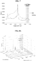
- the alkyne peak is negligible due to non-existence of induced lipid peroxidation process, confirming the specificity of the detection system. It is worth noting that the alkynes possess a unique Raman peak which is isolated from the signature region of Raman peaks characteristic of cells, thus enabling specific and interference-free detection.
- SERS mapping was performed by monitoring the SERS intensity of the alkyne peak at 2113 cm -1 .
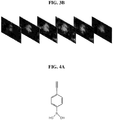
- the SERS mapping was performed in an area 50 ⁇ m ⁇ 40 ⁇ m as shown in FIG. 3A , where one cell was mapped and the mapping focus distances was scanned from -10 ⁇ m to 15 ⁇ m with a 5 ⁇ m steps.
- the SERS map indicates that the alkyne signal was localized within the cell, confirming that LAA was able to specifically modify proteins with alkyne group in cell upon lipid peroxidation.
- This result also indicated that after mixing the cells with high concentration of AuNPs, gold nanoparticles may be found on the surface and inside the cell, thus signal from LAA within a whole cell was vastly enhanced, demonstrating that the sensor concept disclosed herein is capable of monitoring cellular lipid peroxidation.
- HC-PCF based SERS platform capable of accurately monitoring lipid peroxidation derived protein modifications in cells with the use of LAA and AuNPs has been demonstrated herein.
- the SERS technique exploited the hollow core of HC-PCF fiber to provide high sensitivity, while the LAA enabled specificity and interference-free detection. More importantly, the detection avoided tedious and time consuming preparation and stain procedures, which rendered it possible to rapidly monitor the lipid peroxidation progress in cells.
- Use of commercial Raman microscopes integrated with the novel designed fiber demonstrated its possible application for rapid fluidic monitoring of lipid peroxidation.
- a HC-PCF was used to achieve sensitive detection and quantification of sialic acid on single living cells.
- the HC-PCF was designed to allow liquid samples to be pumped through the center air hole while light was confined and transmitted in. It was worth noting that the size of the center air hole was comparative with a cell (about 25 ⁇ m by 25 ⁇ m), thus permitting single cell loading within the channel, thereby making it possible for SERS detection of single living cell.
- the cells were mixed with AuNP solution and pumped into the air channel of HC-PCF, thus each cell is surrounded volumetrically with large amounts of AuNP. This mixture is squeezed in the air channel, therefore achieving '3D' SERS enhancement with extremely high sensitivity which is ideal for sialic acid detection on single cell.
- PBA phenyl-boronic acid
- 1,2 or 1,3 diols which are abundant in glycan structures, to form five- or six-membered cyclic complexes at a pH value around 8 to 9.
- Kataoka et al. reported that PBA can form favorable binding with sialic acid among common carbohydrates (mannose, glucose, galactose, and sialic acid) on surface of cell at the physiological pH of 7.4, which make it possible for living cell labeling.
- Liu et al proposed the use of phenyl-boronic acid-tagged quantum dots to label sialic acid on cell membrane for fluorescence imaging.
- HC-PCF based system for SERS detection and quantification of sialic acid on single living cancer cells is proposed.
- SERS intensity of DBA signal with different sizes of AuNP was studied to find the AuNP with optimized SERS enhancement.
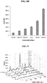
- concentrations of DBA solution were pumped in HC-PCF, and SERS intensities were obtained to plot the calibration curve.
- Sialic acid detection was performed with HeLa cells, as they are known to be sialic acid positive. Four conditions simulating the different levels of sialic acid on cells were tested in the study.
- Finite element method (FEM) modeling using commercial software COMSOL Multiphysics was utilized. 3D RF module was adopted to calculate the enhancement factor of AuNP with different sizes. E-field distribution of AuNP was calculated within a square box, while port boundary condition was set at the top and bottom face of the box with 633 nm light excitation at the top propagated along z axis, while E-field was set to vibrate along x-axis. Two faces parallel to the E-field (x-axis) was set to perfect magnetic conductor (PMC) boundary condition and another two faces perpendicular to the E-field is set to perfect electric conductor (PEC) boundary condition.
- PMC perfect magnetic conductor
- PEC perfect electric conductor
- the refractive index of the subdomain surrounding the AuNP was 1.33, while the refractive index of AuNP was set to 0.19683-j ⁇ 3.0905. It is generally agreed that the SERS enhancement of AuNP may be approximated by the electromagnetic field enhanced from localized surface plasmon resonance effect.
- the enhancement factor may be described as follow EF SERS ⁇ E loc ⁇ i 2 E loc ⁇ s 2 E 0 ⁇ i 2 E 0 ⁇ s 2
- E loc ( ⁇ i ) and E 0 ( ⁇ i ) are the local and incident electric fields at the laser excitation frequency.
- the emitted Raman radiation is enhanced by the plasmon resonance which is proportional to the local electric fields at the frequency of the Raman scattered frequency.
- E loc ( ⁇ s ) and E 0 ( ⁇ s ) refer to the local and incident electric fields at the Raman scattered frequency.
- the HC-PCF fibers were cut into 7-centimeter lengths and both ends were cleaved using an optical fiber cleaver.
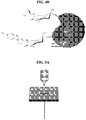
- One end of the fiber was plugged into the tip of a 0.3 mm syringe needle (BD PrecisionGlide) and the connection was sealed with a strong adhesive. Therefore, the liquid sample may be pumped into the fiber through a connected 1 ml tuberculin syringe (BD PrecisionGlide) for SERS measurement, as shown in FIG. 5B . It is worth noting that the maximum volume of liquid in the air channel of this fiber segment is about 46.2 nL.
- HeLa cells were cultured in Dulbecco's modified eagle medium (DMEM) with 1% penicillin streptomycin (Gibco) and 10 % fetal bovine serum (Gibco) in the dark with 5 % CO2 and 37 °C until they were confluent. The cells were then harvested by adding 1 ⁇ EDTA trypsin of appropriate volume for 4 min, followed by addition of DMEM of double that volume to neutralize the trypsin. The harvested cells were centrifuged at 1,500 rpm for 3 mins. The supernatant was removed and the cells were suspended with DMEM to achieve a cell concentration of 5000 cells/ml. Two milliliter of cell suspension was seeded per well in a six-well plate and allowed to grow for 48 hours.
- DMEM Dulbecco's modified eagle medium
- DMEM Dulbecco's modified eagle medium
- Gibco penicillin streptomycin
- DMEM fetal bovine serum
- Post-incubation washing was performed for all the wells and 10 mM of DBA (Sigma Aldrich) dissolved in DMEM and 1% DMSO was added into wells A, B and C, with incubation time of 4 hours. All four wells were washed twice with 1 ⁇ PBS to remove excess compounds and unbound cells, and then harvested by trypsinization and centrifugation. Supernatant were removed after centrifugation and the cells are ready for SERS sample preparation.
- DBA Sigma Aldrich
- TEM Transmission Electron Microscope
- SERS spectra were recorded using a Raman microscope system (Renishaw InVia) with a 633 nm excitation laser and equipped with a grating (spectral resolution of about 1.8 cm -1 ).
- a microscope (Leica) and a CCD detector cooled at -70° C were connected to the system.
- the laser was coupled through 50 ⁇ objective lens, which was used to collect the Stokes-shifted Raman signal. Rayleigh scattering was blocked with a notch filter.
- the samples were excited with laser power of about 3.5 mW.
- the instrument was calibrated with a silicon standard at a Raman peak of 520 cm -1 .
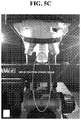
- the fiber with liquid sample pumped in was mounted on the microscope stage using a SMA connector and light was coupled into the free-end of the fiber through the objective lens, as shown in FIG. 5A and FIG. 5C .
- the SERS signal from the entire length of the fiber was collected in a backscattering geometry.
- the HeLa cell and AuNP mixture in fiber was pumped out by air using an empty syringe and loaded onto a glass slide.
- the samples were visualized using an enhanced dark-field illumination system (CytoViva, Auburn, AL) attached to a Nikon LV100 microscope.
- the system consists of a dark-field condenser (CytoViva) attached via a fiber optic light guide to a 24-watt metal halide light source (Solarc Lighting Technology). Images were acquired at 300 ms exposure time using a Nikon objective lens (100 ⁇ , NA 1.25 and WD 0.23, oil lens) and a Nikon DS-Fi1 camera with associated software (NIS-Elements D).
- AuNP were used in the SERS detection as they were able to dramatically enhance Raman signals.
- the inventors investigated the correlation of DBA intensity enhancement with different diameter of AuNP ranging from 5 to 250 nm in aqueous solutions. The sample solutions were tested on glass slide and in HC-PCF fiber to compare their enhancement capability.
- the signals obtained from glass slide were much weaker than that those of HC-PCF fiber. This was because laser light was scattered diffusely after impinging upon the sample on glass slide, thus only a portion of the scattered light was able to be collected by the objective lens. While for HC-PCF, the scattered light may be conducted back to the objective lens. More importantly, the laser light was able to interact with sample solution for a much longer distance through the whole length of fiber than on glass slide, therefore resulting in much stronger signal intensity.
- DBA has three major Raman peaks at 1,174 cm -1 , 1,592 cm -1 and 2,000 cm -1 that may potentially be used for intensity calibration.
- Raman signal from cells overlaps with DBA signal below 1800 cm -1 , therefore interfering with the intensities for DBA peak at 1,174 cm -1 and 1,592 cm -1 , resulting in incorrect detection value.
- DBA peak at 2,000 cm -1 from its alkyne functional group is beyond the aforementioned range and within a relative clean region. Therefore, it is the choice peak for the calibration study.
- the number of DBA molecules for each liquid sample in the 46.2 nL air channel of the fiber were 2.31 fmoles, 4.62 fmoles, 23.1 fmoles, 46.2 fmoles, 462 fmoles and 4.62 pmoles, respectively.
- the inventors manipulated the sialic acid level by incubating cell in the presence of prednisolone (which encourages production of sialic acid) and PBA (which binds sialic acid competitively with DBA, depending on the sequence in which the compounds were added to the cells).
- the sialic acid level in the HeLa cells may be elevated through treatment with prednisolone, a chemical which can inhibit the shedding of surface sialopeptides into the medium.
- prednisolone a chemical which can inhibit the shedding of surface sialopeptides into the medium.
- Normal HeLa cells tagged with and without DBA were used for positive (Sialic acid) and negative (Cell only) controls.
- FIG. 5A to FIG. 5C these samples were pumped by syringe into the HC-PCF and mounted on the stage of Raman system by a fiber connector, therefore enabling the transmission of laser light into the fiber for SERS detection.
- the inventors fixed the concentration of cells in each sample to be 20,000 cells/mL, and the cells were uniformly dispersed before pumping them into the fiber.
- the resultant estimated number of cells in a 46.2 nL air channel of the fiber was less than one (about 0.862). This ensured that the detection of sialic acid was restricted to single cell.
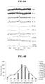
- the inventors use an empty syringe to pump out the liquid sample from fiber on to a glass slide after SERS measurement. Dark-field imaging was used to exam the number of cells in such sample, as shown in FIG. 5B . It was observed that only one HeLa cell was found in the field.
- Sialic acid a2-6Gal N-glycan Integrins Some carcinom as Alteration of integrin function Enhancement of invasion Poor prognosis in some cancers
- Sialic acid a2-6GalNAc Tn Mucins Some carcinom as Enhancement of invasion Tumor marker.
- N-CAM neural cell adhesion molecule
- Tn Siaa2-3GalNAca-Ser/Thr.
- the inventors successfully demonstrated a novel SERS based platform that can detect and quantify sialic acid on single living cancer cells using HC-PCF based system.
- this is the first reported SERS-based fiber platform that can accurately quantify components on single living cell.
- our method is much simpler and easier to perform.
- the inventors were able to achieve highly sensitive detection of compounds on a single cell.
- the system may be used in sialic acid detection, and may also be developed into clinical diagnostic tool, which will find broad applications in highly sensitive detection of biomolecules from various types of liquid samples such as blood, serum, plasma, urine, saliva, sweat and tears.
Landscapes
- Health & Medical Sciences (AREA)
- Chemical & Material Sciences (AREA)
- Life Sciences & Earth Sciences (AREA)
- Immunology (AREA)
- Organic Chemistry (AREA)
- Engineering & Computer Science (AREA)
- Molecular Biology (AREA)
- Analytical Chemistry (AREA)
- Pathology (AREA)
- Physics & Mathematics (AREA)
- General Physics & Mathematics (AREA)
- Biochemistry (AREA)
- General Health & Medical Sciences (AREA)
- Biomedical Technology (AREA)
- Hematology (AREA)
- Urology & Nephrology (AREA)
- Nuclear Medicine, Radiotherapy & Molecular Imaging (AREA)
- Tropical Medicine & Parasitology (AREA)
- Microbiology (AREA)
- Cell Biology (AREA)
- Food Science & Technology (AREA)
- Medicinal Chemistry (AREA)
- Biotechnology (AREA)
- Investigating, Analyzing Materials By Fluorescence Or Luminescence (AREA)
Claims (14)
- Procédé destiné à évaluer l'état d'une cellule vivante en utilisant une spectroscopie de Raman exaltée de surface « SERS », le procédé comprenant les étapes suivantes :a) modifier une ou plusieurs cellules vivantes avec un composé qui contient un alcyne afin de former une ou plusieurs cellules vivantes modifiées,b) mélanger la ou les cellules vivantes modifiées avec un matériau actif SERS afin de former un mélange,c) injecter le mélange dans un conduit défini par une paroi intérieure d'une fibre à cristal photonique à cœur creux, etd) détecter un signal de Raman exalté de surface en provenance du mélange dans le conduit,
dans lequel le conduit est dimensionné (i) afin de recevoir seulement une cellule vivante modifiée à chaque emplacement sur sa longueur, espacée d'une autre cellule vivante modifiée, et (ii) pour que chaque cellule vivante modifiée soit entourée de manière volumétrique avec le matériau actif SERS. - Procédé selon la revendication 1, dans lequel la modification d'une ou plusieurs cellules vivantes avec le composé qui contient un alcyne comprend de mettre en incubation la ou les cellules vivantes avec le composé qui contient un alcyne, pendant une période de temps comprise d'environ 1 heure à environ 5 heures.
- Procédé selon la revendication 1, dans lequel le composé qui contient un alcyne est sélectionné dans : (i) le groupe constitué par un acide gras insaturé modifié par alcyne, un acide boronique fonctionnalisé par alcyne, et des combinaisons de ceux-ci, ou (ii) le groupe constitué par un alcyne de linoléamide, un acétylène 4-(dihydroxyborophényle), et des combinaisons de ceux-ci.
- Procédé selon la revendication 1, dans lequel le matériau actif SERS comprend : (i) des nanoparticules de matériau actif SERS, ou (ii) des nanoparticules d'or.
- Procédé selon la revendication 4, dans lequel chaque nanoparticule de matériau actif SERS présente un diamètre qui se situe dans une plage (i) de 5 nm environ à 250 nm environ, ou (ii) de 60 nm environ à 100 nm environ.
- Procédé selon la revendication 4, dans lequel la concentration des nanoparticules de matériau actif SERS dans le mélange se situe dans une plage de 1 × 10¹⁰ particules/ml environ à 1 × 10¹² particules/ml environ.
- Procédé selon la revendication 1, dans lequel la concentration de la ou des cellules vivantes modifiées dans le mélange se situe dans une plage de 20 000 cellules/ml environ à 30 000 cellules/ml environ.
- Procédé selon la revendication 1, dans lequel le conduit est dimensionné afin de recevoir une cellule vivante modifiée.
- Procédé selon la revendication 1, dans lequel détecter un signal de Raman exalté de surface provenant du mélange dans le conduit comprend de détecter des changements de motif et / ou d'intensité du signal de Raman exalté de surface dans la région de 1800 cm-1 à 2200 cm-1.
- Procédé selon la revendication 1, dans lequel détecter un signal de Raman exalté de surface en provenance du mélange dans le conduit comprend de diriger un rayonnement dans le conduit pendant une période de temps de 8 secondes environ à 12 secondes environ.
- Procédé selon la revendication 1, dans lequel détecter un signal de Raman exalté de surface en provenance du mélange dans le conduit comprend de collecter le signal de Raman exalté de surface dans une géométrie de rétrodiffusion.
- Procédé selon la revendication 1, dans lequel évaluer un état de la cellule vivante comprend de déterminer la présence et / ou l'ampleur de la peroxydation de lipide de la cellule vivante.
- Procédé selon la revendication 1, dans lequel (i) évaluer un état de la cellule vivante comprend de déterminer la présence et / ou la quantité d'acide sialique sur la cellule vivante, ou (ii) évaluer un état de la cellule vivante comprend de déterminer la présence et / ou la quantité d'acide sialique sur la cellule vivante, dans lequel la quantité d'acide sialique sur la cellule vivante est corrélée avec le signal de Raman exalté de surface en provenance du mélange dans le conduit.
- Procédé selon la revendication 1, dans lequel la ou les cellules vivantes sont contenues dans un échantillon et la détection s'effectue in vitro.
Applications Claiming Priority (2)
| Application Number | Priority Date | Filing Date | Title |
|---|---|---|---|
| SG10201600721R | 2016-01-29 | ||
| PCT/SG2017/050014 WO2017131583A1 (fr) | 2016-01-29 | 2017-01-12 | Procédé pour évaluer l'état d'une cellule vivante |
Publications (3)
| Publication Number | Publication Date |
|---|---|
| EP3408655A1 EP3408655A1 (fr) | 2018-12-05 |
| EP3408655A4 EP3408655A4 (fr) | 2019-08-28 |
| EP3408655B1 true EP3408655B1 (fr) | 2021-06-23 |
Family
ID=59398486
Family Applications (1)
| Application Number | Title | Priority Date | Filing Date |
|---|---|---|---|
| EP17744653.1A Active EP3408655B1 (fr) | 2016-01-29 | 2017-01-12 | Procédé pour évaluer l'état d'une cellule vivante |
Country Status (5)
| Country | Link |
|---|---|
| US (1) | US10955352B2 (fr) |
| EP (1) | EP3408655B1 (fr) |
| CN (1) | CN108603842B (fr) |
| SG (2) | SG10201913138TA (fr) |
| WO (1) | WO2017131583A1 (fr) |
Families Citing this family (7)
| Publication number | Priority date | Publication date | Assignee | Title |
|---|---|---|---|---|
| WO2020180710A1 (fr) | 2019-03-01 | 2020-09-10 | The Board Of Regents Of The University Of Oklahoma | Analyse automatique de diffusion raman exaltée de surface (sers) en temps réel |
| US11624742B2 (en) | 2019-10-18 | 2023-04-11 | The Board Of Regents Of The University Of Oklahoma | Photonic crystal gas sensor |
| CN114235773B (zh) * | 2020-09-09 | 2024-02-09 | 南京大学 | 一种用于细胞膜修复过程动态监测的拉曼成像方法 |
| CN112255215B (zh) * | 2020-09-16 | 2021-08-17 | 清华大学 | 检测分析活体细胞生命活动状态的修饰二聚体表面增强拉曼的方法 |
| CN113624736B (zh) * | 2021-07-07 | 2023-12-08 | 张鑫 | 一种基于拉曼效应的微生物浓度快速检测方法 |
| CN115707955A (zh) * | 2021-08-20 | 2023-02-21 | 南开大学 | 一种基于温度控制高碘酸钠选择性氧化的细胞唾液酸原位成像方法 |
| US12332151B2 (en) | 2021-08-30 | 2025-06-17 | Ernst-Ulrich Berndt | Near real-time, hands-off detection of airborne particulate contaminants and bioburden |
Family Cites Families (7)
| Publication number | Priority date | Publication date | Assignee | Title |
|---|---|---|---|---|
| US20070134733A1 (en) * | 2003-07-10 | 2007-06-14 | Parallax Biosystems, Inc. | Raman signature probes and their use in the detection and imaging of molecular processes and structures |
| CN1815197A (zh) * | 2006-02-24 | 2006-08-09 | 清华大学 | 基于纳米颗粒表面增强拉曼谱的光子晶体光纤探针传感器 |
| US8717558B2 (en) * | 2007-09-04 | 2014-05-06 | The Regents Of The University Of California | Liquid core photonic crystal fiber biosensors using surface enhanced Raman scattering and methods for their use |
| US7595882B1 (en) * | 2008-04-14 | 2009-09-29 | Geneal Electric Company | Hollow-core waveguide-based raman systems and methods |
| CN102947738B (zh) * | 2010-06-09 | 2015-07-15 | 新加坡科技研究局 | 光子晶体光纤传感器 |
| CN102661941B (zh) * | 2012-04-19 | 2015-04-22 | 湖南大学 | 基于拉曼活性纳米颗粒混合组装的循环肿瘤细胞的耦合增强sers高通量生物传感方法 |
| WO2015038077A1 (fr) * | 2013-09-16 | 2015-03-19 | Agency For Science, Technology And Research | Procédé de détection de peroxyde d'hydrogène |
-
2017
- 2017-01-12 SG SG10201913138TA patent/SG10201913138TA/en unknown
- 2017-01-12 EP EP17744653.1A patent/EP3408655B1/fr active Active
- 2017-01-12 CN CN201780008826.4A patent/CN108603842B/zh not_active Expired - Fee Related
- 2017-01-12 US US16/071,728 patent/US10955352B2/en not_active Expired - Fee Related
- 2017-01-12 WO PCT/SG2017/050014 patent/WO2017131583A1/fr not_active Ceased
- 2017-01-12 SG SG11201805912XA patent/SG11201805912XA/en unknown
Non-Patent Citations (1)
| Title |
|---|
| None * |
Also Published As
| Publication number | Publication date |
|---|---|
| SG10201913138TA (en) | 2020-02-27 |
| EP3408655A4 (fr) | 2019-08-28 |
| EP3408655A1 (fr) | 2018-12-05 |
| US10955352B2 (en) | 2021-03-23 |
| CN108603842B (zh) | 2021-06-25 |
| WO2017131583A1 (fr) | 2017-08-03 |
| SG11201805912XA (en) | 2018-08-30 |
| CN108603842A (zh) | 2018-09-28 |
| US20190033218A1 (en) | 2019-01-31 |
Similar Documents
| Publication | Publication Date | Title |
|---|---|---|
| EP3408655B1 (fr) | Procédé pour évaluer l'état d'une cellule vivante | |
| Gong et al. | Highly sensitive SERS detection and quantification of sialic acid on single cell using photonic-crystal fiber with gold nanoparticles | |
| Koike et al. | Quantitative drug dynamics visualized by alkyne-tagged plasmonic-enhanced Raman microscopy | |
| Bekmurzayeva et al. | Ultra-wide, attomolar-level limit detection of CD44 biomarker with a silanized optical fiber biosensor | |
| Lima et al. | The role of Raman spectroscopy within quantitative metabolomics | |
| Ren et al. | A fast responsive two-photon fluorescent probe for imaging H2O2 in lysosomes with a large turn-on fluorescence signal | |
| Dipalo et al. | 3D plasmonic nanoantennas integrated with MEA biosensors | |
| Cialla-May et al. | Recent progress in surface-enhanced Raman spectroscopy for biological and biomedical applications: from cells to clinics | |
| Datta et al. | Fluorescence lifetime imaging of endogenous biomarker of oxidative stress | |
| Sigaeva et al. | Optical detection of intracellular quantities using nanoscale technologies | |
| Zhang et al. | SERS nanotags and their applications in biosensing and bioimaging | |
| Li et al. | A SERS nano-tag-based fiber-optic strategy for in situ immunoassay in unprocessed whole blood | |
| Shen et al. | Interference-free surface-enhanced Raman scattering nanosensor for imaging and dynamic monitoring of reactive oxygen species in mitochondria during photothermal therapy | |
| Ma et al. | Individual plasmonic nanoprobes for biosensing and bioimaging: recent advances and perspectives | |
| Chen et al. | Kill two birds with one stone: A near-infrared ratiometric fluorescent probe for simultaneous detection of β-galactosidase in senescent and cancer cells | |
| Al-Sammarraie et al. | Silver nanoparticles-based substrate for blood serum analysis under 785 nm laser excitation | |
| Amouzadeh Tabrizi et al. | Remote biosensor for the determination of trypsin by using nanoporous anodic alumina as a three-dimensional nanostructured material | |
| Sloan-Dennison et al. | From Raman to SESORRS: moving deeper into cancer detection and treatment monitoring | |
| Moorthy et al. | Optical biosensors for detection of cancer biomarkers: current and future perspectives | |
| Liu et al. | Tracking endocytosis and intracellular distribution of spherical nucleic acids with correlative single-cell imaging | |
| Rodríguez-Sevilla et al. | Multichannel fluorescence microscopy: advantages of going beyond a single emission | |
| CN115046986B (zh) | 一种基于空芯反谐振光纤和表面增强拉曼光谱的生化传感器 | |
| Alijanianzadeh et al. | Detection of methamphetamine using aptamer-based biosensor chip and cyclic voltammetry technique | |
| Wu et al. | A 3D-printed SERS bionic taster for dynamic tumor metabolites detection | |
| Amani et al. | Smartphone-assisted quantitative colorimetric identification of thrombin based on peroxidase mimetic features of fibrinogen-gold nanozymes |
Legal Events
| Date | Code | Title | Description |
|---|---|---|---|
| STAA | Information on the status of an ep patent application or granted ep patent |
Free format text: STATUS: THE INTERNATIONAL PUBLICATION HAS BEEN MADE |
|
| PUAI | Public reference made under article 153(3) epc to a published international application that has entered the european phase |
Free format text: ORIGINAL CODE: 0009012 |
|
| STAA | Information on the status of an ep patent application or granted ep patent |
Free format text: STATUS: REQUEST FOR EXAMINATION WAS MADE |
|
| 17P | Request for examination filed |
Effective date: 20180828 |
|
| AK | Designated contracting states |
Kind code of ref document: A1 Designated state(s): AL AT BE BG CH CY CZ DE DK EE ES FI FR GB GR HR HU IE IS IT LI LT LU LV MC MK MT NL NO PL PT RO RS SE SI SK SM TR |
|
| AX | Request for extension of the european patent |
Extension state: BA ME |
|
| DAV | Request for validation of the european patent (deleted) | ||
| DAX | Request for extension of the european patent (deleted) | ||
| A4 | Supplementary search report drawn up and despatched |
Effective date: 20190731 |
|
| RIC1 | Information provided on ipc code assigned before grant |
Ipc: B82Y 15/00 20110101ALN20190725BHEP Ipc: G01N 21/65 20060101AFI20190725BHEP Ipc: G02B 6/02 20060101ALN20190725BHEP |
|
| STAA | Information on the status of an ep patent application or granted ep patent |
Free format text: STATUS: EXAMINATION IS IN PROGRESS |
|
| 17Q | First examination report despatched |
Effective date: 20200518 |
|
| RIC1 | Information provided on ipc code assigned before grant |
Ipc: G01N 21/65 20060101AFI20201216BHEP Ipc: B82Y 20/00 20110101ALN20201216BHEP Ipc: G02B 6/02 20060101ALN20201216BHEP |
|
| GRAP | Despatch of communication of intention to grant a patent |
Free format text: ORIGINAL CODE: EPIDOSNIGR1 |
|
| STAA | Information on the status of an ep patent application or granted ep patent |
Free format text: STATUS: GRANT OF PATENT IS INTENDED |
|
| INTG | Intention to grant announced |
Effective date: 20210122 |
|
| GRAS | Grant fee paid |
Free format text: ORIGINAL CODE: EPIDOSNIGR3 |
|
| GRAA | (expected) grant |
Free format text: ORIGINAL CODE: 0009210 |
|
| STAA | Information on the status of an ep patent application or granted ep patent |
Free format text: STATUS: THE PATENT HAS BEEN GRANTED |
|
| AK | Designated contracting states |
Kind code of ref document: B1 Designated state(s): AL AT BE BG CH CY CZ DE DK EE ES FI FR GB GR HR HU IE IS IT LI LT LU LV MC MK MT NL NO PL PT RO RS SE SI SK SM TR |
|
| REG | Reference to a national code |
Ref country code: GB Ref legal event code: FG4D |
|
| REG | Reference to a national code |
Ref country code: CH Ref legal event code: EP |
|
| REG | Reference to a national code |
Ref country code: DE Ref legal event code: R096 Ref document number: 602017040801 Country of ref document: DE Ref country code: AT Ref legal event code: REF Ref document number: 1404709 Country of ref document: AT Kind code of ref document: T Effective date: 20210715 |
|
| REG | Reference to a national code |
Ref country code: IE Ref legal event code: FG4D |
|
| REG | Reference to a national code |
Ref country code: LT Ref legal event code: MG9D |
|
| PG25 | Lapsed in a contracting state [announced via postgrant information from national office to epo] |
Ref country code: BG Free format text: LAPSE BECAUSE OF FAILURE TO SUBMIT A TRANSLATION OF THE DESCRIPTION OR TO PAY THE FEE WITHIN THE PRESCRIBED TIME-LIMIT Effective date: 20210923 Ref country code: HR Free format text: LAPSE BECAUSE OF FAILURE TO SUBMIT A TRANSLATION OF THE DESCRIPTION OR TO PAY THE FEE WITHIN THE PRESCRIBED TIME-LIMIT Effective date: 20210623 Ref country code: LT Free format text: LAPSE BECAUSE OF FAILURE TO SUBMIT A TRANSLATION OF THE DESCRIPTION OR TO PAY THE FEE WITHIN THE PRESCRIBED TIME-LIMIT Effective date: 20210623 Ref country code: FI Free format text: LAPSE BECAUSE OF FAILURE TO SUBMIT A TRANSLATION OF THE DESCRIPTION OR TO PAY THE FEE WITHIN THE PRESCRIBED TIME-LIMIT Effective date: 20210623 |
|
| REG | Reference to a national code |
Ref country code: AT Ref legal event code: MK05 Ref document number: 1404709 Country of ref document: AT Kind code of ref document: T Effective date: 20210623 |
|
| PG25 | Lapsed in a contracting state [announced via postgrant information from national office to epo] |
Ref country code: NO Free format text: LAPSE BECAUSE OF FAILURE TO SUBMIT A TRANSLATION OF THE DESCRIPTION OR TO PAY THE FEE WITHIN THE PRESCRIBED TIME-LIMIT Effective date: 20210923 Ref country code: SE Free format text: LAPSE BECAUSE OF FAILURE TO SUBMIT A TRANSLATION OF THE DESCRIPTION OR TO PAY THE FEE WITHIN THE PRESCRIBED TIME-LIMIT Effective date: 20210623 Ref country code: RS Free format text: LAPSE BECAUSE OF FAILURE TO SUBMIT A TRANSLATION OF THE DESCRIPTION OR TO PAY THE FEE WITHIN THE PRESCRIBED TIME-LIMIT Effective date: 20210623 Ref country code: GR Free format text: LAPSE BECAUSE OF FAILURE TO SUBMIT A TRANSLATION OF THE DESCRIPTION OR TO PAY THE FEE WITHIN THE PRESCRIBED TIME-LIMIT Effective date: 20210924 Ref country code: LV Free format text: LAPSE BECAUSE OF FAILURE TO SUBMIT A TRANSLATION OF THE DESCRIPTION OR TO PAY THE FEE WITHIN THE PRESCRIBED TIME-LIMIT Effective date: 20210623 |
|
| REG | Reference to a national code |
Ref country code: NL Ref legal event code: MP Effective date: 20210623 |
|
| PG25 | Lapsed in a contracting state [announced via postgrant information from national office to epo] |
Ref country code: SK Free format text: LAPSE BECAUSE OF FAILURE TO SUBMIT A TRANSLATION OF THE DESCRIPTION OR TO PAY THE FEE WITHIN THE PRESCRIBED TIME-LIMIT Effective date: 20210623 Ref country code: SM Free format text: LAPSE BECAUSE OF FAILURE TO SUBMIT A TRANSLATION OF THE DESCRIPTION OR TO PAY THE FEE WITHIN THE PRESCRIBED TIME-LIMIT Effective date: 20210623 Ref country code: PT Free format text: LAPSE BECAUSE OF FAILURE TO SUBMIT A TRANSLATION OF THE DESCRIPTION OR TO PAY THE FEE WITHIN THE PRESCRIBED TIME-LIMIT Effective date: 20211025 Ref country code: RO Free format text: LAPSE BECAUSE OF FAILURE TO SUBMIT A TRANSLATION OF THE DESCRIPTION OR TO PAY THE FEE WITHIN THE PRESCRIBED TIME-LIMIT Effective date: 20210623 Ref country code: NL Free format text: LAPSE BECAUSE OF FAILURE TO SUBMIT A TRANSLATION OF THE DESCRIPTION OR TO PAY THE FEE WITHIN THE PRESCRIBED TIME-LIMIT Effective date: 20210623 Ref country code: ES Free format text: LAPSE BECAUSE OF FAILURE TO SUBMIT A TRANSLATION OF THE DESCRIPTION OR TO PAY THE FEE WITHIN THE PRESCRIBED TIME-LIMIT Effective date: 20210623 Ref country code: AT Free format text: LAPSE BECAUSE OF FAILURE TO SUBMIT A TRANSLATION OF THE DESCRIPTION OR TO PAY THE FEE WITHIN THE PRESCRIBED TIME-LIMIT Effective date: 20210623 Ref country code: CZ Free format text: LAPSE BECAUSE OF FAILURE TO SUBMIT A TRANSLATION OF THE DESCRIPTION OR TO PAY THE FEE WITHIN THE PRESCRIBED TIME-LIMIT Effective date: 20210623 Ref country code: EE Free format text: LAPSE BECAUSE OF FAILURE TO SUBMIT A TRANSLATION OF THE DESCRIPTION OR TO PAY THE FEE WITHIN THE PRESCRIBED TIME-LIMIT Effective date: 20210623 |
|
| PG25 | Lapsed in a contracting state [announced via postgrant information from national office to epo] |
Ref country code: PL Free format text: LAPSE BECAUSE OF FAILURE TO SUBMIT A TRANSLATION OF THE DESCRIPTION OR TO PAY THE FEE WITHIN THE PRESCRIBED TIME-LIMIT Effective date: 20210623 |
|
| REG | Reference to a national code |
Ref country code: DE Ref legal event code: R097 Ref document number: 602017040801 Country of ref document: DE |
|
| PG25 | Lapsed in a contracting state [announced via postgrant information from national office to epo] |
Ref country code: DK Free format text: LAPSE BECAUSE OF FAILURE TO SUBMIT A TRANSLATION OF THE DESCRIPTION OR TO PAY THE FEE WITHIN THE PRESCRIBED TIME-LIMIT Effective date: 20210623 |
|
| PGFP | Annual fee paid to national office [announced via postgrant information from national office to epo] |
Ref country code: DE Payment date: 20220107 Year of fee payment: 6 |
|
| PLBE | No opposition filed within time limit |
Free format text: ORIGINAL CODE: 0009261 |
|
| STAA | Information on the status of an ep patent application or granted ep patent |
Free format text: STATUS: NO OPPOSITION FILED WITHIN TIME LIMIT |
|
| 26N | No opposition filed |
Effective date: 20220324 |
|
| PG25 | Lapsed in a contracting state [announced via postgrant information from national office to epo] |
Ref country code: AL Free format text: LAPSE BECAUSE OF FAILURE TO SUBMIT A TRANSLATION OF THE DESCRIPTION OR TO PAY THE FEE WITHIN THE PRESCRIBED TIME-LIMIT Effective date: 20210623 |
|
| PGFP | Annual fee paid to national office [announced via postgrant information from national office to epo] |
Ref country code: FR Payment date: 20220120 Year of fee payment: 6 |
|
| PG25 | Lapsed in a contracting state [announced via postgrant information from national office to epo] |
Ref country code: IT Free format text: LAPSE BECAUSE OF FAILURE TO SUBMIT A TRANSLATION OF THE DESCRIPTION OR TO PAY THE FEE WITHIN THE PRESCRIBED TIME-LIMIT Effective date: 20210623 |
|
| PG25 | Lapsed in a contracting state [announced via postgrant information from national office to epo] |
Ref country code: MC Free format text: LAPSE BECAUSE OF FAILURE TO SUBMIT A TRANSLATION OF THE DESCRIPTION OR TO PAY THE FEE WITHIN THE PRESCRIBED TIME-LIMIT Effective date: 20210623 |
|
| REG | Reference to a national code |
Ref country code: CH Ref legal event code: PL |
|
| GBPC | Gb: european patent ceased through non-payment of renewal fee |
Effective date: 20220112 |
|
| REG | Reference to a national code |
Ref country code: BE Ref legal event code: MM Effective date: 20220131 |
|
| PG25 | Lapsed in a contracting state [announced via postgrant information from national office to epo] |
Ref country code: LU Free format text: LAPSE BECAUSE OF NON-PAYMENT OF DUE FEES Effective date: 20220112 Ref country code: GB Free format text: LAPSE BECAUSE OF NON-PAYMENT OF DUE FEES Effective date: 20220112 |
|
| PG25 | Lapsed in a contracting state [announced via postgrant information from national office to epo] |
Ref country code: BE Free format text: LAPSE BECAUSE OF NON-PAYMENT OF DUE FEES Effective date: 20220131 |
|
| PG25 | Lapsed in a contracting state [announced via postgrant information from national office to epo] |
Ref country code: LI Free format text: LAPSE BECAUSE OF NON-PAYMENT OF DUE FEES Effective date: 20220131 Ref country code: CH Free format text: LAPSE BECAUSE OF NON-PAYMENT OF DUE FEES Effective date: 20220131 |
|
| PG25 | Lapsed in a contracting state [announced via postgrant information from national office to epo] |
Ref country code: IE Free format text: LAPSE BECAUSE OF NON-PAYMENT OF DUE FEES Effective date: 20220112 |
|
| REG | Reference to a national code |
Ref country code: DE Ref legal event code: R119 Ref document number: 602017040801 Country of ref document: DE |
|
| PG25 | Lapsed in a contracting state [announced via postgrant information from national office to epo] |
Ref country code: DE Free format text: LAPSE BECAUSE OF NON-PAYMENT OF DUE FEES Effective date: 20230801 |
|
| PG25 | Lapsed in a contracting state [announced via postgrant information from national office to epo] |
Ref country code: FR Free format text: LAPSE BECAUSE OF NON-PAYMENT OF DUE FEES Effective date: 20230131 |
|
| PG25 | Lapsed in a contracting state [announced via postgrant information from national office to epo] |
Ref country code: HU Free format text: LAPSE BECAUSE OF FAILURE TO SUBMIT A TRANSLATION OF THE DESCRIPTION OR TO PAY THE FEE WITHIN THE PRESCRIBED TIME-LIMIT; INVALID AB INITIO Effective date: 20170112 |
|
| PG25 | Lapsed in a contracting state [announced via postgrant information from national office to epo] |
Ref country code: MK Free format text: LAPSE BECAUSE OF FAILURE TO SUBMIT A TRANSLATION OF THE DESCRIPTION OR TO PAY THE FEE WITHIN THE PRESCRIBED TIME-LIMIT Effective date: 20210623 Ref country code: CY Free format text: LAPSE BECAUSE OF FAILURE TO SUBMIT A TRANSLATION OF THE DESCRIPTION OR TO PAY THE FEE WITHIN THE PRESCRIBED TIME-LIMIT Effective date: 20210623 |
|
| PG25 | Lapsed in a contracting state [announced via postgrant information from national office to epo] |
Ref country code: TR Free format text: LAPSE BECAUSE OF FAILURE TO SUBMIT A TRANSLATION OF THE DESCRIPTION OR TO PAY THE FEE WITHIN THE PRESCRIBED TIME-LIMIT Effective date: 20210623 |
|
| PG25 | Lapsed in a contracting state [announced via postgrant information from national office to epo] |
Ref country code: MT Free format text: LAPSE BECAUSE OF FAILURE TO SUBMIT A TRANSLATION OF THE DESCRIPTION OR TO PAY THE FEE WITHIN THE PRESCRIBED TIME-LIMIT Effective date: 20210623 |