EP3417052B1 - Procédés d'expansion et de différenciation de cellules b destinés à la production d'anticorps - Google Patents
Procédés d'expansion et de différenciation de cellules b destinés à la production d'anticorps Download PDFInfo
- Publication number
- EP3417052B1 EP3417052B1 EP17753829.5A EP17753829A EP3417052B1 EP 3417052 B1 EP3417052 B1 EP 3417052B1 EP 17753829 A EP17753829 A EP 17753829A EP 3417052 B1 EP3417052 B1 EP 3417052B1
- Authority
- EP
- European Patent Office
- Prior art keywords
- cells
- cell line
- cell
- human
- polypeptide
- Prior art date
- Legal status (The legal status is an assumption and is not a legal conclusion. Google has not performed a legal analysis and makes no representation as to the accuracy of the status listed.)
- Active
Links
Images




Classifications
-
- C—CHEMISTRY; METALLURGY
- C07—ORGANIC CHEMISTRY
- C07K—PEPTIDES
- C07K16/00—Immunoglobulins [IG], e.g. monoclonal or polyclonal antibodies
-
- C—CHEMISTRY; METALLURGY
- C12—BIOCHEMISTRY; BEER; SPIRITS; WINE; VINEGAR; MICROBIOLOGY; ENZYMOLOGY; MUTATION OR GENETIC ENGINEERING
- C12N—MICROORGANISMS OR ENZYMES; COMPOSITIONS THEREOF; PROPAGATING, PRESERVING, OR MAINTAINING MICROORGANISMS; MUTATION OR GENETIC ENGINEERING; CULTURE MEDIA
- C12N5/00—Undifferentiated human, animal or plant cells, e.g. cell lines; Tissues; Cultivation or maintenance thereof; Culture media therefor
- C12N5/06—Animal cells or tissues; Human cells or tissues
- C12N5/0602—Vertebrate cells
- C12N5/0634—Cells from the blood or the immune system
- C12N5/0635—B lymphocytes
-
- C—CHEMISTRY; METALLURGY
- C07—ORGANIC CHEMISTRY
- C07K—PEPTIDES
- C07K2317/00—Immunoglobulins specific features
- C07K2317/20—Immunoglobulins specific features characterized by taxonomic origin
- C07K2317/21—Immunoglobulins specific features characterized by taxonomic origin from primates, e.g. man
-
- C—CHEMISTRY; METALLURGY
- C12—BIOCHEMISTRY; BEER; SPIRITS; WINE; VINEGAR; MICROBIOLOGY; ENZYMOLOGY; MUTATION OR GENETIC ENGINEERING
- C12N—MICROORGANISMS OR ENZYMES; COMPOSITIONS THEREOF; PROPAGATING, PRESERVING, OR MAINTAINING MICROORGANISMS; MUTATION OR GENETIC ENGINEERING; CULTURE MEDIA
- C12N2501/00—Active agents used in cell culture processes, e.g. differentation
- C12N2501/20—Cytokines; Chemokines
- C12N2501/23—Interleukins [IL]
- C12N2501/2302—Interleukin-2 (IL-2)
-
- C—CHEMISTRY; METALLURGY
- C12—BIOCHEMISTRY; BEER; SPIRITS; WINE; VINEGAR; MICROBIOLOGY; ENZYMOLOGY; MUTATION OR GENETIC ENGINEERING
- C12N—MICROORGANISMS OR ENZYMES; COMPOSITIONS THEREOF; PROPAGATING, PRESERVING, OR MAINTAINING MICROORGANISMS; MUTATION OR GENETIC ENGINEERING; CULTURE MEDIA
- C12N2501/00—Active agents used in cell culture processes, e.g. differentation
- C12N2501/20—Cytokines; Chemokines
- C12N2501/23—Interleukins [IL]
- C12N2501/2304—Interleukin-4 (IL-4)
-
- C—CHEMISTRY; METALLURGY
- C12—BIOCHEMISTRY; BEER; SPIRITS; WINE; VINEGAR; MICROBIOLOGY; ENZYMOLOGY; MUTATION OR GENETIC ENGINEERING
- C12N—MICROORGANISMS OR ENZYMES; COMPOSITIONS THEREOF; PROPAGATING, PRESERVING, OR MAINTAINING MICROORGANISMS; MUTATION OR GENETIC ENGINEERING; CULTURE MEDIA
- C12N2501/00—Active agents used in cell culture processes, e.g. differentation
- C12N2501/20—Cytokines; Chemokines
- C12N2501/23—Interleukins [IL]
- C12N2501/231—Interleukin-10 (IL-10)
-
- C—CHEMISTRY; METALLURGY
- C12—BIOCHEMISTRY; BEER; SPIRITS; WINE; VINEGAR; MICROBIOLOGY; ENZYMOLOGY; MUTATION OR GENETIC ENGINEERING
- C12N—MICROORGANISMS OR ENZYMES; COMPOSITIONS THEREOF; PROPAGATING, PRESERVING, OR MAINTAINING MICROORGANISMS; MUTATION OR GENETIC ENGINEERING; CULTURE MEDIA
- C12N2501/00—Active agents used in cell culture processes, e.g. differentation
- C12N2501/20—Cytokines; Chemokines
- C12N2501/23—Interleukins [IL]
- C12N2501/2321—Interleukin-21 (IL-21)
-
- C—CHEMISTRY; METALLURGY
- C12—BIOCHEMISTRY; BEER; SPIRITS; WINE; VINEGAR; MICROBIOLOGY; ENZYMOLOGY; MUTATION OR GENETIC ENGINEERING
- C12N—MICROORGANISMS OR ENZYMES; COMPOSITIONS THEREOF; PROPAGATING, PRESERVING, OR MAINTAINING MICROORGANISMS; MUTATION OR GENETIC ENGINEERING; CULTURE MEDIA
- C12N2502/00—Coculture with; Conditioned medium produced by
- C12N2502/13—Coculture with; Conditioned medium produced by connective tissue cells; generic mesenchyme cells, e.g. so-called "embryonic fibroblasts"
- C12N2502/1323—Adult fibroblasts
-
- C—CHEMISTRY; METALLURGY
- C12—BIOCHEMISTRY; BEER; SPIRITS; WINE; VINEGAR; MICROBIOLOGY; ENZYMOLOGY; MUTATION OR GENETIC ENGINEERING
- C12N—MICROORGANISMS OR ENZYMES; COMPOSITIONS THEREOF; PROPAGATING, PRESERVING, OR MAINTAINING MICROORGANISMS; MUTATION OR GENETIC ENGINEERING; CULTURE MEDIA
- C12N2502/00—Coculture with; Conditioned medium produced by
- C12N2502/13—Coculture with; Conditioned medium produced by connective tissue cells; generic mesenchyme cells, e.g. so-called "embryonic fibroblasts"
- C12N2502/1352—Mesenchymal stem cells
Definitions
- the invention relates to a method for producing and supporting B cells, including human B cells, which can be used to produce antigen-specific antibodies, and more particularly, monoclonal antibodies. More specifically, the invention relates to culturing B cells to produce monoclonal antibodies without using hybridoma technology or EBV-transformed B cells.
- Monoclonal antibodies have found particular utility in medicine, such as through the development of antibody-based biologicals or pharmaceuticals.
- Current methods for monoclonal antibody production involve the mouse hybridoma method; i.e., fusing antibody producing cells with myeloma cells.
- Other methods include EBV-transformed B cell lines and phage display.
- Each of the different methods used to produce human monoclonal antibodies suffers from technical limitations that render it difficult to use. For example, with hybridoma technology there is a need to "humanize" the antibody so as to reduce the frequency of promoting an immune response to nonhuman portions of the monoclonal antibody. Additionally, immune responses to a particular antigen or epitope may be species-specific.
- Other methods have drawbacks that limit their wide application for use in developing monoclonal antibodies for use in therapy and diagnosis for humans.
- EP 2 495 312 A1 provides a method for producing an antigen-specific B cell population comprising IgG-positive B cells specific to a specific antigen.
- Masahiro Kitabatke et al., J Immunol. 2015 Feb 15;194(4):1480-8 discloses B1a cells from G5PR transgenic mice that differentiate into IgM and IgG autoantibody-secreting plasmablasts.
- the disclosure is based on the discovery of a highly efficient method for expanding and differentiating B cells in culture that enables single cell cloning of B cells.
- the discovery relates to a highly efficient method for expanding and differentiating human B cells in culture that enables single cell cloning of human B cells and production of antibodies.
- the disclosure is also based on the discovery of a highly efficient method for expanding and differentiating B cells in culture that enables single cell cloning of B cells (seeding a single B cell, and expanding that B cell into a clone of B cells) in amount and time period sufficient to generate monoclonal antibody in an amount that facilitates characterization of the monoclonal antibody produced.
- human B cells are expanded and differentiated as single clones to produce human monoclonal antibody.
- the method does not require and/or no antigen is added to the expansion culture.
- the antigen-specificity of the B cells is unknown.
- the B cells are not exposed to antigen in vitro.
- an in vitro method for the production of monoclonal antibodies particularly of the IgG type, is provided.
- feeder cell lines comprising modified mesenchymal stromal cells or modified thymic epithelial cells, which can efficiently promote and support high numbers of B cells in culture, thereby allowing higher levels of monoclonal antibody production than that observed in prior attempts of culturing B cells with modified mouse 3T3 cells.
- the feeder cell lines comprising mesenchymal stromal cells or thymic epithelial cells, modified to express a combination comprising CD154 (also known as CD40L or CD40 Ligand) and BLyS (B Lymphocyte Stimulator, also known as BAFF (B-cell activating factor)), or a combination comprising CD154, BLyS, and IL-21.
- the feeder cells of the disclosure support significant proliferation and differentiation of B cells of mammalian species, including human B cells and murine B cells, without the need to add (i.e., in the absence of) exogenous antigen or exogenous IL-4 when B cells are cultured in the presence of the modified feeder cells in vitro.
- a method for producing monoclonal antibodies that bind to a particular antigen typically a known antigen.
- the method includes the steps of isolating the B cells that are either na ⁇ ve to antigen, or have been exposed to an antigen (e.g., either by in vitro or in vivo exposure); separating the isolated B cells into single cells; culturing an isolated B cell (single B cell) in the presence of modified feeder cells (i.e.
- feeder cells expressing CD154 or another CD40 agonist and BLyS or a comparable B cell activating factor according to the disclosure in vitro under conditions and for a sufficient time to achieve at least an average of 10 4 -fold expansion in number (e.g., expansion from a single cell to 10,000 cells, average taken over wells of a multiwell plate, such as a 96 well plate) in less than 2 weeks in culture.
- a monoclonal antibody is produced from the expanded B cell clone in culture.
- Further steps may comprise characterization of the monoclonal antibody such as one or more of (a) determining the antigen specificity of the monoclonal antibody using methods known in the art; and (b) isolating the monoclonal antibody comprising (i) purification of the monoclonal antibody from the culture medium using methods known in the art, and/or (ii) isolating the total RNA from the B cell clone to produce cDNA encoding the variable-heavy (VH) antibody chains and cDNA encoding variable-light (VL) antibody chains which then can be cloned into a eukaryotic expression vector for recombinant production of the monoclonal antibody using methods known in the art.
- characterization of the monoclonal antibody such as one or more of (a) determining the antigen specificity of the monoclonal antibody using methods known in the art; and (b) isolating the monoclonal antibody comprising (i) purification of the monoclonal antibody from the culture medium using
- the selected B cell clones producing antigen-specific monoclonal antibodies can be immortalized by hybridoma technology, EBV-transformation, immortalized using the amphotropic retrovirus LXSN16E6E7 produced by the PA317 LXSN 16E6E7 cell line (American Type Culture Collection, CRL-2203 TM ), or other methods known in the art.
- kits for expanding and differentiating mammalian B cells in culture may also be used for producing monoclonal antibodies from B cells expanded using the kit.
- the kit comprises modified feeder cells of the disclosure, reagents for isolating B cells from a biological sample, and packaging for holding the modified feeder cells and for holding the reagents.
- the kit may further comprise one or more of reagents necessary for culturing the modified feeder cells and B cells, and characterization of monoclonal antibodies produced from B cells co-cultured with the modified feeder cells using the kit.
- the disclosure herein is based on the discovery of an efficient method for expanding and differentiating B cells in culture, from single cell clones to an amount of B cells that produce sufficient quantities of monoclonal antibody enabling further characterization without relying on first cloning and expression of the immunoglobulin genes. Further characterization may comprise characterization of one or more of antigen specificity, function, binding, and or neutralization using one or more high throughput screening assays known in the art.
- the disclosure overcomes the deficiencies of feeder cell lines that were used previously to expand and differentiate murine B cells, such as the NIH-3T3 cell line (murine fibroblast cell line), in expanding human B cells in culture, particularly in an amount and state of differentiation to be useful in production of monoclonal antibodies.
- B cell is used herein to mean a mammalian B lymphocyte including, but not limited to, human B cell, and murine B cell. Any B cell may be expanded in a method of the present disclosure, and for methods of the disclosure involving antibody production, such as (a) naive B cells, which comprise a diverse antibody repertoire generated by DNA rearrangements during B cell development, and (b) B cells that have been exposed to, or have memory of the exposure to, a specific antigen.
- B cells include B cell populations, subpopulations, or subsets thereof, including but not limited to, na ⁇ ve B cells, memory B cells, activated B cells, B1 cells, germinal center B cells, marginal zone B cells, regulatory B cells, and follicular B cells.
- the term “monoclonal antibody” is used herein to mean an antibody that displays a single binding specificity and affinity for an epitope of an antigen.
- "Recombinant monoclonal antibodies” is a term that refers to monoclonal antibodies that are produced by recombinant means, such as by using a recombinant expression vector containing immunoglobulin gene sequences (e.g., VH and VL cDNA) and transfected into a host cell.
- a recombinant expression vector may comprise human immunoglobulin gene sequences (e.g., human VH and human VL cDNA), which are then transfected into a host cell.
- Recombinant monoclonal antibodies also comprise murine VH and VL cDNA, which are engineered to produce "humanized” antibodies that can be used for therapeutic, diagnostic, or theranostic applications in humans.
- antigen is used herein to mean a substance that can stimulate the production of antibody, and can be bound by antibody specific for the substance (i.e., an antibody can specifically bind an antigen for which it has binding specificity).
- Antigens may be comprised of a substance comprising one or more of protein, peptide, lipid, carbohydrate, nucleic acid, and small molecule (organic or inorganic).
- Antigens may include: a substance foreign to the human body, viral antigens, bacterial antigens, parasite antigens, tumor antigens, toxin antigens, fungal antigens, self antigens, altered self antigens (self antigens that are altered or modified as the result of a disease state), modified antigens (misfolded or oxidized or with altered glycosylation or overexpression or mutated, as a result of a disease state and as compared to the antigen in a healthy or non-disease state).
- modified feeder cell and "feeder cell” are used herein to mean mesenchymal stromal cells or thymic epithelial cells, modified to express a combination comprising a CD40 agonists such as CD154 (also known as CD40L or CD40 Ligand) and a B cell survival promoter such as BLyS (B Lymphocyte Stimulator, also known as BAFF (B-cell activating factor), or a combination comprising CD154, BLyS, and IL-21.
- CD40 agonists such as CD154 (also known as CD40L or CD40 Ligand)
- B cell survival promoter such as BLyS (B Lymphocyte Stimulator, also known as BAFF (B-cell activating factor)
- BAFF B-cell activating factor
- modified feeder cells are modified mesenchymal stromal cell lines (e.g., MS-5 cells available as catalog number ACC 441 from Deutsche Sammlung von Mikroorganismen und Zellkulturen (DSMZ), Braunschweig, Germany) and modified thymic epithelial cell lines (e.g., TEC cells).
- MS-5 is a murine mesenchymal stromal cell line that is commercially available.
- mesenchymal stromal cell lines are distinguished from fibroblast cell lines, such as the NIH-3T3 cell line (murine fibroblast cell line NIH/3T3, deposited as ATCC CRL-1658), in that mesenchymal stromal cell lines secrete different factors and can support different functions (including vasculogenesis, angiogenesis) than fibroblast cell lines.
- NIH-3T3 cell line murine fibroblast cell line NIH/3T3, deposited as ATCC CRL-1658
- MS-5 cells secrete significantly higher levels of CXCL12 (SDF-1), CXCL-16, Angiopoetin-1, MCP-1 (monocyte chemoattractant protein-1, also known as CCL2), NOV (nephroblastoma overexpressed, also known as CCN3) and HGF (hepatocyte growth factor) than NIH 3T3 cells ( Zhou et al., 2012, Br. J. Haematology, 157:297-311 ).
- TEC or thymic epithelial cell lines e.g., TEC-84, TEC-D11
- support human lymphopoiesis Beaudette-Ziatanovo, Exp. Hematol., 39(5): 570-579 ).
- Thymic epithelial cell lines can be induced to undergo biological changes to assume a mesenchymal cell phenotype (epithelial-mesenchymal transition) such as by treatment with transforming growth factor in culture.
- the mesenchymal stromal cells or thymic epithelial cells are engineered to express a combination comprising a CD40 agonist such as a CD154 polypeptide and a B cell survival promoter such as BLyS, or a combination comprising these two polypeptides and IL-21, in forming the modified feeder cells of the disclosure. If the feeder cells are not engineered to express IL-21, then IL-21 must be added exogenously to the culture medium during the culture period.
- the culture period is suitably less than 3 weeks, suitably less than 2 weeks.
- the culture period may be 5, 6, 7, 8, 9, 10, 11, 12 or more days.
- BLys may be expressed on the surface or may be soluble after cleaved from the cell surface. Alternatively the BLyS is replaced by a different factor(s) that promotes B cell survival in culture including BLyS fragments, APRIL, CD22 ligand, CD22 monoclonal antibody, or fragments thereof.
- BLyS is a 285 amino acid glycoprotein (SEQ ID NO: 69) encoded by a nucleic acid sequence comprising SEQ ID NO:01 or a similar nucleotide sequence encoding the same amino acids.
- BLyS produced recombinantly by the modified feeder cells of the disclosure, may vary in length as long as it can bind and signal the BLyS receptor, and promote expansion of human B cells. In that regard, BLyS can be produced as a membrane bound type, or in soluble form. For example, a soluble form comprised of amino acids 136-285 has been found to be functionally active.
- the CD154 polypeptide may be replaced by any CD40 agonist including but not limited to CD40 antibodies and fragments thereof, the CD40 ligand, CD154 polypeptide and polypeptide fragments thereof, small molecules, synthetic drugs, peptides (including cyclic peptides), polypeptides, proteins, nucleic acids, aptamers, synthetic or natural inorganic molecules, mimetic agents, and synthetic or natural organic molecules capable of activating CD40.
- the CD40 agonist is a CD40 antibody.
- the CD40 antibodies can be of any form. Antibodies to CD40 are known in the art ( see, e.g., Buhtoiarov et al., 2005, J. Immunol.
- the CD40 agonists may be CD154 polypeptides and may be expressed on the surface of feeder cells or expressed in soluble form.
- Human CD154 polypeptide was used in the Examples and can be found as a 261 amino membrane bound protein (SEQ ID NO: 68) or 149 amino acid soluble protein, and can be encoded by a nucleotide sequence comprising SEQ ID NO:02 or SEQ ID NO:03, or a similar nucleotide sequence encoding the same amino acids.
- CD154 can be produced as a membrane bound type, or in soluble form, of varying lengths. It has been shown that amino acids important for CD40L and CD40 interactions include amino acids 128 to 258.
- Human IL-21 (mature polypeptide) comprises 131 amino acids (SEQ ID NO: 70), and can be encoded by a nucleotide sequence comprising SEQ ID NO:04, or a similar nucleotide sequence encoding the same amino acids.
- IL-21 can be produced in various lengths as long as binding and induction of signaling through the IL-21 receptor is maintained, and it has been shown that amino acids important for IL-21 binding and functional interactions include, but are not limited to, amino acids 33, 145, and 148.
- the feeder cells can be engineered to express IL-21 or the IL-21 can be added to the culture exogenously.
- the IL-21 can be added at between 2ng/mL and 1000 ng/mL, suitably between 5 and 500 ng/mL, suitably between 10 and 100 ng/mL.
- B cells may be exposed to antigen in vivo including, but not limited to, infection, injection, vaccination, immunization, and circulation (e.g., tumor antigen, altered self antigen, self antigen). In another embodiment, B cells may be exposed to and activated by a specific antigen in vitro.
- US published patent application US2015/0299655 discloses a method for in vitro immunization of human B cells by a specific (e.g., known) antigen comprising culturing a total population of human peripheral blood mononuclear cells in the presence of an antigenic composition comprising at least one known antigen that is coupled to a Tat protein and a ligand to a receptor of an antigen-presenting cell (the latter including saccharides that bind C-lectin type receptors, immunoglobulins or fragments thereof (e.g., containing Fc portion) that bind Fc receptors) for sufficient time and under sufficient conditions for B cells present in the human peripheral blood mononuclear cells to be immunized by the antigenic composition.
- an antigenic composition comprising at least one known antigen that is coupled to a Tat protein and a ligand to a receptor of an antigen-presenting cell (the latter including saccharides that bind C-lectin type receptors, immunoglobulins or fragments thereof (e.g
- B cells, exposed to antigen in vivo may be isolated from a biological sample comprising peripheral blood or other body fluids (e.g., synovial fluid, or exudates) or from tissues (e.g., any tissue from the body of an individual, including but not limited to bone marrow, cardiac tissue, nervous tissue, tumor tissue, diseased tissue, connective tissue, spleen tissue, lymph node tissue, connective tissue, thymus tissue, and other lymphoid tissues) using methods known in the art. Such methods include fractionation using antibody coated magnetic beads and fluorescence-activated cell sorting. In immunomagnetic sorting, positive and/or negative selections may be performed to isolate the B cells.
- peripheral blood or other body fluids e.g., synovial fluid, or exudates
- tissues e.g., any tissue from the body of an individual, including but not limited to bone marrow, cardiac tissue, nervous tissue, tumor tissue, diseased tissue, connective tissue, spleen tissue, lymph node tissue, connective
- Reagents for isolating B cells include one or more antibody preparations for isolating B cells.
- Antibodies which are specific for (can bind to) cell surface markers on B cells include antibodies specific for IgM, IgD, IgG, IgA, IgE, CD19, CD20, CD21, CD22, CD24, CD40, CD72, CD79a, CD79b, or combinations (particularly for isolating B cell subpopulations or subsets) of these with, or combinations including, additional cell surface molecules such as CD5, CD9, CD10, CD23, CD38, CD48, CD80, CD86, CD138 or CD148.
- Antibodies or an antibody combination may be bound to magnetic beads in forming reagents for isolating B cells by immunomagnetic separation or sorting.
- the antibodies may be bound to a fluorescent label, as well known in the art, to form reagents for isolating B cells by fluorescence-activated cell sorting.
- Isolating B cells for seeding single B cell cultures can be done using methods known in the art which include, but are not limited to, limiting dilution or dilution cloning, and fluorescence activated cell sorting.
- the methods of expanding B cells in vitro or ex vivo include isolating at least one B cell from a subject.
- the B cells used in the methods may be harvested from various areas of the subject, including but not limited to the blood, spleen, peritoneal cavity, lymph nodes, bone marrow, site of autoimmune disease, site of inflammation or tissue undergoing transplant rejection in the subject.
- the cells may be harvested from the subject by any means available to those of skill in the art.
- the harvested population of cells should contain B cells, but may be a mixed cellular population.
- the subject may be any animal with B lymphocytes, suitably a mammal, suitably a domesticated animal such as a horse, cow, pig, cat, dog, or chicken, or suitably a human.
- the cells may be derived from stem cells, including but not limited to B cell stem cells, bone marrow stem cells, embryonic stem cells and induced pluripotent stem cells, which have been appropriately differentiated in vitro to develop into B cells or B cell progenitors prior to use in the methods described herein. See e.g., Carpenter et al., 2011, Blood 117: 4008-4011 .
- the B cells may be na ⁇ ve or antigen-exposed cells.
- the B cells may be isolated using the isolation and selection methods described above or other methods known to those of skill in the art and may include both positive and negative selection steps.
- the B cells may be less than 100% B cells. Isolating is used to indicate that a group of cells is separated from incubation media, feeder cells or other non-B cells. Isolating is not meant to convey that the resulting isolated cells have a certain level of purity or homogeneity.
- the cells may be harvested, isolated or selected using any means available to those of skill in the art. For example, B cells may be harvested from adherent cells by selecting for non-adherent cells after an appropriate incubation. Cells may also be selected for expression of cell surface markers by FACS sorting or by the differential ability to bind antibody coated magnetic beads. Means of selecting cells in a mixed population are well known to those skilled in the art.
- the isolated B cells are then cultured with a feeder cell line comprising a stromal cell line modified to express a CD40 agonist and a B cell survival promoter and optionally IL-21.
- the IL-21 may be added exogenously to the culture media.
- the culturing step is carried out without adding additional exogenous IL-4.
- the term "without additional exogenous IL-4" indicates that the culture comprises less than 200pg/mL IL-4, less than 100pg/mL IL-4, less than 50pg/mL IL-4 or suitably less than 5pg/mL IL-4 added exogenously to the culture. This is distinct from the mouse B cell expansion methods reported by either U.S. Patent No. 8,815,543 or U.S.
- expansion of B cells includes stimulation of proliferation of the cells as well as prevention of apoptosis or other death of the cells.
- “culturing” and “incubation” are used to indicate that the cells are maintained in cell culture medium at 37°C and 5% CO 2 for a period of time with the indicated additives (feeder cells, cytokines, agonists, other stimulatory molecules or media, which may include buffers, salts, sugars, serum or various other constituents).
- the incubation or culturing periods used herein is at least 48 hours, but may be for any amount of time up to eight or more days.
- the culturing or incubation time may be varied to allow proper expansion, to adjust for different cell densities or frequencies of individual subsets, and to allow an investigator to properly time use of the cells.
- the precise culture length may be determined empirically by one of skill in the art.
- the culturing period may also be carried out without addition of any exogenous cytokines if the feeder cells are modified to express Il-21 or Il-21 may be the only exogenous cytokine added to the cultures.
- the methods may allow from two fold to over 5 ⁇ 10 6 fold expansion of B cells.
- the B cells are expanded at least an average of 10 3 , 10 4 , or 10 5 fold in less than 2 weeks in culture.
- the cells may be selected after the culture period to remove any non-B cells or to positively select for B cells or for a particular B cell subset.
- the culturing step may include an antigen or may be completed without addition of an antigen. Without addition of an antigen is similar to without addition of IL-4 as defined above.
- the expanded B cells may have undergone class-switching during the culturing period.
- the isolated B cells were positive for IgM and after expansion in the methods described herein at least 10%, 15%, 20%, 25% of the expanded B cells express IgG.
- the expanded B cells express IgA. If the cells are separated into single B cells prior to the culturing step the resulting expanded B cells may be used to produce and isolate a monoclonal antibody.
- the monoclonal antigen specificity can be determined using methods available to those of skill in the art and the monoclonal antibodies can be purified from the B cells.
- RNA Unless otherwise specified or indicated by context, the terms “a”, “an”, and “the” mean “one or more.”
- a protein or “an RNA” should be interpreted to mean “one or more proteins” or “one or more RNAs,” respectively.
- PBMCs Peripheral blood mononuclear cells
- CD19 + B cells were isolated by positive selection using CD19 magnetic beads according to the manufacturer's instructions. Briefly, isolated mononuclear cells were incubated with CD19 monoclonal antibodycoated microbeads (60 ⁇ L microbeads/mL of PBMCs) at 4 °C for 15 minutes, diluted 10-fold, pelleted again by centrifugation, and resuspended in 1 mL MACS buffer. The cells were loaded on a prewetted LS column (a column comprising a matrix composed of ferromagnetic spheres, which are covered with a coating allowing fast and gentle separation of cells) under a magnetic field. The column was then washed with an additional 6 mL of MACS buffer.
- LS column a column comprising a matrix composed of ferromagnetic spheres, which are covered with a coating allowing fast and gentle separation of cells
- CD19 + cells were eluted by removing the column from the magnetic field and adding 3 mL of RPMI 1640 medium containing 10% FCS (fetal calf serum) to the column and collecting the eluate in a separate conical tube. Viable cell concentrations were determined using a hemocytometer under a microscope with Trypan Blue staining of dead cells. B cell purities were determined by immunofluorescence staining and flow cytometric analysis with CD19 + cell frequencies of ⁇ 98%. Additional rounds of selection can be used to enhance purification.
- FCS fetal calf serum
- Expanding human B cells in vitro was first attempted using culture conditions that induced murine B cell expansion.
- Purified human B cells were cultured on stromal cell monolayers comprising NIH-3T3 cells expressing BLyS and CD154 (NIH-3T3-m154/hBLyS) with added human IL-21 under culture conditions mimicking mouse B cell cultures (patent application US20140065118 ).
- Expanding purified human B cells in vitro was subsequently attempted using 3T3-CD154 EAT or 3T3-CD154 BEAT cells under culture conditions mimicking mouse B cell cultures in the presence of exogenous recombinant cytokine mixtures, including IL-2, IL-4, IL-21, IL-10 and APRIL.
- NIH-3T3-m154/hBLyS cells were supertransfected with cDNAs encoding mouse CD154 and mouse BLyS to generate NIH-3T3-CD154 EAT cells, or cDNAs encoding mouse CD154, mouse BLyS and mouse IL-21 to generate NIH-3T3-CD154 BEAT cells.
- Stromal cell lines expressing these cDNAs were isolated, subcloned and assayed for their ability to support mouse and human B cell expansion.
- Stromal cells were seeded in 24-well plates at a density of 3 ⁇ 10 5 cells/well in 1 mL of culture media for at least 12 hours prior to the addition of purified human B cells (3 ⁇ 10 3 /well) to the cultures.
- NIH-3T3 cells were transfected with human BLyS and human CD154 cDNAs, with CD154 + BLyS + cells isolated, expanded and subsequently subcloned. Under these conditions, maximal human B cell expansion within a 9 to 12 day culture period was not increased significantly beyond that observed using NIH-3T3-mCD154/hBLyS cell monolayers (patent application US20140065118 ) as human B cell expansion within a 12 day period was only 130 ⁇ 17-fold (mean ⁇ s.e.m.; Fig. 1A ).
- Human B cell expansion was not significantly improved by the addition of exogenous human recombinant cytokine mixtures, including IL-2, IL-4, IL-21, IL-10 and APRIL. Therefore, a series of additional human and mouse stromal cells were assessed for their ability to support human B cell expansion.
- cDNA encoding human BLyS was used to generate a BLyS-IRES-eGFP DNA construct in order to provide a (GFP - green fluorescent protein) selection marker independent of immunofluorescence staining for CD154 expression.
- Human CD154 cDNA and human BLyS-IRES-eGFP DNA were independently cloned into a retroviral-based expression vector having LTR as the promoter (pMSCVpuro vectors) and transfected into the stromal cell lines by retroviral transduction using a commercially available retroviral packaging cell line.
- Stromal cells tested for their ability to support and expand human B cells included: mouse MS-5 cells, a bone marrow stromal cell line; AFT024 cells, a mouse fetal liver stromal cell line; OP9 cells, a mouse bone marrow stromal cell line; human A549 cells, an adenocarcinomic alveolar basal epithelial cell line; human EA-Hy926 cells, a human endothelial cell line; HS-5 cells, a human fibroblastoid stromal cell line; TEC-84 cells, a human thymic stromal cell line; human foreskin fibroblasts immortalized using the amphotropic retrovirus LXSN16E6E7 produced by the PA317 LXSN 16E6E7 cell line (American Type Culture Collection, CRL-2203 TM ); and similarly-transformed human umbilical vein endothelial cells.
- MS-5 cells and TEC-84 cells demonstrated an ability to support and expand human B cells in culture, with subsequent MS-5 cell clones having the most optimal growth characteristics for supporting human B cell expansion without a significant need to arrest stromal cell proliferation by treating the cells with mitomycin C to prevent cell division.
- Human IL-21 cDNA was used to generate an IL-21-2A-peptide-mBFP2 DNA construct enabling expression of Blue Fluorescent Protein (mBFP2) via a cleavable 2A peptide sequence.
- IL-21-2A-peptide-mBFP2 DNA was inserted in place of the puromycin gene of the pMSCVpuro vector. Stable puromycin (5 ⁇ g/mL)-resistant cells were subsequently selected during culture.
- Modified feeder cells that were APC + GFP + BFP + (CD154 + BLyS + IL-21 + ) were isolated using fluorescence-activated cell sorting, and single cell clones of each stromal cell line were subsequently isolated and tested for their ability to support human B cell proliferation.
- Modified feeder cells of the disclosure were compared with NIH-3T3 cells transfected with human BLyS and mouse CD154 cDNAs (NIH-3T3-mCD154/hBLyS) for the ability, as feeder cells, to support and induce expansion of human B cells isolated from blood.
- viable human CD19 + B cells were added to the respective cultures of fresh feeder cell monolayers.
- Human B cells were cultured on NIH-3T3-mCD154/hBLyS cell monolayers with exogenous human IL-4 (2 ng/ml) for 7 days. Additional medium containing IL-4 (2 ng/ml) was added to the cultures on days 2 and 4.
- B cells were isolated on day 7 and transferred onto fresh NIH-3T3-mCD154/hBLyS cell monolayers with exogenous human IL-21 (10 ng/ml) for 5 days.
- MS-5 cells transfected with human CD154 and BLyS MS5 Duo cells
- B cells were cultured on MS5 Duo cells with the addition of IL-4 plus IL-21 for 6 days, then cultured with exogenous IL-21 until day 14.
- MS5 Trio cells B cells were cultured on MS5 Trio cells with the addition of IL-4 for 6 days, then cultured without exogenous cytokines until day 14.
- Fig. 1 shows a comparison of modified feeder cells comprising MS-5 clones that expressed human CD154 and BLyS (MS5 Duo ) or MS-5 clones that expressed human CD154, BLyS and IL-21 (MS5 Trio ) with NIH-3T3-mCD154/hBLyS cells, in relation to the expansion of human CD19 + B cells in vitro (expansion fold is relative to numbers of B cells present in the cultures at the initiation of the cultures).
- MS5 Duo cells or MS5 Trio cells as the modified feeder cells, human B cells expanded by 12,018 ⁇ 5,523-and 21,611 ⁇ 3,576-fold at day 10, and 61,547 ⁇ 16,134- and 80,761 ⁇ 28,979-fold by day 12, respectively ( Fig. 1A ).
- NIH-3T3-mCD154/hBLyS feeder cells human B cells expanded by 3.4-fold by day 7 and ⁇ 130-fold by day 12. It is also noted that cultures of B cell with either MS5 Duo cells or MS5 Trio cells can be performed in the absence of IL-4 with similar or better results for amplification and differentiation ( Figs. 1B and 2B-C ).
- modified feeder cells of the disclosure can significantly promote expansion of human B cells as compared to NIH-3T3-mCD154/hBLyS cells as feeder cells.
- the modified feeder cells of the disclosure provide a significantly more optimal stromal cell monolayer for human B cell expansion in comparison with the majority of mouse and human cell lines that were tested for this capacity.
- Illustrated in this Example is the ability of the modified feeder cells of the invention to induce differentiation in human B cells expanded in vitro.
- Human B cells expanded in vitro in the MS5 Duo or MS5 Trio culture systems were analyzed for markers indicative of differentiation.
- Human B cell phenotypes were assessed using PerCP-, FITC-, PE/Cy7, FITC, and PE/Cy7-conjugated CD19 (HIB19), IgM (MHM-88), IgG (HP6017), CD38 (HIT2), and CD138 (MI15) monoclonal antibodies. Viable cells were analyzed by flow cytometry.
- Fig. 2A Less than 10% of freshly isolated B cells expressed CD38 or CD138 prior to being cultured with the modified feeder cells ( FIG. 2A ), which are activation markers expressed on activated B cells and expressed at high densities on plasma cells.
- Fig. 2B By day 6-7 of culture, 16% of B cells expanded in the MS5 Trio culture system expressed the CD38 activation marker ( Fig. 2B ).
- Fig. 2C By day 14 of culture, up to 56% of these B cells expressed CD38, with 23% of the B cells expressing both CD38 and CD138 ( Fig. 2C ). Similar, if not identical results were obtained with B cells expanded in the MS5 Duo culture system.
- Some of the human B cells cultured with modified feeder cells of the disclosure acquired a phenotype consistent with their activation and differentiation into antibody-secreting plasmablasts.
- Illustrated in this Example is the ability of the modified feeder cells of the disclosure to induce human B cells expanded in vitro to produce antibody.
- Human B cells expanded in vitro in the MS5 Duo or MS5 Trio culture systems were analyzed for the production of antibody.
- Human IgG, IgM, and IgA antibody levels were determined by enzyme-linked immunoassays (ELISA). Plates were blocked with tris-buffered saline containing 1% BSA before cell culture supernatant fluid (diluted 1:10 from bulk B cell cultures or undiluted if from single B cell cultures) was added to the plates.
- Alkaline-phosphatase-conjugated anti-IgG 1 antibody was used to detect bound antibody, and 1 M diethanolamine/0.5 M MgCl 2 with 4-nitrophenyl phosphate disodium salt hexahydrate was used as the detection reagent. Absorbance was read at 405 nm. Control plates were coated using 1% BSA in phosphate buffered saline (PBS), and blocked as above before the addition of culture supernatant fluid and detection reagents. Antibody concentrations were quantified based on standard curves obtained for each ELISA using commercially available human IgM, IgG and IgA standards.
- human B cells cultured on NIH-3T3-mCD154/hBLyS, NIH-3T3-CD154 EAT , or 3T3-CD154 BEAT stromal cell monolayers under similar conditions did not secrete measurable antibody into the tissue culture supernatant fluid.
- a method for producing human monoclonal antibodies that bind to a particular antigen, typically a known antigen includes the steps of isolating human B cells, separating the isolated B cells into single cells, culturing an isolated B cell (single cell) in the presence of modified feeder cells according to the disclosure under conditions and for a sufficient time to achieve at least a 10 4 -fold expansion in number (e.g., expansion from a single cell to 10,000 cells, average expansion number over multiple wells of a multiwell plate) in less than 2 weeks in culture, wherein produced from the expanded B cell clone in culture is monoclonal antibody.
- a 10 4 -fold expansion in number e.g., expansion from a single cell to 10,000 cells, average expansion number over multiple wells of a multiwell plate
- This aspect excludes the addition of antigen, in attempts to guide antigen selection or further induce antigen-sensitization, directly to the coculture of isolated B cells with the modified feeder cells of the disclosure (i.e., exogenous antigen is absent when isolated B cells are cultured in the presence of modified feeder cells, as this is not necessary to produce monoclonal antibodies according to the method of the disclosure).
- single human B cells were cultured on MS5 Trio stromal cell monolayers in 96-well plates without the addition of exogenous cytokines.
- human B cell colonies were observed in 60 to 70% of wells; reflecting cloning efficiencies of 60% to 70%.
- single B cells expanded 46,689 ⁇ 4,105-fold on average after 12 days ( Fig. 4A ).
- Tissue culture supernatant fluid IgM concentrations on days 10 and 12 of culture were 5.5 ⁇ 1.2 ⁇ g/mL and 9.3 ⁇ 1.4 ⁇ g/mL, respectively ( Fig. 4B ).
- IgG concentrations on days 10 and 12 of culture were 3.5 ⁇ 0.5 ⁇ g/mL and 10.5 ⁇ 0.8 ⁇ g/mL, respectively.
- Further steps of the method of the disclosure may comprise characterization of the monoclonal antibody produced by each B cell clone such as determining the specificity of the monoclonal antibody for antigen using methods known in the art.
- characterization of the monoclonal antibody produced by each B cell clone such as determining the specificity of the monoclonal antibody for antigen using methods known in the art.
- any one of several methods known in the art may be used.
- antigen arrays are now available which permit the screening of up to 100 different antigens with small volumes of undiluted tissue culture supernatant fluid from B cell clones which significantly increases the efficiency of identification of antigens for which a monoclonal antibody has specificity in parallel to monoclonal antibody production number.
- biotinylated antigens are spotted in streptavidin-coated 96-well microtiter plates by direct contact printing using 0.2 or 0.4 mm solid printing pins. Printed plates are left unwashed and plates are stored at 4 °C until ready for use.
- B cell clone supernatant fluid is added directly to the wells, with an optional blocking step if desired (e.g., BSA).
- a biotinylated human antibody, specific for an antigen in the printed array is used as a positive control and orientation marker. B cell clone supernatants are added to the antigen arrays and incubated overnight at 4 °C.
- arrays After washing with buffer (PBS + 0.1% Tween), arrays are incubated for 2 hours with an anti-human antibody (for detecting human antibody), or an anti-murine antibody (for detecting murine antibody), labeled with a fluorophore for fluorescence detection, or an enzyme (e.g., alkaline phosphatase) and colorimetric substrate for colorimetric detection.
- Antigen arrays can then be analyzed with high throughput microscopy, with image capture and use of imaging software to optimize and quantitate detection.
- antigen microarrays Similar high throughput antigen microarrays have been described in which more than 25,000 antigen-antibody reactivity tests can be performed in less than a week.
- arrays of protein antigens are covalently bound to aldehyde-coated glass slides. Binding of the antigens onto aldehyde glass slides required 24 hours to become stable, and stability was demonstrated for at least six months.
- the antigen microarray chips were printed on glass slides using solid pin deposition technology and a commercially available robotic system. Using this system allows as little as 20 ⁇ l of culture supernatant fluid to be incubated with antigen on the array surface.
- the presence of human antibodies bound to the array may be made with a mixture of anti-human IgG and anti-human IgM antibodies, conjugated to two different and distinguishable detection molecules if determination of the immunoglobulin class of the monoclonal antibody is desired simultaneously with determination of antigen specificity (e.g., laser scanning by an array reader which can incorporate three to four different lasers for differential detection).
- Further steps of the method of the disclosure may comprise characterization of the monoclonal antibody such as isolating the monoclonal antibody comprising (i) purification of the monoclonal antibody from the culture medium using methods known in the art, and/or (ii) comprise isolating the total RNA from the B cell clone to produce cDNA encoding the variable-heavy (VH) antibody chains and cDNA encoding variable-light (VL) antibody chains which then can be cloned into an eukaryotic expression vector for recombinant production of the monoclonal antibody using methods known in the art.
- characterization of the monoclonal antibody such as isolating the monoclonal antibody comprising (i) purification of the monoclonal antibody from the culture medium using methods known in the art, and/or (ii) comprise isolating the total RNA from the B cell clone to produce cDNA encoding the variable-heavy (VH) antibody chains and cDNA encoding variable-
- human B cell clones expanded and differentiated using the method of the disclosure may be harvested, followed by extraction of total RNA from cells of a B cell clone. Any one of a number of methods known in the art to isolate total RNA may be used, such as using a commercially available miniprep kit.
- Human VH and VL chain genes may be selectively amplified using primers known in the art (see, e.g., SEQ ID NOs: 5-12) and reverse transcriptasepolymerase chain reaction to make respective cDNA molecules.
- Human immunoglobulin (Ig) heavy (H) and light (L) chain transcripts can be amplified using nested PCR primers.
- Immunoglobulinspecific cDNA is then used as a template in a nested PCR amplification. Briefly, 1-2 ⁇ l of cDNA from each monoclonal B cell expansion is used as a template with the external VH PCR primers and appropriate constant region primer (see, e.g., SEQ ID NOs: 42-67) to amplify isotype-specific BCR transcripts. If necessary, PCR products from the external amplification can be used as templates for a second round of internal amplification (see, e.g., SEQ ID NOs: 13-41).
- plasmidspecific sequences can be added to the forward and reverse primers to generate antibody transcripts that can be easily cloned into a vector of choice for re-expression analyses.
- Productive Ig rearrangements could be compared against germline Ig sequences using publicly available software, and analyzed using commercially available software to determine V(D)J gene family usage. Mutation frequencies could be determined using germline V, D, and J sequences.
- V H -D-J H , V K -J K and V -J transcript alignments and phylogenetic trees based on average percent identity could be constructed using commercially available software.
- VH and VL sequences could be used to generate recombinant human or mouse monoclonal antibodies of the appropriate isotypes as needed, with subsequent expression and monoclonal antibody protein production in eukaryotic cells.
- VH chain cDNA and VL cDNA may be cloned into expression vectors selected for the host cell to be used for expression, and the expression vectors are co-transfected into the host cell.
- Any number of host cells may be used to produce recombinant human or murine monoclonal antibody including, but not limited to mammalian cells (e.g., 293T cells, Chinese Hamster Ovary cells, and the like), baculovirus, insect cells, bacteria cells, and plant cells.
- the transfected host cells are then cloned and grown under sufficient conditions and for a sufficient time to produce antibodies.
- the production process can be scaled up to make large quantities of antibody, using methods known in the art and standard industrial systems.
- Monoclonal antibody may then be purified using standard immunopurification techniques known in the art, e.g. such as by using protein A and/or protein G chromatography (for IgG) or using anti-immunoglobulin antibody specific for IgA, IgM, or IgD as desired.
- an individual may have a pathological condition, disorder, or disease process that results in exposure of that individual's B cells to one or more antigens associated with or caused by the pathological condition, disorder, or disease process.
- An antibody produced by such exposure may either contribute to the development or progression of the condition, disorder, or disease process ("pathological antibody”) or, to the contrary, may be produced in an attempt to inhibit or prevent the development of the condition or disease (“therapeutic antibody”).
- the method comprises the steps of isolating B cells from such a human individual, separating the isolated B cells into single cells, culturing in vitro an isolated B cell (single cell) in the presence of modified feeder cells according to the disclosure under conditions and for a sufficient time to achieve at least a 10 4 -fold expansion in number in less than 2 weeks in culture, wherein produced from the expanded B cell clone in culture is monoclonal antibody. Each monoclonal antibody, produced from this co-culture, may then be assessed for antigenic specificity.
- MS5 Trio cells were cultured in 96-well plates for at least 12 hours prior to the addition of the isolated and purified blood B cells from pemphigus patients. For limiting dilution, 0.2 mL of fresh tissue culture medium containing B cells (10 cells/mL) was added to each well without any addition of exogenous cytokines. Half of the culture medium was replaced with fresh medium on days 6 and 9. Culture supernatant fluid was collected on day 12 from each well, and the supernatant fluid was assessed for the presence of autoantibody associated with pemphigus. In that regard, both forms of pemphigus (PV and PF) are caused by autoantibodies to cell surface antigens of stratified epithelia or mucous membranes and skin.
- these pathological antibodies bind to calcium-dependent adhesion molecules on cell surface desmosomes, notably desmoglein 1 (DSG1) in PF, and desmoglein 3 (DSG3) and/or desmogelin 1 in PV.
- DSG1 desmoglein 1
- DSG3 desmoglein 3
- ELISA enzyme linked immunosorbent assays
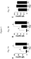
- Fig. 5B Based on the number of positive wells from all the eleven plates and the cloning efficiency of the single B cell culture system, the proportion of antigen-specific blood B cells was calculated. As shown in Fig. 5B , about 0.3% to 0.5% of circulating B cells have antigen specificity for DSG1 or DSG3 in for pemphigus patients ( Fig. 5B , PV and PF). B cell clones reactive with DSG1 and DSG3 in ELISA were also isolated from a healthy individual ("Healthy Control", HC) without pemphigus who did not have measurable anti-DSG serum antibodies ( Fig.
Landscapes
- Health & Medical Sciences (AREA)
- Engineering & Computer Science (AREA)
- Chemical & Material Sciences (AREA)
- Life Sciences & Earth Sciences (AREA)
- Organic Chemistry (AREA)
- Biomedical Technology (AREA)
- Immunology (AREA)
- Genetics & Genomics (AREA)
- Bioinformatics & Cheminformatics (AREA)
- Wood Science & Technology (AREA)
- Biotechnology (AREA)
- Zoology (AREA)
- General Health & Medical Sciences (AREA)
- Biochemistry (AREA)
- Hematology (AREA)
- Cell Biology (AREA)
- Microbiology (AREA)
- General Engineering & Computer Science (AREA)
- Proteomics, Peptides & Aminoacids (AREA)
- Molecular Biology (AREA)
- Medicinal Chemistry (AREA)
- Biophysics (AREA)
- Micro-Organisms Or Cultivation Processes Thereof (AREA)
- Preparation Of Compounds By Using Micro-Organisms (AREA)
- Peptides Or Proteins (AREA)
Claims (14)
- Procédé d'expansion de cellules B in vitro, le procédé comprenant les étapes consistant à :(a) isoler au moins une cellule B, et(b) mettre en culture la au moins une cellules B de l'étape (a) avec une lignée de cellules nourricières comprenant une lignée de cellules épithéliales thymiques modifiée pour exprimer un polypeptide CD154 (ou un agoniste CD40) et un polypeptide BLyS (ou un facteur de survie de cellules B), en combinaison avec de l'IL-21 et sans IL-4 exogène supplémentaire, dans lequel les cellules B sont cultivées avec la lignée de cellules nourricières sous des conditions suffisantes et pendant un temps suffisant pour induire une expansion en nombre des cellules B humaines, dans lequel les cellules B sont facultativement des cellules B humaines, dans lequel de l'IL-21 est facultativement ajoutée de manière exogène à la culture ou la lignée de cellules nourricières est modifiée pour exprimer un polypeptide IL-21, et dans lequel les une ou plusieurs cellules B ont été exposées à un antigène avant un isolement lors de l'étape (a), dans lequel une cellule B est isolée lors de l'étape (a) pour être utilisée lors de l'étape (b).
- Procédé selon la revendication 1, dans lequel les cellules B sont expansées en moyenne d'au moins 104 fois en nombre et facultativement dans lequel la mise en culture de la au moins une cellule B avec la lignée de cellules nourricières dans l'étape (b) est effectuée en moins de 2 semaines.
- Procédé selon l'une quelconque des revendications 1 à 2, dans lequel la mise en culture de la au moins une cellule B avec la lignée de cellules nourricières dans l'étape (b) est effectuée en présence d'un antigène ou dans lequel la culture de l'étape (b) est effectuée en l'absence d'antigène ajouté.
- Procédé selon l'une quelconque des revendications 1 à 3, dans lequel au moins une cellule B est isolée par liaison à un antigène.
- Procédé selon l'une quelconque des revendications 1 à 4, dans lequel, après l'étape (b), au moins 10 % des cellules B expansées expriment de l'IgG ou dans lequel, après l'étape (b), au moins 2,5 % des cellules B expansées expriment de l'IgA.
- Procédé selon l'une quelconque des revendications 1 à 5, dans lequel les cellules nourricières sont une lignée de cellules nourricières comprenant une lignée de cellules épithéliales thymiques modifiée pour exprimer un polypeptide CD154, un polypeptide BLyS, et, facultativement, un polypeptide IL-21 et facultativement dans lequel la lignée de cellules nourricières est capable d'expanser des cellules B humaines à au moins une moyenne de 104 fois en nombre en moins de 2 semaines en culture ; et facultativement dans lequel la lignée de cellules nourricières comprenant le polypeptide CD154 comprend SEQ ID NO : 68 ou un fragment fonctionnel ou un variant de celui-ci, dans lequel le polypeptide BLyS comprend SEQ ID NO : 69 ou un fragment fonctionnel ou un variant de celui-ci, et dans lequel le polypeptide IL-21 comprend SEQ ID NO : 70 ou un fragment fonctionnel ou un variant de celui-ci.
- Procédé in vitro de production d'un anticorps monoclonal, comprenant les étapes consistant à :(a) isoler des cellules B ;(b) séparer les cellules B de l'étape (a) en cellules B uniques ;(c) mettre en culture les cellules B uniques avec une lignée de cellules nourricières comprenant une lignée de cellules stromales modifiée pour exprimer un polypeptide CD154 (ou un agoniste de CD40) et un polypeptide BLyS (ou un facteur de survie de cellules B), en combinaison avec de l'IL-21 et sans IL-4 exogène supplémentaire pour produire une pluralité de clones de cellule B,dans lequel les cellules B uniques sont mises en culture avec la lignée de cellules nourricières sous des conditions suffisantes et pendant un temps suffisant pour induire une expansion en nombre pour différentier en un clone de cellule B produisant un anticorps monoclonal, dans lequel les cellules nourricières sont une lignée de cellules nourricières comprenant une lignée de cellules épithéliales stromales ou thymiques modifiée pour exprimer un polypeptide CD154, un polypeptide BLyS, et, facultativement, un polypeptide IL-21 et facultativement dans lequel la lignée de cellules nourricières est capable d''expanser des cellules B humaines à au moins une moyenne de 104 fois en nombre en moins de 2 semaines en culture ; et facultativement dans lequel la lignée de cellules nourricières comprenant le polypeptide CD154 comprend SEQ ID NO : 68 ou un fragment fonctionnel ou un variant de celui-ci, dans lequel le polypeptide BLyS comprend SEQ ID NO : 69 ou un fragment fonctionnel ou un variant de celui-ci, et dans lequel le polypeptide IL-21 comprend SEQ ID NO : 70 ou un fragment fonctionnel ou un variant de celui-ci et comprenant en outre facultativement l'étape consistant à(d) évaluer la spécificité antigénique d'au moins un des anticorps monoclonaux produits par la pluralité de clones de cellules B, et comprenant facultativement en outre l'étape consistant à(e) purifier au moins un des anticorps monoclonaux produits par la pluralité de clones de cellule B.
- Procédé selon la revendication 7, dans lequel les cellules B ont été exposées à un antigène avant isolement dans l'étape (a).
- Procédé selon l'une quelconque des revendications 7 à 8, dans lequel les cellules B sont des cellules B humaines ou dans lequel les cellules B sont des cellules B murines ou un sous-ensemble de cellules B murines.
- Procédé selon l'une quelconque des revendications 7 à 9, dans lequel l'antigène n'est pas inclus dans l'étape (c) .
- Procédé selon l'une quelconque des revendications 7 à 10, dans lequel après l'étape (c) au moins 10 % des cellules B expansées expriment de l'IgG ou dans lequel après l'étape (c), au moins 2,5 % des cellules B expansées expriment de l'IgA.
- Kit pour l'expansion et la différenciation de cellules B de mammifère in vitro comprenant des cellules nourricières, au moins un anticorps pour isoler des cellules B à partir d'un échantillon biologique et des réactifs pour collecter les cellules B à partir d'un échantillon sanguin périphérique, dans lequel les cellules nourricières sont une lignée de cellules nourricières comprenant une lignée de cellules épithéliales ou stromales thymiques modifiée pour exprimer un polypeptide CD154, un polypeptide BLyS, et, facultativement, un polypeptide IL-21 et facultativement dans lequel la lignée de cellules nourricières est capable d'expanser des cellules B humaines à au moins une moyenne de 104 fois en nombre en moins de 2 semaines en culture ; et facultativement dans lequel la lignée de cellules nourricières comprenant le polypeptide CD154 comprend SEQ ID NO : 68 ou un fragment fonctionnel ou un variant de celui-ci, dans lequel le polypeptide BLyS comprend SEQ ID NO : 69 ou un fragment fonctionnel ou un variant de celui-ci, et dans lequel le polypeptide IL-21 comprend SEQ ID NO : 70 ou un fragment fonctionnel ou un variant de celui-ci, dans lequel l'anticorps est facultativement un anticorps CD19, comprenant facultativement en outre un ou plusieurs réactifs nécessaires à la culture des cellules nourricières modifiées et des cellules B isolées, facultativement de l'IL-21, comprenant facultativement en outre un ou plusieurs réactifs pour une caractérisation d'anticorps monoclonaux produits à partir de cellules B, facultativement un anticorps spécifique pour IgM, IgG, IgA ou IgE.
- Lignée de cellules nourricières comprenant une lignée de cellules épithéliales thymiques modifiée pour exprimer un polypeptide CD154, un polypeptide BLyS et, facultativement, un polypeptide IL-21 et facultativement dans laquelle la lignée de cellules nourricières est capable d'expanser des cellules B humaines en moyenne au moins 104 fois en nombre en moins de 2 semaines en culture.
- Lignée de cellules nourricière selon la revendication 13, dans lequel le polypeptide CD154 comprend SEQ ID NO : 68 ou un fragment fonctionnel ou un variant de celui-ci, dans lequel le polypeptide BLyS comprend SEQ ID NO : 69 ou un fragment fonctionnel ou un variant de celui-ci, et dans lequel le polypeptide IL-21 comprend SEQ ID NO : 70 ou un fragment fonctionnel ou un variant de celui-ci.
Applications Claiming Priority (2)
| Application Number | Priority Date | Filing Date | Title |
|---|---|---|---|
| US201662295728P | 2016-02-16 | 2016-02-16 | |
| PCT/US2017/018155 WO2017143052A1 (fr) | 2016-02-16 | 2017-02-16 | Procédés d'expansion et de différenciation de cellules b destinés à la production d'anticorps |
Publications (3)
| Publication Number | Publication Date |
|---|---|
| EP3417052A1 EP3417052A1 (fr) | 2018-12-26 |
| EP3417052A4 EP3417052A4 (fr) | 2019-09-25 |
| EP3417052B1 true EP3417052B1 (fr) | 2023-02-15 |
Family
ID=59626268
Family Applications (1)
| Application Number | Title | Priority Date | Filing Date |
|---|---|---|---|
| EP17753829.5A Active EP3417052B1 (fr) | 2016-02-16 | 2017-02-16 | Procédés d'expansion et de différenciation de cellules b destinés à la production d'anticorps |
Country Status (5)
| Country | Link |
|---|---|
| US (1) | US20190352607A1 (fr) |
| EP (1) | EP3417052B1 (fr) |
| JP (1) | JP7018203B2 (fr) |
| CN (1) | CN109072192B (fr) |
| WO (1) | WO2017143052A1 (fr) |
Families Citing this family (7)
| Publication number | Priority date | Publication date | Assignee | Title |
|---|---|---|---|---|
| GB201707238D0 (en) * | 2017-05-05 | 2017-06-21 | Univ Oxford Innovation Ltd | Composition |
| CN111518765B (zh) * | 2020-05-12 | 2021-01-26 | 优睿赛思(武汉)生物科技有限公司 | 一种b淋巴细胞体外培养体系及应用 |
| EP4305190A4 (fr) | 2021-03-11 | 2025-03-26 | Iggenix, Inc. | Procédés et systèmes pour prédire une réponse allergique |
| WO2022238890A1 (fr) * | 2021-05-10 | 2022-11-17 | Bazaz Zohre | Procédé de production d'anticorps à partir de cellules plasmatiques uniques |
| CN115873800A (zh) * | 2021-09-26 | 2023-03-31 | 谛邈生物科技(北京)有限公司 | 一种全人源体外生发中心培养模型及其制备方法和应用 |
| EP4569088A1 (fr) * | 2022-08-11 | 2025-06-18 | Imperial College Innovations Limited | Isolement d'anticorps monoclonaux |
| WO2025081199A2 (fr) * | 2023-10-13 | 2025-04-17 | Northwestern University | Production et expansion de lymphocytes b |
Family Cites Families (14)
| Publication number | Priority date | Publication date | Assignee | Title |
|---|---|---|---|---|
| US20040096971A1 (en) * | 2000-12-22 | 2004-05-20 | Blackburn Catherine Clare | Thymic epithelial progenitor cells and uses thereof |
| WO2005037218A2 (fr) * | 2003-10-17 | 2005-04-28 | Actis Biologics, Inc. | Administration d'il-21 par vecteurs lentiviraux pour le traitement du cancer |
| AU2003274535A1 (en) * | 2003-10-30 | 2005-05-19 | Michaela Slanicka Krieger | Cell culture supplement for the in vitro cultivation of cells and a method for culturing cells |
| CN101218351A (zh) * | 2005-02-15 | 2008-07-09 | 杜克大学 | 抗cd19抗体及其在肿瘤学中的应用 |
| KR20080074136A (ko) * | 2005-10-28 | 2008-08-12 | 센토코 인코포레이티드 | 항체의 생성에 있어서의 b 세포 팽창제의 사용 |
| JP5550132B2 (ja) | 2009-10-30 | 2014-07-16 | 学校法人東京理科大学 | 抗原特異的b細胞集団の製造方法 |
| US9074223B2 (en) | 2010-01-08 | 2015-07-07 | Immusoft Corporation | Vectors and methods for transducing B cells |
| JP2012029685A (ja) * | 2010-06-29 | 2012-02-16 | Advance Co Ltd | 体外免疫方法及び同方法を用いた抗体作製方法 |
| ES2617488T3 (es) * | 2011-11-23 | 2017-06-19 | F. Hoffmann-La Roche Ag | Células de mamífero que expresan CD40L y su uso |
| US10017739B2 (en) * | 2012-09-06 | 2018-07-10 | Duke University | Methods of expanding and assessing B cells and using expanded B cells to treat disease |
| FR2998579B1 (fr) | 2012-11-27 | 2015-06-19 | Commissariat Energie Atomique | Methode pour obtenir des anticorps humains specifiques d'un antigene par immunisation in vitro |
| EP3197558A4 (fr) * | 2014-09-28 | 2018-05-16 | Duke University | Compositions et méthodes de modulation de la réponse immunitaire transitoire pendant la vaccination |
| JP7653759B2 (ja) * | 2016-01-20 | 2025-03-31 | フェイト セラピューティクス,インコーポレイテッド | 養子免疫療法における免疫細胞調節のための組成物および方法 |
| CN109668819A (zh) * | 2019-02-01 | 2019-04-23 | 上海市公共卫生临床中心 | 一种机体免疫功能评价的试剂盒及其使用方法和应用 |
-
2017
- 2017-02-16 WO PCT/US2017/018155 patent/WO2017143052A1/fr not_active Ceased
- 2017-02-16 US US15/998,919 patent/US20190352607A1/en not_active Abandoned
- 2017-02-16 EP EP17753829.5A patent/EP3417052B1/fr active Active
- 2017-02-16 JP JP2018543365A patent/JP7018203B2/ja not_active Expired - Fee Related
- 2017-02-16 CN CN201780022683.2A patent/CN109072192B/zh not_active Expired - Fee Related
Also Published As
| Publication number | Publication date |
|---|---|
| JP7018203B2 (ja) | 2022-02-10 |
| CN109072192A (zh) | 2018-12-21 |
| EP3417052A4 (fr) | 2019-09-25 |
| JP2019509729A (ja) | 2019-04-11 |
| US20190352607A1 (en) | 2019-11-21 |
| WO2017143052A1 (fr) | 2017-08-24 |
| EP3417052A1 (fr) | 2018-12-26 |
| CN109072192B (zh) | 2024-02-09 |
Similar Documents
| Publication | Publication Date | Title |
|---|---|---|
| EP3417052B1 (fr) | Procédés d'expansion et de différenciation de cellules b destinés à la production d'anticorps | |
| KR102069394B1 (ko) | Cd40l 발현 포유류 세포 및 그의 용도 | |
| US20060063256A1 (en) | Human regulatory T cells and uses thereof | |
| CN102164957A (zh) | 利用抗体库阵列(ara)发现靶标特异性抗体的方法和组合物 | |
| US20140287952A1 (en) | Protocol for identifying and isolating antigen-specific b cells and producing antibodies to desired antigens | |
| WO2001070817A1 (fr) | Anticorps monoclonal et methode et kit d'immunodosage de st2 humaine soluble l'utilisant | |
| US20230228739A1 (en) | Method for determining potency of chimeric antigen receptor expressing immune cells | |
| CN107727843A (zh) | 检测抗血小板表面受体特异性自身抗体的试剂及其制备方法与应用 | |
| CN110121554B (zh) | B细胞培养方法 | |
| US20160046907A1 (en) | In vitro system for generation of antigen-specific immune responses | |
| JP2020520650A (ja) | 胸腺細胞上清を産生するための方法 | |
| JP6881801B2 (ja) | 細胞の選抜方法、核酸の製造方法、組換え細胞の製造方法、目的物質の製造方法、医薬組成物の製造方法、及び試薬 | |
| US8435530B2 (en) | Methods for suppressing activity of activated interferon-producing cells | |
| KR20260016919A (ko) | 제시된 hpv16-, mart1-, cmv-, ebv-, 또는 인플루엔자-펩티드에 결합하는 t 세포 수용체 | |
| CN109504661B (zh) | 一种抗原特异性的t淋巴细胞杂交瘤及其制备方法和应用 | |
| Lin et al. | A novel strategy to investigate the factors regulating the Treg to Tfr transition during acute viral infection | |
| Rodriguez et al. | B cell responses specific for polyomavirus-derived oncoprotein are predictive of Merkel cell carcinoma tumor control | |
| WO2026009937A1 (fr) | Procédé de culture de lymphocytes t | |
| JP2026511106A (ja) | 抗クローディン-1抗体を使用したがん治療のためのバイオマーカー | |
| HK40012870B (zh) | B细胞培养方法 | |
| Bergendahl et al. | Belinky, P., see Schlesinger, J.(282) 175 Bercovici, N., Givan, AL, Waugh, MG, Fisher, JL, Vernel-Pauillac, F., Ernstoff, MS, Abastado, J.-P. and Wallace, PK, Multiparameter precursor analysis of T-cell responses to antigen (276) 5 | |
| Arumugakani | Phenotypic and Functional Characterization of B-lineage cells Associated With Relapse and Response to B-cell Depletion Therapy for Rheumatoid Arthritis | |
| JPWO2006054748A1 (ja) | 腎炎の治療剤 |
Legal Events
| Date | Code | Title | Description |
|---|---|---|---|
| STAA | Information on the status of an ep patent application or granted ep patent |
Free format text: STATUS: THE INTERNATIONAL PUBLICATION HAS BEEN MADE |
|
| PUAI | Public reference made under article 153(3) epc to a published international application that has entered the european phase |
Free format text: ORIGINAL CODE: 0009012 |
|
| STAA | Information on the status of an ep patent application or granted ep patent |
Free format text: STATUS: REQUEST FOR EXAMINATION WAS MADE |
|
| 17P | Request for examination filed |
Effective date: 20180821 |
|
| AK | Designated contracting states |
Kind code of ref document: A1 Designated state(s): AL AT BE BG CH CY CZ DE DK EE ES FI FR GB GR HR HU IE IS IT LI LT LU LV MC MK MT NL NO PL PT RO RS SE SI SK SM TR |
|
| AX | Request for extension of the european patent |
Extension state: BA ME |
|
| DAV | Request for validation of the european patent (deleted) | ||
| DAX | Request for extension of the european patent (deleted) | ||
| A4 | Supplementary search report drawn up and despatched |
Effective date: 20190827 |
|
| RIC1 | Information provided on ipc code assigned before grant |
Ipc: C12N 5/0781 20100101AFI20190821BHEP Ipc: C12N 5/077 20100101ALI20190821BHEP |
|
| STAA | Information on the status of an ep patent application or granted ep patent |
Free format text: STATUS: EXAMINATION IS IN PROGRESS |
|
| 17Q | First examination report despatched |
Effective date: 20210426 |
|
| GRAP | Despatch of communication of intention to grant a patent |
Free format text: ORIGINAL CODE: EPIDOSNIGR1 |
|
| STAA | Information on the status of an ep patent application or granted ep patent |
Free format text: STATUS: GRANT OF PATENT IS INTENDED |
|
| INTG | Intention to grant announced |
Effective date: 20220825 |
|
| GRAS | Grant fee paid |
Free format text: ORIGINAL CODE: EPIDOSNIGR3 |
|
| GRAA | (expected) grant |
Free format text: ORIGINAL CODE: 0009210 |
|
| STAA | Information on the status of an ep patent application or granted ep patent |
Free format text: STATUS: THE PATENT HAS BEEN GRANTED |
|
| AK | Designated contracting states |
Kind code of ref document: B1 Designated state(s): AL AT BE BG CH CY CZ DE DK EE ES FI FR GB GR HR HU IE IS IT LI LT LU LV MC MK MT NL NO PL PT RO RS SE SI SK SM TR |
|
| REG | Reference to a national code |
Ref country code: CH Ref legal event code: EP Ref country code: GB Ref legal event code: FG4D |
|
| REG | Reference to a national code |
Ref country code: DE Ref legal event code: R096 Ref document number: 602017066041 Country of ref document: DE |
|
| REG | Reference to a national code |
Ref country code: AT Ref legal event code: REF Ref document number: 1548228 Country of ref document: AT Kind code of ref document: T Effective date: 20230315 Ref country code: IE Ref legal event code: FG4D |
|
| REG | Reference to a national code |
Ref country code: LT Ref legal event code: MG9D |
|
| REG | Reference to a national code |
Ref country code: NL Ref legal event code: MP Effective date: 20230215 |
|
| P01 | Opt-out of the competence of the unified patent court (upc) registered |
Effective date: 20230524 |
|
| REG | Reference to a national code |
Ref country code: AT Ref legal event code: MK05 Ref document number: 1548228 Country of ref document: AT Kind code of ref document: T Effective date: 20230215 |
|
| PG25 | Lapsed in a contracting state [announced via postgrant information from national office to epo] |
Ref country code: RS Free format text: LAPSE BECAUSE OF FAILURE TO SUBMIT A TRANSLATION OF THE DESCRIPTION OR TO PAY THE FEE WITHIN THE PRESCRIBED TIME-LIMIT Effective date: 20230215 Ref country code: PT Free format text: LAPSE BECAUSE OF FAILURE TO SUBMIT A TRANSLATION OF THE DESCRIPTION OR TO PAY THE FEE WITHIN THE PRESCRIBED TIME-LIMIT Effective date: 20230615 Ref country code: NO Free format text: LAPSE BECAUSE OF FAILURE TO SUBMIT A TRANSLATION OF THE DESCRIPTION OR TO PAY THE FEE WITHIN THE PRESCRIBED TIME-LIMIT Effective date: 20230515 Ref country code: NL Free format text: LAPSE BECAUSE OF FAILURE TO SUBMIT A TRANSLATION OF THE DESCRIPTION OR TO PAY THE FEE WITHIN THE PRESCRIBED TIME-LIMIT Effective date: 20230215 Ref country code: LV Free format text: LAPSE BECAUSE OF FAILURE TO SUBMIT A TRANSLATION OF THE DESCRIPTION OR TO PAY THE FEE WITHIN THE PRESCRIBED TIME-LIMIT Effective date: 20230215 Ref country code: LT Free format text: LAPSE BECAUSE OF FAILURE TO SUBMIT A TRANSLATION OF THE DESCRIPTION OR TO PAY THE FEE WITHIN THE PRESCRIBED TIME-LIMIT Effective date: 20230215 Ref country code: HR Free format text: LAPSE BECAUSE OF FAILURE TO SUBMIT A TRANSLATION OF THE DESCRIPTION OR TO PAY THE FEE WITHIN THE PRESCRIBED TIME-LIMIT Effective date: 20230215 Ref country code: ES Free format text: LAPSE BECAUSE OF FAILURE TO SUBMIT A TRANSLATION OF THE DESCRIPTION OR TO PAY THE FEE WITHIN THE PRESCRIBED TIME-LIMIT Effective date: 20230215 Ref country code: AT Free format text: LAPSE BECAUSE OF FAILURE TO SUBMIT A TRANSLATION OF THE DESCRIPTION OR TO PAY THE FEE WITHIN THE PRESCRIBED TIME-LIMIT Effective date: 20230215 |
|
| PG25 | Lapsed in a contracting state [announced via postgrant information from national office to epo] |
Ref country code: SE Free format text: LAPSE BECAUSE OF FAILURE TO SUBMIT A TRANSLATION OF THE DESCRIPTION OR TO PAY THE FEE WITHIN THE PRESCRIBED TIME-LIMIT Effective date: 20230215 Ref country code: PL Free format text: LAPSE BECAUSE OF FAILURE TO SUBMIT A TRANSLATION OF THE DESCRIPTION OR TO PAY THE FEE WITHIN THE PRESCRIBED TIME-LIMIT Effective date: 20230215 Ref country code: IS Free format text: LAPSE BECAUSE OF FAILURE TO SUBMIT A TRANSLATION OF THE DESCRIPTION OR TO PAY THE FEE WITHIN THE PRESCRIBED TIME-LIMIT Effective date: 20230615 Ref country code: GR Free format text: LAPSE BECAUSE OF FAILURE TO SUBMIT A TRANSLATION OF THE DESCRIPTION OR TO PAY THE FEE WITHIN THE PRESCRIBED TIME-LIMIT Effective date: 20230516 Ref country code: FI Free format text: LAPSE BECAUSE OF FAILURE TO SUBMIT A TRANSLATION OF THE DESCRIPTION OR TO PAY THE FEE WITHIN THE PRESCRIBED TIME-LIMIT Effective date: 20230215 |
|
| REG | Reference to a national code |
Ref country code: CH Ref legal event code: PL |
|
| REG | Reference to a national code |
Ref country code: BE Ref legal event code: MM Effective date: 20230228 |
|
| PG25 | Lapsed in a contracting state [announced via postgrant information from national office to epo] |
Ref country code: SM Free format text: LAPSE BECAUSE OF FAILURE TO SUBMIT A TRANSLATION OF THE DESCRIPTION OR TO PAY THE FEE WITHIN THE PRESCRIBED TIME-LIMIT Effective date: 20230215 Ref country code: RO Free format text: LAPSE BECAUSE OF FAILURE TO SUBMIT A TRANSLATION OF THE DESCRIPTION OR TO PAY THE FEE WITHIN THE PRESCRIBED TIME-LIMIT Effective date: 20230215 Ref country code: LU Free format text: LAPSE BECAUSE OF NON-PAYMENT OF DUE FEES Effective date: 20230216 Ref country code: LI Free format text: LAPSE BECAUSE OF NON-PAYMENT OF DUE FEES Effective date: 20230228 Ref country code: EE Free format text: LAPSE BECAUSE OF FAILURE TO SUBMIT A TRANSLATION OF THE DESCRIPTION OR TO PAY THE FEE WITHIN THE PRESCRIBED TIME-LIMIT Effective date: 20230215 Ref country code: DK Free format text: LAPSE BECAUSE OF FAILURE TO SUBMIT A TRANSLATION OF THE DESCRIPTION OR TO PAY THE FEE WITHIN THE PRESCRIBED TIME-LIMIT Effective date: 20230215 Ref country code: CZ Free format text: LAPSE BECAUSE OF FAILURE TO SUBMIT A TRANSLATION OF THE DESCRIPTION OR TO PAY THE FEE WITHIN THE PRESCRIBED TIME-LIMIT Effective date: 20230215 Ref country code: CH Free format text: LAPSE BECAUSE OF NON-PAYMENT OF DUE FEES Effective date: 20230228 |
|
| PGFP | Annual fee paid to national office [announced via postgrant information from national office to epo] |
Ref country code: GB Payment date: 20230825 Year of fee payment: 7 |
|
| REG | Reference to a national code |
Ref country code: DE Ref legal event code: R097 Ref document number: 602017066041 Country of ref document: DE |
|
| PG25 | Lapsed in a contracting state [announced via postgrant information from national office to epo] |
Ref country code: SK Free format text: LAPSE BECAUSE OF FAILURE TO SUBMIT A TRANSLATION OF THE DESCRIPTION OR TO PAY THE FEE WITHIN THE PRESCRIBED TIME-LIMIT Effective date: 20230215 |
|
| REG | Reference to a national code |
Ref country code: IE Ref legal event code: MM4A |
|
| PLBE | No opposition filed within time limit |
Free format text: ORIGINAL CODE: 0009261 |
|
| STAA | Information on the status of an ep patent application or granted ep patent |
Free format text: STATUS: NO OPPOSITION FILED WITHIN TIME LIMIT |
|
| PG25 | Lapsed in a contracting state [announced via postgrant information from national office to epo] |
Ref country code: MC Free format text: LAPSE BECAUSE OF FAILURE TO SUBMIT A TRANSLATION OF THE DESCRIPTION OR TO PAY THE FEE WITHIN THE PRESCRIBED TIME-LIMIT Effective date: 20230215 |
|
| 26N | No opposition filed |
Effective date: 20231116 |
|
| PG25 | Lapsed in a contracting state [announced via postgrant information from national office to epo] |
Ref country code: SI Free format text: LAPSE BECAUSE OF FAILURE TO SUBMIT A TRANSLATION OF THE DESCRIPTION OR TO PAY THE FEE WITHIN THE PRESCRIBED TIME-LIMIT Effective date: 20230215 Ref country code: MC Free format text: LAPSE BECAUSE OF FAILURE TO SUBMIT A TRANSLATION OF THE DESCRIPTION OR TO PAY THE FEE WITHIN THE PRESCRIBED TIME-LIMIT Effective date: 20230215 Ref country code: IE Free format text: LAPSE BECAUSE OF NON-PAYMENT OF DUE FEES Effective date: 20230216 |
|
| PG25 | Lapsed in a contracting state [announced via postgrant information from national office to epo] |
Ref country code: BE Free format text: LAPSE BECAUSE OF NON-PAYMENT OF DUE FEES Effective date: 20230228 |
|
| PGFP | Annual fee paid to national office [announced via postgrant information from national office to epo] |
Ref country code: DE Payment date: 20240228 Year of fee payment: 8 |
|
| PG25 | Lapsed in a contracting state [announced via postgrant information from national office to epo] |
Ref country code: IT Free format text: LAPSE BECAUSE OF FAILURE TO SUBMIT A TRANSLATION OF THE DESCRIPTION OR TO PAY THE FEE WITHIN THE PRESCRIBED TIME-LIMIT Effective date: 20230215 |
|
| PGFP | Annual fee paid to national office [announced via postgrant information from national office to epo] |
Ref country code: FR Payment date: 20240226 Year of fee payment: 8 |
|
| GBPC | Gb: european patent ceased through non-payment of renewal fee |
Effective date: 20240216 |
|
| PG25 | Lapsed in a contracting state [announced via postgrant information from national office to epo] |
Ref country code: BG Free format text: LAPSE BECAUSE OF FAILURE TO SUBMIT A TRANSLATION OF THE DESCRIPTION OR TO PAY THE FEE WITHIN THE PRESCRIBED TIME-LIMIT Effective date: 20230215 |
|
| PG25 | Lapsed in a contracting state [announced via postgrant information from national office to epo] |
Ref country code: BG Free format text: LAPSE BECAUSE OF FAILURE TO SUBMIT A TRANSLATION OF THE DESCRIPTION OR TO PAY THE FEE WITHIN THE PRESCRIBED TIME-LIMIT Effective date: 20230215 |
|
| PG25 | Lapsed in a contracting state [announced via postgrant information from national office to epo] |
Ref country code: GB Free format text: LAPSE BECAUSE OF NON-PAYMENT OF DUE FEES Effective date: 20240216 |
|
| PG25 | Lapsed in a contracting state [announced via postgrant information from national office to epo] |
Ref country code: GB Free format text: LAPSE BECAUSE OF NON-PAYMENT OF DUE FEES Effective date: 20240216 |
|
| PG25 | Lapsed in a contracting state [announced via postgrant information from national office to epo] |
Ref country code: CY Free format text: LAPSE BECAUSE OF FAILURE TO SUBMIT A TRANSLATION OF THE DESCRIPTION OR TO PAY THE FEE WITHIN THE PRESCRIBED TIME-LIMIT; INVALID AB INITIO Effective date: 20170216 |
|
| PG25 | Lapsed in a contracting state [announced via postgrant information from national office to epo] |
Ref country code: HU Free format text: LAPSE BECAUSE OF FAILURE TO SUBMIT A TRANSLATION OF THE DESCRIPTION OR TO PAY THE FEE WITHIN THE PRESCRIBED TIME-LIMIT; INVALID AB INITIO Effective date: 20170216 |
|
| REG | Reference to a national code |
Ref country code: DE Ref legal event code: R119 Ref document number: 602017066041 Country of ref document: DE |
|
| PG25 | Lapsed in a contracting state [announced via postgrant information from national office to epo] |
Ref country code: TR Free format text: LAPSE BECAUSE OF FAILURE TO SUBMIT A TRANSLATION OF THE DESCRIPTION OR TO PAY THE FEE WITHIN THE PRESCRIBED TIME-LIMIT Effective date: 20230215 |
|
| PG25 | Lapsed in a contracting state [announced via postgrant information from national office to epo] |
Ref country code: DE Free format text: LAPSE BECAUSE OF NON-PAYMENT OF DUE FEES Effective date: 20250902 |
|
| PG25 | Lapsed in a contracting state [announced via postgrant information from national office to epo] |
Ref country code: FR Free format text: LAPSE BECAUSE OF NON-PAYMENT OF DUE FEES Effective date: 20250228 |