EP3431992A1 - Marqueur pd de facteur de croissance des hépatocytes (hgf) - Google Patents
Marqueur pd de facteur de croissance des hépatocytes (hgf) Download PDFInfo
- Publication number
- EP3431992A1 EP3431992A1 EP17766767.2A EP17766767A EP3431992A1 EP 3431992 A1 EP3431992 A1 EP 3431992A1 EP 17766767 A EP17766767 A EP 17766767A EP 3431992 A1 EP3431992 A1 EP 3431992A1
- Authority
- EP
- European Patent Office
- Prior art keywords
- apoa4
- hgf
- antibody
- administered
- protein
- Prior art date
- Legal status (The legal status is an assumption and is not a legal conclusion. Google has not performed a legal analysis and makes no representation as to the accuracy of the status listed.)
- Withdrawn
Links
Images



Classifications
-
- G—PHYSICS
- G01—MEASURING; TESTING
- G01N—INVESTIGATING OR ANALYSING MATERIALS BY DETERMINING THEIR CHEMICAL OR PHYSICAL PROPERTIES
- G01N33/00—Investigating or analysing materials by specific methods not covered by groups G01N1/00 - G01N31/00
- G01N33/48—Biological material, e.g. blood, urine; Haemocytometers
- G01N33/50—Chemical analysis of biological material, e.g. blood, urine; Testing involving biospecific ligand binding methods; Immunological testing
- G01N33/92—Chemical analysis of biological material, e.g. blood, urine; Testing involving biospecific ligand binding methods; Immunological testing involving lipids, e.g. cholesterol, lipoproteins, or their receptors
-
- G—PHYSICS
- G01—MEASURING; TESTING
- G01N—INVESTIGATING OR ANALYSING MATERIALS BY DETERMINING THEIR CHEMICAL OR PHYSICAL PROPERTIES
- G01N33/00—Investigating or analysing materials by specific methods not covered by groups G01N1/00 - G01N31/00
- G01N33/48—Biological material, e.g. blood, urine; Haemocytometers
- G01N33/50—Chemical analysis of biological material, e.g. blood, urine; Testing involving biospecific ligand binding methods; Immunological testing
- G01N33/68—Chemical analysis of biological material, e.g. blood, urine; Testing involving biospecific ligand binding methods; Immunological testing involving proteins, peptides or amino acids
- G01N33/6893—Chemical analysis of biological material, e.g. blood, urine; Testing involving biospecific ligand binding methods; Immunological testing involving proteins, peptides or amino acids related to diseases not provided for elsewhere
-
- C—CHEMISTRY; METALLURGY
- C12—BIOCHEMISTRY; BEER; SPIRITS; WINE; VINEGAR; MICROBIOLOGY; ENZYMOLOGY; MUTATION OR GENETIC ENGINEERING
- C12Q—MEASURING OR TESTING PROCESSES INVOLVING ENZYMES, NUCLEIC ACIDS OR MICROORGANISMS; COMPOSITIONS OR TEST PAPERS THEREFOR; PROCESSES OF PREPARING SUCH COMPOSITIONS; CONDITION-RESPONSIVE CONTROL IN MICROBIOLOGICAL OR ENZYMOLOGICAL PROCESSES
- C12Q1/00—Measuring or testing processes involving enzymes, nucleic acids or microorganisms; Compositions therefor; Processes of preparing such compositions
- C12Q1/68—Measuring or testing processes involving enzymes, nucleic acids or microorganisms; Compositions therefor; Processes of preparing such compositions involving nucleic acids
- C12Q1/6876—Nucleic acid products used in the analysis of nucleic acids, e.g. primers or probes
- C12Q1/6883—Nucleic acid products used in the analysis of nucleic acids, e.g. primers or probes for diseases caused by alterations of genetic material
-
- G—PHYSICS
- G01—MEASURING; TESTING
- G01N—INVESTIGATING OR ANALYSING MATERIALS BY DETERMINING THEIR CHEMICAL OR PHYSICAL PROPERTIES
- G01N33/00—Investigating or analysing materials by specific methods not covered by groups G01N1/00 - G01N31/00
- G01N33/48—Biological material, e.g. blood, urine; Haemocytometers
- G01N33/50—Chemical analysis of biological material, e.g. blood, urine; Testing involving biospecific ligand binding methods; Immunological testing
- G01N33/68—Chemical analysis of biological material, e.g. blood, urine; Testing involving biospecific ligand binding methods; Immunological testing involving proteins, peptides or amino acids
-
- C—CHEMISTRY; METALLURGY
- C12—BIOCHEMISTRY; BEER; SPIRITS; WINE; VINEGAR; MICROBIOLOGY; ENZYMOLOGY; MUTATION OR GENETIC ENGINEERING
- C12Q—MEASURING OR TESTING PROCESSES INVOLVING ENZYMES, NUCLEIC ACIDS OR MICROORGANISMS; COMPOSITIONS OR TEST PAPERS THEREFOR; PROCESSES OF PREPARING SUCH COMPOSITIONS; CONDITION-RESPONSIVE CONTROL IN MICROBIOLOGICAL OR ENZYMOLOGICAL PROCESSES
- C12Q2600/00—Oligonucleotides characterized by their use
- C12Q2600/106—Pharmacogenomics, i.e. genetic variability in individual responses to drugs and drug metabolism
-
- C—CHEMISTRY; METALLURGY
- C12—BIOCHEMISTRY; BEER; SPIRITS; WINE; VINEGAR; MICROBIOLOGY; ENZYMOLOGY; MUTATION OR GENETIC ENGINEERING
- C12Q—MEASURING OR TESTING PROCESSES INVOLVING ENZYMES, NUCLEIC ACIDS OR MICROORGANISMS; COMPOSITIONS OR TEST PAPERS THEREFOR; PROCESSES OF PREPARING SUCH COMPOSITIONS; CONDITION-RESPONSIVE CONTROL IN MICROBIOLOGICAL OR ENZYMOLOGICAL PROCESSES
- C12Q2600/00—Oligonucleotides characterized by their use
- C12Q2600/158—Expression markers
-
- G—PHYSICS
- G01—MEASURING; TESTING
- G01N—INVESTIGATING OR ANALYSING MATERIALS BY DETERMINING THEIR CHEMICAL OR PHYSICAL PROPERTIES
- G01N2333/00—Assays involving biological materials from specific organisms or of a specific nature
- G01N2333/435—Assays involving biological materials from specific organisms or of a specific nature from animals; from humans
- G01N2333/475—Assays involving growth factors
- G01N2333/4753—Hepatocyte growth factor; Scatter factor; Tumor cytotoxic factor II
-
- G—PHYSICS
- G01—MEASURING; TESTING
- G01N—INVESTIGATING OR ANALYSING MATERIALS BY DETERMINING THEIR CHEMICAL OR PHYSICAL PROPERTIES
- G01N2333/00—Assays involving biological materials from specific organisms or of a specific nature
- G01N2333/435—Assays involving biological materials from specific organisms or of a specific nature from animals; from humans
- G01N2333/775—Apolipopeptides
-
- G—PHYSICS
- G01—MEASURING; TESTING
- G01N—INVESTIGATING OR ANALYSING MATERIALS BY DETERMINING THEIR CHEMICAL OR PHYSICAL PROPERTIES
- G01N2405/00—Assays, e.g. immunoassays or enzyme assays, involving lipids
-
- G—PHYSICS
- G01—MEASURING; TESTING
- G01N—INVESTIGATING OR ANALYSING MATERIALS BY DETERMINING THEIR CHEMICAL OR PHYSICAL PROPERTIES
- G01N2800/00—Detection or diagnosis of diseases
- G01N2800/08—Hepato-biliairy disorders other than hepatitis
- G01N2800/085—Liver diseases, e.g. portal hypertension, fibrosis, cirrhosis, bilirubin
-
- G—PHYSICS
- G01—MEASURING; TESTING
- G01N—INVESTIGATING OR ANALYSING MATERIALS BY DETERMINING THEIR CHEMICAL OR PHYSICAL PROPERTIES
- G01N2800/00—Detection or diagnosis of diseases
- G01N2800/52—Predicting or monitoring the response to treatment, e.g. for selection of therapy based on assay results in personalised medicine; Prognosis
Definitions
- the present invention relates to a PD marker of hepatocyte growth factor (hereinafter also referred to as HGF) and utilization thereof.
- HGF hepatocyte growth factor
- HGF is a factor having hepatic parenchymal cell proliferation activity that is purified from the blood plasma of human fulminant hepatitis patients (Patent Literature 1 and Non-Patent Literature 1), and is reported as having various pharmacological effects such as antitumoral effect, enhancement of cell-mediated immunity, wound therapeutic effect, and tissue regeneration promotional effect (such as Patent Literature 2).
- Non-Patent Literature 1 J. Clin. Invest., 81, 414(1988 )
- the object of the present invention is to provide a method for evaluating the pharmacological action of HGF that utilizes a PD marker in which fluctuation is recognized by administration of HGF in a broader range of subjects.
- apolipoprotein A-IV (ApoA4) as one of the markers where expression is fluctuated by administration of HGF.
- ApoA4 apolipoprotein A-IV
- the present inventors found that expression of ApoA4 gene is induced by interaction between HGF and c-Met, induction of ApoA4 gene expression can be detected with ApoA4 protein in blood, and increase in ApoA4 gene expression by exposure to HGF is a recognized marker also in normal human hepatocytes.
- the present invention is based on the above knowledge, and encompasses the following aspects.
- a method for evaluating the pharmacodynamic action of HGF that utilizes ApoA4 as a PD marker in which fluctuation is recognized by administration of HGF in a broader range of subjects is provided.
- the present invention relates to a method for evaluating the pharmacodynamic action of HGF.
- the evaluation method of the present invention is based on a new finding that the ApoA4 level in a subject is fluctuated by administration of HGF. Accordingly, the evaluation method of the present invention is characterized in that it comprises a step of measuring the ApoA4 level in a biological sample collected from a test subject who was administered HGF.
- HGF used in the present invention may be that commercially available or manufactured by recombinant technology.
- Manufacture of HGF by recombinant technology can be done for example by cloning the gene encoding HGF, creating a vector comprising the aforementioned gene, introducing this into a host cell to allow transformation to thereby obtain a cell expressing HGF, and subjecting this to cell culture.
- the cell, the type of vector, the type of cell, culturing condition, and the like used for this process are within the technical range of those skilled in the art, and they can set appropriate conditions as required.
- the test subject to be the target for evaluation is not particularly limited, and includes for example a test subject who has developed liver disease that is to be the target of HGF administration, a test subject who has the risk of developing liver disease, and a healthy individual.
- liver disease may be any liver disease, but is preferably severe hepatopathy.
- a test subject typically refers to a human, but may also be for example other primates, rodents, and the like as long as it is an animal in which ApoA4 expression is recognized.
- the administration route of HGF is not particularly limited, but in light of the fact that HGF is a protein product, it is typically by intravenous administration.
- said "biological sample collected from a test subject” is not particularly limited as long as it can be collected from a test subject, and can be for example tissues (formed by cells, the basic units configuring animals, that have differentiated and formed clusters so as to exert particular functions, is configured by cells and intercellular substances, and achieves a certain function as a whole), organs (created by orderly combination of tissues), or body fluids.
- Tissues or organs include, nonlimitingly, ovary, uterus, breast, thyroid, brain, esophagus, tongue, lung, pancreas, stomach, small intestine, duodenum, large intestine, bladder, kidney, liver, prostate, gallbladder, pharynx, muscle, bone, skin, and the like, but are not limited thereto.
- Body fluids include, nonlimitingly, blood such as serum, blood plasma or whole blood, lymph, tissue fluid, spinal fluid, body cavity fluid, digestive juice, nasal discharge, lacrimal fluid, sweat, urine, and the like, but are not limited thereto. In terms of ease of acquirement and treatment, it is preferred to employ serum or blood plasma as said biological sample.
- said body fluid may be a body fluid as it is collected from a subject, or those subjected to treatments ordinarily performed on collected body fluids such as dilution and concentration.
- the person who collects or prepares the biological sample derived from a test subject employed in the present invention may be the same or different individual than who carries out the steps of the present invention.
- the biological sample derived from a test subject employed in the present invention may be collected or prepared upon carrying out the present invention, or may be collected or prepared and stored in advance.
- the present invention further comprises a step of comparing the ApoA4 level in a biological sample collected from a test subject who was administered HGF with the ApoA4 level in said biological sample collected from a test subject who was not administered HGF or who has a disappeared influence from the administration of HGF.
- a test subject who has a disappeared influence from the administration of HGF refers to a test subject who has a history of HGF administration but a length of time sufficient for the pharmacodynamic action by HGF to disappear has passed.
- the present invention further comprises a step of determining that said administered HGF is administered in an amount that yields pharmacological action when the ApoA4 level of a test subject who was administered HGF is elevated compared to the ApoA4 level of a test subject who was not administered HGF or who has a disappeared influence from the administration of HGF.
- administered in an amount that yields pharmacodynamic action means that some kind of pharmacodynamic action is provided by administration of HGF, and the magnitude of the action is not necessarily an issue. For example, both of when the ApoA4 level had largely elevated or when the ApoA4 level had only slightly elevated after administration of HGF falls under “administered in an amount that yields pharmacodynamic action.”
- administered in an amount that yields pharmacodynamic action is not synonymous with "administered in a therapeutically effective amount,” and depending on the degree of elevation of the ApoA4 level, may be determined to be “administered in a therapeutically effective amount” or may be determined not to be “administered in a therapeutically effective amount.”
- HGF may include HGF derived from human, mouse, rat, pig, and other animals, but is preferably HGF derived from human.
- human HGF includes a polypeptide having the amino acid sequence shown in SEQ ID NO. 1 or a variant thereof.
- Variants of human HGF include a polypeptide having an amino acid sequence having addition, deletion, or substitution of one or multiple amino acids to the amino acid sequence shown in SEQ ID NO. 1, as well as having HGF activity similar to or more than the polypeptide having the amino acid sequence shown in SEQ ID NO. 1.
- Multiple as used herein is 2 - 150, more preferably 2 - 80, more preferably 2 - 70, more preferably 2 - 60, more preferably 2 - 50, more preferably 2 - 40, more preferably 2 - 30, more preferably 2 - 20, more preferably 2 - 10, or more preferably 2 - 5.
- Said variants also include a polypeptide having an amino acid sequence showing at least 80%, more preferably at least 85%, and more preferably at least 90%, 91%, 92%, 93%, 94%, 95%, 96%, 97%, 98%, or 99% sequence identity with the amino acid sequence shown in SEQ ID NO. 1, as well as having HGF activity similar to or more than the polypeptide having the amino acid sequence shown in SEQ ID NO.
- a "stringent condition” can include those where in the post hybridization washing, hybridization is achieved with washing at for example a condition of "2 X SSC, 0.1% SDS, 50°C," a condition of “2 X SSC, 0.1% SDS, 42°C, “ or a condition of "1 X SSC, 0.1% SDS, 37°C, " and a more stringent condition can include those where hybridization is achieved with washing at for example conditions of "2 X SSC, 0.1% SDS, 65°C,” “0.5 X SSC, 0.1% SDS, 42°C,” “0.2 X SSC, 0.1% SDS, 65°C,” or "0.1 X SSC, 0.1% SDS, 65°C” (1 X SSC is 150 mM NaCl, 15 mM sodium citrate, pH 7.0).
- a solution comprising 5 X SSC, 7% (W/V) SDS, 100 ⁇ g/ml denatured salmon sperm DNA, and 5 X Denhardt's solution (1 X Denhardt's solution comprises 0.2% polyvinylpyrrolidone, 0.2% bovine serum albumin, and 0.2% Ficoll) is employed as prehybridization and hybridization solutions, prehybridization is performed at 65°C for 30 minutes to 1 hour, and hybridization is performed at the same temperature overnight (6 - 8 hours).
- prehybridization in Expresshyb Hybridization Solution (CLONTECH) at 55°C for 30 minutes or more, add a labeled probe and incubate at 37 - 55°C for 1 hour or more, and three washes in 2 X SSC and 0.1% SDS at room temperature for 20 minutes and then one washing in 1 X SSC and 0.1% SDS at 37°C for 20 minutes.
- a more stringent condition can be achieved for example by raising the temperature for prehybridization, hybridization, or second washing.
- the temperature for prehybridization and hybridization can be 60°C, or 65°C or 68°C for a further stringent condition.
- ApoA4 gene is the gene encoding apolipoprotein A-IV, and is known to be expressed in human as well as in rodents such as mouse, rat, and the like.
- the gene sequence of human ApoA4 and the amino acid sequence corresponding thereto are registered as GenBank Accession No. NM_000482 and GenBank Accession No. NP_000473, respectively.
- human ApoA4 protein may comprise a variant thereof.
- Variants of human ApoA4 protein include a polypeptide having an amino acid sequence having addition, deletion, or substitution of one or multiple amino acids to the amino acid sequence shown in SEQ ID NO. 3, as well as having functional property equivalent to that of human ApoA4 protein having the amino acid sequence shown in SEQ ID NO. 3.
- "Multiple" as used herein is 2 - 80, more preferably 2 - 60, more preferably 2 - 40, more preferably 2 - 35, more preferably 2 - 30, more preferably 2 - 25, more preferably 2 - 20, more preferably 2 - 15, more preferably 2 - 10, or more preferably 2 - 5.
- Said variants also include a polypeptide having an amino acid sequence showing at least 80%, more preferably at least 85%, and more preferably at least 90%, 91%, 92%, 93%, 94%, 95%, 96%, 97%, 98%, or 99% sequence identity with the amino acid sequence shown in SEQ ID NO. 3, as well as having functional property equivalent to that of human ApoA4 protein having the amino acid sequence shown in SEQ ID NO. 3, or a polypeptide having an amino acid sequence encoded by a gene that hybridizes under stringent conditions to the gene encoding the amino acid sequence shown in SEQ ID NO. 3 (such as SEQ ID NO. 2), as well as having functional property equivalent to that of human ApoA4 protein having the amino acid sequence shown in SEQ ID NO. 3.
- variants of human ApoA4 protein have the amino acid sequence shown in SEQ ID NO. 5, or an amino acid sequence encoded by the base sequence shown in SEQ ID NO. 4.
- the measurement of the ApoA4 level in said biological sample is not particularly limited as long as it is a method that can quantitatively detect ApoA4. Accordingly, it may be by measuring the expression level of ApoA4 protein or by measuring the expression level of ApoA4 gene (i.e. ApoA4 mRNA). Since fluctuation of ApoA4 protein due to HGF administration can be confirmed by protein in the blood, measurement of the amount of ApoA4 protein in the serum or blood plasma may be advantageous in terms of convenience.
- the method for measuring the expression level of ApoA4 protein is not particularly limited as long as it is a method that can specifically detect and measure ApoA4 protein, and methods ordinarily employed for measuring can be applied.
- Such a method can include, but is not limited to, for example, a method for measuring ApoA4 protein or a fragment thereof such as a method that employs an antibody that binds to ApoA4 protein, ion exchange chromatography, mass spectrometry, LC/MS, and LC/MS/MS having a mass spectrometer tandemly connected to LC/MS.
- the device employed for the detection of ApoA4 protein is not particularly limited, and can be suitably selected depending on the method for measuring ApoA4 protein.
- this can include, for example, an HPLC instrument, a mass spectrometry instrument (mass spectrometry), a LC/MS instrument having liquid chromatography and mass spectrometry instruments connected, LC/MS/MS instrument having a mass spectrometer tandemly connected to an LC/MS instrument, an electrophoresis instrument (such as a capillary electrophoresis device), an automatic or semi-automatic enzyme immunoassay instrument, a cell washer, an automatic or semi-automatic chemiluminescent immunoassay instrument, a luminescence measuring device, an automatic or semi-automatic electrochemiluminescent immunoassay instrument, an optical measuring device, a plate reader, a CCD camera, an automatic or semi-automatic fluorescent immunoassay instrument, a fluorescence measuring device, an automatic or semi-automatic radioimmuno
- an antibody that binds to ApoA4 protein also referred to herein as an "anti-ApoA4 antibody”
- an immunoassay that employs an anti-ApoA4 antibody for measuring the expression level of ApoA4 protein.
- the anti-ApoA4 antibody is not particularly limited as long as it is an antibody that can recognize and bind ApoA4.
- the anti-ApoA4 antibody including both monoclonal and polyclonal antibodies can be created according to well-known methods.
- a monoclonal antibody can be obtained by for example isolating an antibody-producing cell from a non-human mammal immunized with ApoA4 or an ApoA4 fragment, fusing this with a myeloma cell etc. to create a hybridoma, and purifying the antibody produced by this hybridoma.
- a polyclonal antibody can be obtained from the serum of an animal immunized with an ApoA4 protein or an ApoA4 fragment.
- An ApoA4 fragment is a partial peptide of the ApoA4 protein, and an anti-ApoA4 fragment antibody recognizes the ApoA4 protein.
- immunogens include, but are not limited to, the ApoA4 protein or an ApoA4 fragment of for example primates such as human or monkey, rodents such as rat or mouse.
- an "antibody” herein refers to a full length immunoglobulin molecule or an immunologically active fragment of an immunoglobulin molecule such as an antibody fragment that occurs in nature or is produced by genetic recombination technology. These antibodies can be created with conventional technology, and may be a polyclonal antibody or a monoclonal antibody. Antibody fragments include F(ab') 2 , F(ab) 2 , Fab', Fab, Fv, scFv, and the like.
- the anti-ApoA4 antibody used in the present invention is preferably an antibody that specifically binds to the ApoA4 protein.
- “Specific binding” refers to an antibody that binds to the ApoA4 protein at a higher binding affinity compared to non-specific interaction.
- “specific binding” may be shown by for example an antibody that has a Kd of at least about 10 -4 M, or at least about 10 -5 M, or at least about 10 -6 M, or at least about 10 -7 M, or at least about 10 -8 M, or at least about 10 -9 M, or at least about 10 -10 M, or at least about 10 -11 M, or at least about 10 -12 M, or more against the ApoA4 protein.
- "specific binding” means binding to ApoA4 without substantially binding to other polypeptides different from the ApoA4 polypeptide.
- An immunoassay employs a detectably labeled anti-ApoA4 antibody, or an antibody (secondary antibody) against a detectably labeled anti-ApoA4 antibody.
- EIA or ELISA enzyme immunoassay
- RIA radioimmunoassay
- FPIA fluorescence immunoassay
- FPIA fluorescence polarization immunoassay
- CLIA electrochemiluminescence immunoassay
- ELISA method can employ an antibody labeled with an enzyme such as peroxidase and alkaline phosphatase, RIA method with a radioactive material such as 125 I, 131 I, 35 S, and 3 H, FPIA method with a fluorescent substance such as fluorescein isothiocyanate, rhodamine, dansyl chloride, phycoerythrin, tetramethylrhodamine isothiocyanate, and near-infrared fluorescent material, and CLIA method can employ an enzyme such as luciferase and ⁇ galactosidase and an antibody labeled with a luminescent substrate that changes into a luminescent substance with the respective enzyme or a luminescent substance such as luciferin and aequorin. Furthermore, an antibody labeled with a nanoparticle such as a gold colloid and a quantum dot can also be detected.
- an enzyme such as peroxidase and alkaline phosphatase
- an anti-ApoA4 antibody in an immunoassay, can be labeled with biotin, and allowed to bind to avidin or streptavidin labeled with for example an enzyme in order to detect and measure the ApoA4.
- ELISA method employing enzyme-labeling is preferred in that it allows convenient and rapid measurement of the target.
- sandwich method can for example be used.
- An anti-ApoA4 antibody is fixed onto a solid phase support, a suitably treated biological sample is added and allowed to react, and further, an anti-ApoA4 antibody that recognizes another epitope labeled with an enzyme is added and allowed to react.
- the expression level of ApoA4 protein can be determined by allowing reaction with the enzyme substrate, and then subjecting to color development and measuring the absorbance. Moreover, after allowing the anti-ApoA4 antibody fixed onto the solid phase support with the ApoA4 protein in the biological sample to react, unlabeled anti-ApoA4 antibody (primary antibody) may be added, and an antibody (secondary antibody) against this unlabeled antibody may be enzyme-labeled and further added.
- primary antibody unlabeled anti-ApoA4 antibody
- an antibody (secondary antibody) against this unlabeled antibody may be enzyme-labeled and further added.
- DAB 3,3'-diaminobenzidine
- TMB 3,3',5,5'-tetramethylbenzidine
- OPD o-phenylenediamine
- NPP p-nitrophenyl phosphate
- the aggregation method is also preferred as a method for conveniently detecting a trace amount of protein.
- An example of the aggregation method includes the latex aggregation method where a latex particle is bound to the antibody.
- the antibody-bound latex particles aggregate if the ApoA4 protein is present.
- Near-infrared light is then irradiated onto the sample, the aggregation mass is quantified by measurement of absorbance (turbidimetry) or measurement of scattered light (nephelometry), and the antigen concentration can be determined.
- the measurement of the expression level of ApoA4 gene is not particularly limited as long as it is a method that can quantitatively detect ApoA4 mRNA, and for example, a nucleic acid amplification method such as PCR method, RT-PCR method, and real-time PCR method employing a primer specific to ApoA4 mRNA can be employed.
- a nucleic acid amplification method such as PCR method, RT-PCR method, and real-time PCR method employing a primer specific to ApoA4 mRNA
- real-time PCR be carried out specifically as follows. In other words, total RNA is acquired from tissue according to conventional means, and cDNA is created with a reverse transcription enzyme and a suitable primer. Based on the created cDNA, the ApoA4 gene can be specifically amplified by employing a primer specific to the ApoA4 gene and a DNA polymerase.
- the amplification curve of the ApoA4 gene can be monitored with a particular instrument (such as PRISM 7700 Sequence Detector 7900HT Sequence Detection System or ViiATM 7 Real-Time PCR System (from Applied Biosystems)) by incorporating a suitable fluorescent dye upon amplification.
- a particular instrument such as PRISM 7700 Sequence Detector 7900HT Sequence Detection System or ViiATM 7 Real-Time PCR System (from Applied Biosystems)
- an internal standard gene such as Hprt
- the present invention relates to a kit for evaluating the pharmacodynamic action of HGF.
- the kit according to the present invention comprises a means for measuring the ApoA4 level, and in one embodiment, such a means is an anti-ApoA4 antibody, an ApoA4 aptamer, an ApoA4 protein, or a fragment thereof, or the same labeled with an isotope, or a primer or a probe consisting of a partial sequence complementary to the ApoA4 gene sequence or the same labeled with a dye.
- the anti-ApoA4 antibody is an antibody that specifically binds to ApoA4
- the ApoA4 aptamer is an aptamer that specifically binds to the ApoA4 protein.
- the kit of the present invention may further comprise reagents and devices necessary for measuring the amount of ApoA4 protein in the biological sample by an immunoassay that utilizes the antigen antibody reaction between the ApoA4 protein and the anti-ApoA4 antibody.
- the kit of the present invention is for measuring the amount of ApoA4 protein by sandwich ELISA method, and comprises a microtiter plate; an anti-ApoA4 antibody for capture; an anti-ApoA4 antibody labeled with alkaline phosphatase or peroxidase; and an alkaline phosphatase substrate (such as NPP) or a peroxidase substrate (such as DAB, TMB, and microtiter plate OPD).
- the capture antibody and the labeled antibody recognize different epitopes.
- the capture antibody is fixed onto a microtiter plate, a suitably treated and diluted biological sample is added thereto and incubated, and the sample is removed and washed.
- the labeled antibody is added and incubated, the substrate is added, and this is subjected to color development.
- the amount of ApoA4 protein can be determined.
- the kit of the present invention is for measuring the amount of ApoA4 protein by sandwich ELISA method that uses a secondary antibody, and comprises a microtiter plate; an anti-ApoA4 antibody for capture; an anti-ApoA4 antibody as the primary antibody; an antibody against the primary anti-ApoA4 antibody labeled with alkaline phosphatase or peroxidase as the secondary antibody; and an alkaline phosphatase (such as NPP) or a peroxidase substrate (such as DAB, TMB, and OPD).
- the capture antibody and the primary antibody recognize different epitopes.
- the capture antibody is fixed onto a microtiter plate, a suitably treated and diluted biological sample is added thereto and incubated, and the sample is removed and washed. Subsequently, the primary antibody is added and incubated and washed, the enzyme-labeled secondary antibody is further added and incubated, and then the substrate is added and subjected to color development. By measuring the color development with a microtiter plate reader etc., the amount of ApoA4 protein can be determined. By employing a secondary antibody, the reaction is amplified and detection sensitivity can be improved.
- kit of the present invention further comprises the necessary buffer, enzyme reaction quenching solution, a microplate reader, product instruction, and the like.
- the labeled antibody is not limited to an enzyme-labeled antibody, and may be an antibody labeled with a radioactive material (such as 125 I, 131 I, 35 S, and 3 H), a fluorescent substance (such as fluorescein isothiocyanate, rhodamine, dansyl chloride, phycoerythrin, tetramethylrhodamine isothiocyanate, and near-infrared fluorescent material), a luminescent substance (such as luciferase, luciferin, and aequorin), a nanoparticle (gold colloid and quantum dot), and the like.
- a biotinylated antibody may also be employed as the labeled antibody, and labeled avidin or streptavidin may be added to the kit.
- the kit of the present invention may further comprise reagents and devices necessary for measuring the amount of ApoA4 in a biological sample in an assay that utilizes the reaction between ApoA4 and ApoA4 aptamer.
- first and second are sometimes employed to express various elements, and it should be recognized that these elements are not to be limited by these terms. These terms are employed solely for the purpose of discriminating one element from another, and it is for example possible to describe a first element as a second element, and similarly, to describe a second element as a first element without departing from the scope of the present invention.
- Example 1 Gene expression analysis employing hepatic tissue of healthy mice administered rh-HGF
- liver which is a target organ to which rh-HGF brings its biological activity as a material
- identification of molecules affected by rh-HGFand identification of a PD marker among the molecules were attempted.
- Microarray and real-time PCR method were employed for molecule identification.
- hepatocyte growth factor activator recombinantly expressed in Chinese hamster ovary (CHO) cells
- human HGF recombinantly expressed in CHO cells were activated to obtain rh-HGF.
- BALB/cAnNCrlCrlj male 6 weeks-old mice (CHARLES RIVER JAPAN) were intravenously administered with Dulbecco's Phosphate buffered saline (PBS) (Wako, #045-29795) or rh-HGF (1.5 mg/kg).
- PBS Dulbecco's Phosphate buffered saline
- rh-HGF 1.5 mg/kg.
- RNA concentration was measured by absorbance, and then quality check was carried out with RNA 6000 Nano Assay Kit (Agilent Technologies, #5067-1511) using 2100 Bioanalyzer (Agilent Technologies).
- the DNA microarray used was SurePrint G3 mouse GE 8 X 60K (Agilent Technologies, #G4852A).
- genes which had a signal value of 100 or more in all three groups were targeted, and those having two-folds or more increase in the signal value in Group 2 or Group 3 compared to Group 1 were temporarily extracted.
- molecules that may appear to possibly exist as soluble proteins those having information of "Extracellular” or "Plasma membrane” in Gene Ontology annotation were extracted. In this way, molecules of which expression was strongly induced by rh-HGF administration were selected as PD marker candidates.
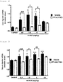
- ApoA4 molecules were extracted by the above molecule selection method (Table 2). ApoA4 molecules are molecules that exist in the blood as apoproteins that configure apolipoproteins. ApoA4 molecules had low expression in mice that were administered PBS only (Group 1), and enhancement of expression was seen with rh-HGF administration. [Table 2] Change in expression (vs 0 hours) Gene code (probe ID) Signal value (average) 8 hours 24 hours 0 hours 8 hours 24 hours Ratio P value Ratio P value ApoA4 (A_51_ P327491) 2436 15738 13453 6.46 0.0002 5.52 0.0016 P value: Welch's test
- RNA was reverse transcripted using High-Capacity cDNA Reverse Transcription Kit with RNase Inhibitor (Applied Biosystems, #4374966) to create cDNA.
- RNase Inhibitor Applied Biosystems, #4374966
- Hprt mRNA was used as the internal control for examination.
- PCR amplification reaction was carried out with TaqMan Fast Advanced Master Mix (Applied Biosystems, #4444557) and TaqMan Assay (ApoA4: Mm00431814_m1, Hprt: Mm03024075_m1, Applied Biosystems) and using ViiA7 Real-time PCR System (Applied Biosystems).
- TaqMan Fast Advanced Master Mix Applied Biosystems, #4444557
- TaqMan Assay ApoA4: Mm00431814_m1, Hprt: Mm03024075_m1, Applied Biosystems
- ViiA7 Real-time PCR System Applied Biosystems
- rh-HGF The biological activity of rh-HGF is achieved via its receptor, c-Met.
- c-Met The biological activity of rh-HGF is achieved via its receptor, c-Met.
- ApoA4 is a PD marker of rh-HGF can be guaranteed by revealing that the induced expression is dependent on c-Met.
- Investigation targets were mRNA derived from liver and protein in the serum.
- mice To BALB/cAnNCrlCrlj male 6 weeks-old mice (CHARLES RIVER JAPAN), c-Met inhibitor PF-04217903 (50 mg/kg) (Selleck Chemicals #S1094, lot. 05) or vehicle (5% DMSO, 10% Tween80, 6.7 mmol/L aqueous hydrochloric acid solution) was orally administered at a dose of 10 mL/kg, and after 1 hour, rh-HGF (0.5, 1.5, 5.0, or 15 mg/kg) or PBS was intravenously administered.
- rh-HGF 0.5, 1.5, 5.0, or 15 mg/kg
- mice Twenty-four hours after administration of rh-HGF or PBS, blood was collected from the abdominal inferior vena cava under anesthesia by 3% isoflurane inhalation, and mice were euthanized by cervical dislocation method. After euthanization, hepatic tissue was collected and immersed in RNA later (Ambion, #AM7021). Serum was collected from the blood collected.
- the Table below shows the test groups employed in this experiment.
- RNA concentration was measured by absorbance.
- ApoA4 protein was quantified by Western blot method.
- PBS PBS
- electrophoresis buffer solution 313 mM Tris-HCl (pH 6.8), 10% SDS, 30% Glycerol, 0.2 mg/mL Bromo-phenol blue, and 25% 2-Mercaptoethnol
- the filter was soaked in the blocking solution (50 mM Tris-HCl (pH 7.5), 150 mM NaCl, 5% skim-milk, 0.01% Tween20, and 0.1% NaN3), shaken at room temperature for 1 hour, then the blocking solution was discarded, the filter was soaked in a reaction solution (50 mM Tris-HCl (pH 7.5), 150 mM NaCl, 5% skim-milk, 4% PEG6000, 0.2% EDTA/3Na, 0.01% Tween20, and 0.2% Proclin150) supplemented with goat anti-mouse Apolipoprotein A-IV antibody (GeneTex, #GTX88533) to 0.42 ⁇ g/mL concentration, and shaken at room temperature for 2 hours.
- the blocking solution 50 mM Tris-HCl (pH 7.5), 150 mM NaCl, 5% skim-milk, 0.01% Tween20, and 0.1% NaN3
- a reaction solution 50 mM Tri
- the filter was soaked in the above reaction solution supplemented with the secondary antibody which is HRP-labeled rabbit anti-goat IgG (H+L) (Invitrogen) to 1/2000 concentration, and shaken at room temperature for 1 hour.
- chemiluminescence solution SuperSignal West Dura Extended Duration Substrate (Thermo Scientific) was added to allow luminescence, and ApoA4 protein was detected with LAS-4000 (FUJIFILM).
- mouse ApoA4 protein was a PD marker of HGF that can be measured in blood.
- ApoA4 protein behaved as a PD marker of HGF in the liver mRNA and blood of healthy mice, then another test with experimental hepatopathy mice (Jo2 liver failure model) was carried out to confirm whether it also behaves as a PD marker of HGF during hepatopathy.
- the Jo2 liver failure model is a hepatopathy model with high severity attributed to direct evoking of hepatocyte apoptosis by administration of anti-Fas monoclonal antibody (Anti-Fas MAb).
- mice To BALB/cAnNCrlCrlj male 6 weeks-old mice (CHARLES RIVER JAPAN), 0.23 mg/kg of anti-Fas monoclonal antibody (clone name: Jo2, BD Pharmingen, #554254, lot#3269634) was administered once into the tail vein to create an acute liver failure model. To this model, rh-HGF dissolved in PBS was administered into the tail vein at 0.15, 1.5, or 15 mg/kg 1 hour before administration of anti-Fas monoclonal antibody. For the control group (Control), PBS was administered into the tail vein.
- anti-Fas monoclonal antibody (clone name: Jo2, BD Pharmingen, #554254, lot#3269634) was administered once into the tail vein to create an acute liver failure model.
- rh-HGF dissolved in PBS was administered into the tail vein at 0.15, 1.5, or 15 mg/kg 1 hour before administration of anti-Fas monoclonal antibody.
- mice Five hours after the administration of anti-Fas monoclonal antibody, mice were retained under anesthesia by 3% isoflurane inhalation, blood was collected from the vein located behind the jaw bone, and Prothrombin time (PT) was measured with CoaguChek XS (Roche-diagnostics, #ES350009) and PT Test strips (Roche-diagnostics, #ES350054). After measuring the Prothrombin time, blood was collected from the abdominal inferior vena cava, and the mice were then euthanized by cervical dislocation method.
- PT Prothrombin time
- AST Aspartate Aminotransferase
- ALT Alanine transaminase
- RNA concentration was measured by absorbance.
- the purified total RNA was reverse transcripted using High-Capacity cDNA Reverse Transcription Kit with RNase Inhibitor (Applied Biosystems, #4374966) to create cDNA.
- the amount of ApoA4 mRNA expressed was examined by real-time PCR method. 18S rRNA was used as the internal control for examination.
- PCR amplification reaction was carried out with TaqMan Fast Advanced Master Mix (Applied Biosystems, #4444557) and TaqMan Assay (ApoA4: Hs00166636_m1, 18S rRNA: Hs99999901_s1, Applied Biosystems) and using ViiA7 Real-time PCR System (Applied Biosystems).
- TaqMan Fast Advanced Master Mix Applied Biosystems, #4444557
- TaqMan Assay ApoA4: Hs00166636_m1, 18S rRNA: Hs99999901_s1, Applied Biosystems
- ViiA7 Real-time PCR System Applied Biosystems
- hepatocyte culture supernatant (the same culture supernatant as that of cells for preparation of mRNA) 24 hours after addition of rh-HGF, ApoA4 protein was quantified by Western blot method.
- the electrophoresis buffer solution 313 mM Tris-HCl (pH 6.8), 10% SDS, 30% Glycerol, 0.2 mg/mL Bromo-phenol blue, 25% 2-Mercuptoethnol
- the filter was soaked in the above reaction solution supplemented with the secondary antibody which is HRP-labeled goat anti-rabbit IgG (H+L) (Jackson) to 1/2000 concentration, and shaken at room temperature for 1 hour.
- chemiluminescence solution SuperSignal West Dura Extended Duration Substrate (Thermo Scientific) was added to allow luminescence, and ApoA4 protein was detected with LAS-4000 (FUJIFILM).
Landscapes
- Life Sciences & Earth Sciences (AREA)
- Health & Medical Sciences (AREA)
- Chemical & Material Sciences (AREA)
- Engineering & Computer Science (AREA)
- Molecular Biology (AREA)
- Immunology (AREA)
- Proteomics, Peptides & Aminoacids (AREA)
- Analytical Chemistry (AREA)
- Organic Chemistry (AREA)
- Hematology (AREA)
- Urology & Nephrology (AREA)
- Biomedical Technology (AREA)
- Biotechnology (AREA)
- Physics & Mathematics (AREA)
- Biochemistry (AREA)
- General Health & Medical Sciences (AREA)
- Microbiology (AREA)
- Pathology (AREA)
- Zoology (AREA)
- Wood Science & Technology (AREA)
- Genetics & Genomics (AREA)
- Biophysics (AREA)
- Medicinal Chemistry (AREA)
- General Physics & Mathematics (AREA)
- Food Science & Technology (AREA)
- Cell Biology (AREA)
- Bioinformatics & Cheminformatics (AREA)
- General Engineering & Computer Science (AREA)
- Endocrinology (AREA)
- Measuring Or Testing Involving Enzymes Or Micro-Organisms (AREA)
- Peptides Or Proteins (AREA)
- Investigating Or Analysing Biological Materials (AREA)
Applications Claiming Priority (2)
| Application Number | Priority Date | Filing Date | Title |
|---|---|---|---|
| JP2016055491 | 2016-03-18 | ||
| PCT/JP2017/010587 WO2017159771A1 (fr) | 2016-03-18 | 2017-03-16 | Marqueur pd de facteur de croissance des hépatocytes (hgf) |
Publications (2)
| Publication Number | Publication Date |
|---|---|
| EP3431992A1 true EP3431992A1 (fr) | 2019-01-23 |
| EP3431992A4 EP3431992A4 (fr) | 2019-10-23 |
Family
ID=59850920
Family Applications (1)
| Application Number | Title | Priority Date | Filing Date |
|---|---|---|---|
| EP17766767.2A Withdrawn EP3431992A4 (fr) | 2016-03-18 | 2017-03-16 | Marqueur pd de facteur de croissance des hépatocytes (hgf) |
Country Status (4)
| Country | Link |
|---|---|
| US (1) | US20200341014A1 (fr) |
| EP (1) | EP3431992A4 (fr) |
| JP (1) | JP6800208B2 (fr) |
| WO (1) | WO2017159771A1 (fr) |
Families Citing this family (3)
| Publication number | Priority date | Publication date | Assignee | Title |
|---|---|---|---|---|
| CN106456695B (zh) | 2014-04-28 | 2019-11-12 | 卫材R&D管理有限公司 | Hgf的冻干配制品 |
| KR102275262B1 (ko) | 2016-03-17 | 2021-07-12 | 에자이 알앤드디 매니지먼트 가부시키가이샤 | 활성 간세포 성장 인자(hgf)를 생성하는 방법 |
| WO2019167874A1 (fr) * | 2018-02-27 | 2019-09-06 | エーザイ・アール・アンド・ディー・マネジメント株式会社 | Anticorps monoclonal dirigé contre apoa4, procédé de dosage immunologique et kit de mesure |
Family Cites Families (5)
| Publication number | Priority date | Publication date | Assignee | Title |
|---|---|---|---|---|
| JP4463885B2 (ja) * | 1996-12-05 | 2010-05-19 | 敏一 中村 | 劇症肝炎疾患治療剤 |
| JP2012051822A (ja) * | 2010-08-31 | 2012-03-15 | Institute Of Physical & Chemical Research | 肺癌診断用ポリペプチド、肺癌の検出方法、および治療効果の評価方法 |
| US9272018B2 (en) * | 2011-04-18 | 2016-03-01 | Kyoto University | Acute hepatic insufficiency depressant and method for evaluating drug efficacy thereof |
| JP5110235B1 (ja) * | 2011-06-27 | 2012-12-26 | コニカミノルタホールディングス株式会社 | 画像処理装置、画像処理方法、およびプログラム |
| WO2013018851A1 (fr) * | 2011-08-02 | 2013-02-07 | 独立行政法人国立がん研究センター | Procédé d'induction de la différentiation hépatique à partir d'une cellule souche hépatique induite et d'une cellule progénitrice hépatique induite |
-
2017
- 2017-03-16 US US16/078,557 patent/US20200341014A1/en not_active Abandoned
- 2017-03-16 JP JP2018505994A patent/JP6800208B2/ja not_active Expired - Fee Related
- 2017-03-16 WO PCT/JP2017/010587 patent/WO2017159771A1/fr not_active Ceased
- 2017-03-16 EP EP17766767.2A patent/EP3431992A4/fr not_active Withdrawn
Also Published As
| Publication number | Publication date |
|---|---|
| EP3431992A4 (fr) | 2019-10-23 |
| WO2017159771A1 (fr) | 2017-09-21 |
| JP6800208B2 (ja) | 2020-12-16 |
| JPWO2017159771A1 (ja) | 2019-01-31 |
| US20200341014A1 (en) | 2020-10-29 |
Similar Documents
| Publication | Publication Date | Title |
|---|---|---|
| Wu et al. | ITGA6 and RPSA synergistically promote pancreatic cancer invasion and metastasis via PI3K and MAPK signaling pathways | |
| US11598776B2 (en) | Biomarkers for predicting and assessing responsiveness of thyroid and kidney cancer subjects to lenvatinib compounds | |
| KR101032607B1 (ko) | 간암 진단용 단백질성 마커 | |
| JP5650871B1 (ja) | そう痒を伴う疾患に罹患した患者のil−31アンタゴニストによる治療に対する応答を予測する方法 | |
| US9482676B2 (en) | Method for screening for a therapeutic agent for idiopathic interstitial pneumonia | |
| KR102742415B1 (ko) | 척수성 근위축증의 치료 방법 | |
| CN103403550B (zh) | 使用与braf v600e特异性结合的抗体诊断癌症的工具和方法 | |
| CN103238069B (zh) | 肺癌和结直肠癌中的bard1同工型及其应用 | |
| EP3431992A1 (fr) | Marqueur pd de facteur de croissance des hépatocytes (hgf) | |
| JPWO2016152352A1 (ja) | メラノーマ特異的バイオマーカー及びその利用 | |
| EP2863224A1 (fr) | Kit permettant de diagnostiquer un mélanome malin | |
| WO2011129427A1 (fr) | Agent de diagnostic et agent thérapeutique pour les cancers | |
| JP2013007724A (ja) | 筋ジストロフィーの病態及び治療評価のための分子マーカー | |
| JP5885243B2 (ja) | 胆管細胞癌の検出方法および予防・治療剤のスクリーニング方法 | |
| US20150218242A1 (en) | TIF1-Gamma for Treating and Diagnosing Inflammatory Diseases | |
| CA2908889A1 (fr) | Traitement de la resistance a l'insuline par des inhibiteurs du facteur de transcription tsc22d4 | |
| WO2021172315A1 (fr) | Variant d'épissage de lamc2-nr6a1 et son produit de traduction | |
| EP3133400B1 (fr) | Utilisation de protéines associées à la fécondité masculine ak6 et gpx5 ou de leur combinaison | |
| EP2631656B1 (fr) | Marqueur de la sclérose latérale amyotrophique, et son utilisation | |
| KR101516716B1 (ko) | 간 섬유화 진단용 바이오마커 rorc | |
| US20260110036A1 (en) | Marker for prognosis of diffuse type gastric cancer and treatment target | |
| Nabeshima et al. | Distinct fibrosis-associated macrophage subsets coordinate iron metabolism in pulmonary fibrosis | |
| JP6065172B2 (ja) | 微小血管障害又はその関連疾患のバイオマーカー | |
| CN119144719A (zh) | 钙结合蛋白s100a1作为肺癌免疫治疗生物标志物的应用 | |
| JPWO2007026960A1 (ja) | Mocs3遺伝子の治療的又は診断的用途 |
Legal Events
| Date | Code | Title | Description |
|---|---|---|---|
| STAA | Information on the status of an ep patent application or granted ep patent |
Free format text: STATUS: THE INTERNATIONAL PUBLICATION HAS BEEN MADE |
|
| PUAI | Public reference made under article 153(3) epc to a published international application that has entered the european phase |
Free format text: ORIGINAL CODE: 0009012 |
|
| STAA | Information on the status of an ep patent application or granted ep patent |
Free format text: STATUS: REQUEST FOR EXAMINATION WAS MADE |
|
| 17P | Request for examination filed |
Effective date: 20180822 |
|
| AK | Designated contracting states |
Kind code of ref document: A1 Designated state(s): AL AT BE BG CH CY CZ DE DK EE ES FI FR GB GR HR HU IE IS IT LI LT LU LV MC MK MT NL NO PL PT RO RS SE SI SK SM TR |
|
| AX | Request for extension of the european patent |
Extension state: BA ME |
|
| DAV | Request for validation of the european patent (deleted) | ||
| DAX | Request for extension of the european patent (deleted) | ||
| A4 | Supplementary search report drawn up and despatched |
Effective date: 20190919 |
|
| RIC1 | Information provided on ipc code assigned before grant |
Ipc: G01N 33/53 20060101AFI20190914BHEP |
|
| STAA | Information on the status of an ep patent application or granted ep patent |
Free format text: STATUS: EXAMINATION IS IN PROGRESS |
|
| 17Q | First examination report despatched |
Effective date: 20200623 |
|
| STAA | Information on the status of an ep patent application or granted ep patent |
Free format text: STATUS: THE APPLICATION IS DEEMED TO BE WITHDRAWN |
|
| 18D | Application deemed to be withdrawn |
Effective date: 20221001 |