EP3443149B1 - Hybriderfassungssequenz zur bestimmung von immunzellenklonalität - Google Patents
Hybriderfassungssequenz zur bestimmung von immunzellenklonalität Download PDFInfo
- Publication number
- EP3443149B1 EP3443149B1 EP17781661.8A EP17781661A EP3443149B1 EP 3443149 B1 EP3443149 B1 EP 3443149B1 EP 17781661 A EP17781661 A EP 17781661A EP 3443149 B1 EP3443149 B1 EP 3443149B1
- Authority
- EP
- European Patent Office
- Prior art keywords
- seq
- sequences
- gene
- cell
- sequencing
- Prior art date
- Legal status (The legal status is an assumption and is not a legal conclusion. Google has not performed a legal analysis and makes no representation as to the accuracy of the status listed.)
- Active
Links
Images










































Classifications
-
- C—CHEMISTRY; METALLURGY
- C12—BIOCHEMISTRY; BEER; SPIRITS; WINE; VINEGAR; MICROBIOLOGY; ENZYMOLOGY; MUTATION OR GENETIC ENGINEERING
- C12Q—MEASURING OR TESTING PROCESSES INVOLVING ENZYMES, NUCLEIC ACIDS OR MICROORGANISMS; COMPOSITIONS OR TEST PAPERS THEREFOR; PROCESSES OF PREPARING SUCH COMPOSITIONS; CONDITION-RESPONSIVE CONTROL IN MICROBIOLOGICAL OR ENZYMOLOGICAL PROCESSES
- C12Q1/00—Measuring or testing processes involving enzymes, nucleic acids or microorganisms; Compositions therefor; Processes of preparing such compositions
- C12Q1/68—Measuring or testing processes involving enzymes, nucleic acids or microorganisms; Compositions therefor; Processes of preparing such compositions involving nucleic acids
- C12Q1/6813—Hybridisation assays
- C12Q1/6834—Enzymatic or biochemical coupling of nucleic acids to a solid phase
- C12Q1/6837—Enzymatic or biochemical coupling of nucleic acids to a solid phase using probe arrays or probe chips
-
- C—CHEMISTRY; METALLURGY
- C12—BIOCHEMISTRY; BEER; SPIRITS; WINE; VINEGAR; MICROBIOLOGY; ENZYMOLOGY; MUTATION OR GENETIC ENGINEERING
- C12Q—MEASURING OR TESTING PROCESSES INVOLVING ENZYMES, NUCLEIC ACIDS OR MICROORGANISMS; COMPOSITIONS OR TEST PAPERS THEREFOR; PROCESSES OF PREPARING SUCH COMPOSITIONS; CONDITION-RESPONSIVE CONTROL IN MICROBIOLOGICAL OR ENZYMOLOGICAL PROCESSES
- C12Q1/00—Measuring or testing processes involving enzymes, nucleic acids or microorganisms; Compositions therefor; Processes of preparing such compositions
- C12Q1/68—Measuring or testing processes involving enzymes, nucleic acids or microorganisms; Compositions therefor; Processes of preparing such compositions involving nucleic acids
- C12Q1/6844—Nucleic acid amplification reactions
- C12Q1/6853—Nucleic acid amplification reactions using modified primers or templates
- C12Q1/6855—Ligating adaptors
-
- C—CHEMISTRY; METALLURGY
- C12—BIOCHEMISTRY; BEER; SPIRITS; WINE; VINEGAR; MICROBIOLOGY; ENZYMOLOGY; MUTATION OR GENETIC ENGINEERING
- C12Q—MEASURING OR TESTING PROCESSES INVOLVING ENZYMES, NUCLEIC ACIDS OR MICROORGANISMS; COMPOSITIONS OR TEST PAPERS THEREFOR; PROCESSES OF PREPARING SUCH COMPOSITIONS; CONDITION-RESPONSIVE CONTROL IN MICROBIOLOGICAL OR ENZYMOLOGICAL PROCESSES
- C12Q1/00—Measuring or testing processes involving enzymes, nucleic acids or microorganisms; Compositions therefor; Processes of preparing such compositions
- C12Q1/68—Measuring or testing processes involving enzymes, nucleic acids or microorganisms; Compositions therefor; Processes of preparing such compositions involving nucleic acids
- C12Q1/6869—Methods for sequencing
-
- C—CHEMISTRY; METALLURGY
- C12—BIOCHEMISTRY; BEER; SPIRITS; WINE; VINEGAR; MICROBIOLOGY; ENZYMOLOGY; MUTATION OR GENETIC ENGINEERING
- C12Q—MEASURING OR TESTING PROCESSES INVOLVING ENZYMES, NUCLEIC ACIDS OR MICROORGANISMS; COMPOSITIONS OR TEST PAPERS THEREFOR; PROCESSES OF PREPARING SUCH COMPOSITIONS; CONDITION-RESPONSIVE CONTROL IN MICROBIOLOGICAL OR ENZYMOLOGICAL PROCESSES
- C12Q1/00—Measuring or testing processes involving enzymes, nucleic acids or microorganisms; Compositions therefor; Processes of preparing such compositions
- C12Q1/68—Measuring or testing processes involving enzymes, nucleic acids or microorganisms; Compositions therefor; Processes of preparing such compositions involving nucleic acids
- C12Q1/6876—Nucleic acid products used in the analysis of nucleic acids, e.g. primers or probes
- C12Q1/6881—Nucleic acid products used in the analysis of nucleic acids, e.g. primers or probes for tissue or cell typing, e.g. human leukocyte antigen [HLA] probes
-
- C—CHEMISTRY; METALLURGY
- C40—COMBINATORIAL TECHNOLOGY
- C40B—COMBINATORIAL CHEMISTRY; LIBRARIES, e.g. CHEMICAL LIBRARIES
- C40B30/00—Methods of screening libraries
- C40B30/04—Methods of screening libraries by measuring the ability to specifically bind a target molecule, e.g. antibody-antigen binding, receptor-ligand binding
-
- C—CHEMISTRY; METALLURGY
- C40—COMBINATORIAL TECHNOLOGY
- C40B—COMBINATORIAL CHEMISTRY; LIBRARIES, e.g. CHEMICAL LIBRARIES
- C40B40/00—Libraries per se, e.g. arrays, mixtures
- C40B40/04—Libraries containing only organic compounds
- C40B40/06—Libraries containing nucleotides or polynucleotides, or derivatives thereof
-
- C—CHEMISTRY; METALLURGY
- C12—BIOCHEMISTRY; BEER; SPIRITS; WINE; VINEGAR; MICROBIOLOGY; ENZYMOLOGY; MUTATION OR GENETIC ENGINEERING
- C12Q—MEASURING OR TESTING PROCESSES INVOLVING ENZYMES, NUCLEIC ACIDS OR MICROORGANISMS; COMPOSITIONS OR TEST PAPERS THEREFOR; PROCESSES OF PREPARING SUCH COMPOSITIONS; CONDITION-RESPONSIVE CONTROL IN MICROBIOLOGICAL OR ENZYMOLOGICAL PROCESSES
- C12Q2525/00—Reactions involving modified oligonucleotides, nucleic acids, or nucleotides
- C12Q2525/10—Modifications characterised by
- C12Q2525/191—Modifications characterised by incorporating an adaptor
-
- C—CHEMISTRY; METALLURGY
- C12—BIOCHEMISTRY; BEER; SPIRITS; WINE; VINEGAR; MICROBIOLOGY; ENZYMOLOGY; MUTATION OR GENETIC ENGINEERING
- C12Q—MEASURING OR TESTING PROCESSES INVOLVING ENZYMES, NUCLEIC ACIDS OR MICROORGANISMS; COMPOSITIONS OR TEST PAPERS THEREFOR; PROCESSES OF PREPARING SUCH COMPOSITIONS; CONDITION-RESPONSIVE CONTROL IN MICROBIOLOGICAL OR ENZYMOLOGICAL PROCESSES
- C12Q2535/00—Reactions characterised by the assay type for determining the identity of a nucleotide base or a sequence of oligonucleotides
- C12Q2535/122—Massive parallel sequencing
-
- C—CHEMISTRY; METALLURGY
- C12—BIOCHEMISTRY; BEER; SPIRITS; WINE; VINEGAR; MICROBIOLOGY; ENZYMOLOGY; MUTATION OR GENETIC ENGINEERING
- C12Q—MEASURING OR TESTING PROCESSES INVOLVING ENZYMES, NUCLEIC ACIDS OR MICROORGANISMS; COMPOSITIONS OR TEST PAPERS THEREFOR; PROCESSES OF PREPARING SUCH COMPOSITIONS; CONDITION-RESPONSIVE CONTROL IN MICROBIOLOGICAL OR ENZYMOLOGICAL PROCESSES
- C12Q2565/00—Nucleic acid analysis characterised by mode or means of detection
- C12Q2565/50—Detection characterised by immobilisation to a surface
- C12Q2565/514—Detection characterised by immobilisation to a surface characterised by the use of the arrayed oligonucleotides as identifier tags, e.g. universal addressable array, anti-tag or tag complement array
-
- C—CHEMISTRY; METALLURGY
- C12—BIOCHEMISTRY; BEER; SPIRITS; WINE; VINEGAR; MICROBIOLOGY; ENZYMOLOGY; MUTATION OR GENETIC ENGINEERING
- C12Q—MEASURING OR TESTING PROCESSES INVOLVING ENZYMES, NUCLEIC ACIDS OR MICROORGANISMS; COMPOSITIONS OR TEST PAPERS THEREFOR; PROCESSES OF PREPARING SUCH COMPOSITIONS; CONDITION-RESPONSIVE CONTROL IN MICROBIOLOGICAL OR ENZYMOLOGICAL PROCESSES
- C12Q2600/00—Oligonucleotides characterized by their use
- C12Q2600/156—Polymorphic or mutational markers
Definitions
- the invention relates to methods of capturing and sequencing immune-associated nucleotide sequences, and more particularly to methods of determining clonality of immune cells.
- lymphocytes The maturation of lymphocytes is a spectacular process that is marked not only by immunophenotypic changes, but also by discrete and regulated molecular events (1-3) .
- T-cells As T-cells mature, an important part of the associated molecular "maturation” involves the somatic alteration of the germline configuration of the T-cell receptor (TR) genes to a semi-unique configuration in order to permit the development of a clone of T-cells with an extracellular receptor specific to a given antigen (1-3) .
- TR T-cell receptor
- B-cells undergo a similar maturation process involving different loci that encode the antibody-containing B-cell receptor (BC).
- lymphoid cell lineage When molecular carcinogenesis occurs in a lymphoid cell lineage, the result is the selective growth and expansion of the tumoural lymphocytes relative to their normal counterparts (2) .
- precursor historically termed "lymphoblastic” lesions are believed to reflect molecular carcinogenesis in lymphoid cells at a relatively immature stage of maturation (2) .
- T-cell receptor gene re-arrangement if molecular carcinogenesis occurs at a point during or after the process of T-cell receptor gene re-arrangement (TRGR), the result is a "mature" (often also termed "peripheral") T-cell lymphoma in which the tumour contains a massively expanded population of malignant T-cells with an immunophenotype reminiscent of mature lymphocytes, most if not all bearing an identical TR gene configuration (4) . It is this molecular "homogeneity" of the TR configuration within a T-cell neoplasm that defines the concept of clonality in T-cell neoplasia (1,2,4) .
- the TRA locus is found on the long arm of chromosome 14 in band 14q11.2 and spans a total of 1000 kilobases (kb) (23) ; interestingly, sandwiched between the TRA V and J domains, is the TRD locus (14q11.2), itself spanning only 60 kb (24) .
- the TRB locus is found on the long arm of chromosome 7 in band 7q35 and spans a total of 620 kb (25) .
- the TRG locus is found on the short arm of chromosome 7 in region 7p15-p14 and spans 160 kb (26) .
- each TR gene locus Within each TR gene locus are a variable number of variable (V) and join (J) segments (23-26) ; additional diversity (D) segments are present within the TRB and TRD loci (24,25) .
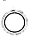
- V, D and J segments are grouped into respective V, D and J regions (see Figure 1-1 ).
- a full complement of V numbering from 4-6 in TRG to 45-47 in TRA
- D (2 in TRB and 3 in TRD
- J numbering as few as 4 in TRD to as many as 61 in TRA
- the specificity of any resulting coding sequence would be uniformly based on inherited variation.
- BIOMED-2 (29) is a product of several years of collaborative expert study, resulting in a thoroughly studied consensus T-cell clonality assay.
- the BIOMED-2 assay includes multiplexed primer sets for both Immunoglobulin (IG) and TR clonality assessment and can be implemented with commercially available electrophoresis systems (e.g. Applied Biosystems fluorescence electrophoresis platforms) (29) . These commercially available primer sets have the advantage of standardization and ease of implementation.
- the BIOMED-2 assay has the well-documented advantage of capturing the mono-clonality of the vast majority of control lymphomas bearing productive T-cell receptors (i.e.
- TRGR testing is subject to a number of technical and practical caveats that dilute the applicability of TRGR testing to the full breadth of real-world contexts.
- BIOMED-2 assay is subject to additional technical challenges. As part of the standard TRGR assay, most laboratories rely on the demonstration of electrophoretic migration patterns for the determination of TR clonality. Interpretation of the assay depends on the demonstration (or lack thereof) of a dominant amplicon of specific (albeit not pre-defined) molecular weight, rather than the normal Gaussian distribution of amplicons of variable size. This approach, as has been described previously (35-37) , is subject to interpretative error and other technical problems. Also, given the large amounts of DNA required for the multitude of multiplex tubes making up the assay, the overall assay can very quickly deplete DNA supplies, especially when obtained from limited sample sources.
- TCR Current T-Cell Receptor
- the simplest method for assessing clonality of T-cells involves qualitative assessment through multiplexed amplification of the individual loci using defined primer sets and interpretation of fragment size distributions according to the BIOMED2 protocol A1,2 .
- Next-generation sequencing can be used as a read-out to provide quantitative assessment of the TCR repertoire including detection of low abundance rearrangements from bulk immune cells, or even pairing of the heterodimeric chain sequences with single cell preparation methods A3,4 .
- Hybrid-capture based library subsetting is an alternative method to PCR-based amplification that can improve coverage uniformity and library complexity when sample is not limiting and allows for targeted enrichment of genetic loci of interest from individual genes to entire exomes A5 .
- hybrid-capture methods the formation of probe-library fragment DNA duplexes are used to recover regions of interest A6 7,8 .
- B-cells involved in adaptive immunity also undergo somatic rearrangement of germline DNA to encode a functional B-cell receptor (BR).
- BR B-cell receptor
- these sequences comprise by discrete V, D, J segments that are rearranged and potentially altered during B-cell maturation to encode a diversity of unique immunoglobulin proteins.
- the clonal diversity of B-cell populations may have clinical utility and, similar to T-cell lymphomas, several cancers are characterized by clonal expansion of specific BR/lg sequences.
- He et al. discloses a method of capturing and characterising rearranged immunoglobulin (IgH) and B-cell DNA fragments in lymphoma and plasma samples of patients with Non-Hodgkin's Lymphoma.
- Illumina Inc. Publication No. 770-2012-008, 2012 ) discloses Illumina Next-Generation sequencing techniques adapted for short read sequencing.
- US2015/0218620 discloses methods to capture and/or remove highly abundant RNAs from a heterogeneous RNA sample.
- US2008/0125333 discloses methods and kits for hybrid capture using a depletion technique to allow isolation of capture-associated nucleotides.
- NGS-based T-cell clonality assay incorporating all four TR loci.
- the assay was both analytically and clinically validated.
- a series of idealized specimens was used, with combined PCR/Electrophoresis and Sanger Sequencing to confirm NGS-data.
- the latter validation compared NGS results to the current gold standard for clinical T-cell clonality testing (i.e. the BIOMED-2 primer PCR method) on an appropriately-sized minimally-biased sample of hematopathology specimens.
- the patterns of T-cell clonality were also correlated with clinical, pathologic, and outcome data.
- a method of capturing a population of T-Cell receptor and/or immunoglobulin sequences with variable regions within a patient sample comprising: extracting/preparing DNA fragments from the patient sample; ligating a nucleic acid adapter to the DNA fragments, the nucleic acid adapter suitable for recognition by a pre-selected nucleic acid probe; capturing DNA fragments existing in the patient sample using a collection of nucleic acid hybrid capture probes, wherein each capture probe is designed to hybridize to a known V gene segment and/or a J gene segment within the T cell receptor and/or immunoglobulin genomic loci, wherein the hybrid capture probes are designed based on all unique V genes and J genes and these probe sets specifically target the 3' regions of V gene coding regions and the 5' regions of J gene coding regions.
- a method of immunologically classifying a population of T-Cell receptor and/or immunoglobulin sequences can comprise:
- a method of identifying CDR3 regions in T-Cell receptor and/or immunoglobulin sequences can comprise:
- sequence-level data in a TRGR assay would likely be much more informative than gross estimates of DNA electrophoretic migration patterns when disease trends are being studied; the high-level analysis of such data might help the identification of heretofore hidden patterns of TR rearrangement in specific T-cell lymphoma subtypes.
- the issue of replicate numbers for establishing test sensitivity/specificity can be easily overcome by exploiting the high-throughput capacity of modern DNA sequencing platforms; for a comparable investment of time (and possibly cost), sequencing-based approach to TRGR could perform a greater number of individual tests, thereby potentially allowing a more statistically robust estimate of test performance.
- library preparation may be generalized to permit the enrichment of specific sequences out of a mix of "all" sequences produced from the primary non-specific amplification step; it is easy to see how this approach may be used to permit multiple separate assays using different enrichment approaches applied to a single input library (40) .
- Hybrid capture is a form of library enrichment in which a library is probed for known sequences of interest using tagged nucleic acid probes followed by a subsequent "pull-down" of the tagged hybrids (38) ; for example, DNA probes tagged with biotin can be efficiently enriched when hybridization is followed by a streptavidin enrichment step (38,40-43) .
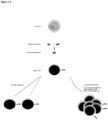
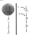
- the biotin/streptavidin enrichment procedure is schematized in Figure 2-1A . In reference to the assessment of TRGR, this approach has the advantage of enriching TR genes based on the available well-defined germline TR gene sequences, which can be performed in a massively parallel fashion using several hundred probes.
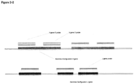
- this approach also allows for enrichment of rearranged sequences as the hybrid-capture probes can also hybridize to (and therefore enrich for) subsequences of the rearrangement product.
- This latter "pull-down" of rearranged TR genes would be difficult using a primer-only approach to library preparation.
- a more prudent sampling rubric might use a "real-world” series of consecutive samples taken from a population as similar to the "test population” as possible.
- a sample might consist of a series of consecutive tissue samples from patients being worked-up by a hematologist and submitted for molecular (i.e. T-cell clonality) assessment.
- the overall sample size could be established based on an estimate of the historical incidence of T-cell lymphomas in such a population, such that the total size of the sample is adequately large to include a sufficient "expected" number of clonal T-cell lymphoproliferative disorders.
- Composite gold-standards might include a number of individual test results or clinical observations logically combined to produce "positive” or “negative” composites (46) ; of key import is that (1) well-defined rules of composition be set out a priori and (2) the number of samples or subjects with each of the composite test results should be well-described (46) . Ideally, all samples or subjects should be evaluated using each of the composite tests (46) .
- T-cell lymphomas are cancers of immune cell development that result in clonal expansion of malignant clones that dominate the T-cell repertoire of affected patients. Therefore, clonality assessment of these cell populations is essential for the identification and monitoring of T-cell lymphomas.
- TCR T-cell receptor
- TCR V(D)J repertoire of monoclonal cancer cell lines, tumor-derived lymphocyte cultures, and peripheral blood mononuclear cells from a healthy donor, as well as a set of 63 clinical isolates sent for clinical clonality testing for suspected T-cell lymphoma.
- PCR amplification and Sanger sequencing confirmed cell line and tumor predominant rearrangements, individual beta locus V and J allele prevalence was well correlated with results from a commercial PCR-based DNA sequencing assay with an r 2 value of 0.94, and BIOMED2 PCR fragment size beta and gamma locus clonotyping of clinical isolates showed 73% and 77% agreement respectively.
- Our method allows for rapid, high-throughput and low cost characterization of TCR repertoires that will enhance sensitivity of tumor surveillance as well as facilitate serial analysis of patient samples with a quantitative read-out during clinical immunotherapy interventions.
- a method of capturing a population of T-Cell receptor and/or immunoglobulin sequences with variable regions within a patient sample comprising: extracting/preparing DNA fragments from the patient sample; ligating a nucleic acid adapter to the DNA fragments, the nucleic acid adapter suitable for recognition by a pre-selected nucleic acid probe; capturing DNA fragments existing in the patient sample using a collection of nucleic acid hybrid capture probes, wherein each capture probe is designed to hybridize to a known V gene segment and/or a J gene segment within the T cell receptor and/or immunoglobulin genomic loci, wherein the hybrid capture probes are designed based on all unique V genes and J genes and these probe sets specifically target the 3' regions of V gene coding regions and the 5' regions of J gene coding regions.
- T-Cell Receptor or "TCR” means a molecule found on the surface of T lymphocytes (or T cells), preferably human, that is responsible for recognizing fragments of antigen as peptides bound to major histocompatibility complex (MHC) molecules.
- MHC major histocompatibility complex
- the TCR is a disulfide-linked membrane-anchored heterodimeric protein normally consisting of the highly variable alpha ( ⁇ ) and beta ( ⁇ ) chains expressed as part of a complex with the invariant CD3 chain molecules. T cells expressing this receptor are referred to as ⁇ : ⁇ (or ⁇ ) T cells, though a minority of T cells express an alternate receptor, formed by variable gamma ( ⁇ ) and delta ( ⁇ ) chains, referred as ⁇ T cells.
- Each chain is composed of two extracellular domains: Variable (V) region and a Constant (C) region.
- the variable domain of both the TCR ⁇ -chain and ⁇ -chain each have three hypervariable or complementarity determining regions (CDRs).
- CDR3 is the main CDR responsible for recognizing processed antigen.
- antibody and "immunoglobulin”, as used herein, refer broadly to any immunological binding agent or molecule that comprises a human antigen binding domain, including polyclonal and monoclonal antibodies. Depending on the type of constant domain in the heavy chains, whole antibodies are assigned to one of five major classes: IgA, IgD, IgE, IgG, and IgM. Several of these are further divided into subclasses or isotypes, such as IgG1, IgG2, IgG3, IgG4, and the like.
- the heavy-chain constant domains that correspond to the difference classes of immunoglobulins are termed ⁇ , ⁇ , ⁇ , ⁇ and ⁇ , respectively.
- the subunit structures and three-dimensional configurations of different classes of immunoglobulins are well known.
- the "light chains" of mammalian antibodies are assigned to one of two clearly distinct types: kappa ( ⁇ ) and lambda ( ⁇ ), based on the amino acid sequences of their constant domains and some amino acids in the framework regions of their variable domains.
- the variable domains comprise the complementarity determining regions (CDRs).
- CDRs complementarity determining regions
- V gene segments refer to the variable (V), joining (J), and diversity (D) gene segments involved in V(D)J recombination, less commonly known as somatic recombination.
- V(D)J recombination is the mechanism of genetic recombination that occurs in developing lymphocytes during the early stages of T and B cell maturation. The process results in the highly diverse immune repertoire of antibodies/immunoglobulins (Igs) and T cell receptors (TCRs) found on B cells and T cells, respectively.
- Igs antibodies/immunoglobulins
- TCRs T cell receptors
- nucleic acid includes DNA and RNA and can be either double stranded or single stranded.
- probe refers to a nucleic acid sequence that will hybridize to a nucleic acid target sequence.
- the probe hybridizes to the RNA biomarker or a nucleic acid sequence complementary thereof.
- the length of probe depends on the hybridization conditions and the sequences of the probe and nucleic acid target sequence. In one embodiment, the probe is at least 8, 10, 15, 20, 25, 50, 75, 100, 150, 200, 250, 400, 500 or more nucleotides in length.
- adapter refers a moiety capable of conjugation to a nucleic acid sequence for a particular purpose.
- the adapter may be used to identify or barcode the nucleic acid.
- the adapter may be a primer which can be used to amplify the nucleic acid sequence.
- hybridize or “hybridizable” refers to the sequence specific non-covalent binding interaction with a complementary nucleic acid.
- the hybridization is under stringent conditions. Appropriate stringency conditions which promote hybridization are known to those skilled in the art, or can be found in Current Protocols in Molecular Biology, John Wiley & Sons, N.Y. (1989), 6.3.1 6.3.6 . For example, 6.0 x sodium chloride/sodium citrate (SSC) at about 45°C, followed by a wash of 2.0 x SSC at 50°C may be employed.
- SSC sodium chloride/sodium citrate
- the method further comprises sequencing the captured DNA fragments, wherein the sequencing can be used to determine clonotypes within the patient sample.
- sequencing techniques are known to the person skilled in the art, such as polymerase chain reaction (PCR) followed by Sanger sequencing.
- PCR polymerase chain reaction
- NGS next-generation sequencing
- high-throughput sequencing which includes various sequencing technologies including: Illumina (Solexa) sequencing, Roche 454 sequencing, Ion torrent: Proton / PGM sequencing, SOLiD sequencing.
- NGS allow for the sequencing of DNA and RNA much more quickly and cheaply than the previously used Sanger sequencing.
- said sequencing is optimized for short read sequencing.
- the method further comprises amplifying the population of sequences using nucleic acid amplification probes/oligonucleotides that recognize the adapter prior to said sequencing.
- the method further comprises fragmenting DNA extracted from the patient sample to generate the DNA fragments.
- the ligating step is performed before the capturing step.
- the capturing step is performed before the ligating step.
- patient refers to any member of the animal kingdom, preferably a human being and most preferably a human being that has AML or that is suspected of having AML.
- sample refers to any fluid, cell or tissue sample from a subject which can be assayed for nucleic acid sequences.
- the patient sample comprises tissue, urine, cerebral spinal fluid, saliva, feces, ascities, pleural effusion, blood or blood plasma.
- the patient sample comprises cell-free nucleic acids in blood plasma.
- the clonality analyses described herein may be use to track clonality across samples types.
- the hybrid capture probes are at least 30bp in length. In a further embodiment, the hybrid capture probes are between 60bp and 150bp in length. In a further embodiment, the hybrid capture probes are between 80bp and 120bp in length. In a further embodiment, the hybrid capture probes are about 100bp in length.
- the hybrid capture probes hybridize to at least 30bp, preferably 50bp, more preferably 100bp of the V gene segment and/or J gene segment.
- the screening probes hybridize to at least a portion of the V gene segment.
- the screening probes hybridize to at least a portion of the V gene segment at the 3' end.
- hybridizing comprises hybridizing under stringent conditions, preferably very stringent conditions.
- the collection of nucleic acid hybrid capture probes comprise at least 2, 5, 10, 20, 30, 80, 100, 300, 400, 500, 600, 700, 800 or 900 unique hybrid capture probes.
- the collection of nucleic acid hybrid capture probes is sufficient to capture at least 50%, 60%, 70%, 80%, 90% or 99% of known T-Cell receptor and/or immunoglobulin loci clonotypes.
- the hybrid capture probes comprise a label.
- the label is used to distinguish between sequences bound to the screening probes and unbound double stranded fragments, and preferably the capture is performed in solution.
- the method further comprises a depletion step, comprising depleting the DNA fragments of non-rearranged sequences using probes that recognize nucleic acid sequences adjacent to V and/or J gene segments in the genome.
- a depletion step comprising depleting the DNA fragments of non-rearranged sequences using probes that recognize nucleic acid sequences adjacent to V and/or J gene segments in the genome.
- the capturing of DNA fragments using V gene segment and J gene segment hybrid capture probes is performed in separate steps, and in any order with the depletion step, preferably in the following order: J gene capture , depletion , then V gene capture.
- a method of immunologically classifying a population of T-Cell receptor and/or immunoglobulin sequences may comprise:
- step (h) may be searching the 6 translated sequences for flanking invariable anchor sequences to define the intervening T-Cell receptor and/or B-cell receptor CDR3 sequences encoded by the DNA fragments.
- This method may further comprise, prior to step (a), aligning left and right reads of overlapping initial DNA fragments to produce the DNA fragments on which step (a) is performed.
- Steps (a), (c), (e) may be performed with BLASTn and step (i) may be performed using expression pattern matching to known sequences and IMGT annotated data.
- a method of identifying CDR3 regions in T-Cell receptor and/or immunoglobulin sequences may comprise:
- step (c) may be if V and J gene segments are identified, then searching the immunoglobulin sequence for flanking invariable anchor sequences to define the intervening T-Cell receptor and/or immunoglobulin CDR3 sequences.
- Steps (a) and (b) may be performed using the Burrows-Wheeler Alignment or other sequence alignment algorithm.
- the method may further comprise determining whether the identified V and J gene segments could be rearranged in the same locus using a heuristic approach.
- the method may further comprise determining if a combination of V(D)J gene segments is present based on Smith Waterman Alignment scores.
- Any of the methods described herein may be used to capture a population of T-Cell receptor sequences, for immunologically classifying a population of T-Cell receptor sequences or for identifying CDR3 regions in T-Cell receptor.
- the methods described herein are for characterizing T-cell clonality for a disease in the subject.
- the T-Cell receptor sequences are from tumour infiltrating lymphocytes.
- the methods described herein are for identifying therapeutic tumour infiltrating lymphocytes for the purposes of expansion and reinfusion into a patient and/or adoptive cell transfer immunotherapy.
- the methods described herein are for monitoring T-cell populations/turnover in a subject, preferably a subject with cancer during cancer therapy, preferably immunotherapy.
- the methods described herein are for characterizing the immune repertoire of a subject, the immune repertoire comprising the subject's B-Cell population.
- the methods described herein are for capturing a population of B-Cell receptor sequences with variable regions within a patient sample.
- the methods described herein may also be used for immunologically classifying a population of B-Cell receptor sequences, or for identifying CDR3 regions in B-Cell receptor sequences.
- the methods described herein are for characterizing B-cell clonality as a feature of a disease in the subject.
- Cancers include adrenal cancer, anal cancer, bile duct cancer, bladder cancer, bone cancer, brain/cns tumors, breast cancer, castleman disease, cervical cancer, colon/rectum cancer, endometrial cancer, esophagus cancer, ewing family of tumors, eye cancer, gallbladder cancer, gastrointestinal carcinoid tumors, gastrointestinal stromal tumor (gist), gestational trophoblastic disease, hodgkin disease, kaposi sarcoma, kidney cancer, laryngeal and hypopharyngeal cancer, leukemia (acute lymphocytic, acute myeloid, chronic lymphocytic, chronic myeloid, chronic myelomonocytic), liver cancer, lung cancer (non-small cell, small cell, lung carcinoid tumor), lymphoma, lymphoma of the skin, malignant mesothelioma, multiple myeloma, myelodysplastic syndrome, nasal cavity
- the subject may have a T-cell related disease, such as a T-cell lymphoma.
- T-cell lymphomas are types of lymphoma affecting T cells, and can include peripheral T-cell lymphoma not otherwise specified, extranodal T cell lymphoma, cutaneous T cell lymphoma, including Sézary syndrome and Mycosis fungoides, anaplastic large cell lymphoma, angioimmunoblastic T cell lymphoma, adult T-cell Leukemia/Lymphoma (ATLL), blastic NK-cell Lymphoma, enteropathy-type T-cell lymphoma, hematosplenic gamma-delta T-cell Lymphoma, lymphoblastic Lymphoma, nasal NK/T-cell Lymphomas, treatment-related T-cell lymphomas.
- peripheral T-cell lymphoma not otherwise specified, extranodal T cell lymphoma, cutaneous T cell lymphoma, including Sézary syndrome and Mycosis fungoides, anaplastic large cell lymphoma, angioimmunoblast
- the subject may have a B-cell related disease, plasma cell disorder, preferably a B-cell lymphoma.
- B-cell are types of lymphoma affecting B cells and can include, diffuse large B-cell lymphoma (DLBCL), follicular lymphoma, marginal zone B-cell lymphoma (MZL) or mucosa-associated lymphatic tissue lymphoma (MALT), small lymphocytic lymphoma (also known as chronic lymphocytic leukemia, CLL), mantle cell lymphoma (MCL), DLBCL variants or sub-types of primary mediastinal (thymic) large B cell lymphoma, T cell/histiocyte-rich large B-cell lymphoma, primary cutaneous diffuse large B-cell lymphoma, leg type (Primary cutaneous DLBCL, leg type), EBV positive diffuse large B-cell lymphoma of the elderly, diffuse large B-cell lymphoma associated with inflammation, Burkitt's lymphoma, lymphoplasmacytic lymphoma, which may manifest as Waldenström's macroglobulinemia, nodal
- the methods described herein are for identifying therapeutic B-cells for the purposes of expansion and reinfusion into a patient.
- the methods described herein are for monitoring B-cell populations/turnover in a subject, preferably a subject with cancer during cancer therapy, preferably immunotherapy.
- the methods described herein are for detecting minimal residual disease, whereby TCR or immunoglobulin rearrangements may be used as a marker of disease.
- a library of probes comprising the depletion probes in Table D or at least one of the V-gene and J-gene probes set forth in any of Tables 2.1, 4, B1, or B2.
- the clonality analyses described herein may be performed serially.
- the clonality analyses described herein may be used to distinguish between samples.
- BIOMED Unlike the current BIOMED approach, we wished to avoid a gene-specific primer-based approach to signal amplification. To accomplish this, we chose a "hybrid capture" target enrichment approach by which input genomic DNA containing the TR genes might be enriched (or “captured”) relative to other segments of the genome.
- hybrid capture target enrichment approach by which input genomic DNA containing the TR genes might be enriched (or “captured") relative to other segments of the genome.
- FFPE Formalin-fixed paraffin-embedded
- the most useful NTRA should allow users to both accurately assess the "clonality" of an input sample (as can be done using BIOMED-2 based assays) but also fully characterize the clonotypes of constituent TRGR configurations.
- the NTRA not simply produce a binary "clonal” vs. "polyclonal” result but also provide a much more robust and quantitative data output, including the genes and CDR3 regions present within identified TRGR configurations.
- Appendix 2.0 outlines a MATLAB script designed to estimate the optimal DNA insert length (a value also generalizable to optimal shearing length and minimal Paired-end rEAd mergeR (PEAR)-assembled sequencing length) for the purposes of the NTRA.
- This optimum is subject to an important restriction: for our purposes, using the Illumina NextSeq platform, read lengths are limited to paired-ended reads of 150 bp each-this translates to ⁇ 300 bp read lengths when paired-ends are joined by overlapping sequence (using, in our case, the PEAR algorithm (52) ).
- BWA Burrow
- DNA probe design was then performed using the IMGT reference sequences (including all annotated V and J gene functional, pseudogene and open reading frame sequences) using the xGen Lockdown probe technology.
- this technology is a hybrid-capture-based technology by which biotin-tagged DNA probes (complementary to known sequences/genomic regions set at a 1x depth of coverage) are allowed to hybridize with sample DNA, followed by a streptavidin elution procedure performed to enrich the target sequences (40-43) .
- each DNA probe was designed to a length as close to 100 bp as possible.
- germline-configuration sequences were extracted for all alleles of all J-genes, with additional leading and trailing IMGT nucleotides added (as necessary) to obtain 100 bp probe lengths; for those instances in which the IMGT data was insufficient to prepare 100 bp probes, additional random nucleotides were added to the leading and trailing ends of the available sequences.
- Table 2.1 outlines the complete list of xGen Lockdown probe design sequences (with relevant associated metadata).
- the NTRA work-flow is summarized in Figures 2-1A & 2-1B .
- the process begins with DNA isolation, performed for the purposes of this study according to the protocol of Appendix 2.1.
- isolated DNA was retrieved from frozen archives and quantified using the Qubit assay, per Appendix 2.2.
- input DNA was shorn using a Covaris sonicator (Appendix 2.3) set to a desired mean DNA length of 200 base pairs; adequate shearing was confirmed using TapeStation assessment.
- Sequence libraries for each specimen were prepared using the protocol outlined in Appendix 2.4; multiplexing was accommodated using either TruSeq or NEXTflex-96 indices (the latter employed in the final validation run to permit large-scale multiplexing).
- the NTRA TRSeq pipeline was designed around three main algorithmic steps.
- the first performs local alignment indexed to the TR V and J genes implemented using the Burrows-Wheeler-Alignment (BWA) algorithm (55) .
- BWA Burrows-Wheeler-Alignment
- two important results are obtained: the first is a "reads-on-target" estimate (since the genes enriched for (i.e. the TR V and J genes) are those genes used as the index reference gene set); second, by way of the resulting Sequence Alignment Map (SAM) file output, the original input reads are filtered to exclude those unlikely to contain any of the TR V or J genes.
- SAM Sequence Alignment Map
- This latter step reduces the informatic burden of input to the (relatively computationally slow) second algorithm step (using either heuristics or the Smith-Waterman Alignment (SWA)).
- SWA Smith-Waterman Alignment
- the BWA algorithm could be implemented on a UNIX-based platform only (55) .
- the second algorithm step is designed to extract CDR3 sequences wherever present. This algorithm was implemented in MATLAB, guided by previous publications (56) , and using a regular-expression (regexp) based search algorithm.
- the third step combined the above alignment and CDR3 data (where present), to decide whether a given read contains a TRGR.
- a CDR3 is identified in a read
- a heuristic approach is employed to decide if the BWA-alignment reference genes could be rearranged within the same locus
- the second in the event that a CDR3 is not detected, relies on the SWA-determined alignment scores to determine if a given combination of V(D)J genes is present.
- Reads-on-target and gene-coverage estimates are also derived using the BWA algorithm, since NTRA input probes consist only of TR V and J genes; this measure is calculated as a percentage of the number of unique reads mapped to the IMGT reference TR V and J gene indices relative to the total number of reads in the input FASTQ-formatted file.
- the TRSeq algorithm was implemented in MATLAB using strategies similar to those employed by the IMGT (56-58) .
- the IMGT/V-QUEST system utilizes a CDR3 sequence extraction algorithm (57,59) and an SWA (60) algorithm performed against the IMGT reference sequences; the IMGT algorithms are all implemented in JAVA and processing is performed on IMGT servers.
- a MATLAB implementation was chosen for convenience, programming familiarity, and because of easy vectorization, parallel computation and object-oriented programming capabilities.
- the MATLAB programming and command-line environments are able to easily incorporate UNIX and PERL-based scripts, including the BWA (Li, 2009) and CIRCOS software (61) suites, respectively.
- the SWA algorithm produces an optimal local alignment (60,64) of two co-input sequences (in this case, a query sequence relative to an IMGT reference sequence), and provides an alignment score (a unit-less measure of the degree to which the alignment perfectly matches an input sequence to its co-input sequence).
- an alignment score a unit-less measure of the degree to which the alignment perfectly matches an input sequence to its co-input sequence.
- the alphabetical highest-scoring alignment was selected as the "correct" alignment, provided that this score was at least greater than the minimum cut-off score.
- the minimum SWA alignment cut-off score was empirically determined for each of the three V, D, and J-gene gene groups using a large set of confirmed-negative sequences evaluated using the IMGT/HighV-QUEST system (56,57) .
- the MATLAB code required for implementation of this algorithm is outlined in Appendix 2.6.1.
- a "practice" set obtained from the IMGT database (65,66) was also employed to test the pipeline, consisting of IMGT PCR-confirmed TRGR sequences with known V-D-J combinations and CDR3 sequences (see Section 3.1.3 for results of this practice set analysis).
- PCR-based experiment was deemed a reasonable orthogonal validation approach, given the gold standard BIOMED-2 assay methodology. Knowing that the number of possible rearrangements detected by the NTRA might be substantially large, the PCR validation was arbitrarily limited to those TRSeq-detected rearrangements in the upper 90 th centile (i.e. percent rearrangement of greater than 10% of total rearrangements). Given this restriction, however, to ensure an adequate denominator of reactions for comparative purposes, all PCR validation experiments were uniformly performed across all 10 first-run samples.
- PCR validation primer sets were constructed modeling the standard V-D-J orientation of rearranged TR genes; specifically, the PCR forward primer was set in the V gene and the reverse primer set in the anti-sense strand of the J gene.
- the V and J genes were identified and the IMGT primer set database searched for gene (not allele) specific primers. While the IMGT primer database did contain a number of suggested primers, many of the TR genes did not have an available appertaining primer.
- the anticipated rearrangement sequence (containing the V gene sequence artificially positioned before the J gene) was used to derive custom primers using the NCBI Primer-Blast tool (68) .
- each resulting theoretical PCR product length was at least 100 bp (the lower limit of fragment size reliably detectable by standard gel electrophoresis) and that a sufficient amount of the anticipated CDR3 region sequence would be preserved in the PCR product.
- the theoretical product length was recorded as an approximate size reference for analysis of the resulting electrophoresis migration patterns.
- Custom primer set production was performed commercially by IDT and the forward and reverse primers were then mixed according to the design outlined in Appendix 2.7.2.
- PCR was performed in a 384-well plate on an Applied Biosystems Veriti thermal cycler using the Thermo Scientific 2X ReddyMix PCR Master Mix kit according to the manufacturer's instructions; several control reactions were included, as highlighted in Appendix 2.7.2.
- the resulting sequencing results were analyzed using the FinchTV v 1.4 software suite, with corrections to sequencing error and reverse-complement sequence corrections performed manually as required.
- the originating TRSeq CDR3 sequences were then compared to the "reference" Sanger Sequence result. This comparison was performed in two ways: first, a basic multi-alignment comparison was performed (using the multialign algorithm of the MATLAB Bioinformatics Toolbox); second, a k-mer based PHRED-quality adjusted comparison was performed.
- the Sanger Sequencing results were taken to represent a "consensus" of sequence data produced over all possible V and J configuration CDR3 sequences for that V-J gene configuration (reflecting the possibility of variable TRGR subclones).
- a quality-based alignment algorithm was employed, based on the methods of (70) .
- Each input TRSeq CDR3 sequence was aligned along a progressive series of k-mers of the Sanger sequence using a custom quality-based alignment algorithm (code outlined in Appendix 2.8). For each alignment result, if the optimal alignment score occurred within the expected sequencing region (thereby representing an optimal alignment within a region of Sanger sequence expected to contain the actual CDR3 based on flanking primer sets), as outlined in Table 3.1A, the CDR3 sequence was classified as correct (and vice-versa). This classification was then used to perform ROC analysis to determine what number of TRSeq CDR3 sequence read counts might be considered a validated cut-off.
- a fully germline TR gene configuration was approximated using a cell lines of embryonic origin and a cell line that has been fully sequenced without any known/reported TR gene derangements.
- the former scenario was approximated using the HEK293 cell line (an embryonic kidney cell line; ATCC CRL-1573) and the latter using a Coriell cell line (whose genome has been well-characterized and is not known to contain TR rearrangements).
- Use of the latter cell line was incorporated given that, in our hands, this cell line had been previously and purposefully degraded by FFPE treatment, representing a scenario of TR gene coverage assessment in the context of degraded DNA.
- Total genomic DNA was extracted from previously cultured HEK293 cells and FFPE treated Coriell cell cultures and subsequently subjected to the NTRA, as outlined in Appendices 2.1 to 2.5. Standard TRSeq analyses were performed for each sample, with special deference paid to the coverage results.
- a rigorous dilution series experiment in the context of this project, might involve a flow-sort spike of cells with a known TR gene configuration into a population previously determined to be "polyclonal"; this might be approximated, for example, using a well-characterized cell line spiked into a population of lymphocytes obtained from normal blood. Rather than undertaking this more complex and expensive approach, an approximation of this dilution experiment was undertaken with DNA obtained from the Jurkat cell line spiked into a known-polyclonal lymphocyte population DNA isolate (the A037 sample; see Results section 3.2).
- Jurkat DNA was spiked in at log-decrements (as outlined in Table 2.3) based on a lymphocyte total DNA complement assumed to be 0.7 pg, given the results of previous publications (71-73) .
- the total DNA of each sample in the dilution series was verified (and compared to expected values) using a Qubit assay; the samples were then subjected to the NTRA, as outlined in Appendices 2.1 to 2.5. Standard TRSeq analyses were performed, with special deference to changes in the raw read counts of Jurkat-specific TRGR configurations across the dilution series.
- TCR T cell receptor
- PBMC peripheral blood mononuclear cells
- Isolated DNA is sheared to ⁇ 275bp fragments by sonication in 130uL volumes (Covaris).
- DNA libraries are generated for illumina platform sequencing from 100-1000ng of sheared DNA by ligation of sequencing library adaptors (NextFlex) using the KAPA library preparation kit with standard conditions. Libraries are visually assessed (Agilent TapeStation) and quantified (Qubit) for quality.
- Hybridization with probes specifically targeting the V and J genes is performed under standard SeqCap (Roche) conditions with xGen blocking oligos (IDT) and human cot-1 blocking DNA (Invitrogen). Hybridization is performed either at 65C overnight.
- the target capture panel consists of 598 probes (IDT) targeting the 3' and 5' 100bp of all TCR V gene regions, and 95 probes targeting the 5' 100bp of all TCR J gene regions as annotated by IMGT (four loci, 1.8Mb, total targeted 36kb).
- Hybridization and capture can be performed as a single step with a combined V/J panel, as a single step with only the V panel, or as a three step process when non-rearranged fragment depletion is desired consisting of a V capture, then depletion, then J capture.
- a modified and truncated SeqCap protocol is employed wherein following incubation with M-270 streptavidin linked magnetic beads (Invitrogen), the hybridization reaction is diluted with wash buffer I, beads are discarded and the supernatant is cleaned up by standard Agencourt AMPure XP SPRI bead purification (Beckman).
- a custom Bash/Python/R pipeline is employed for analysis of paired read sequencing data generated by Illumina NextSeq 2500 instrument from the hybrid-capture products.
- this pipeline consists of four major steps: (1) Merging of the paired reads; (2) identification of specific V, J, and D genes within the fragment sequence; (3) identification of the V/J junction position as well as the antigen specificity determining Complementarity Determining Region 3 (CDR3) sequence at this site; (4) Calculation and visualization of capture efficiency and clone frequency within and across individual samples.
- CDR3 Complementarity Determining Region 3
- the custom pipeline is not dependent on our hybrid-capture protocol and can be performed on non-target captured whole genome or RNA-seq data.
- an in silico capture is performed by extracting reads aligning to the four TCR loci (7:38250000-38450000, 7:141950000-142550000, 14:22000000-23100000) or Ig loci (chr2:89,100,000-90,350,000, chr14:106,400,000-107,300,000, chr22:22,350,000-23,300,000) from DNA (BWA) or RNA (STAR) sequence data (SamTools), followed by paired-end nucleotide sequencing data extraction (PicardTools). These reads are then inserted in to the previously described computational pipeline.
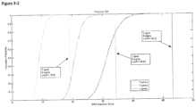
- Figure 3-1A details the DNA Insert Length Simulation results.
- the analysis suggested a plateau of sensitivity of greater than 99,1% reached after 182 bp.
- an adequately "evidence-based" insert length and informatics read length goal of 200 bp was chosen for the NTRA.
- the TRSeq algorithm identified 50 of the 55 reads as containing evidence of TRGR; the 5 remaining reads were identified by the IMGT as containing rearrangements within the TRD locus, each with a TRSeq CDR3 region correctly identified.
- Table 2.5 outlines the flow-cytometric features of the 6 patient lymphocyte samples. These immunophenotypic features were in keeping with the lymphocyte sample sources of origin (also documented in Table 2.5), varying from normal patient peripheral blood mononuclear cells to highly immuno-sensitized lymphocyte cultures from tumour infiltrating lymphocyte specimens. Notably, the A037 sample served as a model of a "polyclonal" lymphocyte population whereas, for the purposes of qualitative assessment at least, the L2D8 sample could be immunophenotypically interpreted as highly "clonal” in nature.
- model "clonal” samples were included, consisting of the Jurkat, CEM, SUPT1 and MOLT4 cell lines. Table 2.6 lists the previously documented rearrangements, as cited in the IMGT database (67) .
- Figures 3-5 , 3-6 , and 3-7A & 3-7B summarize the TRSeq metadata for the first-run sample series, including input reads, reads-on-target, summary coverage statistics, and a histogram of read counts for the proportion of each locus contributing to identified TRGR's, respectively.
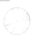
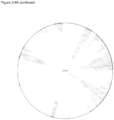
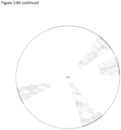
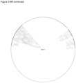
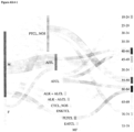
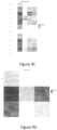
- Figures 3-8A and 3-8B display composites of the circos plots obtained from the 10 first-run samples. Much as the coverage profiles differed across the samples (as seen in Figures 3-8C & 3-8D), the resulting circos plots demonstrated a clear aesthetic difference from polyclonal to clonal/oligoclonal samples, with emphasis on the number and relative width of the composite circos links (i.e. fewer and broader in width in the more clonal cases and vice versa). Also of note, the color distributions were distinctly different with the more polyclonal cases, containing a larger number of smaller-quantity "subclones" involving a more disparate number of TR genes.
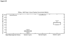
- the high D-gene concordance relative to the V and J-gene values may relate to both the shorter reference sequences of the D-genes relative to the V and J genes, as well as the lower number of reference D-genes available for rearrangement. It is important to point out the possibility of a theoretical bias against D-gene identification in input reads, given that TRGR reads containing D-genes require 3 rather than 2 composite genes, which could be more difficult to detect in the context of restricted average read lengths.
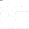
- PCR primers were mixed according to the design of Figure 3-10 and the results by Agarose gel electrophoresis are shown in Figure 3-11 . Note that results obtained from PCR reactions using the TRGJ2 reverse primer are excluded, as noted in Section 2.2.2. Two classification approaches may then be entertained, one based on dark-staining PCR bands only, and the other based on any staining (assuming bands to be of appropriate molecular weights, as set out in Table 3.1A). When these classifiers are compared with the read-count-normalized results of the TRSeq algorithm (as set out in Table 3.1A), the ROC curves of Figures 3-12A & 3-12B are obtained, respectively.
- the ROC Area-Under-the-Curve (AUC) 0.91 and p-value ⁇ 0.001, with a TRSeq normalized read count of 6.7 or more.
- AUC Area-Under-the-Curve
- Figure 3-10 details those PCR reactions that were post-PCR purified and submitted for Sanger Sequencing.
- Figure 3-13 denotes the alignment of each corresponding TRSeq CDR3 sequence (and associated raw read count) in relation to the manually-verified/corrected Sanger Sequencing Result; only those Sanger Sequencing specimens containing TRSeq-identified CDR3 regions, those of sufficient quality for interpretation, and those not rejected based on use of the TRGJ2 reverse primer were further considered.
- the NTRA was tested on samples of previously cultured HEK293 and Coriell cell lines; these analyses aimed mainly at estimating coverage ceilings for the NTRA, but also served as added negative control specimens (i.e. specimens known or expected not to contain any TRGRs).
- TRGRs were identified in 0 of 5,729,205 total input reads in the HEK293 cell line and only 7 of 2,761,466 total input reads in the Coriell cell line). This was in keeping with the anticipated fully-germline configuration of each of these non-lymphoid origin cell types.
- the percent V and J genes at or above 100x coverage was 100%; the overall TR V gene coverage averaged 29960x; and the overall TR J gene coverage averaged 8789x.
- the percent V and J genes at or above 100x coverage was 100%; the overall TR V gene coverage averaged 13379x; and the overall TR J gene coverage averaged 3925x.
- a dilution experiment was performed at log-reduction intervals, set up according to the design of Table 2.3, and adjusted according to Table 3.2 to account for Jurkat DNA concentration discrepancies.
- Three Jurkat cell line unique TRGR configurations were selected for inter-dilution comparison, namely the TRAV8-4 - TRAJ3, TRGV11 - TRGJ1 and TRGV8 - TRGJ2 rearrangements identified & confirmed in Section 3.3.
- the above configurations were confirmed absent in the polyclonal (A037) sample.
- each of these configurations showed a specific dominant CDR3 sequence.
- Figure 3-16A details the mean of the raw read-counts (i.e. not normalized) across the three tracked V-J configurations (with error bars for standard deviation) vs. expected approximate Jurkat cell numbers (with adjustments for significant digits) from Table 3.2.
- This limited sensitivity speaks to the sensitivity of the TRSeq junction finder to sequencing error. Indeed, if even a single base is changed relative to the canonical regular expression required for detection of a CDR3 sequence, the junction finder will not identify the sequence correctly; likewise, any non-triplicate base insertion will not be detected as an in-frame CDR3 sequence.
- the TRSeq V and J gene enumeration scheme uses alignment-based algorithms, the TRSeq results relating to V and J gene enumeration are much more forgiving of higher the higher likelihood of sequencing error in clonotypes with low read counts, thus substantially improving the assay sensitivity for characteristically unique V-J gene configurations.
- the NTRA TRB and TRG clonotype tables were analyzed to compare the ratio of the dominant clonotype read count relative to the "background” read count.
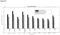
- the largest read count not satisfying the normalized TRSeq read count according to the results of Section 3.3 was taken as the background read count value; alternatively, in the case where the dominant clonotype did not satisfy the normalized TRSeq read count cut-off of Section 3.3, the next largest clonotype read count was taken as "background”. From among each of the TRB and TRG loci, the largest dominant clonotype-to-background ratios were compared to the overall BIOMED-2 results using a ROC analysis.
- Section 3.4 The clear discordance between these results and those of Section 3.4 likely relates to several factors.
- the sample size in Section 3.4 is one-sixth that of the clinical validation set, making the results of Section 3.4 much more vulnerable to the effects of outliers.
- the overall coverage in the analytical validation set was lower, owing to base-output restrictions using the mid-output NextSeq kit; as such, coverage correlations made in Section 3.4 might not necessarily be applicable to experiments performed using the high-output NextSeq kit.
- the clinical validation experiment was not subject to bias of assumption as to the clonality of each input specimen; rather clonality was specifically assayed using an orthogonal method.
- Described above is the first hybrid-capture-based T-cell clonality assay designed to assess clonality and provide clonotype data over all four T-cell gene loci.
- a custom MATLAB-based analysis pipeline was implemented using optimized object-oriented programming integrating the ultra-fast BWA alignment system and the aesthetically-pleasing circos-based genomic data visualization suite.
- the latter visualization was designed with current methods in mind, in which electropherographic plots serve as the primary means by which clonotypes are visualized.
- the use of hybrid capture makes the process platform-agnostic.
- the laboratory work-flow can be integrated into any standard library preparation work-flow with the addition of a single hybridization step, capable of enriching for sequences containing T-cell genes of a several specimens at a time.
- the probes used as part of the NTRA are amenable to "spike-in" combined hybridization reactions, provided that there is no significant probe-set sequence overlap or complementarity.
- the NTRA adds a dearth of extra data, especially as pertaining to clonotype data from the gene-rich alpha-locus. This locus has traditionally been too diffusely distributed within the genome to be amenable to primer-based amplification, a challenge easily overcome using a hybrid-capture approach.
- the NTRA outputs a clonotype table containing data specific to the best aligned allele.
- visualized data is restricted to gene-level only, thereby providing a means of visualization comparable to electropherographic output.
- included with the latter is the in-frame CDR3 sequence (where detected), data currently not available using either standard PCR-based techniques or the mainstream sequencing-based solutions (e.g. Invivoscribe).
- the NTRA was also shown to be theoretically sensitive to low-level clonotypes. This latter observation is an important boon to the hybrid-capture approach, suggesting that carefully performed hybrid-capture methods can provide signal amplification comparable to flow-cytometric (81) and molecular approaches (32)(82)(83) .
- the assay may be considered cost effective, depending on the specific scenario of interest.
- the use of a hybrid-capture approach allows for spike-ins of additional probes for other genomic regions of interest. This allows the possibility of running multiple assays from a single library preparation step, requiring only bioinformatic separation of the resulting enriched sequences.
- lymphocyte clonality is integral to the diagnosis of diseases and cancer affecting the immune system.
- sequencing of the T-cell repertoire of a patient has gained clinical value with the recent understanding of T-cell mediated recognition and destruction of neoplasms.
- the development of adoptive cell therapy and recombinatorial engineering of T-cell receptors requires high-throughput molecular characterization of in vitro T-cell populations before transplant. PCR-based methods such as BIOMED-2 and Immunoseq are currently in use for TCR characterization however their costs and complexity remain barriers for clinical deployment requiring high-throughput multi-patient, multi-sample work-flows at low cost.
- TCR clonality and CDR3 prevalence can be rapidly assessed in a three-day turn-around time with an automated pipeline generating summary figures that can be rapidly assessed by clinicians.
- Adaptive T-cell immunotherapy has become a field of great interest in the treatment of multiple solid-tumor cancer types.
- Non-childhood cancers particularly those linked to chronic exposure of known carcinogens, are driven by the accumulation of mutations. Some of these mutations drive pro-tumorigenic changes, while others result in non-tumorigenic changes to proteins expressed by the carrier cell.
- MHC I molecular surveillance molecules
- these modified polypeptides are recognized as foreign neo-antigens by the host immune system, and in the context of other signals, lead to the activation of T-cells that direct the destruction of cells expressing these modified proteins.
- TIL tumor infiltrating lymphocytes
- T-cell lymphocytes are fundamental to this process, however due to their extraordinar specificity, only neo-antigen specific T-cells are capable of driving anti-tumor activity.
- Our method provides a high-throughput, low cost and rapid turn-around method for T-cell receptor characterization in order to facilitate clinical deployment and uptake of adoptive cell transfer immunotherapy.
- This method is not only of use in immunotherapy applications, as any disease involving expansion of T-cell clones would benefit from its use.
- the symptoms of autoimmune diseases are driven largely by T-cell mediated cytotoxicity of "self" tissue and therefore the identification and expansion of specific T-cell clones can be monitored using this method.
- This method would also be useful to follow immune challenges such as infection or immunization in the development of anti-infectives or vaccines.
- Probe design All annotated V, D, J gene segments were retrieved from the IMGT / LIGM-DB website (www.imgt.org 9 ). The 100bp of annotated 3' V gene coding regions and up to 100bp, when available, of annotated 5' J gene coding regions were selected as baits. Probes with duplicate sequences were not included.
- DNA isolation - CD3+ T cells were isolated by flow assisted cell sorting of PBMC populations separated from whole blood.
- Peripheral blood mononuclear cells PBMC
- Qiagen Gentra Puregene kit
- a Qiagen Allprep (Qiagen) kit was employed, according to the manufacturer's instructions.
- FFPE samples a previously optimized in-house approach was used.
- sample FFPE tissue blocks were cored with a sterilized Tissue-Tek Quick-Ray punch (Sakura) in a pre-selected area of representative tissue; alternatively, under sterile conditions, 10 x 10 ⁇ m DNA curls/unstained slides were obtained for each submitted block of FFPE tissue.
- a fumehood 400-1000 ⁇ L xylene was aliquot into each tube (volume increased for larger FFPE fragments), followed by vigorous vortexing for 10 sec, incubation in a 65°C water bath for 5 min, and centrifugation at 13200 rpm for 2 min. The supernatant was then discarded and step an additional xylene treatment step was performed.
- Hybrid capture - isolated genomic DNA was diluted in TE buffer to 130uL volumes. Shearing to ⁇ 275bp was then performed on either a Covaris M220 Focused-ultrasonicator or E220 Focused-ultrasonicator, depending on sample throughput, with the following settings: for a sample volume of 130 ⁇ L and desired peak length of 200 bp, Peak Incident Power was set to 175 W; duty factor was set to 10%; cycles per burst was set to 200; treatment time was set to 180 s. in addition, temperature and water levels were carefully held to manufacturer's recommendations given the instrument in use.
- the V/J junction position is extracted from the previous search data for those fragments containing both a V and J search result. 80bp of DNA sequence flanking this junction is translated to amino acid sequence in all six open reading frames and sequences lacking stop codons are searched for invariable anchor residues using regular expressions specific for each TR class as determined by sequence alignments of polyclonal hybrid-captured data from rearranged TR polypeptides annotated by IMGT 9
- the CapTCR-seq method employs hybrid capture biotinylated probe sets designed based on all unique Variable (V) gene and Joining (J) gene annotations retrieved from the IMGT database version 1.1, LIGMDB_V12 9 .
- V Variable
- J Joining
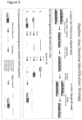
- These probe sets specifically target the 3' regions of V gene coding regions and the 5' regions of J gene coding regions that together flank the short Diversity (D) gene fragment in heavy chain encoding loci and which together form the antigen specificity conferring CDR3 ( Figure 6A ).
- D regions abent in alpha and gamma rearrangements
- a 250bp fragment size was selected as mid-range between the maximum length of a merged fragment from 150bp paired-end read sequencing (275bp) and a lower limit of 182bp based on alignments of simulated reads centered at the VJ junction with variable insert sizes that had successful V and J alignment sensitivity of > 99%.
- V(D)J and CDR3 calling algorithms are available A10-16 and these may be used in addition or in lieu of our pipeline to analyze V(D)J fragments captured by our laboratory approach. A head-to-head comparison of these methods is beyond the scope of this proof-of-principle report.
- TIL Tumor Infiltrating Lymphocytes
- STIM1 is MART1-specific cell line made from peptide stimulation of healthy donor PBMCs, FACS sorting and expansion of tetramer+ cells from which we found 195 unique VJ rearrangements.
- the cell lines were found to encode previously reported gene rearrangements at the TCR beta and gamma loci, and additional rearrangements not previously reported (Supplemental Table 2) A17 . Targeted PCR amplification of V/J rearrangement pairs, including the most frequently observed for each sample, was performed on these samples.
- BIOMED2 proliferatively-derived clonal based on relative prevalence (predominant rearrangement relative proportion of 25.9%, 18.6%, 6.5%) with a median unique VJ rearrangement count of 191. These discrepancies could be resolved with deeper sequencing of these libraries to determine whether insufficient depth was distorting the interpretation or whether these represent incorrect interpretations by the BIOMED2 protocol. Improvements in the BIOMED2 primer sets have led to reduced false positives compared to previous generations, and can be further diminished through the use of higher resolution gel separation and additional analyses A2 , however if available, sequencing-based methods provide a more quantitative assessment and relative comparison between all rearrangements.
- A037 and the lymphoma collection had similar VJ usage profiles with few individual unique VJ rearrangement proportion enriched in A037 of up to 1% and more enrichments amongst the lymphoma set of up to 3% as expected given the clonal enrichment of select rearrangements in T-cell lymphomas.
- CapTR-Seq allows for rapid, inexpensive and high-throughput profiling of all four loci from multiple samples of diverse types from a given DNA sequencing library with fragment size of 250bp and sequencing length of 150bp.
- This method will permit intensive monitoring of TR repertoires of patients with T-cell malignancies as well as monitoring of tumor-infiltrating lymphocytes in tumors from patients undergoing immune checkpoint blockade, adoptive cell transfer and other immunotherapies.
- TIL Tumour-Infiltrating Lymphocytes
- the CD8+ effector T-cell repertoire consists of alpha/beta and gamma/delta subtypes, both polyclonal and skewing in the incidence of an antigen-specific response or malignancy B3 .
- the MHC molecule In high mutation load neoplasms, the MHC molecule often presents tumour-associated neo-antigens generated as a result of mutation that lead to clonal expansion and infiltration of tumour-infiltrating lymphocytes (TILs) B4 .
- TILs tumour-infiltrating lymphocytes
- TILs While these TILs are capable of driving an effective anti-tumour response in vitro, they are often exhausted within the tumour microenvironment as a result of expression of immunosuppressive cell-surface proteins by the tumour but their activities can be restored with immune checkpoint blockade therapy B6 .
- immunodepletion, TIL ACT and checkpoint blockade together present an effective treatment for many patients but have a disruptive effect on the endogenous immune repertoire and therefore proper patient care would benefit from longitudinal monitoring of the T-cell repertoire during the course of disease and treatment.
- the TCR repertoire consists of cell-specific heterodimeric receptors uniquely rearranged and expressed from either the alpha/beta or gamma/delta genomic loci B8 .
- the TCR has unique specificity for an antigen presented in the context of the an MHC molecule as defined by the combined interactions of the amino acid residues encoded at the V-(D)-J junction known as the complementarity determining region 3 (CDR3), and by the CDR1 and CDR2 regions in the upstream V gene fragment.
- CDR3 complementarity determining region 3
- V-panel All annotated V (V-panel), D, J (J panel) gene segments and V 3'-UTR (depletion panel) sequences were retrieved from the IMGT / LIGM-DB website (www.imgt.org).
- the 100bp of annotated 3' V gene coding regions, up to 100bp, when available, of annotated 5' J gene coding regions, and 120bp of V 3'-UTR sequences were selected as baits. Probes with duplicate sequences were not included.
- the V-panel consists of 299 probes (IDT) targeting the 3' and 5' 100bp of all TR V gene regions
- the J-panel consists of 95 probes targeting the 5' 100bp of all TR J gene regions as annotated by IMGT (four loci, 1.8Mb, total targeted 36kb).
- the depletion-panel consists of 131 probes targeting the 5' 120bp of 3'-UTR Immunoglobulin V regions, and 107 probes tareting the 5' 120bp of 3'-UTR TCR V regions.
- DNA isolation - CD3+ T cells were isolated by flow assisted cell sorting of PBMC populations separated from whole blood.
- Peripheral blood mononuclear cells PBMC
- PBMC Peripheral blood mononuclear cells
- Qiagen Gentra Puregene kit
- a Qiagen Allprep kit Qiagen was employed to extract DNA and RNA, according to the manufacturer's instructions.
- the whole blood plasma fraction was then treated with red blood cell lysis buffer and circulating DNA (cfDNA) was extracted using the Qiagen Nucleic Acid kit (Qiagen) according to manufacturer protocol.
- Library preparation Isolated genomic DNA or synthesized cDNA was diluted in TE buffer to 130uL volumes. Shearing to ⁇ 275bp was then performed on either a Covaris M220 Focused-ultrasonicator or E220 Focused-ultrasonicator, depending on sample throughput, with the following settings: for a sample volume of 130 ⁇ L and desired peak length of 200 bp, Peak Incident Power was set to 175 W; duty factor was set to 10%; cycles per burst was set to 200; treatment time was set to 180 s. in addition, temperature and water levels were carefully held to manufacturer's recommendations given the instrument in use.
- Illumina DNA libraries were generated from 100 - 1000 ng of fragmented DNA using the KAPA HyperPrep Kit (Sigma) library preparation kit following manufacturer's protocol version 5.16 employing NEXTFlex sequencing library adapters (BIOO Scientific). Library fragment size distribution was determined using the Agilent TapeStation D1000 kit and quantified by fluorometry using the Invitrogen Qubit.
- Hybrid capture For cDNA derived libraries, hybridization was performed with a pooled panel of probes targeting V and J loci in equimolar concentrations. For genomic DNA derived libraries, hybridization and capture was performed iteratively with probes specifically targeting V loci, 3'-UTR sequences, or J loci under standard SeqCap (Roche) conditions with xGen blocking oligos (IDT) and human Cot-1 blocking DNA (Invitrogen). Hybridization is performed at 50C overnight. The Capture process consisting of bead incubations and washes are performed at 50C.
- the first J hybridization and capture is performed in completion with terminal PCR amplification with 4 steps. Following clean-up by Agencourt AMPure XP SPRI bead purification (Beckman) this product is used as input for a subsequent depletion step.
- a modified and truncated SeqCap protocol is employed wherein following incubation of the hybridization mixture with M-270 streptavidin linked magnetic beads (Invitrogen), the 15uL hybridization reaction is separated on a magnetic rack, the supernatant is recovered and diluted to 100uL with TE buffer, followed by clean up by standard Agencourt AMPure XP SPRI bead purification (Beckman).
- the depletion-probe-target-beads are discarded.
- the purified supernatant is then used as input for a subsequent V-panel capture and hybridization as described above, but with terminal PCR amplification with 16 or amplifications steps to achieve sufficient library for sequencing.
- a custom Bash/Python/R pipeline was employed for analysis of paired read sequencing data generated by Illumina NextSeq 2500 instrument from the hybrid-capture products.
- 150 bp paired reads were merged using PEAR 0.9.6 with a 25bp overlap parameter. This results in a single 275 bp sequence for each sequenced fragment.
- specific V, J, and D genes within the fragment sequence were identified by aligning regions against a reference sequence database.
- individual BLAST databases were created using all annotated V, D, J gene segments retrieved from the IMGT / LIGM-DB website (www.imgt.org), as these full-length gene sequences were the source of probes used to design the hybrid-capture probe panel.
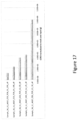
- the one-step combined VJ-panel capture increased signal by 6.84x
- the two-step J and V iterative capture increased signal by 12x (no significant difference was observed for J-V or V-J iterative order)
- the three-step J-depletion-V iterative capture increased signal by 31.2x ( Figure 13 ).
- TIL Tumor Infiltrating Lymphocyte
- PBMCs Tumor Infiltrating Lymphocyte
- Repertoire profiling reveals a polyclonal and diverse baseline repertoire before treatment, a less complex oligoclonal TIL derived culture, less complex oligoclonal repertoires following chemodepletion and transfusion of the TIL infusion, and finally restoration of a more complex polyclonal repertoire over time.
- Illumina cDNA sequencing libraries were generated from FFPE-derived total RNA and subjected to sequencing followed by analysis using the TCR annotation pipeline to identify unique TCR CDR3 sequences (bulk unique CDR3). Residual library then underwent CapTCR-Seq to identify unique TCR CDR3 sequences (capture unique CDR3).
- Tumor tissue extracted DNA and RNA
- pre-treatment blood extracted PBMC DNA, PBMC RNA, and plasma cfDNA
- on-treatment blood extracted plasma cfDNA
- TCR signal is defined as the total number of counts of fragments containing both a V and J gene region (non-unique, reads normalized) while diversity is defined as the total number of unique CDR3 sequences detected (unique, reads normalized).
- diversity increased with signal ( Figure 27 ).
- cfDNA samples had the lowest signal, genomic DNA samples had intermediate signal, while cDNA samples had the highest signal.
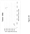
- Blood sample signal and diversity is similar for all five patients, however tumor signal and diversity varied. Two patients had ten-fold higher TCR signal and diversity in their tumors likely reflecting increased infiltration of immune cells ( Figure 28 ).
- Tumors with clonal infiltration have a larger than expected population of one or more VJ rearrangements, the population of which are significantly greater than the next most prevalent clone.
- Patient A appears to have a large alpha rearrangement population in its tumor compared to baseline blood, while the most prevalent beta rearrangement is only slightly enriched ( Figure 29-30 ).
- the tumor sample for patient B showed both greatly enriched top alpha and beta VJ rearrangements compared to baseline blood ( Figure 31-32 ).
- the tumor sample for patient C showed both greatly enriched top alpha and beta VJ rearrangements compared to baseline blood ( Figure 33-34 ).
- the tumor sample for patient D showed both greatly enriched top alpha (2) and beta VJ (1) rearrangements compared to baseline blood ( Figure 35-36 ).
- the tumor sample for patient E showed only a slightly enriched top beta VJ rearrangement compared to baseline blood ( Figure 37-38 ).
- V(D)J rearrangements and translocations detected through this approach have been validated in three well-characterized cell-lines with publically available whole genome data; an additional 67 MM cell lines have been annotated for V(D)J rearrangements and translocations into IGH, IGL and IGK genes. The limit of detection was established with a cell-line dilution series. We were also able to translate these techniques to cell-free DNA. These methods are applicable to the detection of MRD in mature B-cell malignancies and immunoglobulin repertoire profiling in a many clinical scenarios including cellular immunotherapy and therapeutics with immunomodulatory effects. V(D)J and complex rearrangement annotations in 70 MM cell-lines are highly relevant in further in-vitro studies.
- the B-cell V-gene and J-gene capture probes used are shown in Tables B1 and B2 respectively.
- Table 1.1 Capture Sample Method Data Sample Sample Protocol Type Library Input (ng) A037 healthy reference Sample_A037_PBMC_TCR_A_all A037_PBMC CapSeq_One-Step V 100 Sample_A037_PBMC_TCR_B_all A037_PBMC CapSeq_One-Step V 200 Sample_A037PBMC_TCR_D_all A037_PBMC CapSeq_One-Step_V 600 Szmple_A037_PBMC_TCR_E_all A037_PBMC CapSeq_One-Step_V 800 Sample_A037_PBMC_TCR_F_all A037_PBMC CapSeq_One-Step_V 1000 Sample_A037_PBMC_TCR_G_all A037_PBMC CapSeq_One-Step_V 200 Sample_A037_PBMC_TCR_H_all A037_PBMC CapSeq_One-Step_V 600 Sample
Landscapes
- Chemical & Material Sciences (AREA)
- Life Sciences & Earth Sciences (AREA)
- Organic Chemistry (AREA)
- Health & Medical Sciences (AREA)
- Proteomics, Peptides & Aminoacids (AREA)
- Engineering & Computer Science (AREA)
- Zoology (AREA)
- Wood Science & Technology (AREA)
- Immunology (AREA)
- Molecular Biology (AREA)
- Biochemistry (AREA)
- Analytical Chemistry (AREA)
- General Health & Medical Sciences (AREA)
- Physics & Mathematics (AREA)
- Biophysics (AREA)
- Microbiology (AREA)
- Bioinformatics & Cheminformatics (AREA)
- General Engineering & Computer Science (AREA)
- Biotechnology (AREA)
- Genetics & Genomics (AREA)
- Chemical Kinetics & Catalysis (AREA)
- General Chemical & Material Sciences (AREA)
- Medicinal Chemistry (AREA)
- Cell Biology (AREA)
- Measuring Or Testing Involving Enzymes Or Micro-Organisms (AREA)
Claims (14)
- Verfahren zum Einfangen einer Population von T-Zell-Rezeptor- und/oder Immunglobulin-Sequenzen mit variablen Regionen in einer Patientenprobe, wobei das Verfahren umfasst:Extrahieren/Präparieren von DNA-Fragmenten aus der Patientenprobe;Ligieren eines Nukleinsäureadapters an die DNA-Fragmente, wobei der Nukleinsäureadapter zur Erkennung durch eine vorgewählte Nukleinsäuresonde geeignet ist;Einfangen von DNA-Fragmenten, die in der Patientenprobe vorhanden sind, unter Verwendung einer Sammlung von Nukleinsäure-Hybrid-Einfangsonden, wobei jede Einfangsonde so ausgelegt ist, dass sie an ein bekanntes V-Gen-Segment und/oder ein J-Gen-Segment innerhalb der genomischen T-Zell-Rezeptor- und/oder Immunglobulin-Loci hybridisiert,wobei die Hybrid-Einfangsonden auf der Grundlage aller einzigartigen V-Gene und J-Gene entworfen werden und diese Sondensätze spezifisch auf die 3'-Regionen von V-Gen-codierenden Regionen und die 5'-Regionen von J-Gencodierenden Regionen abzielen.
- Verfahren nach Anspruch 1, zudem umfassend das Sequenzieren der eingefangenen DNA-Fragmente, wobei die Sequenzierung zur Bestimmung von Klonotypen innerhalb der Patientenprobe verwendet werden kann.
- Verfahren nach Anspruch 1, wobei die Sequenzierung für Short-Read-Sequenzierung optimiert ist, vorzugsweise zudem umfassend die Amplifikation der Population von Sequenzen unter Verwendung von Nukleinsäure-Amplifikationssonden, die den Adapter erkennen, vor der Sequenzierung.
- Verfahren nach einem der Ansprüche 1 bis 3, zudem umfassend das Fragmentieren der aus der Patientenprobe extrahierten DNA zur Erzeugung der DNA-Fragmente.
- Verfahren nach einem der Ansprüche 1 bis 4, wobei der Ligationsschritt vor dem Einfangschritt durchgeführt wird oder der Einfangschritt vor dem Ligationsschritt durchgeführt wird.
- Verfahren nach einem der Ansprüche 1 bis 5, wobei die Patientenprobe Gewebe, Urin, Cerebrospinal-Flüssigkeit, Speichel, Fäkalien, Aszites, Pleuraerguss, Blut oder Blutplasma umfasst, und wobei die Patientenprobe vorzugsweise zellfreie Nukleinsäuren im Blutplasma umfasst.
- Verfahren nach einem der Ansprüche 1 bis 6, wobei die Hybrid-Einfangsonden mindestens 30 bp lang sind, wobei die Hybrid-Einfangsonden vorzugsweise zwischen 60 bp und 150 bp lang sind, vorzugsweise zwischen 80 bp und 120 bp lang sind und weiter vorzugsweise etwa 100 bp lang sind.
- Verfahren nach einem der Ansprüche 1 bis 7, wobei das Hybridisieren das Hybridisieren unter stringenten Bedingungen, vorzugsweise hochstringenten Bedingungen, umfasst.
- Verfahren nach einem der Ansprüche 1 bis 8, wobei die Hybrid-Einfangsonden eine Markierung umfassen, wobei die Markierung vorzugsweise zur Unterscheidung zwischen an die Screening-Sonden gebundenen Sequenzen und ungebundenen doppelsträngigen Fragmenten verwendet wird.
- Verfahren nach einem der Ansprüche 1 bis 9, wobei der Adapter so gestaltet ist, dass er die Sequenzierung des DNA-Fragments und/oder die Strichcodierung des DNA-Fragments ermöglicht.
- Verfahren nach einem der Ansprüche 1 bis 10, wobei die Herstellung der DNA-Fragmente die Extraktion von RNA aus der Patientenprobe und die Herstellung der entsprechenden cDNA umfasst.
- Verfahren nach einem der Ansprüche 1 bis 11, zudem umfassend einen Abreicherungsschritt, umfassend die Abreicherung der DNA-Fragmente von nicht umgelagerten Sequenzen unter Verwendung von Sonden, die Nukleinsäuresequenzen erkennen, die an V- und/oder J-Gensegmente im Genom angrenzen.
- Das Verfahren nach Anspruch 12, wobei das Einfangen von DNA-Fragmenten unter Verwendung von V-Gensegment- und J-Gensegment-Hybrid-Einfangsonden in getrennten Schritten und in beliebiger Reihenfolge mit dem Abreicherungsschritt durchgeführt wird, vorzugsweise in der folgenden Reihenfolge: J-Gen-Einfang, Abreicherung, dann V-Gen-Einfang.
- Verfahren zur Charakterisierung des Immunrepertoires eines Individuums, wobei das Immunrepertoire die T-Zell-Population des Individuums umfasst, wobei das Verfahren das Verfahren nach einem der Ansprüche 1 bis 13 umfasst, gefolgt von einem Verfahren, umfassend:(j) Identifizierung aller Sequenzen, die ein V-Gensegment enthalten, aus den Sequenzen der DNA-Fragmente durch Alignment der Sequenzen der DNA-Fragmente mit einer Bibliothek bekannter V-Gensegmentsequenzen;(k) Trimmen der identifizierten Sequenzen in (j), um alle Sequenzen zu entfernen, die V-Gensegmenten entsprechen, um eine Sammlung von V-getrimmten Nukleotidsequenzen zu erzeugen;(l) Identifizierung aller Sequenzen, die ein J-Gensegment in der Population der V-getrimmten Nukleotidsequenzen enthalten, durch Alignment der V-getrimmten Nukleotidsequenzen mit einer Bibliothek bekannter J-Gensegmentsequenzen;(m) Trimmen der in (l) identifizierten V-getrimmten Nukleotidsequenzen, um alle Sequenzen zu entfernen, die J-Gensegmenten entsprechen, um VJ-getrimmte Nukleotidsequenzen zu erzeugen;(n) Identifizierung jedes D-Gensegments, das in den in (m) identifizierten VJ-getrimmten Nukleotidsequenzen enthalten ist, durch Alignment der VJ-getrimmten Nukleotidsequenzen mit einer Bibliothek bekannter D-Gensegmentsequenzen;(o) für jede in (m) identifizierte VJ-getrimmte Nukleotidsequenz Assemblieren einer Nukleotidsequenz, umfassend das V-Gensegment, irgendein D-Gensegment und das J-Gensegment, die in den Schritten (j), (n) bzw. (l) identifiziert wurden,;(p) Auswählen aus der in Schritt (o) assemblierten Nukleotidsequenz einer Verbindungsnukleotidsequenz, die mindestens die Verbindung zwischen dem V-Gensegment und dem J-Gensegment, einschließlich eines beliebigen D-Gensegments, umfasst, wobei die Verbindungsnukleotidsequenz zwischen 18 bp und 140 bp umfasst,und gegebenenfalls (q) und (r):(q) Translatieren jedes Leserasters der Verbindungsnukleotidsequenz und ihres komplementären Strangs, um 6 translatierte Sequenzen zu erzeugen; und(r) Vergleichen der 6 translatierten Sequenzen mit einer Bibliothek bekannter CDR3-Regionen von T-Zell-Rezeptor- und/oder Immunglobulinsequenzen, um die CDR3-Region in den DNA-Fragmenten zu identifizieren,
Applications Claiming Priority (2)
| Application Number | Priority Date | Filing Date | Title |
|---|---|---|---|
| US201662322999P | 2016-04-15 | 2016-04-15 | |
| PCT/CA2017/000084 WO2017177308A1 (en) | 2016-04-15 | 2017-04-13 | Hybrid-capture sequencing for determining immune cell clonality |
Publications (3)
| Publication Number | Publication Date |
|---|---|
| EP3443149A1 EP3443149A1 (de) | 2019-02-20 |
| EP3443149A4 EP3443149A4 (de) | 2019-12-18 |
| EP3443149B1 true EP3443149B1 (de) | 2024-08-14 |
Family
ID=60041289
Family Applications (1)
| Application Number | Title | Priority Date | Filing Date |
|---|---|---|---|
| EP17781661.8A Active EP3443149B1 (de) | 2016-04-15 | 2017-04-13 | Hybriderfassungssequenz zur bestimmung von immunzellenklonalität |
Country Status (4)
| Country | Link |
|---|---|
| US (1) | US11149312B2 (de) |
| EP (1) | EP3443149B1 (de) |
| CA (1) | CA3020814A1 (de) |
| WO (1) | WO2017177308A1 (de) |
Families Citing this family (21)
| Publication number | Priority date | Publication date | Assignee | Title |
|---|---|---|---|---|
| CA2916662C (en) | 2013-06-25 | 2022-03-08 | Prognosys Biosciences, Inc. | Methods and systems for determining spatial patterns of biological targets in a sample |
| EP3901282B1 (de) | 2015-04-10 | 2023-06-28 | Spatial Transcriptomics AB | Räumlich getrennte multiplex-nukleinsäureanalyse von biologischen proben |
| EP3443149B1 (de) | 2016-04-15 | 2024-08-14 | University Health Network (UHN) | Hybriderfassungssequenz zur bestimmung von immunzellenklonalität |
| US10704082B2 (en) | 2016-09-15 | 2020-07-07 | ArcherDX, Inc. | Methods of nucleic acid sample preparation |
| WO2018053365A1 (en) | 2016-09-15 | 2018-03-22 | ArcherDX, Inc. | Methods of nucleic acid sample preparation for analysis of cell-free dna |
| ES2940620T3 (es) * | 2016-11-02 | 2023-05-09 | Archerdx Llc | Métodos de preparación de muestras de ácido nucleico para la secuenciación del repertorio inmunitario |
| EP3631053A4 (de) * | 2017-05-30 | 2021-05-19 | University of Health Network | Hybriderfassungssequenz zur bestimmung von immunzellenklonalität |
| GB201718238D0 (en) * | 2017-11-03 | 2017-12-20 | Univ Oxford Innovation Ltd | Method and system for determining the disease status of a subject |
| WO2019191459A1 (en) * | 2018-03-28 | 2019-10-03 | Berkeley Lights, Inc. | Methods for preparation of nucleic acid sequencing libraries |
| WO2020243579A1 (en) | 2019-05-30 | 2020-12-03 | 10X Genomics, Inc. | Methods of detecting spatial heterogeneity of a biological sample |
| CA3145114A1 (en) * | 2019-08-16 | 2021-02-25 | Chris Raymond | Targeted hybrid capture methods for determination of t cell repertoires |
| WO2021053008A1 (en) * | 2019-09-20 | 2021-03-25 | F. Hoffmann-La Roche Ag | Immune repertoire profiling by primer extension target enrichment |
| EP4093887A1 (de) * | 2020-01-22 | 2022-11-30 | Life Technologies Corporation | Immunrepertoire-biomarker bei autoimmunerkrankungen und immunschwächeerkrankungen |
| CA3174332A1 (en) * | 2020-04-21 | 2021-10-28 | Jason PERERA | Tcr/bcr profiling |
| AU2021283174A1 (en) | 2020-06-02 | 2023-01-05 | 10X Genomics, Inc. | Nucleic acid library methods |
| EP4600376A3 (de) * | 2020-06-02 | 2025-10-22 | 10X Genomics, Inc. | Räumliche transkriptomik für antigenrezeptoren |
| EP4421186B1 (de) | 2020-06-08 | 2025-08-13 | 10X Genomics, Inc. | Verfahren zur bestimmung eines chirurgischen randes und verfahren zur verwendung davon |
| CN117203349A (zh) * | 2021-04-05 | 2023-12-08 | Ncan基因组学有限公司 | 增强的链接靶标捕获 |
| WO2023288018A2 (en) * | 2021-07-14 | 2023-01-19 | Ultima Genomics, Inc. | Barcode selection |
| CN117133357A (zh) * | 2022-05-18 | 2023-11-28 | 京东方科技集团股份有限公司 | Igk基因重排的检测方法、装置、电子设备及存储介质 |
| WO2025175065A1 (en) * | 2024-02-13 | 2025-08-21 | The Board Of Trustees Of The Leland Stanford Junior University | Systems and methods for assessment of immune response and applications thereof |
Family Cites Families (14)
| Publication number | Priority date | Publication date | Assignee | Title |
|---|---|---|---|---|
| US2392551A (en) | 1943-05-10 | 1946-01-08 | Albert Kahn | Interlocking building block |
| US6189282B1 (en) | 1998-06-24 | 2001-02-20 | Building Works, Inc. | Mortarless concrete block |
| US20080125333A1 (en) | 2006-05-31 | 2008-05-29 | Antara Biosciences Inc. | Devices and kits for detecting one or more target agents |
| US7410328B2 (en) | 2006-06-14 | 2008-08-12 | Transpavé Inc. | Concrete block system |
| EP2118393A4 (de) | 2007-02-02 | 2015-11-04 | Matériaux De Construction Oldcastle Canada Inc | Wand mit dekorativer verkleidung |
| EP2245198A1 (de) * | 2008-02-04 | 2010-11-03 | Massachusetts Institute of Technology | Selektion von nukleinsäuren durch lösungshybridisierung an oligonukleotidköder |
| CA3106531A1 (en) | 2009-09-29 | 2011-04-07 | Keystone Retaining Wall Systems, Inc. | Wall blocks, veneer panels for wall blocks and method of constructing walls |
| ES2769796T3 (es) * | 2012-07-03 | 2020-06-29 | Integrated Dna Tech Inc | Oligonucleótidos de bloqueo aumentados en Tm y señuelos para un enriquecimiento de diana mejorado y una selección fuera de diana reducida |
| US9783593B2 (en) | 2013-05-02 | 2017-10-10 | Kymab Limited | Antibodies, variable domains and chains tailored for human use |
| EP3102678A2 (de) | 2014-02-03 | 2016-12-14 | Integrated DNA Technologies Inc. | Verfahren zur erfassung und/oder entfernung von hochabundanten rnas aus einer heterogenen rna-probe |
| EP3227464B1 (de) * | 2014-12-05 | 2022-04-20 | Foundation Medicine, Inc. | Multigenanalyse von tumorproben |
| GB201512869D0 (en) | 2015-07-21 | 2015-09-02 | Almac Diagnostics Ltd | Gene signature for minute therapies |
| US9453341B1 (en) | 2015-08-18 | 2016-09-27 | Hengestone Holdings, Inc. | Wall system having core supporting blocks and decorative fascia blocks |
| EP3443149B1 (de) | 2016-04-15 | 2024-08-14 | University Health Network (UHN) | Hybriderfassungssequenz zur bestimmung von immunzellenklonalität |
-
2017
- 2017-04-13 EP EP17781661.8A patent/EP3443149B1/de active Active
- 2017-04-13 WO PCT/CA2017/000084 patent/WO2017177308A1/en not_active Ceased
- 2017-04-13 US US16/093,825 patent/US11149312B2/en active Active
- 2017-04-13 CA CA3020814A patent/CA3020814A1/en active Pending
Also Published As
| Publication number | Publication date |
|---|---|
| US20190390273A1 (en) | 2019-12-26 |
| CA3020814A1 (en) | 2017-10-19 |
| EP3443149A4 (de) | 2019-12-18 |
| WO2017177308A1 (en) | 2017-10-19 |
| US11149312B2 (en) | 2021-10-19 |
| EP3443149A1 (de) | 2019-02-20 |
Similar Documents
| Publication | Publication Date | Title |
|---|---|---|
| EP3443149B1 (de) | Hybriderfassungssequenz zur bestimmung von immunzellenklonalität | |
| Duncavage et al. | Genomic profiling for clinical decision making in myeloid neoplasms and acute leukemia | |
| US11788136B2 (en) | Hybrid-capture sequencing for determining immune cell clonality | |
| US11875899B2 (en) | Analyzing copy number variation in the detection of cancer | |
| US11697846B2 (en) | Detecting and classifying copy number variation | |
| US20260055472A1 (en) | Microsatellite instability detection in cell-free dna | |
| US20200219588A1 (en) | Detecting and classifying copy number variation | |
| US9323888B2 (en) | Detecting and classifying copy number variation | |
| US9411937B2 (en) | Detecting and classifying copy number variation | |
| US20190385700A1 (en) | METHODS AND SYSTEMS FOR DETERMINING The CELLULAR ORIGIN OF CELL-FREE NUCLEIC ACIDS | |
| US20220028494A1 (en) | Methods and systems for determining the cellular origin of cell-free dna | |
| Hansmann et al. | Clonal expansion and interrelatedness of distinct B-lineage compartments in multiple myeloma bone marrow | |
| US20260120797A1 (en) | Cell-free dna blood-based test for cancer screening | |
| Pereira et al. | Cancer genomics in precision oncology: applications, challenges, and prospects | |
| JP2025538871A (ja) | 非小細胞肺がん(NSCLC)患者の薬物応答または死亡もしくはがん進行までの時間を、循環腫瘍DNA(ctDNA)から、ベースラインctDNAレベルおよびctDNAレベルの経時的な縦断的変化の両方からのシグナルを利用して予測する方法 | |
| Islam et al. | CRIS: complete reconstruction of immunoglobulin VDJ sequences from RNA-seq data | |
| Vormittag-Nocito et al. | The impact of next-generation sequencing for diagnosis and disease understanding of myeloid malignancies | |
| US20240203601A1 (en) | Analyzing copy number variation in the detection of cancer | |
| US20250243550A1 (en) | Minimum residual disease (mrd) detection in early stage cancer using urine | |
| Kositsky | Profiling Blood Cancer Drivers through Large-Scale Genomics | |
| Chakravorty et al. | Implementation of exome sequencing assay | |
| Kurucová | IDENTIFICATION OF CANCER SUBPOPULATIONS BY SINGLE-CELL METHODS | |
| Mahe | Design & Validation of a Hybrid-Capture Next-Generation Sequencing-Based T-cell Clonality Assay |
Legal Events
| Date | Code | Title | Description |
|---|---|---|---|
| STAA | Information on the status of an ep patent application or granted ep patent |
Free format text: STATUS: THE INTERNATIONAL PUBLICATION HAS BEEN MADE |
|
| PUAI | Public reference made under article 153(3) epc to a published international application that has entered the european phase |
Free format text: ORIGINAL CODE: 0009012 |
|
| STAA | Information on the status of an ep patent application or granted ep patent |
Free format text: STATUS: REQUEST FOR EXAMINATION WAS MADE |
|
| 17P | Request for examination filed |
Effective date: 20181115 |
|
| AK | Designated contracting states |
Kind code of ref document: A1 Designated state(s): AL AT BE BG CH CY CZ DE DK EE ES FI FR GB GR HR HU IE IS IT LI LT LU LV MC MK MT NL NO PL PT RO RS SE SI SK SM TR |
|
| AX | Request for extension of the european patent |
Extension state: BA ME |
|
| DAV | Request for validation of the european patent (deleted) | ||
| DAX | Request for extension of the european patent (deleted) | ||
| A4 | Supplementary search report drawn up and despatched |
Effective date: 20191118 |
|
| RIC1 | Information provided on ipc code assigned before grant |
Ipc: C12N 15/12 20060101ALI20191112BHEP Ipc: C12N 15/13 20060101ALI20191112BHEP Ipc: C40B 40/06 20060101AFI20191112BHEP Ipc: C12Q 1/6881 20180101ALI20191112BHEP Ipc: C12Q 1/6855 20180101ALI20191112BHEP Ipc: C12Q 1/68 20180101ALI20191112BHEP Ipc: C40B 30/04 20060101ALI20191112BHEP Ipc: C12N 15/10 20060101ALI20191112BHEP |
|
| STAA | Information on the status of an ep patent application or granted ep patent |
Free format text: STATUS: EXAMINATION IS IN PROGRESS |
|
| 17Q | First examination report despatched |
Effective date: 20210924 |
|
| REG | Reference to a national code |
Ref legal event code: R079 Ipc: C12Q0001686900 Ref country code: DE Ref legal event code: R079 Ref document number: 602017084087 Country of ref document: DE Free format text: PREVIOUS MAIN CLASS: C40B0040060000 Ipc: C12Q0001686900 |
|
| RIC1 | Information provided on ipc code assigned before grant |
Ipc: C12Q 1/6881 20180101ALI20240125BHEP Ipc: C12Q 1/6855 20180101ALI20240125BHEP Ipc: C12Q 1/6869 20180101AFI20240125BHEP |
|
| GRAP | Despatch of communication of intention to grant a patent |
Free format text: ORIGINAL CODE: EPIDOSNIGR1 |
|
| STAA | Information on the status of an ep patent application or granted ep patent |
Free format text: STATUS: GRANT OF PATENT IS INTENDED |
|
| INTG | Intention to grant announced |
Effective date: 20240305 |
|
| GRAS | Grant fee paid |
Free format text: ORIGINAL CODE: EPIDOSNIGR3 |
|
| GRAA | (expected) grant |
Free format text: ORIGINAL CODE: 0009210 |
|
| STAA | Information on the status of an ep patent application or granted ep patent |
Free format text: STATUS: THE PATENT HAS BEEN GRANTED |
|
| AK | Designated contracting states |
Kind code of ref document: B1 Designated state(s): AL AT BE BG CH CY CZ DE DK EE ES FI FR GB GR HR HU IE IS IT LI LT LU LV MC MK MT NL NO PL PT RO RS SE SI SK SM TR |
|
| REG | Reference to a national code |
Ref country code: GB Ref legal event code: FG4D |
|
| REG | Reference to a national code |
Ref country code: CH Ref legal event code: EP |
|
| REG | Reference to a national code |
Ref country code: DE Ref legal event code: R096 Ref document number: 602017084087 Country of ref document: DE |
|
| REG | Reference to a national code |
Ref country code: IE Ref legal event code: FG4D |
|
| REG | Reference to a national code |
Ref country code: LT Ref legal event code: MG9D |
|
| REG | Reference to a national code |
Ref country code: NL Ref legal event code: MP Effective date: 20240814 |
|
| PG25 | Lapsed in a contracting state [announced via postgrant information from national office to epo] |
Ref country code: NO Free format text: LAPSE BECAUSE OF FAILURE TO SUBMIT A TRANSLATION OF THE DESCRIPTION OR TO PAY THE FEE WITHIN THE PRESCRIBED TIME-LIMIT Effective date: 20241114 |
|
| REG | Reference to a national code |
Ref country code: AT Ref legal event code: MK05 Ref document number: 1713338 Country of ref document: AT Kind code of ref document: T Effective date: 20240814 |
|
| PG25 | Lapsed in a contracting state [announced via postgrant information from national office to epo] |
Ref country code: PL Free format text: LAPSE BECAUSE OF FAILURE TO SUBMIT A TRANSLATION OF THE DESCRIPTION OR TO PAY THE FEE WITHIN THE PRESCRIBED TIME-LIMIT Effective date: 20240814 Ref country code: FI Free format text: LAPSE BECAUSE OF FAILURE TO SUBMIT A TRANSLATION OF THE DESCRIPTION OR TO PAY THE FEE WITHIN THE PRESCRIBED TIME-LIMIT Effective date: 20240814 Ref country code: NL Free format text: LAPSE BECAUSE OF FAILURE TO SUBMIT A TRANSLATION OF THE DESCRIPTION OR TO PAY THE FEE WITHIN THE PRESCRIBED TIME-LIMIT Effective date: 20240814 Ref country code: PT Free format text: LAPSE BECAUSE OF FAILURE TO SUBMIT A TRANSLATION OF THE DESCRIPTION OR TO PAY THE FEE WITHIN THE PRESCRIBED TIME-LIMIT Effective date: 20241216 Ref country code: GR Free format text: LAPSE BECAUSE OF FAILURE TO SUBMIT A TRANSLATION OF THE DESCRIPTION OR TO PAY THE FEE WITHIN THE PRESCRIBED TIME-LIMIT Effective date: 20241115 |
|
| PG25 | Lapsed in a contracting state [announced via postgrant information from national office to epo] |
Ref country code: BG Free format text: LAPSE BECAUSE OF FAILURE TO SUBMIT A TRANSLATION OF THE DESCRIPTION OR TO PAY THE FEE WITHIN THE PRESCRIBED TIME-LIMIT Effective date: 20240814 |
|
| PG25 | Lapsed in a contracting state [announced via postgrant information from national office to epo] |
Ref country code: LV Free format text: LAPSE BECAUSE OF FAILURE TO SUBMIT A TRANSLATION OF THE DESCRIPTION OR TO PAY THE FEE WITHIN THE PRESCRIBED TIME-LIMIT Effective date: 20240814 |
|
| PG25 | Lapsed in a contracting state [announced via postgrant information from national office to epo] |
Ref country code: AT Free format text: LAPSE BECAUSE OF FAILURE TO SUBMIT A TRANSLATION OF THE DESCRIPTION OR TO PAY THE FEE WITHIN THE PRESCRIBED TIME-LIMIT Effective date: 20240814 Ref country code: IS Free format text: LAPSE BECAUSE OF FAILURE TO SUBMIT A TRANSLATION OF THE DESCRIPTION OR TO PAY THE FEE WITHIN THE PRESCRIBED TIME-LIMIT Effective date: 20241214 |
|
| PG25 | Lapsed in a contracting state [announced via postgrant information from national office to epo] |
Ref country code: HR Free format text: LAPSE BECAUSE OF FAILURE TO SUBMIT A TRANSLATION OF THE DESCRIPTION OR TO PAY THE FEE WITHIN THE PRESCRIBED TIME-LIMIT Effective date: 20240814 |
|
| PG25 | Lapsed in a contracting state [announced via postgrant information from national office to epo] |
Ref country code: RS Free format text: LAPSE BECAUSE OF FAILURE TO SUBMIT A TRANSLATION OF THE DESCRIPTION OR TO PAY THE FEE WITHIN THE PRESCRIBED TIME-LIMIT Effective date: 20241114 Ref country code: ES Free format text: LAPSE BECAUSE OF FAILURE TO SUBMIT A TRANSLATION OF THE DESCRIPTION OR TO PAY THE FEE WITHIN THE PRESCRIBED TIME-LIMIT Effective date: 20240814 |
|
| PG25 | Lapsed in a contracting state [announced via postgrant information from national office to epo] |
Ref country code: RS Free format text: LAPSE BECAUSE OF FAILURE TO SUBMIT A TRANSLATION OF THE DESCRIPTION OR TO PAY THE FEE WITHIN THE PRESCRIBED TIME-LIMIT Effective date: 20241114 Ref country code: PT Free format text: LAPSE BECAUSE OF FAILURE TO SUBMIT A TRANSLATION OF THE DESCRIPTION OR TO PAY THE FEE WITHIN THE PRESCRIBED TIME-LIMIT Effective date: 20241216 Ref country code: PL Free format text: LAPSE BECAUSE OF FAILURE TO SUBMIT A TRANSLATION OF THE DESCRIPTION OR TO PAY THE FEE WITHIN THE PRESCRIBED TIME-LIMIT Effective date: 20240814 Ref country code: NO Free format text: LAPSE BECAUSE OF FAILURE TO SUBMIT A TRANSLATION OF THE DESCRIPTION OR TO PAY THE FEE WITHIN THE PRESCRIBED TIME-LIMIT Effective date: 20241114 Ref country code: NL Free format text: LAPSE BECAUSE OF FAILURE TO SUBMIT A TRANSLATION OF THE DESCRIPTION OR TO PAY THE FEE WITHIN THE PRESCRIBED TIME-LIMIT Effective date: 20240814 Ref country code: LV Free format text: LAPSE BECAUSE OF FAILURE TO SUBMIT A TRANSLATION OF THE DESCRIPTION OR TO PAY THE FEE WITHIN THE PRESCRIBED TIME-LIMIT Effective date: 20240814 Ref country code: IS Free format text: LAPSE BECAUSE OF FAILURE TO SUBMIT A TRANSLATION OF THE DESCRIPTION OR TO PAY THE FEE WITHIN THE PRESCRIBED TIME-LIMIT Effective date: 20241214 Ref country code: HR Free format text: LAPSE BECAUSE OF FAILURE TO SUBMIT A TRANSLATION OF THE DESCRIPTION OR TO PAY THE FEE WITHIN THE PRESCRIBED TIME-LIMIT Effective date: 20240814 Ref country code: GR Free format text: LAPSE BECAUSE OF FAILURE TO SUBMIT A TRANSLATION OF THE DESCRIPTION OR TO PAY THE FEE WITHIN THE PRESCRIBED TIME-LIMIT Effective date: 20241115 Ref country code: FI Free format text: LAPSE BECAUSE OF FAILURE TO SUBMIT A TRANSLATION OF THE DESCRIPTION OR TO PAY THE FEE WITHIN THE PRESCRIBED TIME-LIMIT Effective date: 20240814 Ref country code: ES Free format text: LAPSE BECAUSE OF FAILURE TO SUBMIT A TRANSLATION OF THE DESCRIPTION OR TO PAY THE FEE WITHIN THE PRESCRIBED TIME-LIMIT Effective date: 20240814 Ref country code: BG Free format text: LAPSE BECAUSE OF FAILURE TO SUBMIT A TRANSLATION OF THE DESCRIPTION OR TO PAY THE FEE WITHIN THE PRESCRIBED TIME-LIMIT Effective date: 20240814 Ref country code: AT Free format text: LAPSE BECAUSE OF FAILURE TO SUBMIT A TRANSLATION OF THE DESCRIPTION OR TO PAY THE FEE WITHIN THE PRESCRIBED TIME-LIMIT Effective date: 20240814 |
|
| PG25 | Lapsed in a contracting state [announced via postgrant information from national office to epo] |
Ref country code: DK Free format text: LAPSE BECAUSE OF FAILURE TO SUBMIT A TRANSLATION OF THE DESCRIPTION OR TO PAY THE FEE WITHIN THE PRESCRIBED TIME-LIMIT Effective date: 20240814 Ref country code: SM Free format text: LAPSE BECAUSE OF FAILURE TO SUBMIT A TRANSLATION OF THE DESCRIPTION OR TO PAY THE FEE WITHIN THE PRESCRIBED TIME-LIMIT Effective date: 20240814 Ref country code: RO Free format text: LAPSE BECAUSE OF FAILURE TO SUBMIT A TRANSLATION OF THE DESCRIPTION OR TO PAY THE FEE WITHIN THE PRESCRIBED TIME-LIMIT Effective date: 20240814 |
|
| PG25 | Lapsed in a contracting state [announced via postgrant information from national office to epo] |
Ref country code: EE Free format text: LAPSE BECAUSE OF FAILURE TO SUBMIT A TRANSLATION OF THE DESCRIPTION OR TO PAY THE FEE WITHIN THE PRESCRIBED TIME-LIMIT Effective date: 20240814 |
|
| PG25 | Lapsed in a contracting state [announced via postgrant information from national office to epo] |
Ref country code: CZ Free format text: LAPSE BECAUSE OF FAILURE TO SUBMIT A TRANSLATION OF THE DESCRIPTION OR TO PAY THE FEE WITHIN THE PRESCRIBED TIME-LIMIT Effective date: 20240814 |
|
| PG25 | Lapsed in a contracting state [announced via postgrant information from national office to epo] |
Ref country code: SK Free format text: LAPSE BECAUSE OF FAILURE TO SUBMIT A TRANSLATION OF THE DESCRIPTION OR TO PAY THE FEE WITHIN THE PRESCRIBED TIME-LIMIT Effective date: 20240814 Ref country code: IT Free format text: LAPSE BECAUSE OF FAILURE TO SUBMIT A TRANSLATION OF THE DESCRIPTION OR TO PAY THE FEE WITHIN THE PRESCRIBED TIME-LIMIT Effective date: 20240814 |
|
| REG | Reference to a national code |
Ref country code: DE Ref legal event code: R097 Ref document number: 602017084087 Country of ref document: DE |
|
| PLBE | No opposition filed within time limit |
Free format text: ORIGINAL CODE: 0009261 |
|
| STAA | Information on the status of an ep patent application or granted ep patent |
Free format text: STATUS: NO OPPOSITION FILED WITHIN TIME LIMIT |
|
| PGFP | Annual fee paid to national office [announced via postgrant information from national office to epo] |
Ref country code: DE Payment date: 20250417 Year of fee payment: 9 |
|
| PGFP | Annual fee paid to national office [announced via postgrant information from national office to epo] |
Ref country code: GB Payment date: 20250417 Year of fee payment: 9 |
|
| PGFP | Annual fee paid to national office [announced via postgrant information from national office to epo] |
Ref country code: FR Payment date: 20250422 Year of fee payment: 9 |
|
| 26N | No opposition filed |
Effective date: 20250515 |
|
| PG25 | Lapsed in a contracting state [announced via postgrant information from national office to epo] |
Ref country code: SE Free format text: LAPSE BECAUSE OF FAILURE TO SUBMIT A TRANSLATION OF THE DESCRIPTION OR TO PAY THE FEE WITHIN THE PRESCRIBED TIME-LIMIT Effective date: 20240814 |
|
| REG | Reference to a national code |
Ref country code: CH Ref legal event code: H13 Free format text: ST27 STATUS EVENT CODE: U-0-0-H10-H13 (AS PROVIDED BY THE NATIONAL OFFICE) Effective date: 20251125 |
|
| PG25 | Lapsed in a contracting state [announced via postgrant information from national office to epo] |
Ref country code: LU Free format text: LAPSE BECAUSE OF NON-PAYMENT OF DUE FEES Effective date: 20250413 |
|
| PG25 | Lapsed in a contracting state [announced via postgrant information from national office to epo] |
Ref country code: MC Free format text: LAPSE BECAUSE OF FAILURE TO SUBMIT A TRANSLATION OF THE DESCRIPTION OR TO PAY THE FEE WITHIN THE PRESCRIBED TIME-LIMIT Effective date: 20240814 |
|
| REG | Reference to a national code |
Ref country code: BE Ref legal event code: MM Effective date: 20250430 |
|
| PG25 | Lapsed in a contracting state [announced via postgrant information from national office to epo] |
Ref country code: BE Free format text: LAPSE BECAUSE OF NON-PAYMENT OF DUE FEES Effective date: 20250430 |
|
| PG25 | Lapsed in a contracting state [announced via postgrant information from national office to epo] |
Ref country code: CH Free format text: LAPSE BECAUSE OF NON-PAYMENT OF DUE FEES Effective date: 20250430 |
|
| PG25 | Lapsed in a contracting state [announced via postgrant information from national office to epo] |
Ref country code: IE Free format text: LAPSE BECAUSE OF NON-PAYMENT OF DUE FEES Effective date: 20250413 |