EP3446716A1 - Transkriptionsfaktor-basierte erzeugung von herzschrittmacherzellen und verwendungsverfahren dafür - Google Patents
Transkriptionsfaktor-basierte erzeugung von herzschrittmacherzellen und verwendungsverfahren dafür Download PDFInfo
- Publication number
- EP3446716A1 EP3446716A1 EP18186583.3A EP18186583A EP3446716A1 EP 3446716 A1 EP3446716 A1 EP 3446716A1 EP 18186583 A EP18186583 A EP 18186583A EP 3446716 A1 EP3446716 A1 EP 3446716A1
- Authority
- EP
- European Patent Office
- Prior art keywords
- cells
- tbx18
- pacemaker
- transcription factors
- several embodiments
- Prior art date
- Legal status (The legal status is an assumption and is not a legal conclusion. Google has not performed a legal analysis and makes no representation as to the accuracy of the status listed.)
- Granted
Links
Images
























Classifications
-
- A—HUMAN NECESSITIES
- A61—MEDICAL OR VETERINARY SCIENCE; HYGIENE
- A61K—PREPARATIONS FOR MEDICAL, DENTAL OR TOILETRY PURPOSES
- A61K38/00—Medicinal preparations containing peptides
- A61K38/16—Peptides having more than 20 amino acids; Gastrins; Somatostatins; Melanotropins; Derivatives thereof
- A61K38/17—Peptides having more than 20 amino acids; Gastrins; Somatostatins; Melanotropins; Derivatives thereof from animals; from humans
- A61K38/1703—Peptides having more than 20 amino acids; Gastrins; Somatostatins; Melanotropins; Derivatives thereof from animals; from humans from vertebrates
- A61K38/1709—Peptides having more than 20 amino acids; Gastrins; Somatostatins; Melanotropins; Derivatives thereof from animals; from humans from vertebrates from mammals
-
- A—HUMAN NECESSITIES
- A61—MEDICAL OR VETERINARY SCIENCE; HYGIENE
- A61K—PREPARATIONS FOR MEDICAL, DENTAL OR TOILETRY PURPOSES
- A61K35/00—Medicinal preparations containing materials or reaction products thereof with undetermined constitution
- A61K35/12—Materials from mammals; Compositions comprising non-specified tissues or cells; Compositions comprising non-embryonic stem cells; Genetically modified cells
- A61K35/34—Muscles; Smooth muscle cells; Heart; Cardiac stem cells; Myoblasts; Myocytes; Cardiomyocytes
-
- A—HUMAN NECESSITIES
- A61—MEDICAL OR VETERINARY SCIENCE; HYGIENE
- A61P—SPECIFIC THERAPEUTIC ACTIVITY OF CHEMICAL COMPOUNDS OR MEDICINAL PREPARATIONS
- A61P43/00—Drugs for specific purposes, not provided for in groups A61P1/00-A61P41/00
-
- A—HUMAN NECESSITIES
- A61—MEDICAL OR VETERINARY SCIENCE; HYGIENE
- A61P—SPECIFIC THERAPEUTIC ACTIVITY OF CHEMICAL COMPOUNDS OR MEDICINAL PREPARATIONS
- A61P9/00—Drugs for disorders of the cardiovascular system
-
- A—HUMAN NECESSITIES
- A61—MEDICAL OR VETERINARY SCIENCE; HYGIENE
- A61P—SPECIFIC THERAPEUTIC ACTIVITY OF CHEMICAL COMPOUNDS OR MEDICINAL PREPARATIONS
- A61P9/00—Drugs for disorders of the cardiovascular system
- A61P9/04—Inotropic agents, i.e. stimulants of cardiac contraction; Drugs for heart failure
-
- A—HUMAN NECESSITIES
- A61—MEDICAL OR VETERINARY SCIENCE; HYGIENE
- A61P—SPECIFIC THERAPEUTIC ACTIVITY OF CHEMICAL COMPOUNDS OR MEDICINAL PREPARATIONS
- A61P9/00—Drugs for disorders of the cardiovascular system
- A61P9/06—Antiarrhythmics
-
- C—CHEMISTRY; METALLURGY
- C07—ORGANIC CHEMISTRY
- C07K—PEPTIDES
- C07K14/00—Peptides having more than 20 amino acids; Gastrins; Somatostatins; Melanotropins; Derivatives thereof
- C07K14/435—Peptides having more than 20 amino acids; Gastrins; Somatostatins; Melanotropins; Derivatives thereof from animals; from humans
- C07K14/46—Peptides having more than 20 amino acids; Gastrins; Somatostatins; Melanotropins; Derivatives thereof from animals; from humans from vertebrates
- C07K14/47—Peptides having more than 20 amino acids; Gastrins; Somatostatins; Melanotropins; Derivatives thereof from animals; from humans from vertebrates from mammals
- C07K14/4701—Peptides having more than 20 amino acids; Gastrins; Somatostatins; Melanotropins; Derivatives thereof from animals; from humans from vertebrates from mammals not used
- C07K14/4702—Regulators; Modulating activity
-
- C—CHEMISTRY; METALLURGY
- C12—BIOCHEMISTRY; BEER; SPIRITS; WINE; VINEGAR; MICROBIOLOGY; ENZYMOLOGY; MUTATION OR GENETIC ENGINEERING
- C12N—MICROORGANISMS OR ENZYMES; COMPOSITIONS THEREOF; PROPAGATING, PRESERVING, OR MAINTAINING MICROORGANISMS; MUTATION OR GENETIC ENGINEERING; CULTURE MEDIA
- C12N5/00—Undifferentiated human, animal or plant cells, e.g. cell lines; Tissues; Cultivation or maintenance thereof; Culture media therefor
- C12N5/06—Animal cells or tissues; Human cells or tissues
- C12N5/0602—Vertebrate cells
- C12N5/0652—Cells of skeletal and connective tissues; Mesenchyme
- C12N5/0657—Cardiomyocytes; Heart cells
-
- A—HUMAN NECESSITIES
- A61—MEDICAL OR VETERINARY SCIENCE; HYGIENE
- A61K—PREPARATIONS FOR MEDICAL, DENTAL OR TOILETRY PURPOSES
- A61K48/00—Medicinal preparations containing genetic material which is inserted into cells of the living body to treat genetic diseases; Gene therapy
-
- C—CHEMISTRY; METALLURGY
- C12—BIOCHEMISTRY; BEER; SPIRITS; WINE; VINEGAR; MICROBIOLOGY; ENZYMOLOGY; MUTATION OR GENETIC ENGINEERING
- C12N—MICROORGANISMS OR ENZYMES; COMPOSITIONS THEREOF; PROPAGATING, PRESERVING, OR MAINTAINING MICROORGANISMS; MUTATION OR GENETIC ENGINEERING; CULTURE MEDIA
- C12N2501/00—Active agents used in cell culture processes, e.g. differentation
- C12N2501/60—Transcription factors
-
- C—CHEMISTRY; METALLURGY
- C12—BIOCHEMISTRY; BEER; SPIRITS; WINE; VINEGAR; MICROBIOLOGY; ENZYMOLOGY; MUTATION OR GENETIC ENGINEERING
- C12N—MICROORGANISMS OR ENZYMES; COMPOSITIONS THEREOF; PROPAGATING, PRESERVING, OR MAINTAINING MICROORGANISMS; MUTATION OR GENETIC ENGINEERING; CULTURE MEDIA
- C12N2506/00—Differentiation of animal cells from one lineage to another; Differentiation of pluripotent cells
- C12N2506/02—Differentiation of animal cells from one lineage to another; Differentiation of pluripotent cells from embryonic cells
-
- C—CHEMISTRY; METALLURGY
- C12—BIOCHEMISTRY; BEER; SPIRITS; WINE; VINEGAR; MICROBIOLOGY; ENZYMOLOGY; MUTATION OR GENETIC ENGINEERING
- C12N—MICROORGANISMS OR ENZYMES; COMPOSITIONS THEREOF; PROPAGATING, PRESERVING, OR MAINTAINING MICROORGANISMS; MUTATION OR GENETIC ENGINEERING; CULTURE MEDIA
- C12N2799/00—Uses of viruses
- C12N2799/02—Uses of viruses as vector
- C12N2799/021—Uses of viruses as vector for the expression of a heterologous nucleic acid
- C12N2799/022—Uses of viruses as vector for the expression of a heterologous nucleic acid where the vector is derived from an adenovirus
Definitions
- pacemaker cells e.g., cardiac cells that have regular, rhythmic electrical activity
- some embodiments of the invention relate to gene and cell therapy methods (and associated compositions) to generate pacemaker cells using transcription factors.
- SAN sinoatrial node
- the sinoatrial node (SAN) is a highly-specialized structure containing fewer than 10,000 pacemaker cells, which function to initiate contractions in the SAN. These SAN contractions then propagate to the rest of the excitable heart tissue and result in a heartbeat. Irregularities of excitable cardiac tissue and/or irregularities of pacemaker cells can lead to abnormalities in heart rhythm. Many cardiac abnormalities typically involve irregular heartbeat, tachycardia (where the heart rate is too high), or bradycardia (where the heart rate is too slow). These abnormalities are collectively known as arrhythmias.
- catheter ablation is less successful in treating more complex cases, such as atrial fibrillation (AF) or ventricular tachycardia (VT).
- catheter ablation is not useful in the treatment of bradycardia.
- Electronic pacemaker devices can sustain heart rate, or deliver shocks to terminate tachycardias.
- the high cost of dcviccs, and complications such as pulmonary collapse, hemorrhage, bacterial infection, and lead/generator failure or other types of malfunction represent limitations of the technology.
- pacemakers typically elicit their effects through the gene delivery of nucleotides that transcribe mutant ion-channel proteins.
- several embodiments of the present invention operate through delivery to cells of polynucleotides that encode one or more transcription factors, which allows direct somatic-to-somatic cell reprogramming (e.g., a non-pacemaker cell is reprogrammed to a pacemaker cell or a malfunctioning pacemaker cell is reprogrammed to a functional pacemaker cell).
- pacemaker cells are generated without the induced alteration expression of ion-channel proteins.
- Additional embodiments involve the conversion of stem cells, or in some embodiments cardiomyocytes or combinations of stem cells and cardiomyocytes, to biological pacemaker cells in vitro (e.g., by administration of one or more transcription factors disclosed herein), followed by subsequent implantation of the converted cells.
- delivery of the transcription factors, or cells converted by those transcription factors treats a variety of cardiac rhythmic abnormalities and/or lessens or obviates the needs for traditional therapies.
- methods for generating a biological pacemaker using transcription factors in order to modify the electrical activity of the cardiac tissue of a subject comprising identifying a subject having cardiac tissue exhibiting abnormal electrical activity, wherein the subject has cardiac tissue comprising quiescent cells, wherein the quiescent cells comprise one or more of cardiomyocytes and stem cells, wherein the quiescent cells do not exhibit spontaneous, repetitive electrical activity; and administering one or more transcription factors to the quiescent cells to generate treated cells, wherein the treated cells exhibit spontaneous, repetitive electrical activity, thereby modifying electrical activity of the cardiac tissue the subject.
- the abnormal cardiac electrical activity is due to a cardiac arrhythmia.
- other abnormalities in cardiac electrical activity, signaling or function can be addressed with the methods disclosed herein.
- the subject is afflicted with a condition selected from the group consisting of sick sinus syndrome, sinus bradycardia, tachycardia-bradycardia syndrome, atrial fibrillation, atrioventricular block, chronotropic incompetence, prolonged QT syndrome, and heart failure.
- the administration occurs in vitro, and the method further comprises administering the treated cells to the subject.
- the methods disclosed herein allow the administration to occur in vivo.
- converting a population of stem cells through the use of transcription factors into cells suitable for generation of a biological pacemaker comprising obtaining a population of stem cells, culturing the stem cells in vitro, wherein the cultured stem cells comprise quiescent cells that do not exhibit spontaneous, repetitive electrical activity, delivering one or more transcription factors to the quiescent cells to generate converted cells, wherein the converted cells exhibit spontaneous, repetitive electrical activity, thereby converting the stem cells into cells capable of generating a biological pacemaker.
- a method of treating a cardiac arrhythmia using transcription factors comprising identifying a subject suffering from cardiac arrhythmia, wherein the subject has cardiac tissue comprising quiescent cells, wherein the quiescent cells comprise one or more of cardiomyocytes and stem cells, wherein the quiescent cells do not exhibit spontaneous, repetitive electrical activity; and administering one or more of Tbx18, Shox-2, Tbx3, Tbx5, functional fragments thereof, or combinations thereof, to the quiescent cells to generate treated cells, wherein the treated cells exhibit spontaneous, repetitive electrical activity; thereby treating the cardiac arrhythmia.
- a method of treating a cardiac arrhythmia by generating a biological pacemaker using transcription factors comprising identifying a subject suffering from cardiac arrhythmia, obtaining a population of converted stem cells, wherein, prior to the conversion, the stem cells comprised quiescent cells that did not exhibit spontaneous, repetitive electrical activity, wherein one or more of Tbx18, Shox-2, Tbx3, Tbx5, functional fragments thereof, or combinations thereof were administered to the quiescent cells to generate converted cells that exhibit spontaneous, repetitive electrical activity; and administering the converted cells to the subject, wherein the administered converted cells engraft into the cardiac tissue of the subject and continue to exhibit spontaneous, repetitive electrical activity, thereby generating a biological pacemaker and treating the cardiac arrhythmia.
- the one or more transcription factors are regulators of embryonic sinoatrial node development and/or promote de novo cellular differentiation into sinoatrial nodal cells.
- the one or more transcription factors define the sinus venosus during development.
- the one or more transcription factors arc negative regulators of Nkx2.5 in the sinus venosus.
- the one or more transcription factors is selected from the group consisting of Tbxl8, Shox-2, Tbx3, Tbx5, functional fragments thereof, and combinations thereof.
- the one or more transcription factors comprises Tbx-18.
- Tbx18 (or other Tbox factors) are used to cause myocytes to generate spontaneous electrical activity.
- the one or more transcription factors comprises Shox-2.
- Shox-2 is used to convert stem cells to cells that generate spontaneous electrical activity.
- Shox-2 may optionally be used to convert myocytes to pacemaker cells, and/or Tbx18 is used to convert stem cells to pacemaker cells.
- the administration of the transcription factors is in vivo, and is to a site selected from the group consisting of the apex of the heart, right branch of the Bundle of His, the left branch of the Bundle of His, the Purkinje fibers, the inter-ventricular septum, the right ventricular free wall, the left ventricular free wall, the SA node, the AV node.
- the administration site is accessed via the right ventricle, via the right atrium, or by accessing the heart directly.
- direct access is achieved by a map guided catheter injection system, by fluoroscopy guidance, by X-ray guidance, by echocardiography guidance, by guidance via magnetic resonance imaging, or combinations thereof.
- Each particular subject may present with symptoms or other relevant medical issues that determine an optimal rout of administration.
- the administration of the transcription factors is achieved by delivering to the target tissue a DNA delivery system comprising a polynucleotide encoding the one or more transcription factors.
- the DNA delivery system comprises a viral vector.
- viral vectors are used, depending on the embodiment, such as, for example, adenovirus, adeno-associated virus, lentivirus, retrovirus, HJV, HIV, and/or HSV.
- the transcription factors can optionally be packed in different viral vectors.
- multiple transcription factors can be packaged in a single viral vector.
- the DNA delivery system comprises a non-viral vector.
- Various non-viral vectors can be used, depending on the embodiment, such as for example, liposomal vectors, a cationic polymers, and/or DNA binding polymers.
- the DNA delivery system comprises naked DNA.
- a patient that is immuno-suppressed or otherwise has deficient immune function may benefit from a non-viral delivery system.
- the treated cells in addition to functioning like pacemaker cells, exhibit characteristics that are similar to those of natural pacemaker cells. However, certain of these characteristics need not be present in order to have the treated cells function as pacemaker cells.
- the treated cells exhibit a length-to-width morphology substantially similar to a length-to-width morphology of native SAN cells. In several embodiments, this length-to-width ratio is at least about 10.
- the treated cells exhibit an increase in spontaneous intracellular Ca ++ oscillations.
- the spontaneous, repetitive electrical activity increases in response to ⁇ -adrenergic stimulation.
- the converted cells do not express atrial natriuretic peptide (ANP) or skeletal ⁇ -actin ( ⁇ SkA).
- the subject has an electronic pacemaker to modify the electrical activity of the cardiac tissue, which in some patients may be implanted in the subject.
- the generation of the biological pacemaker supplements the function of the electronic pacemaker (e.g., the workload of the electronic pacemaker is decreased due to the generation of the biological pacemaker).
- the generation of the pacemaker cells serves as a temporary bridge until a replacement or alternative electronic pacemaker can be implanted in a subject.
- the generation of the biological pacemaker functionally replaces the electronic pacemaker. In several embodiments, this functional replacement allows short-term, medium-term, long-term, or even permanent shut-off (or explanting) of the electronic pacemaker.
- a method of generating a biological pacemaker using transcription factors to treat a cardiac arrhythmia comprising identifying a subject suffering from cardiac arrhythmia, wherein the subject has cardiac tissue comprising quiescent cells, wherein the quiescent cells comprise one or more of cardiomyocytes and stem cells, wherein the quiescent cells do not exhibit spontaneous, repetitive electrical activity; and administering one or more transcription factors to the quiescent cells to generate treated cells, wherein the treated cells exhibit spontaneous, repetitive electrical activity; thereby treating the cardiac arrhythmia.
- the subject is mammalian, and in several embodiments is a human.
- the stem cells that are converted into pacemaker cells are embryonic stem cells, while in additional embodiments, they are adult stem cells, induced stem cells, or resident stem cells.
- a method of treating a dysfunction in cardiac electrical activity comprising obtaining cells converted into pacemaker cells according to the methods disclosed herein and administering the converted cells to a subject suffering from a dysfunction in cardiac electrical activity, wherein the converted cells exhibit spontaneous, repetitive electrical activity, thereby treating the dysfunction in cardiac electrical activity.
- the dysfunction in cardiac electrical activity comprises a cardiac arrhythmia.
- the method further comprises isolating the converted cells prior to the administration.
- the spontaneous, repetitive electrical activity is within about 65% to about 100% of the normal activity of pacemaker cells.
- the administration of transcription factors causes a change in the rhythm of the heart of the subject.
- the changed rhythm of the heart corresponds to a new heart rate within about 25% to about 35% of a normal heart rate.
- compositions for the generation of a biological pacemaker comprising: a DNA delivery system, the system comprising, a viral vector encoding Tbx18, Shox-2, Tbx3, Tbx5, functional fragments thereof, or combinations thereof, and a eukaryotic promoter.
- the viral vector is an adenoviral vector.
- the adenoviral vector further comprises the following operably linked components in sequence, a first inverted terminal repeat sequence (ITR), a first lox P site, a packaging site ( ⁇ , psi), a cytomeglovirus promoter, a sequence encoding Tbx18, Shox2, or combinations thereof, an internal ribosome entry site (IRES), a polyadenylation signal (An), a second lox P site, a sequence encoding the adenovirus early region 2 and early region 4 genes; and a second inverted repeat sequence (ITR).
- a population of cells for the generation of a biological pacemaker comprising a plurality of stem cells, wherein the stem cells have been contacted with one or more transcription factors selected from the group consisting of Tbx18, Shox-2, Tbx3, Tbx5, functional fragments thereof, and combinations thereof, wherein the one or more transcription factors induce an increase in the spontaneous, repetitive electrical activity of the cells, wherein the increase in the spontaneous, repetitive electrical activity of the cells is capable of generating an ectopic contraction of the cells, and wherein the stem cells are suitable for administration to a subject in need of biological pacemaker function.
- the stem cells are selected from the group consisting of embryonic stem cells, non-embryonic stem cells, bone marrow-derived stem cells, adipose-derived stem cells, induced pluripotent stem cells, and cardiac stem cells.
- transcription factors selected from the group consisting of Tbx18, Shox-2, Tbx3, Tbx5, functional fragments thereof, and combinations thereof to convert quiescent cells that do not exhibit spontaneous, repetitive electrical activity into pacemaker cells that exhibit spontaneous, repetitive electrical activity.
- methods for generating a biological pacemaker using transcription factors comprising identifying a subject having cardiac tissue comprising quiescent cells that do not exhibit spontaneous, repetitive electrical activity and administering one or more transcription factors to the quiescent cells to generate treated cells, wherein the treated cells exhibit spontaneous, repetitive electrical activity, thereby treating said cardiac arrhythmia.
- the subject was previously suffering from cardiac arrhythmia.
- a method for treating cardiac arrhythmia comprising identifying a subject having cardiac tissue comprising quiescent cells, the subject suffering from cardiac arrhythmia, wherein the quiescent cells do not exhibit spontaneous repetitive electrical activity, and administering one or more of Tbx18 and/or Shox2 (or fragments thereof) to the quiescent cells to generate treated cells, wherein the treated cells exhibit spontaneous repetitive electrical activity, thereby treating the cardiac arrhythmia
- the quiescent cells comprise cardiomyocytes. In other embodiments, the quiescent cells comprise stem cells. In still additional embodiments, the quiescent cells comprise malfunctioning sinoatrial node cells (e.g. sinoatrial node cells that signal at levels that are too fast or too slow relative to normal functioning pacemaker cells). In other embodiments, the quiescent cells comprise other cells found in the heart and/or other somatic cells not found in the heart.
- a method of converting a population of stem cells into cells suitable for generation of a biological paccmakcr comprising obtaining a population of stem cells, culturing the stem cells in vitro, wherein the cultured stem cells comprise quiescent cells that do not exhibit spontaneous repetitive electrical activity, and administering one or more transcription factors to the quiescent cells to generate converted cells.
- the converted cells exhibit spontaneous repetitive electrical activity, and thus are capable of generating a biological pacemaker in vivo.
- the stem cells comprise cardiac stem cells.
- in vitro conversion can be performed on cardiomyocytes, other cells found in the heart, and/or other somatic cells not found in the heart.
- methods of treating a cardiac arrhythmia by generating a biological pacemaker using transcription factors comprising identifying a subject suffering from cardiac arrhythmia, obtaining a population of converted stem cells, wherein, prior to the conversion, the stem cells comprised quiescent cells that did not exhibit spontaneous repetitive electrical activity, wherein one or more of Tbx18 and Shox2 (or fragments thereof) were administered to the quiescent cells to generate converted cells, the converted cells exhibiting spontaneous repetitive electrical activity, and administering the converted cells to the subject, wherein the converted cells engraft into the cardiac tissue of the subject and continue to exhibit spontaneous repetitive electrical activity, thereby generating a biological pacemaker and treating the cardiac arrhythmia.
- the converted cells are isolated, purified, selected for, or otherwise concentrated prior to administration to the subject.
- other quiescent cells types are converted to pacemaker cells.
- cells converted in vitro to cells that exhibit spontaneous repetitive electrical activity include cardiomyocytes.
- the methods provided herein utilize mammalian stem cells to treat mammalian subjects.
- the subject is a human.
- the one or more transcription factors are regulators of embryonic sinoatrial node development. In several embodiments, the one or more transcription factors promote de novo cellular differentiation into sinoatrial nodal cells. In still additional embodiments, the one or more transcription factors not only regulate embryonic sinoatrial node development but also promote de novo cellular differentiation of precursor cells into sinoatrial nodal cells. In further embodiments, the one or more transcription factors provide a positive or negative regulatory input on such development or differentiation pathways. In several embodiments, the one or more transcription factors define the sinus venosus during cardiac development. In some embodiments, the one or more transcription factors are negative regulators of Nkx2.5 in the sinus venosus (or other regions of the developing heart).
- the one or more transcription factors may negatively regulate a pathway in which Nkx2.5 signaling is involved. Further, the one more transcription factors may negatively regulate the expression (either at the RNA or protein level) of Nkx2.5. Moreover, the one or more transcription factors may indirectly regulate Nkx2.5, or a signaling pathway related thereto (e.g., the one or more transcription factors regulate a pathway that thereafter negatively regulates Nkx2.5).
- the one or more transcription factors is selected from the group consisting of Tbx18, Shox2, Tbx3, Tbx5 or combinations thereof.
- variants of Tbx18, Shox2, Tbx3, Tbx5 are used alone or in combination.
- functional fragments of Tbx18, Shox2, Tbx3, Tbx5 are used either alone or in combination.
- related family members of these transcription factors may also be used.
- human transcription factors are used, while in other embodiments homologs from different species are used (either in place of or in conjunction with the human transcription factor(s)).
- additional transcription factors, proteins, biologic molecules, or pharmacological agents are administered prior to, concurrently with, or subsequently to the one or more transcription factors in order to enhance the effect of the one or more transcription factors in generation of a biological pacemaker or conversion of stem cells.
- a traditional pharmacological agent that is used to treat cardiac arrhythmia is administered concurrently with the one or more transcription factors in order to provide a short-term bridge to allow the generated biological pacemaker to take full functional effect in a subject.
- the one or more transcription factors comprises Tbx18 while in some embodiments, the one or more transcription factors comprises Shox-2. In still additional embodiments, Tbx18 and Shox2 are both used.
- these two transcription factors are administered at discrete time frames (that optionally overlap in some embodiments).
- Tbx18 and Shox2 are administered separately in some embodiments (e.g., different delivery systems are used).
- a functional fragment of one transcription factor is used in conjunction with one (or more) full length transcription factors.
- combinations of functional fragments of transcription factors are used.
- administration of the one or more transcription factors comprises administration of a DNA delivery system comprising a polynucleotide encoding the one or more transcription factors.
- the DNA delivery system comprises a viral vector.
- a variety of different viral vectors are used, depending on the embodiment, based on the transcription factor(s) to be delivered, the size of the polynucleotide(s), the route of administration, and the general health status of the patient, among other variables.
- the viral vector is selected from the group consisting of adenovirus, adeno-associated virus, lentivirus, retrovirus, HJV, HIV, and HSV. As discussed above, more than a single transcription factor is administered in certain embodiments.
- each of the transcription factors delivered is delivered in the same type of virus. In some embodiments, however, a first transcription factor is delivered in a first type of virus, while a second transcription factor is delivered in a second type of virus. In this manner, a desired expression profile of each of the transcription factors can be generated based on the characteristics of the virus chosen (e.g., the time from infection with the virus to expression of the transgene carried by the virus). For example, certain viruses provide longer-term expression of the polynucleotides that they carry as compared to other viruses. In still other embodiments, more than one viral vector can be used to deliver a single transcription factor.
- the DNA delivery system comprises a non-viral vector.
- Non-viral vectors in some embodiments, are selected from liposomal vectors, cationic polymers, and/or DNA binding polymers.
- naked DNA encoding the one or more transcription factors is delivered.
- a combination of viral and non-viral delivery systems may be used.
- the methods disclosed herein result in the generation of cells that exhibit spontaneous, repetitive, electrical activity.
- that spontaneous, repetitive, electrical activity is between about 50% to about 100% of the normal activity of healthy pacemaker cells.
- a biological pacemaker can be generated even when the generated cells exhibit spontaneous, repetitive, electrical activity that is between about 5% to about 50% of the normal activity of healthy pacemaker cells.
- the methods disclosed herein are suitable for generating a biological pacemaker when the generated cells exhibit spontaneous, repetitive, electrical activity between about 5% to about 15%, about 15% to about 25%, about 25% to about 35%, about 35% to about 45%, about 45% to about 55%, about 55% to about 65%, about 65% to about 75%, about 75% to about 85%, about 85% to about 95%, or about 95% to about 100% of the normal activity of healthy pacemaker cells.
- the generated cells exhibit spontaneous, repetitive, electrical activity of greater than 100% of the normal activity of healthy pacemaker cells.
- administering causes a subsequent change in the rhythm of the heart.
- the resultant heart rhythm is within about 5% to about 15%, about 15% to about 25%, about 25% to about 35%, about 35% to about 45%, about 45% to about 55%, about 55% to about 65%, about 65% to about 75%, about 75% to about 85%, about 85% to about 95%, or about 95% to about 100% of normal heart rhythm (taking into account the age and health history of a particular patient).
- increases in heart rhythm of greater than 100% are achieved.
- the administration of one or more transcription factors is to a subject having an implanted electronic pacemaker.
- the generation of a biological pacemaker due to set administration reduces the dependence of the subject on the implanted electronic pacemaker.
- generation of the biological pacemaker is for the purpose of providing a bridge therapy during which time all or a portion of the electronic pacemaker can be replaced (e.g., if a portion of the electronic pacemaker has become infected).
- the generation of a biological pacemaker obviates the need for an implanted electronic pacemaker, and as such, the implanted electronic pacemaker can be permanently removed from the subject.
- an electronic pacemaker may be implanted in conjunction with administration of one or more transcription factors (or converted biological pacemaker cells) such that reliance on the electronic pacemaker is reduced.
- administration is into the apex of the heart.
- administration is to the inter-ventricular septum.
- administration is to the right ventricular free wall.
- administration is to the left ventricular free wall.
- administration is via the right ventricle.
- administration is via the right atrium.
- a right-sided approach advantageously reduces the risk of embolism and/or stroke.
- administration is made via more than one of the above routes, either concurrently or at different times.
- the methods disclosed herein are used to restore (either partially or fully) the function of an existing pacemaker cell, or a cell within the cardiac conduction system.
- administration is to the right or left branch of the Bundle of His.
- administration is to the Purkinje fibers.
- administration is to the SA node and/or to the AV node.
- intravenous administration is used.
- intra-coronary administration is used.
- administration is made by accessing the heart directly (e.g., injection during removal of an electronic pacemaker).
- a catheter is used for administration.
- the catheter comprises a map guided catheter injection system.
- fluoroscopy guidance is used to guide a catheter (or other administration system) to the target tissue within the heart.
- x-ray guidance is used.
- Echocardiography guidance is used in additional embodiments as well as magnetic resonance imaging, in certain embodiments. In some embodiments, more than one of the above modes of guidance vehicles are used concurrently or at different times during administration.
- composition for the generation of a biological pacemaker comprising a DNA delivery system, the system comprising a viral vector encoding Tbx18, Shox2, or combinations thereof, and a eukaryotic promoter.
- the viral vector is an adenoviral vector however, as discussed above a variety of different viral vectors may also be used, depending on the embodiment.
- the adenoviral vector further comprises the following operably linked components in sequence: a first inverted terminal repeat sequence (ITR), a first lox P site, a packaging site ( ⁇ , psi), a cytomeglovirus promoter, a sequence encoding Tbx18, Shox2, or combinations thereof, an internal ribosome entry site (IRES), a polyadenylation signal (An), a second lox P site, a sequence encoding the adenovirus early region 2 and early region 4 genes, and a second inverted repeat sequence (ITR).
- ITR inverted terminal repeat sequence
- a first lox P site a packaging site ( ⁇ , psi), a cytomeglovirus promoter, a sequence encoding Tbx18, Shox2, or combinations thereof
- IRS internal ribosome entry site
- An polyadenylation signal
- second lox P site a sequence encoding the adenovirus early region 2 and early region 4 genes
- ITR second inverted
- the dose of a viral construct to be administered is based on plaque-forming units, which is a well-established unit of measurement in the viral arts.
- a dose of between about 1 x 10 8 and 1 x10 10 pfu (in volumes ranging from 50 to 200 microliters) are used.
- doses of cells range between 1x10 5 and 1x10 8 cells. Higher or lower doses may be used, depending (among other factors) on the severity of cardiac arrhythmia in the subject, the presence or absence of an electronic pacemaker, and/or the size of the heart of the subject.
- a population of cells for the generation of a biological pacemaker comprising a plurality of stem cells, wherein the stem cells have been contacted with one or more transcription factors selected from the group consisting of Tbx18, Shox2, and combinations thereof, wherein the one or more transcription factors induce an increase in the spontaneous, repetitive electrical activity of the cells, wherein the increase in the spontaneous, repetitive electrical activity of the cells generates spontaneous, repetitive ectopic contractions, and wherein the stem cells are suitable for administration to a subject in need of biological pacemaker function.
- the stem cells are selected from the group consisting of embryonic stem cells, non-embryonic stem cells, bone marrow-derived stem cells, adipose-derived stem cells, induced pluripotent stem cells, and cardiac stem cells.
- the population of cells comprises harvested adult cardiac stem cells that have been converted to cells with the biological pacemaker phenotype.
- transcription factors selected from the group consisting of Tbx18 and Shox2 to convert quiescent cells that do not exhibit spontaneous, repetitive electrical activity into pacemaker cells that exhibit spontaneous, repetitive electrical activity.
- Cardiac arrhythmias belong to a heterogeneous group of conditions in which there is abnormal electrical activity in the heart. As the result of an arrhythmia, heart rate may be too fast, too slow and/or may be regular or irregular.
- Normal electrical activity in the heart results from an electrical impulse that originates from the right atrium of the heart, in particular the sinus node (also referred to as the sino-atrial node, the SA node and/or SAN). This impulse induces contraction of both atria. The impulse then passes through the atrioventricular (or AV) node and through both ventricles via the Bundle of His and the Purkinje fibers. The result is a synchronized contraction of the heart muscle, and thus, blood flow. Normal adult heart rates range from 60 to 80 beats per minute, while the resting heart rate in children is typically much faster.
- Bradycardias have multiple possible etiologies, namely slowed signals from the sinus node (sinus bradycardia), pauses in the normal activity of the sinus node (sinus arrest), or blockages of the electrical impulse from the atria to the ventricles (AV block).
- Tachycardias resting HR of >100 bpm
- may cause mere palpitations a subject becomes abnormally aware their heart beat
- Tachycardia that is not sinus tachycardia may result from abnormal impulses in addition to the normal cardiac cycle.
- Abnormal impulses can begin by one of three mechanisms: automaticity, reentry or triggered activity.
- Certain cardiac tissues are capable of initiating an electrical impulse on their own, which is known as automaticity.
- automatic cells are located in the conduction system of the heart (the SA node, AV node, Bundle of His and Purkinje fibers).
- the sinoatrial node is a single specialized location in the atrium which has a higher automaticity (a faster pacemaker) than the rest of the heart, and therefore is usually responsible for setting the heart rate, and initiating each heartbeat.
- Re-entry arrhythmias occur when an electrical impulse recurrently circulates through a small region of the heart, rather than propagating from the atria to the ventricles. If conduction is abnormally slow in some areas of the heart, for example due to damaged or diseased cardiac tissue, impulse propagation times will vary, and certain impulses may potentially be treated as an entirely new impulse. Such disjointed impulse propagation can produce sustained abnormal circuit rhythms, which are responsible for atrial flutter, most paroxysmal supraventricular tachycardia, and dangerous ventricular tachycardia.
- the chamber When an entire chamber of the heart has multiple reentry circuits, the chamber is considered to be in fibrillation, and quivers due to the chaotic electrical stimulation, rather than smoothly contracting and delivering blood.
- the lack of smooth and sustained blood and chaotic contraction can result in cardiogenic shock, cessation of effective blood circulation, and sudden cardiac death. Fibrillation can affect the atrium (atrial fibrillation) or the ventricle (ventricular fibrillation); ventricular fibrillation is imminently life-threatening.
- Implanted pacemakers and ICDs may cause complications from device implantation, malfunction or hardware infection.
- Pharmacological therapies may not be tolerated well in some patients, and have the capacity to induce additional arrhythmias during treatment.
- quiescent cells that are reprogrammed to become biological pacemaker cells.
- Several embodiments of the invention are particularly advantageous because the reprogramming of quiescent cells converts cells to their natural state, rather than treating cells with genetic sequences that did not exist naturally in the cell make-up.
- several embodiments are advantageous because the reprogramming of the quiescent cells with transcription factors (e.g., not with ion channels) reduces the risk of induced abnormalities in the reprogrammed cell.
- the transcription factors are less bulky (e.g., smaller genetic sequences can be used) which reduces logistical complications and opens up additional delivery options.
- Some embodiments of the invention are particularly beneficial because, by converting a cell to a natural (or native) pacemaker state, that cell will have a higher likelihood of success in generating and maintaining an appropriate pacemaker rhythm with reduced possibilities of side effects that may occur with other approaches (e.g., induction of arrhythmias due to the treatment itself). Further, several embodiments of the invention are beneficial in that they reduce the need for "fine-tuning" of the desired effect (e.g., requiring increased dose or number of treatments to achieve a particular result) as the conversion of the cells to a natural pacemaker-state enables the cells to operate at naturally defined, and therefore balanced, frequencies.
- the methods disclosed herein may be less invasive to a patient, requiring fewer administration procedures, thereby presenting fewer risks to the patient and lowering morbidity due to the therapy itself.
- inventions of the methods disclosed herein are advantageous in that they do not rely on modification (by "non-native" sequences) of existing complex functional units of cardiac cells (e.g., the ion channels), rather, the conversion of the cells to a pacemaker state results in the generation of a complete complement of functional endogenous pacemaker channels.
- the reduced requirement for modification of existing channels reduces the likelihood that adverse results occur (e.g., mis-formation of channels or formation of channels that produce a greater or lesser impact on function than anticipated).
- the delivery of transcription factors involved in the early natural development of pacemaker cells can unexpectedly reprogram non-pacemaker cells into pacemaker cells.
- These approaches are unexpected because the general view of cardiac cells is that they are terminally differentiated (e.g., once a contractile cell, always a contractile cell).
- several embodiments of the present methods allow the reprogramming of these cells, without direct exogenous alteration of their ion channels.
- the methods disclosed herein change the functional fate and functional identity of the cells into pacemaker cells.
- several embodiments of the methods and compositions disclosed herein result in the generation of a reprogrammed biological pacemaker, which lessens or obviates the need for such traditional pharmacological therapies, ablation, or artificial pacemakers.
- transcription factor based biological pacemakers reduces and/or obviates the need for traditional arrhythmia therapies.
- generation of a biological pacemaker is used to supplement traditional therapies for bradycardias or other arrhythmias.
- generation of a biological pacemaker reduces dependence (e.g., patients are weaned from) on traditional therapies.
- cardiac arrhythmias are treated by generating a biological pacemaker that drives the heart at a normal or substantially normal rhythm that was not possible when a subject was untreated or when treated with a non-biological therapy (e.g., pharmaceutical or electronic pacemaker therapy).
- generation of a biological pacemaker is used as a bridge therapy (e.g., for patients with cardiac damage sufficient to necessitate a transplant).
- the generation of a biological pacemaker comprises inducing the conversion of quiescent cardiac cells into pacemaker cells by transfer of transcription factors to cells (transfer can occur, for example, through the use of gene delivery techniques used to deliver polynucleotides that encode one or more of the transcription factors disclosed herein).
- quiescent cardiac cells shall be given its ordinary meaning and shall also refer to cardiac cells that exhibit no, little, or an inappropriate firing rate and/or cardiac cells are not spontaneously active. It shall be appreciated that identification of quiescent cardiac cells depends, in some embodiments, on the cell type being targeted. For example, ventricular and/or atrial myocardium normally responds to electrical signals generated by pacemaker cells, and thus typically has lower spontaneous firing rates as compared to normal pacemaker cells.
- targeting the ventricular and/or atrial myocardium quiescent cells having little firing rate comprise, in some embodiments, cells having less than about 20%, less than about 15%, or less than about 10% of the spontaneous firing as compared to normal pacemaker cells.
- certain embodiments disclosed herein target a malfunctioning region (or regions) of the conduction system of the heart.
- a malfunctioning region of the sinoatrial node is target, in for example, sick sinus syndrome.
- the atrioventricular (AV) node is targeted, for example, in patients having AV block.
- the secondary conduction pathways of the heart e.g., the His-Purkinje system and/or Bundle of His
- the secondary conduction pathways of the heart are targeted.
- quiescent cells are those that fire at a reduced rate compared to a normal cell in that region of the heart, with said reduced rate being insufficient to maintain an appropriate cardiac firing rate and/or cardiac output.
- quiescent cells in some embodiments, are recognized as those cells which, if responsible for driving the electrical activity of the heart, would do so at a level of electrical firing that is insufficient to maintain appropriate blood flow throughout the cardiovascular system (e.g., those cells firing at a hemodynamically non-sustainable rate).
- the generation of a biological pacemaker results from delivery of transcription factors to cardiac tissue in vivo, resulting in the conversion of quiescent cardiomyocytes, endogenous cardiac stem cells, or combinations thereof, to pacemaker cells.
- the generation of a biological pacemaker is performed by delivery of transcription factors in vitro resulting in the conversion of cultured somatic cells, cardiomyocytes, stem cells (including embryonic, induced pluripotent, pluripotent, multipotent, unipotent and/or adult stem cells), or combinations thereof to pacemaker cells.
- these generated pacemaker cells can subsequently be implanted in vivo to treat abnormalities of cardiac rhythm.
- the conversion of somatic cells, cardiomyocytes, and/or stem cells to pacemaker cells is achieved using transcription factors that arc regulators of embryonic sinoatrial node (SAN) development.
- SAN embryonic sinoatrial node
- Potential transcription factors that are regulators of embryonic SAN development include, but are not limited to: Tbx18, Shox2, Tbx3, and/or Tbx5.
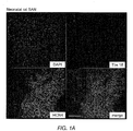
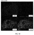
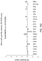
- Tbx18 is a transcription factor that is required for embryonic development of the SAN head area, but, as discussed in more detail below, typically becomes undetectable by birth and remains undetectable in adulthood ( Figure 1 ).
- Shox2 is a negative regulator of Nkx2.5 in the sinus venosus. This protein is critical for tissue differentiation.
- Tbx3 is a potent regulator of SAN specialization, with developmental errors resulting from either deficiency or ectopic expression.
- Tbx5 which shows an inverse correlation between its dosage and abnormal cardiac morphogenesis in Holt-Oram syndrome, is a positive regulator of Shox2 and Tbx3.
- SAN regulating transcription factors or transcription factors that are related to cardiac development, but not specifically SAN regulation
- Those other transcription factors include, but are not limited to Tbx3 and Tbx5. Sequences for Tbx3, Tbx5, Tbx18, Shox2, and variants thereof are shown in Appendix A, B, C, and D, respectively.
- Tbx18, Shox2, or a combination of Tbx-18 and Shox2 are delivered to cardiomyocytes or stem cells in vitro, which, as discussed below, yields cultured pacemaker cells. These cells can subsequently be administered to patients as a cell therapy for biological pacemaker generations.
- administration comprises direct administration of the cells to the heart of a subject (e.g., injection). Other administration routes disclosed herein are used. For example, in some embodiments, catheter-based administration is employed.
- the generated pacemaker cells are incorporated into a matrix, graft, or other biomaterial that aids in cell retention at a target site within the heart.
- Cells used for in vitro generation of biological pacemakers are, in some embodiments, harvested from healthy tissue of the patient that they are to be transplanted into (e.g., autologous transplant of induced pacemaker cells). Then these cells can be converted to pacemaker cells by transcription factors in vitro and can be reinserted into the same patient for the treatment of arrhythmias.
- allogeneic transplant of induced pacemaker cells is performed (e.g., cells are harvested from a first subject, cultured in vitro, contacted with one or more of the transcription factors disclosed herein, and then transplanted to a second subject).
- SAN regulating transcription factors or combinations of such transcription factors are used (e.g., transcription factors in addition to or in place of Tbxl8, Shox2, and/or Tbx-18+Shox2) to convert cells in vitro to pacemaker cells.
- transcription factors include, but are not limited to Tbx3 and Tbx5.
- Patients having abnormalities of excitable tissue that may be treated with the methods disclosed herein include mammals and in particular humans. Patients include those suffering from or diagnosed with one or more of the cardiac conditions disclosed herein or other conditions known in the art to affect cardiac activity, conductivity, rhythm, and the like.
- Reprogramming cardiomyocytes is accomplished, in several embodiments disclosed herein by using gene delivery as a means of delivering exogenous genetic material to somatic cells, cardiomyocytes, stem cells, or combinations thereof.
- polynucleotides are administered in a nucleic acid delivery system.
- a nucleic acid delivery system comprises a non-viral vector linked to the polynucleotide. Examples of such non-viral vectors include the polynucleotide alone (e.g., naked DNA) or the polynucleotide in combination with a suitable protein, polysaccharide or lipid formulation.
- the nucleic acid delivery systems comprise one or more viral vectors, including but not limited to adenovirus, adenovirus-associated virus (AAV), helper-dependent adenovirus, lentivirus, retrovirus, or hemaglutinating virus of Japan-liposome (HVJ) complex.
- viral vectors including but not limited to adenovirus, adenovirus-associated virus (AAV), helper-dependent adenovirus, lentivirus, retrovirus, or hemaglutinating virus of Japan-liposome (HVJ) complex.
- AAV adenovirus-associated virus
- helper-dependent adenovirus helper-dependent adenovirus
- lentivirus lentivirus
- retrovirus hemaglutinating virus of Japan-liposome (HVJ) complex.
- HVJ Japan-liposome
- adenovirus and/or AAV hemaglutinating virus of Japan-liposome
- Additional vectors include retroviral vectors such as moloney murine leukemia viruses and HIV-based viruses.
- an HIV-based viral vector comprises at least two vectors wherein the gag and pol genes are from an HIV genome and the env gene is from another virus.
- DNA viral vectors are used in some embodiments, which include, but are not limited to pox vectors such as orthopox or avipox vectors, herpes virus vectors such as a herpes simplex I virus (HSV) vector.
- pox vectors such as orthopox or avipox vectors
- herpes virus vectors such as a herpes simplex I virus (HSV) vector.
- HSV herpes simplex I virus
- polynucleotides e.g. those encoding one or more transcription factors
- polynucleotides that encode a functional fragment of the transcription factor are delivered in addition to, or in place, of the entire transcription factor.
- fragment As used herein, the terms “fragment”, “functional fragment” or similar terms shall be given their ordinary meaning and shall refer to a portion of an amino acid sequence (or polynucleotide encoding that sequence) that has at least about 70%, preferably at least about 80%, more preferably at least about 90%, 95%, 96%, 97%, 98% or 99% of the function of the corresponding full-length amino acid sequence (or polynucleotide encoding that sequence). Methods of detecting and quantifying functionality of such fragments are established in the art.
- administration of the compositions (being transcription factors or cells) disclosed herein to modulate cardiac electrical activity is via direct cardiac injection (e.g., during electronic pacemaker implantation or explantation).
- systemic injection is used.
- Intracoronary injection is used in some embodiments.
- catheter-directed administration is used.
- a map-guided catheter system e.g., NOGA®
- Other mapping or guidance techniques are used in some embodiments. For example, in several embodiments fluoroscopy-based guidance is used. Electroanatomical guidance is also used in some embodiments.
- Mapping of specific structures including but not limited to the His Bundle, the right or left portions of the bundle, the Purkinje fibers, etc) by intracardiac electrograms are also used in some embodiments.
- X-rays or magnetic catheters are also used in some embodiments to guide delivery of a catheter, needle, or other delivery device(s) to a desired target location.
- a focal delivery approach advantageously reduces the time to generation of an active biological pacemaker.
- the tissue-specific (or cell type-specific) delivery of several of the constructs disclosed herein is advantageous in that the construct is particularly suited for facilitating the generation of a biological pacemaker based on the expression profile of endogenous tissues.
- combinations of transcription factors are delivered to the same target site, while in other embodiments, individual constructs are delivered to distinct target sites, with the overall effect resulting in biological pacemaker generation.
- transduction is achieved by focal injection into the apex of the heart. In several embodiments, transduction is achieved by focal injection to the left ventricular apex.
- a right-sided (e.g., right side of the heart, either atrium or ventricle) approach is used, in order to reduce the risk of stroke or other embolism.
- left-sided approaches are used.
- an injection catheter is introduced via the right atrium (rather than the right ventricle), in order to access the Bundle of His or AV node from above.
- trans-septal catheter methods are used to introduce an injection catheter into the left atrium or left ventricle without the need for arterial access, thereby reducing stroke risk.
- an injection catheter is by way of the cardiac veins via the sinus of Valsalva for injection of a biologic as disclosed herein into various targets of the ventricles.
- a biologic as disclosed herein into various targets of the ventricles.
- compositions as disclosed herein can be used to deliver one or more transcription factors (or cells that have been previously contacted with the transcription factors) to either the right atrium, right ventricle, SA node, AV node, bundle of his, and/or left and right bundle branches.
- transcription factors or cells that have been previously contacted with the transcription factors
- cannulation of the coronary sinus and its venous branches delivery to multiple left ventricular sites is achieved in several embodiments.
- access to the left side is achieved, in several embodiments, from the right side through a trans-septal puncture which allows direct access to left sided structures without the need of arterial access.
- Supplemental methods are used in several embodiments and include administration of compounds to increase the microvascular permeability of the cardiac tissue.
- Suitable vascular permeability agents include a solution having less than about 500 micromolar calcium: substance P, histamine, acetylcholine, an adenosine nucleotide, arachidonic acid, bradykinin, endothelin, endotoxin, interleukin-2, nitroglycerin, nitric oxide, nitroprusside, a leukotriene, an oxygen radical, phospholipase, platelet activating factor, protamine, serotonin, tumor necrosis factor, vascular endothelial growth factor, a venom, a vasoactive amine, or a nitric oxide synthase inhibitor, serotonin, vascular endothelial growth factor (VEGF), or a functional VEGF fragment.
- substance P substance P
- histamine histamine
- administering induces or otherwise causes the spontaneous repetitive electrical signals to be generated in cells that previously responded to such signals, but did not generate them.
- myocardial cells that exhibited little (e.g., an index of automaticity between about 40% to about 30%, about 30% to about 20%, about 20% to about 10%, or about 10% to about 0%, or overlapping ranges thereof) or no firing rate, exhibit an increased frequency of firing rate or electrical signal output post-administration (e.g., an index of automaticity of between about 5% to about 15%, about 15% to about 25%, about 25% to about 35%, about 35% to about 45%, about 45% to about 55%, about 55% to about 65%, about 65% to about 75%, about 75% to about 85%, about 85% to about 95%, about 95% to about 100%, or more) as compared to the cells pre-administration.
- the resultant changes in cardiac contraction and/or an electrical property of converted pacemaker cells modulate cardiac rhythm in several embodiments.
- the methods and compositions disclosed herein achieve a heart rate within about 25%, within about 20%, within about 15%, within about 10%, within about 5%, within about 2%, or within about 1% of a clinically desired heart rate.
- the methods and compositions disclosed herein are used to treat subjects suffering from or susceptible to a disease or disorder such as a cardiac-related syncope (e.g., Stokes-Adam syncope), an abnormality of sinus node function such as persistent sinus bradycardia, sino-atrial (S-A) block manifested as S-A Wenckebach, complete S-A block or sinus arrest, and high-grade atrioventricular block; or bradycardiatachycardia syndrome or other bradycardia related condition.
- a cardiac-related syncope e.g., Stokes-Adam syncope
- an abnormality of sinus node function such as persistent sinus bradycardia, sino-atrial (S-A) block manifested as S-A Wenckebach, complete S-A block or sinus arrest, and high-grade atrioventricular block
- S-A sino-atrial
- modulation is used to increase or slow down the function of an implanted pacemaker (e.g., to achieve
- the administration of Tbx18, Shox2, or a combination thereof produces several physiological changes to the contacted cells in addition to or separate from the generation of spontaneous repetitive electrical signals discussed above. In some embodiments, these physiological changes even if first seen in vitro, are also detectable in vivo, where they may serve as supplemental markers of the efficacy of biological pacemaker generation (e.g., they are recognized as characteristics or hallmarks of pacemaker cells). In some embodiments, the administration of one or more transcription factors results in an increased percentage of spontaneously beating monolayer cultures compared to control. In several embodiments, the presence, or amount, of spontaneous beating is use to screen cultures for functionality prior to transplant or to evaluate other combinations of transcription factors for their utility in generating pacemaker cells in vitro.
- the administration of one or more transcription factors results in spontaneous intracellular Ca 2+ oscillations of myocardial cells are administered. In some embodiments, the administration of one or more transcription factors results in a gradual phase-4 depolarization. In some embodiments, the delivery of one or more transcription factors disclosed herein increases or decreases the expression of HCN4 in cells. Because calcium flux is a primary component of cardiac electrical signaling and IICN4 expression is important in pacemaker cell function, changes in these endpoints correspond, in some embodiments, to more SAN-like behavior in induced biological pacemaker cells. In some embodiments, the administration of one or more transcription factors results in modulation of sub-sarcolemmal, spontaneous localized Ca 2+ release events.
- the administration of one or more transcription factors results in modulation of intracellular cAMP levels.
- cardiac electrical activity can be modulated and abnormalities in excitable cardiac tissue can be treated.
- the administration of Tbx18, Shox2, or a combination thereof will result in cells new phenotypes of the contacted cells in addition to or separate from the generation of spontaneous repetitive electrical signals.
- the administration of one or more transcription factors results in disorganized and markedly lower sarcomeric ⁇ -actin expression in transduced cells, which is indicative of pacemaker cells.
- the administration of one or more transcription factors results in a change in cell size.
- the administration of one or more transcription factors results in changes to the chromatin state.
- the administration of one or more transcription factors results in chromatin modification and will cause lower or higher expression and or activity of one or more of the following genes: Cx43, Kir2.1, Actc2, and HCN4.
- Cx43, Kir2.1, Actc2, and HCN4 genes that are involved in the chromatin modification.
- the administration of the one or more transcription factors to the heart will result in frequent ectopic ventricular beats that originate from the site of gene injection resulting in targeted generation of pacemaker activity.
- the administration of one or more transcription factors results in gene-related epigenetic changes in cells and de novo pacemaker activity.
- This pacemaker activity in some embodiments, is a result of somatic reprogramming, and not due to dedifferentiation to a progenitor state. Somatic to somatic transdifferentiation will lower the threat of neoplasia from transduced cells (e.g., teratoma formation is reduced).
- the administration of one or more transcription factors may result in dedifferentiation to a progenitor state.
- differentiation into pacemaker cells is induced using one or more of the transcription factors disclosed herein. Using such methods, biological pacemaker cells can safely and effectively be made from a wide variety of cells.
- the human Tbx18 gene with a C-terminal myc/FLAG tag was excised from pCMV6-Tbx18 (Origene, Rockville, MD) by digestion with Fse I and Sal I .
- the Tbx18 gene was then sub cloned into a Not I - and Xho I digested lentiviral expression vector with the desired reporter gene, pLVX-IRES-ZsGreenl (Clontech, Mountain View, CA), thus generating the pLV-Tbx18-IRES-ZsGreen1 (-10.1 kb) vector.
- ZsGreenl was used as the reporter protein for Tbx18-transduced cardiomyocytes, due to its similar spectral characteristics as the commonly used GFP.
- the recombinant target gene was then introduced to an adenovirus vector backbone by Gateway recombination cloning using Gateway-adapted vectors (Invitrogen, Carlsbad, CA).
- An LR recombination reaction was performed between the entry clone and the destination vector, pAd/CMV/V5-DEST (-36.7 kb), to generate the desired adenoviral expression construct, pAd-CMV-TBX18-IRES-GFP (-39.8 kb). Positive constructs were verified to have the correct target gene by DNA sequencing (Laragen, Los Angeles, CA).
- the micropipette electrode solution was composed of (mmol/L): K-glutamate 130, KCl 9, NaCl 8, MgCl 2 0.5, HEPES 10, EGTA 2, and Mg-ATP 5; pH 7.2.
- Adenoviruses were plaque-purified, amplified, and affinity-column purified using Adenopure kit (Puresyn, Inc), and stored at - 80°C.
- NRVMs were isolated from 1-2 day old rat pups and cultured as a monolayer using established culture methods. Only the lower one third of the heart (from the apex to the midline) was excised in order to minimize contaminating atrioventricular nodal cells. In some embodiments, other portions of the heart are used, including the lower third, the middle third, and combinations of these portions, or whole hearts (or portions thereof) where endogenous pacemaker cells are selectively removed. In some embodiments, biopsies (e.g., guided biopsies) are used to obtain non-pacemaker tissue).
- biopsies e.g., guided biopsies
- Tbx20 known to be critical for cardiac chamber differentiation, was employed in order to control for non-specific, embryonic transcription factor-related effects.
- SA nodal myocytes were isolated from adult Sprague-Dawley rats. Animals were anesthetized with isoflurane. Hearts were quickly removed, the atria separated from the ventricles, and the sinoatrial node region dissected in Tyrode solution, which consisted of (in mM) 140 NaCl, 5.4 KCl, 1.2 KH 2 PO 4 , 5 HEPES, 5.55 glucosc, 1 MgCl 2 , 1.8 CaCl 2 ; pH adjusted to 7.4 with NaOH.
- the rat sinoatrial node region was defined by the borders of the crista terminalis, the interatrial septum, and the superior and inferior vena cavae.
- Mouse embyronic stem cells were transfected with an adenoviral vector expressing Shox2 or a control gene. Established embryoid body methods were used for differentiation. Pooled data are n ⁇ 3 with p ⁇ 0.05 for all reported differences.
- 2x10 6 NRVMs were plated in 35-mm glass bottom dishes (MatTek Cultureware) or 22 mm fibronectin-coated glass coverslips, transduced, and analyzed 4 days post-transduction.
- Cells were loaded with Rhod2-AM (2 ⁇ mol/L) (Molecular Probes) for 18 minutes, then washed once and subsequently placed in normal Tyrode's solution with 2 mmol/L calcium.
- Calcium transients were recorded at 37°C from AdTbx18IS-IRES-GFP and Ad-GFP transduced NRVMs.
- Frozen sections of neonatal rat sinoatrial node and NRVMs 4 days post adenoviral transduction were fixed with 4% paraformaldehyde and permeabilized with 0.1 % Triton-X 100 and then incubated with the appropriate primary antibody: sarcomeric ⁇ -actinin (Sigma-Aldrich; A5044; 1:800), ANP (AbCam;ab-14348; 1:1000), HCN4 (Abcam; ab85023; 1:500) and Alexa Fluor-conjugated secondary antibodies (Invitrogen).
- the transferred membrane was incubated with a primary antibody overnight at 4°C, followed by 1-hour incubation with a peroxidase-conjugated secondary antibody. Immunoreactivity was detected by chemiluminescence (ECL Western Blotting Analysis System, Amersham). Equal protein loading of the gels was assessed by re-probing the membrane with monoclonal anti-GAPDH antibody (Abeam; ab9482; 1:10000) or anti- ⁇ -actin (Sigma-Aldrich; A3848: 1:25000).
- Adenoviruses were injected into the left ventricular apex of guinea pigs.
- Adult female guinea pigs (weight, 250 to 300 g; Charles River) were anesthetized with 4% isoflurane, intubated, and placed on a ventilator with a vaporizer supplying 1.5% to 2% isoflurane.
- a 30-gauge needle was inserted at the free wall apex of the left ventricle.
- Ad-Tbx18-IRES-GFP Ad-GFP
- ECGs In vivo and ex vivo electrocardiographs
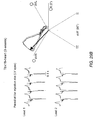
- Methacholine (0.1-0.5 mg per kg of body weight in saline, Sigma-Aldrich, St. Louis, MO) was delivered via the jugular vein in order to slow the animals' sinus rhythm prior to ECG recordings under general anesthesia (2% isoflurane, 98% O2). ECGs were recorded using a 2-lead digital ECG system at 2 kHz (Lead I and Lead 3, BIOPAC Systems, Goleta,CA) and Lead 2 was offline calculated by Einthoven's triangle using Acqknowlege 3.7.3 software (BIOPAC Systems, Goleta, CA). In all animals, methacholine was administered until complete heart block was achieved. Heart block was accompanied by a reduction of the animals' sinus rhythm to ⁇ 100 bpm.
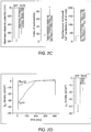
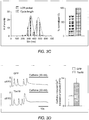
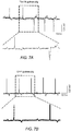
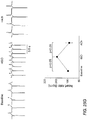
- Fig. 3D In most of the Tbx18-injected guinea pigs, ectopic ventricular rhythms were manifested well before the sinus rhythm reached 100 bpm ( FIG. 3D , right panel). In contrast, control animals exhibited no evidence of such ectopic ventricular beats even when the sinus rhythm was brought to ⁇ 100 bpm ( Fig. 3D , left panel). Fig. 3D highlights the lack of any ectopic ventricular rhythm in the control animals even at a very slow escape rate compared to Tbx18-injected animals.
- the heart was retrograde-perfused via the aorta at 60 mmHg with oxygenated Tyrode's solution at 36°C.
- the perfused heart was placed in a sylgard-coated plate filled with warm Tyrode's solution.
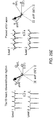
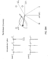
- ECG leads were stationed at appropriate sites to record leads I and II ( Figure 28 ).
- the region of the atrioventricular node was ablated with a cryogun (Brymill Cryogenic Systems, Ellington, CT) filled with liquid N 2 .
- Electrode-pacing was performed at the site of transgene injection (anterior, left ventricular apex) at 200-ms intervals with a platinum electrode connected to an isolated pulse stimulator (Model 2100, AM Systems, Carlsborg, WA).
- Rat sinoatrial node, left ventricle and Tbx18- and GFP-transduced NRVMs (4 days post transduction) were collected and mRNA was extracted (Qiagen mRNA Isolation Kit,).
- the mRNA samples were converted to first strand cDNA, using the RT2 First Strand Kit (SA Biosciences).
- the cDNA template was mixed with the RT2 qPCR Master Mix and the mixture was aliquoted into each well of the same plate containing pre-dispensed gene specific primer sets.
- PCR was performed on a 7900HT Fast Real-Time PCR System (Applied Biosystems/Life Technologies Corporation, Carlsbad, CA) and the relative expression of the genes was calculated.
- the Cyclic AMP Elisa Assay Kit (catalog # STA-501; Cell Biolabs, INC) was used to determine cAMP levels in NRVMs transduced with Ad-Tbx18 or Ad-GFP. Briefly, 50 ⁇ l Tbx18- and GFP-NRVM cell lysates were added to the Goat Anti-Rabbit Antibody Coated Plate 96 well plate. 25 ⁇ l of diluted Peroxidase cAMP Tracer Conjugate was added to each tested well. Then, 50 ⁇ l of diluted Rabbit Anti-cAMP Polyclonal Antibody was added to each tested well and the plate was incubated at room temperature for 30 minutes with shaking.
- Ryanodine and Protein kinase I (PKI) were purchased from Tocris biosciences and caffeine was purchased from Sigma. Rhod-2/AM and was purchased from Invitrogen.
- NRVMs transduced with either Tbx18 or GFP were fixed two- to four-days after viral vector transduction with 10% formaldehyde for 8 min at room temperature.
- Cells were sheared using a sonicator with ten pulses of 20 seconds each, with a 30-second rest on ice between each pulse.
- Chromatin immunoprecipitation was performed using ChIP-IT® Express Chromatin Immunoprecipitation kit (Active Motif, Carlsbad, CA) following the manufacturer's protocol.
- Primary antibodies for the H3K4me3 and H3K27me3 were purchased from Active Motif.
- Single myocytes were collected in PBS with a wide-opening patch pipette, placed on dry ice and then stored at -80°C. Tbx18 mRNA levels in individual myocytes were examined by quantitative real-time PCR with an Ambion® Single Cell-to-CTTM Kit (Life Technologies) according to manufacturer's instructions. Briefly, single cells were treated with cell lysis solution and DNase I for 5 minutes at room temperature. Reverse transcription was performed at 25°C for 10 minutes, at 42°C for 60 minutes and at 85°C for 5 minutes after addition of SupersScript RT and VILO RT mix.
- Preamplification was performed with 14 cycles of 95°C for 15 seconds and 60°C for 4 minutes with addition of PreAmp mix and 0.2x TaqMan Gene Expression Assays, containing primers for human Tbx18 (assay ID:Hs01385457_m1), guinea pig GAPDH (assay ID:Cp03755742_g1), and guinea pig TnnT2 (assay ID:Cp04182357_g1).
- the custom primers were synthesized by Applied Biosystems (Carlsbad, CA).
- Preamplification products (1:20 dilution) were used for real-time PCR with TaqMan Gene Expression Assays using an Applied Biosystems 7900HT Fast Real-Time PCR System.
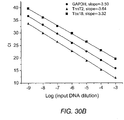
- Standard curves for each of the 3 primer sets were constructed with serial dilutions of input DNA templates ( Fig. 30 ) and validated comparable amplification efficiencies (curve slopes: -3.32 to -3.64). Relative mRNA levels of Tbx18, GAPDH, and TnnT2 were obtained by extrapolation of Ct values with the slopes of the standard curve for each primer sets. Tbx18 mRNA expression in each cell was then normalized to GAPDH or TnnT2 level.
- Example 1 Pacemaker cells created by gene delivery to NVRMs
- Tbx18-transfected NRVMs Tbx18-NRVMs
- Fig. 2A Tbx18-transfected NRVMs
- Tbx18 is used to create a biological pacemaker.
- other transcription factors are used, including one or more of those screened in the current experiment.
- combinations of two or more transcription factors will be used.
- Shox2 will be used in conjugation with Tbx18 to achieve these results.
- one or more of the following transcription factors will be selected for use: Tbx18, Shox 2, Tbx3, and Tbx5.
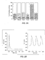
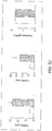
- FIG. 2B shows representative action potential (AP) traces.
- AP action potential
- Tbx18 could be used instead of or in conjunction with Tbx18.
- one or more of the following transcription factors will be selected for use: Tbx18, Shox2, Tbx3, and Tbx5.
- the Tbx18-NRVMs exhibited spontaneous APs with prominent gradual phase-4 depolarization.
- Gradual phase-4 depolarization underlies automaticity in SAN pacemaker cells, and is prominent in Tbx-NRVMs ( Fig. 2B ).
- This increase in autonomous beats and gradual depolarization indicates that these cells are functioning in a manner consistent with natural sinoatrial node cells, which suggests that Tbx18 (alone or in combination with other transcription factors) is generating pacemaker cells.
- similar functions are detected in stem cells, and which therefore indicates that the cells are useful in the generation of biological pacemakers and in the treatment of arrhythmias.
- MDP maximum diastolic potential
- RMP resting membrane potential
- index of automaticity shall be given its ordinary meaning and shall also be defined as the percentage of autonomously-beating cells multiplied by the frequency of action potential (AP) oscillations in those cells, was much larger in Tbx-NRVMs (70 ⁇ 16% bpm) compared to control (13 ⁇ 4% bpm, Fig. 2C , middle). As shown in Fig. 2C middle, automaticity increased relative to the GFP control by around 60%.
- the increase in index of automaticity will be 5% to about 15%, about 15% to about 25%, about 25% to about 35%, about 35% to about 45%, about 45% to about 55%, about 55% to about 65%, about 65% to about 75%, about 75% to about 85%, about 85% to about 95%, about 95% to about 100%, and overlapping ranges thereof, relative to the automaticity of normal pacemaker cells.
- the number of cells having spontaneous Ca 2+ oscillations also increased relative to the controls ( Fig. 2C , right).
- the number of Tbx18-NRVMs exhibiting spontaneous intracellular Ca 2+ oscillations was about 6-fold higher relative to control ( Fig. 2C , right). Because Ca 2+ is necessary for the generation of an action potential in pacemaker cells, these data also demonstrate that transduction of cells with Tbx18 (alone or in combination with other transcription factors) can be successfully used in the generation of pacemaker cells.
- the reduction in I K1 density will be between about 5% to about 15%, about 15% to about 25%, about 25% to about 35%, about 35% to about 45%, about 45% to about 55%, about 55% to about 65%, about 65% to about 75%, about 75% to about 85%, about 85% to about 95%, about 95% to about 100%, and overlapping ranges thereof.
- the reduction in I K1 density also indicates that the transduced cells will fire with higher frequency relative to untreated cells (e.g., non-pacemaker cells). This increase in firing indicates that these transduced cells are functioning as pacemaker cells.
- the methods used to generate these cells, or methods used to generate pacemaker cells from stem cells can be used in the treatment of cardiac arrhythmias, or other maladies that are associated with a disrupted cardiac cell firing rate.
- HCN4 is the molecular correlate of the hyperpolarization-activated current, If, which contributes to pacemaker activity in the SAN.
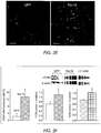
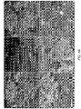
- Tbx18 transduction led to a 3.8-fold increase in the number of cells expressing HCN4 ( Fig. 2E ).
- HCN4 immunostaining image HCN4-white, nuclei-blue
- Scale bar 200 ⁇ m.
- Right panel summary of the percent HCN4-positive cells per GFP- or Tbx18-transduced cells.
- Tbx18 expression also led to a 1.4-fold increase in HCN4 protein level ( Fig. 2F ) in Tbx18-NRVMs.
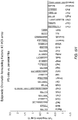
- Tbx18-NRVMs Changes of relative mRNA levels of selected genes comparing Tbx18-NRVMs normalized to GFP-NRVMs (left) and SAN normalized to LV (right). SAN and Tbx18-NRVMs demonstrate similar pattern of normalized transcript levels. Thus, in several embodiments, Tbx18 also elicits gene and protein expression changes that are associated with biological pacemaker formation.
- FIG. 2B shows Tbx18 cells were more likely to fire and fire in a manner (with slow depolarization) like that of native SAN cells.
- Figure 2C shows that Tbx18 cells have a lower diastolic potential (middle) resulting in a higher index of automaticity (middle) and more spontaneous whole-cell oscillations of Ca 2+ (left).
- Figure 2D shows that the current density of the Tbx18 cells have less repolarization time, and thus a higher chance of firing.
- Tbx18 changes the electrical properties of cells and make them SAN-like.
- Each of these results indicates a higher likelihood that these cells will fire automatically when implanted into or generated in the living heart.
- these induced pacemaker cells are able to beat and can modulate heart rhythm to treat cardiac arrhythmias, or other maladies associated with abnormal cardiac rhythm.
- LCRs spontaneous localized Ca 2+ release events
- IRCX Na+-Ca 2+ exchanger currents
- LCRs preceding the whole cell Ca 2+ transient only in the Tbx18-NRVMs whereas occasional Ca 2+ sparks could be detected in the GFP-NRVMs.
- the LCR period is defined as the period between the start of the Ca 2+ transient to the beginning of the subsequent LCR.
- Cycle length is defined as the period between the start of the whole cell Ca 2+ transient to the start of the subsequent Ca 2+ transient.
- the LCRs in Tbx18-NRVMs occurred at an average period of 343 ⁇ 8 ms, which was 72 ⁇ 1 % of the whole-cell Ca 2+ transient cycle length (474 ⁇ 7 ms, Fig. 3D ).
- the LCR will occur between about 5% to about 15%, about 15% to about 25%, about 25% to about 35%, about 35% to about 45%, about 45% to about 55%, about 55% to about 65%, about 65% to about 75%, about 75% to about 85%, about 85% to about 95%, about 95% to about 100%, and overlapping ranges thereof, of the whole-cell Ca2 + transient cycle length.
- Phospholamban in its unphosphorylated state, inhibits sarcoplasmic reticulum Ca 2+ -ATPasc 2a (SERCA2a), thereby suppressing the rcuptake of Ca 2+ by internal stores. Such inhibition is relieved upon phosphorylation of the protein (P-PLB).
- the relative p-PLN (Ser16) level was 65-fold higher in Tbx-NRVMs in comparison to GFP-NRVMs ( Fig. 3F , left panel), mimicking the augmentation of p PLN found in the SAN compared to that in the ventricular myocardium ( Fig. 3F , right panel).
- Tbx18 expression in NRVMs produced changes in key Ca 2+ cycling components which recapitulate key features of sinoatrial node pacemaker cells, thereby allowing, in several embodiments, transduction of cells with Tbx18 (alone or in combination with other transcription factors, depending on the embodiment) to be used in therapeutic methods for treating cardiac arrhythmias.
- cellular reprogramming replicates key features of cell structure in pacemaker tissue.
- Native sinoatrial node pacemaker cells are distinctive in their morphology: they are smaller and exhibit less-organized myofibrils than working cardiomyocytes.
- Tbx18-NRVMs had phenotypic changes, the morphology of the cells was investigated.
- Sections of neonatal rat heart demonstrate that cardiac sarcomeric ⁇ -actinin (a-SA) expression is markedly lower and disorganized in the sinoatrial node compared to the adjacent right atrium (RA, Fig. 4A , top left and bottom left).
- Tbx18-NRVMs resembled native sinoatrial node cells in their morphology, with myofibrillar disorganization and weak expression of a-SA ( Fig. 4A , bottom right).
- Neonatal rat sinoatrial node, demarcated by HCN4 expression top middle
- exhibits weaker and unstructured sarcomeric ⁇ -actinin (a-SA) expression top panel).
- Bottom left Zoom-in image of the boxed area in top left.
- Tbx18-NRVMs The pattern is faithfully recapitulated in Tbx18-NRVMs compared to GFP-NRVMs (bottom right and middle, respectively). Scale bar: 30 ⁇ m.
- the data are corroborated by the observed downregulation of transcript levels in Tbx18-NRVMs ( Fig. 2H ).
- Cell size measured by two complementary methods, is 28-33% smaller in Tbx18-NRVMs than in control NRVMs ( Fig. 4B ), recapitulating the smaller size of SAN cells relative to working cardiomyocytes.
- Tbx18-NRVMs underwent both structural (morphology) as well as functional (electrophysiology) reprogramming.
- Tbx18-NRVMs Trimethylation of lysine 27 in histone 3 (H3K27mc3) is a heterochromatin mark which promotes the recruitment of Polycomb group proteins for gene silencing. Conversely, trimethylation of lysine 4 (H3K4me3) marks genes transcriptionally active. Histone modification profiles in the promoter regions of four genes, Cx43, Kir2.1, Actc2, and HCN4, were then investigated.
- Tbx18 is a transcription factor that is required for embryonic development of the sinoatrial node head area.
- Shox2 is a negative regulator of Nkx2.5 in the sinus venosus (as discussed above).
- Tbx3 is a potent regulator of sinoatrial node specialization, with developmental errors resulting from either deficiency or ectopic expression.
- Tbx5 is a positive regulator of Shox2 and Tbx3. These genes exhibit relevant molecular and functional changes in Tbx18-NRVMs. Tri-methylation level on H3K27 indicates that Tbx18 increased inactivity of Cx43, Kir2.1, and a-SA promoters while relieving its repressive epigenetic pressure on HCN4 promoter normalized to control. These results were measured by chromatin immunoprecipitation followed by qPCR.
- H3K4me3 ( Figure 4C right) levels indicate that ratio of active HCN4 promoter regions increased upon Tbx18 expression while the transcriptionally active promoter regions of Cx43, Kir2.1, and a-SA have decreased upon Tbx18 expression.
- Cx43, Kir2.1, and Actc2 became epigenetically inactive (higher H3K27me3 and lower H3K4me3) in Tbxl8-NRVMs compared to control ( Fig. 4C ).
- Tbx18 expression would reduce one or more of Cx43, Kir2.1, and a-SA expression while HCN4 would be upregulated.
- HCN4 is involved in repetitive signals, these data are consistent with biological pacemaker formation.
- the chromatin histone modifications are consistent with durable epigenetic reprogramming rather than transient functional re-engineering.
- At least one of Cx43, Kir2.1, and Actc2 may become epigenetically inactive in iSAN cells as compared to the quiescent cells.
- expression of sarcomeric ⁇ -actinin in iSAN cells may be weak as compared to the quiescent cells.
- the iSAN cells may exhibit myofibrillar disorganization. Only one of such changes may occur, or combinations thereof may occur, depending on the embodiment; however, each of the above are consistent with a shift of a cell towards a sinoatrial node-like phenotype.
- pacemaker cells are generated without the induced alteration expression of ion-channel proteins, the genetic introduction of which may be contrary to the data above (e.g., the reduction in Kir2.1 that contributes, at least in part, to iSAN formation, would potentially be offset by genetic introduction of Kir2.1 channels).
- Tbx18 The prior example established the ability of Tbx18 to induce changes in gene/protein expression and function in vitro.
- An adenoviral vector encoding Tbx18 was directly and focally injected into the apex of guinea pig hearts.
- the injection will be to other areas of the heart and sometimes in several areas at once.
- the guinea pig is an accepted model for use in cardiovascular studies, and data can be readily extrapolated to other mammals, including the human.
- the administration of the Tbx18 adenoviral vector will be performed on various mammalian hearts, including that of the human heart.
- the rate of beating of the pacemaker cells will be between about 5% to about 15%, about 15% to about 25%, about 25% to about 35%, about 35% to about 45%, about 45% to about 55%, about 55% to about 65%, about 65% to about 75%, about 75% to about 85%, about 85% to about 95%, about 95% to about 100%, and overlapping ranges thereof, of the normal beating rate of healthy pacemaker cells.
- This beating will correspond to a new heart rate of the mammal, including the human, of about 5% to about 15%, about 15% to about 25%, about 25% to about 35%, about 35% to about 45%, about 45% to about 55%, about 55% to about 65%, about 65% to about 75%, about 75% to about 85%, about 85% to about 95%, about 95% to about 100%, or overlapping ranges thereof, of the normal heart.
- Electrocardiographic vector analysis revealed that ectopic beats originated at the site of gene injection (apex) and propagated towards the base ( Fig. 6A , right).
- stem cells, progenitor cells, somatic cells, and/or cardiomyocytes could be transfected with genes for the above transcription factors. Then these cells, after verifying generation of pacemaker activity, can be implanted in vivo to treat abnormalities of excitable tissues.
- Tbx18-VMs Tbx18-transduced ventricular myocytes
- GFP-transduced ventricular myocytes GFP-transduced ventricular myocytes
- native SAN cells native SAN cells.
- LtW length-to-width
- p ⁇ 0.05 7.6 ⁇ 0.7
- Tbx18-VMs had stable resting potentials at -76mV, with action potentials elicited only upon electrical stimulation.
- the Tbx18 transduced cells function more like pacemaker cells.
- the electrophysiological and morphological features of Tbx18-VMs generally resembled those of native SAN cells ( Fig. 8 and 9 ).
- iSAN induced SAN
- the in situ reprogramming was effective and rapid in speed (as little as five days), offering a novel approach to creating a biological pacemaker as an alternative to electronic devices.
- the data indicates that in several embodiments the iSAN cells will have a length-to-width ratio greater than the nontransduced ventricular myocytes. In several embodiments, this LtW ratio will be greater than about 2, about 4, about 6, about 8, or about 10. In several other embodiments, this LtW ratio will be about equal to 15.
- the ratio will range from about 10 to about 12, about 12 to about 14, about 14 to about 16, about 16 to about 20, or greater.
- other phenotypic characteristics of the iSAN cells resemble that of native pacemaker cells (e.g., more closely than nontransduced ventricular cells).
- Tbx18 is a marker of multipotent progenitors in cardiac development, and has been associated with neoplasia.
- Pluripotent stem cells embryonic or induced
- Pluripotent stem cells are known to differentiate spontaneously into pacemaker cells.
- testing was performed to ensure that Tbx18 transduction did not produce pacemaker cells via a pluripotent/neoplastic state, which could occur by accelerating the dedifferentiation known to occur in cultured cardiomyocytes.
- the data presented herein indicate that the SAN-like morphology of Tbx18-VMs is not indicative of nonspecific regression.
- dedifferentiating adult VMs lose the longitudinal, 'bricklike' shape and become rather circular, similar to their neonatal counterparts, but do not become thinner.
- Tbx18 induces specific re-engineering rather than reversion to a fetal state are the following.
- dedifferentiation is accompanied by re-expression of genes characteristic of the fetal heart, including atrial natriuretic peptide (ANP) and skeletal ⁇ -actin ( ⁇ SkA).
- ANP nor ⁇ SkA was re-expressed in Tbx18-NRVMs (iSAN cells); in fact, ANP expression was strongly suppressed ( Fig. 5C and 5A , B, respectively).
- dedifferentiation would bring the transduced myocytes closer to a progenitor state, and expression of early ventricular transcription factors such as Nkx2-5 would be enhanced. Instead, Tbx18 lowered the transcript level of Nkx2-5 ( Fig. 2I ), consistent with conversion to mature pacemaker cells but not reversion to a fetal state.
- Tbx18-NRVMs were a consequence of dedifferentiation to an embryonic/fetal state, the proliferative index of those cells would be expected to increase. This is because the embryonic ventricle grows by hyperplasia, an ability which plummets after birth.
- Shox2 could be used instead of or in conjugation with Tbx18.
- one or more of the following transcription factors will be selected for use to achieve transdifferentiation: Tbx18, Shox2, Tbx3, and Tbx5.
- the quiescent cells will transdifferentiate to iSAN cells without first dedifferentiating to an embryonic/fetal state.
- the iSAN cells may exhibit similar morphology to that of native SAN cells consistent with conversion from quiescent cells to iSAN cells without dedifferentiation.
- expression of ANP and ⁇ SkA will be suppressed in the iSAN cells.
- iSAN cells may exhibit transcript levels of Nkx2.5 similar to that of mature native pacemaker cells.
- the iSAN cells will remain differentiated without any discernible increase in stemness factors. In several embodiments this transdifferentiation may occur in vitro, while in other embodiments this transdifferentiation may occur in vivo.
- the quiescent cells will be mammalian, including several embodiments in which the quiescent cell is human.
- Embryonic stem cells can spontaneously differentiate into heterogeneous aggregates of cardiomyocytes with atrial, ventricular, and pacemaker properties. Experiments were thus performed to test whether Embryonic stem cells could be biased from random cardiogenesis toward a dominant pacemaker phenotype.
- Shox2 is an embryonic transcription factor essential for the patterning of pacemaker cells in the sinoatrial node. Using Shox2 overexpression, the developmental program for the embryonic stem cells was tilted and dominated toward pacemaker myocytes. It will be appreciated that, in several embodiments, Tbx18 could be used instead of or in conjugation with Shox2. In several embodiments, one or more of the following transcription factors could be selected for use: Tbx18, Shox2, Tbx3, and Tbx5.
- Mouse embryonic stem cells were cultured on a MEFs feeder layer. Embryoid body formation was cultivated using established hanging drop culture methods. On day 3, cultivation of embryoid bodies was performed. On day 6, plating of embryoid bodies on tissue culture plates was performed ( Fig. 11 ).
- Endogenous levels of transcription factor RNA were measured at several time points. As shown in Figure 12 , endogenous levels of Tbx18 RNA increased sharply upon formation of embryoid bodies and leveled after plating of the embryoid bodies. Endogenous Tbx3 RNA however, decreased relative to mouse embryonic stem cells.
- Figure 13 shows the administration of Shox2 expression vectors. As shown by the graph in 13B, the level of mRNA relative to control mouse embryonic stem cells increased after the third administration of Shox2-vector to levels higher than for control mouse embryonic stem cells.
- Cx43 transcript and protein levels were reduced in Shox2-embryoid bodies by 33 ⁇ 5% and 30 ⁇ 10%, respectively, compared to control ( Fig. 20 ).
- This decrease in Cx43 transcript remained below the control for the 14 day time period of the experiment ( Figure 21A ).
- Cx45 remained increased relative to the control until day 14, with a decline at day 7 after the administration of the Shox2 vector ( Fig. 21B ).
- the increased level of HCN4 expression and Cx45 is also apparent from confocal imaging of cells that were transduced ( Fig. 22 ).
- the Shox2 expression after transfection also remained at elevated levels relative to control HMBS and GFP transfected cells ( Fig. 23 ).
- hShox2 mRNA decreased after transduction of the Shox2 gene eventually falling below the level of control cells.
- Tbx18 and Tbx3 mRNA levels relative to HMBS are shown in Fig. 24 .
- HCN4 is known to be important for pacemaker function and its upregulation the transduced cells indicates that these cells i) are taking on characteristics of pacemakers due to Shox2 and/or, ii) may be useful themselves as biological pacemaker cells (e.g., transplanted cells).
- SAN pacemaker cells respond to autonomic inputs with altered firing rates.
- Tbx18- and GFP-NRVMs were plated on multi-electrode arrays (MEAs) to record extracellular field potentials from spontaneously-beating cells ( Fig. 25A ).
- Tbx18-NRVMs respond appropriately to adrenergic and muscarinic stimuli.
- a disease model of atrioventricular (AV) block which is a common indication for the placement of an electronic pacemaker, was created to investigate autonomic regulation of induced biological pacemakers in the intact heart.
- Electrocardiographic recordings of the beating hearts, perfused ex vivo revealed ectopic ventricular beats in 8 of 8 Tbx18-injected hearts, at a rate of 154 ⁇ 6 bpm.
- the polarity and morphology of the ectopic beats was identical to that of electrode-paced beats at the location of transgene injection ( Fig. 25E ), linking the origin of the ectopic beats to the site of Tbx18 transduction.
- Shox2 could be used instead of or in conjugation with Tbx18.
- one or more of the following transcription factors could be selected for use: Tbx18, Shox2, Tbx3, and Tbx5.
- the iSAN cells may generate ectopic ventricular beats.
- the Tbx18-NRVM cells iSAN cells
- iSAN cells will respond to autonomic regulation such as ⁇ -adrenergic stimulation and cholinergic suppression in vivo.
- iSAN cells converted from quiescent cells in vitro may be capable of responding to autonomic regulation.
- the iSAN cells may generate ectopic beats at a rhythm controlled by autonomic regulation. It shall be appreciated that in several embodiments in which iSAN cells respond to autonomic regulation, the iSAN cells can function to treat a cardiac arrhythmia in a patient suffering from said arrhythmia. In several embodiments, iSAN cells responding to autonomic regulation can replace or supplement electronic pacemaker devices in a patient.
- Tbx18-VMs were isolated 6-8 weeks after the initial in vivo gene transfer, and the transcript levels of Tbx18 were quantified by single-cell, quantitative RT-PCR.
- Tbx18 transcripts exhibited a wide dynamic detection range in Tbx18-VMs isolated just 3 days after in vivo gene transfer ( Fig. 26A , bottom panel).
- 5 spontaneously-beating ventricular myocytes (114 ⁇ 10 bpm, identified by live-cell video recording or whole-cell patch-clamp) isolated 6-8 weeks after in vivo gene transfer were examined.
- PCR primer sets were designed to detect Tbx18, cardiac troponin T (TnT2, to verify cardiomyocyte identity) and GAPDH.
- the levels of Tbx18 transcript in 4 of the 5 spontaneously-beating myocytes were negligible ( Fig. 26B , cell 1, 2, 3 and 4, bottom panel), close to the negative control level ( Fig. 26C ) in ventricular myocytes expressing GFP alone.
- L-to-W cell length-to-width
- the percentage of SAN-like Tbx18-VMs up to six weeks after initial gene transfer was examined. A significant proportion of iSAN cells persists, although the percentage tends to decline over time ( Fig. 9C ).
- the persistent positive effects due to administration of iSAN cells persists for an extended period of time after administration.
- the effects last from about 2 to about 4 weeks, about 3 to about 5 weeks, about 4 to about 6 weeks, about 5 to about 7 weeks, about 6 to about 8 weeks, post-administration, and overlapping ranges thereof.
- longer periods of persistence are realized, depending on the embodiment.
- the iSAN cells will not continue to express Tbx18 after conversion from quiescent cells to pacemaker-like cells.
- a single administration of Tbx18 to the quiescent cells may be sufficient to convert the quiescent cells to iSAN cells and for the iSAN cells to maintain their pacemaker function for a period of at least 6-8 weeks in vivo even if Tbx18 expression wanes in the iSAN cells.
- this period during which the converted iSAN cells maintain pacemaker function could be a period of less than 6 weeks. In some embodiments, this period could be a period of more than 8 weeks.
- Tbx18 transcript may be administered to the quiescent cells.
- Shox2 could be used instead of or in conjugation with Tbx18.
- one or more of the following transcription factors could be selected for use: Tbx18, Shox2, Tbx3, and Tbx5 and/or functional fragments or combinations thereof.
- the administration of the transcription factor or transcription factors could occur in vitro.
- the ability of the iSAN cell to generate an ectopic rhythm after expression of Tbx18 in the iSAN cell has waned indicates that the iSAN cell may be suitable to replace or supplement an electronic pacemaker in a subject with a cardiac arrhythmia or act to treat said cardiac arrhythmia in said subject.
Landscapes
- Health & Medical Sciences (AREA)
- Life Sciences & Earth Sciences (AREA)
- Chemical & Material Sciences (AREA)
- Engineering & Computer Science (AREA)
- Organic Chemistry (AREA)
- General Health & Medical Sciences (AREA)
- Bioinformatics & Cheminformatics (AREA)
- Zoology (AREA)
- Medicinal Chemistry (AREA)
- Biomedical Technology (AREA)
- Veterinary Medicine (AREA)
- Pharmacology & Pharmacy (AREA)
- Public Health (AREA)
- Animal Behavior & Ethology (AREA)
- Cardiology (AREA)
- Genetics & Genomics (AREA)
- Biotechnology (AREA)
- Cell Biology (AREA)
- Gastroenterology & Hepatology (AREA)
- Proteomics, Peptides & Aminoacids (AREA)
- Wood Science & Technology (AREA)
- Biochemistry (AREA)
- Chemical Kinetics & Catalysis (AREA)
- Nuclear Medicine, Radiotherapy & Molecular Imaging (AREA)
- General Chemical & Material Sciences (AREA)
- Immunology (AREA)
- Epidemiology (AREA)
- Heart & Thoracic Surgery (AREA)
- Developmental Biology & Embryology (AREA)
- Marine Sciences & Fisheries (AREA)
- Microbiology (AREA)
- General Engineering & Computer Science (AREA)
- Rheumatology (AREA)
- Toxicology (AREA)
- Biophysics (AREA)
- Molecular Biology (AREA)
- Vascular Medicine (AREA)
- Virology (AREA)
- Hospice & Palliative Care (AREA)
- Medicines Containing Material From Animals Or Micro-Organisms (AREA)
Applications Claiming Priority (3)
| Application Number | Priority Date | Filing Date | Title |
|---|---|---|---|
| US201161557812P | 2011-11-09 | 2011-11-09 | |
| PCT/US2012/064204 WO2013070952A1 (en) | 2011-11-09 | 2012-11-08 | Transcription factor-based generation of pacemaker cells and methods of using same |
| EP12847179.4A EP2776071B1 (de) | 2011-11-09 | 2012-11-08 | Transkriptionsfaktor-basierte erzeugung von herzschrittmacherzellen und verwendungsverfahren dafür |
Related Parent Applications (1)
| Application Number | Title | Priority Date | Filing Date |
|---|---|---|---|
| EP12847179.4A Division EP2776071B1 (de) | 2011-11-09 | 2012-11-08 | Transkriptionsfaktor-basierte erzeugung von herzschrittmacherzellen und verwendungsverfahren dafür |
Publications (2)
| Publication Number | Publication Date |
|---|---|
| EP3446716A1 true EP3446716A1 (de) | 2019-02-27 |
| EP3446716B1 EP3446716B1 (de) | 2020-03-18 |
Family
ID=48290559
Family Applications (2)
| Application Number | Title | Priority Date | Filing Date |
|---|---|---|---|
| EP12847179.4A Active EP2776071B1 (de) | 2011-11-09 | 2012-11-08 | Transkriptionsfaktor-basierte erzeugung von herzschrittmacherzellen und verwendungsverfahren dafür |
| EP18186583.3A Active EP3446716B1 (de) | 2011-11-09 | 2012-11-08 | Transkriptionsfaktor-basierte erzeugung von herzschrittmacherzellen und verwendungsverfahren dafür |
Family Applications Before (1)
| Application Number | Title | Priority Date | Filing Date |
|---|---|---|---|
| EP12847179.4A Active EP2776071B1 (de) | 2011-11-09 | 2012-11-08 | Transkriptionsfaktor-basierte erzeugung von herzschrittmacherzellen und verwendungsverfahren dafür |
Country Status (5)
| Country | Link |
|---|---|
| US (3) | US9763999B2 (de) |
| EP (2) | EP2776071B1 (de) |
| JP (3) | JP6236393B2 (de) |
| ES (1) | ES2685319T3 (de) |
| WO (1) | WO2013070952A1 (de) |
Cited By (1)
| Publication number | Priority date | Publication date | Assignee | Title |
|---|---|---|---|---|
| US10973876B2 (en) | 2011-11-09 | 2021-04-13 | Cedars-Sinai Medical Center | Transcription factor-based generation of pacemaker cells and methods of using same |
Families Citing this family (15)
| Publication number | Priority date | Publication date | Assignee | Title |
|---|---|---|---|---|
| DE102013114671B4 (de) * | 2013-12-20 | 2015-10-29 | Universität Rostock | Verfahren zur Erzeugung von Sinusknotenzellen ("Herz-Schrittmacherzellen") aus Stammzellen |
| US9913865B2 (en) * | 2015-12-28 | 2018-03-13 | Ingeneron Inc. | Induced pacemaker and Purkinje cells from adult stem cells |
| US10172517B2 (en) | 2016-02-25 | 2019-01-08 | Samsung Electronics Co., Ltd | Image-analysis for assessing heart failure |
| US11164596B2 (en) | 2016-02-25 | 2021-11-02 | Samsung Electronics Co., Ltd. | Sensor assisted evaluation of health and rehabilitation |
| US10362998B2 (en) | 2016-02-25 | 2019-07-30 | Samsung Electronics Co., Ltd. | Sensor-based detection of changes in health and ventilation threshold |
| US10420514B2 (en) | 2016-02-25 | 2019-09-24 | Samsung Electronics Co., Ltd. | Detection of chronotropic incompetence |
| WO2017192602A1 (en) | 2016-05-02 | 2017-11-09 | Emory University | Uses of epithelial-to-mesenchymal inhibitors in generating pacemaker cells |
| DE102016223423B4 (de) * | 2016-11-25 | 2018-06-07 | Universität Rostock | Verfahren, Vorrichtung und Kit zur Bestimmung der kardialen Erregungsleitung |
| GB2571704A (en) * | 2018-02-22 | 2019-09-11 | Georg August Univ Gottingen Stiftung Offentlichen Rechts Univer Sitatsmedzin Goettingen | Prevention or treatment of cardiac arrhytmia and sudden cardiac death |
| EP3758720A4 (de) * | 2018-03-01 | 2022-03-23 | Georgia Tech Research Corporation | Verfahren und zusammensetzungen zur mrna-basierten modulation und detektion von zellphänotypen |
| DE102018212005B4 (de) | 2018-07-18 | 2020-08-06 | Universität Rostock | Herzschrittmacherzellen auf Chip zur Etablierung eines autonom regulierbaren elektrischen Schrittmachers |
| WO2021113382A1 (en) * | 2019-12-02 | 2021-06-10 | Cornell University | Compositions and methods for generation of sinoatrial node-like cells and their use in drug discovery |
| CN113801842A (zh) * | 2021-09-18 | 2021-12-17 | 江苏省苏北人民医院 | 一种促进胚胎样干细胞向起搏样干细胞分化方法 |
| WO2024020587A2 (en) | 2022-07-22 | 2024-01-25 | Tome Biosciences, Inc. | Pleiopluripotent stem cell programmable gene insertion |
| EP4403196A1 (de) | 2023-01-23 | 2024-07-24 | Universität Rostock | Herzschrittmacher-konstrukt, verfahren zur herstellung eines herzschrittmacher-konstrukts und dessen verwendung |
Citations (2)
| Publication number | Priority date | Publication date | Assignee | Title |
|---|---|---|---|---|
| US20040254134A1 (en) * | 2001-04-27 | 2004-12-16 | Eduardo Marban | Biological pacemaker |
| WO2008088882A2 (en) * | 2007-01-19 | 2008-07-24 | The J. David Gladstone Institutes | Methods of generating cardiomyocytes |
Family Cites Families (17)
| Publication number | Priority date | Publication date | Assignee | Title |
|---|---|---|---|---|
| JPS5660038A (en) | 1980-10-20 | 1981-05-23 | Nec Corp | Semiconductor device |
| CA2257148A1 (en) | 1996-05-31 | 1997-12-04 | Wei-Wei Zhang | Mini-adenoviral vector |
| KR100545945B1 (ko) | 2000-07-03 | 2006-01-25 | 갈라 디자인, 인크. | 발현 벡터 |
| WO2003008599A2 (de) * | 2001-07-18 | 2003-01-30 | DeveloGen Aktiengesellschaft für entwicklungsbiologische Forschung | System zur erzeugung klonaler oder komplexer populationen rekombinanter adenoviren und deren anwendung |
| US20040214182A1 (en) | 2003-04-25 | 2004-10-28 | Vinod Sharma | Genetic modification of targeted regions of the cardiac conduction system |
| EP1931783A1 (de) * | 2005-09-09 | 2008-06-18 | The Trustees of Columbia University in the City of New York | Chimäre hcn-kanäle |
| JP4595786B2 (ja) * | 2005-11-01 | 2010-12-08 | セイコーエプソン株式会社 | 印刷装置 |
| US7787949B2 (en) | 2006-10-30 | 2010-08-31 | Medtronic, Inc. | Biological pacemaker compositions and systems incorporating interstitial cells of Cajal |
| US8076305B2 (en) * | 2006-11-30 | 2011-12-13 | Medtronic, Inc. | Biological pacemakers including mutated hyperpolarization-activated cyclic nucleotide-gated (HCN) channels |
| US20090233991A1 (en) * | 2007-11-01 | 2009-09-17 | Hee Cheol Cho | Generation of biological pacemaker activity |
| WO2009065001A2 (en) * | 2007-11-15 | 2009-05-22 | The Regents Of The University Of California | Cardiac conduction system (ccs) progenitor cells and uses thereof |
| US8603817B2 (en) * | 2008-11-03 | 2013-12-10 | University Of Washington | Induction of human embryonic stem cell derived cardiac pacemaker or chamber-type cardiomyocytes by manipulation of neuregulin signaling |
| WO2010059806A2 (en) * | 2008-11-20 | 2010-05-27 | Cedars-Sinai Medical Center | Generation of induced pluripotent stem cells without the use of viral vectors |
| WO2010108126A2 (en) * | 2009-03-19 | 2010-09-23 | Fate Therapeutics, Inc. | Reprogramming compositions and methods of using the same |
| JP5988961B2 (ja) * | 2010-04-28 | 2016-09-07 | ザ ジェイ. デヴィッド グラッドストーン インスティテューツ | 心筋細胞を発生させるための方法 |
| AU2012220702B2 (en) * | 2011-02-22 | 2016-12-15 | The Board Of Regents Of The University Of Texas System | Cardiac repair by reprogramming of cardiac fibroblasts into cardiomyocytes |
| EP2776071B1 (de) | 2011-11-09 | 2018-08-08 | Cedars-Sinai Medical Center | Transkriptionsfaktor-basierte erzeugung von herzschrittmacherzellen und verwendungsverfahren dafür |
-
2012
- 2012-11-08 EP EP12847179.4A patent/EP2776071B1/de active Active
- 2012-11-08 ES ES12847179T patent/ES2685319T3/es active Active
- 2012-11-08 WO PCT/US2012/064204 patent/WO2013070952A1/en not_active Ceased
- 2012-11-08 JP JP2014541269A patent/JP6236393B2/ja active Active
- 2012-11-08 US US14/357,195 patent/US9763999B2/en active Active
- 2012-11-08 EP EP18186583.3A patent/EP3446716B1/de active Active
-
2017
- 2017-08-16 US US15/678,973 patent/US10973876B2/en active Active
- 2017-10-30 JP JP2017209274A patent/JP6584475B2/ja active Active
-
2019
- 2019-09-03 JP JP2019160154A patent/JP2019218394A/ja not_active Withdrawn
-
2021
- 2021-03-08 US US17/194,543 patent/US20210260158A1/en not_active Abandoned
Patent Citations (2)
| Publication number | Priority date | Publication date | Assignee | Title |
|---|---|---|---|---|
| US20040254134A1 (en) * | 2001-04-27 | 2004-12-16 | Eduardo Marban | Biological pacemaker |
| WO2008088882A2 (en) * | 2007-01-19 | 2008-07-24 | The J. David Gladstone Institutes | Methods of generating cardiomyocytes |
Non-Patent Citations (8)
| Title |
|---|
| CIRCULATION, vol. 126, no. 21, Suppl. S, November 2012 (2012-11-01), AMERICAN-HEART-ASSOCIATION RESUSCITATION SCIENCE SYMPOSIUM; LOS ANGELES, CA, USA; NOVEMBER 03 -04, 2012, pages 14678, ISSN: 0009-7322(print) * |
| DATABASE BIOSIS [online] BIOSCIENCES INFORMATION SERVICE, PHILADELPHIA, PA, US; November 2012 (2012-11-01), CHO HEE CHEOL ET AL: "Transcription Factor-Driven Conversion of Quiescent Cardiomyocytes to Pacemaker Cells", XP002738717, Database accession no. PREV201400455837 * |
| IONTA VITTORIA ET AL: "Enhanced Embryonic Stem Cell Differentiation to Cardiac Pacemaker Cells by Transduction with a Single Transcription Factor", BIOPHYSICAL JOURNAL, vol. 102, no. 3, 31 January 2012 (2012-01-31), XP028892341, ISSN: 0006-3495, DOI: 10.1016/J.BPJ.2011.11.3666 * |
| KAPOOR N ET AL: "Transcriptional suppression of connexin43 by Tbx18 undermines cell-cell electrical coupling in postnatal cardiomyocytes", JOURNAL OF BIOLOGICAL CHEMISTRY, vol. 286, no. 16, 22 April 2011 (2011-04-22), AMERICAN SOCIETY FOR BIOCHEMISTRY AND MOLECULAR BIOLOGY INC. USA, pages 14073 - 14079, XP002738799, DOI: 10.1074/JBC.M110.185298 * |
| KAPOOR NIDHI ET AL: "Lineage Reprogramming from Cardiomyocytes to Pacemaker Cells via a Single Transcription Factor", BIOPHYSICAL JOURNAL, vol. 102, no. 3, 31 January 2012 (2012-01-31), XP028893265, ISSN: 0006-3495, DOI: 10.1016/J.BPJ.2011.11.3665 * |
| LIANG WENBIN ET AL: "Induced Pacemaker Cells Created by In Vivo Somatic Reprogramming: Phenotypic Comparison with Native Sinoatrial Node Cells", BIOPHYSICAL JOURNAL, vol. 102, no. 3, 31 January 2012 (2012-01-31), XP028892753, ISSN: 0006-3495, DOI: 10.1016/J.BPJ.2011.11.3664 * |
| M. L. BAKKER ET AL: "T-box transcription factor TBX3 reprogrammes mature cardiac myocytes into pacemaker-like cells", CARDIOVASCULAR RESEARCH, vol. 94, no. 3, 1 June 2012 (2012-06-01), pages 439 - 449, XP055163087, ISSN: 0008-6363, DOI: 10.1093/cvr/cvs120 * |
| NIDHI KAPOOR ET AL: "Direct conversion of quiescent cardiomyocytes to pacemaker cells by expression of Tbx18", NATURE BIOTECHNOLOGY, vol. 31, no. 1, 16 December 2012 (2012-12-16), pages 54 - 62, XP055184482, ISSN: 1087-0156, DOI: 10.1038/nbt.2465 * |
Cited By (1)
| Publication number | Priority date | Publication date | Assignee | Title |
|---|---|---|---|---|
| US10973876B2 (en) | 2011-11-09 | 2021-04-13 | Cedars-Sinai Medical Center | Transcription factor-based generation of pacemaker cells and methods of using same |
Also Published As
| Publication number | Publication date |
|---|---|
| US20180071364A1 (en) | 2018-03-15 |
| JP6584475B2 (ja) | 2019-10-02 |
| US10973876B2 (en) | 2021-04-13 |
| EP3446716B1 (de) | 2020-03-18 |
| JP2019218394A (ja) | 2019-12-26 |
| US20210260158A1 (en) | 2021-08-26 |
| JP2014532767A (ja) | 2014-12-08 |
| EP2776071B1 (de) | 2018-08-08 |
| JP6236393B2 (ja) | 2017-11-22 |
| ES2685319T3 (es) | 2018-10-08 |
| JP2018019718A (ja) | 2018-02-08 |
| EP2776071A1 (de) | 2014-09-17 |
| US9763999B2 (en) | 2017-09-19 |
| US20150359845A1 (en) | 2015-12-17 |
| WO2013070952A1 (en) | 2013-05-16 |
| EP2776071A4 (de) | 2015-07-29 |
Similar Documents
| Publication | Publication Date | Title |
|---|---|---|
| US20210260158A1 (en) | Transcription factor-based generation of pacemaker cells and methods of using same | |
| Kapoor et al. | Direct conversion of quiescent cardiomyocytes to pacemaker cells by expression of Tbx18 | |
| JP2005527482A (ja) | 心臓伝導障害の矯正のための方法および組成物 | |
| US20100189701A1 (en) | Methods and compositions to treat arrhythmias | |
| WO2006018836A2 (en) | Nucleic acid constructs and cells, and methods utilizing same for modifying the electrophysiological function of excitable tissues | |
| Fernandes et al. | Cardiac cell therapy: overexpression of connexin43 in skeletal myoblasts and prevention of ventricular arrhythmias | |
| Savi et al. | Antiarrhythmic effect of growth factor-supplemented cardiac progenitor cells in chronic infarcted heart | |
| JP2019506895A (ja) | 成人幹細胞から誘導されたペースメーカ細胞及びプルキンエ細胞 | |
| US20070099268A1 (en) | Chimeric HCN channels | |
| US20090053180A1 (en) | Tandem cardiac pacemaker system | |
| US11371020B2 (en) | Uses of epithelial-to-mesenchymal inhibitors in generating pacemaker cells | |
| EP1509252A2 (de) | Verfahren und zusammensetzungen zur korrektur von herzüberleitungsstörungen | |
| Landi et al. | Lack of the transcription factor Nfix causes tachycardia in mice sinus node and rats neonatal cardiomyocytes | |
| Hu et al. | Genetically modified porcine mesenchymal stem cells by lentiviral Tbx18 create a biological pacemaker | |
| US9506032B2 (en) | Engineered biological pacemakers | |
| Wolfson | Use of Cell-and Gene-Based Therapies for Providing Cardiac Biological Pacing | |
| Choudhury | Biopacemaking: New Targets and New Mechanisms | |
| Yu et al. | Future of Cardiac Pacemaker-an Update of Biological Pacemaker Patents | |
| Savi et al. | Growth Factor-Induced Mobilization of Cardiac Progenitor Cells Reduces the Risk of Arrhythmias, in a Rat Model of Chronic Myocardial Infarction | |
| Hameed | Abstract 23526 Opposite Effects of Genetic Deletion of the IL-1 Receptor and IL-1 Receptor Antagonist in an Experimental Mouse Model of Pulmonary Hypertension |
Legal Events
| Date | Code | Title | Description |
|---|---|---|---|
| PUAI | Public reference made under article 153(3) epc to a published international application that has entered the european phase |
Free format text: ORIGINAL CODE: 0009012 |
|
| STAA | Information on the status of an ep patent application or granted ep patent |
Free format text: STATUS: THE APPLICATION HAS BEEN PUBLISHED |
|
| AC | Divisional application: reference to earlier application |
Ref document number: 2776071 Country of ref document: EP Kind code of ref document: P |
|
| AK | Designated contracting states |
Kind code of ref document: A1 Designated state(s): AL AT BE BG CH CY CZ DE DK EE ES FI FR GB GR HR HU IE IS IT LI LT LU LV MC MK MT NL NO PL PT RO RS SE SI SK SM TR |
|
| STAA | Information on the status of an ep patent application or granted ep patent |
Free format text: STATUS: REQUEST FOR EXAMINATION WAS MADE |
|
| 17P | Request for examination filed |
Effective date: 20190802 |
|
| RBV | Designated contracting states (corrected) |
Designated state(s): AL AT BE BG CH CY CZ DE DK EE ES FI FR GB GR HR HU IE IS IT LI LT LU LV MC MK MT NL NO PL PT RO RS SE SI SK SM TR |
|
| GRAP | Despatch of communication of intention to grant a patent |
Free format text: ORIGINAL CODE: EPIDOSNIGR1 |
|
| STAA | Information on the status of an ep patent application or granted ep patent |
Free format text: STATUS: GRANT OF PATENT IS INTENDED |
|
| INTG | Intention to grant announced |
Effective date: 20191021 |
|
| GRAS | Grant fee paid |
Free format text: ORIGINAL CODE: EPIDOSNIGR3 |
|
| GRAA | (expected) grant |
Free format text: ORIGINAL CODE: 0009210 |
|
| STAA | Information on the status of an ep patent application or granted ep patent |
Free format text: STATUS: THE PATENT HAS BEEN GRANTED |
|
| AC | Divisional application: reference to earlier application |
Ref document number: 2776071 Country of ref document: EP Kind code of ref document: P |
|
| AK | Designated contracting states |
Kind code of ref document: B1 Designated state(s): AL AT BE BG CH CY CZ DE DK EE ES FI FR GB GR HR HU IE IS IT LI LT LU LV MC MK MT NL NO PL PT RO RS SE SI SK SM TR |
|
| REG | Reference to a national code |
Ref country code: GB Ref legal event code: FG4D |
|
| REG | Reference to a national code |
Ref country code: DE Ref legal event code: R096 Ref document number: 602012068678 Country of ref document: DE |
|
| REG | Reference to a national code |
Ref country code: AT Ref legal event code: REF Ref document number: 1245169 Country of ref document: AT Kind code of ref document: T Effective date: 20200415 Ref country code: IE Ref legal event code: FG4D |
|
| REG | Reference to a national code |
Ref country code: CH Ref legal event code: NV Representative=s name: TR-IP CONSULTING LLC, CH |
|
| PG25 | Lapsed in a contracting state [announced via postgrant information from national office to epo] |
Ref country code: RS Free format text: LAPSE BECAUSE OF FAILURE TO SUBMIT A TRANSLATION OF THE DESCRIPTION OR TO PAY THE FEE WITHIN THE PRESCRIBED TIME-LIMIT Effective date: 20200318 Ref country code: FI Free format text: LAPSE BECAUSE OF FAILURE TO SUBMIT A TRANSLATION OF THE DESCRIPTION OR TO PAY THE FEE WITHIN THE PRESCRIBED TIME-LIMIT Effective date: 20200318 Ref country code: NO Free format text: LAPSE BECAUSE OF FAILURE TO SUBMIT A TRANSLATION OF THE DESCRIPTION OR TO PAY THE FEE WITHIN THE PRESCRIBED TIME-LIMIT Effective date: 20200618 |
|
| REG | Reference to a national code |
Ref country code: NL Ref legal event code: MP Effective date: 20200318 |
|
| PG25 | Lapsed in a contracting state [announced via postgrant information from national office to epo] |
Ref country code: BG Free format text: LAPSE BECAUSE OF FAILURE TO SUBMIT A TRANSLATION OF THE DESCRIPTION OR TO PAY THE FEE WITHIN THE PRESCRIBED TIME-LIMIT Effective date: 20200618 Ref country code: HR Free format text: LAPSE BECAUSE OF FAILURE TO SUBMIT A TRANSLATION OF THE DESCRIPTION OR TO PAY THE FEE WITHIN THE PRESCRIBED TIME-LIMIT Effective date: 20200318 Ref country code: SE Free format text: LAPSE BECAUSE OF FAILURE TO SUBMIT A TRANSLATION OF THE DESCRIPTION OR TO PAY THE FEE WITHIN THE PRESCRIBED TIME-LIMIT Effective date: 20200318 Ref country code: LV Free format text: LAPSE BECAUSE OF FAILURE TO SUBMIT A TRANSLATION OF THE DESCRIPTION OR TO PAY THE FEE WITHIN THE PRESCRIBED TIME-LIMIT Effective date: 20200318 Ref country code: GR Free format text: LAPSE BECAUSE OF FAILURE TO SUBMIT A TRANSLATION OF THE DESCRIPTION OR TO PAY THE FEE WITHIN THE PRESCRIBED TIME-LIMIT Effective date: 20200619 |
|
| REG | Reference to a national code |
Ref country code: LT Ref legal event code: MG4D |
|
| PG25 | Lapsed in a contracting state [announced via postgrant information from national office to epo] |
Ref country code: NL Free format text: LAPSE BECAUSE OF FAILURE TO SUBMIT A TRANSLATION OF THE DESCRIPTION OR TO PAY THE FEE WITHIN THE PRESCRIBED TIME-LIMIT Effective date: 20200318 |
|
| PG25 | Lapsed in a contracting state [announced via postgrant information from national office to epo] |
Ref country code: PT Free format text: LAPSE BECAUSE OF FAILURE TO SUBMIT A TRANSLATION OF THE DESCRIPTION OR TO PAY THE FEE WITHIN THE PRESCRIBED TIME-LIMIT Effective date: 20200812 Ref country code: EE Free format text: LAPSE BECAUSE OF FAILURE TO SUBMIT A TRANSLATION OF THE DESCRIPTION OR TO PAY THE FEE WITHIN THE PRESCRIBED TIME-LIMIT Effective date: 20200318 Ref country code: SM Free format text: LAPSE BECAUSE OF FAILURE TO SUBMIT A TRANSLATION OF THE DESCRIPTION OR TO PAY THE FEE WITHIN THE PRESCRIBED TIME-LIMIT Effective date: 20200318 Ref country code: IS Free format text: LAPSE BECAUSE OF FAILURE TO SUBMIT A TRANSLATION OF THE DESCRIPTION OR TO PAY THE FEE WITHIN THE PRESCRIBED TIME-LIMIT Effective date: 20200718 Ref country code: RO Free format text: LAPSE BECAUSE OF FAILURE TO SUBMIT A TRANSLATION OF THE DESCRIPTION OR TO PAY THE FEE WITHIN THE PRESCRIBED TIME-LIMIT Effective date: 20200318 Ref country code: CZ Free format text: LAPSE BECAUSE OF FAILURE TO SUBMIT A TRANSLATION OF THE DESCRIPTION OR TO PAY THE FEE WITHIN THE PRESCRIBED TIME-LIMIT Effective date: 20200318 Ref country code: SK Free format text: LAPSE BECAUSE OF FAILURE TO SUBMIT A TRANSLATION OF THE DESCRIPTION OR TO PAY THE FEE WITHIN THE PRESCRIBED TIME-LIMIT Effective date: 20200318 Ref country code: LT Free format text: LAPSE BECAUSE OF FAILURE TO SUBMIT A TRANSLATION OF THE DESCRIPTION OR TO PAY THE FEE WITHIN THE PRESCRIBED TIME-LIMIT Effective date: 20200318 |
|
| REG | Reference to a national code |
Ref country code: AT Ref legal event code: MK05 Ref document number: 1245169 Country of ref document: AT Kind code of ref document: T Effective date: 20200318 |
|
| REG | Reference to a national code |
Ref country code: DE Ref legal event code: R097 Ref document number: 602012068678 Country of ref document: DE |
|
| PLBE | No opposition filed within time limit |
Free format text: ORIGINAL CODE: 0009261 |
|
| STAA | Information on the status of an ep patent application or granted ep patent |
Free format text: STATUS: NO OPPOSITION FILED WITHIN TIME LIMIT |
|
| PG25 | Lapsed in a contracting state [announced via postgrant information from national office to epo] |
Ref country code: IT Free format text: LAPSE BECAUSE OF FAILURE TO SUBMIT A TRANSLATION OF THE DESCRIPTION OR TO PAY THE FEE WITHIN THE PRESCRIBED TIME-LIMIT Effective date: 20200318 Ref country code: DK Free format text: LAPSE BECAUSE OF FAILURE TO SUBMIT A TRANSLATION OF THE DESCRIPTION OR TO PAY THE FEE WITHIN THE PRESCRIBED TIME-LIMIT Effective date: 20200318 Ref country code: AT Free format text: LAPSE BECAUSE OF FAILURE TO SUBMIT A TRANSLATION OF THE DESCRIPTION OR TO PAY THE FEE WITHIN THE PRESCRIBED TIME-LIMIT Effective date: 20200318 Ref country code: ES Free format text: LAPSE BECAUSE OF FAILURE TO SUBMIT A TRANSLATION OF THE DESCRIPTION OR TO PAY THE FEE WITHIN THE PRESCRIBED TIME-LIMIT Effective date: 20200318 |
|
| 26N | No opposition filed |
Effective date: 20201221 |
|
| PG25 | Lapsed in a contracting state [announced via postgrant information from national office to epo] |
Ref country code: PL Free format text: LAPSE BECAUSE OF FAILURE TO SUBMIT A TRANSLATION OF THE DESCRIPTION OR TO PAY THE FEE WITHIN THE PRESCRIBED TIME-LIMIT Effective date: 20200318 |
|
| PG25 | Lapsed in a contracting state [announced via postgrant information from national office to epo] |
Ref country code: SI Free format text: LAPSE BECAUSE OF FAILURE TO SUBMIT A TRANSLATION OF THE DESCRIPTION OR TO PAY THE FEE WITHIN THE PRESCRIBED TIME-LIMIT Effective date: 20200318 |
|
| PG25 | Lapsed in a contracting state [announced via postgrant information from national office to epo] |
Ref country code: MC Free format text: LAPSE BECAUSE OF FAILURE TO SUBMIT A TRANSLATION OF THE DESCRIPTION OR TO PAY THE FEE WITHIN THE PRESCRIBED TIME-LIMIT Effective date: 20200318 |
|
| PG25 | Lapsed in a contracting state [announced via postgrant information from national office to epo] |
Ref country code: LU Free format text: LAPSE BECAUSE OF NON-PAYMENT OF DUE FEES Effective date: 20201108 |
|
| REG | Reference to a national code |
Ref country code: BE Ref legal event code: MM Effective date: 20201130 |
|
| PG25 | Lapsed in a contracting state [announced via postgrant information from national office to epo] |
Ref country code: IE Free format text: LAPSE BECAUSE OF NON-PAYMENT OF DUE FEES Effective date: 20201108 |
|
| PG25 | Lapsed in a contracting state [announced via postgrant information from national office to epo] |
Ref country code: TR Free format text: LAPSE BECAUSE OF FAILURE TO SUBMIT A TRANSLATION OF THE DESCRIPTION OR TO PAY THE FEE WITHIN THE PRESCRIBED TIME-LIMIT Effective date: 20200318 Ref country code: MT Free format text: LAPSE BECAUSE OF FAILURE TO SUBMIT A TRANSLATION OF THE DESCRIPTION OR TO PAY THE FEE WITHIN THE PRESCRIBED TIME-LIMIT Effective date: 20200318 Ref country code: CY Free format text: LAPSE BECAUSE OF FAILURE TO SUBMIT A TRANSLATION OF THE DESCRIPTION OR TO PAY THE FEE WITHIN THE PRESCRIBED TIME-LIMIT Effective date: 20200318 |
|
| PG25 | Lapsed in a contracting state [announced via postgrant information from national office to epo] |
Ref country code: MK Free format text: LAPSE BECAUSE OF FAILURE TO SUBMIT A TRANSLATION OF THE DESCRIPTION OR TO PAY THE FEE WITHIN THE PRESCRIBED TIME-LIMIT Effective date: 20200318 Ref country code: AL Free format text: LAPSE BECAUSE OF FAILURE TO SUBMIT A TRANSLATION OF THE DESCRIPTION OR TO PAY THE FEE WITHIN THE PRESCRIBED TIME-LIMIT Effective date: 20200318 |
|
| REG | Reference to a national code |
Ref country code: CH Ref legal event code: PL |
|
| PG25 | Lapsed in a contracting state [announced via postgrant information from national office to epo] |
Ref country code: BE Free format text: LAPSE BECAUSE OF NON-PAYMENT OF DUE FEES Effective date: 20201130 |
|
| PG25 | Lapsed in a contracting state [announced via postgrant information from national office to epo] |
Ref country code: LI Free format text: LAPSE BECAUSE OF NON-PAYMENT OF DUE FEES Effective date: 20211130 Ref country code: CH Free format text: LAPSE BECAUSE OF NON-PAYMENT OF DUE FEES Effective date: 20211130 |
|
| PG25 | Lapsed in a contracting state [announced via postgrant information from national office to epo] |
Ref country code: LI Free format text: LAPSE BECAUSE OF NON-PAYMENT OF DUE FEES Effective date: 20211130 Ref country code: CH Free format text: LAPSE BECAUSE OF NON-PAYMENT OF DUE FEES Effective date: 20211130 |
|
| PGRI | Patent reinstated in contracting state [announced from national office to epo] |
Ref country code: LI Effective date: 20220715 Ref country code: CH Effective date: 20220715 |
|
| P01 | Opt-out of the competence of the unified patent court (upc) registered |
Effective date: 20230830 |
|
| REG | Reference to a national code |
Ref country code: CH Ref legal event code: U11 Free format text: ST27 STATUS EVENT CODE: U-0-0-U10-U11 (AS PROVIDED BY THE NATIONAL OFFICE) Effective date: 20251201 |
|
| PGFP | Annual fee paid to national office [announced via postgrant information from national office to epo] |
Ref country code: DE Payment date: 20251118 Year of fee payment: 14 |
|
| PGFP | Annual fee paid to national office [announced via postgrant information from national office to epo] |
Ref country code: GB Payment date: 20251118 Year of fee payment: 14 |
|
| PGFP | Annual fee paid to national office [announced via postgrant information from national office to epo] |
Ref country code: FR Payment date: 20251125 Year of fee payment: 14 |
|
| PGFP | Annual fee paid to national office [announced via postgrant information from national office to epo] |
Ref country code: CH Payment date: 20251201 Year of fee payment: 14 |