EP3447154A1 - Procédé de détection de mutations, de polymorphismes et de séquences d'adn spécifiques sur des matrices d'adn à l'aide de techniques d'imagerie d'adn à utiliser dans le diagnostic médical et la génétique médico-légal - Google Patents
Procédé de détection de mutations, de polymorphismes et de séquences d'adn spécifiques sur des matrices d'adn à l'aide de techniques d'imagerie d'adn à utiliser dans le diagnostic médical et la génétique médico-légal Download PDFInfo
- Publication number
- EP3447154A1 EP3447154A1 EP17460047.8A EP17460047A EP3447154A1 EP 3447154 A1 EP3447154 A1 EP 3447154A1 EP 17460047 A EP17460047 A EP 17460047A EP 3447154 A1 EP3447154 A1 EP 3447154A1
- Authority
- EP
- European Patent Office
- Prior art keywords
- dna
- amino
- sequence
- mutations
- matrix
- Prior art date
- Legal status (The legal status is an assumption and is not a legal conclusion. Google has not performed a legal analysis and makes no representation as to the accuracy of the status listed.)
- Withdrawn
Links
Images





Classifications
-
- C—CHEMISTRY; METALLURGY
- C12—BIOCHEMISTRY; BEER; SPIRITS; WINE; VINEGAR; MICROBIOLOGY; ENZYMOLOGY; MUTATION OR GENETIC ENGINEERING
- C12Q—MEASURING OR TESTING PROCESSES INVOLVING ENZYMES, NUCLEIC ACIDS OR MICROORGANISMS; COMPOSITIONS OR TEST PAPERS THEREFOR; PROCESSES OF PREPARING SUCH COMPOSITIONS; CONDITION-RESPONSIVE CONTROL IN MICROBIOLOGICAL OR ENZYMOLOGICAL PROCESSES
- C12Q1/00—Measuring or testing processes involving enzymes, nucleic acids or microorganisms; Compositions therefor; Processes of preparing such compositions
- C12Q1/68—Measuring or testing processes involving enzymes, nucleic acids or microorganisms; Compositions therefor; Processes of preparing such compositions involving nucleic acids
- C12Q1/6813—Hybridisation assays
- C12Q1/6827—Hybridisation assays for detection of mutation or polymorphism
-
- C—CHEMISTRY; METALLURGY
- C12—BIOCHEMISTRY; BEER; SPIRITS; WINE; VINEGAR; MICROBIOLOGY; ENZYMOLOGY; MUTATION OR GENETIC ENGINEERING
- C12Q—MEASURING OR TESTING PROCESSES INVOLVING ENZYMES, NUCLEIC ACIDS OR MICROORGANISMS; COMPOSITIONS OR TEST PAPERS THEREFOR; PROCESSES OF PREPARING SUCH COMPOSITIONS; CONDITION-RESPONSIVE CONTROL IN MICROBIOLOGICAL OR ENZYMOLOGICAL PROCESSES
- C12Q1/00—Measuring or testing processes involving enzymes, nucleic acids or microorganisms; Compositions therefor; Processes of preparing such compositions
- C12Q1/68—Measuring or testing processes involving enzymes, nucleic acids or microorganisms; Compositions therefor; Processes of preparing such compositions involving nucleic acids
- C12Q1/6813—Hybridisation assays
- C12Q1/6834—Enzymatic or biochemical coupling of nucleic acids to a solid phase
- C12Q1/6837—Enzymatic or biochemical coupling of nucleic acids to a solid phase using probe arrays or probe chips
Definitions
- the invention relates to the detection of mutations, polymorphisms and specific DNA sequences on DNA matrices with DNA IMAGING technology for use in medical diagnosis and forensic genetics, in particular the method relates to the detection of mutations in the CFTR, BRCA1 and/or BRCA2 genes.
- the invention also relates to starters for use in the detection of mutations in the CFTR, BRCA1 and BRCA2 genes.
- mutations in the DNA can be detected with non-specific methods, such as SSCP, DNA heteroduplexes analysis, temperature or denaturing gradient electrophoresis. These methods are currently going out of use due to their non-specificity and relatively small possibility of multiplexing.
- a variant of PCR method for the detection of mutations has been developed, which consists in designing amplification starters at the mutation site, assuming that the mutation would cause incapability of DNA amplification, however, further research has shown that this assumption was wrong and significantly affected the specificity of the method. Also possibilities of multiplexing of the allelospecific method were limited.
- a different variation of the PCR reaction has shown much more specificity, estimated at 100%, namely real-time PCR, in which even a few mutations can be detected in one reaction through the use of fluorescent labeled probes with a silencing molecule binding to the area that contains the specific sequence. Possibility of multiplexing is a limitation of this method, which in the case of numerous mutations in multigenic disorders renders this method useless for genetic diagnostics.
- DNA sequencing methods that employ base by base reading of nucleotide sequences, where not only known mutations can be detected but also those not described before, are described as most accurate. In a standard variant, DNA sequencing method does not allow for multiplexing. So called next generation DNA sequencing methods allow for mass determination of hundreds and thousands of DNA sequence fragments. Complexity, labouriousness, time consumption and high costs as well as sequencing errors occurring with this method are limitations to popularizing it in genetic diagnostics for standard use. Over the course of years many different molecular genetics methods dedicated for mutation detection have been developed, however, they have not found a broader application in genetic diagnostics.
- Hybridization methods offer great possibilities for the detection of mutations in DNA, where the analyzed DNA fragments obtained with PCR amplification are subject to hybridization, that is, DNA sequence conditioned binding to short DNA fragments immobilized on a substrate.
- hybridization DNA sequence conditioned binding to short DNA fragments immobilized on a substrate.
- One approach assumes obtaining through amplification fluorescently labeled DNA fragments, which are hybridized with probes - normal type, unchanged (in genetics referred to as wild type) DNA fragments and probes containing a mutation.
- the disadvantage of this approach is the need to control the conditions of the process of binding DNA fragments to the probes, so as to obtain high specificity and reliability of research.
- the specificity of binding DNA segments should be ensured by: temperature and ionic strength of the solution in which hybridisation is carried out.
- Another problem is ensuring access of DNA fragments to all areas on which the probes are located.
- This method has a very high potential of multiplexing, however, it has limited specificity and requires a long process of hybridization and a large quantity of analyzed and labeled DNA fragments.
- the second approach is employing in research a variant of DNA sequencing, where on a solid substrate are bound short sections of DNA, located below the mutation site, and using a variant of DNA polymerase, which after joining to that probe a complementary DNA fragment inserts in the order conditioned by the matrix sequence one of four dideoxyanalogues nucleotide triphosphates, each of which labelled with a different fluorochrome.
- the reaction is single-stage and ends with the addition of only one nucleotide, whose type depends on the matrix sequence.
- the result is read with a matrix scanner using four different fluorescence excitation wavelengths and four different cut-off filters.
- the method requires a large amount of matrix to provide an appropriate signal level and a relatively large amount of reagents, because the whole reaction takes place in one stage.
- the present invention provides a solution to said problems.
- Disclosed herein is a method for the detection of mutations, polymorphisms and specific DNA sequences on DNA matrices, which runs a reaction of binding oligonucleotides with sequences complementary to the 3' region directly preceding the detected sequence to a probe of such a sequence that after binding one nucleotide the mutated variant and the wild type variant would differ in the kind of the complement nucleotide successive in the 3' direction of the probe sequence, and then during matrix analysis bound molecules are amplified by PCR with thermostable DNA polymerase specific for dideoxynucleotide phosphates, which performs matrix sequence conditioned elongation by one nucleotide of oligonucleotides bound to the vehicle, in cycles of alternating denaturation and addition, after which matrix minisequencing is being performed on the collected and purified amplification products in an amplification reaction and based on that a multicolor scan is performed, which results in labeling mutations in the examined sequence.
- the examined sequence is a mutation, particularly said mutation is a mutation in CFTR or BRCA1 and/or BRCA2 gene.
- each probe at the 5' end has C12-AMINO modification and/or 18dT sequence preceding the proper oligonucleotide sequence.
- thermostable DNA polymerase in matrix analysis, said polymerase being specific for dideoxynucleotide phosphates, and said polymerase performing matrix sequence conditioned elongation by one nucleotide of oligonucleotides bound to the vehicle, in 60 cycles of alternating denaturation and addition.
- matrix sequence conditioned elongation by one nucleotide of oligonucleotides bound to the vehicle in 60 cycles of alternating denaturation and addition.
- matrix sequence conditioned elongation by one nucleotide of oligonucleotides bound to the vehicle in 60 cycles of alternating denaturation and addition.
- matrix sequence conditioned elongation by one nucleotide of oligonucleotides bound to the vehicle in 60 cycles of alternating denaturation and addition.
- Due to periodicity of the reaction already high specificity of the reaction was increased even more, as the elongation of probes with a fluorescently labeled dideoxynucleotide takes place
- the method of the invention comprises preparation of a matrix on a glass slide with SUPEREPOXY surface. Then, a 73 x 26 mm mask is prepared using 0.35 mm thick self-adhesive PVC foil, cut with a laser plotter and forming 4 separate areas on the slide: in each fragment of the foil a 14 mm diameter circle is cut out, 2.5 mm from the edge of the slide and at regular distances of 4 mm (from the edges of circles).
- the matrices are printed with contact method on a matrix printer SpotBot 2 Array Printer (Arraylt) (sequences of oligonucleotide probes are shown in tables 1 and 3).
- Arraylt SpotBot 2 Array Printer
- Such prepared matrices are dried by centrifugation for 1 min., 500xg in a Microarray High Speed Centrifuge.
- To block background fluorescence matrices are incubated for 60 min.
- the DNA is isolated on an automated workstation QIAsymphony (QIAGEN) with a reagent kit QIAsymphony DNA Investigator Kit (Qiagen) and the DNA concentration is determined by a fluorimetric method with a Qubit 2.0 apparatus (Life Technologies) using a Qubit ®dsDNA BR assay kit (Life Technologies).
- PCR amplification is performed in 8 multiplex PCR reactions in 50 ⁇ l volume, in 200 ⁇ l PCR tubes (amplification conditions in the text, PCR starter sequences in multiplex reactions are shown in tables 2 and 4). PCR products are assessed for throughput with capillary electrophoresis on a QIAxcelAdvanced device (QIAGEN).
- reaction mixtures are applied on submatrices and the matrix is sealed with PCR plates sealing foil cut to matrix size (76 x 26 mm). Cyclic reaction is performed in a thermocycler for amplification on microscope slides . After the amplification the sealing foil is taken off and the matrix is placed in a wash station and washed 2x2 min. in deionized water and then dried with centrifugation in a minicentrifuge for matrix centrifugation.
- minisequencing is performed, where the matrix is analyzed with a multicolor scanner GenePix 4300A (Molecular Devices).
- GenePix 4300A Molecular Devices
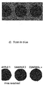
- the presence of fluorescent signal in one color shows the presence of only one sequence variant, corresponding to said color.
- the presence of signal in two colors in one probe confirms the presence of a mutation.
- each probe has to be designed in such a way that after adding one nucleotide the mutated variant and the wild type variant differ in the kind of complementary nucleotide successive in the 3' direction of the probe sequence.
- each probe has C12-AMINO modification and 18dT sequence preceding the proper oligonucleotide sequence.
- a matrix preparation control of sequence 5'-C12-AMINO-(dT) 18 -ACG TAC GTA CGT ACG TAC GTA CGT-Cy5-3' is deposited on every submatrix.
- DNA fragments containing the analyzed regions of BRCA1 and BRCA2 genes are amplified by PCR starters in 8 multiplex reactions (A-H) - table 3: Table 3 gene exon name sequence PCR reaction Concentration nM BRCA1 2 1020014 GACGTTGTCATTAGTTCTTTGG A 600 1020029 GGTCAATTCTGTTCATTTGC 600 5 1050017 CTCTTAAGGGCAGTTGTGAG B 500 1050023 ATGGTTTTATAGGAACGCTATG 500 8 1080012 TGTTAGCTGACTGATGATGGT C 700 1080026 AACCCAGCAATTATTATTAAATAC 700 11 1110012 GATTTCCACCTCCAAGGTGTATGA A 600 1110024 CATGGCTCCACATGCAAG 600 1110031 CAGCTGAGAGGCATCCA B 800 1110043 GGATTCTCTGAGCATGGCA 800 1110052 GATCCCCTGTGTGAGAA C 400 1110069 TTACTCTCTACTGATTTGGAGTGA 400 1110073 AGTGATCCTCATGAGG
- Reactions are performed under the following conditions: PCR reaction volume: 50 ⁇ l, 1x PCR buffer, MgCl 2 2.5 mM, dNTPs 250 ⁇ M, BSA fraction V 0.16 mg/ml, Glycerol (10% w/v), 5U TaqPol, starters see table 3, and 10 ⁇ l DNA(1 ng/ ⁇ l), in multiplex reactions of table 3.
- Reaction temperature profile initial denaturation 95°C - 3 min., then 35 cycles: denaturation 94°C - 1 min., annealing 55°C - 1 min., elongation 72°C - 1 min., final elongation 60°C - 15 min.
- Reactions are carried out under the following conditions: PCR reaction volume: 50 ⁇ l, 1x PCR buffer, MgCl 2 2,5 mM, dNTPs 250 ⁇ M, BSA fraction V 0,16 mg/ml, glycerol (10% w/v), 5U TaqPol, starters cf. table 4, and 10 ⁇ l DNA (1 ng/ ⁇ l), in multiplex reactions of table 4.
- Reactions temperature profile initial denaturation 95°C - 3 min., followed by 35 cycles: denaturation 94°C - 1 min., annealing 55°C - 1 min., elongation 72°C - 1 min., final elongation 60°C - 15 min.
- DNA for examination can be obtained according to many available protocols, according to user's preferences.
- Each analysis involves amplification of 4 samples:: a positive control containing DNA of known genotype, a negative control containing water instead of DNA solution, and two examined samples, which fulfills sample double analysis criteria for a reliable result.
- reaction products of the individual multiplex reactions for the same sample are mixed together: in the case of mutation analysis in BRCA1 and BRCA2 genes, products of 8 multiplex PCR reactions are mixed together for each sample, in the case of CFTR gene, products of 4 multiplex PCR reactions are mixed together.
- the mixed amplification products are subjected to purification from starters, ions, nucleotides and polymerase by a method which guarantees yield of at least 20 ⁇ l of purified product.
- Reaction on the matrix is prepared in 4 test tubes (negative control, positive control, examined sample 1, examined sample 2) in a volume of 100 ⁇ l, said volume containing: 50 mM Tris-HCl, ph 9,5, 3 mM MgCl 2 , 0,75 ⁇ m 7-Propargylamino-7-deaza-2',3'-dideoxyadenosine-5'thiphosphate-6-FAM, 5-Propargylamino-2',3'-dideoxycytidine+5'thiphosphate-5/6-TAMRA, 7-Propargylamino-7-deaza-2'3'dideoxyguanosine-5'triphosphate-6-ROX, 5-Propargylamino-2',3'-dideoxyuridine-5'triphosphate-6-JOE, 5U thermostable DNA polymerase adding dideoxynucleotides, and 20 ⁇ l of purified PCR product.
- Reaction mixtures are placed in proper matrix fields and sealed with foil.
- the matrix is placed in a thermocycler for amplification on a microscope slide and amplification is run under the following conditions: 35 cycles: denaturation 94°C - 1 min., annealing/elongation 50°C - 1 min., cover temperature 50°C.
- the sealing foil is removed and the matrix is placed in a wash station.
- the matrices are washed 2x 2 min. in deionized water and then dried by centrifugation in a minicentrifuge for matrix centrifugation.
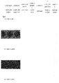
- the matrix is analyzed on a multicolor scanner. 4 scans are conducted under the following conditions:
- Oligonucleotides for matrix preparation were purchased from GENOMED S.A. Synthesis was carried out in 0.2 ⁇ mol scale, HPLC purified. Sequences of the individual probes are shown in table 5. Oligonucleotides were dissolved in water in an appropriate water volume for obtaining 200 ⁇ M concentration. Table 5. Nucleotide sequence of matrix probes for the detection of CFTR gene mutations.
- Matrices were prepared on slides with SUPEREPOXY surface. Before taking out of the box, the case and the box containing slides were cleaned with Microarray Cleanroom Wipes and a 95% ethanol solution and with air from a Microarray Air Jet compressor. Slides were unpacked in a class II laminar air flow chamber and a "mask" with dimensions of 76 x 26 mm, cut out from self-adhesive PVC foil of 0.35 mm thickness with a laser plotter, was applied to the active surface. In every fragment of the foil a circle of 14 mm diameter was cut out, 2,5 mm from the edge of the slide and at regular distances of 4 mm (from the edges of circles).
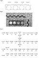
- oligonucleotide solutions in buffer were prepared in a 96-well plate by adding 25 ⁇ l of 200 ⁇ M oligonucleotide solution and 25 ⁇ l of 2x Micro Spotting Solution Plus (Arraylt) solution. Oligonucleotide solutions were mixed by 10x pipetting with 40 ⁇ l volume and the plate was centrifuged in a centrifuge for plate centrifugation (2 min., 4000 rpm). Matrices were printed with contact method. Each submatrix was printed in the same probe layout. Said probe layout is shown in fig. 2 .
- the printed matrices were placed in a climatic chamber with relative humidity ⁇ 30%, at room temperature ( ⁇ 25°C) for 12 hours, after which the matrices were incubated 1 hour in 80°C in a laboratory incubator.
- To wash out unbound DNA matrices were washed twice for 2 min. at room temperature (22-25°C) in 500 ml of 0,1% SDS solution and once in 500 ml of deionized water with intense mixing in a High Throughput Wash Station.
- Matrices were dried by 1 min. centrifugation, 500xg in a Microarray High Speed Centrifuge.
- To block background fluorescence the matrices were incubated for 60 min. at 60°C in 500 ml Blocklt Buffer and dried by 1 min. centrifugation, 500xg in a Microarray High Speed Centrifuge.
- the examined material was a dried blood specimen on a FTA card for proficiency testing as part of "Newborn Screening Quality Assurance Program", Cystic Fibrosis DNA Mutation Detection Proficiency Testing (CFDNAPT), PT Quarter 3 - 2016, denoted as: 316C1 and 316C2.
- Blood dried on a FTA card denoted as EXDNAPC was used as positive control, which was verified in the course of a test with DNA sequencing method to contain no CFTR gene mutations.
- Two fragments were cut out from the FTA card containing the blood specimen with a chisel of 3 mm diameter to two test tubes, and denoted as "Control +".
- DNA was isolated on a QIAsymphony (QIAGEN) automatic workstation using a QIAsymphony DNA Investigator Kit (Qiagen) reagent kit, according to the manufacturer instructions. Isolation was performed in presence of the negative control, containing all reagent components without the addition of biological material, denoted containing no DNA, denoted "Control -”.
- DNA concentration was assayed with fluorymetric method with a Qubit 2.0 (Life Technologies) apparatus, using a Qubit ®dsDNA BR (Life Technologies) kit, according to the manufacturer instructions.
- PCR amplification was carried out in 4 multiplex PCR reactions in 50 ⁇ l volume in PCR tubes of 200 ⁇ l volume, said tubes denoted as A, B, C, D, containing: 1x PCR buffer, MgCl 2 2,5 mM, dNTPs 250 ⁇ M (A&A Biotechnology), BSA fraction V 0,16 mg/ml (Sigma), glycerol (10% w/v), 5U TaqPol (A&A Biotechnology), starters as in table 6, and 10 ⁇ l DNA, in multiplex reactions of table 6; PCR reaction temperature profile: initial denaturation 95°C - 3 min., followed by 35 cycles: denaturation 94°C - 1 min., annealing 55°C - 1 min., elongation 72°C - 1 min., final elongation 60°C - 15 min.
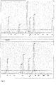
- PCR products were assessed for yield by capillary electrophoresis with a QIAxcelAdvanced (QIAGEN) analyzer and a QIAxcel DNA High Resolution Kit, and markers: QX Intensity Calibration Marker and QX Alignment Marker, according to manufacturer instructions.
- Amplification yield was assessed based on the positive control. The condition to continue the examination was to achieve the examined samples amplification yield at least at the level of the positive control yield.
- Amplification products of multiplex reactions A, B, C, D, control -, control +, sample 1 and sample 2 were mixed in one tube, obtaining around 200 ⁇ l of PCR amplification products of each sample.
- Amplification products were purified from starters and nucleotides with a precipitation method on Clean-up columns (A&A Biotechnology), according to manufacturer instructions. Purified products were collected from the column in volume of 50 ⁇ l.
- Matrix minisequencing mixtures were prepared in 4 tubes (negative control, positive control, examined sample 1, examined sample 2) in 70 ⁇ l volume comprising: 50 mM Tris-HCl, pH 9,5, 3 mM MgCl 2 , 0,75 ⁇ M 7-Propargylamino-7-deaza-2',3'-dideoxyadenosine-5'thiphosphate-6-FAM, 5-Propargylamino-2',3'-dideoxycytidine+5'thiphosphate-5/6-TAMRA, 7-Propargylamino-7-deaza-2'3'dideoxyguanosine-5'triphosphate-6-ROX, 5-Propargylamino-2',3'-dideoxyuridine-5'triphosphate-6-JOE, 5U thermostable DNA polymerase adding dideoxynucleotides, and 20 ⁇ l of purified PCR product.
- Reaction mixtures were placed in the submatrix fields and sealed with foil cut to matrix dimensions (76 x 26 mm).
- the matrix was placed in a thermocycler for amplification on a microscopic slide and amplification was run under the following conditions: 35 cycles: denaturation 94°C - 1 min., annealing/elongation 50°C - 1 min., cover temperature 50°C.
- the sealing foil was removed and the matrix was placed in a wash station and washed 2x 2 min. in deionized water and then dried by centrifugation in a minicentrifuge for matrix centrifugation.
- the matrix was analysed on a multicolor scanner GenePix 4300A (Molecular Devices) with 4 scans conducted under the following conditions:
- the presence of fluorescence signal in one color evidences presence of only one sequence variant, corresponding to the color.
- the presence of signal in two colors in one probe confirms presence of a mutation.
- the matrix was constructed for identification of mutations in germline and thus signal intensity should be similar; signal intensity variation for one probe is admissible at the level of +/- 20%.
- Samples were also analyzed by DNA IMAGING method with use of CFTR matrices, which resulted in: Table 7 Sample Detected mutations DNA sequencing method Detected mutations DNA IMAGING method 316C1 F508del (c.1521_1523delCTT) / F508del (c.1521_1523delCTT) F508del (c.1521_1523delCTT) / F508del / (c.1521_1523delCTT) 316C2 F508del (c.1521_1523delCTT) / WT F508del (c.1521_1523delCTT) / WT
- the obtained results are consistent for the DNA method and the reference (DNA sequencing) method.
- the obtained results are consistent with the sample characteristics.
- Oligonucleotides for matrix preparation were purchased from GENOMED S.A. Synthesis was carried out in 0.2 ⁇ mol scale, HPLC purified. Sequences of the individual probes are shown in table 8. Oligonucleotides were dissolved in water in an appropriate water volume for obtaining 200 ⁇ M concentration. Table 8.
- Matrices were prepared on slides with SUPEREPOXY surface. Before taking out of the box, the case and the box containing slides were cleaned with Microarray Cleanroom Wipes and a 95% ethanol solution and with air from a Microarray Air Jet compressor. Slides were unpacked in a class II laminar air flow chamber and a "mask" with dimensions of 76 x 26 mm, cut out with a laser plotter from self-adhesive PVC foil of 0.35 mm thickness, was applied to the active surface. In every fragment of the foil a circle of 14 mm diameter was cut out, 2.5 mm from the edge of the slide and at regular distances of 4 mm (from the edges of circles).
- oligonucleotide solutions in buffer were prepared in a 96-well plate by adding 25 ⁇ l of 200 ⁇ M oligonucleotide solution and 25 ⁇ l of 2x Micro Spotting Solution Plus (Arraylt) solution. Oligonucleotide solutions were mixed by 10x pipetting with 40 ⁇ l volume and the plate was centrifuged in a centrifuge for plate centrifugation (2 min., 4000 rpm). Matrices were printed with contact method. Each submatrix was printed in the same probe layout. Said probe layout is shown in fig. 2 .
- the printed matrices were placed in a climatic chamber with relative humidity ⁇ 30%, at room temperature ( ⁇ 25°C) for 12 hours after which the matrices were incubated 1 hour in 80°C in laboratory incubator.
- To wash out unbound DNA matrices were washed twice for 2 min. at room temperature (22-25°C) in 500 ml of 0,1% SDS solution and once in 500 ml of deionized water with intense mixing in a High Throughput Wash Station.
- Matrices were dried with 1 min. centrifugation, 500xg in a Microarray High Speed Centrifuge.
- the matrices were incubated for 60 min. at 60°C in 500 ml Blocklt Buffer and dried with 1 min. centrifugation, 500xg in a Microarray High Speed Centrifuge.
- Testing material comprised cheek mucosa swab samples protected on swab pearls for diagnostic examination in respect of BRCA1 and BRCA2 gene mutations, denoted as D832 and D994. Swabs of cheek mucosa were collected on two swab sticks for each sample. Blood dried on a FTA card denoted as EXDNAPC was used as positive control, confirmed in the course of examination by DNA sequencing method to contain no BRCA1 or BRCA2 gene mutations. Swab sticks were broken away to test tubes denoted: "Sample 1" and "Sample 2". 2 fragments of the EXDNAPC card were collected with a chisel of 3 mm diameter and denoted as "Control +".
- DNA was isolated on a QIAsymphony (QIAGEN) automatic workstation using a QIAsymphony DNA Investigator Kit (Qiagen) reagent kit according to the manufacturer instructions. Isolation was performed in the presence of the negative control, containing all reagent components without the addition of biological material, denoted as containing no DNA, denoted "Control -”.
- DNA concentration was assayed with fluorimetric method with a Qubit 2.0 (Life Technologies) apparatus, using a Qubit ®dsDNA BR (Life Technologies) kit, according to the manufacturer instructions.
- PCR amplification was carried out in 8 multiplex PCR reactions in 50 ⁇ l volume in PCR tubes of 200 ⁇ l volume, said tubes denoted as A, B, C, D, E, F, G, H containing: 1x PCR buffer, MgCl 2 2,5 mM, dNTPs 250 ⁇ M (A&A Biotechnology), BSA fraction V 0,16 mg/ml (Sigma), glycerol (10% w/v), 5U TaqPol (A&A Biotechnology), starters according to table and 10 ⁇ l DNA, in multiplex reactions of table 9; PCR reaction temperature profile: initial denaturation 95°C - 3 min., followed by 35 cycles: denaturation 94°C - 1 min., annealing 55°C - 1 min., elongation 72°C - 1 min., final elongation 60°C - 15 min.
- PCR products were assessed for yield with capillary electrophoresis with a QIAxcelAdvanced (QIAGEN) analyzer and a QIAxcel DNA High Resolution Kit, and markers: QX Intensity Calibration Marker and QX Alignment Marker, according to manufacturer instructions.
- Amplification yield was assessed based on positive control. The condition to continue the examination was to achieve the examined samples amplification yield at least at the level of the positive control yield.
- Amplification products of multiplex reactions A, B, C, D, E, F, G, H, control -, control +, sample 1 and sample 2 were mixed in one tube, obtaining around 200 ⁇ l of PCR amplification products of each sample.
- Amplification products were purified from starters and nucleotides with a precipitation method on Clean-up columns (A&A Biotechnology), according to manufacturer instructions. Purified products were collected from the column in volume of 50 ⁇ l
- Matrix minisequencing mixtures were prepared in 4 tubes (negative control, positive control, examined sample 1, examined sample 2) in 70 ⁇ l volume comprising: 50 mM Tris-HCl, pH 9,5, 3 mM MgCl 2 , 0,75 ⁇ M 7-Propargylamino-7-deaza-2',3'-dideoxyadenosine-5'thiphosphate-6-FAM, 5-Propargylamino-2',3'-dideoxycytidine+5'thiphosphate-5/6-TAMRA, 7-Propargylamino-7-deaza-2'3'dideoxyguanosine-5'triphosphate-6-ROX, 5-Propargylamino-2',3'-dideoxyuridine-5'triphosphate-6-JOE, 5U thermostable DNA polymerase (DynaSeq DNA Polymerase, Finnzyme) adding dideoxynucleotides, and 20 ⁇ l of purified PCR product.
- Reaction mixtures were placed in the submatrix fields and sealed with foil cut to matrix dimensions (76 x 26 mm).
- the matrix was placed in a thermocycler for amplification on microscope slides and amplification was run under the following conditions: 35 cycles: denaturation 94°C - 1 min., annealing/elongation 50°C - 1 min., cover temperature 50°C.
- the sealing foil was removed and the matrix was placed in a wash station and washed 2x 2 min. in deionized water and then dried by centrifugation in a minicentrifuge for matrix centrifugation.
- the matrix was analyzed on a multicolor scanner GenePix 4300A (Molecular Devices) with 4 scans conducted under the following conditions:
- the presence of fluorescence signal in one color evidences presence of only one sequence variant, corresponding to the color.
- the presence of signal in two colors in one probe confirms the presence of a mutation.
- the matrix was constructed for identification of mutations in germline and thus signal intensity should be similar; signal intensity variation for one probe is admissible at the level of +/- 20%.
- Samples D832 and D994 were analyzed for presence of mutations in coding part of BRCA1 and BRCA2 genes by enzymatic DNA sequencing with reaction termination by fluorescently labeled dideoxy terminators. The samples were also analyzed with DNA IMAGING using BRCA1/BRCA2 matrices, which resulted in: Table 10 Sample Detected mutations DNA sequencing method Detected mutations DNA IMAGING method D832 c.5266dupC/ WT c.5266dupC / WT D994 c.68_69deIAG / WT c.68_69delAG / WT
- the obtained results are consistent for the DNA method and the reference (DNA sequencing) method.
- the obtained results are consistent with the sample characteristics.
Landscapes
- Chemical & Material Sciences (AREA)
- Organic Chemistry (AREA)
- Life Sciences & Earth Sciences (AREA)
- Zoology (AREA)
- Wood Science & Technology (AREA)
- Proteomics, Peptides & Aminoacids (AREA)
- Health & Medical Sciences (AREA)
- Engineering & Computer Science (AREA)
- Microbiology (AREA)
- Immunology (AREA)
- Physics & Mathematics (AREA)
- Molecular Biology (AREA)
- Biotechnology (AREA)
- Biophysics (AREA)
- Analytical Chemistry (AREA)
- Biochemistry (AREA)
- Bioinformatics & Cheminformatics (AREA)
- General Engineering & Computer Science (AREA)
- General Health & Medical Sciences (AREA)
- Genetics & Genomics (AREA)
- Measuring Or Testing Involving Enzymes Or Micro-Organisms (AREA)
Priority Applications (1)
| Application Number | Priority Date | Filing Date | Title |
|---|---|---|---|
| EP17460047.8A EP3447154A1 (fr) | 2017-08-23 | 2017-08-23 | Procédé de détection de mutations, de polymorphismes et de séquences d'adn spécifiques sur des matrices d'adn à l'aide de techniques d'imagerie d'adn à utiliser dans le diagnostic médical et la génétique médico-légal |
Applications Claiming Priority (1)
| Application Number | Priority Date | Filing Date | Title |
|---|---|---|---|
| EP17460047.8A EP3447154A1 (fr) | 2017-08-23 | 2017-08-23 | Procédé de détection de mutations, de polymorphismes et de séquences d'adn spécifiques sur des matrices d'adn à l'aide de techniques d'imagerie d'adn à utiliser dans le diagnostic médical et la génétique médico-légal |
Publications (1)
| Publication Number | Publication Date |
|---|---|
| EP3447154A1 true EP3447154A1 (fr) | 2019-02-27 |
Family
ID=59955522
Family Applications (1)
| Application Number | Title | Priority Date | Filing Date |
|---|---|---|---|
| EP17460047.8A Withdrawn EP3447154A1 (fr) | 2017-08-23 | 2017-08-23 | Procédé de détection de mutations, de polymorphismes et de séquences d'adn spécifiques sur des matrices d'adn à l'aide de techniques d'imagerie d'adn à utiliser dans le diagnostic médical et la génétique médico-légal |
Country Status (1)
| Country | Link |
|---|---|
| EP (1) | EP3447154A1 (fr) |
Cited By (2)
| Publication number | Priority date | Publication date | Assignee | Title |
|---|---|---|---|---|
| CN111154874A (zh) * | 2020-01-16 | 2020-05-15 | 安徽省第二人民医院(安徽医学高等专科学校附属医院、安徽省职业病防治院) | 一种乳腺癌易感基因BRCA2位点g.32332272G>A突变体及其应用 |
| CN112831564A (zh) * | 2021-02-09 | 2021-05-25 | 南京医科大学 | 一种与原发性肺癌辅助诊断相关的遗传变异区域的基因组合及其应用 |
Citations (7)
| Publication number | Priority date | Publication date | Assignee | Title |
|---|---|---|---|---|
| JP2002507883A (ja) * | 1996-11-06 | 2002-03-12 | シークエノム・インコーポレーテツド | マススペクトロメトリーに基づくdna診断法 |
| US20030096236A1 (en) * | 1996-02-12 | 2003-05-22 | Oncormed Inc. | Determining common functional alleles in a population and uses therefore |
| WO2007088537A2 (fr) * | 2006-01-31 | 2007-08-09 | Medical Research Fund Of Tel Aviv Sourasky Medical Center | Procédés et trousses pour la détection précoce du cancer ou d'une prédisposition au cancer |
| WO2011126869A2 (fr) * | 2010-03-30 | 2011-10-13 | Trustees Of Boston University | Outils et procédé pour séquençage d'acide nucléique dépendant du dégrafage de nanopores |
| WO2013040429A1 (fr) * | 2011-09-14 | 2013-03-21 | Rana Therapeutics Inc. | Composés oligonucléotidiques multimères |
| WO2014070233A1 (fr) * | 2012-10-29 | 2014-05-08 | Boston Medical Center Corporation | Mutations de brca1 en tant que marqueurs prédictifs d'inhibiteurs de la topoisomérase i |
| EP2784165A1 (fr) * | 2013-03-29 | 2014-10-01 | Centrum Badan DNA Sp. z o.o. | Procédé pour la détection d'une prédisposition génétique au cancer de la prostate |
-
2017
- 2017-08-23 EP EP17460047.8A patent/EP3447154A1/fr not_active Withdrawn
Patent Citations (7)
| Publication number | Priority date | Publication date | Assignee | Title |
|---|---|---|---|---|
| US20030096236A1 (en) * | 1996-02-12 | 2003-05-22 | Oncormed Inc. | Determining common functional alleles in a population and uses therefore |
| JP2002507883A (ja) * | 1996-11-06 | 2002-03-12 | シークエノム・インコーポレーテツド | マススペクトロメトリーに基づくdna診断法 |
| WO2007088537A2 (fr) * | 2006-01-31 | 2007-08-09 | Medical Research Fund Of Tel Aviv Sourasky Medical Center | Procédés et trousses pour la détection précoce du cancer ou d'une prédisposition au cancer |
| WO2011126869A2 (fr) * | 2010-03-30 | 2011-10-13 | Trustees Of Boston University | Outils et procédé pour séquençage d'acide nucléique dépendant du dégrafage de nanopores |
| WO2013040429A1 (fr) * | 2011-09-14 | 2013-03-21 | Rana Therapeutics Inc. | Composés oligonucléotidiques multimères |
| WO2014070233A1 (fr) * | 2012-10-29 | 2014-05-08 | Boston Medical Center Corporation | Mutations de brca1 en tant que marqueurs prédictifs d'inhibiteurs de la topoisomérase i |
| EP2784165A1 (fr) * | 2013-03-29 | 2014-10-01 | Centrum Badan DNA Sp. z o.o. | Procédé pour la détection d'une prédisposition génétique au cancer de la prostate |
Non-Patent Citations (5)
| Title |
|---|
| INMACULADA DE JUAN ET AL: "High-resolution melting analysis for rapid screening of BRCA1 and BRCA2 Spanish mutations", BREAST CANCER RESEARCH AND TREATMENT., vol. 115, no. 2, 5 June 2008 (2008-06-05), US, pages 405 - 414, XP055571499, ISSN: 0167-6806, DOI: 10.1007/s10549-008-0073-7 * |
| LAN-SZU CHOU ET AL: "Complete Gene Scanning by Temperature Gradient Capillary Electrophoresis Using the Cystic Fibrosis Transmembrane Conductance Regulator Gene as a Model", THE JOURNAL OF MOLECULAR DIAGNOSTICS, vol. 7, no. 1, 1 February 2005 (2005-02-01), US, pages 111 - 120, XP055571508, ISSN: 1525-1578, DOI: 10.1016/S1525-1578(10)60016-5 * |
| LILJEDAHL ULRIKA ET AL: "Detecting imbalanced expression of SNP alleles by minisequencing on microarrays", BMC BIOTECHNOLOGY, BIOMED CENTRAL LTD, vol. 4, no. 1, 22 October 2004 (2004-10-22), pages 24, XP021005941, ISSN: 1472-6750, DOI: 10.1186/1472-6750-4-24 * |
| PODDER MOHUA ET AL: "Robust SNP genotyping by multiplex PCR and arrayed primer extension", BMC MEDICAL GENOMICS, BIOMED CENTRAL LTD, LONDON UK, vol. 1, no. 1, 31 January 2008 (2008-01-31), pages 5, XP021040918, ISSN: 1755-8794 * |
| TOST J ET AL: "GENOTYPING SINGLE NUCLEOTIDE POLYMORPHISMS BY MASS SPECTROMETRY", MASS SPECTROMETRY REVI, JOHN WILEY AND SONS, NEW YORK, NY, US, vol. 21, no. 6, 1 November 2002 (2002-11-01), pages 388 - 418, XP009019382, ISSN: 0277-7037, DOI: 10.1002/MAS.1009 * |
Cited By (3)
| Publication number | Priority date | Publication date | Assignee | Title |
|---|---|---|---|---|
| CN111154874A (zh) * | 2020-01-16 | 2020-05-15 | 安徽省第二人民医院(安徽医学高等专科学校附属医院、安徽省职业病防治院) | 一种乳腺癌易感基因BRCA2位点g.32332272G>A突变体及其应用 |
| CN112831564A (zh) * | 2021-02-09 | 2021-05-25 | 南京医科大学 | 一种与原发性肺癌辅助诊断相关的遗传变异区域的基因组合及其应用 |
| CN112831564B (zh) * | 2021-02-09 | 2023-01-03 | 南京医科大学 | 一种与原发性肺癌辅助诊断相关的遗传变异区域的基因组合及其应用 |
Similar Documents
| Publication | Publication Date | Title |
|---|---|---|
| JP4175884B2 (ja) | 複数の遺伝子座で複数のサンプルを遺伝子タイピングするマイクロアレイ方法 | |
| EP2007901B1 (fr) | Analyse rapide de genotypage et dispositif associe | |
| AU2002218740A1 (en) | Microarray method of genotyping multiple samples at multiple loci | |
| CZ293278B6 (cs) | Způsob přípravy komplexních DNA methylačních peptidových map | |
| CN105936940B (zh) | 检测耳聋基因的核酸序列及其应用 | |
| US10337066B2 (en) | Methods for PCR and HLA typing using unpurified samples | |
| US7049104B2 (en) | Genetic analysis method | |
| JPH06504427A (ja) | 多重核酸配列の存在または不存在の検出による識別および父性決定 | |
| KR20110094041A (ko) | 정상 안압 녹내장 질환 감수성 유전자 및 그 이용 | |
| EP3447154A1 (fr) | Procédé de détection de mutations, de polymorphismes et de séquences d'adn spécifiques sur des matrices d'adn à l'aide de techniques d'imagerie d'adn à utiliser dans le diagnostic médical et la génétique médico-légal | |
| BRPI0614994A2 (pt) | quantificaÇço da hibridizaÇço de suspensço de micro esferas e usos das mesmas | |
| Vijg et al. | Two‐dimensional gene scanning: Exploring human genetic variability | |
| KR20140000439A (ko) | 유전성 난청 관련 유전자의 돌연변이 유무를 감별하기 위한 올리고뉴클레오티드, dna칩, 진단 키트 및 감별 방법 | |
| US10294512B2 (en) | Method and apparatus for analyzing biomolecules by using oligonucleotide | |
| WO2008102924A1 (fr) | Microréseau pour la détection d'une mutation d'adn mitochondrial et méthode de diagnostic du diabète faisant appel audit microréseau | |
| JPH07508407A (ja) | 分子生物学的診断のための方法およびシステム | |
| US20090143245A1 (en) | Microarrays for genotyping and methods of use | |
| US7659064B2 (en) | PNA chip using zip-codes and fabrication method thereof | |
| RU2402771C2 (ru) | Способ скрининга сердечно-сосудистых заболеваний и биочип для осуществления этого способа | |
| KR100443569B1 (ko) | 길이다형성 dna 염기서열을 이용한 다중 증폭중합효소연쇄반응 조건의 최적화에 의한 개인식별 및친자감별 방법 | |
| RU2402612C2 (ru) | Способ скрининга новорожденных на моногенные заболевания и биочип для осуществления этого способа | |
| Zou et al. | DNA microarrays: applications, future trends, and the need for standardization | |
| KR101075392B1 (ko) | Fga 유전자로부터 유래된 단일염기다형을 포함하는 폴리뉴클레오티드, 이를 포함하는 마이크로어레이 및 진단키트, 이를 이용한 검출 방법 | |
| US20070099221A1 (en) | Method of analyzing nucleic acid | |
| Gluba et al. | Microarray technology in the study of genetic determinants of cardiovascular diseases |
Legal Events
| Date | Code | Title | Description |
|---|---|---|---|
| PUAI | Public reference made under article 153(3) epc to a published international application that has entered the european phase |
Free format text: ORIGINAL CODE: 0009012 |
|
| STAA | Information on the status of an ep patent application or granted ep patent |
Free format text: STATUS: REQUEST FOR EXAMINATION WAS MADE |
|
| 17P | Request for examination filed |
Effective date: 20180921 |
|
| AK | Designated contracting states |
Kind code of ref document: A1 Designated state(s): AL AT BE BG CH CY CZ DE DK EE ES FI FR GB GR HR HU IE IS IT LI LT LU LV MC MK MT NL NO PL PT RO RS SE SI SK SM TR |
|
| AX | Request for extension of the european patent |
Extension state: BA ME |
|
| STAA | Information on the status of an ep patent application or granted ep patent |
Free format text: STATUS: EXAMINATION IS IN PROGRESS |
|
| 17Q | First examination report despatched |
Effective date: 20190327 |
|
| RAP1 | Party data changed (applicant data changed or rights of an application transferred) |
Owner name: INSTYTUT GENETYKI SADOWEJ SPOLKA Z OGRANICZONA ODPOWIEDZIALNOSCIA |
|
| GRAP | Despatch of communication of intention to grant a patent |
Free format text: ORIGINAL CODE: EPIDOSNIGR1 |
|
| STAA | Information on the status of an ep patent application or granted ep patent |
Free format text: STATUS: GRANT OF PATENT IS INTENDED |
|
| INTG | Intention to grant announced |
Effective date: 20231110 |
|
| RIN1 | Information on inventor provided before grant (corrected) |
Inventor name: CZARNY, JAKUB |
|
| STAA | Information on the status of an ep patent application or granted ep patent |
Free format text: STATUS: THE APPLICATION IS DEEMED TO BE WITHDRAWN |
|
| 18D | Application deemed to be withdrawn |
Effective date: 20240312 |