EP3456246B1 - Classificateur binaire en cascade permettant d'identifier des rythmes dans un signal d'électrocardiogramme (ecg) à fil unique - Google Patents
Classificateur binaire en cascade permettant d'identifier des rythmes dans un signal d'électrocardiogramme (ecg) à fil unique Download PDFInfo
- Publication number
- EP3456246B1 EP3456246B1 EP18153347.2A EP18153347A EP3456246B1 EP 3456246 B1 EP3456246 B1 EP 3456246B1 EP 18153347 A EP18153347 A EP 18153347A EP 3456246 B1 EP3456246 B1 EP 3456246B1
- Authority
- EP
- European Patent Office
- Prior art keywords
- features
- rhythms
- signal
- classifier
- ecg
- Prior art date
- Legal status (The legal status is an assumption and is not a legal conclusion. Google has not performed a legal analysis and makes no representation as to the accuracy of the status listed.)
- Active
Links
Images


Classifications
-
- G—PHYSICS
- G06—COMPUTING OR CALCULATING; COUNTING
- G06F—ELECTRIC DIGITAL DATA PROCESSING
- G06F18/00—Pattern recognition
- G06F18/20—Analysing
- G06F18/21—Design or setup of recognition systems or techniques; Extraction of features in feature space; Blind source separation
- G06F18/214—Generating training patterns; Bootstrap methods, e.g. bagging or boosting
- G06F18/2148—Generating training patterns; Bootstrap methods, e.g. bagging or boosting characterised by the process organisation or structure, e.g. boosting cascade
-
- A—HUMAN NECESSITIES
- A61—MEDICAL OR VETERINARY SCIENCE; HYGIENE
- A61B—DIAGNOSIS; SURGERY; IDENTIFICATION
- A61B5/00—Measuring for diagnostic purposes; Identification of persons
- A61B5/24—Detecting, measuring or recording bioelectric or biomagnetic signals of the body or parts thereof
- A61B5/316—Modalities, i.e. specific diagnostic methods
- A61B5/318—Heart-related electrical modalities, e.g. electrocardiography [ECG]
- A61B5/346—Analysis of electrocardiograms
- A61B5/349—Detecting specific parameters of the electrocardiograph cycle
-
- A—HUMAN NECESSITIES
- A61—MEDICAL OR VETERINARY SCIENCE; HYGIENE
- A61B—DIAGNOSIS; SURGERY; IDENTIFICATION
- A61B5/00—Measuring for diagnostic purposes; Identification of persons
- A61B5/24—Detecting, measuring or recording bioelectric or biomagnetic signals of the body or parts thereof
- A61B5/316—Modalities, i.e. specific diagnostic methods
- A61B5/318—Heart-related electrical modalities, e.g. electrocardiography [ECG]
- A61B5/346—Analysis of electrocardiograms
- A61B5/347—Detecting the frequency distribution of signals
-
- A—HUMAN NECESSITIES
- A61—MEDICAL OR VETERINARY SCIENCE; HYGIENE
- A61B—DIAGNOSIS; SURGERY; IDENTIFICATION
- A61B5/00—Measuring for diagnostic purposes; Identification of persons
- A61B5/24—Detecting, measuring or recording bioelectric or biomagnetic signals of the body or parts thereof
- A61B5/316—Modalities, i.e. specific diagnostic methods
- A61B5/318—Heart-related electrical modalities, e.g. electrocardiography [ECG]
- A61B5/346—Analysis of electrocardiograms
- A61B5/349—Detecting specific parameters of the electrocardiograph cycle
- A61B5/352—Detecting R peaks, e.g. for synchronising diagnostic apparatus; Estimating R-R interval
-
- A—HUMAN NECESSITIES
- A61—MEDICAL OR VETERINARY SCIENCE; HYGIENE
- A61B—DIAGNOSIS; SURGERY; IDENTIFICATION
- A61B5/00—Measuring for diagnostic purposes; Identification of persons
- A61B5/24—Detecting, measuring or recording bioelectric or biomagnetic signals of the body or parts thereof
- A61B5/316—Modalities, i.e. specific diagnostic methods
- A61B5/318—Heart-related electrical modalities, e.g. electrocardiography [ECG]
- A61B5/346—Analysis of electrocardiograms
- A61B5/349—Detecting specific parameters of the electrocardiograph cycle
- A61B5/361—Detecting fibrillation
-
- A—HUMAN NECESSITIES
- A61—MEDICAL OR VETERINARY SCIENCE; HYGIENE
- A61B—DIAGNOSIS; SURGERY; IDENTIFICATION
- A61B5/00—Measuring for diagnostic purposes; Identification of persons
- A61B5/24—Detecting, measuring or recording bioelectric or biomagnetic signals of the body or parts thereof
- A61B5/316—Modalities, i.e. specific diagnostic methods
- A61B5/318—Heart-related electrical modalities, e.g. electrocardiography [ECG]
- A61B5/346—Analysis of electrocardiograms
- A61B5/349—Detecting specific parameters of the electrocardiograph cycle
- A61B5/366—Detecting abnormal QRS complex, e.g. widening
-
- A—HUMAN NECESSITIES
- A61—MEDICAL OR VETERINARY SCIENCE; HYGIENE
- A61B—DIAGNOSIS; SURGERY; IDENTIFICATION
- A61B5/00—Measuring for diagnostic purposes; Identification of persons
- A61B5/72—Signal processing specially adapted for physiological signals or for diagnostic purposes
- A61B5/7203—Signal processing specially adapted for physiological signals or for diagnostic purposes for noise prevention, reduction or removal
-
- A—HUMAN NECESSITIES
- A61—MEDICAL OR VETERINARY SCIENCE; HYGIENE
- A61B—DIAGNOSIS; SURGERY; IDENTIFICATION
- A61B5/00—Measuring for diagnostic purposes; Identification of persons
- A61B5/72—Signal processing specially adapted for physiological signals or for diagnostic purposes
- A61B5/7235—Details of waveform analysis
- A61B5/7264—Classification of physiological signals or data, e.g. using neural networks, statistical classifiers, expert systems or fuzzy systems
-
- G—PHYSICS
- G06—COMPUTING OR CALCULATING; COUNTING
- G06F—ELECTRIC DIGITAL DATA PROCESSING
- G06F18/00—Pattern recognition
- G06F18/20—Analysing
- G06F18/24—Classification techniques
- G06F18/243—Classification techniques relating to the number of classes
- G06F18/2431—Multiple classes
-
- G—PHYSICS
- G16—INFORMATION AND COMMUNICATION TECHNOLOGY [ICT] SPECIALLY ADAPTED FOR SPECIFIC APPLICATION FIELDS
- G16H—HEALTHCARE INFORMATICS, i.e. INFORMATION AND COMMUNICATION TECHNOLOGY [ICT] SPECIALLY ADAPTED FOR THE HANDLING OR PROCESSING OF MEDICAL OR HEALTHCARE DATA
- G16H50/00—ICT specially adapted for medical diagnosis, medical simulation or medical data mining; ICT specially adapted for detecting, monitoring or modelling epidemics or pandemics
- G16H50/20—ICT specially adapted for medical diagnosis, medical simulation or medical data mining; ICT specially adapted for detecting, monitoring or modelling epidemics or pandemics for computer-aided diagnosis, e.g. based on medical expert systems
Definitions
- the disclosure herein generally relate to electrocardiogram (ECG) signal analysis, and, more particularly, to systems and methods for identifying rhythms in a single-lead electrocardiogram (ECG) signal using a cascaded binary classifier.
- ECG electrocardiogram
- Atrial Fibrillation is a common type of heart disease that leads to stroke, heart failure or other complications. Millions of people get affected by AF every year and the prevalence of the disease is likely to increase. Noninvasive detection of AF is a popular area of research for quite a long time. Irregularities in heart beat is considered to be the most common symptom of AF and can be traced in an ECG. However, being an episodic event an accurate detection of AF is not always trivial.
- Conventional AF detectors have been mostly of atrial activity analysis based or ventricular response analysis-based methods. The absence of P waves or the presence of f waves in the TQ interval are searched in atrial activity analysis-based AF detectors.
- time, frequency and morphological features are extracted from RR intervals to identity the heart beat irregularity in ventricular response analysis based methods.
- the traditional methods have certain limitations regarding real time deployments, firstly, most of them are validated on clinically accepted multiples of lead ECG signals, recorded for a relatively longer duration. Secondly, algorithms are mostly applied on carefully selected clean data. However, in practical scenario, ECG signals are often noisy in nature. Thirdly, size of the test dataset may often not be adequate for making a conclusion (or decision) thereby result in misclassification. Lastly, most traditional or conventional methods perform binary classification between AF and normal recordings only.
- non-AF abnormal rhythms e.g., tachycardia, bradycardia, arrhythmia etc.
- these non-AF abnormal rhythms are not considered for classification. Even if considered them in the dataset makes the classification task more challenging.
- Embodiments of the present disclosure present technological improvements as solutions to one or more of the above-mentioned technical problems recognized by the inventors in conventional systems. For example, in one aspect, a processor implemented method for identifying rhythms in a single-lead electrocardiogram (ECG) signal using a cascaded binary classifier according to claim 1 is provided.
- ECG electrocardiogram
- the step of applying in real-time, via the one or more hardware processors, a spectrogram based noisy data removal technique on the acquired single-lead ECG signal may comprise: dividing in real-time, the acquired single-lead ECG signal into a plurality of windows; computing in real-time, a spectrogram of each of the plurality of windows; performing in real-time, a comparison of the computed spectrogram of each of the plurality of windows with a dynamically computed threshold, wherein the dynamically computed threshold is based on a signal to noise ratio (SNR); determining in real-time, noise in at least a subset of the plurality of windows based on the comparison; and extracting the clean ECG signal based on the determined noise in the at least a subset of the plurality of windows.
- SNR signal to noise ratio
- the noise is determined in the at least a subset of the plurality of windows when each window from the subset have power that is greater than a threshold power.
- the clean ECG signal is extracted by applying in real-time, the spectrogram based noisy data removal technique on the acquired single-lead ECG signal by: dividing in real-time, the acquired single-lead ECG signal into a plurality of windows; computing in real-time, a spectrogram of each of the plurality of windows; performing in real-time, a comparison of the computed spectrogram of each of the plurality of windows with a dynamically computed threshold, wherein the dynamically computed threshold is based on a signal to noise ratio (SNR); determining in real-time, noise in at least a subset of the plurality of windows based on the comparison; and extracting in real-time, the clean ECG signal based on the determined noise in the at least a subset of the plurality of windows.
- SNR signal to noise ratio
- noise is determined in the at least a subset of the plurality of windows when each window from the subset have power that is greater than a threshold power.
- the step of applying, via the one or more hardware processors, a spectrogram based noisy data removal technique on the acquired single-lead ECG signal may comprise: dividing the acquired single-lead ECG signal into a plurality of windows; computing a spectrogram of each of the plurality of windows; performing a comparison of the computed spectrogram of each of the plurality of windows with a dynamically computed threshold, wherein the dynamically computed threshold is based on a signal to noise ratio (SNR); determining noise in at least a subset of the plurality of windows based on the comparison; and extracting the clean ECG signal based on the determined noise in the at least a subset of the plurality of windows.
- SNR signal to noise ratio
- the noise is determined in the at least a subset of the plurality of windows when each window from the subset have power that is greater than a threshold power.
- Embodiments of the present disclosure provide systems and methods for a robust and more efficient technique for classifying normal, AF, other abnormal rhythms and noisy ECG recordings. More particularly, embodiments of the present disclosure provide systems and methods for identifying rhythms in a single-lead electrocardiogram (ECG) signal using a cascaded binary classifier. Embodiments of the present disclosure provide experiment results of a diverse ECG dataset (provided in Physionet challenge 2017) that was used for internal performance evaluation and creating the training models.
- ECG electrocardiogram
- the embodiments of the present disclosure identify rhythms in a single-lead electrocardiogram (ECG) signal by implementing 1) a multi-layer cascaded binary classifier instead of a single multiclass classifier, 2) front end noise removal and 3) optimum feature selection at each layer of classification from a pool of features.
- ECG electrocardiogram
- FIGS. 1 through 5 where similar reference characters denote corresponding features consistently throughout the figures, there are shown preferred embodiments and these embodiments are described in the context of the following exemplary system and/or method.
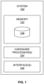
- FIG. 1 illustrates an exemplary block diagram of a system 100 for identifying rhythms in a single-lead electrocardiogram (ECG) signal using a cascaded binary classifier in accordance with an embodiment of the present disclosure.
- the system 100 includes one or more processors 104, communication interface device(s) or input/output (I/O) interface(s) 106, and one or more data storage devices or memory 102 operatively coupled to the one or more processors 104.
- the one or more processors 104 may be one or more software processing modules and/or hardware processors.
- the I/O interface device(s) 106 can include a variety of software and hardware interfaces, for example, a web interface, a graphical user interface, and the like and can facilitate multiple communications within a wide variety of networks N/W and protocol types, including wired networks, for example, LAN, cable, etc., and wireless networks, such as WLAN, cellular, or satellite.
- the I/O interface device(s) can include one or more ports for connecting a number of devices to one another or to another server.
- the memory 102 may store the classifiers (e.g., cascaded binary classifier(s)), one or more techniques, for example, spectrogram based noisy data removal technique, feature extraction technique, optimum feature selection technique(s) such as minimum redundancy maximum relevancy (mRMR) technique, and/or maximal Information Coefficient (MIC) technique and the like, which are executed by the one or more hardware processors 104 (or by the system 100) to perform the methodology described herein.
- the classifiers e.g., cascaded binary classifier(s)
- one or more techniques for example, spectrogram based noisy data removal technique, feature extraction technique, optimum feature selection technique(s) such as minimum redundancy maximum relevancy (mRMR) technique, and/or maximal Information Coefficient (MIC) technique and the like, which are executed by the one or more hardware processors 104 (or by the system 100) to perform the methodology described herein.
- mRMR minimum redundancy maximum relevancy
- MIC maximal Information Coefficient
- FIG. 2 illustrates an exemplary flow diagram of a method for identifying rhythms in the single-lead electrocardiogram (ECG) signal using the cascaded binary classifier executed by the system 100 of FIG. 1 in accordance with an embodiment of the present disclosure.
- the system(s) 100 comprises one or more data storage devices or the memory 102 operatively coupled to the one or more hardware processors 104 and is configured to store instructions for execution of steps of the method by the one or more processors 104.
- the system 100 stores values/information (and/or parameters, features, rhythms, and the like) associated with ECG signal(s).
- ECG signals recorded using single-lead, non-medical grade equipment(s) are inherently noisy in nature. Body movement of the user during recording, voltage fluctuation of the sensor device or improper contact between subject body and the sensor electrodes can heavily corrupt the signals. A low frequency component is also present due breathing. Locating and discarding the noisy portions inside a signal is considered a prerequisite before feature extraction and classification.
- the one or more hardware processors 104 apply in real-time, a spectrogram based noisy data removal technique on the acquired single-lead ECG signal to obtain a clean ECG signal.
- the threshold that is dynamically computed (or computed in real-time) is based on a signal to noise ratio (SNR).
- SNR is measured for all the windows in a recording (or an ECG signal). If the difference between maximum and minimum SNR values is 'n' times (e.g., say 3 times) the minimum value, x % of the maximum SNR is measured as threshold (e.g., say 75% of the maximum SNR is measured as threshold - which is also referred as dynamically computed threshold) for discarding the noisy windows. In other words, 75% of the maximum SNR may be referred as dynamically computed threshold.
- noise is determined in the at least a subset of the plurality of windows when each window from the subset have power (e.g., high spectral power) that is greater than a threshold power above 'X' Hertz (e.g., 50 Hz).
- a threshold power above 'X' Hertz e.g. 50 Hz.
- each window from the subset comprising portion of the ECG signal e.g., the acquired single-lead ECG signal
- threshold power also referred as threshold 'P x '
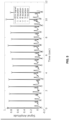
- FIG. 4 illustrates a graphical representation of a spectrogram based noise removal in accordance with an example embodiment of the present disclosure.
- the clean signal is passed through a high pass filter (cut-off frequency 0.5 Hz) to remove the baseline movement of the signal. If more than X% (e.g., say 80%) of a certain waveform (e.g., the single-lead ECG signal) is discarded in this process, it is marked as noisy.
- X% e.g., say 80%
- a certain waveform e.g., the single-lead ECG signal
- Identifying the differentiating feature set is considered the most important task of any classification problem.
- An accurate detection P, Q, R, S, T points are the primary requirement for an accurate feature extraction.
- the system 100 implements one or more techniques (e.g., a modified Pan-Tompkins technique) to identify QRS complex and R peaks.
- the QRS complex is a name for the combination of three of the graphical deflections seen on a typical electrocardiogram (EKG or ECG). It is usually the central and most visually obvious part of the tracing. It corresponds to the depolarization of the right and left ventricles of the human heart.
- P, Q, S and T points are located, taking R peaks as reference points (as shown in FIG. 5 ). More particularly, FIG.
- the Morphological ECG features are derived from the PQRST points detected in an ECG waveform as depicted in FIG. 5 .
- these features may comprise, but are not limited to, median, range and variance of the corrected QT interval (QTc), QR and QRS widths, slopes of QR, RS and ST intervals, depth of the Q and S points with respect to R, amplitude difference of the TR wave; ratio of the number of P waves to the number of R waves and distance of the ST segment crossing from the S point among others.
- QTc corrected QT interval
- QR and QRS widths slopes of QR, RS and ST intervals
- depth of the Q and S points with respect to R amplitude difference of the TR wave
- ratio of the number of P waves to the number of R waves and distance of the ST segment crossing from the S point among others are widely used by clinicians for identifying cardiac abnormalities. It is to be noted that some of these features are indicative of AF while others are for detecting the other abnormal rhythms.
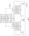
- FIG. 3 illustrates an exemplary architecture of the system 100 of FIG. 1 in accordance with an example embodiment of the present disclosure.
- Irregular RR intervals is a very common symptom present in AF patients. There are several metrics to identify the same. Several measurements have been proposed such as for example, AFEvidence, Original Count, Irregularity Evidence, Pace Count, Density Evidence, Anisotropy Evidence, AFEvidence from Lorentz plot of RR intervals. Certain features are derived from the inter beat intervals using Poincare plots also reported commending accuracy in identifying AF. Other features include approximate and sample entropy based features for AF detection as well as coefficient of variation of RR and delta RR intervals.
- HRV related features are also incorporated in the proposed analysis.
- pNNx number of NN intervals above x, normalized by duration of recording, where x lies between 20 and 500 ms
- SDNN standard deviation of NN intervals
- SDSD standard deviation of successive differences
- RMSSD normalized root mean square of successive differences
- Embodiments of the present disclosure have also explored certain frequency domain features used in biomedical and other applications.
- Raw time signal is broken into small windows of 'n' seconds duration (e.g., 2 seconds duration) having Y% (e.g., 50%) overlapping using hamming window.
- Frequency analysis is performed by computing Short Time Fourier Transform (STFT) of each window.
- Extracted features comprise but are not limited to, mean spectral centroid, spectral roll-off, spectral flux along with normalized spectral power between 0-10 Hz and 10-20 Hz across all windows in a measurement.
- Statistical features comprise but are not limited to, mean, median, variance, range, kurtosis and skewness of RR intervals and the probability density estimate (PDE) of the RR intervals and the delta RR intervals. Additionally, the number of peaks on the PDE of the RR and delta RR intervals along with the variation of energy in between the RR peaks were also used as features.
- the Shannon, Tsallis and Renyi entropy, Linear Predictive Coefficients (LPC) of the raw time series data were also used as extracted features by the embodiments of the present disclosure for identifying rhythms in the clean ECG signal.
- Embodiments of the present disclosure attempt to extract some of the major features to detect arrhythmia or other non-AF related abnormalities present in an ECG signal.
- the heart rate was estimated using an adaptive frequency tracking algorithm (AMM) to derive the features as mean of RR interval, decrease of HR, max SPI index, average HR, abnormal HR etc.
- AMM adaptive frequency tracking algorithm
- Embodiments of the present disclosure use domain dependent time and frequency features along with certain statistical features that exploit the rise and fall in the morphology of the ECG signal for improved noise detection. These features discriminate well between the regularities of the clean ECG signal versus the randomness in a noisy waveform.
- the one or more hardware processors 104 select, using an optimum feature selection technique, one or more optimum features from the one or more extracted features.
- Feature selection often improves classification accuracy by removing the noisy (irrelevant) features and also reduces the computation time by removing redundant features.
- a separate feature set is used in each of the three classifiers of the proposed methodology, efficiently chosen using one or more statistical feature selection technique(s) (also referred hereinafter optimum feature selection technique(s)), for example, but are not limited to, a Maximal Information Coefficient (MIC) technique and minimum Redundancy Maximum Relevance (mRMR) technique(s) and are finalized during training phase.
- MIC Maximal Information Coefficient
- mRMR Minimum Redundancy Maximum Relevance
- the one or more hardware processors 104 identifying based on the selected one or more optimum features, using a binary cascade classifier, at least one of one or more normal rhythms (Normal), a first set of abnormal rhythms (others), and a second set of abnormal rhythms (AF and noisy) in at least one of the single-lead electrocardiogram (ECG) signal, and the clean ECG signal.
- a first binary classifier of the cascaded binary classifier (as depicted in Layer 1) outputs or identifies 'A' set of rhythms (e.g., Normal and Others), and 'B' set of rhythms (e.g., AF and noisy).
- 'A' set of rhythms may be referred as one or more normal rhythms and other rhythms, and wherein 'B' set of rhythms may be referred as AF rhythms and noisy.
- the system 100 classifies an unknown short single-lead ECG signal into one of the four classes including normal, AF, other noisy rhythms and noisy recordings.
- the major problem of the task is that there is not a single feature exists that can separate four classes at a single shot (or at a single instance). For example, irregular HRV is a common differentiator between normal and AF. However, other rhythms also have the same properties, which introduces ambiguity in a classifier.
- the embodiments of the present disclosure and the system 100 comprise a cascaded binary classifier that when executed by the one or more hardware processors 104 identifies 'A' set of rhythms may be referred as one or more normal rhythms and other rhythms, and wherein 'B' set of rhythms may be referred as AF rhythms and noisy in the clean ECG signal.
- the cascaded binary classifier implemented and executed in the system 100 is a three level classifier, connected in two acceded layer as depicted in FIG. 3 .
- a binary classification is done at each layer.
- the corresponding discriminative features are selected at each level using one or more statistical feature extraction technique(s) as described above.
- AF and noisy rhythms contain high frequency noise components.
- normal and AF shows similar frequency feature patterns.
- features like spectral centroid, spectral roll off spectral flux etc. can very well separate them into two classes "normal + others" and "AF + noisy”.
- normal ECG signal has a regular pattern in RR intervals and heart rate value lies within a certain limit.
- HRV features are not (or may not be) very stable.
- These kind of features are selected using the optimum feature selection technique(s) when passed through the three level classifier as depicted in FIG. 3 .
- P wave are often found to be missing, that is not found in noisy recordings, moreover noisy recordings have high frequency spectral components which are not present in AF.
- These kind of features are selected at the third level of classifier (e.g., classifier 3 as depicted in FIG. 3 ), for classification.
- AdaBoost Adaptive Boosting
- AdaBoost adaptive boosting
- Adaboost is an ensemble learning approach, where many weak learners are iteratively added in each round of training and a weighting vector is adjusted to reduce the misclassification rate. This technique is less prone to overfitting, more sensitive to noisy data and outliers and can handle the class label imbalance in the training set.
- two parameters of the ensemble classifiers namely, number of learning cycles and learning rate are optimized using Bayesian optimization function.
- Table 1 F norm F af F oth F T Average 0.9095 0.7978 0.7719 0.8264 Standard 0.0022 0.0062 0.0040 0.0026
- Embodiments of the present disclosure and the system 100 identify one or more rhythms in a (short) single-lead ECG recordings, (e.g., say 30 seconds recorded ECG signal) using a series of multi-layer cascaded binary classifiers (also referred as the cascaded binary classifier).
- the proposed methodology has been successfully validated on Physionet (2017) dataset as mentioned above.
- the hardware device can be any kind of device which can be programmed including e.g. any kind of computer like a server or a personal computer, or the like, or any combination thereof.
- the device may also include means which could be e.g. hardware means like e.g. an application-specific integrated circuit (ASIC), a field-programmable gate array (FPGA), or a combination of hardware and software means, e.g.
- ASIC application-specific integrated circuit
- FPGA field-programmable gate array
- the means can include both hardware means and software means.
- the method embodiments described herein could be implemented in hardware and software.
- the device may also include software means.
- the embodiments may be implemented on different hardware devices, e.g. using a plurality of CPUs.
- the embodiments herein can comprise hardware and software elements.
- the embodiments that are implemented in software include but are not limited to, firmware, resident software, microcode, etc.
- the functions performed by various modules described herein may be implemented in other modules or combinations of other modules.
- a computerusable or computer readable medium can be any apparatus that can comprise, store, communicate, propagate, or transport the program for use by or in connection with the instruction execution system, apparatus, or device.
- a computer-readable storage medium refers to any type of physical memory on which information or data readable by a processor may be stored.
- a computer-readable storage medium may store instructions for execution by one or more processors, including instructions for causing the processor(s) to perform steps or stages consistent with the embodiments described herein.
- the term "computer-readable medium” should be understood to include tangible items and exclude carrier waves and transient signals, i.e., be non-transitory. Examples include random access memory (RAM), read-only memory (ROM), volatile memory, nonvolatile memory, hard drives, CD ROMs, DVDs, flash drives, disks, and any other known physical storage media.
Landscapes
- Health & Medical Sciences (AREA)
- Life Sciences & Earth Sciences (AREA)
- Engineering & Computer Science (AREA)
- Cardiology (AREA)
- Physics & Mathematics (AREA)
- Public Health (AREA)
- Biophysics (AREA)
- Medical Informatics (AREA)
- Heart & Thoracic Surgery (AREA)
- Veterinary Medicine (AREA)
- General Health & Medical Sciences (AREA)
- Animal Behavior & Ethology (AREA)
- Biomedical Technology (AREA)
- Surgery (AREA)
- Pathology (AREA)
- Molecular Biology (AREA)
- Artificial Intelligence (AREA)
- Computer Vision & Pattern Recognition (AREA)
- Signal Processing (AREA)
- Evolutionary Computation (AREA)
- Theoretical Computer Science (AREA)
- Data Mining & Analysis (AREA)
- Physiology (AREA)
- Psychiatry (AREA)
- Fuzzy Systems (AREA)
- Mathematical Physics (AREA)
- Bioinformatics & Cheminformatics (AREA)
- General Physics & Mathematics (AREA)
- General Engineering & Computer Science (AREA)
- Bioinformatics & Computational Biology (AREA)
- Evolutionary Biology (AREA)
- Measurement And Recording Of Electrical Phenomena And Electrical Characteristics Of The Living Body (AREA)
- Measuring And Recording Apparatus For Diagnosis (AREA)
Claims (5)
- Système (100) pour identifier des rythmes dans un signal électrocardiographique (ECG) à une seule dérivation en utilisant un classificateur binaire en cascade comprenant :une mémoire (102) stockant des instructions ;une ou plusieurs interfaces de communication (106) ; etun ou plusieurs processeurs matériels (104) couplés à la mémoire (102) via les une ou plusieurs interfaces de communication (106),un ou plusieurs dispositifs portables configurés pour acquérir un signal électrocardiographique (ECG) à une seule dérivation qui est enregistré pendant un intervalle de temps prédéfini, caractérisé en ce que :
dans lequel les un ou plusieurs processeurs matériels sont configurés par les instructions pour :appliquer en temps réel, une technique d'élimination de données bruitées basée sur un spectrogramme, avec un tracé de spectrogramme sur l'axe du temps, sur le signal ECG à une seule dérivation acquis pour obtenir un signal ECG propre et le signal propre est passé à travers un filtre passe-haut pour éliminer un mouvement de ligne de base du signal propre ;extraire une ou plusieurs caractéristiques du signal ECG propre, classées en une ou plusieurs catégories incluant des caractéristiques ECG morphologiques, des caractéristiques de fibrillation auriculaire (AF) des caractéristiques de variabilité de fréquence cardiaque (HRV) des caractéristiques de fréquence, des caractéristiques statistiques, des caractéristiques d'anomalies, des caractéristiques pour détecter des enregistrements bruités ;sélectionner, en utilisant une technique de sélection de caractéristique optimale, une ou plusieurs caractéristiques optimales parmi les une ou plusieurs caractéristiques extraites ;identifier, sur la base des une ou plusieurs caractéristiques optimales sélectionnées, en utilisant le classificateur binaire en cascade mis en œuvre et exécuté en tant que classificateur à trois niveaux connecté dans deux couches accédées, au moins l'un parmi un ou plusieurs rythmes normaux, un premier ensemble de rythmes anormaux, et un second ensemble de rythmes anormaux dans au moins l'un parmi le signal électrocardiographique (ECG) à une seule dérivation, et le signal ECG propre, dans lequel à chaque niveau du classificateur à trois niveaux, des caractéristiques qui séparent le premier ensemble de rythmes anormaux, et le second ensemble de rythmes anormaux sont sélectionnés en utilisant une technique d'extraction de caractéristique statistique ;produire, par un premier classificateur binaire dans la couche 1 du classificateur binaire en cascade, un ensemble « A » de rythmes en tant que rythmes normaux et anormaux (autres), et un ensemble « B » de rythmes en tant que rythmes AF et de rythmes bruités dans le signal ECG propre sur la base de caractéristiques de fréquence telles que le centroïde, le déclin spectral, le flux spectral ;effectuer, par un deuxième classificateur binaire dans la couche 2 du classificateur binaire en cascade, une classification fine en sélectionnant d'autres rythmes avec des caractéristiques HRV comme étant instables ;sélectionner des caractéristiques, par un troisième classificateur binaire dans la couche 2 du classificateur binaire en cascade, pour des signaux AF dans lesquels une onde P est souvent absente et des rythmes bruités ont des composantes de haute fréquence absents dans les rythmes AF,dans lequel pour chacun des trois classificateurs binaires, deux paramètres d'un ensemble de classificateurs, à savoir le nombre de cycles d'apprentissage, et le taux d'apprentissage, sont optimisés en utilisant une fonction d'optimisation bayésienne ; etappliquer le classificateur binaire en cascade sur un ensemble de données d'entraînement de signaux ECG à une seule dérivation en utilisant une validation croisée à 5 volets, produisant un score F1 avec une valeur moyenne de 0,9 pour la classification de rythmes normaux, et un score F1 proche de 0,77 pour la détection d'autres rythmes, suivi par le test du classificateur binaire en cascade avec un sous-ensemble d'ensemble de données de test caché. - Système selon la revendication 1, dans lequel le signal ECG propre est extrait en appliquant la technique d'élimination de données bruitées basée sur un spectrogramme sur le signal ECG à une seule dérivation acquis en :divisant le signal ECG à une seule dérivation acquis en une pluralité de fenêtres ;calculant un spectrogramme de chacune de la pluralité de fenêtres ;effectuant une comparaison du spectrogramme calculé de chacune de la pluralité de fenêtres avec un seuil calculé dynamiquement ;déterminant un bruit dans au moins un sous-ensemble de la pluralité de fenêtres sur la base de la comparaison ; etextrayant le signal ECG propre sur la base du bruit déterminé dans l'au moins un sous-ensemble de la pluralité de fenêtres.
- Système selon la revendication 2, dans lequel le bruit est déterminé dans l'au moins un sous-ensemble de la pluralité de fenêtres lorsque chaque fenêtre du sous-ensemble a une puissance qui est supérieure à une puissance seuil.
- Système selon la revendication 1, dans lequel la technique de sélection de caractéristique optimale est au moins l'une d'une technique de pertinence maximale et de redondance minimale (mRMR) et d'une technique de coefficient d'information maximal (MIC) et les caractéristiques sont sélectionnées lorsqu'elles passent à travers le classificateur à plusieurs niveaux.
- Système selon la revendication 2, dans lequel le seuil calculé dynamiquement est basé sur un rapport signal/bruit (SNR) mesuré pour la pluralité de fenêtres du signal ECG à une seule dérivation si la différence entre un SNR maximal et un SNR minimal est « n » fois la valeur minimale, alors x % du SNR maximal est mesuré en tant que seuil.
Applications Claiming Priority (1)
| Application Number | Priority Date | Filing Date | Title |
|---|---|---|---|
| IN201721033210 | 2017-09-19 |
Publications (3)
| Publication Number | Publication Date |
|---|---|
| EP3456246A1 EP3456246A1 (fr) | 2019-03-20 |
| EP3456246B1 true EP3456246B1 (fr) | 2025-03-05 |
| EP3456246C0 EP3456246C0 (fr) | 2025-03-05 |
Family
ID=61163478
Family Applications (1)
| Application Number | Title | Priority Date | Filing Date |
|---|---|---|---|
| EP18153347.2A Active EP3456246B1 (fr) | 2017-09-19 | 2018-01-25 | Classificateur binaire en cascade permettant d'identifier des rythmes dans un signal d'électrocardiogramme (ecg) à fil unique |
Country Status (6)
| Country | Link |
|---|---|
| US (1) | US10750968B2 (fr) |
| EP (1) | EP3456246B1 (fr) |
| JP (1) | JP6786536B2 (fr) |
| CN (1) | CN109522916B (fr) |
| AU (1) | AU2018200751B2 (fr) |
| SG (1) | SG10201800886TA (fr) |
Families Citing this family (33)
| Publication number | Priority date | Publication date | Assignee | Title |
|---|---|---|---|---|
| US11553843B2 (en) * | 2017-10-18 | 2023-01-17 | Nxgen Partners Ip, Llc | Topological features and time-bandwidth signature of heart signals as biomarkers to detect deterioration of a heart |
| US10755188B2 (en) | 2017-10-18 | 2020-08-25 | Nxgen Partners Ip, Llc | Unified nonlinear modeling approach for machine learning and artificial intelligence (attractor assisted AI) |
| FR3079405B1 (fr) * | 2018-03-30 | 2023-10-27 | Substrate Hd | Dispositif informatique de detection de troubles du rythme cardiaque |
| US12053287B2 (en) * | 2018-10-26 | 2024-08-06 | Mayo Foundation For Medical Education And Research | Neural networks for atrial fibrillation screening |
| CN111743530A (zh) * | 2019-03-29 | 2020-10-09 | 丽台科技股份有限公司 | 心电图信号判断装置及方法 |
| CN109770862B (zh) * | 2019-03-29 | 2022-03-08 | 广州视源电子科技股份有限公司 | 心电信号分类方法、装置、电子设备和存储介质 |
| CN109907753B (zh) * | 2019-04-23 | 2022-07-26 | 杭州电子科技大学 | 一种多维度ecg信号智能诊断系统 |
| EP3735894B1 (fr) * | 2019-05-09 | 2022-11-30 | Tata Consultancy Services Limited | Classification de fibrillation auriculaire basée sur une architecture de réseau neuronal utilisant un ecg à dérivation unique |
| CN110226921B (zh) * | 2019-06-27 | 2022-07-29 | 广州视源电子科技股份有限公司 | 心电信号检测分类方法、装置、电子设备和存储介质 |
| CN110458245B (zh) * | 2019-08-20 | 2021-11-02 | 图谱未来(南京)人工智能研究院有限公司 | 一种多标签分类模型训练方法、数据处理方法及装置 |
| JP7601549B2 (ja) * | 2019-08-29 | 2024-12-17 | 日本光電工業株式会社 | 被検者判別装置、被検者判別方法、コンピュータプログラム、および非一時的コンピュータ可読媒体 |
| US20220313098A1 (en) * | 2019-09-06 | 2022-10-06 | Valencell, Inc. | Wearable biometric waveform analysis systems and methods |
| CN110638430B (zh) * | 2019-10-23 | 2022-08-09 | 苏州大学 | 级联神经网络ecg信号心律失常分类模型的搭建方法 |
| CN112826514B (zh) * | 2019-11-22 | 2022-07-22 | 华为技术有限公司 | 一种房颤信号的分类方法、装置、终端以及存储介质 |
| CN111259820B (zh) * | 2020-01-17 | 2023-05-05 | 上海乐普云智科技股份有限公司 | 一种基于r点的心搏数据分类方法和装置 |
| KR102461702B1 (ko) * | 2020-02-18 | 2022-11-01 | 주식회사 에이티센스 | 심전도 신호 처리 방법 |
| EP3881767B1 (fr) * | 2020-03-19 | 2024-12-25 | Tata Consultancy Services Limited | Système de détection de fibrillation atriale (fa) et de troubles cardiaques à partir de signaux biologiques |
| CN111407261B (zh) * | 2020-03-31 | 2024-05-21 | 京东方科技集团股份有限公司 | 生物信号的周期信息的测量方法及装置、电子设备 |
| US11709844B2 (en) * | 2020-09-11 | 2023-07-25 | Volvo Car Corporation | Computerized smart inventory search methods and systems using classification and tagging |
| WO2022073220A1 (fr) * | 2020-10-10 | 2022-04-14 | 上海市第一人民医院 | Dispositif et procédé de détection de fibrillation atriale, et système et support d'informations |
| EP3996002A1 (fr) * | 2020-11-05 | 2022-05-11 | Tata Consultancy Services Limited | Système et procédé de génération d'étiquette pour la classification des séries chronologiques |
| CN112842342B (zh) * | 2021-01-25 | 2022-03-29 | 北京航空航天大学 | 一种结合希尔伯特曲线和集成学习的心电磁信号分类方法 |
| CN113052229B (zh) * | 2021-03-22 | 2023-08-29 | 武汉中旗生物医疗电子有限公司 | 一种基于心电数据的心脏病症分类方法及装置 |
| CN113177514B (zh) * | 2021-05-20 | 2023-06-16 | 浙江波誓盾科技有限公司 | 无人机信号检测方法、装置及计算机可读存储介质 |
| KR102798586B1 (ko) * | 2021-08-17 | 2025-04-22 | 주식회사 메디컬에이아이 | 딥러닝 알고리즘을 기반으로 복수개의 표준 심전도 데이터를 생성하는 방법 |
| KR102460058B1 (ko) * | 2021-12-02 | 2022-10-28 | (주)씨어스테크놀로지 | 딥 러닝을 이용하여 심방세동을 감지하는 방법 및 장치 |
| CN114469126B (zh) * | 2022-03-09 | 2023-06-23 | 平安科技(深圳)有限公司 | 心电数据的分类处理方法、装置、存储介质及计算机设备 |
| CN114767125A (zh) * | 2022-04-12 | 2022-07-22 | 五邑大学 | 一种心律失常检测方法、装置、电子设备及存储介质 |
| WO2024044711A1 (fr) * | 2022-08-25 | 2024-02-29 | Vektor Medical, Inc. | Classification automatique de fibrillation et identification des périodes de fibrillation |
| EP4331488A1 (fr) * | 2022-09-05 | 2024-03-06 | Tata Consultancy Services Limited | Procédé et système de génération de représentation 2d de signaux d'électrocardiogramme (ecg) |
| US12232902B1 (en) | 2023-08-01 | 2025-02-25 | The Regents Of The University Of California | Targeting coronary revascularization based on myocardial viability |
| WO2025205718A1 (fr) * | 2024-03-26 | 2025-10-02 | 東レ株式会社 | Système d'analyse d'informations biologiques et programme d'analyse d'informations biologiques |
| CN119174613B (zh) * | 2024-11-22 | 2025-04-01 | 纳龙健康科技股份有限公司 | 一种心搏诊断结果反向确认方法、终端设备及存储介质 |
Citations (2)
| Publication number | Priority date | Publication date | Assignee | Title |
|---|---|---|---|---|
| US20140187989A1 (en) * | 2013-01-02 | 2014-07-03 | Boston Scientific Scimed, Inc. | Estimating restitution curves in an anatomical mapping system |
| US20150327781A1 (en) * | 2014-05-13 | 2015-11-19 | Sensium Healthcare Limited | Method for confidence level determination of ambulatory hr algorithm based on a three-way rhythm classifier |
Family Cites Families (14)
| Publication number | Priority date | Publication date | Assignee | Title |
|---|---|---|---|---|
| DE60233157D1 (de) * | 2001-12-03 | 2009-09-10 | Medtronic Inc | Zweikammer-herzschrittmacher-system zur diagnose und behebung von arrhythmien |
| US7751873B2 (en) * | 2006-11-08 | 2010-07-06 | Biotronik Crm Patent Ag | Wavelet based feature extraction and dimension reduction for the classification of human cardiac electrogram depolarization waveforms |
| US8512240B1 (en) * | 2007-11-14 | 2013-08-20 | Medasense Biometrics Ltd. | System and method for pain monitoring using a multidimensional analysis of physiological signals |
| US20120123232A1 (en) * | 2008-12-16 | 2012-05-17 | Kayvan Najarian | Method and apparatus for determining heart rate variability using wavelet transformation |
| CN101449971A (zh) * | 2008-12-30 | 2009-06-10 | 南京大学 | 基于节律模式的便携式心电诊断监测设备 |
| US9392948B2 (en) * | 2011-12-09 | 2016-07-19 | The Regents Of The University Of California | System and method of identifying sources for biological rhythms |
| US9314181B2 (en) * | 2009-11-03 | 2016-04-19 | Vivaquant Llc | Method and apparatus for detection of heartbeat characteristics |
| US20130096447A1 (en) * | 2011-09-27 | 2013-04-18 | Akshay Dhawan | System and methods for serial analysis of electrocardiograms |
| US9545227B2 (en) * | 2013-12-13 | 2017-01-17 | Vital Connect, Inc. | Sleep apnea syndrome (SAS) screening using wearable devices |
| US10154794B2 (en) * | 2014-04-25 | 2018-12-18 | Medtronic, Inc. | Implantable cardioverter-defibrillator (ICD) tachyarrhythmia detection modifications responsive to detected pacing |
| WO2016007410A1 (fr) * | 2014-07-07 | 2016-01-14 | Zoll Medical Corporation | Système et procédé pour distinguer un événement cardiaque du bruit dans un signal d'électrocardiogramme (ecg) |
| ES3063638T3 (en) * | 2015-10-07 | 2026-04-17 | Precordior Oy | Method and apparatus for producing information indicative of cardiac condition |
| JP6468986B2 (ja) * | 2015-10-27 | 2019-02-13 | 日本電信電話株式会社 | ノイズ判定装置、方法、およびプログラム |
| EP4523616A3 (fr) * | 2015-11-23 | 2025-05-21 | Mayo Foundation for Medical Education and Research | Traitement de données électriques physiologiques pour évaluations de substance à analyser |
-
2018
- 2018-01-25 EP EP18153347.2A patent/EP3456246B1/fr active Active
- 2018-01-30 US US15/883,712 patent/US10750968B2/en active Active
- 2018-02-01 SG SG10201800886TA patent/SG10201800886TA/en unknown
- 2018-02-01 AU AU2018200751A patent/AU2018200751B2/en active Active
- 2018-02-13 CN CN201810149362.5A patent/CN109522916B/zh active Active
- 2018-02-14 JP JP2018023660A patent/JP6786536B2/ja active Active
Patent Citations (2)
| Publication number | Priority date | Publication date | Assignee | Title |
|---|---|---|---|---|
| US20140187989A1 (en) * | 2013-01-02 | 2014-07-03 | Boston Scientific Scimed, Inc. | Estimating restitution curves in an anatomical mapping system |
| US20150327781A1 (en) * | 2014-05-13 | 2015-11-19 | Sensium Healthcare Limited | Method for confidence level determination of ambulatory hr algorithm based on a three-way rhythm classifier |
Also Published As
| Publication number | Publication date |
|---|---|
| CN109522916B (zh) | 2023-04-28 |
| EP3456246A1 (fr) | 2019-03-20 |
| AU2018200751B2 (en) | 2020-04-02 |
| CN109522916A (zh) | 2019-03-26 |
| US10750968B2 (en) | 2020-08-25 |
| AU2018200751A1 (en) | 2019-04-04 |
| JP6786536B2 (ja) | 2020-11-18 |
| JP2019055173A (ja) | 2019-04-11 |
| US20190082988A1 (en) | 2019-03-21 |
| EP3456246C0 (fr) | 2025-03-05 |
| SG10201800886TA (en) | 2019-04-29 |
Similar Documents
| Publication | Publication Date | Title |
|---|---|---|
| EP3456246B1 (fr) | Classificateur binaire en cascade permettant d'identifier des rythmes dans un signal d'électrocardiogramme (ecg) à fil unique | |
| Datta et al. | Identifying normal, AF and other abnormal ECG rhythms using a cascaded binary classifier | |
| Zhao et al. | An explainable attention-based TCN heartbeats classification model for arrhythmia detection | |
| Mjahad et al. | Ventricular Fibrillation and Tachycardia detection from surface ECG using time-frequency representation images as input dataset for machine learning | |
| US12220262B2 (en) | Systems and methods for atrial fibrillation (AF) and cardiac disorders detection from biological signals | |
| EP3735894B1 (fr) | Classification de fibrillation auriculaire basée sur une architecture de réseau neuronal utilisant un ecg à dérivation unique | |
| Eerikäinen et al. | Decreasing the false alarm rate of arrhythmias in intensive care using a machine learning approach | |
| US11051741B2 (en) | Non-invasive detection of coronary heart disease from short single-lead ECG | |
| Di Marco et al. | Evaluation of an algorithm based on single-condition decision rules for binary classification of 12-lead ambulatory ECG recording quality | |
| Zhu et al. | Feature extraction from a novel ECG model for arrhythmia diagnosis | |
| Peimankar et al. | Ensemble learning for detection of short episodes of atrial fibrillation | |
| De Giovanni et al. | A patient-specific methodology for prediction of paroxysmal atrial fibrillation onset | |
| Mykoliuk et al. | Machine learning methods in ECG classification | |
| Anas et al. | Exploiting correlation of ECG with certain EMD functions for discrimination of ventricular fibrillation | |
| Sultana et al. | MSVM-based classifier for cardiac arrhythmia detection | |
| Karthika et al. | Detection of life-threatening arrhythmias using temporal, spectral and wavelet features | |
| Abdeldayem et al. | Automatically detecting arrhythmia-related irregular patterns using the temporal and spectro-temporal textures of ECG signals | |
| KR102264569B1 (ko) | 클래스 확률 출력망 기반 심장 상태 분류 방법 및 장치 | |
| US11304663B2 (en) | Systems and methods for detecting anomaly in a cardiovascular signal using hierarchical extremas and repetitions | |
| Mohammad-Taheri et al. | Slope analysis based methods for detection of ventricular fibrillation and ventricular tachycardia | |
| Basil et al. | Automatic classification of heartbeats | |
| Raj | An efficient method for computer-aided diagnosis of cardiac arrhythmias | |
| Nguyen | Advanced Ensemble Machine Learning for ECG Arrhythmia Classification | |
| Chandrakar et al. | A real time approach for classification of ECG beats using repetition-based pattern detection and cardiac profiling scheme | |
| Tobón Cardona | Automatic detection of early repolarization pattern in ECG signals with waveform prototype-based learning |
Legal Events
| Date | Code | Title | Description |
|---|---|---|---|
| PUAI | Public reference made under article 153(3) epc to a published international application that has entered the european phase |
Free format text: ORIGINAL CODE: 0009012 |
|
| STAA | Information on the status of an ep patent application or granted ep patent |
Free format text: STATUS: THE APPLICATION HAS BEEN PUBLISHED |
|
| AK | Designated contracting states |
Kind code of ref document: A1 Designated state(s): AL AT BE BG CH CY CZ DE DK EE ES FI FR GB GR HR HU IE IS IT LI LT LU LV MC MK MT NL NO PL PT RO RS SE SI SK SM TR |
|
| AX | Request for extension of the european patent |
Extension state: BA ME |
|
| STAA | Information on the status of an ep patent application or granted ep patent |
Free format text: STATUS: REQUEST FOR EXAMINATION WAS MADE |
|
| 17P | Request for examination filed |
Effective date: 20190920 |
|
| RBV | Designated contracting states (corrected) |
Designated state(s): AL AT BE BG CH CY CZ DE DK EE ES FI FR GB GR HR HU IE IS IT LI LT LU LV MC MK MT NL NO PL PT RO RS SE SI SK SM TR |
|
| STAA | Information on the status of an ep patent application or granted ep patent |
Free format text: STATUS: EXAMINATION IS IN PROGRESS |
|
| 17Q | First examination report despatched |
Effective date: 20220519 |
|
| REG | Reference to a national code |
Ref country code: DE Ref legal event code: R079 Free format text: PREVIOUS MAIN CLASS: A61B0005000000 Ipc: A61B0005349000 Ref document number: 602018079755 Country of ref document: DE |
|
| RIC1 | Information provided on ipc code assigned before grant |
Ipc: A61B 5/00 20060101ALI20230728BHEP Ipc: A61B 5/352 20210101ALI20230728BHEP Ipc: A61B 5/349 20210101AFI20230728BHEP |
|
| GRAP | Despatch of communication of intention to grant a patent |
Free format text: ORIGINAL CODE: EPIDOSNIGR1 |
|
| STAA | Information on the status of an ep patent application or granted ep patent |
Free format text: STATUS: GRANT OF PATENT IS INTENDED |
|
| INTG | Intention to grant announced |
Effective date: 20241203 |
|
| GRAS | Grant fee paid |
Free format text: ORIGINAL CODE: EPIDOSNIGR3 |
|
| GRAA | (expected) grant |
Free format text: ORIGINAL CODE: 0009210 |
|
| STAA | Information on the status of an ep patent application or granted ep patent |
Free format text: STATUS: THE PATENT HAS BEEN GRANTED |
|
| AK | Designated contracting states |
Kind code of ref document: B1 Designated state(s): AL AT BE BG CH CY CZ DE DK EE ES FI FR GB GR HR HU IE IS IT LI LT LU LV MC MK MT NL NO PL PT RO RS SE SI SK SM TR |
|
| REG | Reference to a national code |
Ref country code: GB Ref legal event code: FG4D |
|
| REG | Reference to a national code |
Ref country code: CH Ref legal event code: EP |
|
| REG | Reference to a national code |
Ref country code: IE Ref legal event code: FG4D |
|
| REG | Reference to a national code |
Ref country code: DE Ref legal event code: R096 Ref document number: 602018079755 Country of ref document: DE |
|
| U01 | Request for unitary effect filed |
Effective date: 20250305 |
|
| U07 | Unitary effect registered |
Designated state(s): AT BE BG DE DK EE FI FR IT LT LU LV MT NL PT RO SE SI Effective date: 20250311 |
|
| PG25 | Lapsed in a contracting state [announced via postgrant information from national office to epo] |
Ref country code: RS Free format text: LAPSE BECAUSE OF FAILURE TO SUBMIT A TRANSLATION OF THE DESCRIPTION OR TO PAY THE FEE WITHIN THE PRESCRIBED TIME-LIMIT Effective date: 20250605 |
|
| PG25 | Lapsed in a contracting state [announced via postgrant information from national office to epo] |
Ref country code: ES Free format text: LAPSE BECAUSE OF FAILURE TO SUBMIT A TRANSLATION OF THE DESCRIPTION OR TO PAY THE FEE WITHIN THE PRESCRIBED TIME-LIMIT Effective date: 20250305 |
|
| PG25 | Lapsed in a contracting state [announced via postgrant information from national office to epo] |
Ref country code: NO Free format text: LAPSE BECAUSE OF FAILURE TO SUBMIT A TRANSLATION OF THE DESCRIPTION OR TO PAY THE FEE WITHIN THE PRESCRIBED TIME-LIMIT Effective date: 20250605 |
|
| PG25 | Lapsed in a contracting state [announced via postgrant information from national office to epo] |
Ref country code: HR Free format text: LAPSE BECAUSE OF FAILURE TO SUBMIT A TRANSLATION OF THE DESCRIPTION OR TO PAY THE FEE WITHIN THE PRESCRIBED TIME-LIMIT Effective date: 20250305 |
|
| PG25 | Lapsed in a contracting state [announced via postgrant information from national office to epo] |
Ref country code: GR Free format text: LAPSE BECAUSE OF FAILURE TO SUBMIT A TRANSLATION OF THE DESCRIPTION OR TO PAY THE FEE WITHIN THE PRESCRIBED TIME-LIMIT Effective date: 20250606 |
|
| PG25 | Lapsed in a contracting state [announced via postgrant information from national office to epo] |
Ref country code: SM Free format text: LAPSE BECAUSE OF FAILURE TO SUBMIT A TRANSLATION OF THE DESCRIPTION OR TO PAY THE FEE WITHIN THE PRESCRIBED TIME-LIMIT Effective date: 20250305 |
|
| PG25 | Lapsed in a contracting state [announced via postgrant information from national office to epo] |
Ref country code: PL Free format text: LAPSE BECAUSE OF FAILURE TO SUBMIT A TRANSLATION OF THE DESCRIPTION OR TO PAY THE FEE WITHIN THE PRESCRIBED TIME-LIMIT Effective date: 20250305 |
|
| PG25 | Lapsed in a contracting state [announced via postgrant information from national office to epo] |
Ref country code: CZ Free format text: LAPSE BECAUSE OF FAILURE TO SUBMIT A TRANSLATION OF THE DESCRIPTION OR TO PAY THE FEE WITHIN THE PRESCRIBED TIME-LIMIT Effective date: 20250305 |
|
| PG25 | Lapsed in a contracting state [announced via postgrant information from national office to epo] |
Ref country code: SK Free format text: LAPSE BECAUSE OF FAILURE TO SUBMIT A TRANSLATION OF THE DESCRIPTION OR TO PAY THE FEE WITHIN THE PRESCRIBED TIME-LIMIT Effective date: 20250305 |
|
| PG25 | Lapsed in a contracting state [announced via postgrant information from national office to epo] |
Ref country code: IS Free format text: LAPSE BECAUSE OF FAILURE TO SUBMIT A TRANSLATION OF THE DESCRIPTION OR TO PAY THE FEE WITHIN THE PRESCRIBED TIME-LIMIT Effective date: 20250705 |
|
| PLBE | No opposition filed within time limit |
Free format text: ORIGINAL CODE: 0009261 |
|
| STAA | Information on the status of an ep patent application or granted ep patent |
Free format text: STATUS: NO OPPOSITION FILED WITHIN TIME LIMIT |
|
| REG | Reference to a national code |
Ref country code: CH Ref legal event code: L10 Free format text: ST27 STATUS EVENT CODE: U-0-0-L10-L00 (AS PROVIDED BY THE NATIONAL OFFICE) Effective date: 20260114 |
|
| REG | Reference to a national code |
Ref country code: CH Ref legal event code: U11 Free format text: ST27 STATUS EVENT CODE: U-0-0-U10-U11 (AS PROVIDED BY THE NATIONAL OFFICE) Effective date: 20260201 |
|
| 26N | No opposition filed |
Effective date: 20251208 |
|
| U20 | Renewal fee for the european patent with unitary effect paid |
Year of fee payment: 9 Effective date: 20260126 |
|
| PGFP | Annual fee paid to national office [announced via postgrant information from national office to epo] |
Ref country code: GB Payment date: 20260126 Year of fee payment: 9 |