EP3456839A1 - Procédé d'enrichissement de micro-organismes dans un flux de travail métagénomique - Google Patents
Procédé d'enrichissement de micro-organismes dans un flux de travail métagénomique Download PDFInfo
- Publication number
- EP3456839A1 EP3456839A1 EP17190993.0A EP17190993A EP3456839A1 EP 3456839 A1 EP3456839 A1 EP 3456839A1 EP 17190993 A EP17190993 A EP 17190993A EP 3456839 A1 EP3456839 A1 EP 3456839A1
- Authority
- EP
- European Patent Office
- Prior art keywords
- micro
- organisms
- sample
- dna
- eukaryotic
- Prior art date
- Legal status (The legal status is an assumption and is not a legal conclusion. Google has not performed a legal analysis and makes no representation as to the accuracy of the status listed.)
- Withdrawn
Links
- 244000005700 microbiome Species 0.000 title claims abstract description 101
- 238000000034 method Methods 0.000 title claims abstract description 62
- 210000003527 eukaryotic cell Anatomy 0.000 claims abstract description 31
- 230000002934 lysing effect Effects 0.000 claims abstract description 6
- 230000002068 genetic effect Effects 0.000 claims abstract description 4
- 238000012545 processing Methods 0.000 claims abstract description 4
- 210000004369 blood Anatomy 0.000 claims description 31
- 239000008280 blood Substances 0.000 claims description 31
- 239000011324 bead Substances 0.000 claims description 22
- 239000011159 matrix material Substances 0.000 claims description 20
- 239000012139 lysis buffer Substances 0.000 claims description 16
- 239000003795 chemical substances by application Substances 0.000 claims description 14
- 102000039446 nucleic acids Human genes 0.000 claims description 13
- 108020004707 nucleic acids Proteins 0.000 claims description 13
- 150000007523 nucleic acids Chemical class 0.000 claims description 13
- 230000009089 cytolysis Effects 0.000 claims description 11
- 230000005298 paramagnetic effect Effects 0.000 claims description 10
- 102000004190 Enzymes Human genes 0.000 claims description 9
- 108090000790 Enzymes Proteins 0.000 claims description 9
- 239000000758 substrate Substances 0.000 claims description 9
- 238000012163 sequencing technique Methods 0.000 claims description 8
- 239000004793 Polystyrene Substances 0.000 claims description 6
- 230000005294 ferromagnetic effect Effects 0.000 claims description 6
- 239000011521 glass Substances 0.000 claims description 6
- 229920002223 polystyrene Polymers 0.000 claims description 6
- 238000002955 isolation Methods 0.000 claims description 5
- 206010036790 Productive cough Diseases 0.000 claims description 4
- 238000005119 centrifugation Methods 0.000 claims description 4
- 210000001175 cerebrospinal fluid Anatomy 0.000 claims description 4
- 230000005291 magnetic effect Effects 0.000 claims description 4
- 210000003802 sputum Anatomy 0.000 claims description 4
- 208000024794 sputum Diseases 0.000 claims description 4
- 210000002700 urine Anatomy 0.000 claims description 3
- FGUUSXIOTUKUDN-IBGZPJMESA-N C1(=CC=CC=C1)N1C2=C(NC([C@H](C1)NC=1OC(=NN=1)C1=CC=CC=C1)=O)C=CC=C2 Chemical compound C1(=CC=CC=C1)N1C2=C(NC([C@H](C1)NC=1OC(=NN=1)C1=CC=CC=C1)=O)C=CC=C2 FGUUSXIOTUKUDN-IBGZPJMESA-N 0.000 claims 1
- 238000004925 denaturation Methods 0.000 claims 1
- 230000036425 denaturation Effects 0.000 claims 1
- 238000011534 incubation Methods 0.000 claims 1
- 239000000523 sample Substances 0.000 description 116
- 108020004414 DNA Proteins 0.000 description 103
- 102000053602 DNA Human genes 0.000 description 103
- 230000003321 amplification Effects 0.000 description 22
- 238000003199 nucleic acid amplification method Methods 0.000 description 22
- 241000222178 Candida tropicalis Species 0.000 description 14
- 210000004027 cell Anatomy 0.000 description 13
- 108090000623 proteins and genes Proteins 0.000 description 11
- 102000004169 proteins and genes Human genes 0.000 description 10
- 102100030802 Beta-2-glycoprotein 1 Human genes 0.000 description 9
- FAPWRFPIFSIZLT-UHFFFAOYSA-M Sodium chloride Chemical compound [Na+].[Cl-] FAPWRFPIFSIZLT-UHFFFAOYSA-M 0.000 description 9
- 108010023562 beta 2-Glycoprotein I Proteins 0.000 description 9
- 239000011780 sodium chloride Substances 0.000 description 9
- 230000003196 chaotropic effect Effects 0.000 description 8
- 239000003599 detergent Substances 0.000 description 8
- 244000052769 pathogen Species 0.000 description 8
- 102000052510 DNA-Binding Proteins Human genes 0.000 description 7
- 238000003753 real-time PCR Methods 0.000 description 7
- HEMHJVSKTPXQMS-UHFFFAOYSA-M Sodium hydroxide Chemical compound [OH-].[Na+] HEMHJVSKTPXQMS-UHFFFAOYSA-M 0.000 description 6
- 102100039869 Histone H2B type F-S Human genes 0.000 description 5
- 101001035372 Homo sapiens Histone H2B type F-S Proteins 0.000 description 5
- 230000000813 microbial effect Effects 0.000 description 5
- 239000008188 pellet Substances 0.000 description 5
- 241000222120 Candida <Saccharomycetales> Species 0.000 description 4
- 108700020911 DNA-Binding Proteins Proteins 0.000 description 4
- 101710163270 Nuclease Proteins 0.000 description 4
- 238000004458 analytical method Methods 0.000 description 4
- 238000013459 approach Methods 0.000 description 4
- 210000000170 cell membrane Anatomy 0.000 description 4
- 238000011143 downstream manufacturing Methods 0.000 description 4
- ZJYYHGLJYGJLLN-UHFFFAOYSA-N guanidinium thiocyanate Chemical compound SC#N.NC(N)=N ZJYYHGLJYGJLLN-UHFFFAOYSA-N 0.000 description 4
- 238000003752 polymerase chain reaction Methods 0.000 description 4
- 238000001712 DNA sequencing Methods 0.000 description 3
- 101710096438 DNA-binding protein Proteins 0.000 description 3
- 102000016911 Deoxyribonucleases Human genes 0.000 description 3
- 108010053770 Deoxyribonucleases Proteins 0.000 description 3
- 230000003115 biocidal effect Effects 0.000 description 3
- 230000001332 colony forming effect Effects 0.000 description 3
- 229920002521 macromolecule Polymers 0.000 description 3
- 239000003550 marker Substances 0.000 description 3
- 239000011148 porous material Substances 0.000 description 3
- 239000006228 supernatant Substances 0.000 description 3
- 108020004463 18S ribosomal RNA Proteins 0.000 description 2
- 108091092584 GDNA Proteins 0.000 description 2
- 102000004240 Glycodelin Human genes 0.000 description 2
- 108010081520 Glycodelin Proteins 0.000 description 2
- 230000027455 binding Effects 0.000 description 2
- 239000012468 concentrated sample Substances 0.000 description 2
- 230000008030 elimination Effects 0.000 description 2
- 238000003379 elimination reaction Methods 0.000 description 2
- 238000001914 filtration Methods 0.000 description 2
- 229910052739 hydrogen Inorganic materials 0.000 description 2
- 239000001257 hydrogen Substances 0.000 description 2
- 210000000265 leukocyte Anatomy 0.000 description 2
- 238000007481 next generation sequencing Methods 0.000 description 2
- 239000002773 nucleotide Substances 0.000 description 2
- 125000003729 nucleotide group Chemical group 0.000 description 2
- 229920002113 octoxynol Polymers 0.000 description 2
- 230000001717 pathogenic effect Effects 0.000 description 2
- 238000005464 sample preparation method Methods 0.000 description 2
- 241000894007 species Species 0.000 description 2
- 108091032973 (ribonucleotides)n+m Proteins 0.000 description 1
- 108090000288 Glycoproteins Proteins 0.000 description 1
- 102000003886 Glycoproteins Human genes 0.000 description 1
- 229920003171 Poly (ethylene oxide) Polymers 0.000 description 1
- 206010040047 Sepsis Diseases 0.000 description 1
- 230000002378 acidificating effect Effects 0.000 description 1
- 150000001413 amino acids Chemical class 0.000 description 1
- 230000000845 anti-microbial effect Effects 0.000 description 1
- 238000003556 assay Methods 0.000 description 1
- 239000000872 buffer Substances 0.000 description 1
- 230000015556 catabolic process Effects 0.000 description 1
- 230000001413 cellular effect Effects 0.000 description 1
- 238000012512 characterization method Methods 0.000 description 1
- 239000003153 chemical reaction reagent Substances 0.000 description 1
- 238000003776 cleavage reaction Methods 0.000 description 1
- 238000001514 detection method Methods 0.000 description 1
- 238000003745 diagnosis Methods 0.000 description 1
- 238000010586 diagram Methods 0.000 description 1
- 230000000694 effects Effects 0.000 description 1
- 230000002708 enhancing effect Effects 0.000 description 1
- 210000003743 erythrocyte Anatomy 0.000 description 1
- 238000013412 genome amplification Methods 0.000 description 1
- 239000003365 glass fiber Substances 0.000 description 1
- 229930182470 glycoside Natural products 0.000 description 1
- 150000002338 glycosides Chemical class 0.000 description 1
- 230000003301 hydrolyzing effect Effects 0.000 description 1
- 230000002209 hydrophobic effect Effects 0.000 description 1
- 150000002632 lipids Chemical class 0.000 description 1
- 239000000463 material Substances 0.000 description 1
- 238000005259 measurement Methods 0.000 description 1
- 239000012528 membrane Substances 0.000 description 1
- 238000012986 modification Methods 0.000 description 1
- 230000004048 modification Effects 0.000 description 1
- 125000002467 phosphate group Chemical group [H]OP(=O)(O[H])O[*] 0.000 description 1
- 150000004713 phosphodiesters Chemical class 0.000 description 1
- 239000004417 polycarbonate Substances 0.000 description 1
- 229920000515 polycarbonate Polymers 0.000 description 1
- -1 polyoxyethylene Polymers 0.000 description 1
- 229920000136 polysorbate Polymers 0.000 description 1
- 238000009877 rendering Methods 0.000 description 1
- 230000007017 scission Effects 0.000 description 1
- 230000003381 solubilizing effect Effects 0.000 description 1
- 230000003313 weakening effect Effects 0.000 description 1
Images




Classifications
-
- C—CHEMISTRY; METALLURGY
- C12—BIOCHEMISTRY; BEER; SPIRITS; WINE; VINEGAR; MICROBIOLOGY; ENZYMOLOGY; MUTATION OR GENETIC ENGINEERING
- C12Q—MEASURING OR TESTING PROCESSES INVOLVING ENZYMES, NUCLEIC ACIDS OR MICROORGANISMS; COMPOSITIONS OR TEST PAPERS THEREFOR; PROCESSES OF PREPARING SUCH COMPOSITIONS; CONDITION-RESPONSIVE CONTROL IN MICROBIOLOGICAL OR ENZYMOLOGICAL PROCESSES
- C12Q1/00—Measuring or testing processes involving enzymes, nucleic acids or microorganisms; Compositions therefor; Processes of preparing such compositions
- C12Q1/68—Measuring or testing processes involving enzymes, nucleic acids or microorganisms; Compositions therefor; Processes of preparing such compositions involving nucleic acids
- C12Q1/6806—Preparing nucleic acids for analysis, e.g. for polymerase chain reaction [PCR] assay
-
- C—CHEMISTRY; METALLURGY
- C12—BIOCHEMISTRY; BEER; SPIRITS; WINE; VINEGAR; MICROBIOLOGY; ENZYMOLOGY; MUTATION OR GENETIC ENGINEERING
- C12Q—MEASURING OR TESTING PROCESSES INVOLVING ENZYMES, NUCLEIC ACIDS OR MICROORGANISMS; COMPOSITIONS OR TEST PAPERS THEREFOR; PROCESSES OF PREPARING SUCH COMPOSITIONS; CONDITION-RESPONSIVE CONTROL IN MICROBIOLOGICAL OR ENZYMOLOGICAL PROCESSES
- C12Q1/00—Measuring or testing processes involving enzymes, nucleic acids or microorganisms; Compositions therefor; Processes of preparing such compositions
- C12Q1/02—Measuring or testing processes involving enzymes, nucleic acids or microorganisms; Compositions therefor; Processes of preparing such compositions involving viable microorganisms
- C12Q1/04—Determining presence or kind of microorganism; Use of selective media for testing antibiotics or bacteriocides; Compositions containing a chemical indicator therefor
Definitions
- the present invention relates to a method of enrichment of micro-organisms, in a metagenomics workflow, in a sample suspected to contain micro-organisms.
- the metagenomics approach of nucleic acid sequencing is an unbiased way to analyze all the nucleic acid present in a sample.
- the metagenomics approach allows for identification of unsuspected micro-organisms. This enables further analysis of such pathogens which are not detected by traditional genome amplification approaches.
- the metagenomics approach also allows for the detection of antimicrobial resistance patterns that are distributed throughout the genome length.
- DNA human/host genomic deoxyribonucleic acid
- the object of the invention is therefore to provide a method for enrichment of micro-organisms in a given sample, thereby rendering an enriched pathogen sample for the metagenomics workflow of genomic sequencing.
- the invention relates to a method of enrichment of micro-organisms, in a metagenomics workflow, from a sample suspected to contain micro-organisms.
- the method includes obtaining a sample suspected to contain micro-organisms. Said sample may be obtained, for example, from a patient.
- the method further comprises concentrating said micro-organisms present in said sample. Concentrating micro-organisms enables removal of eukaryotic DNA background and efficient analysis of said micro-organisms.
- the method comprises processing said micro-organisms to identify genetic information pertaining to said micro-organisms. Obtaining genetic information of said micro-organisms enables determining, for example, the antibiotic resistance profile of said micro-organisms.
- the method further comprises selectively lysing eukaryotic cells present in said sample.
- Selective lysis is a method of breaking down cells of a particular type and/or species without damaging the cells of another type and/or species. Therefore, the eukaryotic cells are selectively lysed such that said micro-organisms that may be present in said sample are not affected.
- eukaryotic nucleic acids are released into said sample.
- Selectively lysing eukaryotic cells is a negative enrichment method.
- a negative enrichment method involves capturing and/or non-target cells such that target cells in the sample are intact and unaffected.
- the method includes a step of removing said eukaryotic nucleic acids from said sample.
- the removal of said eukaryotic nucleic acid is essential so as to enable enrichment of micro-organisms present in said sample.
- the concentration of micro-organisms in said given sample may be very low. Therefore, any hindrance from eukaryotic nucleic acids is removed.
- the method further includes a step of isolating said micro-organisms from said sample. Said micro-organisms are separated from said sample for further enrichment. Isolation of micro-organisms from said sample enables elimination of eukaryotic DNA background, thereby enhancing downstream processing of said micro-organisms.
- said eukaryotic cells may be lysed using a selective lysis buffer.
- said selective lysis buffer is a non-ionic detergent.
- Detergents create pores in the cell-membrane of the cells by solubilizing lipids and proteins in the membrane.
- Non-ionic detergents are characterized by their uncharged, hydrophilic head-groups.
- Non-ionic detergents are typically based on polyoxyethylene or a glycoside.
- Some of the non-ionic detergents that may be used for selective lysis include but are not limited to Triton-X, Tween, and the Brij series. The pH of said non-ionic detergent may be around 8.0 and above.
- the selective lysis buffer may be used to lyse the eukaryotic cells along with a chaotropic agent.
- a chaotropic agent is a molecule that can disrupt the hydrogen bonding in macromolecules. Chaotropic agents affect the stability of macromolecules by weakening the hydrophobic effect.
- a chaotropic agent that may be used along with the selective lysis buffer may be, for example but not limited to, guanidine thiocyanate.
- said eukaryotic DNA which is released from said lysed eukaryotic cells may be removed using one or more enzymes that lyse DNA. Removal of said eukaryotic DNA forms a part of negative enrichment of micro-organisms.
- an enzyme may be, for example, nuclease and more particularly, deoxyribonuclease (DNAse).
- DNAse is an enzyme that catalyzes the hydrolytic cleavage of phosphodiester linkages in the DNA backbone, thereby breaking down DNA.
- the use of enzymes that break down nucleic acids eliminates eukaryotic DNA background and provides for efficient characterization of said micro-organisms that may be present in said sample.
- said broken down eukaryotic DNA may be removed by centrifuging the sample.
- Said sample may be centrifuged at a relative centrifugal force ranging between 5000 x g and 7000 x g.
- said eukaryotic DNA settles down in the pellet formed at the bottom of the centrifuge tube.
- the supernatant containing said micro-organisms is extracted and may be subjected to further enrichment.
- said eukaryotic DNA released from said selectively lysed eukaryotic cells may be denatured in alkaline conditions.
- DNA may be denatured using alkaline agents such as, but not limited to, sodium hydroxide (NaOH).
- alkaline agent may have a pH of 8.0 or above.
- Alkaline agents denature DNA by changing the pH and removing protons that contribute to hydrogen-bonds, thereby enabling removal of eukaryotic DNA background from said sample.
- said denatured eukaryotic DNA may be removed from said sample by filtering said denatured sample.
- Said denatured sample may be filtered using filters made of, for example, glass fiber or polycarbonate, with a pore size in the range between 0.1 ⁇ m to 0.5 pm.
- Said intact micro-organisms are captured on said filter, whereas said eukaryotic genomic DNA passes through said filter.
- Said micro-organisms captured on said filter may be lysed and used for further downstream processing.
- said lysed eukaryotic cells may be removed from said sample by using one or more DNA binding proteins which bind specifically to eukaryotic DNA.
- Said DNA binding proteins may be, for example, histone proteins or proteins that bind to methylated DNA.
- Said DNA binding proteins may be coated on to a matrix such as paramagnetic beads such that once eukaryotic DNA is bound to said proteins, such formed complex is removed using a magnet.
- the use of DNA binding proteins enables removal of any eukaryotic DNA that may be remaining in said sample.
- said micro-organisms are isolated from said enriched sample by incubating said sample with at least one matrix coated with a substrate having affinity for micro-organisms. Isolation of micro-organisms from said sample is a positive enrichment method.
- a positive enrichment method involves isolation of target cells from a given sample, leaving behind the non-target cells and cellular components in the sample.
- Said substrate may be, for example, Apolipoprotein H.
- said matrix on to which said substrate is coated is chosen from a group which includes but is not limited to paramagnetic beads, polystyrene beads, and glass beads.
- said complex formed is isolated from said sample using a magnet. Said paramagnetic bead is attracted to said magnet, thereby enabling easy isolation of said complex.
- said complex formed is isolated from said sample using centrifugation. On centrifugation, said complex settles down in the pellet. Therefore, said complex may be extracted to obtain said complex.
- said sample is chosen from a group comprising but not limited to whole blood, sputum, urine, cerebrospinal fluid and bronchoalveolar lavage.
- the present invention also provides for a kit for carrying out the abovementioned method.
- said kit includes a selective lysis buffer, one or more enzymes for lysis of eukaryotic DNA, an alkaline agent, at least one filter, and one or more matrices coated with a substrate having affinity for micro-organisms, which can be used for the enrichment of micro organisms present in a sample.
- FIG 1 illustrates a flowchart of an embodiment of a method 1 of enrichment of micro-organisms, in a metagenomics workflow, according to the invention.
- a whole blood sample suspected to contain micro-organisms is used for enrichment.
- the sample volume may be in the range between 8 milliliter and 15 milliliter.
- a sample volume of 8 milliliter is considered for enrichment.
- Said whole blood sample contains eukaryotic cells such as red blood cells and white blood cells among other components.
- said eukaryotic cells present in said whole blood sample are selectively lysed.
- the step of selective lysis is performed such that said micro-organisms present in said sample are not affected.
- Said eukaryotic cells are lysed using a selective lysis buffer.
- said selective lysis buffer is a non-ionic detergent such as Triton X having a pH around 8.0 or above.
- Non-ionic detergents affect the cell membrane of eukaryotic cells by creating pores and thereby disintegrating the cell membrane.
- the eukaryotic cells are selectively lysed by using a chaotropic agent, for example, guanidine thiocyanate, in combination with a selective lysis buffer. Guanidine thiocyanate breaks down the hydrogen bonds thereby affecting the stability of the native state of macromolecules.
- a selective lysis buffer for example, guanidine thiocyanate
- Said eukaryotic DNA released into said sample is removed at step 20 of said method 1, so as to eliminate the eukaryotic DNA background.
- one or more enzymes such as nucleases, that breakdown eukaryotic DNA are used to enable removal of eukaryotic DNA. Nucleases break the bonds between nucleotides, particularly the phosphodiester bonds, in nucleic acids. Nucleotides are small subunits that form DNA or RNA. Nucleases are non specific and cut the DNA strand at any random location in an unbiased manner. DNAses that work in the presence of guanidine thiocyanate are used for breaking down eukaryotic DNA released on lysis of said eukaryotic cells.
- Said broken down DNA is then removed from said sample by centrifuging said sample at a relative centrifugal force ranging between 5000 x g and 7000 x g.
- said micro-organisms settle down in the pellet at the bottom of the centrifuge tube. Therefore, said eukaryotic DNA settles down in a pellet that is formed as a result of centrifugation.
- Said supernatant is removed from said sample so as to obtain an enriched sample containing micro-organisms.
- removal of said eukaryotic DNA is achieved by using an alkaline selective lysis buffer so as to denature said eukaryotic DNA.
- Said denatured eukaryotic DNA is removed from the said sample, by filtering the selectively lysed sample to capture the intact pathogens on a filter for further downstream analysis.
- Said supernatant containing micro-organisms is brought in contact with one or matrix coated with at least one DNA binding protein so as to remove any micro organisms that may be left out in said sample.
- Said DNA binding protein used in the present embodiment is histone proteins. Histone proteins have basic amino acids which bind to the acidic phosphate groups of DNA. Alternatively, said DNA binding protein may be any protein that binds to methylated DNA such as methyl-CpG-binding domains (MBD Proteins) or immune proteins known to bind micro-organisms.
- Said histone proteins are coated onto a matrix. Said matrix preferably is spherical is shape, thereby providing higher surface area for binding of said eukaryotic DNA.
- a paramagnetic bead is chosen as a matrix for capturing said eukaryotic DNA that may be present in said sample.
- said matrix is chosen from a group comprising, but not limited to, magnetic, ferromagnetic, ferromagnetic, polystyrene or glass bead. Said histone proteins when brought in contact with said eukaryotic DNA, bind to said eukaryotic DNA to form a complex.
- said matrix used is a paramagnetic bead, said complex is removed from the sample using a magnet.
- said micro-organisms present in said pellet are re-suspended into a buffer.
- said micro-organisms are incubated with at least one matrix coated with one or more proteins that specifically bind to micro-organisms.
- said protein is Apolipoprotein H.
- Apolipoprotein H is also known as beta2-glycoprotein I and is a plasma glycoprotein.
- Apolipoprotein H has high affinity for micro-organisms or their proteins.
- said one or more proteins coated on the beads can be any immune protein that binds non-specifically to any micro-organism.
- Said matrix is preferably spherical in shape and is a paramagnetic bead.
- said matrix is chosen from a group comprising, but not limited to, magnetic, ferromagnetic, ferromagnetic, polystyrene or glass bead.
- Said paramagnetic bead coated with Apolipoprotein H is brought in contact with said micro-organisms and incubated for a time period ranging between 5 minutes and 30 minutes, and more particularly between 5 minutes and 15 minutes.
- Said micro-organisms present in said sample bind to said matrix and form a complex.
- Said complex is removed from the sample using a magnet.
- Said micro-organisms bound to Apolipoprotein H can be lysed so as to extract micro-organism DNA for further downstream processing at step 40.
- Such further processing may involve, for example, using quantitative polymerase chain reaction.
- One of the significant steps in downstream processing of said micro-organism DNA could be identifying the sequence of said micro-organism DNA.
- Such sequencing may be performed using nanopore based sequencer, for example. In one instance, genomic sequencing enables identification of the antibiotic resistance profile of said micro-organisms.
- FIG 2A illustrates a graph 45 depicting a change in the amount of eukaryotic DNA with employment of enrichment steps according to the invention.
- Said enriched sample was checked for the presence of eukaryotic DNA by performing a quantitative polymerase chain reaction.
- the primer used for the amplification of eukaryotic DNA is progestagen-associated endo-metrial protein (PAEP) human DNA marker.
- PAEP progestagen-associated endo-metrial protein
- the samples used in the quantitative polymerase chain reaction include a 100 micro liter of non-enriched whole blood sample, wherein said 100 micro liter of non-enriched whole blood sample represents said starting 8 milliliter blood volume; 100 micro liter of enriched whole blood sample (8 milliliter blood sample is concentrated to 100 micro liter after enrichment workflow) containing Candida tropicalis at a concentration of 100 colony forming units (CFU)/milliliter; and 10 micro liter of enriched whole blood sample containing Candida tropicalis at a concentration of 100 CFU/milliliter.
- the X-axis of the graph 45 represents the number of cycles of polymerase chain reaction for amplification of DNA present in the sample.
- the Y-axis of the graph 45 represents the amount of fluorescence produced and detected as a result of amplification of nucleic acids.
- the amount of fluorescence generated is directly proportional to the amount of DNA present in the sample being amplified. Therefore, as the polymerase chain reaction progresses, the amount of nucleic acids in the sample increases. If the amount of nucleic acid present in the sample is low, more number of cycles of polymerase chain reaction would be required to produce sufficient amount of amplified DNA. Therefore, greater number of amplification cycles would be required for generation of fluorescence beyond a certain threshold. In the present embodiment, the threshold of fluorescence is 0.109051.
- the number of amplification cycles required to produce fluorescence above the threshold value is 23.16 and 23.3. Therefore, when the quantity of non-enriched whole blood sample is scaled up to 8000 micro liter, the number of amplification cycles required to produce fluorescence above threshold level is 16.98. When a 100 micro liter enriched whole blood sample containing Candida tropicalis at a concentration of 100 CFU/milliliter is amplified, the number of amplification cycles required to produce fluorescence above the threshold level is 39.79. The delta in the threshold cycle between amplification of non-enriched whole blood sample and enriched whole blood sample is 22.81. Therefore, an enrichment fold (ratio of eukaryotic DNA before enrichment and eukaryotic DNA after enrichment) of 7.36X10 6 is achieved.
- the graph 45 further depicts that when an enriched whole blood sample containing Candida tropicalis at a concentration of 10 CFU/milliliter is amplified, the number of amplification cycles required to produce fluorescence above the threshold level is 40.19.
- the delta in the threshold cycle between amplification of non-enriched whole blood sample CT value has been estimated for 8ml non-enriched whole blood sample from the CT value of the 100 micro liter fraction
- enriched whole blood sample is 23.21. Therefore, an enrichment fold (ratio of eukaryotic DNA before enrichment and eukaryotic DNA after enrichment) of 9.72X10 6 is achieved.
- the depletion fold achieved for eukaryotic DNA is around the same.
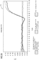
- FIG 2B illustrates a graph 50 depicting a change in the amounts of micro-organism DNA between a saline sample and enriched sample having micro-organism concentration of 100 CFU/milliliter.
- the enriched sample was checked for the presence of micro-organism DNA by performing a quantitative polymerase chain reaction.
- the micro organism in the present embodiment is Candida tropicalis.
- the primer used for the amplification of micro-organism DNA is Candida tropicalis 18S rRNA marker.
- the samples used in the quantitative polymerase chain reaction include a 100 micro liter of saline sample containing Candida tropicalis at a concentration of 100 CFU/milliliter, 100 micro liter of enriched whole blood sample containing Candida tropicalis at a concentration of 100 colony forming units (CFU)/milliliter and negative template controls.
- the X-axis of the graph 50 represents the number of cycles of polymerase chain reaction for amplification of DNA present in the sample.
- the Y-axis of the graph 50 represents the amount of fluorescence produced and detected as a result of amplification of nucleic acids.
- the number of amplification cycles required to produce fluorescence above the threshold value is 34.75 and 34.59.
- the number of amplification cycles required to produce fluorescence above the threshold level is 34.59 and 34.35. Therefore, the enrichment steps do not affect the quantity of micro-organism cells and therefore micro-organism DNA. Thus, the loss of micro-organisms as a result of the enrichment steps is negligible.
- FIG 2C illustrates a graph 60 depicting a comparison of change in the amounts of micro-organism DNA between a saline sample and enriched sample having micro-organism concentration of 10 CFU/milliliter.
- the enriched sample was checked for the presence of micro-organism DNA by performing a quantitative polymerase chain reaction.
- the primer used for the amplification of micro-organism DNA is Candida tropicalis 18S rRNA marker.
- the samples used in the quantitative polymerase chain reaction include a 100 micro liter of saline sample containing Candida tropicalis at a concentration of 10 CFU/milliliter, 100 micro liter of enriched whole blood sample containing Candida tropicalis at a concentration of 100 colony forming units (CFU)/milliliter and negative template controls.
- CFU colony forming units
- the number of amplification cycles required to produce fluorescence above the threshold value is 36.92 and 37.23.
- the number of amplification cycles required to produce fluorescence above the threshold level is 36.92 and 36.97.
- FIG 3 illustrates a graph 70 depicting the change in the amounts of eukaryotic DNA to microbial DNA with the employment of enrichment steps according to the invention.
- the X-axis of graph 70 depicts the progression of the enrichment steps and the Y-axis depicts length of DNA (in base pairs) of DNA present in the sample.
- the amount of eukaryotic DNA or human genomic DNA is reduced.
- the concentration of micro-organism DNA or Candida DNA is not affected by the enrichment steps.
- the ratio of human genomic DNA to Candida DNA after the treatment of said sample with selective lysis buffer, reduces from 10 9 :1 to 10 5 :1. Further, after said sample is treated with Apolipoprotein H, the ratio of human genomic DNA to Candida DNA further reduces from 10 5 :1 to 10 2 :1.
- the enriched Candida DNA is further used for genomic amplification and DNA sequencing, at step 40.
- kits designed to expedite performance of the subject methods.
- Kits serve to expedite the performance of the methods of interest by assembling two or more components required for carrying out the disclosed methods.
- Kits may contain components in pre-measured unit amounts to minimize the need for measurements by end-users.
- Kits may include instructions for performing one or more of the disclosed methods.
- said kit components are optimized to operate in conjunction with one another.
- kits comprise a selective lysis buffer for lysing one or more eukaryotic cells in said sample.
- Said kit further includes one or more enzymes to lyse the released eukaryotic DNA.
- said kit comprises an alkaline reagent to denature eukaryotic DNA.
- Said kit further comprises a filter for removal of broken down or denatured eukaryotic DNA.
- Said kit also includes one or matrices coated with a substrate that has an affinity for micro-organisms.
- said kit may further comprise a chaotropic agent. Said chaotropic agent, when use along with said selective lysis buffer, enables efficient lysis of eukaryotic cells.
- the abovementioned method of enrichment of micro-organisms involves a combination of negative and positive enrichment methods. Negative enrichment ensures elimination of non-target cells from the sample. Therefore, the non-target background is removed thereby providing a concentrated sample containing target cells. When followed by a positive enrichment step, the target cells are separated from any non-target cells that may have been left out in the process of negative enrichment. Therefore, the combination of negative and positive enrichment steps yields a highly enriched and concentrated sample of target cells, which are micro-organisms, in the present embodiment.
- the abovementioned method of enrichment of micro-organisms can be integrated efficiently with the downstream sample preparation methods.
- the method enables nearly complete removal of eukaryotic DNA background, thereby making microbial genomic amplification more efficient.
- the method also reduces the time taken for DNA sequencing as most of sequences being analyzed are of microbial origin.
- this enrichment method can be integrated with the sample preparation methods of any gene sequencing workflow that is used for metagenomics analysis of micro-organisms in a complex sample such as whole blood, sputum, urine, and cerebrospinal fluid.
Landscapes
- Chemical & Material Sciences (AREA)
- Life Sciences & Earth Sciences (AREA)
- Organic Chemistry (AREA)
- Health & Medical Sciences (AREA)
- Zoology (AREA)
- Wood Science & Technology (AREA)
- Proteomics, Peptides & Aminoacids (AREA)
- Engineering & Computer Science (AREA)
- Analytical Chemistry (AREA)
- Molecular Biology (AREA)
- Biochemistry (AREA)
- Microbiology (AREA)
- Biotechnology (AREA)
- Biophysics (AREA)
- Physics & Mathematics (AREA)
- Genetics & Genomics (AREA)
- Immunology (AREA)
- Bioinformatics & Cheminformatics (AREA)
- General Engineering & Computer Science (AREA)
- General Health & Medical Sciences (AREA)
- Chemical Kinetics & Catalysis (AREA)
- Toxicology (AREA)
- Measuring Or Testing Involving Enzymes Or Micro-Organisms (AREA)
- Apparatus Associated With Microorganisms And Enzymes (AREA)
Priority Applications (1)
| Application Number | Priority Date | Filing Date | Title |
|---|---|---|---|
| EP17190993.0A EP3456839A1 (fr) | 2017-09-14 | 2017-09-14 | Procédé d'enrichissement de micro-organismes dans un flux de travail métagénomique |
Applications Claiming Priority (1)
| Application Number | Priority Date | Filing Date | Title |
|---|---|---|---|
| EP17190993.0A EP3456839A1 (fr) | 2017-09-14 | 2017-09-14 | Procédé d'enrichissement de micro-organismes dans un flux de travail métagénomique |
Publications (1)
| Publication Number | Publication Date |
|---|---|
| EP3456839A1 true EP3456839A1 (fr) | 2019-03-20 |
Family
ID=59858952
Family Applications (1)
| Application Number | Title | Priority Date | Filing Date |
|---|---|---|---|
| EP17190993.0A Withdrawn EP3456839A1 (fr) | 2017-09-14 | 2017-09-14 | Procédé d'enrichissement de micro-organismes dans un flux de travail métagénomique |
Country Status (1)
| Country | Link |
|---|---|
| EP (1) | EP3456839A1 (fr) |
Cited By (2)
| Publication number | Priority date | Publication date | Assignee | Title |
|---|---|---|---|---|
| CN111518869A (zh) * | 2020-01-22 | 2020-08-11 | 微岩医学科技(北京)有限公司 | 一种细胞表面脆弱微生物的核酸富集方法及在高通量测序中的应用 |
| EP4389911A1 (fr) * | 2022-12-21 | 2024-06-26 | Mobidiag Oy | Procédés et systèmes pour isoler un analyte |
Citations (4)
| Publication number | Priority date | Publication date | Assignee | Title |
|---|---|---|---|---|
| US20080160528A1 (en) * | 2005-03-02 | 2008-07-03 | Lorenz Michael G | Use of Nucleases for Degrading Nucleic Acids in the Presence of Chaotropic Agents and/or Surfactants |
| EP2333105A1 (fr) * | 2009-12-08 | 2011-06-15 | Koninklijke Philips Electronics N.V. | Lyse sélective de cellules |
| WO2017042819A1 (fr) * | 2015-09-11 | 2017-03-16 | Molecular Detection Israel Ltd. | Procédés d'isolement de celllules microbiennes présentes dans un échantillon de sang |
| CN106987583A (zh) * | 2017-02-20 | 2017-07-28 | 南昌大学 | 一种新型的分离败血症中革兰氏阳性病原菌的方法革兰氏阳性病原菌 |
-
2017
- 2017-09-14 EP EP17190993.0A patent/EP3456839A1/fr not_active Withdrawn
Patent Citations (4)
| Publication number | Priority date | Publication date | Assignee | Title |
|---|---|---|---|---|
| US20080160528A1 (en) * | 2005-03-02 | 2008-07-03 | Lorenz Michael G | Use of Nucleases for Degrading Nucleic Acids in the Presence of Chaotropic Agents and/or Surfactants |
| EP2333105A1 (fr) * | 2009-12-08 | 2011-06-15 | Koninklijke Philips Electronics N.V. | Lyse sélective de cellules |
| WO2017042819A1 (fr) * | 2015-09-11 | 2017-03-16 | Molecular Detection Israel Ltd. | Procédés d'isolement de celllules microbiennes présentes dans un échantillon de sang |
| CN106987583A (zh) * | 2017-02-20 | 2017-07-28 | 南昌大学 | 一种新型的分离败血症中革兰氏阳性病原菌的方法革兰氏阳性病原菌 |
Non-Patent Citations (5)
| Title |
|---|
| "SepsiTest(TM) Pathogen DNA Extraction and PCR Analysis Version 3.0", 1 October 2013 (2013-10-01), pages 1 - 40, XP055146023, Retrieved from the Internet <URL:http://www.goffinmoleculartechnologies.com/wp-content/uploads/2014/01/SepsiTest_V3-0_IVD_CE.pdf> [retrieved on 20141013] * |
| FRANCISCO VEAS: "ApoH as a tool for ultrasensitive detection of pernicious microorganisms and to extract metagenomic data in low concentration pathogens", 1 June 2015 (2015-06-01), Bethesda, XP055225928, Retrieved from the Internet <URL:http://www.apohtech.com/fileadmin/documents/CR/PRCL15009_Bethesda_KF-CHI_ApoH-Present.pdf> [retrieved on 20151104] * |
| MOLZYM: "Kit for the selective isolation of bacterial DNA from bacteremic blood Molysis Complete5 manual 03/2008", 1 January 2008 (2008-01-01), pages 1 - 11, XP055417161, Retrieved from the Internet <URL:http://peramed.com/peramed/docs/D-321-050_EN.pdf> [retrieved on 20171019] * |
| P. GYARMATI ET AL: "Metagenomic analysis of bloodstream infections in patients with acute leukemia and therapy-induced neutropenia", SCIENTIFIC REPORTS, vol. 6, no. 1, 21 March 2016 (2016-03-21), XP055417157, DOI: 10.1038/srep23532 * |
| YANG, J CLINICAL MICROBIOLOGY, vol. 49, pages 3463 - 3469 |
Cited By (6)
| Publication number | Priority date | Publication date | Assignee | Title |
|---|---|---|---|---|
| CN111518869A (zh) * | 2020-01-22 | 2020-08-11 | 微岩医学科技(北京)有限公司 | 一种细胞表面脆弱微生物的核酸富集方法及在高通量测序中的应用 |
| CN111518869B (zh) * | 2020-01-22 | 2023-03-28 | 微岩医学科技(北京)有限公司 | 一种细胞表面脆弱微生物的核酸富集方法及在高通量测序中的应用 |
| EP4389911A1 (fr) * | 2022-12-21 | 2024-06-26 | Mobidiag Oy | Procédés et systèmes pour isoler un analyte |
| US12460197B2 (en) | 2022-12-21 | 2025-11-04 | Gen-Probe Incorporated | Methods and compositions for isolating an analyte |
| JP2025542198A (ja) * | 2022-12-21 | 2025-12-25 | ジェン-プローブ・インコーポレーテッド | 分析物を単離するための方法、システム、および組成物 |
| EP4623097A4 (fr) * | 2022-12-21 | 2026-03-18 | Gen Probe Inc | Procédés, systèmes et compositions pour isoler un analyte |
Similar Documents
| Publication | Publication Date | Title |
|---|---|---|
| EP2872523B1 (fr) | Purification d'acide nucléique d'un micro-organisme à partir d'échantillons-hôtes | |
| JP6324962B2 (ja) | 標的rna枯渇化組成物を調製するための方法およびキット | |
| JP6204664B2 (ja) | Rnaおよびdnaの並行単離および/または並行精製のためのプロセス | |
| JP2006517298A (ja) | 生物学的試料からの核酸の抽出のための前処理方法、およびそのためのキット | |
| CN110462053A (zh) | 使用基于水凝胶的液滴进行单细胞基因组测序 | |
| CN102776173B (zh) | 使用聚多卡醇和衍生物的核酸分离 | |
| JP5259579B2 (ja) | 血液試料中に存在し得る微生物からのデオキシリボ核酸(dna)の抽出方法 | |
| JP2017523772A (ja) | 液滴ソートによるヌクレオチド配列排除富化(needls) | |
| KR20120115242A (ko) | 세포의 선택적 용균 | |
| CN104854246A (zh) | 无限制酶的靶标富集 | |
| EP2115123A2 (fr) | Lyse selective de cellules de sperme | |
| JP2021519591A5 (fr) | ||
| CA2170967A1 (fr) | Methode pour le conditionnement d'echantillons de sang entier | |
| US20250122557A1 (en) | Mitochondrial nucleic acid depletion and detection | |
| CN105120986A (zh) | 用于核酸纯化的一步程序 | |
| EP4163371A1 (fr) | Procédé et application d'extraction d'acide nucléique | |
| CN112322701A (zh) | 一种病原体微生物宏基因组去宿主方法及试剂盒 | |
| Fernandez-Cassi et al. | Viral concentration and amplification from human serum samples prior to application of next-generation sequencing analysis | |
| US12139752B2 (en) | Methods for RNA sequencing | |
| Thatcher | Nucleic acid isolation | |
| WO2020113173A1 (fr) | Analyse multi-omique d'analytes cellulaires à l'aide de systèmes microfluidiques | |
| JP2010532483A (ja) | 生物学的試料の成分を抽出および精製するための方法 | |
| US20210363561A1 (en) | Method of enrichment of micro-organisms in whole blood | |
| JP2001078761A (ja) | 核酸結合性磁性シリカ粒子担体 | |
| Susheel | The importance of nucleic acid purification in sequencing preparation |
Legal Events
| Date | Code | Title | Description |
|---|---|---|---|
| PUAI | Public reference made under article 153(3) epc to a published international application that has entered the european phase |
Free format text: ORIGINAL CODE: 0009012 |
|
| STAA | Information on the status of an ep patent application or granted ep patent |
Free format text: STATUS: THE APPLICATION HAS BEEN PUBLISHED |
|
| STAA | Information on the status of an ep patent application or granted ep patent |
Free format text: STATUS: THE APPLICATION HAS BEEN WITHDRAWN |
|
| AK | Designated contracting states |
Kind code of ref document: A1 Designated state(s): AL AT BE BG CH CY CZ DE DK EE ES FI FR GB GR HR HU IE IS IT LI LT LU LV MC MK MT NL NO PL PT RO RS SE SI SK SM TR |
|
| AX | Request for extension of the european patent |
Extension state: BA ME |
|
| 18W | Application withdrawn |
Effective date: 20190313 |