EP3475257B1 - Dérivés d'acétalanthraquinone utiles pour la coloration cellulaire - Google Patents
Dérivés d'acétalanthraquinone utiles pour la coloration cellulaire Download PDFInfo
- Publication number
- EP3475257B1 EP3475257B1 EP17730252.8A EP17730252A EP3475257B1 EP 3475257 B1 EP3475257 B1 EP 3475257B1 EP 17730252 A EP17730252 A EP 17730252A EP 3475257 B1 EP3475257 B1 EP 3475257B1
- Authority
- EP
- European Patent Office
- Prior art keywords
- compound
- bst
- cells
- sample
- cell
- Prior art date
- Legal status (The legal status is an assumption and is not a legal conclusion. Google has not performed a legal analysis and makes no representation as to the accuracy of the status listed.)
- Active
Links
Images



Classifications
-
- C—CHEMISTRY; METALLURGY
- C09—DYES; PAINTS; POLISHES; NATURAL RESINS; ADHESIVES; COMPOSITIONS NOT OTHERWISE PROVIDED FOR; APPLICATIONS OF MATERIALS NOT OTHERWISE PROVIDED FOR
- C09B—ORGANIC DYES OR CLOSELY-RELATED COMPOUNDS FOR PRODUCING DYES, e.g. PIGMENTS; MORDANTS; LAKES
- C09B1/00—Dyes with anthracene nucleus not condensed with any other ring
- C09B1/16—Amino-anthraquinones
- C09B1/20—Preparation from starting materials already containing the anthracene nucleus
- C09B1/26—Dyes with amino groups substituted by hydrocarbon radicals
- C09B1/262—Dyes with no other substituents than the substituted amino groups
-
- C—CHEMISTRY; METALLURGY
- C07—ORGANIC CHEMISTRY
- C07C—ACYCLIC OR CARBOCYCLIC COMPOUNDS
- C07C225/00—Compounds containing amino groups and doubly—bound oxygen atoms bound to the same carbon skeleton, at least one of the doubly—bound oxygen atoms not being part of a —CHO group, e.g. amino ketones
- C07C225/24—Compounds containing amino groups and doubly—bound oxygen atoms bound to the same carbon skeleton, at least one of the doubly—bound oxygen atoms not being part of a —CHO group, e.g. amino ketones the carbon skeleton containing carbon atoms of quinone rings
- C07C225/26—Compounds containing amino groups and doubly—bound oxygen atoms bound to the same carbon skeleton, at least one of the doubly—bound oxygen atoms not being part of a —CHO group, e.g. amino ketones the carbon skeleton containing carbon atoms of quinone rings having amino groups bound to carbon atoms of quinone rings or of condensed ring systems containing quinone rings
- C07C225/32—Compounds containing amino groups and doubly—bound oxygen atoms bound to the same carbon skeleton, at least one of the doubly—bound oxygen atoms not being part of a —CHO group, e.g. amino ketones the carbon skeleton containing carbon atoms of quinone rings having amino groups bound to carbon atoms of quinone rings or of condensed ring systems containing quinone rings of condensed quinone ring systems formed by at least three rings
- C07C225/34—Amino anthraquinones
-
- C—CHEMISTRY; METALLURGY
- C07—ORGANIC CHEMISTRY
- C07C—ACYCLIC OR CARBOCYCLIC COMPOUNDS
- C07C225/00—Compounds containing amino groups and doubly—bound oxygen atoms bound to the same carbon skeleton, at least one of the doubly—bound oxygen atoms not being part of a —CHO group, e.g. amino ketones
- C07C225/24—Compounds containing amino groups and doubly—bound oxygen atoms bound to the same carbon skeleton, at least one of the doubly—bound oxygen atoms not being part of a —CHO group, e.g. amino ketones the carbon skeleton containing carbon atoms of quinone rings
- C07C225/26—Compounds containing amino groups and doubly—bound oxygen atoms bound to the same carbon skeleton, at least one of the doubly—bound oxygen atoms not being part of a —CHO group, e.g. amino ketones the carbon skeleton containing carbon atoms of quinone rings having amino groups bound to carbon atoms of quinone rings or of condensed ring systems containing quinone rings
- C07C225/32—Compounds containing amino groups and doubly—bound oxygen atoms bound to the same carbon skeleton, at least one of the doubly—bound oxygen atoms not being part of a —CHO group, e.g. amino ketones the carbon skeleton containing carbon atoms of quinone rings having amino groups bound to carbon atoms of quinone rings or of condensed ring systems containing quinone rings of condensed quinone ring systems formed by at least three rings
- C07C225/34—Amino anthraquinones
- C07C225/36—Amino anthraquinones the carbon skeleton being further substituted by singly-bound oxygen atoms
-
- C—CHEMISTRY; METALLURGY
- C09—DYES; PAINTS; POLISHES; NATURAL RESINS; ADHESIVES; COMPOSITIONS NOT OTHERWISE PROVIDED FOR; APPLICATIONS OF MATERIALS NOT OTHERWISE PROVIDED FOR
- C09B—ORGANIC DYES OR CLOSELY-RELATED COMPOUNDS FOR PRODUCING DYES, e.g. PIGMENTS; MORDANTS; LAKES
- C09B1/00—Dyes with anthracene nucleus not condensed with any other ring
- C09B1/50—Amino-hydroxy-anthraquinones; Ethers and esters thereof
- C09B1/51—N-substituted amino-hydroxy anthraquinone
-
- C—CHEMISTRY; METALLURGY
- C12—BIOCHEMISTRY; BEER; SPIRITS; WINE; VINEGAR; MICROBIOLOGY; ENZYMOLOGY; MUTATION OR GENETIC ENGINEERING
- C12Q—MEASURING OR TESTING PROCESSES INVOLVING ENZYMES, NUCLEIC ACIDS OR MICROORGANISMS; COMPOSITIONS OR TEST PAPERS THEREFOR; PROCESSES OF PREPARING SUCH COMPOSITIONS; CONDITION-RESPONSIVE CONTROL IN MICROBIOLOGICAL OR ENZYMOLOGICAL PROCESSES
- C12Q1/00—Measuring or testing processes involving enzymes, nucleic acids or microorganisms; Compositions therefor; Processes of preparing such compositions
- C12Q1/68—Measuring or testing processes involving enzymes, nucleic acids or microorganisms; Compositions therefor; Processes of preparing such compositions involving nucleic acids
- C12Q1/6813—Hybridisation assays
- C12Q1/6841—In situ hybridisation
-
- G—PHYSICS
- G01—MEASURING; TESTING
- G01N—INVESTIGATING OR ANALYSING MATERIALS BY DETERMINING THEIR CHEMICAL OR PHYSICAL PROPERTIES
- G01N1/00—Sampling; Preparing specimens for investigation
- G01N1/28—Preparing specimens for investigation including physical details of (bio-)chemical methods covered elsewhere, e.g. G01N33/50, C12Q
- G01N1/30—Staining; Impregnating ; Fixation; Dehydration; Multistep processes for preparing samples of tissue, cell or nucleic acid material and the like for analysis
-
- G—PHYSICS
- G01—MEASURING; TESTING
- G01N—INVESTIGATING OR ANALYSING MATERIALS BY DETERMINING THEIR CHEMICAL OR PHYSICAL PROPERTIES
- G01N33/00—Investigating or analysing materials by specific methods not covered by groups G01N1/00 - G01N31/00
- G01N33/48—Biological material, e.g. blood, urine; Haemocytometers
- G01N33/50—Chemical analysis of biological material, e.g. blood, urine; Testing involving biospecific ligand binding methods; Immunological testing
- G01N33/5005—Chemical analysis of biological material, e.g. blood, urine; Testing involving biospecific ligand binding methods; Immunological testing involving human or animal cells
- G01N33/5008—Chemical analysis of biological material, e.g. blood, urine; Testing involving biospecific ligand binding methods; Immunological testing involving human or animal cells for testing or evaluating the effect of chemical or biological compounds, e.g. drugs, cosmetics
-
- G—PHYSICS
- G01—MEASURING; TESTING
- G01N—INVESTIGATING OR ANALYSING MATERIALS BY DETERMINING THEIR CHEMICAL OR PHYSICAL PROPERTIES
- G01N33/00—Investigating or analysing materials by specific methods not covered by groups G01N1/00 - G01N31/00
- G01N33/48—Biological material, e.g. blood, urine; Haemocytometers
- G01N33/50—Chemical analysis of biological material, e.g. blood, urine; Testing involving biospecific ligand binding methods; Immunological testing
- G01N33/58—Chemical analysis of biological material, e.g. blood, urine; Testing involving biospecific ligand binding methods; Immunological testing involving labelled substances
- G01N33/582—Chemical analysis of biological material, e.g. blood, urine; Testing involving biospecific ligand binding methods; Immunological testing involving labelled substances with fluorescent label
-
- C—CHEMISTRY; METALLURGY
- C07—ORGANIC CHEMISTRY
- C07C—ACYCLIC OR CARBOCYCLIC COMPOUNDS
- C07C2603/00—Systems containing at least three condensed rings
- C07C2603/02—Ortho- or ortho- and peri-condensed systems
- C07C2603/04—Ortho- or ortho- and peri-condensed systems containing three rings
- C07C2603/22—Ortho- or ortho- and peri-condensed systems containing three rings containing only six-membered rings
- C07C2603/24—Anthracenes; Hydrogenated anthracenes
-
- G—PHYSICS
- G01—MEASURING; TESTING
- G01N—INVESTIGATING OR ANALYSING MATERIALS BY DETERMINING THEIR CHEMICAL OR PHYSICAL PROPERTIES
- G01N1/00—Sampling; Preparing specimens for investigation
- G01N1/28—Preparing specimens for investigation including physical details of (bio-)chemical methods covered elsewhere, e.g. G01N33/50, C12Q
- G01N1/30—Staining; Impregnating ; Fixation; Dehydration; Multistep processes for preparing samples of tissue, cell or nucleic acid material and the like for analysis
- G01N2001/302—Stain compositions
Definitions
- the present invention relates to acetalanthraquinone-based compounds capable of cellular staining.
- the present invention also relates to the use of such compounds in the detection of biological materials, methods of analysing biological samples, and kits containing said compounds.
- Biological staining by fluorescent dyes is a principle used extensively in cytometric assays, including those designed to detect cells or subcellular components by flow cytometry and in vivo and in vitro imaging applications.
- Cell-based assays frequently exploit the capacity to differentiate cellular states through the use of fluorescent molecular probes which are either attached to affinity agents, such as antibodies, or through the intrinsic properties of a given compound enabling it to bind to cellular structures or to become differentially dispersed or accumulated throughout various cellular structures.
- Fluorescent dyes with longer wavelength absorption and emission are particularly advantageous since they can be used in situations where biological materials have a background or inherent fluorescence or absorption in the visible range.
- Dyes that have differential abilities to distribute within biological structures, for example nuclear versus cytoplasmic compartments, can also provide multiple levels of cell identification and tracking.
- a cell-based assay is the Aldefluor assay (StemCell Technologies, as described in EP 1348124 B1 ). This is based on a reagent that is an acetal-derivative of a fluorescent molecule, which upon extracellular hydrolysis is able to enter an intact cell and undergo intracellular metabolism, thereby providing a determination of intracellular aldehyde dehydrogenase (ALDH) activity.
- Aldefluor assay StemCell Technologies, as described in EP 1348124 B1 .
- the Aldefluor reagent, BODIPY-aminoacetaldehyde diethyl acetal (BAAA-DA)
- BAAA-DA BODIPY-aminoacetaldehyde diethyl acetal
- BAAA BODIPY-aminoacetaldehyde
- Anthraquinone (AQ) structures are also used in cell-based assays. AQ structures have near-IR fluorescence properties. They also have DNA affinity, although this is in varying degrees and is not necessarily readily predictable from their structures. The lack of fluorescence of specific AQ compounds has enabled the development of AQ compounds as quencher molecules that modify the fluorescent properties of other fluors when present in appropriate combinations.
- WO2004/026804 A1 describes non-fluorescing anthraquinone compositions with strong fluorescence quenching properties.
- Real-time cell viability assays have also been described using the AQ derivative DRAQ7, a modified anthraquinone that is excluded from live cells but stains the nuclear material of dead or fixed cells (as described in Akagi, J., et al., Real-time cell viability assays using a new anthracycline derivative DRAQ7®. Cytometry Part A, 2013, 83 A(2), 227-234 ).
- the cellular uptake and subcellular localisation of molecules can provide important information on the state of a cell or inform the pharmacokinetic properties of the molecule itself in a biological system of interest.
- the AQ anticancer drug mitoxantrone provides an example of how AQ molecules can offer advantageous properties for different detection modalities.
- Mitoxantrone demonstrates a far-red fluorescence signature, permitting its uptake and subcellular distribution to be analysed by fluorescence microscopy and flow cytometry. This principle has been used to reveal drug resistant phenotypes of human tumour cells (as described in Smith PJ et al., Subcellular distribution of the anticancer drug mitoxantrone in human and drug-resistant murine cells analyzed by flow cytometry and confocal microscopy and its relationship to the induction of DNA damage, Cancer Research 1992; 52:4000-8 ). Alternative detection modalities can be employed beyond the property of intrinsic fluorescence.
- a property of molecules of interest in cell-based analyses and in diagnostic assays is that of Raman scattering.
- Raman active molecule such as mitoxantrone
- a Raman active molecule such as mitoxantrone
- Raman scattering occurs, whereby a minority of the incident photons interact with a vibrational mode of the irradiated molecule and are inelastically scattered.
- the inelastically scattered photons are shifted in frequency.
- the shift can be to a higher frequency (anti-Stokes) or to a lower frequency (Stokes).
- the Raman spectrum of frequency versus intensity provides a unique detection signal for the molecule.
- SERS surface-enhanced Raman scattering
- the magnitude of the SERS-enhancement depends on a number of parameters, including the position and orientation of various bonds present in the Raman-active molecule with respect to the electromagnetic field at the metal surface.
- Bright signals, photostability, and narrow spectral features of SERS-active materials offer certain advantages for cell-based analyses, as described in Nolan JP et al, Surface-enhanced Raman scattering (SERS) cytometry, Methods in cell biology 2011; 102:515-32 .
- SERS spectroscopy can be used to reveal drug adsorption to the membrane environment of living cells.
- SERS has been enabled by the addition of silver colloid to mitoxantrone-treated cells and provided a determination of the presence of the anticancer drug at the plasma membrane microenvironment of human tumour cells and its interaction with exogenously supplied nucleic acid. This is described in Breuzard, G. et al., Surface-enhanced Raman scattering reveals adsorption of mitoxantrone on plasma membrane of living cells, Biochem Biophys Res Commun., 2004, 320(2), pp. 615-621 .
- Molecules with intrinsic fluorescence and advantageous SERS properties can be analysed by combining modalities. For example, this is described in relation to the label-free bimodal imaging by confocal Raman and fluorescence spectroscopy of the intracellular drug release of mitoxantrone in Ganbold EO et al, Nonidentical intracellular drug release rates in Raman and fluorescence spectroscopic determination, Physical Chemistry Chemical Physics, 2015; 17:3019-23 .
- WO2008/154332 describes various near-infrared dyes and their use as surface-enhanced Raman scattering (SERS) reporter molecules. These are described as having enhanced SERS signals, allowing for their use in applications such as diagnostic assays using Raman spectroscopy as a detection method, optical imaging of tissues and cells, and other applications, where high sensitivity is required.
- SERS surface-enhanced Raman scattering
- An alternative method of detection is one that exploits the "photoacoustic" effect.
- Absorption of laser light by chromophore(s) within a sample induces a thermoelastic expansion realized as an initial pressure rise, which then propagates as an acoustic wave.
- An ultrasonic transducer or transducer array detects the acoustic wave to form an image, which maps the original optical energy deposition in the tissue or cell sample.
- a given percentage change in the optical absorption coefficient yields the same percentage change in the photoacoustic amplitude with advantages for imaging in microscopy, cell evaluation and further biomedical applications.
- US 2010/093004 describes assays for detecting and monitoring autophagy and phospholipidosis, including the progression of lysosomal storage diseases. This document also provides methods for distinguishing between phospholipidosis activators and autophagy pathway perturbation agents.
- US 2010/062460 and WO 2010/028349 each describe compounds, methods and kits for identifying, in cells of interest, organelles including nuclei and a wide variety of organelles other than nuclei, as well as cell regions or cell domains.
- WO 2010/062499 describes near IR emitting fluorescent compounds, methods of making and kits containing the described compounds, and their use in fluorescence-based detection of biological materials.
- the present invention addresses the continuing need for molecular probes for fluorometric or other light-based detection analyses, and related assays employing different detection modalities.
- the compounds of the present invention have various beneficial properties that allow them to address deficiencies in the art.
- the present invention provides acetalanthraquinone-based compounds that are useful for cellular staining and for light-based detection of biological material, e.g. fluorescence-based detection of biological material.
- the invention provides a compound of Formula (I), (Ib), (Id), (II) or (IV), as defined in claim 1.
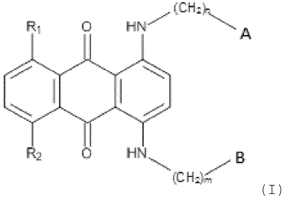
- the invention provides a compound of Formula (I), (Ib), (Id), (II) or (IV):
- R 1 and R 2 when present, are each independently selected from -OH or H, or CH 3 , halo or OR, wherein R is selected from -CH 3 , -CH 2 OCH 3 , -CH(CH 3 ) 2 , -COCH 3 , -CH 2 CHCH 2 , benzene and benzyl; and either: (i) A and B are each independently of formula: -R a CH 2 CH(OR b ) 2 ; or (ii) one of A and B is of formula: -R a CH 2 CH(OR b ) 2 and the other one is of formula: -NR b 2 .
- the or each R b is independently selected from -CH 3 , -CH 2 CH 3 , -CH(CH 3 ) 2 , and -CH 2 CHCH 2 .
- the present compounds have an affinity for nucleic acid molecules and can form a complex with nucleic acids, which can be detected optically.
- the present compounds may be used in fluorescence-based technologies and assays or other light-based technologies and assays, e.g. based on Raman scattering or photoacoustic detection.
- the compounds may be used to directly stain or label a sample so that the sample can be identified or quantified.
- such compounds may be added as part of an assay for a biological target analyte or as a detectable tracer element in a biological or non-biological fluid.
- the present compounds generally are utilized by combining the compound with a sample of interest under conditions selected to yield a detectable optical response.
- the sample may be then illuminated at a wavelength selected to elicit the optical response.
- the present compounds have in particular been determined to have the following properties:
- the invention provides, in a second aspect, the use of a compound of the first aspect in light-based detection of biological materials, e.g. fluorescence-based detection of biological materials. It may be colorimetric, or photoacoustic, or Raman Scattering -based detection of biological materials.
- these biological materials comprise cellular and/or subcellular structures. In one embodiment, the biological materials comprise biological materials containing nucleic acid.
- the invention provides, in a third aspect, the use of a compound of the first aspect in a biological assay, e.g. in an image-based assay.
- the compound may provide one or more of the following functions in the assay: cell detection; cell tracking; cell enumeration; and determination of cellular integrity, state and function.
- the invention provides, in a fourth aspect, the use of a compound of the first aspect as a nuclear and cytoplasmic differentiating counterstain.
- the invention provides a fluorescent complex comprising a nucleic acid and a compound of the first aspect.
- the nucleic acid is DNA. In one embodiment, the DNA is present in a cell. In one embodiment, the DNA is present in a non-intact cell.
- the invention provides a method of analysing a biological sample comprising cells, or other biological material containing nucleic acid, the method comprising the steps of:
- Step c) may include exciting the compound with electromagnetic radiation, and detecting an emitted fluorescence signal.
- the spectroscopic property associated with absorption of electromagnetic radiation by the compound is a colorimetric property.
- the spectroscopic property associated with absorption of electromagnetic radiation by the compound is a photoacoustic property.
- the spectroscopic property associated with absorption of electromagnetic radiation by the compound is a Raman Scattering property.
- the method of analysing a biological sample further discriminates cellular nuclei in the cells, and step b) is performed to cause binding of nucleic acid in cellular nuclei by the compound, and the discrimination of the cellular nuclei is based at least in part on the spectroscopic property detected in step c).
- step b) is performed to stain the cells with the compound.
- Cell death accruement may be monitored such that step b) is performed prior to or during an assay period thereby enabling a continuous or frequent readout of cell death accruement during the assay period.
- Step c) may include detecting fluorescence emitted by individual cells by flow cytometry, or step c) may include intra-cellular location detection by fluorescence microscopy.
- detection of emitted fluorescence signals may be made with a variety of instruments including, but not limited to, microscopes, flow cytometers, plate readers and any variations thereof.
- the biological sample comprises cells, which may be fixed cells or permeabilised cells.
- the method therefore may analyse fixed or permeabilised cells, in which the cells are fixed by treatment with a fixative or permeabilising agent.
- step b) of the method further comprises treating the sample with at least one other fluorochrome or light- emitting compound, and step c) further comprises detecting a spectroscopic property associated with the absorption of electromagnetic radiation by the fluorochrome or light-emitting compound.
- a method of staining a biological sample comprising cells or other biological material containing nucleic acid comprises contacting the biological sample with a compound of the first aspect.
- the method comprises contacting the biological sample with the compound and then:
- the method of staining a biological sample comprising cells or other biological material containing nucleic acid comprises the steps of:
- the sample may include a combination of the compound and other fluorophores or dyes.
- additional compound(s) may be present that emit in the UV or visible region of the spectrum upon excitation with an appropriate light source.
- the fluorescence spectra of the compounds described herein by virtue of their relatively long emission wavelengths, make the compounds particularly useful in multiplex applications with fluorophores that emit in the UV or visible portion of the spectrum.
- the biological sample comprises fixed cells or permeabilized cells. In one embodiment, the biological sample comprises live cells, which may be in a form of a cell suspension or tissue sample.
- the methods as described herein may use live cells.
- the methods may use dead or fixed cells. It may be that the methods use a combination of cell types, e.g. selected from live and dead cells, fixed cells, permeabilized cells, and/or free cell nuclei.
- Cells may be treated with a cell fixative reagent, or a cell fixative reagent combined with a cell permeabilizing reagent, or a cell permeabilizing reagent.
- the cells may be suspended in a fluid (e.g. a biological fluid or an aqueous fluid, such as buffer or water) or may be in a solid form, such as cells adhered to plates, coverslips, dishes, flasks, or the like or solid tissue samples that have been disaggregated.
- a fluid e.g. a biological fluid or an aqueous fluid, such as buffer or water
- solid form such as cells adhered to plates, coverslips, dishes, flasks, or the like or solid tissue samples that have been disaggregated.
- a compound of the first aspect, as described herein is added to a sample containing living cells.
- the sample can include cells suspended in a buffer or media, or cells adhered to a glass or plastic surface, bathed in a buffer or media.
- the compound can be added to the buffer or media containing living cells, incubated, and data acquired without washing the dye out of the sample.
- a dead cell dye or a live cell dye may be included in the testing for gating out of dead cells or gating on living cells.
- the present methods for detecting the presence of nucleic acids in a sample may further include quantification of the nucleic acid detected.
- a method of quantifying nucleic acid content in live cells comprising: combining a compound of the first aspect with a sample to form a mixture, wherein the sample comprises a nucleic acid molecule; incubating the mixture for a sufficient amount of time for the compound to associate with the nucleic acid in the sample; illuminating the incubated sample with an appropriate wavelength to generate a detectable optical response or photoacoustic response; and detecting the presence of nucleic acid in the sample by flow cytometric analysis, image cytometry analysis, image analysis, or high content image analysis.
- the present invention provides a kit for carrying out an assay, which includes a compound as defined in the first aspect.
- kits that facilitate the practice of various assays.
- the kits may be packaged with the compound in a dry form or with the compound in solution.
- the kits may optionally further include one or more buffering agents, typically present as an aqueous solution, sample preparation reagents, additional detection reagents, organic solvent, other fluorescent detection probes, standards, microspheres, specific cell lines, antibodies and/or instructions for carrying out an assay. Additional optional agents include components for testing of other cell functions in conjunction with the compound.
- the present invention provides di-substituted acetalanthraquinone compounds.
- these are 1,4-di-substituted (Formulae (I), (Ib), (Id)).
- these are 1,5-di-substituted (Formula (II)).
- these are 1,8-di-substituted (Formula (IV)).
- hydrolysis is acid-based, e.g. by exposure to 2M HCl for 30 minutes.
- These compounds may optionally be provided in deuterated form.
- the skilled person would be readily able to deuterate the compounds as provided herein. This may be by applying the teachings of Matthaus C et al, New ways of imaging uptake and intracellular fate of liposomal drug carrier systems inside individual cells, based on Raman microscopy, Molecular pharmaceutics 2008; 5:287-93 .
- the compound when it is chiral, it may be provided in the form of a single enantiomer or it may be provided in the form of a racemic mixture.
- R 1 and R 2 are each independently selected from -OH or H, or CH 3 , a halo group (F, Cl, Br or 1), or OR, wherein R is selected from -CH 3 , -CH 2 OCH 3 , -CH(CH 3 ) 2 , - COCH 3 , -CH 2 CHCH 2 , benzene and benzyl. It may be that the halo group is F or Cl, e.g. Cl.
- the compounds are only di-substituted, i.e. in Formula (I) and Formula (II) R 1 and R 2 are both H.
- the compounds may include additional substitution, i.e. at least one of R 1 and R 2 is not H. In one embodiment both R 1 and R 2 are not H.
- R 1 and R 2 may each independently selected from -OH, CH 3 , and Cl. In one embodiment one or both of R 1 and R 2 is -OH.
- R 1 and R 2 are the same. In one preferred embodiment, R 1 and R 2 are both -OH or are both H.
- n and m are each independently an integer from 1 to 6, e.g. 1, 2, 3, 4 or 5. It may be that n and m are each independently an integer from 1 to 3.
- n and n are the same. In one embodiment m and n are both 1. In another embodiment m and n are both 2.
- option (i) applies and A and B are each independently of formula: -R a CH 2 CH(OR b ) 2 ; and m and n are the same. In one such embodiment m and n are 1 or 2 or 3 or 4 or 5. In one such embodiment m and n are 1 or 2. In one such embodiment m and n are 1.
- option (ii) applies and A is of formula: -NR b 2 and B is of formula -R a CH 2 CH(OR b ) 2 , m is 1 or 2 or 3 or 4 or 5 and n is 1 or 2 or 3.
- A is of formula: -NR b 2 and B is of formula -R a CH 2 CH(OR b ) 2 , m is 1 or 2 or 3 or 4 or 5 and n is 2.
- the or each R b is independently selected from -CH 3 , - CH 2 CH 3 , and -CH 2 CHCH 2 . In one such embodiment the or each R b is independently selected from -CH 3 , and -CH 2 CH 3 . In one embodiment the or each R b is-CH 3 .
- option (i) applies and A and B have the same R a group.
- option (i) applies and A and B have the same R b group.
- each R b is -CH 3 .
- option (i) applies A and B are the same.
- option (ii) applies and A and B have the same R b group.
- R b is -CH 3 .
- the invention provides a compound of Formula (Ia): wherein
- R 1 and R 2 are the same.
- the compound is of the following formula (Ib): wherein m is an integer selected from 1, 2, 3, 4 and 5, e.g. it may be 1 or 2.
- the compound of Formula (I) is of the following formula: wherein
- R 1 and R 2 are the same.
- R a is-CH 2 -.
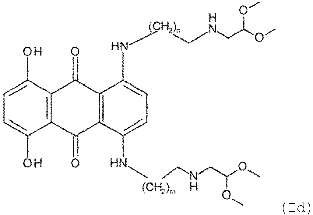
- the compound is of the following formula (Id): wherein n and m are the same and are an integer selected from 1 to 6.
- n and m are the same and are an integer selected from 1, 2, 3, 4 and 5. It may be that n and m are the same and are an integer selected from 1, 2, and 3. In one embodiment both n and m are 1.
- the compounds of the present invention may have hydrolysable groups.
- the compounds may be hydrolysed in use.
- the hydrolysis of compounds is known in the art and the skilled person would be able to hydrolyse the compounds using known techniques.
- Acid-catalysed hydrolysis and base-catalysed hydrolysis are well known, e.g. refluxing in the presence of dilute hydrochloric acid or dilute sulphuric acid or sodium hydroxide solution.
- the compounds of the invention can contain asymmetric carbon atoms and can therefore exist in racemic and optically active forms.
- optical isomers or enantiomers, racemates, tautomers, and diastereomers are envisaged as being able to be used in the present invention.
- the methods of present invention include the use of all such isomers and mixtures thereof.
- Methods of separation of enantiomeric and diastereomeric mixtures are well known to one skilled in the art.
- racemic forms can be resolved by physical methods, such as, e.g., fractional crystallization, separation or crystallization of diastereomeric derivatives or separation by chiral column chromatography.
- the individual optical isomers can be obtained from the racemates using conventional methods, such as, e.g., salt formation with an optically active acid followed by crystallization.
- the present invention encompasses any isolated racemic or optically active form of compounds of the invention, or any mixture thereof.
- aryl refers to a carbocyclic aromatic system containing one ring, or two or three rings fused together where in the ring atoms are all carbon.
- aryl includes, but is not limited to, groups such as phenyl, naphthyl, or anthracenyl.
- alkyl refers to a straight-chain or branched-chain alkyl group containing from 1 to 12 carbon atoms.
- a C1-z alkyl is an alkyl from 1 to z carbon atoms; thus, a C1-8 alkyl has from 1 to 8 carbon atoms, a C1-4 alkyl has from 1 to 4 carbon atoms and a C1-2 alkyl has from 1 to 2 carbon atoms.
- alkyl groups include, but are not limited to, methyl, ethyl, n-propyl, isopropyl, n-butyl, isobutyl, sec-butyl, tert-butyl, pentyl, isopentyl, neo-pentyl, iso-amyl, hexyl, heptyl, octyl, or nonyl.
- alkylaryl refers to a straight-chain or branched-chain alkyl group containing from 1 to 12 carbon atoms, e.g. from 1 to 10 carbon atoms, onto which an aryl group (such as defined above) is attached.
- Specific examples of the alkylaryl groups are those having 7 to 30 carbon atoms in total and in particular those wherein the alkyl group is a C1-6 straight-chain or branched alkyl.
- the alkylaryl group may be benzyl, phenylethyl, phenylpropyl, phenylbutyl, phenylpentyl or phenylhexyl.
- N N , N' -(2,2'-(5,8-Dihydroxy-9,10-dioxo-9,10-dihydroanthracene-1,4-diyl)bis(azanediyl)bis(ethane-2,1-diyl))bis(3,3-dimethoxypropanamide) (BST-105)
- the compounds were exited at 540 nm and at the wavelength of their absorption peak 1 ( ⁇ 1 nm).
- the far-red fluorescent DNA dye DRAQ5 was used as reference.
- absorbance measurements were obtained using a Beckmann Coulter DU 800 UV/Visible spectrophotometer.
- the excitation wavelengths were scanned from 280 to 800 nm. Fluorescent spectral scans were performed using a Perkin Elmer LS-50B Luminescence spectrophotometer. To obtain emission scans, the excitation wavelength was set accordingly to the maximum values for each agent or another designated excitation wavelength that matches typical laer lines; and the emission wavelength were scanned from 400 nm to 900 nm in 1 nm increments.
- a Xenon flash lamp was used as the light source, with the lamp energy set to high. For each data point ten measurements were acquired with the photomultiplier tube sensitivity set at 125.
- Typical absorption spectra and fluorescence spectra of compounds BST-108, BST-109, BST-110 and BST-111 and BST-100, BST-101 and BST-106 can be seen in Figure 1A and Figure 1B respectively.
- the absorption and fluorescence spectra show that the compounds have absorption and emission in the long-wavelength spectral region (>650 nm).
- Table 1 shows the summarised fluorescence emission data for tested compounds in PBS and HEPES.
- Compound BST-203 is described but not claimed.
- Table 1 Compound ⁇ 1 nm (Ex in PBS) 540 nm (Ex in PBS) ⁇ 1 nm (Ex in HEPES) DRAQ5 >665 635 >665 BST-104 684 712 726 BST-108 685 692 684 BST-111 684 684 684 BST-200 688 690 684 BST-201 690.5 684 684 BST-202 - - 680 BST-203 678 690 696 BST-100 747.5 - 740 BST-101 - 712 - BST-102 661 670 670 663 BST-106 660 670 664 BST-109 666 660 660 BST-110 665.5 662 660
- DNA melting temperature (variation in Tm) was used to assess interaction between CT-DNA and compounds of Formula (I). Studies were carried out using a thermal DNA denaturation assay.
- CT-DNA calf thymus DNA
- CT-DNA melting buffer was prepared by making up a 1 L solution A of (10 mM NaH 2 PO 4 and 1 mM Na 2 EDTA) and a separate 1 L solution B (10 mM Na 2 HPO 4 and 1 mM Na 2 EDTA), and then solution A was added to solution B until pH 7.0 was achieved.
- Calf thymus DNA (CT-DNA, Sigma) solutions were prepared for use by dissolving 21-23 mg in 20 mL of the CT-DNA melting buffer and stored at 4°C.
- Mitoxantrone (Sigma) was solubilized in DMSO, diluted to desired concentrations in 10 mM Tris-HCl buffer (pH 7.4) and used as control.
- CT-DNA thermal melting assay mixtures were prepared by adding sufficient CT-DNA stock solution to CT-DNA melting buffer, to ensure the preparation of a 50 ⁇ M CT-DNA solution. Compound stock solution was then added to the 50 ⁇ M CT-DNA solution, to ensure the final concentration of compound in the mixture was 10 ⁇ M.
- the temperature of the test solution was increased at 1°C/min till 95°C was reached. Melting curves generated by computer software were printed out and the melting points were determined using the published graphical method by Brown and Fox (1997; Brown, P. M. and Fox, K. R. 1997. Foot-printing studies with nucleosome-bound DNA.Methods in Molecular Biology; Drug-DNA interaction protocols. Vol. 90. pp. 81-93 ). The Tm was the mean of two separate determinations within 0.2 °C.
- the assay was performed initially by incubating 50 ⁇ M of calf thymus DNA (CT-DNA), in the presence of 10 ⁇ M of mitoxantrone, to validate the assay and to have a positive control for comparison. 10 ⁇ M solutions of compound counterparts were incubated with 50 ⁇ M CT-DNA.
- CT-DNA calf thymus DNA
- Table 2 shows results of CT-DNA/compound Formula (I) melting temperatures before and after acidic hydrolysis.
- Compound BST-203 is described but not claimed.
- Table 2 Compound Compound Tm (°C) Hydrolysed product of compound Tm (°C) Mitoxantrone >95 - BST-101 69.0 69.7 BST-107 74.0 71.9 BST-102 69.0 70.0 BST-103 69.4 70.1 BST-104 69.6 ** BST-105 69.0 ** BST-106 72.1 * BST-108 76.4 * BST-109 72.0 72.9 BST-110 72.0 71.6 BST-111 71.6 74.6 BST-200 74.2 70.4 BST-201 70.8 * BST-202 70.0 70.0 BST-203 73.0 72.6 * Excess of interference, hence any calculation of Tm was inaccurate. ** Solubility issues in the working buffer, therefore no experiment was performed.
- This basic protocol was the standard for testing the patterns of AAQ agents at 20 ⁇ M, compared to 20 ⁇ M of DRAQ5 as the benchmark reference (both in terms of cellular location and ability to detect the red fluorescence).
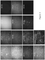
- BST-101 and BST-106 showed diverse staining when put into cells.
- BST-101 and BST-106 have a diffuse cytoplasmic staining, with some perinuclear and nucleoli labelling. Moreover, very bright background was observed in BST-106 compared to BST-101.
- Compounds BST-108 and BST-109 showed heterogeneity in cytoplasmic staining and positive spots in the nuclei due to DNA binding. Moreover, BST-110 and BST-111 displayed very high background fluorescence, especially in BST-110, with cytoplasmic staining and heterogeneity on labelling of mitotic figures.
- BST-101 and BST-106 showed heterogeneous cytoplasmic staining, with BST-106 in particular displaying a high background.
- the BST-108, BST-109 and BST-110 analogues revealed very bright cellular fluorescence, whereas BST-111 has a greater contrast and diffuse cellular staining.
- BST-200 showed heterogeneity on cytoplasmic load on, with bright fluorescence signal and staining of mitotic figures, whereas BST-201 had perinuclear accumulation.
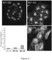
- Confocal laser scanning microscopy was used to analyse the localisation of compounds BST-100, BST-101 and BST-106 with the U-2 OS cells, as it permitted the acquisition of volume limited optical sections through the cell and media. Excitation (637 nm) and emission (>665 nm) values for the probe analysis were the optimal conditions. The data sets obtained were processed and assembled into a single maximum projection image.
- the patterns were analysed after 30 min incubation at different z depths and eliminating the solution background.
- the nucleus/cytoplasm ratio was calculated to quantify the localisation of the probe per cell.
- Optical sectioning enabled detection of low levels of probe in the perinuclear region, with probable overload in Golgi body and large vesicles for BST-100 and BST-101.
- the accumulation area appeared similarly to the MTOC (microtubule organizing centre); however BST-106 gave an additional nuclear signal.
- BST-108, BST-109, BST-110 and BST-111 showed a strong nuclear uptake and perinuclear staining, with possible ER accumulation.
- a whole cell staining was a specific feature of BST-110 and BST-109, whereas BST-111 was mainly loaded in the nuclear compartment.
- Timelapse imaging in accordance with Errington RJ et al, Time-lapse microscopy approaches to track cell cycle and lineage progression at the single-cell level, Curr Protoc Cytom. 2013 Apr; Chapter 12: Unit 12.4 , was carried out in relation to cells with exposure to probe compounds.
- A549 cells were seeded in a 24 well coverslip bottomed -plate, cultured overnight in McCoys media for 24 or 48 hours.
- the plates were placed onto an inverted automated microscope, with the capacity for acquiring an image sequence of different wells (therefore exposure to different agent labelling).
- An Axiovert 100 microscope (Carl Zeiss, Welwyn Garden City, UK) was fitted with an incubator for 37°C/5% CO2 maintenance (Solent Scientific, Portsmouth, UK), and an ORCA-ER CCD camera (Hamamatsu, Reading, UK). Illumination was controlled by a shutter in front of the transmission lamp, and an x, y positioning stage with separate z-focus (Prior Scientific, Cambridge, UK) controlled multi-field acquisition. Image capture was controlled using Metamorph software.
- Cells were exposed to a constant dose (0-5 ⁇ M) or a pulsed dose of 0 or 20 ⁇ M agent for 15 minutes followed by washout with warmed full media.
- A549 cell lines were monitored using time-lapse collection and lineage progression, in constant exposure to compounds of the invention. The effects on cells were also determined.
- A549 cells were treated with probe compounds BST-101, BST-109, BST-110 and put on the time-lapse.
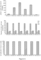
- Non-fluorescent images were collected over time. 72 hours later the cells were harvested and assayed using conventional flow cytometry and conventional methods for fluorescence intensity measurements to obtain relative fluorescence intensity (for example in units of channel number) and electronic gating (for example to determine the relative frequency of events) understood by those practised in the art, for (a) total signal of probe per cell determined by relative fluorescence intensity (arb. Units) and (b) cell cycle distribution across the cell compartments G1, S and G2 based on relative DNA content determined fluorometrically, and (c) cell viability determined by % total cells showing vital dye exclusion.
- the BST-109 and BST-110 are predominantly in a membrane compartment of the cytoplasm.
- the nucleus is now a low signal - the nuclear/cytoplasmic ratio is reversed.
- BST-101 in A549 cells dividing, and signal retained in the same membrane compartment as at the start.
- BST-109 in A549 cells showed dividing, and signal retained but now relocated in a nonnuclear compartment perinuclear.
- BST-110 in A549 cells gave dividing, and signal now relocated in a nonnuclear compartment perinuclear.
- A549 or MCF7 in response to compounds was determined using the MTT (3-(4,5-dimethylthiazolyl-2-)-2, 5-diphenyltetrazolium bromide) assay according to the protocol set out in Mosmann T., Rapid colorimetric assay for cellular growth and survival: application to proliferation and cytotoxicity assays. Journal of immunological methods 1983; 65:55-63 .
- cells were seeded into a rounded-bottom 96 well plate (Corning) at six different cell seeding densities (0.5x10⁴, 1x10⁴, 2x10⁴, 3x10⁴, 4x10⁴, and 5x10⁴) per mL of R10 media (cells/mL) in order to optimise the assay and establish the most suitable seeding assay. Concentration of 1x10⁴ cells/mL was determined to be the optimum seeding density for both cell lines for 96 h total incubation. A control lane contained supplemented R10 only. The 96 well plates were incubated overnight at 37°C, 5% CO2 and 100% humidity to allow cell adhesion.
- the IC 50 values are shown in Tables 3 and 4.
- Table 3 Compound MCF7 ( ⁇ M) R 2 value A549 ( ⁇ M) R 2 value Doxorubicin 0.028 0.1831 0.05 0.1759 BST-101 0.750 0.7865 >100 0.9034 BST-102 1.75 0.6174 >100 0.2725 BST-106 11.3 0.5697 11.7 0.568 BST-107 1.66 0.5541 17.5 0.9475 BST-108 1.44 0.4437 1.7 0.7563 BST-109 15.70 0.7976 22.5 0.7371 BST-110 19.5 0.7258 22.3 0.9473 BST-111 1.8 0.757 38.2 0.9546
- Table 4 Compound MCF-7 ( ⁇ M) R 2 value A549 ( ⁇ M) R 2 value BST-101 hydrolysed 56.73* 0.9434 >100 0.9621 BST-102 hydrolysed ⁇ 0.250* 0.8793 >100* 0.8352 BST-106 hydrolysed 40* 0.933 >100* 0.8502 B
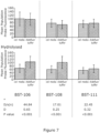
- Flow cytometry analysis was performed to evaluate the effect of pre-treatment with HCl to generate an active aldehyde on the cell uptake properties.
- A549 cells were incubated for 30 min with compounds of Formula (I) and its hydrolysed forms (20 ⁇ M) prior to analysis.
- the rate of uptake was tested in A549 cell culture media and in Aldefluor buffer, containing the MDR-blocker verapamil (Pearce and Bonnet 2007), to evaluate the effect in uptake with/without the Aldefluor assay system. Analysis was conducted in the viable subset of cells, corresponding to ⁇ 95% of the total amount under investigation.
- Figure 7 shows the uptake profiles of BST-106, BST-108, BST-111 compounds and their hydrolysed forms (generated after exposure to 2M HCl for 30 min) in A549 cell line versus Aldefluor buffer.
- Aldefluor buffer contained verapamil vs. cell media. The cells were incubated with 20 ⁇ M of the respective AAQ/HAAQs (30 min) before analysis
- anthraquinones typically have very low quantum yields (as described in Njoh KL et al , Spectral analysis of the DNA targeting bisalkylaminoanthraquinone DRAQ5 in intact living cells, Cytometry A 2006; 69:805-14 ) this can make them ideal probes for performance as a contrast agent for both fluorescence detection due to cell accumulation and for photoacoustic imaging. Accordingly, low quantum yield molecules such as those described in the present invention, including their hydrolysed and deuterated forms, that also have differential cellular uptake, subcellular localization and persistence profiles can co-display advantageous photoacoustic for use in cell-based assays. AQ-derived molecules can also provide advantageous properties for detection by SERS in cell-based assays.
- NIR emissive compounds have advantages in the development of fluorescent probes and labelling for bio-imaging in living systems since fluorescence in the long-wavelength region would generate minimum photo-toxicity to biological components, deep tissue penetration and minimal background from auto-fluorescence by bio-molecules.
- compounds of the invention are useful for analysing biological samples comprising cells, which may be dead, fixed or living or other biological material containing nucleic acid.
- the compounds of the present invention showed intrinsic ability to bind to nucleic acids.
- compounds of the invention may be used in nuclear discrimination assays.
- the compounds of the invention can be used as nuclear counterstains.
- Nuclear counterstains are important tools for many fluorescence-based cell analyses, including but not limited to microscopy, flow cytometry and in-cell western analysis.
- the compounds of the invention provide stable fluorophores with structure-dependent subcellular localization and a lack of toxic effects consistent with differential accumulation in endoplasmic reticulum, Golgi or nucleus.
Landscapes
- Chemical & Material Sciences (AREA)
- Organic Chemistry (AREA)
- Health & Medical Sciences (AREA)
- Life Sciences & Earth Sciences (AREA)
- Engineering & Computer Science (AREA)
- Immunology (AREA)
- Molecular Biology (AREA)
- Biomedical Technology (AREA)
- Physics & Mathematics (AREA)
- Analytical Chemistry (AREA)
- General Health & Medical Sciences (AREA)
- Biochemistry (AREA)
- Hematology (AREA)
- Pathology (AREA)
- Zoology (AREA)
- Microbiology (AREA)
- Urology & Nephrology (AREA)
- Biotechnology (AREA)
- Wood Science & Technology (AREA)
- Proteomics, Peptides & Aminoacids (AREA)
- General Physics & Mathematics (AREA)
- Bioinformatics & Cheminformatics (AREA)
- Cell Biology (AREA)
- Genetics & Genomics (AREA)
- Biophysics (AREA)
- General Engineering & Computer Science (AREA)
- Food Science & Technology (AREA)
- Medicinal Chemistry (AREA)
- Toxicology (AREA)
- Tropical Medicine & Parasitology (AREA)
- Investigating Or Analysing Biological Materials (AREA)
- Measuring Or Testing Involving Enzymes Or Micro-Organisms (AREA)
- Heterocyclic Carbon Compounds Containing A Hetero Ring Having Oxygen Or Sulfur (AREA)
- Heterocyclic Compounds That Contain Two Or More Ring Oxygen Atoms (AREA)
- Investigating, Analyzing Materials By Fluorescence Or Luminescence (AREA)
Claims (15)
- Composé de formule (I), (Ib), (Id), (II) ou (IV) :
 dans laquelle n et m sont chacun indépendamment un entier de 1 à 6,
 dans laquelle m est un entier choisi parmi 1, 2, 3, 4 et 5,
 dans laquelle n et m sont identiques et sont un entier choisi parmi 1 à 6,
 dans laquelle n et m sont chacun indépendamment un entier de 1 à 6,
 dans laquelle n et m sont chacun indépendamment un entier de 1 à 6,ou une forme deutérée de celui-ci,dans lequel R1 et R2, lorsqu'ils sont présents, sont chacun indépendamment choisis parmi -OH ou H, ou CH3, halogéno ou OR, dans lequel R est choisi parmi -CH3,-CH2OCH3, -CH(CH3)2, -COCH3, -CH2CHCH2, benzène et benzyle ; et soit :(i) A et B sont chacun indépendamment de formule :-RaCH2CH (ORb)2 ;
ou(ii) l'un de A et B est de formule : -RaCH2CH(ORb)2 et l'autre est de formule : -NRb 2 ; dans lequelle ou chaque Ra est indépendamment choisi parmi -CH2-,-C(=O)NH-, -NHC(=O)-, -C(=O)-, -C(=CH2)-, -C(=CH2CH3)-,-N(CH3)- et -N(alkylaryl)-,dans lequel alkylaryle désigne un groupe alkyle à chaîne linéaire ou à chaîne ramifiée contenant de 1 à 12 atomes de carbone dans lequel un groupe aryle est lié,et le ou chaque Rb est indépendamment choisi parmi -CH3, -CH2CH3, -CH(CH3)2 et -CH2CHCH2. - Composé selon la revendication 1, dans lequel :a) R1 et R2 sont identiques ; et/oub) le ou chaque Ra est indépendamment choisi parmi-CH2-, -C(=O)NH-, -NHC(=O)-, -C(=O)-, -C(=CH2)- et-C(=CH2CH3)- ; et/ouc) le ou chaque Rb est indépendamment choisi parmi-CH3, -CH2CH3 et -CH2CHCH2.
- Composé selon la revendication 1 ou la revendication 2, dans lequel l'option (i) selon la revendication 1 s'applique et dans lequel :a) m et n sont identiques ; et/oub) A et B ont le même groupe Ra ; et/ouc) A et B ont le même groupe Rb ; et/oud) A et B sont identiques.
- Composé selon la revendication 1 ou la revendication 2, dans lequel l'option (ii) selon la revendication 1 s'applique et dans lequel :a) A est de formule : -NRb 2 et B est de formule - RaCH2CH (ORb)2, m est 1 ou 2 ou 3 ou 4 ou 5 et n est 1 ou 2 ou 3 ; et/oub) A et B ont le même groupe Rb.
- Utilisation d'un composé tel que défini dans l'une quelconque des revendications 1 à 4, l'utilisation étanta) dans la détection de matériaux biologiques basée sur la lumière ; oub) dans un dosage biologique ; ouc) en tant que contre-colorant de différenciation nucléaire et cytoplasmique.
- Utilisation d'un composé selon la revendication 5, option a), dans laquelle le composé est utilisé dans :(i) la détection de matériaux biologiques basée sur la fluorescence, ou(ii) la détection de matériaux biologiques basée sur la colorimétrie, ou(iii) la détection de matériaux biologiques basée sur la photoacoustique, ou(iv) la détection de matériaux biologiques basée sur la diffusion Raman.
- Complexe fluorescent comprenant un acide nucléique et un composé tel que défini dans l'une quelconque des revendications 1 à 4.
- Procédé d'analyse d'un échantillon biologique comprenant des cellules, ou un autre matériau biologique contenant un acide nucléique, le procédé comprenant les étapes de :a) préparation d'une solution biologiquement compatible contenant un composé tel que défini dans l'une quelconque des revendications 1 à 4 ;b) traitement d'un échantillon biologique avec la solution biologiquement compatible ;
etc) détection d'une propriété spectroscopique associée à l'absorption de rayonnement électromagnétique par le composé. - Procédé selon la revendication 8 dans lequel la propriété spectroscopique associée à l'absorption de rayonnement électromagnétique par le composé est :(i) la fluorescence, ou(ii) une propriété colorimétrique, ou(iii) une propriété photoacoustique, ou(iv) une propriété de diffusion Raman.
- Procédé de coloration d'un échantillon biologique comprenant des cellules ou un autre matériau biologique contenant un acide nucléique, ledit procédé comprenant la mise en contact de l'échantillon biologique avec un composé tel que défini dans l'une quelconque des revendications 1 à 4.
- Procédé selon la revendication 10, le procédé comprenant la mise en contact de l'échantillon biologique avec un composé tel que défini dans l'une quelconque des revendications 1 à 4 et ensuite :- la conduite d'une étape de détection basée sur la fluorescence ; ou- la conduite d'une étape de détection basée sur la diffusion Raman ; ou- la conduite d'une étape de détection basée sur la photoacoustique.
- Procédé selon la revendication 10, qui comprend les étapes de :a) préparation d'une solution biologiquement compatible contenant un composé tel que défini dans l'une quelconque des revendications 1 à 4 ;b) traitement d'un échantillon biologique avec la solution biologiquement compatible pendant un temps suffisant pour causer une coloration ;c) éclairage de l'échantillon coloré avec une source de lumière suffisante pour exciter le composé ; etd) détection de la fluorescence émise depuis l'échantillon au moyen d'un instrument de détection de fluorescence.
- Procédé de quantification de la teneur en acide nucléique dans des cellules vivantes, comprenant :la combinaison d'un composé tel que défini dans l'une quelconque des revendications 1 à 4 avec un échantillon pour former un mélange, dans lequel l'échantillon comprend une molécule d'acide nucléique ;l'incubation du mélange pendant une durée suffisante pour que le composé s'associe à l'acide nucléique dans l'échantillon ;l'éclairage de l'échantillon incubé avec une longueur d'onde appropriée pour générer une réponse optique ou une réponse photoacoustique détectable ; etla détection de la présence d'acide nucléique dans l'échantillon par analyse par cytométrie en flux, analyse par cytométrie en image, analyse d'image ou analyse d'image à haut contenu.
- Kit pour effectuer un dosage, qui comprend un composé tel que défini dans l'une quelconque des revendications 1 à 4.
- Kit selon la revendication 14, comprenant en outre un ou plusieurs des suivants : agents tampons, réactifs de préparation d'échantillons, réactifs de détection supplémentaires, solvant organique, autres sondes de détection fluorescentes, étalons, microsphères, lignées cellulaires spécifiques, anticorps et/ou instructions pour effectuer un dosage.
Applications Claiming Priority (2)
| Application Number | Priority Date | Filing Date | Title |
|---|---|---|---|
| GBGB1610971.2A GB201610971D0 (en) | 2016-06-23 | 2016-06-23 | Compounds |
| PCT/GB2017/051702 WO2017220969A1 (fr) | 2016-06-23 | 2017-06-09 | Dérivés d'acétoanthraquinone utilisés pour la coloration cellulaire |
Publications (3)
| Publication Number | Publication Date |
|---|---|
| EP3475257A1 EP3475257A1 (fr) | 2019-05-01 |
| EP3475257B1 true EP3475257B1 (fr) | 2023-06-07 |
| EP3475257C0 EP3475257C0 (fr) | 2023-06-07 |
Family
ID=56891479
Family Applications (1)
| Application Number | Title | Priority Date | Filing Date |
|---|---|---|---|
| EP17730252.8A Active EP3475257B1 (fr) | 2016-06-23 | 2017-06-09 | Dérivés d'acétalanthraquinone utiles pour la coloration cellulaire |
Country Status (6)
| Country | Link |
|---|---|
| US (1) | US11447638B2 (fr) |
| EP (1) | EP3475257B1 (fr) |
| ES (1) | ES2951774T3 (fr) |
| GB (1) | GB201610971D0 (fr) |
| PL (1) | PL3475257T3 (fr) |
| WO (1) | WO2017220969A1 (fr) |
Family Cites Families (13)
| Publication number | Priority date | Publication date | Assignee | Title |
|---|---|---|---|---|
| JPS60172591A (ja) | 1984-02-17 | 1985-09-06 | Mitsubishi Chem Ind Ltd | アントラキノン系感熱転写記録用色素 |
| US4597905A (en) * | 1984-08-27 | 1986-07-01 | Polaroid Corporation | Method for acylating aminoanthraquinones |
| JPS61255897A (ja) | 1985-05-10 | 1986-11-13 | Mitsui Toatsu Chem Inc | 記録用色素 |
| JPH11100523A (ja) * | 1997-09-29 | 1999-04-13 | Mitsubishi Chemical Corp | 気化性色素及びこれを用いた記録方法 |
| GB9813062D0 (en) * | 1998-06-18 | 1998-08-19 | Univ Wales Medicine | An anthraquinone and its derivatives |
| EP1348124B1 (fr) | 2000-12-22 | 2004-05-19 | Cellomics, Inc. | Essai automatise servant a identifier des cellules individuelles pendant des essais cinetiques |
| CA2498320A1 (fr) | 2002-09-20 | 2004-04-01 | Integrated Dna Technologies, Inc. | Colorants extincteurs de fluorescence a base d'anthraquinone et procedes de fabrication et d'utilisation |
| ES2535717T3 (es) | 2007-06-06 | 2015-05-14 | Becton, Dickinson And Company | Colorantes del infrarrojo cercano como indicadores de la dispersión raman potenciada en la superficie |
| US8715944B2 (en) * | 2008-09-08 | 2014-05-06 | Enzo Life Sciences, Inc. | Fluorochromes for organelle tracing and multi-color imaging |
| US9250249B2 (en) | 2008-09-08 | 2016-02-02 | Enzo Biochem, Inc. | Autophagy and phospholipidosis pathway assays |
| US9334281B2 (en) * | 2008-09-08 | 2016-05-10 | Enzo Life Sciences, Inc. | Fluorochromes for organelle tracing and multi-color imaging |
| EP2362890B1 (fr) * | 2008-10-27 | 2014-07-16 | Life Technologies Corporation | Composes a base d'anthraquinone emettant un infrarouge proche et utilisations associees |
| GB201005939D0 (en) | 2010-04-09 | 2010-05-26 | Biostatus Ltd | Method of analysing a cell or other biological material containing nucleic acid |
-
2016
- 2016-06-23 GB GBGB1610971.2A patent/GB201610971D0/en not_active Ceased
-
2017
- 2017-06-09 WO PCT/GB2017/051702 patent/WO2017220969A1/fr not_active Ceased
- 2017-06-09 ES ES17730252T patent/ES2951774T3/es active Active
- 2017-06-09 EP EP17730252.8A patent/EP3475257B1/fr active Active
- 2017-06-09 PL PL17730252.8T patent/PL3475257T3/pl unknown
- 2017-06-09 US US16/312,652 patent/US11447638B2/en active Active
Also Published As
| Publication number | Publication date |
|---|---|
| ES2951774T3 (es) | 2023-10-24 |
| EP3475257A1 (fr) | 2019-05-01 |
| WO2017220969A1 (fr) | 2017-12-28 |
| PL3475257T3 (pl) | 2023-11-27 |
| US11447638B2 (en) | 2022-09-20 |
| GB201610971D0 (en) | 2016-08-10 |
| US20190249011A1 (en) | 2019-08-15 |
| EP3475257C0 (fr) | 2023-06-07 |
Similar Documents
| Publication | Publication Date | Title |
|---|---|---|
| JP6586070B2 (ja) | 核酸を含有する細胞または別の生体材料を分析する方法 | |
| Xiong et al. | Construction of long-wavelength fluorescein analogues and their application as fluorescent probes. | |
| US9315465B2 (en) | Photostable AIE luminogens for specific mitochondrial imaging and its method of manufacturing thereof | |
| US8344158B2 (en) | Fluorescent polymethine cyanine dyes | |
| Song et al. | Improving brightness and stability of Si-rhodamine for super-resolution imaging of mitochondria in living cells | |
| Liu et al. | Two-photon fluorescence imaging of RNA in nucleoli and cytoplasm in living cells based on low molecular weight probes | |
| WO2016165487A1 (fr) | Procédé de surveillance en temps réel de la mithophagie au moyen d'une sonde biologique spécifique à la mitochondrie à fluorescence photostable dotée de caractéristique d'aie | |
| Mader et al. | Structure property analysis of pentamethine indocyanine dyes: identification of a new dye for life science applications | |
| Botti et al. | Fine structural tuning of styryl-based dyes for fluorescence and CD-based sensing of various ds-DNA/RNA sequences | |
| Li et al. | Next-generation red ultra-bright fluorescent dyes for nuclear imaging and peripheral blood leukocytes sorting | |
| Kurutos et al. | Bright green-emitting ds-DNA labeling employed by dicationic monomethine cyanine dyes: Apoptosis assay and fluorescent bio-imaging | |
| CN105906619B (zh) | 一种双光子荧光探针及其制备方法和用途 | |
| US9709557B2 (en) | Anthraquinone based near IR emitting compounds and uses thereof | |
| WO2008155593A2 (fr) | Composés et compositions pour marquer des gouttelettes de lipide, et procédés pour la visualisation de cellules et/ou d'organelles cellulaires | |
| CN116375692A (zh) | 一种用于检测半胱氨酸的近红外荧光分子探针及其制备方法和试剂盒 | |
| EP3475257B1 (fr) | Dérivés d'acétalanthraquinone utiles pour la coloration cellulaire | |
| Nikolova et al. | New fluorogenic dyes for analysis of cellular processes by flow cytometry and confocal microscopy | |
| CN109369565A (zh) | 一种苯并噻唑衍生物及其制备方法和应用 | |
| Espinar Barranco | Estudio fotofísico de nuevas sondas fluorescentes y su aplicación en sistemas biológicos de interés biomédico | |
| US8653261B2 (en) | Modified actinomycin-based nucleic acid stains and methods of their use | |
| Yeo et al. | The Applications of Responsive Fluorescent Sensors to Biological Systems | |
| 張帥 | Development of solvatochromic fluorophores incorporating boron complex and application for biothiols detection in live-cell imaging | |
| Chang et al. | Quantitative molecular imaging in living cells via FLIM | |
| JP2021038336A (ja) | 蛍光色素 | |
| DAS | SOLID PHASE SYNTHESIS OF FLUORESCENT DYE LIBRARIES AND THEIR APPLICATIONS IN BIO-IMAGING |
Legal Events
| Date | Code | Title | Description |
|---|---|---|---|
| STAA | Information on the status of an ep patent application or granted ep patent |
Free format text: STATUS: UNKNOWN |
|
| STAA | Information on the status of an ep patent application or granted ep patent |
Free format text: STATUS: THE INTERNATIONAL PUBLICATION HAS BEEN MADE |
|
| PUAI | Public reference made under article 153(3) epc to a published international application that has entered the european phase |
Free format text: ORIGINAL CODE: 0009012 |
|
| STAA | Information on the status of an ep patent application or granted ep patent |
Free format text: STATUS: REQUEST FOR EXAMINATION WAS MADE |
|
| 17P | Request for examination filed |
Effective date: 20190109 |
|
| AK | Designated contracting states |
Kind code of ref document: A1 Designated state(s): AL AT BE BG CH CY CZ DE DK EE ES FI FR GB GR HR HU IE IS IT LI LT LU LV MC MK MT NL NO PL PT RO RS SE SI SK SM TR |
|
| AX | Request for extension of the european patent |
Extension state: BA ME |
|
| DAV | Request for validation of the european patent (deleted) | ||
| DAX | Request for extension of the european patent (deleted) | ||
| STAA | Information on the status of an ep patent application or granted ep patent |
Free format text: STATUS: EXAMINATION IS IN PROGRESS |
|
| 17Q | First examination report despatched |
Effective date: 20200422 |
|
| GRAP | Despatch of communication of intention to grant a patent |
Free format text: ORIGINAL CODE: EPIDOSNIGR1 |
|
| STAA | Information on the status of an ep patent application or granted ep patent |
Free format text: STATUS: GRANT OF PATENT IS INTENDED |
|
| RIC1 | Information provided on ipc code assigned before grant |
Ipc: G01N 33/58 20060101ALI20220827BHEP Ipc: G01N 1/30 20060101ALI20220827BHEP Ipc: C12Q 1/6841 20180101ALI20220827BHEP Ipc: C09B 1/54 20060101ALI20220827BHEP Ipc: C09B 1/515 20060101ALI20220827BHEP Ipc: C09B 1/28 20060101ALI20220827BHEP Ipc: C09B 1/26 20060101ALI20220827BHEP Ipc: C07C 225/36 20060101ALI20220827BHEP Ipc: C07C 225/34 20060101AFI20220827BHEP |
|
| INTG | Intention to grant announced |
Effective date: 20220929 |
|
| GRAS | Grant fee paid |
Free format text: ORIGINAL CODE: EPIDOSNIGR3 |
|
| GRAA | (expected) grant |
Free format text: ORIGINAL CODE: 0009210 |
|
| STAA | Information on the status of an ep patent application or granted ep patent |
Free format text: STATUS: THE PATENT HAS BEEN GRANTED |
|
| AK | Designated contracting states |
Kind code of ref document: B1 Designated state(s): AL AT BE BG CH CY CZ DE DK EE ES FI FR GB GR HR HU IE IS IT LI LT LU LV MC MK MT NL NO PL PT RO RS SE SI SK SM TR |
|
| REG | Reference to a national code |
Ref country code: GB Ref legal event code: FG4D |
|
| REG | Reference to a national code |
Ref country code: CH Ref legal event code: EP Ref country code: AT Ref legal event code: REF Ref document number: 1574678 Country of ref document: AT Kind code of ref document: T Effective date: 20230615 |
|
| REG | Reference to a national code |
Ref country code: DE Ref legal event code: R096 Ref document number: 602017069529 Country of ref document: DE |
|
| U01 | Request for unitary effect filed |
Effective date: 20230706 |
|
| U07 | Unitary effect registered |
Designated state(s): AT BE BG DE DK EE FI FR IT LT LU LV MT NL PT SE SI Effective date: 20230718 |
|
| U20 | Renewal fee for the european patent with unitary effect paid |
Year of fee payment: 7 Effective date: 20230810 |
|
| REG | Reference to a national code |
Ref country code: LT Ref legal event code: MG9D |
|
| REG | Reference to a national code |
Ref country code: ES Ref legal event code: FG2A Ref document number: 2951774 Country of ref document: ES Kind code of ref document: T3 Effective date: 20231024 |
|
| PG25 | Lapsed in a contracting state [announced via postgrant information from national office to epo] |
Ref country code: NO Free format text: LAPSE BECAUSE OF FAILURE TO SUBMIT A TRANSLATION OF THE DESCRIPTION OR TO PAY THE FEE WITHIN THE PRESCRIBED TIME-LIMIT Effective date: 20230907 |
|
| PG25 | Lapsed in a contracting state [announced via postgrant information from national office to epo] |
Ref country code: RS Free format text: LAPSE BECAUSE OF FAILURE TO SUBMIT A TRANSLATION OF THE DESCRIPTION OR TO PAY THE FEE WITHIN THE PRESCRIBED TIME-LIMIT Effective date: 20230607 Ref country code: HR Free format text: LAPSE BECAUSE OF FAILURE TO SUBMIT A TRANSLATION OF THE DESCRIPTION OR TO PAY THE FEE WITHIN THE PRESCRIBED TIME-LIMIT Effective date: 20230607 Ref country code: GR Free format text: LAPSE BECAUSE OF FAILURE TO SUBMIT A TRANSLATION OF THE DESCRIPTION OR TO PAY THE FEE WITHIN THE PRESCRIBED TIME-LIMIT Effective date: 20230908 |
|
| PG25 | Lapsed in a contracting state [announced via postgrant information from national office to epo] |
Ref country code: SK Free format text: LAPSE BECAUSE OF FAILURE TO SUBMIT A TRANSLATION OF THE DESCRIPTION OR TO PAY THE FEE WITHIN THE PRESCRIBED TIME-LIMIT Effective date: 20230607 |
|
| PG25 | Lapsed in a contracting state [announced via postgrant information from national office to epo] |
Ref country code: IS Free format text: LAPSE BECAUSE OF FAILURE TO SUBMIT A TRANSLATION OF THE DESCRIPTION OR TO PAY THE FEE WITHIN THE PRESCRIBED TIME-LIMIT Effective date: 20231007 |
|
| PG25 | Lapsed in a contracting state [announced via postgrant information from national office to epo] |
Ref country code: SM Free format text: LAPSE BECAUSE OF FAILURE TO SUBMIT A TRANSLATION OF THE DESCRIPTION OR TO PAY THE FEE WITHIN THE PRESCRIBED TIME-LIMIT Effective date: 20230607 Ref country code: SK Free format text: LAPSE BECAUSE OF FAILURE TO SUBMIT A TRANSLATION OF THE DESCRIPTION OR TO PAY THE FEE WITHIN THE PRESCRIBED TIME-LIMIT Effective date: 20230607 Ref country code: RO Free format text: LAPSE BECAUSE OF FAILURE TO SUBMIT A TRANSLATION OF THE DESCRIPTION OR TO PAY THE FEE WITHIN THE PRESCRIBED TIME-LIMIT Effective date: 20230607 Ref country code: IS Free format text: LAPSE BECAUSE OF FAILURE TO SUBMIT A TRANSLATION OF THE DESCRIPTION OR TO PAY THE FEE WITHIN THE PRESCRIBED TIME-LIMIT Effective date: 20231007 Ref country code: CZ Free format text: LAPSE BECAUSE OF FAILURE TO SUBMIT A TRANSLATION OF THE DESCRIPTION OR TO PAY THE FEE WITHIN THE PRESCRIBED TIME-LIMIT Effective date: 20230607 |
|
| REG | Reference to a national code |
Ref country code: DE Ref legal event code: R097 Ref document number: 602017069529 Country of ref document: DE |
|
| PG25 | Lapsed in a contracting state [announced via postgrant information from national office to epo] |
Ref country code: MC Free format text: LAPSE BECAUSE OF FAILURE TO SUBMIT A TRANSLATION OF THE DESCRIPTION OR TO PAY THE FEE WITHIN THE PRESCRIBED TIME-LIMIT Effective date: 20230607 |
|
| PG25 | Lapsed in a contracting state [announced via postgrant information from national office to epo] |
Ref country code: MC Free format text: LAPSE BECAUSE OF FAILURE TO SUBMIT A TRANSLATION OF THE DESCRIPTION OR TO PAY THE FEE WITHIN THE PRESCRIBED TIME-LIMIT Effective date: 20230607 |
|
| PLBE | No opposition filed within time limit |
Free format text: ORIGINAL CODE: 0009261 |
|
| STAA | Information on the status of an ep patent application or granted ep patent |
Free format text: STATUS: NO OPPOSITION FILED WITHIN TIME LIMIT |
|
| 26N | No opposition filed |
Effective date: 20240308 |
|
| U20 | Renewal fee for the european patent with unitary effect paid |
Year of fee payment: 8 Effective date: 20240604 |
|
| PGFP | Annual fee paid to national office [announced via postgrant information from national office to epo] |
Ref country code: PL Payment date: 20250602 Year of fee payment: 9 |
|
| U20 | Renewal fee for the european patent with unitary effect paid |
Year of fee payment: 9 Effective date: 20250603 |
|
| PGFP | Annual fee paid to national office [announced via postgrant information from national office to epo] |
Ref country code: GB Payment date: 20250603 Year of fee payment: 9 |
|
| PG25 | Lapsed in a contracting state [announced via postgrant information from national office to epo] |
Ref country code: CY Free format text: LAPSE BECAUSE OF FAILURE TO SUBMIT A TRANSLATION OF THE DESCRIPTION OR TO PAY THE FEE WITHIN THE PRESCRIBED TIME-LIMIT; INVALID AB INITIO Effective date: 20170609 |
|
| PGFP | Annual fee paid to national office [announced via postgrant information from national office to epo] |
Ref country code: IE Payment date: 20250617 Year of fee payment: 9 |
|
| PG25 | Lapsed in a contracting state [announced via postgrant information from national office to epo] |
Ref country code: HU Free format text: LAPSE BECAUSE OF FAILURE TO SUBMIT A TRANSLATION OF THE DESCRIPTION OR TO PAY THE FEE WITHIN THE PRESCRIBED TIME-LIMIT; INVALID AB INITIO Effective date: 20170609 |
|
| PGFP | Annual fee paid to national office [announced via postgrant information from national office to epo] |
Ref country code: ES Payment date: 20250710 Year of fee payment: 9 |
|
| PGFP | Annual fee paid to national office [announced via postgrant information from national office to epo] |
Ref country code: CH Payment date: 20250701 Year of fee payment: 9 |
|
| PG25 | Lapsed in a contracting state [announced via postgrant information from national office to epo] |
Ref country code: TR Free format text: LAPSE BECAUSE OF FAILURE TO SUBMIT A TRANSLATION OF THE DESCRIPTION OR TO PAY THE FEE WITHIN THE PRESCRIBED TIME-LIMIT Effective date: 20230607 |