EP3480780A1 - Détermination d'une transformation entre des trames de coordonnées d'ensembles de données d'image - Google Patents
Détermination d'une transformation entre des trames de coordonnées d'ensembles de données d'image Download PDFInfo
- Publication number
- EP3480780A1 EP3480780A1 EP18169912.5A EP18169912A EP3480780A1 EP 3480780 A1 EP3480780 A1 EP 3480780A1 EP 18169912 A EP18169912 A EP 18169912A EP 3480780 A1 EP3480780 A1 EP 3480780A1
- Authority
- EP
- European Patent Office
- Prior art keywords
- image data
- model
- data
- dimensional
- intelligent agent
- Prior art date
- Legal status (The legal status is an assumption and is not a legal conclusion. Google has not performed a legal analysis and makes no representation as to the accuracy of the status listed.)
- Ceased
Links
Images



Classifications
-
- G—PHYSICS
- G06—COMPUTING OR CALCULATING; COUNTING
- G06T—IMAGE DATA PROCESSING OR GENERATION, IN GENERAL
- G06T3/00—Geometric image transformations in the plane of the image
- G06T3/14—Transformations for image registration, e.g. adjusting or mapping for alignment of images
-
- G—PHYSICS
- G06—COMPUTING OR CALCULATING; COUNTING
- G06T—IMAGE DATA PROCESSING OR GENERATION, IN GENERAL
- G06T7/00—Image analysis
- G06T7/30—Determination of transform parameters for the alignment of images, i.e. image registration
-
- G—PHYSICS
- G06—COMPUTING OR CALCULATING; COUNTING
- G06T—IMAGE DATA PROCESSING OR GENERATION, IN GENERAL
- G06T7/00—Image analysis
- G06T7/30—Determination of transform parameters for the alignment of images, i.e. image registration
- G06T7/33—Determination of transform parameters for the alignment of images, i.e. image registration using feature-based methods
- G06T7/344—Determination of transform parameters for the alignment of images, i.e. image registration using feature-based methods involving models
-
- G—PHYSICS
- G06—COMPUTING OR CALCULATING; COUNTING
- G06T—IMAGE DATA PROCESSING OR GENERATION, IN GENERAL
- G06T2207/00—Indexing scheme for image analysis or image enhancement
- G06T2207/10—Image acquisition modality
- G06T2207/10072—Tomographic images
- G06T2207/10081—Computed x-ray tomography [CT]
-
- G—PHYSICS
- G06—COMPUTING OR CALCULATING; COUNTING
- G06T—IMAGE DATA PROCESSING OR GENERATION, IN GENERAL
- G06T2207/00—Indexing scheme for image analysis or image enhancement
- G06T2207/10—Image acquisition modality
- G06T2207/10072—Tomographic images
- G06T2207/10088—Magnetic resonance imaging [MRI]
-
- G—PHYSICS
- G06—COMPUTING OR CALCULATING; COUNTING
- G06T—IMAGE DATA PROCESSING OR GENERATION, IN GENERAL
- G06T2207/00—Indexing scheme for image analysis or image enhancement
- G06T2207/10—Image acquisition modality
- G06T2207/10116—X-ray image
-
- G—PHYSICS
- G06—COMPUTING OR CALCULATING; COUNTING
- G06T—IMAGE DATA PROCESSING OR GENERATION, IN GENERAL
- G06T2207/00—Indexing scheme for image analysis or image enhancement
- G06T2207/10—Image acquisition modality
- G06T2207/10132—Ultrasound image
-
- G—PHYSICS
- G06—COMPUTING OR CALCULATING; COUNTING
- G06T—IMAGE DATA PROCESSING OR GENERATION, IN GENERAL
- G06T2207/00—Indexing scheme for image analysis or image enhancement
- G06T2207/20—Special algorithmic details
- G06T2207/20081—Training; Learning
-
- G—PHYSICS
- G06—COMPUTING OR CALCULATING; COUNTING
- G06T—IMAGE DATA PROCESSING OR GENERATION, IN GENERAL
- G06T2207/00—Indexing scheme for image analysis or image enhancement
- G06T2207/20—Special algorithmic details
- G06T2207/20084—Artificial neural networks [ANN]
-
- G—PHYSICS
- G06—COMPUTING OR CALCULATING; COUNTING
- G06T—IMAGE DATA PROCESSING OR GENERATION, IN GENERAL
- G06T2207/00—Indexing scheme for image analysis or image enhancement
- G06T2207/30—Subject of image; Context of image processing
- G06T2207/30004—Biomedical image processing
Definitions
- the invention relates in one aspect to a method for determining a transformation between coordinate frames of sets of image data.
- the invention relates to a medical imaging device and a computer program product.
- Intensity values, contrast levels, resolution, and field of view can be significantly different between different imaging modalities and, in some cases, the same structures may not be visible in both modalities.
- soft tissue structures that are observable in a magnetic resonance image may not easily be observable in an X-ray fluoroscopy image. Finding a transformation between two image datasets that were acquired using fundamentally different imaging modalities can therefore be challenging.
- preoperative data acquisition protocols are often designed for diagnostic purposes and are not optimized for being registered with images acquired using a different imaging modality. Consequently, images acquired for preoperative diagnostic purposes may have a different field of view and resolution to images that are used intraoperatively.
- an example of an application which uses both preoperative and intraoperative images is a minimally invasive cardiac intervention, such as cardiac resynchronization therapy.
- differences between preoperative and intraoperative image data can be significant.
- the preoperative data is acquired using a magnetic resonance imaging technique and the intraoperative data is acquired using a two-dimensional X-ray fluoroscopy technique.
- the preoperative magnetic resonance acquisition may comprise a stack of images each showing the soft tissue anatomy with high in-plane resolution but low out-of-plane resolution.
- Each of the stack of magnetic resonance images may have a relatively small field of view in order to concentrate on the anatomy of interest (for example, the ventricles of the heart), showing only a few surrounding structures.
- X-ray fluoroscopy performed intraoperatively may have a wider field of view and show dense structure, such as bones or instruments, but may not show the soft tissue anatomy clearly.
- fiducial markers may be used.
- use of fiducial markers for registration requires the preoperative imaging to be performed immediately before the procedure (and the corresponding intraoperative imaging), to ensure that the fiducial markers are consistent between imaging modalities. This may require a change in clinical procedures and may also require additional imaging hardware in the operating room.
- the invention relates in one aspect to a method of determining a transformation between coordinate frames of sets of image data, the method comprising:
- One embodiment of the invention relates in one aspect to a method comprising segmenting the first source image data to extract the model.
- One embodiment of the invention relates in one aspect to a method comprising generating a polygon mesh based on the segmented first source image data.
- One embodiment of the invention relates in one aspect to a method wherein the first source image data comprises three-dimensional image data and the first target image data comprises two-dimensional image data.
- One embodiment of the invention relates in one aspect to a method wherein the reward for each of the plurality of actions is determined based on a translation and/or a rotation of the projection image data.
- One embodiment of the invention relates in one aspect to a method wherein the first source image data comprises one of: magnetic resonance image data, computed tomography image data, and ultrasound image data and/or the first target image data comprises X-ray image data.
- One embodiment of the invention relates in one aspect to a method comprising performing a training process to train the intelligent agent, the training process comprising:
- One embodiment of the invention relates in one aspect to a method wherein the two-dimensional projection has a field of view that is smaller than a field of view of the digitally reconstructed two-dimensional image data.
- One embodiment of the invention relates in one aspect to a method wherein the training data comprises computed tomography image data.
- the invention relates in one aspect to a medical imaging device, comprising: a processor arranged to:
- One embodiment of the invention relates in one aspect to a medical imaging device comprising a display unit, wherein the processor is arranged to:
- the invention relates in one aspect to a computer program, the computer program being loadable into a memory unit of a data processing system, including program code sections to make a data processing system execute the method according to an aspect of the invention when the computer program is executed in said data processing system.
- the computer program product can be, for example, a computer program or comprise another element apart from the computer program.
- This other element can be hardware, for example a memory device, on which the computer program is stored, a hardware key for using the computer program and the like, and/or software, for example a documentation or a software key for using the computer program.
- the medical imaging device can, for example, be selected from the group consisting of an X-ray fluoroscopy device, a computed tomography device, a magnetic resonance imaging device, a molecular imaging device, a SPECT-device, a PET-device and combinations thereof.
- the medical imaging device can be, for example, a combination of an imaging modality and a therapy modality, in particular a radiation therapy modality.
- Fig. 1 is a diagram illustrating an imaging device 100 according to an embodiment of the present invention.
- the imaging device 100 comprises an imaging unit 102 arranged to generate image data according to a particular imaging modality.
- the imaging unit 102 may be an X-ray fluoroscopy unit arranged to generate X-ray fluoroscopy images and present them on a display 104.
- the imaging device 100 may comprise a controller 106 for controlling one or more parameters of the imaging device 100.
- the controller 106 may control images presented on the display 104 and may be arranged to generate display data, based on image data generated by the imaging unit 102, for controlling display parameters of the display 104.
- the controller 106 may be implemented using hardware and/or software.
- the controller 106 may comprise a processor 108 programmed to perform the functions of the controller 106.
- the controller 106 may include a memory 110 arranged to store data in the form of an intelligent agent 112 that is trained by implementing a machine learning algorithm prior to installation and use of the imaging device 100 in an operation setting.
- the intelligent agent 112 may be trained by supplying training data to the intelligent agent 112 and the machine learning algorithm may learn to determine transformations between different sets of image data.
- the machine learning algorithm may be any suitable algorithm implementing an artificial neural network for extracting a model of a structure from image data.
- the machine learning algorithm may be a convolutional neural network (CNN) algorithm, which enables the intelligent agent 112 to be trained to determine transformations between coordinate frames of sets of image data.
- the machine learning algorithm may be a fully convolutional neural network (FCN) algorithm.
- the machine learning algorithm may be a multilayer perceptron (MLP) algorithm.
- the memory 110 may store a computer program executable by the processor 108, to perform the methods described herein, and specifically the methods described below with reference to Figures 2 , 5 and 7 .
- the memory 110 may be any suitable form of memory.
- the memory 110 may comprise volatile memory, such as random access memory (RAM) and/or non-volatile memory such as read only memory (ROM) or flash memory.
- RAM random access memory
- ROM read only memory
- the memory 110 might comprise multiple, separate, memory devices and may comprise a combination of volatile and non-volatile memory.
- certain component of the invention such as the computer program and/or the model, may be stored in one memory device, while other components may be stored in another memory device.
- Figure 2 is a flow diagram depicting a method 200 of determining a transformation between coordinate frames of sets of image data.
- the method 200 may, for example, be implemented by software or firmware executed by the controller 106 using the intelligent agent 112 described above with reference to Figure 1 .
- the intelligent agent 112 receives a model of a structure extracted from first source image data.
- the first source image data is generated according to a first imaging modality having a first data format.
- the first source image data may be generated by a magnetic resonance imaging technique and have a data format appropriate for storing and handling magnetic resonance images.
- the first source image data may be three-dimensional image data.
- the first source image data may be generated by a computer tomography imaging technique or an ultrasound imaging technique.
- the model has a second data format, different from the first data format.
- the model can be generated with the same format irrespective of the imaging modality with which the first source image data is generated or the format of the first source image data.
- the intelligent agent 112 determines a transformation between coordinate frames of the model and first target image data.
- the first target image data is generated according to a second imaging modality different to the first imaging modality.
- the first target image data may comprise two-dimensional image data.
- the first target image data may be generated using an X-ray fluoroscopy technique and have a data format appropriate for displaying X-ray fluoroscopy images on a display.
- a transformation is determined between coordinate frames of a model extracted from the first source image data and the first target image data, instead of a transformation between coordinate frames of the first source image data itself and the first target image data.
- first source image data The only requirement on the first source image data is that a structure of interest (such as an anatomical structure) can be segmented to extract the model. A transformation between coordinate frames of the model and the first target image data can then be determined using the method 200 applied by an intelligent agent 112.
- a structure of interest such as an anatomical structure
- the method 200 is indifferent to the modality by which preoperative image data is acquired.
- a transformation between coordinate frames of the model and the first target image data i.e. the registration result
- the result of the registration can be used, for example, to guide medical procedures where only the first target image data is available in real time during the procedure, but where the real time image data is acquired using an imaging modality that does not show the anatomy of interest for the procedure.
- the intelligent agent 112 receives a further model of a structure extracted from second source image data.
- the second source image data is generated according to a third imaging modality, different to the first modality, and has a third data format different to the first data format.
- the second source image data may be generated by an ultrasound technique and have a data format appropriate for storing and handling ultrasound images.
- the further model has the second data format.
- the model extracted from the second source image data has the same format and the model extracted from the first source image data.
- the intelligent agent 112 determines a transformation between coordinate frames of the further model and second target image data.
- the second target image data is generated according to a fourth imaging modality different to the first imaging modality.
- the fourth imaging modality may be the same as the second imaging modality.
- the fourth imaging modality may be different to the second imaging modality.
- the first target image data and the second target image data may both be generated using an X-ray fluoroscopy technique and have a data format appropriate for displaying X-ray fluoroscopy images on a display.
- FIG 3 is a schematic diagram of a processing system 300 for determining a transformation between coordinate frames of sets of image data in accordance with the method 200 described above with reference to Figure 2 .
- the processing system 300 comprises an intelligent agent such as the intelligent agent 112 described above with reference to Figure 1 .
- the intelligent agent 112 is trained to determine transformations between sets of image data.
- the intelligent agent 112 is arranged to receive a model 302 of a structure extracted from source image data.
- the model 302 may be in the form of a two-dimensional projection of a three-dimensional model extracted from source image data generated according to a first imaging modality.
- the first imaging modality may be one of: magnetic resonance imaging, computed tomography imaging, and ultrasound imaging.
- the intelligent agent 112 is also arranged to receive target image data 304 generated according to a second imaging modality.
- the intelligent agent 112 may receive imaging data generated from the imaging unit 102 described above with reference to Figure 1 , which may be an X-ray fluoroscopy imaging unit, for example.
- the intelligent agent 112 is arranged to determine a transformation between coordinate frames of the model 302 and the target image data 304 using the method 200 described above with reference to Figure 2 . Based on the determined transformation, the intelligent agent 112 may generate an overlay 306 and apply the overlay 306 to the target image data 304 to generate an annotated image 308 for display on a display unit, such as the display unit 104 described above with reference to Figure 1 .
- the display unit 104 may display a real time X-ray fluoroscopy image with an overlay 306 corresponding to the structure extracted from the source image data, registered to the target image data 304.
- the display unit 104 may display a real time chest X-ray image 308 including an overlay 306 showing the position and orientation of a portion of a heart located within the chest.
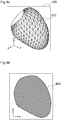
- Figure 4a is an image depicting a three-dimensional model 400 extracted from source image data (such as preoperative image data).
- the three-dimensional model 400 may be, for example, extracted from the source image data by a segmentation process.
- the three-dimensional model 400 may be a model of an anatomical structure.
- the three-dimensional model 400 is a model of the left ventricle of a human heart.
- Segmentation is a process of partitioning a digital image into multiple segments (i.e. sets of pixels) to simplify and/or change the representation of an image into something that is easier to analyse. Segmentation of the source image data is used to locate structures (such as objects and their boundaries) in the source image data. In some examples, source image data may be segmented by a combination of object localization with marginal space learning and a multi-step non-rigid deformation estimation.
- the result of image segmentation is a set of segments that collectively cover the entire image.
- Each of the pixels in a segment are similar with respect to some characteristic and adjacent segments are different with respect to the same characteristic.
- the segments may be defined by annotation.
- the resulting contours after image segmentation can be used to create a three-dimensional reconstructed model using, for example, an interpolation algorithm.
- the segments may define a three-dimension mesh model comprising a set of polygons 402 arranged to define a polygon mesh model of a structure in the source image data.
- Such a model 400 is independent of the parameters of the source image data, such as voxel intensities, resolution, field of view and other image acquisition parameters.
- the format of the mesh model 400 is independent of the imaging modality with which the source image data is generated.
- An intelligent agent trained using models generated from one imaging modality can be applied to models extracted from source image data generated using other imaging modalities without retraining the intelligent agent for the specific other modalities. Therefore, using a mesh model 400 with a format that is imaging modality-independent simplifies the training process, because it removes the need to train the intelligent agent for each combination of imaging modalities.
- Figure 4b is an image depicting an example of a two-dimensional projection 404, or binary mask, of the three-dimensional model 400 shown in Figure 4a .
- the format of the two-dimensional projection 404 is independent of the imaging modality with which the source image data is generated.
- the intelligent agent 112 may record or specify imaging geometry associated with the first target image data. For example, if the first target image data is acquired using X-ray fluoroscopy, parameters relating to collimator settings, field of view, a position and/or an orientation of the radiation source, and/or a detector type may be recorded or specified. Two-dimensional projection image data may then be generated using the same imaging geometry as that used to acquire the first target image data. This, in turn may improve the efficiency of the registration process.
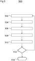
- Figure 5 is a flow diagram depicting a method 500 of determining a transformation between coordinate frames of sets of image data.
- projection image data is generated based on a two-dimensional projection of the model.
- the projection image data (binary mask) may be generated by the intelligent agent 112 as described above with reference to Figure 4b , for example.
- the intelligent agent 112 receives the projection image data and the first target image data.
- the first target image data may be X-ray fluoroscopy data acquired during an intervention procedure.
- the first target image data may be considered to be fixed image data and the projection image data may be considered to be moving image data since, in the process of performing the method 500, the projection image data may be transformed to correspond with the target image data.
- the projection image data i.e. the moving image
- the target image data may result in an image having a field of view of 300 mm x 300 mm
- the projection image data may result in an image having a field of view of 100 mm x 100 mm.
- Generating the projection image data (i.e. the moving image) to have a smaller field of view than the fixed image ensures that the moving image is within the boundaries of the fixed image.
- the intelligent agent 112 determines a reward for each of a plurality of actions applicable to the projection image data.
- the reward for each of the plurality of actions may be determined based on a translation and/or rotation of the projection image data.
- the actions may include translations in each of three orthogonal directions and rotations about axes (x, y, z) defining those directions.
- one or more of the axes may be fixed.
- depth (z axis) may be assumed to be approximately correct if the centre of the model and the centre of the first target image data are co-registered (i.e. the centres can be assumed to be collocated).
- actions may be restricted to translations along the x and y axes and rotation about the z axis, reducing the number of degrees of freedom by which the intelligent agent 112 must determine rewards for actions to three degrees of freedom.
- a reward is determined based on the effectiveness of the action. Higher rewards are determined for improvements to an alignment between the two-dimensional projection image data (of the model) and the two-dimensional target image data. Lower, or negative, rewards are determined for deterioration of alignment between the two-dimensional projection image data (of the model) and the two-dimensional target image data.
- an action is selected based on the determined rewards.
- the intelligent agent 112 may evaluate each of the rewards associated with each of the plurality of actions and determine which action has the highest associated reward. The intelligent agent 112 may thus determine that the action having the highest associated reward is the action most likely to move the model closer to a transformation to the first target image data. In other words, the intelligent agent 112 may determine that the action having the highest associated reward is the most likely to result in a registration with the target image data.
- the projection image data is transformed according to the selected action.
- the selected action may be applied to the model to generate a transformed model and further projection image data may be generated based on a two-dimensional projection of the transformed model.
- the intelligent agent 112 determines whether there is convergence. In other words, the intelligent agent 112 may determine whether a difference between an iteration of the method 500 and a previous iteration (i.e. the change brought about by transforming the model according to the selected action) is below a certain threshold. If the intelligent agent 112 determines that there is not convergence, the method 500 returns to block 502 and projection image data is regenerated based on a two-dimensional projection of a transformed model. If the intelligent agent 112 determines that there is convergence, the intelligent agent 112 moves to block 514 where the method 500 ends.
- the intelligent agent 112 To train the intelligent agent 112 to enable it to accurately and robustly determine a transformation between coordinate frames of different images (i.e. to perform the registration process), the intelligent agent 112 is provided with training data.
- the training data ideally comprises three-dimensional models (representative of the types of models extracted from source image data) that are aligned to two dimensional images representative of the target image data.
- the intelligent agent 112 trains the intelligent agent 112 to train.
- computed tomography image data is used.
- three-dimensional models and two-dimensional image data can be extracted from the same three-dimensional computed tomography image data set.
- the intelligent agent 112 is trained to determine a transformation between coordinate frames of an imaging modality-independent model and target image data
- the intelligent agent 112 can be trained using three-dimensional models and two-dimensional image data extracted from the three-dimensional computed tomography image data set, and then used in an operation setting to determine transformations between a coordinate frame of another model having an imaging modality-independent format and a coordinate frame of target image data generated using other imaging modalities.
- the three-dimensional models can be extracted by segmentation, as described above with reference to Figures 4a and 4b , to produce a mesh model (similar to that shown in Figure 4a ) and a binary mask (similar to that shown in Figure 4b ).
- the two-dimensional images are digitally reconstructed radiographs, which are artificial X-ray images generated based on an X-ray attenuation model.
- the digitally reconstructed radiographs are projections of the three-dimensional computed tomography data onto a two-dimensional plane and represent equivalent X-ray fluoroscopy data.
- the projections are generated using a ray-casting method.
- the centre of projection is deemed to be the centre point of the model (extracted from the same computed tomography image data).
- the registration task performed by the intelligent agent 112 can be formulated as a reinforcement learning problem. Steps performed by the intelligent agent 112 can be modelled as a Markov decision process: ⁇ S, A, ⁇ , r, ⁇ where S represents possible states, A represents possible actions, ⁇ represents a probability of an action taken from a state at a certain time step, r represents a reward for an action and ⁇ represents a discount factor, defining an importance of long term rewards.
- the intelligent agent 112 is in a single state, or alignment S t at a certain time step t, and actions a t in every direction along each degree of freedom are rewarded, depending on the effectiveness (i.e. better or worse alignment) of the respective action a t .
- the intelligent agent 112 can be modelled by an artificial neural network and, by training the artificial neural network, the policy ⁇ is stored in the neural network. Once the intelligent agent 112 is trained, and the policy ⁇ is stored in the neural network, the policy ⁇ may be implemented by the intelligent agent 112 in an operation setting.
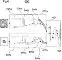
- the intelligent agent 112 is modelled by a pair of convolution neural networks 602a, 602b to encode the input images i.e. the binary masks of the mesh models and the digitally reconstructed radiographs, extracted from the computed tomography data.
- a further neural network 604 decodes the structures to determine the rewards 606.
- the input images may each be resampled to be the same resolution. For example, each input image may have a 128 x 128 pixel resolution.
- Each of the convolutional neural networks 602a, 602b may comprise 4 convolution layers 604a, 604b.
- the convolutional layers 604a, 604b are a set of low-level, shift-invariant filter functions that are learned during training of the intelligent agent 112.
- Each of the convolutional layers 604a, 604b is followed by rectified linear unit layers 606a, 606b and a pooling layer 608a, 608b.
- the rectified linear unit layers 606a, 606b set negative inputs from the convolutional layers 604a, 604b to 0 and retain positive responses from the convolutional layers 604a, 604b.
- the pooling layers 608a, 608b accumulate inputs from the rectified linear unit layers 606a, 606b to a single output.
- Figure 7 is a flow diagram depicting a method 700 of performing a training process to train the intelligent agent 112.
- training data is provided to the intelligent agent 112.
- the training data comprises three-dimensional image data, as described above.
- the training data is processed to extract a three-dimensional model of a structure from the training data and to generate digitally reconstructed two-dimensional image data.
- the intelligent agent 112 determines a reward for each of a plurality of actions applicable to the two-dimensional projection.
- the rewards may be defined as described above with reference to Figure 5 .
- the intelligent agent 112 selects an action based on the determined rewards. For example, the intelligent agent 112 may evaluate each of the rewards associated with each of the plurality of actions and determine which action has the highest associated reward. The intelligent agent 112 may thus determine that the action having the highest associated reward is the action most likely to move the model closer to a transformation to the first target image data. In other words, the intelligent agent 112 may determine that the action having the highest associated reward is the most likely to result in a registration with the target image data. In some examples, the intelligent agent 112 may be provided with an optimal reward in a so-called supervised learning process to increase the efficiency of the training process.
- the extracted three-dimensional model is transformed according to the selected action.
- the selected action may be applied to model to generate a transformed model and further projection image data may be generated based on a two-dimensional projection of the transformed model.
- the intelligent agent 112 determines whether there is convergence. In other words, the intelligent agent 112 may determine whether a difference between an iteration of the method 700 and a previous iteration (i.e. the change brought about by transforming the model according to the selected action) is below a certain threshold. If the intelligent agent 112 determines that there is not convergence, the method 700 returns to block 706 and projection image data is regenerated based on a two-dimensional projection of a transformed model. If the intelligent agent 112 determines that there is convergence, the intelligent agent 112 moves to block 716 where the method 700 ends.
Landscapes
- Engineering & Computer Science (AREA)
- Physics & Mathematics (AREA)
- General Physics & Mathematics (AREA)
- Theoretical Computer Science (AREA)
- Computer Vision & Pattern Recognition (AREA)
- Apparatus For Radiation Diagnosis (AREA)
- Image Processing (AREA)
- Magnetic Resonance Imaging Apparatus (AREA)
Priority Applications (2)
| Application Number | Priority Date | Filing Date | Title |
|---|---|---|---|
| US16/173,853 US10929989B2 (en) | 2017-11-01 | 2018-10-29 | Determining a transformation between coordinate frames of sets of image data |
| CN201811294702.XA CN109754369A (zh) | 2017-11-01 | 2018-11-01 | 确定图像数据集的坐标系之间的变换 |
Applications Claiming Priority (1)
| Application Number | Priority Date | Filing Date | Title |
|---|---|---|---|
| GBGB1718078.7A GB201718078D0 (en) | 2017-11-01 | 2017-11-01 | 2D/3D model- and image-based registration with reinforcement learning for cardiac procedures |
Publications (1)
| Publication Number | Publication Date |
|---|---|
| EP3480780A1 true EP3480780A1 (fr) | 2019-05-08 |
Family
ID=60580124
Family Applications (1)
| Application Number | Title | Priority Date | Filing Date |
|---|---|---|---|
| EP18169912.5A Ceased EP3480780A1 (fr) | 2017-11-01 | 2018-04-27 | Détermination d'une transformation entre des trames de coordonnées d'ensembles de données d'image |
Country Status (4)
| Country | Link |
|---|---|
| US (1) | US10929989B2 (fr) |
| EP (1) | EP3480780A1 (fr) |
| CN (1) | CN109754369A (fr) |
| GB (1) | GB201718078D0 (fr) |
Cited By (1)
| Publication number | Priority date | Publication date | Assignee | Title |
|---|---|---|---|---|
| CN113689476A (zh) * | 2021-08-26 | 2021-11-23 | 中国科学院植物研究所 | 一种点云配准方法、装置、存储介质和计算机设备 |
Families Citing this family (7)
| Publication number | Priority date | Publication date | Assignee | Title |
|---|---|---|---|---|
| US11475565B2 (en) * | 2018-12-21 | 2022-10-18 | GE Precision Healthcare LLC | Systems and methods for whole-body spine labeling |
| EP4390766A3 (fr) * | 2019-09-20 | 2024-09-11 | Brainlab AG | Apprentissage d'un algorithme d'apprentissage automatique à l'aide de radiographies reconstruites numériquement |
| US12142365B2 (en) * | 2019-12-10 | 2024-11-12 | Siemens Healthineers Ag | Method for registration of image data and for provision of corresponding trained facilities, apparatus for doing so and corresponding computer program product |
| EP4115389B1 (fr) * | 2020-03-05 | 2024-04-24 | Koninklijke Philips N.V. | Enregistrement d'image médicale à modes multiples, et dispositifs, systèmes et procédés associés |
| CN112308764B (zh) * | 2020-10-12 | 2024-11-26 | 杭州三坛医疗科技有限公司 | 图像配准方法及装置 |
| CN113963037B (zh) * | 2021-09-29 | 2025-03-25 | 上海联影医疗科技股份有限公司 | 图像配准方法、装置、计算机设备和存储介质 |
| CN115393514A (zh) * | 2022-08-26 | 2022-11-25 | 北京百度网讯科技有限公司 | 三维重建模型的训练方法、三维重建方法、装置、设备 |
Citations (3)
| Publication number | Priority date | Publication date | Assignee | Title |
|---|---|---|---|---|
| EP3121789A1 (fr) * | 2015-07-22 | 2017-01-25 | Siemens Medical Solutions USA, Inc. | Procédé et système de recalage 2d/3d d'images régression à base d'un réseau neuronal convolutif |
| US20170217102A1 (en) * | 2016-01-29 | 2017-08-03 | Siemens Medical Solutions Usa, Inc. | Multi-Modality Image Fusion for 3D Printing of Organ Morphology and Physiology |
| EP3246875A2 (fr) * | 2016-05-18 | 2017-11-22 | Siemens Healthcare GmbH | Procédé et système d'enregistrement d'image à l'aide d'un agent artificiel intelligent |
Family Cites Families (3)
| Publication number | Priority date | Publication date | Assignee | Title |
|---|---|---|---|---|
| US8160345B2 (en) * | 2008-04-30 | 2012-04-17 | Otismed Corporation | System and method for image segmentation in generating computer models of a joint to undergo arthroplasty |
| US9165362B2 (en) * | 2013-05-07 | 2015-10-20 | The Johns Hopkins University | 3D-2D image registration for medical imaging |
| US9569736B1 (en) * | 2015-09-16 | 2017-02-14 | Siemens Healthcare Gmbh | Intelligent medical image landmark detection |
-
2017
- 2017-11-01 GB GBGB1718078.7A patent/GB201718078D0/en not_active Ceased
-
2018
- 2018-04-27 EP EP18169912.5A patent/EP3480780A1/fr not_active Ceased
- 2018-10-29 US US16/173,853 patent/US10929989B2/en active Active
- 2018-11-01 CN CN201811294702.XA patent/CN109754369A/zh active Pending
Patent Citations (3)
| Publication number | Priority date | Publication date | Assignee | Title |
|---|---|---|---|---|
| EP3121789A1 (fr) * | 2015-07-22 | 2017-01-25 | Siemens Medical Solutions USA, Inc. | Procédé et système de recalage 2d/3d d'images régression à base d'un réseau neuronal convolutif |
| US20170217102A1 (en) * | 2016-01-29 | 2017-08-03 | Siemens Medical Solutions Usa, Inc. | Multi-Modality Image Fusion for 3D Printing of Organ Morphology and Physiology |
| EP3246875A2 (fr) * | 2016-05-18 | 2017-11-22 | Siemens Healthcare GmbH | Procédé et système d'enregistrement d'image à l'aide d'un agent artificiel intelligent |
Non-Patent Citations (2)
| Title |
|---|
| MIAO SHUN ET AL: "A CNN Regression Approach for Real-Time 2D/3D Registration", IEEE TRANSACTIONS ON MEDICAL IMAGING, IEEE SERVICE CENTER, PISCATAWAY, NJ, US, vol. 35, no. 5, 1 May 2016 (2016-05-01), pages 1352 - 1363, XP011607918, ISSN: 0278-0062, [retrieved on 20160429], DOI: 10.1109/TMI.2016.2521800 * |
| RUI LIAO ET AL: "An Artificial Agent for Robust Image Registration", ARXIV.ORG, CORNELL UNIVERSITY LIBRARY, 201 OLIN LIBRARY CORNELL UNIVERSITY ITHACA, NY 14853, 30 November 2016 (2016-11-30), XP080735862 * |
Cited By (1)
| Publication number | Priority date | Publication date | Assignee | Title |
|---|---|---|---|---|
| CN113689476A (zh) * | 2021-08-26 | 2021-11-23 | 中国科学院植物研究所 | 一种点云配准方法、装置、存储介质和计算机设备 |
Also Published As
| Publication number | Publication date |
|---|---|
| US10929989B2 (en) | 2021-02-23 |
| US20190130587A1 (en) | 2019-05-02 |
| CN109754369A (zh) | 2019-05-14 |
| GB201718078D0 (en) | 2017-12-13 |
Similar Documents
| Publication | Publication Date | Title |
|---|---|---|
| US10929989B2 (en) | Determining a transformation between coordinate frames of sets of image data | |
| US12094116B2 (en) | Method and system for image registration using an intelligent artificial agent | |
| JP7120584B2 (ja) | 被検体の位置決め装置、被検体の位置決め方法、被検体の位置決めプログラムおよび放射線治療システム | |
| EP4270313A1 (fr) | Enregistrement d'images de projection en images volumétriques | |
| CN112020747B (zh) | 用于跟踪程序的系统和方法 | |
| CN111340859B (zh) | 用于图像配准的方法、学习装置和医学成像装置 | |
| Al Arif et al. | Fully automatic cervical vertebrae segmentation framework for X-ray images | |
| EP3145420B1 (fr) | Procédé de suivi preopératoire | |
| JP2021522931A (ja) | 機械学習を使用する手術中の自動腫瘍識別 | |
| Jaganathan et al. | Self-supervised 2D/3D registration for X-ray to CT image fusion | |
| CN113129308A (zh) | 图像分割方法、装置和非暂态计算机可读存储介质 | |
| JP2021520236A (ja) | 解剖学的構造に基づくフレームレス二次元/三次元画像レジストレーション | |
| Gu et al. | Extended capture range of rigid 2D/3D registration by estimating riemannian pose gradients | |
| CN108430376B (zh) | 提供投影数据集 | |
| CN113597288B (zh) | 基于影像匹配确定手术路径的方法与系统 | |
| JP7233409B2 (ja) | 対象の動的多次元画像に対する方法及びシステム | |
| EP4522267B1 (fr) | Détermination de radiographies reconstruites numériquement et physiquement correctes | |
| US20250000583A1 (en) | Atlas-based trajectory planning | |
| WO2019091883A1 (fr) | Étalonnage de procédures de suivi basées sur l'enregistrement d'images | |
| EP4300414A1 (fr) | Transfert des positions des marqueurs d'une image de référence à une image médicale de suivi | |
| Kung | Intraoperative Vertebra Segmentation from 3D Point Clouds for Radiation-Free Spinal Curve Tracking in Scoliosis Surgery | |
| WO2025228504A1 (fr) | Localisation de tissu mou dans un traitement par rayonnement à l'aide de motifs de mouvement similaires |
Legal Events
| Date | Code | Title | Description |
|---|---|---|---|
| PUAI | Public reference made under article 153(3) epc to a published international application that has entered the european phase |
Free format text: ORIGINAL CODE: 0009012 |
|
| STAA | Information on the status of an ep patent application or granted ep patent |
Free format text: STATUS: THE APPLICATION HAS BEEN PUBLISHED |
|
| AK | Designated contracting states |
Kind code of ref document: A1 Designated state(s): AL AT BE BG CH CY CZ DE DK EE ES FI FR GB GR HR HU IE IS IT LI LT LU LV MC MK MT NL NO PL PT RO RS SE SI SK SM TR |
|
| AX | Request for extension of the european patent |
Extension state: BA ME |
|
| STAA | Information on the status of an ep patent application or granted ep patent |
Free format text: STATUS: REQUEST FOR EXAMINATION WAS MADE |
|
| 17P | Request for examination filed |
Effective date: 20190704 |
|
| RBV | Designated contracting states (corrected) |
Designated state(s): AL AT BE BG CH CY CZ DE DK EE ES FI FR GB GR HR HU IE IS IT LI LT LU LV MC MK MT NL NO PL PT RO RS SE SI SK SM TR |
|
| STAA | Information on the status of an ep patent application or granted ep patent |
Free format text: STATUS: EXAMINATION IS IN PROGRESS |
|
| 17Q | First examination report despatched |
Effective date: 20210419 |
|
| RAP1 | Party data changed (applicant data changed or rights of an application transferred) |
Owner name: SIEMENS HEALTHINEERS AG |
|
| APBN | Date of receipt of notice of appeal recorded |
Free format text: ORIGINAL CODE: EPIDOSNNOA2E |
|
| APBR | Date of receipt of statement of grounds of appeal recorded |
Free format text: ORIGINAL CODE: EPIDOSNNOA3E |
|
| APAF | Appeal reference modified |
Free format text: ORIGINAL CODE: EPIDOSCREFNE |
|
| APAV | Appeal reference deleted |
Free format text: ORIGINAL CODE: EPIDOSDREFNE |
|
| APBT | Appeal procedure closed |
Free format text: ORIGINAL CODE: EPIDOSNNOA9E |
|
| STAA | Information on the status of an ep patent application or granted ep patent |
Free format text: STATUS: THE APPLICATION HAS BEEN REFUSED |
|
| 18R | Application refused |
Effective date: 20260120 |