EP3501400A1 - Procédé et dispositif permettant d'assurer un positionnement correct pour une image radiographique - Google Patents
Procédé et dispositif permettant d'assurer un positionnement correct pour une image radiographique Download PDFInfo
- Publication number
- EP3501400A1 EP3501400A1 EP17209022.7A EP17209022A EP3501400A1 EP 3501400 A1 EP3501400 A1 EP 3501400A1 EP 17209022 A EP17209022 A EP 17209022A EP 3501400 A1 EP3501400 A1 EP 3501400A1
- Authority
- EP
- European Patent Office
- Prior art keywords
- positioning
- image
- preview image
- recording
- unit
- Prior art date
- Legal status (The legal status is an assumption and is not a legal conclusion. Google has not performed a legal analysis and makes no representation as to the accuracy of the status listed.)
- Granted
Links
Images

Classifications
-
- A—HUMAN NECESSITIES
- A61—MEDICAL OR VETERINARY SCIENCE; HYGIENE
- A61B—DIAGNOSIS; SURGERY; IDENTIFICATION
- A61B6/00—Apparatus or devices for radiation diagnosis; Apparatus or devices for radiation diagnosis combined with radiation therapy equipment
- A61B6/08—Auxiliary means for directing the radiation beam to a particular spot, e.g. using light beams
-
- A—HUMAN NECESSITIES
- A61—MEDICAL OR VETERINARY SCIENCE; HYGIENE
- A61B—DIAGNOSIS; SURGERY; IDENTIFICATION
- A61B5/00—Measuring for diagnostic purposes; Identification of persons
- A61B5/0059—Measuring for diagnostic purposes; Identification of persons using light, e.g. diagnosis by transillumination, diascopy, fluorescence
- A61B5/0062—Arrangements for scanning
- A61B5/0064—Body surface scanning
-
- A—HUMAN NECESSITIES
- A61—MEDICAL OR VETERINARY SCIENCE; HYGIENE
- A61B—DIAGNOSIS; SURGERY; IDENTIFICATION
- A61B6/00—Apparatus or devices for radiation diagnosis; Apparatus or devices for radiation diagnosis combined with radiation therapy equipment
- A61B6/04—Positioning of patients; Tiltable beds or the like
-
- A—HUMAN NECESSITIES
- A61—MEDICAL OR VETERINARY SCIENCE; HYGIENE
- A61B—DIAGNOSIS; SURGERY; IDENTIFICATION
- A61B6/00—Apparatus or devices for radiation diagnosis; Apparatus or devices for radiation diagnosis combined with radiation therapy equipment
- A61B6/04—Positioning of patients; Tiltable beds or the like
- A61B6/0487—Motor-assisted positioning
-
- A—HUMAN NECESSITIES
- A61—MEDICAL OR VETERINARY SCIENCE; HYGIENE
- A61B—DIAGNOSIS; SURGERY; IDENTIFICATION
- A61B6/00—Apparatus or devices for radiation diagnosis; Apparatus or devices for radiation diagnosis combined with radiation therapy equipment
- A61B6/46—Arrangements for interfacing with the operator or the patient
- A61B6/467—Arrangements for interfacing with the operator or the patient characterised by special input means
-
- A—HUMAN NECESSITIES
- A61—MEDICAL OR VETERINARY SCIENCE; HYGIENE
- A61B—DIAGNOSIS; SURGERY; IDENTIFICATION
- A61B6/00—Apparatus or devices for radiation diagnosis; Apparatus or devices for radiation diagnosis combined with radiation therapy equipment
- A61B6/46—Arrangements for interfacing with the operator or the patient
- A61B6/467—Arrangements for interfacing with the operator or the patient characterised by special input means
- A61B6/469—Arrangements for interfacing with the operator or the patient characterised by special input means for selecting a region of interest [ROI]
-
- A—HUMAN NECESSITIES
- A61—MEDICAL OR VETERINARY SCIENCE; HYGIENE
- A61B—DIAGNOSIS; SURGERY; IDENTIFICATION
- A61B6/00—Apparatus or devices for radiation diagnosis; Apparatus or devices for radiation diagnosis combined with radiation therapy equipment
- A61B6/48—Diagnostic techniques
- A61B6/488—Diagnostic techniques involving pre-scan acquisition
-
- A—HUMAN NECESSITIES
- A61—MEDICAL OR VETERINARY SCIENCE; HYGIENE
- A61B—DIAGNOSIS; SURGERY; IDENTIFICATION
- A61B6/00—Apparatus or devices for radiation diagnosis; Apparatus or devices for radiation diagnosis combined with radiation therapy equipment
- A61B6/54—Control of apparatus or devices for radiation diagnosis
-
- A—HUMAN NECESSITIES
- A61—MEDICAL OR VETERINARY SCIENCE; HYGIENE
- A61B—DIAGNOSIS; SURGERY; IDENTIFICATION
- A61B6/00—Apparatus or devices for radiation diagnosis; Apparatus or devices for radiation diagnosis combined with radiation therapy equipment
- A61B6/54—Control of apparatus or devices for radiation diagnosis
- A61B6/542—Control of apparatus or devices for radiation diagnosis involving control of exposure
- A61B6/544—Control of apparatus or devices for radiation diagnosis involving control of exposure dependent on patient size
-
- A—HUMAN NECESSITIES
- A61—MEDICAL OR VETERINARY SCIENCE; HYGIENE
- A61B—DIAGNOSIS; SURGERY; IDENTIFICATION
- A61B6/00—Apparatus or devices for radiation diagnosis; Apparatus or devices for radiation diagnosis combined with radiation therapy equipment
- A61B6/54—Control of apparatus or devices for radiation diagnosis
- A61B6/545—Control of apparatus or devices for radiation diagnosis involving automatic set-up of acquisition parameters
-
- G—PHYSICS
- G03—PHOTOGRAPHY; CINEMATOGRAPHY; ANALOGOUS TECHNIQUES USING WAVES OTHER THAN OPTICAL WAVES; ELECTROGRAPHY; HOLOGRAPHY
- G03B—APPARATUS OR ARRANGEMENTS FOR TAKING PHOTOGRAPHS OR FOR PROJECTING OR VIEWING THEM; APPARATUS OR ARRANGEMENTS EMPLOYING ANALOGOUS TECHNIQUES USING WAVES OTHER THAN OPTICAL WAVES; ACCESSORIES THEREFOR
- G03B42/00—Obtaining records using waves other than optical waves; Visualisation of such records by using optical means
- G03B42/02—Obtaining records using waves other than optical waves; Visualisation of such records by using optical means using X-rays
- G03B42/026—Obtaining records using waves other than optical waves; Visualisation of such records by using optical means using X-rays for obtaining three-dimensional pictures
-
- G—PHYSICS
- G06—COMPUTING OR CALCULATING; COUNTING
- G06T—IMAGE DATA PROCESSING OR GENERATION, IN GENERAL
- G06T17/00—Three-dimensional [3D] modelling for computer graphics
-
- G—PHYSICS
- G06—COMPUTING OR CALCULATING; COUNTING
- G06T—IMAGE DATA PROCESSING OR GENERATION, IN GENERAL
- G06T7/00—Image analysis
- G06T7/20—Analysis of motion
- G06T7/292—Multi-camera tracking
-
- G—PHYSICS
- G06—COMPUTING OR CALCULATING; COUNTING
- G06T—IMAGE DATA PROCESSING OR GENERATION, IN GENERAL
- G06T2207/00—Indexing scheme for image analysis or image enhancement
- G06T2207/20—Special algorithmic details
- G06T2207/20081—Training; Learning
-
- G—PHYSICS
- G06—COMPUTING OR CALCULATING; COUNTING
- G06T—IMAGE DATA PROCESSING OR GENERATION, IN GENERAL
- G06T2210/00—Indexing scheme for image generation or computer graphics
- G06T2210/41—Medical
Definitions
- the invention relates to a method and a device for positioning a body region for recording by a radiographic system, in particular for real-time alignment of camera image with simulated X-ray.
- the invention further relates to a control device for a radiography system and a corresponding radiography system.
- the object to be photographed e.g. a knee, wrist, or ankle
- the object to be photographed e.g. a knee, wrist, or ankle
- the orientation or positioning of the object or the receiving elements was not optimal, so that the object in the receptacle is e.g. is shifted, a part of the object is cut off, or the object has been mapped out from a wrong shooting angle.
- a radiographic system is understood to mean a system for projection radiography and not for tomographic imaging.
- areas of the patient's body are irradiated with X-rays from one direction.
- the radiation is irradiated with suitable materials, e.g. Film or a spatially resolving detector) and thereby creates a, usually two-dimensional, projection image.
- suitable materials e.g. Film or a spatially resolving detector
- the principle of the invention is limited to digital radiography systems.
- Preferred systems in this respect are floor-mounted systems, systems with ceiling-mounted spotlights, possibly with table and raster wall unit, systems with radiator and detector on robotic support arms, but also mobile X-ray systems.
- the method according to the invention for positioning a body region of a patient, who may be a human but also an animal, for a, in particular two-dimensional, radiograph recording by a radiography system comprises the following steps:
- the method is informed about which body area, e.g. which bone or which organ it is and which recording should be performed.
- the examination request may simply include an indication of the body area, e.g. "Knee AP", "Knee Lateral".
- an organ program which includes an examination request.
- the organ program is called e.g. Knee AP, but includes all necessary parameters for a specific radiograph, e.g. Information about the generator, the image processing, the image representation and / or the device position.
- the body area for the radiographic image is prepositioned.
- the patient is positioned so that the relevant body region is aligned over an image detector.
- the recording unit and possibly also the image detector of the radiographic system for radiography recording is positioned appropriately. This positioning should be done according to the examination request. If the examination request comprises corresponding control commands or if the method has access to a database with control commands which are linked with information of an examination request in terms of data, this pre-positioning can be carried out quite automatically.
- a three-dimensional positioning image of the body area is created using a 3D camera system.
- This 3D camera system can include a 3D camera or 2 cameras whose image information is combined. The positioning capture is made while the body region is in its position taken in step b). Should the patient have moved, too this can be understood as pre-positioning within the meaning of the invention, since step b) can certainly also be carried out before step a) or after step c).
- a patient model is first generated from the positioning image, e.g. a three-dimensional body or bone model. From this patient model, the preview image is then created, e.g. as a projection of a three-dimensional patent model on a two-dimensional plane or as a section of this patient model.
- the preview image shows a representation of how it would have been done with the recording unit of the radiography system in the pre-positioning assumed in step c).
- the last point which is basically a simulation of the later radiographic image, can be realized in that the relative position of the 3D camera system relative to the recording unit is known or at least very easily determined or calibrated. Since these positions are known, a relationship between the positioning receptacle and the viewing angle and receiving area of the receiving unit can be established. From pre-positioning data of the recording unit and the positioning recording this can be transformed by calculations in space so that it is displayed from the viewpoint of the recording unit. Basically, only the transformation matrix of the viewing direction of the 3D camera system to the viewing direction of the recording unit has to be known. Thus, the patient model can be automatically aligned in position and fade on the body area.
- the patient model may be a physical model of the object, a type of voxel phantom with defined properties.
- X-ray quanta can be simulated, which pass through the object, are scattered or absorbed there, and at the end create a projection image on the detector, eg by means of ray tracing or Monte Carlo simulation.
- the parameters of the X-ray quanta should correspond to the real parameters (those for recording the planned radiographic image), in particular with regard to the beam energy, the beam current and / or the magnitude kV / mAs.
- an over or underexposure can be detected in advance.
- the preview image and / or a positioning information based on the preview image is output.
- an automatic examination of the preview image can be made as to whether it correctly displays the body region within the scope of the examination request for the radiographic image.
- An operator can use the preview image to decide if the system settings or pre-positions need to be adjusted or corrected. Radiography must not be done and the patient should not be exposed to any dose until the decision is made.
- the method can also automatically inform the operator by means of the positioning information whether the preview image is acceptable or not.
- an automatic examination of the preview image serves to generate the positioning information.
- a database is built in the system with X-ray images at different system settings (projections) correlated to the camera view of the body part of the patient.
- the camera view is the view of a 3D avatar of the patient; ie specifics of the current patient image such as clothing, hair, exposure, ... in the room are calculated.
- This database thus consists of the three parameters X-ray image, device position and camera view on Avatar.
- the database grows with each x-ray (could be supplemented by X-ray images of other systems). Now, if the device is positioned and the avatar image made, you can search in the database, the correlating X-ray image and display as a preview image.
- the method can be improved by considering further parameters: planned recording parameters (KV, mAs, dose, ...), image processing, volume of the avatar, ...
- the device also comprises a pre-positioning display unit, by means of which the desired positioning of the body region can be displayed.
- a radiographic system according to the invention comprises a control device according to the invention.
- a large part of the aforementioned components of the device or of the control device can be implemented completely or partially in the form of software modules in a processor of a corresponding device or control device.
- a largely software implementation has the advantage that even previously used computing systems or control devices can be retrofitted in a simple manner by a software update to work in the manner of the invention.
- the object is also achieved by a corresponding computer program product with a computer program which is directly loadable into a computing system or a storage device of a control device of a radiography system, with program sections to perform all steps of the inventive method, if the program in the computing system or the Control device is executed.
- Such a computer program product in addition to the computer program optionally additional components such.
- documentation and / or additional components may include hardware components, such as hardware.
- Hardware keys (dongles, etc.) for using the software include
- a computer readable medium for example a memory stick, a hard disk or another transportable or permanently installed data carrier may be used on which the data from a computing system or a computer unit of the control device readable and executable program sections of the computer program are stored.
- the Computing unit may, for example, this one or more cooperating microprocessors or the like.
- a three-dimensional surface image of the body region is taken or created as a positioning shot by the 3D camera system.
- the patient model is theoretically, e.g. As described above, from a set of many potential models can be selected from a database, it is preferable that a three-dimensional patient model is generated from the surface image and three-dimensional organ data objects of an organ database.
- the organ database preferably contains three-dimensional graphical objects, in particular the group organs, bones, cartilage, muscles, vessels, nerves and other tissue, and additionally markers with which their positioning in the surface image or registration with the surface image is possible.
- the three-dimensional surface information From the three-dimensional surface information, it can be made of e.g. be calculated using the principle of machine-learning an avatar.
- the preview image is then created from a combination of the position of the prepositioned recording unit and the three-dimensional patient model. This can be done, for example, by moving or rotating the patient model in space so that it reproduces a view as if the 3D camera system was identical to Aligned recording position. Then, preferably, a projection of the patient model in the direction of the receiving unit is made.
- a device of this kind comprises an organ database or a data interface to such an organ database, wherein the generation unit is designed to create a three-dimensional patient model from the positioning recording and three-dimensional organ data objects of the organ database.
- the patient model is created based on the principle of machine learning and / or statistical body models. It prefers the form of a (personalized) avatar.
- a neural network with a large amount of data, e.g. Camera and X-rays are trained.
- the examination request preferably defines additional acquisition parameters for the acquisition unit, e.g. Energy, exposure time, collimator setting.
- additional acquisition parameters for the acquisition unit e.g. Energy, exposure time, collimator setting.
- the preview image simulates a Radiographiem that would have been recorded with these recording parameters.
- the preview image is displayed on a screen, eg in the room or even remotely, or as a projection, eg on a table surface.
- the operator can be told what the target position looks like or what should be done (eg, an arrow to the right could mean that the body area should be moved to the right until an indicator eg turns green).
- the target position is a position in which the preview image is acceptable and thus later, the final radiographic image is likely to become acceptable. Acceptable means that a physician would view this image as acceptable for diagnostics usability.
- a recording can be considered acceptable if, for example, the examined body area is fully imaged and correctly positioned (eg the joint space is visible in a knee shot), there is sufficient contrast in the image, no irrelevant areas in the radiation field or quite fundamentally the right one Body area is displayed (eg no left / right confusion exists).
- the patient may also be the operator himself and perform the positioning without background knowledge from the positioning information.
- the positioning information preferably contains information as to whether or not the preview image is acceptable according to the examination request and also which settings should be adapted, e.g. Collimator settings or position / rotation of the display unit or the body area.
- the output is realized with an optical and / or acoustic display. It preferably contains information, in particular pictorial representations or voice commands, about what the target position looks like, whether the target position has been reached and / or navigation information.
- the output comprises the representation of a change of a light field of the radiography system, wherein preferably the brightness and / or the color of the light field changes as soon as a correct positioning is achieved.
- the radiography system is controlled, wherein at least the steps of the method according to the invention with different prepositions of the recording unit and / or the body region are performed two or more times become.
- this multiple implementation can be carried out purely automatically, in that the body region is not moved and only the pre-positioning of the acquisition unit is automatically changed.
- a change in the pre-positioning of the detector can be done. This should always be positioned so that the beam of the recording unit falls onto the detector.
- the pre-positioning of the body area can change alone.
- automated navigational aids could be indicated to the patient, eg, by a voice announcing the positioning (eg, "farther to the left") or corresponding characters that are displayed.
- a presetting is thus made again or the existing presettings are changed and a positioning recording is made again and a preview image is automatically generated and output. If the preview image is correct, the radiographic image can be taken.
- the data base for a trained neural network can be continuously supplemented by newly acquired images (X-ray and positioning recordings, or all relevant parameters).
- the evaluation of the newly acquired images can be carried out manually by the skilled personnel or carried out automatically.
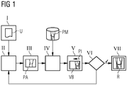
- FIG. 1 1 shows a schematic block diagram of a preferred method sequence for positioning a body region K of a patient P for a radiographic image R by means of a radiography system 1 (see also here Fig. 3 ).
- step I provision is made of an examination request U of the body region K.
- the system is provided with e.g. communicated to which institution it is and which recording is to be performed.
- the example case is assumed that the examination request U has a radiographic image R of the knee to the goal.
- step II a pre-positioning of both the body region K in the radiography system 1 and a recording unit 3 of the radiography system 1 for the radiographic image (R).
- the patient is asked to place his knee in a certain position, the receiving unit 3 is prepositioned above the knee so that the knee gap would most likely be correctly imaged in a radiography receptacle R. Even if the pre-positioning may look correct from the outside, this need not necessarily be true and there is a "dose-free" review according to the invention.
- step III a three-dimensional positioning shot PA of the body region K is made with a 3D camera system 6, while the body region K is in its position taken in step II.
- a three-dimensional surface image OB of the knee is obtained.
- the description is attached FIG. 2 directed.
- step IV follows the creation of a preview image VB from the positioning recording PA.
- a patient model PM is generated from the positioning acquisition PA and possibly also from the examination request U (see FIG. Fig. 2 ).
- a three-dimensional model of the knee with visible bones and a skin and muscle layer shown transparently is taken from a database and registered with the surface image OB.
- the resulting image then represents the patient model PM.
- the preview image VB possibly by adjusting the spatial orientation or positioning of the patient model PM is created so that it reproduces a representation as would have been done with the intended in step II recording unit 3 of the radiography 1.
- a two-dimensional projection of the patient model PM is created, which looks as if it had been taken with the recording unit 3.
- step V the output of the preview image VB and a positioning information PI based on the preview image VB takes place.
- a positioning information PI based on the preview image VB.
- step VI an examination is made as to whether or not there is a correct positioning. This is not the case in the example case. Therefore, part of the process is re-run from step II. In the example case, not the knee is moved to the left, but the receiving unit to the right and steps III to VI are run through again.
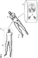
- FIG. 2 shows a schematic representation of the formation of a patient model PM.
- the patient model PM of a whole body is displayed.
- the positioning shot PA is shown with the surface image OB as it is detected by the 3D camera system 6 (see FIG. FIG. 3 ) could have been recorded.
- the surface image OB only shows the surface of the patient P without internal structures.
- These structures here referred to as objects O, may comprise all possible internal structures in the body, eg bones, organs, vessels, connective tissue or nerves.
- the objects are present here as three-dimensional graphic data objects in an object database OD.
- organs and bones are used from already existing models, eg from statistical body models.
- three-dimensional CT data sets from comparable patients can also be inserted into the patient model PM (into the avatar).
- the objects are fitted into the three-dimensional surface image, e.g. by means of a graphic registration.
- This patient model PM provides the basis for creating the preview image VB.
- the preview image VB may be the image of the patient model PM, however, it is more advantageous if the preview image VB is in the manner of displaying the planned image Radiology image R is similar or even equivalent.
- the preview image VB is preferably calculated from the patient model PM by simulating an X-ray projection image resulting from the positioning of the acquisition unit 3 and detector 4 to the body region K and the adjusted parameters, such as e.g. Beam energy, collimator, etc. would result. It is therefore preferred that the preview image VB is a two-dimensional projection of the three-dimensional patient model PM with a transparent surface image OB.
- radiographic system 1 is a digital X-ray machine. In principle, however, the method can also be used on other radiographic systems 1.
- FIG. 3 shows roughly schematically a radiography system 1 with a control device 12 for carrying out the method according to the invention.
- the radiography system 1 has in the usual way a recording unit 3, which represents an X-ray source here, and during a Radiography analysis (R) a patient (P) radiates through, so that the radiation impinges on the recording unit 3 each opposite detector 4.
- the receiving unit 3 can be moved by means of a movement mechanism 2, in this case a swivel arm, eg raised / lowered, pivoted or rotated.
- the movement mechanism 2 allows a plurality of different movements, so that the receiving unit can be optimally moved with respect to its height, its lateral position and its angle of inclination.
- control device 12 only the components are shown, which are essential for the explanation of the invention. In principle, conventional radiography systems and associated control devices are known to the person skilled in the art and therefore need not be explained in detail.
- the radiography system 1 can be controlled by the controller 12, i. it is e.g. the movement mechanism 2 is controlled to move the recording unit 3, or a radiography recording is started.
- the receiving unit 3 may also experience adjustment via the control interface 8, e.g. in terms of exposure time or beam energy.
- a user interface, an acquisition interface for the captured image data or an image data reconstruction unit are not shown, but of course the control unit 12 may also comprise such elements.
- the control device 12 comprises a device 5 for positioning a body region K of a patient P for a radiographic image R. In the case shown, this device 5 does not act directly on the radiography system 1 but via the control interface 8 of the control device 12.
- the device 5 comprises an interface 11 for providing an examination request U of the body region K. In the illustrated case, it receives an examination request U from the outside, which can be input eg via a user interface. This may well be on a User interface of the controller 12 are used.
- a pre-positioning of a recording unit 3 of the radiographic system 1 can be achieved.
- the interface 7 sends the data for positioning to the control interface 8 of the control device 12 and this controls the movement mechanism 2 and thus positions the receiving unit 3 in accordance with the specifications of the interface 7.
- a 3D camera system 6 is not part of the device 5, which however may well be the case in an alternative embodiment.
- the device 5 for establishing a positioning capture PA of the body region K accesses a 3D camera system 6 via a camera interface 6a, controls its function, starts a capture and receives its three-dimensional positioning capture PA.
- This positioning recording PA is then provided for the creation unit 9, as well as the examination request U is provided by means of the interface 11 of the creation unit 9.
- a patient model PM is generated by selecting it from a database 13 which is optionally part of the device 5 here.
- a preview image VB is created by means of the creation unit 9 and the generated patient model PM, which reproduces a representation of how it would have been carried out with the acquisition unit 3 of the radiography system 1 as intended.
- This can be achieved, for example, by first producing a three-dimensional patient model PM, which is displaced or rotated correspondingly in space according to the relative position of the 3D camera system to the acquisition unit 3, and then a projection of the patient model is made. Alternatively, of course, the projection direction can be adjusted accordingly.
- the patient model PM could be oriented in space as if it had been recorded by the 3D camera system at the location of the recording unit 3. From this, a suitable preview image can be created from a corresponding vertical projection.
- the preview image VB or a positioning information PI based on the preview image VB can be output.
- the output unit 10 may thus be a display or an interface for data exchange with a computer.
- the output unit 10 is (also) designed to send data to the control interface 8 and thus, e.g. to achieve a further pre-positioning of the receiving unit 3.
Landscapes
- Health & Medical Sciences (AREA)
- Life Sciences & Earth Sciences (AREA)
- Engineering & Computer Science (AREA)
- Medical Informatics (AREA)
- Physics & Mathematics (AREA)
- Molecular Biology (AREA)
- Animal Behavior & Ethology (AREA)
- Biophysics (AREA)
- Nuclear Medicine, Radiotherapy & Molecular Imaging (AREA)
- Veterinary Medicine (AREA)
- Pathology (AREA)
- Radiology & Medical Imaging (AREA)
- Biomedical Technology (AREA)
- Heart & Thoracic Surgery (AREA)
- Public Health (AREA)
- Surgery (AREA)
- General Health & Medical Sciences (AREA)
- High Energy & Nuclear Physics (AREA)
- Optics & Photonics (AREA)
- General Physics & Mathematics (AREA)
- Human Computer Interaction (AREA)
- Theoretical Computer Science (AREA)
- Computer Graphics (AREA)
- Geometry (AREA)
- Software Systems (AREA)
- Multimedia (AREA)
- Computer Vision & Pattern Recognition (AREA)
- Apparatus For Radiation Diagnosis (AREA)
Priority Applications (3)
| Application Number | Priority Date | Filing Date | Title |
|---|---|---|---|
| EP17209022.7A EP3501400B1 (fr) | 2017-12-20 | 2017-12-20 | Procédé et dispositif permettant d'assurer un positionnement correct pour une image radiographique |
| US16/223,286 US10779774B2 (en) | 2017-12-20 | 2018-12-18 | Method and apparatus for ensuring correct positioning for a radiography acquisition |
| CN201811557115.5A CN109938763A (zh) | 2017-12-20 | 2018-12-19 | 用于确保针对放射成像采集的正确定位的方法和装置 |
Applications Claiming Priority (1)
| Application Number | Priority Date | Filing Date | Title |
|---|---|---|---|
| EP17209022.7A EP3501400B1 (fr) | 2017-12-20 | 2017-12-20 | Procédé et dispositif permettant d'assurer un positionnement correct pour une image radiographique |
Publications (2)
| Publication Number | Publication Date |
|---|---|
| EP3501400A1 true EP3501400A1 (fr) | 2019-06-26 |
| EP3501400B1 EP3501400B1 (fr) | 2022-06-08 |
Family
ID=60813618
Family Applications (1)
| Application Number | Title | Priority Date | Filing Date |
|---|---|---|---|
| EP17209022.7A Active EP3501400B1 (fr) | 2017-12-20 | 2017-12-20 | Procédé et dispositif permettant d'assurer un positionnement correct pour une image radiographique |
Country Status (3)
| Country | Link |
|---|---|
| US (1) | US10779774B2 (fr) |
| EP (1) | EP3501400B1 (fr) |
| CN (1) | CN109938763A (fr) |
Cited By (1)
| Publication number | Priority date | Publication date | Assignee | Title |
|---|---|---|---|---|
| EP4335374A1 (fr) * | 2022-09-09 | 2024-03-13 | Canon Medical Systems Corporation | Appareil de diagnostic à rayons x et procédé de diagnostic à rayons x |
Families Citing this family (20)
| Publication number | Priority date | Publication date | Assignee | Title |
|---|---|---|---|---|
| JP7350519B2 (ja) * | 2019-05-29 | 2023-09-26 | キヤノン株式会社 | 放射線撮影システム、放射線撮影制御装置及びその制御方法、並びに、プログラム |
| CN110507338B (zh) * | 2019-08-30 | 2022-12-27 | 东软医疗系统股份有限公司 | 定位方法、装置、设备及数字化x线摄影系统 |
| IT201900018230A1 (it) * | 2019-10-08 | 2021-04-08 | Zoeen S R L | Dispositivo di visualizzazione di strutture sub-superficiali e metodo di visualizzazione di dette strutture sub-superficiali |
| EP3822845A1 (fr) * | 2019-11-12 | 2021-05-19 | Koninklijke Philips N.V. | Classification de pose de sujet à l'aide de coordonnées d'emplacement conjointes |
| US11721014B2 (en) * | 2019-11-15 | 2023-08-08 | GE Precision Healthcare LLC | Methods and systems for a field-of-view preview |
| JP7397968B2 (ja) * | 2020-03-30 | 2023-12-13 | 富士フイルム株式会社 | 撮影支援装置、その作動方法、及び作動プログラム |
| DE102020207753A1 (de) * | 2020-06-23 | 2021-12-23 | Siemens Healthcare Gmbh | Optimierung der Lagerung eines Patienten auf einer Patientenliege für die medizinische Bildgebung |
| EP4178446B1 (fr) | 2020-08-10 | 2024-11-20 | Shanghai United Imaging Healthcare Co., Ltd. | Systèmes et procédés d'imagerie |
| US20220084239A1 (en) * | 2020-09-17 | 2022-03-17 | Gerd Bodner | Evaluation of an ultrasound-based investigation |
| DE102020213690B4 (de) * | 2020-10-30 | 2022-01-20 | Siemens Healthcare Gmbh | Verfahren zum Einstellen einer Form einer Kopfunterlage, Verfahren zum Bereitstellen von Bildgebungsdaten und System mit einer Kopfunterlage |
| EP4000525A1 (fr) * | 2020-11-23 | 2022-05-25 | Koninklijke Philips N.V. | Guide pour positionner un patient en imagerie médicale |
| EP4137058A1 (fr) * | 2021-08-17 | 2023-02-22 | Koninklijke Philips N.V. | Procédé de fourniture de données de guidage dans un système à rayons x |
| EP4144298A1 (fr) | 2021-09-02 | 2023-03-08 | Koninklijke Philips N.V. | Visualisation d'objet en imagerie à rayons x |
| EP4329618A4 (fr) * | 2021-09-08 | 2024-11-13 | Shanghai United Imaging Healthcare Co., Ltd. | Dispositifs médicaux, procédés et systèmes de surveillance des dispositifs médicaux |
| EP4159138A1 (fr) * | 2021-09-30 | 2023-04-05 | Koninklijke Philips N.V. | Évaluation prospective de la qualité pour l'examen d'imagerie avant l'acquisition |
| JP7806510B2 (ja) | 2022-01-18 | 2026-01-27 | 株式会社島津製作所 | X線撮影装置 |
| JP2024086619A (ja) * | 2022-12-16 | 2024-06-27 | キヤノンメディカルシステムズ株式会社 | X線診断装置、及び画像処理方法 |
| EP4447810B1 (fr) * | 2022-12-22 | 2025-04-30 | Koninklijke Philips N.V. | Détection automatique optimale de la position d'un émetteur de rayons x pour des dispositifs mobiles à rayons x |
| US20240081772A1 (en) * | 2023-09-08 | 2024-03-14 | Canon Medical Systems Corporation | X-ray diagnostic apparatus and x-ray diagnostic method |
| US20250375178A1 (en) * | 2024-06-07 | 2025-12-11 | Canon Medical Systems Corporation | X-ray imaging system, information processing apparatus, medical imaging system, and image processing method |
Citations (5)
| Publication number | Priority date | Publication date | Assignee | Title |
|---|---|---|---|---|
| DE102012201798A1 (de) * | 2012-02-07 | 2013-08-08 | Siemens Aktiengesellschaft | Verfahren und Vorrichtung zur Planung einer Röntgenbildgebung mit geringer Strahlenbelastung |
| US20160089094A1 (en) * | 2014-09-30 | 2016-03-31 | Fujifilm Corporation | Radiological image photographing apparatus and operating method of radiological image photographing apparatus |
| DE102015201070A1 (de) * | 2015-01-22 | 2016-07-28 | Siemens Healthcare Gmbh | Röntgenaufnahmesystem |
| US20170119338A1 (en) * | 2014-06-13 | 2017-05-04 | Agfa Healthcare | Method and system for configuring an x-ray imaging system |
| US20170322484A1 (en) * | 2014-11-19 | 2017-11-09 | Koninklijke Philips N.V. | X-ray pre-exposure control device |
Family Cites Families (17)
| Publication number | Priority date | Publication date | Assignee | Title |
|---|---|---|---|---|
| US6827489B2 (en) | 2001-11-01 | 2004-12-07 | Ge Medical Systems Global Technology Company, Llc | Low-dose exposure aided positioning (LEAP) for digital radiography |
| WO2007046372A1 (fr) * | 2005-10-17 | 2007-04-26 | J. Morita Manufacturing Corporation | Dispositif numerique medical de formation d’image aux rayons x et capteur numerique medical de rayons x |
| US8730314B2 (en) * | 2010-04-13 | 2014-05-20 | Varian Medical Systems, Inc. | Systems and methods for monitoring radiation treatment |
| CN102232835A (zh) * | 2010-04-21 | 2011-11-09 | 清华大学 | 一种影像引导放射治疗的定位方法 |
| US20120155609A1 (en) | 2010-12-20 | 2012-06-21 | General Electric Company | System and method of low dose exposure aided positioning (leap) for digital radiography |
| DE102014218557B4 (de) * | 2014-09-16 | 2023-04-27 | Siemens Healthcare Gmbh | Benutzerschnittstelle und Verfahren zur automatisierten Positionierung eines Patiententisches relativ zu einer medizintechnischen Anlage |
| US9763631B2 (en) * | 2014-09-17 | 2017-09-19 | General Electric Company | Systems and methods for imaging plural axial locations |
| US10154239B2 (en) * | 2014-12-30 | 2018-12-11 | Onpoint Medical, Inc. | Image-guided surgery with surface reconstruction and augmented reality visualization |
| US9895131B2 (en) | 2015-10-13 | 2018-02-20 | Siemens Healthcare Gmbh | Method and system of scanner automation for X-ray tube with 3D camera |
| JP2017080042A (ja) | 2015-10-27 | 2017-05-18 | 東芝メディカルシステムズ株式会社 | X線画像診断装置 |
| US20170312032A1 (en) * | 2016-04-27 | 2017-11-02 | Arthrology Consulting, Llc | Method for augmenting a surgical field with virtual guidance content |
| US10575809B2 (en) | 2016-07-25 | 2020-03-03 | Canon Medical Systems Corporation | X-ray diagnosis apparatus |
| US11000254B2 (en) * | 2016-11-22 | 2021-05-11 | General Electric Company | Methods and systems for patient scan setup |
| US11464474B2 (en) | 2016-12-12 | 2022-10-11 | Canon Medical Systems Corporation | Medical image processing apparatus, X-ray diagnostic apparatus, and medical image processing method |
| DE102016226230B4 (de) | 2016-12-27 | 2018-07-12 | Siemens Healthcare Gmbh | Automatisierte Bildprüfung in der Röntgenbildgebung |
| EP3387997B1 (fr) | 2017-04-13 | 2020-02-26 | Siemens Healthcare GmbH | Dispositif d'imagerie médicale et procédé de réglage d'un ou de plusieurs paramètres d'un dispositif d'imagerie médicale |
| US20190000407A1 (en) * | 2017-06-30 | 2019-01-03 | General Electric Company | Variable distance imaging |
-
2017
- 2017-12-20 EP EP17209022.7A patent/EP3501400B1/fr active Active
-
2018
- 2018-12-18 US US16/223,286 patent/US10779774B2/en active Active
- 2018-12-19 CN CN201811557115.5A patent/CN109938763A/zh active Pending
Patent Citations (5)
| Publication number | Priority date | Publication date | Assignee | Title |
|---|---|---|---|---|
| DE102012201798A1 (de) * | 2012-02-07 | 2013-08-08 | Siemens Aktiengesellschaft | Verfahren und Vorrichtung zur Planung einer Röntgenbildgebung mit geringer Strahlenbelastung |
| US20170119338A1 (en) * | 2014-06-13 | 2017-05-04 | Agfa Healthcare | Method and system for configuring an x-ray imaging system |
| US20160089094A1 (en) * | 2014-09-30 | 2016-03-31 | Fujifilm Corporation | Radiological image photographing apparatus and operating method of radiological image photographing apparatus |
| US20170322484A1 (en) * | 2014-11-19 | 2017-11-09 | Koninklijke Philips N.V. | X-ray pre-exposure control device |
| DE102015201070A1 (de) * | 2015-01-22 | 2016-07-28 | Siemens Healthcare Gmbh | Röntgenaufnahmesystem |
Cited By (1)
| Publication number | Priority date | Publication date | Assignee | Title |
|---|---|---|---|---|
| EP4335374A1 (fr) * | 2022-09-09 | 2024-03-13 | Canon Medical Systems Corporation | Appareil de diagnostic à rayons x et procédé de diagnostic à rayons x |
Also Published As
| Publication number | Publication date |
|---|---|
| US10779774B2 (en) | 2020-09-22 |
| EP3501400B1 (fr) | 2022-06-08 |
| CN109938763A (zh) | 2019-06-28 |
| US20190183439A1 (en) | 2019-06-20 |
Similar Documents
| Publication | Publication Date | Title |
|---|---|---|
| EP3501400B1 (fr) | Procédé et dispositif permettant d'assurer un positionnement correct pour une image radiographique | |
| DE102015201070B4 (de) | Röntgenaufnahmesystem | |
| DE102014219667B3 (de) | Verfahren zur Auswahl eines Aufnahmebereiches und System zur Auswahl eines Aufnahmebereichs | |
| EP3449830B1 (fr) | Commande d'un système technique médicale de capteur d'images | |
| DE102016210131B4 (de) | Positionieren eines Untersuchungsobjekts für ein Bildgebungsverfahren | |
| DE102018212389B3 (de) | Verfahren zum Betrieb einer Röntgeneinrichtung, Röntgeneinrichtung, Computerprogramm und elektronisch lesbares Speichermedium | |
| DE102010020781B4 (de) | Bestimmung und Überprüfung der Koordinatentransformation zwischen einem Röntgensystem und einem Operationsnavigationssystem | |
| EP3210537B1 (fr) | Fabrication d'une image tridimensionnelle d'une partie de corps au moyen d'un appareil à rayons x | |
| DE102011005777B4 (de) | Angiographiesystem zur angiographischen Untersuchung eines Patienten und angiographisches Untersuchungsverfahren | |
| DE102012201798A1 (de) | Verfahren und Vorrichtung zur Planung einer Röntgenbildgebung mit geringer Strahlenbelastung | |
| DE102017223440A1 (de) | Verfahren und Vorrichtung zur Sicherstellung einer korrekten Positionierung für eine Radiographieaufnahme | |
| DE102014203492A1 (de) | Einstellen einer Röntgenstrahlungseinheit | |
| EP1208796A1 (fr) | Procédé de détermination du volume des poumons | |
| DE102009021239A1 (de) | Verfahren zur Überwachung der einem Patienten durch eine Strahlungsquelle verabreichten Röntgendosis bei einer Röntgeneinrichtung und Röntgeneinrichtung | |
| DE102015204449A1 (de) | Verfahren zum Bestimmen eines Röntgenröhrenstromprofils, Computerprogramm, Datenträger sowie Röntgenbildaufnahmevorrichtung | |
| EP2755051A2 (fr) | Appareil de formation d'image et procédé d'imagerie nucléaire | |
| DE102014226756A1 (de) | Bildgebungsanordnung sowie Verfahren zum Positionieren eines Patienten in einer Bildgebungsmodalität | |
| DE102015014908A1 (de) | Vorrichtung, Verfahren und Programm zum Verarbeiten eines medizinischen Bildes, und Röntgentherapievorrichtung | |
| DE102019201227A1 (de) | Bildgebendes Gerät und Verfahren zum Erzeugen eines bewegungskompensierten Bildes oder Videos, Computerprogrammprodukt und computerlesbares Speichermedium | |
| DE102011083854B4 (de) | Zeitaufgelöste Tomosynthesebildgebung | |
| DE112020004854T5 (de) | Medizinische Bildverarbeitungsvorrichtung, medizinisches Bildverarbeitungsprogramm, medizinische Vorrichtung und Behandlungssystem | |
| EP3255608A1 (fr) | Procédé et système de détection d'un changement de position d'un objet | |
| WO2008061565A1 (fr) | Installation de radiographie et procédé de production de radiographies | |
| EP1348394A1 (fr) | Assistance à la planification ou navigation par des données génériques obtenues de patients avec adaptation bi-dimensionelle | |
| DE102005029242A1 (de) | Verfahren zur Aufnahme und Auswertung von Bilddaten eines Untersuchungsobjekts |
Legal Events
| Date | Code | Title | Description |
|---|---|---|---|
| PUAI | Public reference made under article 153(3) epc to a published international application that has entered the european phase |
Free format text: ORIGINAL CODE: 0009012 |
|
| STAA | Information on the status of an ep patent application or granted ep patent |
Free format text: STATUS: REQUEST FOR EXAMINATION WAS MADE |
|
| 17P | Request for examination filed |
Effective date: 20181205 |
|
| AK | Designated contracting states |
Kind code of ref document: A1 Designated state(s): AL AT BE BG CH CY CZ DE DK EE ES FI FR GB GR HR HU IE IS IT LI LT LU LV MC MK MT NL NO PL PT RO RS SE SI SK SM TR |
|
| AX | Request for extension of the european patent |
Extension state: BA ME |
|
| STAA | Information on the status of an ep patent application or granted ep patent |
Free format text: STATUS: EXAMINATION IS IN PROGRESS |
|
| 17Q | First examination report despatched |
Effective date: 20191112 |
|
| GRAP | Despatch of communication of intention to grant a patent |
Free format text: ORIGINAL CODE: EPIDOSNIGR1 |
|
| STAA | Information on the status of an ep patent application or granted ep patent |
Free format text: STATUS: GRANT OF PATENT IS INTENDED |
|
| INTG | Intention to grant announced |
Effective date: 20220120 |
|
| GRAS | Grant fee paid |
Free format text: ORIGINAL CODE: EPIDOSNIGR3 |
|
| GRAA | (expected) grant |
Free format text: ORIGINAL CODE: 0009210 |
|
| STAA | Information on the status of an ep patent application or granted ep patent |
Free format text: STATUS: THE PATENT HAS BEEN GRANTED |
|
| AK | Designated contracting states |
Kind code of ref document: B1 Designated state(s): AL AT BE BG CH CY CZ DE DK EE ES FI FR GB GR HR HU IE IS IT LI LT LU LV MC MK MT NL NO PL PT RO RS SE SI SK SM TR |
|
| REG | Reference to a national code |
Ref country code: AT Ref legal event code: REF Ref document number: 1496375 Country of ref document: AT Kind code of ref document: T Effective date: 20220615 Ref country code: CH Ref legal event code: EP |
|
| REG | Reference to a national code |
Ref country code: DE Ref legal event code: R096 Ref document number: 502017013276 Country of ref document: DE |
|
| REG | Reference to a national code |
Ref country code: IE Ref legal event code: FG4D Free format text: LANGUAGE OF EP DOCUMENT: GERMAN |
|
| REG | Reference to a national code |
Ref country code: LT Ref legal event code: MG9D |
|
| REG | Reference to a national code |
Ref country code: NL Ref legal event code: MP Effective date: 20220608 |
|
| PG25 | Lapsed in a contracting state [announced via postgrant information from national office to epo] |
Ref country code: SE Free format text: LAPSE BECAUSE OF FAILURE TO SUBMIT A TRANSLATION OF THE DESCRIPTION OR TO PAY THE FEE WITHIN THE PRESCRIBED TIME-LIMIT Effective date: 20220608 Ref country code: NO Free format text: LAPSE BECAUSE OF FAILURE TO SUBMIT A TRANSLATION OF THE DESCRIPTION OR TO PAY THE FEE WITHIN THE PRESCRIBED TIME-LIMIT Effective date: 20220908 Ref country code: LT Free format text: LAPSE BECAUSE OF FAILURE TO SUBMIT A TRANSLATION OF THE DESCRIPTION OR TO PAY THE FEE WITHIN THE PRESCRIBED TIME-LIMIT Effective date: 20220608 Ref country code: HR Free format text: LAPSE BECAUSE OF FAILURE TO SUBMIT A TRANSLATION OF THE DESCRIPTION OR TO PAY THE FEE WITHIN THE PRESCRIBED TIME-LIMIT Effective date: 20220608 Ref country code: GR Free format text: LAPSE BECAUSE OF FAILURE TO SUBMIT A TRANSLATION OF THE DESCRIPTION OR TO PAY THE FEE WITHIN THE PRESCRIBED TIME-LIMIT Effective date: 20220909 Ref country code: FI Free format text: LAPSE BECAUSE OF FAILURE TO SUBMIT A TRANSLATION OF THE DESCRIPTION OR TO PAY THE FEE WITHIN THE PRESCRIBED TIME-LIMIT Effective date: 20220608 Ref country code: ES Free format text: LAPSE BECAUSE OF FAILURE TO SUBMIT A TRANSLATION OF THE DESCRIPTION OR TO PAY THE FEE WITHIN THE PRESCRIBED TIME-LIMIT Effective date: 20220608 Ref country code: BG Free format text: LAPSE BECAUSE OF FAILURE TO SUBMIT A TRANSLATION OF THE DESCRIPTION OR TO PAY THE FEE WITHIN THE PRESCRIBED TIME-LIMIT Effective date: 20220908 |
|
| PG25 | Lapsed in a contracting state [announced via postgrant information from national office to epo] |
Ref country code: RS Free format text: LAPSE BECAUSE OF FAILURE TO SUBMIT A TRANSLATION OF THE DESCRIPTION OR TO PAY THE FEE WITHIN THE PRESCRIBED TIME-LIMIT Effective date: 20220608 Ref country code: LV Free format text: LAPSE BECAUSE OF FAILURE TO SUBMIT A TRANSLATION OF THE DESCRIPTION OR TO PAY THE FEE WITHIN THE PRESCRIBED TIME-LIMIT Effective date: 20220608 |
|
| PG25 | Lapsed in a contracting state [announced via postgrant information from national office to epo] |
Ref country code: NL Free format text: LAPSE BECAUSE OF FAILURE TO SUBMIT A TRANSLATION OF THE DESCRIPTION OR TO PAY THE FEE WITHIN THE PRESCRIBED TIME-LIMIT Effective date: 20220608 |
|
| PG25 | Lapsed in a contracting state [announced via postgrant information from national office to epo] |
Ref country code: SM Free format text: LAPSE BECAUSE OF FAILURE TO SUBMIT A TRANSLATION OF THE DESCRIPTION OR TO PAY THE FEE WITHIN THE PRESCRIBED TIME-LIMIT Effective date: 20220608 Ref country code: SK Free format text: LAPSE BECAUSE OF FAILURE TO SUBMIT A TRANSLATION OF THE DESCRIPTION OR TO PAY THE FEE WITHIN THE PRESCRIBED TIME-LIMIT Effective date: 20220608 Ref country code: RO Free format text: LAPSE BECAUSE OF FAILURE TO SUBMIT A TRANSLATION OF THE DESCRIPTION OR TO PAY THE FEE WITHIN THE PRESCRIBED TIME-LIMIT Effective date: 20220608 Ref country code: PT Free format text: LAPSE BECAUSE OF FAILURE TO SUBMIT A TRANSLATION OF THE DESCRIPTION OR TO PAY THE FEE WITHIN THE PRESCRIBED TIME-LIMIT Effective date: 20221010 Ref country code: EE Free format text: LAPSE BECAUSE OF FAILURE TO SUBMIT A TRANSLATION OF THE DESCRIPTION OR TO PAY THE FEE WITHIN THE PRESCRIBED TIME-LIMIT Effective date: 20220608 Ref country code: CZ Free format text: LAPSE BECAUSE OF FAILURE TO SUBMIT A TRANSLATION OF THE DESCRIPTION OR TO PAY THE FEE WITHIN THE PRESCRIBED TIME-LIMIT Effective date: 20220608 |
|
| PG25 | Lapsed in a contracting state [announced via postgrant information from national office to epo] |
Ref country code: PL Free format text: LAPSE BECAUSE OF FAILURE TO SUBMIT A TRANSLATION OF THE DESCRIPTION OR TO PAY THE FEE WITHIN THE PRESCRIBED TIME-LIMIT Effective date: 20220608 Ref country code: IS Free format text: LAPSE BECAUSE OF FAILURE TO SUBMIT A TRANSLATION OF THE DESCRIPTION OR TO PAY THE FEE WITHIN THE PRESCRIBED TIME-LIMIT Effective date: 20221008 |
|
| REG | Reference to a national code |
Ref country code: DE Ref legal event code: R097 Ref document number: 502017013276 Country of ref document: DE |
|
| PG25 | Lapsed in a contracting state [announced via postgrant information from national office to epo] |
Ref country code: AL Free format text: LAPSE BECAUSE OF FAILURE TO SUBMIT A TRANSLATION OF THE DESCRIPTION OR TO PAY THE FEE WITHIN THE PRESCRIBED TIME-LIMIT Effective date: 20220608 |
|
| PLBE | No opposition filed within time limit |
Free format text: ORIGINAL CODE: 0009261 |
|
| STAA | Information on the status of an ep patent application or granted ep patent |
Free format text: STATUS: NO OPPOSITION FILED WITHIN TIME LIMIT |
|
| PG25 | Lapsed in a contracting state [announced via postgrant information from national office to epo] |
Ref country code: DK Free format text: LAPSE BECAUSE OF FAILURE TO SUBMIT A TRANSLATION OF THE DESCRIPTION OR TO PAY THE FEE WITHIN THE PRESCRIBED TIME-LIMIT Effective date: 20220608 |
|
| 26N | No opposition filed |
Effective date: 20230310 |
|
| PG25 | Lapsed in a contracting state [announced via postgrant information from national office to epo] |
Ref country code: SI Free format text: LAPSE BECAUSE OF FAILURE TO SUBMIT A TRANSLATION OF THE DESCRIPTION OR TO PAY THE FEE WITHIN THE PRESCRIBED TIME-LIMIT Effective date: 20220608 |
|
| REG | Reference to a national code |
Ref country code: CH Ref legal event code: PL |
|
| REG | Reference to a national code |
Ref country code: BE Ref legal event code: MM Effective date: 20221231 |
|
| PG25 | Lapsed in a contracting state [announced via postgrant information from national office to epo] |
Ref country code: LU Free format text: LAPSE BECAUSE OF NON-PAYMENT OF DUE FEES Effective date: 20221220 |
|
| PG25 | Lapsed in a contracting state [announced via postgrant information from national office to epo] |
Ref country code: LI Free format text: LAPSE BECAUSE OF NON-PAYMENT OF DUE FEES Effective date: 20221231 Ref country code: IE Free format text: LAPSE BECAUSE OF NON-PAYMENT OF DUE FEES Effective date: 20221220 Ref country code: CH Free format text: LAPSE BECAUSE OF NON-PAYMENT OF DUE FEES Effective date: 20221231 |
|
| PG25 | Lapsed in a contracting state [announced via postgrant information from national office to epo] |
Ref country code: BE Free format text: LAPSE BECAUSE OF NON-PAYMENT OF DUE FEES Effective date: 20221231 |
|
| PG25 | Lapsed in a contracting state [announced via postgrant information from national office to epo] |
Ref country code: IT Free format text: LAPSE BECAUSE OF FAILURE TO SUBMIT A TRANSLATION OF THE DESCRIPTION OR TO PAY THE FEE WITHIN THE PRESCRIBED TIME-LIMIT Effective date: 20220608 |
|
| REG | Reference to a national code |
Ref country code: AT Ref legal event code: MM01 Ref document number: 1496375 Country of ref document: AT Kind code of ref document: T Effective date: 20221220 |
|
| REG | Reference to a national code |
Ref country code: DE Ref legal event code: R081 Ref document number: 502017013276 Country of ref document: DE Owner name: SIEMENS HEALTHINEERS AG, DE Free format text: FORMER OWNER: SIEMENS HEALTHCARE GMBH, 80333 MUENCHEN, DE |
|
| PG25 | Lapsed in a contracting state [announced via postgrant information from national office to epo] |
Ref country code: HU Free format text: LAPSE BECAUSE OF FAILURE TO SUBMIT A TRANSLATION OF THE DESCRIPTION OR TO PAY THE FEE WITHIN THE PRESCRIBED TIME-LIMIT; INVALID AB INITIO Effective date: 20171220 |
|
| PG25 | Lapsed in a contracting state [announced via postgrant information from national office to epo] |
Ref country code: AT Free format text: LAPSE BECAUSE OF NON-PAYMENT OF DUE FEES Effective date: 20221220 |
|
| PG25 | Lapsed in a contracting state [announced via postgrant information from national office to epo] |
Ref country code: CY Free format text: LAPSE BECAUSE OF FAILURE TO SUBMIT A TRANSLATION OF THE DESCRIPTION OR TO PAY THE FEE WITHIN THE PRESCRIBED TIME-LIMIT Effective date: 20220608 Ref country code: AT Free format text: LAPSE BECAUSE OF NON-PAYMENT OF DUE FEES Effective date: 20221220 |
|
| PG25 | Lapsed in a contracting state [announced via postgrant information from national office to epo] |
Ref country code: MK Free format text: LAPSE BECAUSE OF FAILURE TO SUBMIT A TRANSLATION OF THE DESCRIPTION OR TO PAY THE FEE WITHIN THE PRESCRIBED TIME-LIMIT Effective date: 20220608 |
|
| PG25 | Lapsed in a contracting state [announced via postgrant information from national office to epo] |
Ref country code: MC Free format text: LAPSE BECAUSE OF FAILURE TO SUBMIT A TRANSLATION OF THE DESCRIPTION OR TO PAY THE FEE WITHIN THE PRESCRIBED TIME-LIMIT Effective date: 20220608 |
|
| PG25 | Lapsed in a contracting state [announced via postgrant information from national office to epo] |
Ref country code: TR Free format text: LAPSE BECAUSE OF FAILURE TO SUBMIT A TRANSLATION OF THE DESCRIPTION OR TO PAY THE FEE WITHIN THE PRESCRIBED TIME-LIMIT Effective date: 20220608 Ref country code: MC Free format text: LAPSE BECAUSE OF FAILURE TO SUBMIT A TRANSLATION OF THE DESCRIPTION OR TO PAY THE FEE WITHIN THE PRESCRIBED TIME-LIMIT Effective date: 20220608 |
|
| PG25 | Lapsed in a contracting state [announced via postgrant information from national office to epo] |
Ref country code: MT Free format text: LAPSE BECAUSE OF FAILURE TO SUBMIT A TRANSLATION OF THE DESCRIPTION OR TO PAY THE FEE WITHIN THE PRESCRIBED TIME-LIMIT Effective date: 20220608 |
|
| PG25 | Lapsed in a contracting state [announced via postgrant information from national office to epo] |
Ref country code: BG Free format text: LAPSE BECAUSE OF FAILURE TO SUBMIT A TRANSLATION OF THE DESCRIPTION OR TO PAY THE FEE WITHIN THE PRESCRIBED TIME-LIMIT Effective date: 20220608 |
|
| PG25 | Lapsed in a contracting state [announced via postgrant information from national office to epo] |
Ref country code: BG Free format text: LAPSE BECAUSE OF FAILURE TO SUBMIT A TRANSLATION OF THE DESCRIPTION OR TO PAY THE FEE WITHIN THE PRESCRIBED TIME-LIMIT Effective date: 20220608 |
|
| PGFP | Annual fee paid to national office [announced via postgrant information from national office to epo] |
Ref country code: FR Payment date: 20251215 Year of fee payment: 9 |
|
| PGFP | Annual fee paid to national office [announced via postgrant information from national office to epo] |
Ref country code: GB Payment date: 20260107 Year of fee payment: 9 |
|
| PGFP | Annual fee paid to national office [announced via postgrant information from national office to epo] |
Ref country code: DE Payment date: 20260219 Year of fee payment: 9 |