EP3515513B1 - Matériau de matrice extracellulaire - Google Patents
Matériau de matrice extracellulaire Download PDFInfo
- Publication number
- EP3515513B1 EP3515513B1 EP17777321.5A EP17777321A EP3515513B1 EP 3515513 B1 EP3515513 B1 EP 3515513B1 EP 17777321 A EP17777321 A EP 17777321A EP 3515513 B1 EP3515513 B1 EP 3515513B1
- Authority
- EP
- European Patent Office
- Prior art keywords
- scaffold
- fluid
- immersing
- contacting
- agent
- Prior art date
- Legal status (The legal status is an assumption and is not a legal conclusion. Google has not performed a legal analysis and makes no representation as to the accuracy of the status listed.)
- Active
Links
Images












Classifications
-
- A—HUMAN NECESSITIES
- A61—MEDICAL OR VETERINARY SCIENCE; HYGIENE
- A61L—METHODS OR APPARATUS FOR STERILISING MATERIALS OR OBJECTS IN GENERAL; DISINFECTION, STERILISATION OR DEODORISATION OF AIR; CHEMICAL ASPECTS OF BANDAGES, DRESSINGS, ABSORBENT PADS OR SURGICAL ARTICLES; MATERIALS FOR BANDAGES, DRESSINGS, ABSORBENT PADS OR SURGICAL ARTICLES
- A61L27/00—Materials for grafts or prostheses or for coating grafts or prostheses
- A61L27/14—Macromolecular materials
- A61L27/22—Polypeptides or derivatives thereof, e.g. degradation products
- A61L27/225—Fibrin; Fibrinogen
-
- A—HUMAN NECESSITIES
- A61—MEDICAL OR VETERINARY SCIENCE; HYGIENE
- A61L—METHODS OR APPARATUS FOR STERILISING MATERIALS OR OBJECTS IN GENERAL; DISINFECTION, STERILISATION OR DEODORISATION OF AIR; CHEMICAL ASPECTS OF BANDAGES, DRESSINGS, ABSORBENT PADS OR SURGICAL ARTICLES; MATERIALS FOR BANDAGES, DRESSINGS, ABSORBENT PADS OR SURGICAL ARTICLES
- A61L27/00—Materials for grafts or prostheses or for coating grafts or prostheses
- A61L27/14—Macromolecular materials
- A61L27/26—Mixtures of macromolecular compounds
-
- A—HUMAN NECESSITIES
- A61—MEDICAL OR VETERINARY SCIENCE; HYGIENE
- A61L—METHODS OR APPARATUS FOR STERILISING MATERIALS OR OBJECTS IN GENERAL; DISINFECTION, STERILISATION OR DEODORISATION OF AIR; CHEMICAL ASPECTS OF BANDAGES, DRESSINGS, ABSORBENT PADS OR SURGICAL ARTICLES; MATERIALS FOR BANDAGES, DRESSINGS, ABSORBENT PADS OR SURGICAL ARTICLES
- A61L27/00—Materials for grafts or prostheses or for coating grafts or prostheses
- A61L27/28—Materials for coating prostheses
- A61L27/30—Inorganic materials
- A61L27/32—Phosphorus-containing materials, e.g. apatite
-
- A—HUMAN NECESSITIES
- A61—MEDICAL OR VETERINARY SCIENCE; HYGIENE
- A61L—METHODS OR APPARATUS FOR STERILISING MATERIALS OR OBJECTS IN GENERAL; DISINFECTION, STERILISATION OR DEODORISATION OF AIR; CHEMICAL ASPECTS OF BANDAGES, DRESSINGS, ABSORBENT PADS OR SURGICAL ARTICLES; MATERIALS FOR BANDAGES, DRESSINGS, ABSORBENT PADS OR SURGICAL ARTICLES
- A61L27/00—Materials for grafts or prostheses or for coating grafts or prostheses
- A61L27/40—Composite materials, i.e. containing one material dispersed in a matrix of the same or different material
- A61L27/44—Composite materials, i.e. containing one material dispersed in a matrix of the same or different material having a macromolecular matrix
- A61L27/46—Composite materials, i.e. containing one material dispersed in a matrix of the same or different material having a macromolecular matrix with phosphorus-containing inorganic fillers
-
- A—HUMAN NECESSITIES
- A61—MEDICAL OR VETERINARY SCIENCE; HYGIENE
- A61L—METHODS OR APPARATUS FOR STERILISING MATERIALS OR OBJECTS IN GENERAL; DISINFECTION, STERILISATION OR DEODORISATION OF AIR; CHEMICAL ASPECTS OF BANDAGES, DRESSINGS, ABSORBENT PADS OR SURGICAL ARTICLES; MATERIALS FOR BANDAGES, DRESSINGS, ABSORBENT PADS OR SURGICAL ARTICLES
- A61L27/00—Materials for grafts or prostheses or for coating grafts or prostheses
- A61L27/50—Materials characterised by their function or physical properties, e.g. injectable or lubricating compositions, shape-memory materials, surface modified materials
- A61L27/56—Porous materials, e.g. foams or sponges
-
- A—HUMAN NECESSITIES
- A61—MEDICAL OR VETERINARY SCIENCE; HYGIENE
- A61L—METHODS OR APPARATUS FOR STERILISING MATERIALS OR OBJECTS IN GENERAL; DISINFECTION, STERILISATION OR DEODORISATION OF AIR; CHEMICAL ASPECTS OF BANDAGES, DRESSINGS, ABSORBENT PADS OR SURGICAL ARTICLES; MATERIALS FOR BANDAGES, DRESSINGS, ABSORBENT PADS OR SURGICAL ARTICLES
- A61L2430/00—Materials or treatment for tissue regeneration
- A61L2430/02—Materials or treatment for tissue regeneration for reconstruction of bones; weight-bearing implants
Definitions
- This invention concerns an extracellular matrix material, and methods for making such a material.
- the material may be used for promoting bone regeneration.
- Natural bone serves as the model for the development of bone graft substitutes.
- the different materials used to develop bone graft substitutes are: 1) natural and synthetic biodegradable polymers; 2) ceramics which include calcium-phosphate (CaP) materials and bioglasses; 3) metals; 4) carbon-based materials, such as carbon nanotubes and graphene; and 5) composites, which are a combination of two or more materials.
- the biomineral present in bone is a highly substituted hydroxyapatite (Wopenka and Pasteris 2005; LeGeros 2008). Implants fabricated from it may interact with the surrounding bone tissue (Garcia-Gareta et al. 2015). Biomaterials incorporating CaP may be osteoconductive, meaning that they promote direct bonding with bone tissue on the biomaterial surface, and may be osteoinductive, which means that they induce the local stem cells to differentiate into bone cells (Garcia-Gareta et al. 2015).
- Fibrin has been used in regeneration of tissues such as bone, cartilage, skin, tendons, ligaments, liver or cardiac tissue. However, for such applications, fibrin is typically in the form of a gel. For example, in the context of bone regeneration, combinations of fibrin glues and cements have been used to create injectable materials (Cui et al. 2010; Dong et al. 2013; Lopez-Heredia et al. 2013; Xiu et al. 2014). Fibrin has also been combined with synthetic polymers, such as polypropylene fumarate) or poly-DL-lactide-co-glucolide to create composite bone graft substitutes (Mishra et al. 2016; Ignjatovic et al. 2005).
- synthetic polymers such as polypropylene fumarate
- IL 154 208 describes a porous homogeneous freeze-dried fibrin matrix which further comprises at least one glycosaminoglycan and at least one bioactive agent.
- the matrix can be used as a component of a two-phase or multi-phase material for tissue repair. Hereby one layer of the matrix can be combined with a layer of a calcium phosphate material.
- This multi-phase construct can be used e.g. for the repair of osteochondral defects.
- fibrin is a naturally occurring biodegradable polymer that shows excellent biocompatibility, pro-angiogenesis and bioactive properties, the mechanical and bio-degradable properties of fibrin are very poor, therefore limiting its use (Ahmed et al. 2008).
- an extracellular matrix material comprising fibrin or fibrinogen; a bulking agent and a ceramic.
- the fibrin or fibrinogen, and the bulking agent are cross-linked.
- Cross-linking may be achieved using methods described herein.
- an extracellular matrix material comprising: a cross-linked scaffold comprising fibrin or fibrinogen, and a bulking agent, wherein the fibrin or fibrinogen and the bulking agent are cross-linked, and the bulking agent is cross-linked to the fibrin or fibrinogen; and a ceramic deposited on the scaffold.
- the ceramic is a calcium phosphate mineral phase.
- a method or process of preparing an extracellular matrix material comprising: depositing a ceramic on to a cross-linked scaffold comprising fibrin or fibrinogen, and a bulking agent, wherein the fibrin or fibrinogen and the bulking agent are cross-linked , and the bulking agent is cross-linked to the fibrin or fibrinogen.
- the scaffold comprises fibrin
- the scaffold does not comprise fibres arranged in distinct layers.
- the scaffold may be obtained, or is obtainable, by a process comprising: (a) mixing an aqueous solution of fibrinogen with a coagulating agent and a bulking agent; (b) incubating the mixture obtained in step (a) with a cross-linking agent; and (c) washing the cross-linked composition obtained in step (b) to remove the cross-linking agent.
- the process of the invention may comprise: (a) mixing an aqueous solution of fibrinogen with a coagulating agent and a bulking agent; (b) incubating the mixture obtained in step (a) with a cross-linking agent; and (c) washing the cross-linked composition obtained in step (b) to remove the cross-linking agent.
- compositions used to form extracellular matrices of the invention e.g. compositions for use in step (a) of the method of the invention, such as compositions comprising fibrinogen), the coagulating agent and the bulking agent (and optionally a foaming agent and/or stabilising agent
- the fibrinogen may be present in an amount of 0.5 to 10%, by weight.
- Fibrinogen may be present as an aqueous solution buffered to a pH of between 4 and 10.
- Fibrinogen may be buffered to a pH of between 7 and 8 (e.g. 7.4) .
- Fibrinogen may be buffered with phosphate buffered saline (PBS) or HEPES buffered saline.
- PBS phosphate buffered saline
- HEPES buffered saline
- Porous, cross-linked fibrin scaffolds have been developed for the treatment of full thickness skin wounds.
- Such scaffolds are described in WO 2007/144644 A1 and WO 2013/164635 A1 , a particular example of which is Smart Matrix®.
- Smart Matrix® is an intrinsically pro-angiogenic biomaterial which is a composite of fibrin with alginate, stabilised by glutaraldehyde cross-linking.
- the basic formulation to make the Smart Matrix® involves an enzymatic reaction between fibrinogen and thrombin, and alginate is used in order to provide bulking and support to the scaffold.
- an extracellular matrix material of the invention may have a reduced degradation rate compared to such a scaffold without a deposited ceramic. This is particularly advantageous in the field of bone regeneration where it has been suggested that new bone formation in vivo may take up to four months (Hadjidakis and Androulakis 2006). Smart Matrix® has been observed in histological section up to about 5-6 weeks after implantation in a full-thickness skin wound in a porcine model.
- the material of the invention may surprisingly maintain its porosity to allow cells, such as fibroblasts (e.g. dermal fibroblasts) and osteoprogenitor cells, to migrate and populate the biomaterial.
- the material may have good bio-compatibility, such that cells may grow, proliferate and differentiate effectively on the material, and yet the pro-angiogenic and bioactive properties of fibrin or fibrinogen may be maintained.
- the materials of the invention may thus maintain some of the beneficial properties provided by fibrin or fibrinogen, whilst overcoming some of the less favourable properties of fibrin and fibrinogen, such as its mechanical and biodegradable properties.
- calcium phosphate mineral phase encompasses a mineral phase comprising calcium ions together with orthophosphates, metaphosphates or pyrophosphates and possibly hydrogen or hydroxide ions.
- the calcium phosphate mineral phase may comprise monocalcium phosphate, dicalcium phosphate and/or tricalcium phosphate.
- the calcium phosphate mineral phase comprises an apatite such as hydroxyapatite.
- the ceramic may comprise octacalcium phosphate (OCP) and/or amorphous calcium phosphate (ACP).
- OCP octacalcium phosphate
- ACP amorphous calcium phosphate
- the ceramic may comprise a crystalline calcium phosphate mineral phase.
- the ceramic may comprise a plurality, or combination, of different mineral phases, as described herein.
- a calcium phosphate mineral phase may comprise one or more other elements, such as magnesium.
- the calcium phosphate mineral phase does not comprise hydroxyapatite. In some embodiments, the calcium phosphate mineral phase may not comprise tricalcium phosphate, such as ⁇ -tricalcium phosphate.
- the calcium phosphate mineral phase may be deposited on the scaffold using biomimetic deposition.
- the scaffold may be contacted with, or immersed in, a fluid or solution.
- the fluid may be a simulated body fluid (SBF) solution (Kokubo et al 1990).
- the fluid or solution may comprise calcium ions (Ca 2+ ) and phosphate ions, such as hydrogen phosphate ions (HPO 4 2- ).
- the fluid or solution preferably further comprises sodium ions (Na + ), potassium ions (K + ), magnesium ions (Mg 2+ ), chloride ions (Cl - ), hydrogen carbonate ions (HCO 3 - ) and/or sulphate ions (mM SO 4 2- ).
- the fluid or solution may comprise Na + , Ca 2+ , HPO 4 2- , K + , Mg 2+ , Cl - , HCO 3 - and SO 4 2- .
- the fluid or solution may comprise Na + , Ca 2+ , HPO 4 2- , K +, Mg 2+ , Cl - and HCO 3 - .
- the fluid or solution may comprise Na + , Ca 2+ , HPO 4 2- , K + ' and Cl - .
- the fluid may comprise 1 to 30 mM Ca 2+ .
- the fluid may comprise 1 to 20 mM Ca 2+ .
- the fluid may comprise 1 to 5 mM Ca 2+ , for example, 2 to 3 mM Ca 2+ , e.g. about 2.5 mM of Ca 2+ .
- the fluid may comprise 10 to 30 mM Ca 2+ , for example, 10 to 20 mM Ca 2+ , e.g. 15 to 19 mM Ca 2+ .
- the fluid may comprise 0.5 to 10 mM HPO 4 2- .
- the fluid may comprise 0.5 to 3 mM HPO 4 2- , for example 0.5 to 2 mM HPO 4 2- e.g. about 1 mM of HPO 4 2- , Alternatively, the fluid may comprise 5 to 10 mM HPO 4 2- e.g. 6 to 8 mM HPO 4 2- .
- the fluid may comprise 100 to 800 mM Na + .
- the fluid may comprise 100 to 200 mM Na + , for example 120 to 160 mM Na + or 140 to 150 mM of Na + , e.g. about 142 mM Na + .
- the fluid may comprise 500 to 800 mM Na + , e.g. 600 to 800 mM Na + .
- the fluid may comprise 1 to 50 mM K + .
- the fluid may comprise 1 to 10 mM K + , for example 3 to 7 mM K + , e.g. about 5 mM of K + .
- the fluid may comprise 10 to 40 mM K + .
- the fluid may comprise 0.5 to 20 mM Mg 2+ .
- the fluid may comprise 0.5 to 3 mM Mg 2+ , for example 1 to 2 mM Mg 2+ ,e.g. about 1.5 mM of Mg 2+ .
- the fluid may comprise 5 to 15 mM Mg 2+ , e.g. 9 to 12 mM Mg 2+ .
- the fluid may comprise 100 to 800 mM Cl - , In some embodiments, the fluid may comprise 100 to 200 mM Cl - , for example 120 to 160 mM Cl - . Preferably, the fluid comprises about 140 to 150 mM of Cl - , e.g. about 148.8 mM Cl - . Alternatively, the fluid may comprise 500 to 800 mM Cl - , e.g. 600 to 800 mM Cl - .
- the fluid may comprise 1 to 100 mM HCO 3 - .
- the fluid may comprise 1 to 10 mM HCO 3 - , for example 2 to 8 mM HCO 3 - .
- the fluid comprises 4 to 5 mM of HCO 3 - , e.g. about 4.2 mM HCO 3 - .
- the fluid may comprise 10 to 100 mM HCO 3 - , e.g. 20 to 80 mM HCO 3 - .
- the fluid may comprise 1 to 10 mM SO 4 2- .
- the fluid may comprise 1 to 30 mM Ca 2+ , 0.5 to 10 mM HPO 4 2- , 100 to 800 mM Na + , 1 to 50 mM K + , 0.5 to 20 mM Mg 2+ , 100 to 800 mM Cl - , 1 to 100 mM HCO 3 - , and 1 to 10 mM SO 4 2- .
- the fluid may comprise 1 to 30 mM Ca 2+ 0.5 to 10 mM HPO 4 2- , 100 to 800 mM Na + , 1 to 50 mM K + , 0.5 to 20 mM Mg 2+ , 100 to 800 mM Cl - and 1 to 100 mM HCO 3 - .
- the fluid may comprise 1 to 30 mM Ca 2+ 0.5 to 10 mM HPO 4 2- , 100 to 800 mM Na + , 1 to 50 mM K + and 100 to 800 mM Cl - .
- the fluid may comprise 1 to 5 mM Ca 2+ and 0.5 to 3 mM HPO 4 2 , and optionally also comprise 100 to 200 mM Na + , 1 to 10 mM K + , 0.5 to 3 mM Mg 2+ , 100 to 200 mM Cl - and 1 to 10 mM HCO 3 - .
- the fluid may comprise 2 to 3 mM Ca 2+ and 0.5 to 2 mM HPO 4 2- and optionally also comprise 120 to 160 mM Na + , 3 to 7 mM K + , 1 to 2 mM Mg 2+ , 120 to 160 mM Cl - and 2 to 8 mM HCO 3 - .
- the fluid may comprise about 2.5 mM Ca 2+ and about 1 mM HPO 4 2 , and optionally also comprise about 142 mM Na + , about 5 mM of K + , about 1.5 mM of Mg 2+ , about 149 mM Na + and about 4 mM HCO 3 - .
- the fluid may comprise 10 to 30 mM Ca 2+ , 5 to 10 mM HPO 4 2- , 500 to 800 mM Na + , 10 to 40 mM K + , 5 to 15 mM Mg 2+ , 500 to 800 mM Cl - , 10 to 100 mM HCO 3 - and 1 to 10 mM SO 4 2- .
- the fluid may comprise 10 to 30 mM Ca 2+ , 5 to 10 mM HPO 4 2- , 500 to 800 mM Na + , 10 to 40 mM K + , 5 to 15 mM Mg 2+ , 500 to 800 mM Cl - and 10 to 100 mM HCO 3 - .
- the fluid may comprise 10 to 30 mM Ca 2+ , 5 to 10 mM HPO 4 2- , 500 to 800 mM Na + , 10 to 40 mM K + and 500 to 800 mM Cl - .
- Scaffolds may be immersed in or contacted with the fluid.
- the scaffolds are immersed in the fluid.
- Scaffolds may be immersed in, or contacted with, the fluid for at least 12 hours, at least 1 day, at least 5 days, at least 6 days or at least 9 days.
- Scaffolds may be immersed in, or contacted with the fluid, for up to 10 days, preferably up to 9 days.
- scaffolds may be immersed for 1 day to 10 days.
- scaffolds may be immersed in, or contacted with, the fluid for up to 48 hours (2 days).
- scaffolds may be immersed in the fluid from 12 hours to 48 hours.
- Immersion in, or contact with, the fluid may occur at 20 and 50°C, preferably 30 to 40°C, more preferably at about 37°C.
- the ceramic such as the calcium phosphate mineral phase
- the ceramic is deposited throughout the scaffold.
- the ceramic is deposited substantially evenly or uniformly on, or throughout, the scaffold. Even or uniform distribution may be assisted by a biomimetic method as described herein, for example by immersing the scaffold in a SBF fluid or solution.
- hydroxyapatite may be synthesised by wet chemical deposition.
- a hydroxyapatite nanocrystal suspension can be prepared by the following reaction: 10Ca(OH) 2 + 6H 3 PO 4 ⁇ Ca 10 (PO 4 ) 6 (OH) 2 + 18H 2 O
- hydroxyapatite may be synthesised by a sol-gel method or by electrocrystallisation.
- the calcium phosphate mineral phase may be in the form of globular mineral crystals and/or plate-like crystals deposited on the scaffold. These may be termed globular or plate-like morphologies. Examples of globular and plate-like morphologies are shown in Figure 10 .
- the crystals may have an average (mean, mode or median) maximum linear dimension of 0.025 to 1 ⁇ m, preferably 0.05 to 0.8 ⁇ m.
- the ceramic such as the calcium phosphate mineral phase, may be at least 10%, at least 20%, at least 30%, at least 50%, by weight, of the extracellular matrix material of the invention.
- the ceramic is at least 40%, by weight, of the extracellular matrix. More preferably, the ceramic is at least 50%, by weight, of the extracellular matrix.
- the extracellular matrix material of the invention is preferably pre-formed or pre-fabricated, such that the matrix material is formed before it is applied or administered to a subject.
- the extracellular matrix material of the invention is preferably sterile.
- the extracellular matrix material of the invention may be contained within packaging, preferably sterile packaging, for storage prior to use.
- Extracellular matrix materials of the invention may have particular rheological or viscoelastic properties.
- the materials may have a shear or storage modulus (G'), at 20°C, of 50 to 100 kPa, for example 50 to 750 kPa, such as 50 to 600 kPa.
- G' may be at least 50 kPa.
- G' may be up to 1000 kPa.
- G' may be at least 200 kPa, or at least 400 kPa.
- references herein to fibrinogen may include human fibrinogen.
- the fibrinogen may be bovine fibrinogen.
- the fibrinogen may have been purified from plasma.
- References to purified fibrinogen include fibrinogen at a purity level of greater than one of 75%, 80%, 85%, 90%, 95%, 97% or 99%. Preferably the purity is greater than 80 %, more preferably greater than 90%.
- the fibrinogen may have been synthesised recombinantly.
- references herein to fibrinogen may also include derivatives of fibrinogen such as fragments of fibrinogen or analogues of fibrinogen. It will be appreciated that any fragment or analogue thereof should retain the angiogenic function of native or naturally-occurring fibrinogen.
- fragments of fibrinogen include truncated forms of fibrinogen, such as fibrin A, fibrin B, fibrin C, fibrin D, fibrin X and fibrin Y.
- the truncated form of fibrinogen is fibrin E.
- analogues of fibrinogen include a modified derivative of fibrinogen wherein one or more amino acid residues of the peptide have been substituted by other naturally occurring or synthetic amino acid residues and/or wherein one or more amino acid residues have been deleted from the peptide and/or wherein one or more amino acid residues have been added to the peptide.
- a fibrinogen analogue may have a sequence identity of at least 70%, at least 80%, preferably at least 90%, most preferably at least 95% of native or naturally occurring fibrinogen, such as human fibrinogen. It will be appreciated that references to fibrinogen may not extend to electroprocessed fibrinogen or a derivative thereof such as those described in US 2004/0229333 .
- the extracellular matrix of the invention comprises fibrin that has been formed following cleavage of fibrinogen by thrombin. Cleavage of fibrinogen by thrombin forms fibrin monomers and the fibrin monomers are able to polymerise to form fibres. Any derivatives of fibrinogen that may be used in the present invention, such as in the processes of the present invention, thus preferably retain the ability to be cleaved by thrombin to form fibrin monomer derivatives that are able to polymerise to form fibers. References herein to fibrin may thus also include fibrin formed from derivatives of fibrin monomers, such as analogues of fibrin monomers that are able to polymerise to form fibrin. A fibrinogen monomer analogue may have a sequence identity of at least 70%, at least 80%, preferably at least 90%, most preferably at least 95% of native or naturally occurring fibrin monomers, such as human fibrin monomers.
- analogues of fibrinogen and fibrin monomers preferably retain the globular structure consisting of two globular nodules at each end (D domains) and one globular nodule in the middle (E domain).
- Analogues of fibrin monomers preferably retain the ability to interact via knob-hole interactions of the ⁇ and ⁇ holes, cavities present in the ⁇ and ⁇ chains of fibrin monomers, and their complementary binding peptides (knobs) that are exposed by the excision of fibrinopeptides A and B from fibrinogen, by thrombin.
- the knobs extend from the central E domain and the ⁇ and ⁇ holes are in the D domains. Consequently, the E domain of one monomer is able to interact with the D domain of a neighbouring monomer.
- the coagulating agent used in processes of the invention may comprise an enzymatic or non-enzymatic coagulating agent.
- the enzymatic coagulating agent is thrombin (IUBMB Enzyme nomenclature EC3.4.21.5) or a thrombin mimetic.
- thrombin IUBMB Enzyme nomenclature EC3.4.21.5
- thrombin mimetic The presence of thrombin or a thrombin mimetic within the extracellular matrix assists with formation of a stable composition in the form of a gel.
- the enzymatic coagulating agent is thrombin, such as human thrombin.
- thrombin is a chymotrypsin family endopeptidase, with trypsin-like substrate specificity. Thrombin converts fibrinogen into fibrin by selectively cleaving Arg-Gly bonds in fibrinogen to release fibrinopeptides A and B.
- Thrombin is also described as a fibrinogenase, thrombase, thrombofort, topical thrombin-C, tropostasin, activated blood-coagulation factor II, blood-coagulation factor Ha, factor Ha, E thrombin, ⁇ -thrombin, and ⁇ -thrombin. Therefore, references to a thrombin mimetic includes any structurally and functionally related agents, analogues and all derivatives thereof which demonstrate these properties.
- thrombin mimetics include: Batroxobin (synonyms: defibrase, reptilase; IUBMB nomenclature SOI.176) ; Crotalase (derived from Crotalus adamanteus venom; synonyms: defibrinzyme; IUBMB nomenclature SOI.177) ; Bothrombin (derived from Bothrops jararaca venom; IUBMB nomenclature SOI.179) ; Atroxin (derived from Bothrops atrox; IUBMB nomenclature U9G.05); Ancrod (derived from Agkistrodon controtix toxin; synonyms Arvin, Protac, Protein C activator; IUBMB nomenclature SOI.178); and Gabonase (derived from Bitis gabonica; IUBMB nomenclature S01.430).
- a non-enzymatic coagulating agent may include protamine or hyaluronan.
- the presence of a bulking agent in the formation of the extracellular matrix material of the invention may provide the advantage of initiating formation of the extracellular matrix and may synergistically control the micro structure of the resultant mixture.
- bulking agents include: alginates; biopolymers including xanthan gum and scleroglucan; carboxymethylcellulose; carrageenans (e.g. galactose sulfate) ; galactomannans i.e. locust bean gum and guar gum flower; hetastarch; a differentially soluble inert micro-bead; glycosaminoglycans (GAG; e.g.
- chondroitin 6-sulfate chondroitin 4-sulfate, heparin, heparin bulphate, keratan sulfate, dermatan sulfate, chitin, chitosan, dextran sulphate or hyaluronan
- locust bean gum refined extracts such as lecithins and pectins.
- the bulking agent is preferably alginate or derivatised alginate.
- the bulking agent may be sodium alginate or sodium propylglycoalginate.
- the presence of alginate within the extracellular matrix composition induces a calcium-independent co-precipitation reaction which provides the advantage of assisting with formation of a stable composition in the form of a gel.
- Alginates are salts of alginic acid, which is a polyuronide made up of a sequence of two hexuronic acid residues: ⁇ -D-mannuronic acid (or M-residue) ; and ⁇ -L-guluronic acid (or G-residue) .
- ⁇ -L-Guluronic acid is formed from enzymic epimerisation of ⁇ -D-mannuronic acid. These monomers can appear in homopolymeric blocks of consecutive G-residues (G-blocks), consecutive M-residues (M-blocks), alternating M and G-residues (MG-blocks) or randomly organized blocks.
- each block type varies both with the origin of the alginate. Alternating blocks form the most flexible chains and are more soluble at lower pH than the other blocks. G- blocks are more suitable as they form stronger gels than M-rich chains on the addition of divalent cations, e.g. Ca 2+ , Ba 2+ , Sr 2+ , Cu 2+ etc. This is because two G-blocks of more than 6 residues can form stable cross-linked junctions with divalent cations leading to a three-dimensional gel network ( Simpson-NE, et al, Biomaterials 25 (2004) 2603-2610 ) .
- divalent cations e.g. Ca 2+ , Ba 2+ , Sr 2+ , Cu 2+ etc. This is because two G-blocks of more than 6 residues can form stable cross-linked junctions with divalent cations leading to a three-dimensional gel network ( Simpson-NE, et al, Biomaterials 25 (2004) 2603-2610 ) .
- the bulking agent may be a glycosaminoglycan (GAG; e.g. chondroitin 6-sulfate, chondroitin 4-sulfate, heparin, heparin sulphate, keratan sulfate, dermatan sulfate, chitin, chitosan, dextran sulphate or hyaluronan).
- GAG glycosaminoglycan
- the presence of a GAG within the composition may provide the advantage of stability enhancement by virtue of possessing amino acid residues which may be covalently cross-linked to fibrinogen during cross-linking of fibrinogen.
- the bulking agent may be selected from hydroxyethylstarch, ethyl cellulose, Xanthan gum and agarose.
- the extracellular matrix material of the invention comprises a cross-linked bulking agent.
- the cross-linked bulking agent is cross-linked to the fibrin or fibrinogen.
- the process for forming the scaffold may comprise mixing an aqueous solution of fibrinogen with a foaming agent. So, the process may comprise mixing an aqueous solution of fibrinogen with a coagulating agent, a bulking agent and a foaming agent (and optionally a stabilising agent).
- a foaming agent examples include a surfactant, a block co-polymer surfactant such as a pluronic surfactant, detergent or the like.
- a foaming agent provides the benefit of creating an effective foam structure while being capable of being easily removed from the composition, for example, by dissolving in ethanol in the case of a surfactant or by reduction of the calcium ion concentration to dissolve out a calcium dependent bead gel in the case of a micro-bead.
- the foaming agent comprises a surfactant, a non-ionic detergent, a thermo-sensitive gelling surfactant, a poloxamer (such as Pluronic®, particularly F68 or F127) or a poloxamine (such as Tetronic®1307), a diphosphatydyl-glycerol type phospholipid or a mixture of an immiscible phase (e.g. isopentane) with the aqueous fibrinogen solution phase.
- the foaming agent preferably comprises a non-ionic surfactant, such as a pluronic surfactant, preferably selected from Pluronic F68 and F127.
- the foaming agent comprises Pluronic F68.
- the foaming agent may consist of or comprise one or more surfactant agent(s) from the class of sugar surfactants, more preferably from the class of sugar-acyl surfactants. If the foaming agent is from the class of sugar acyl surfactants, it may have an acyl chain length from C 8 to C 12 .
- Sugar-acyl surfactants may be selected from the class of pyranoside (particularly glucopyranoside), maltoside, and acyl-sucrose surfactants.
- the sugar-acyl surfactants may be selected from the group consisting of OGP, ODM, DGP and DdGP, TGP, HGP, DMP, decyl sucrose (nDS), dodecylsucrose (nDdS).
- the sugar-acyl surfactants comprise or consist of DMP, DdGP and/or ODM.
- the foaming agent may comprise or consist of at least two, or at least three sugar surfactants.
- the foaming agent may comprise a plurality of sugar surfactants and at least one pluronic surfactant.
- the foaming agent comprises DMP, DdGP, ODM and Pluronic F68.
- the process for forming the scaffold may additionally comprise mixing an aqueous solution of fibrinogen with a stabilising agent, such as a protein stabilising agent.
- a stabilising agent such as a protein stabilising agent.
- the process of the invention may comprise mixing an aqueous solution of fibrinogen with a coagulating agent, a bulking agent, a stabilising agent and optionally a foaming agent.
- a suitable stabilising agent is trehalose.
- Other suitable stabilising agents may include small carbohydrates, poly-ols, such as glycerol, sorbitol, glucose and sucrose trehalose and raffinose.
- a preferred stabiliser is trehalose.
- trehalose is present in an amount of 10-11% wt. with respect to fibrinogen, and preferably in an amount of 4-7.5% wt. in the mixture comprising fibrinogen, coagulating agent, bulking agent (and optionally foaming agent and/or stabilising agent).
- the process step of mixing the aqueous solution of fibrinogen with the coagulating agent and a bulking agent (and optionally a foaming agent and/or stabilising agent) may alternatively or additionally comprise a casting, phase separation casting, foaming, lyophilising, extrusion, textiling, felting, spray coating or rapid manufacture step.
- the mixture is cast, frozen and optionally lyophilised prior to the incubation step.
- the casting step typically comprises procedures known to those skilled in the art of preparing extracellular matrix compositions.
- the casting step comprises incubation of the mixture obtained in step (a) at 37°C for 15 minutes .
- incubation may occur for at least 30 mins, or at least an hour.
- incubation may occur at 37°C for an hour.
- the freezing step will typically comprise storage of the cast mixture obtained in step (a) at below 0°C (e.g. from -200°C to -70°C) from between several hours to overnight.
- the cast mixture obtained in step (a) is frozen at -20°C for 1 hour followed by freezing at -70°C overnight.
- lyophilisation step typically comprises procedures known to those skilled in the art of lyophilisation.
- lyophilisation of the cast, frozen mixture obtained in step (a) will typically comprise lyophilisation from between overnight to several days (e.g. 24h) at a suitable pressure (e.g. 10 -2 mBar) and at a suitable temperature (e.g. -600°C).
- the process for preparing an extracellular matrix may comprise a step of causing the mixture of the aqueous solution of fibrinogen, the coagulating agent, the bulking agent (and optionally the foaming agent and/or stabilising agent) to foam and coagulate.
- Foaming may be achieved by mixing with aeration.
- foaming is achieved using an aerator (e.g. for 30s).
- Foaming may be achieved by whisking or blending.
- cross-linking agent used in the process of the invention to form the extracellular matrix material of the invention may be any one of a number of cross-linking agents or cross-linking techniques commonly known to those skilled in the art, such as chemical, radiation and dehydrothermal methods.
- cross-linking concern covalent cross-linking.
- Cross-linking therefore does not encompass the process by which fibrin monomers associate and polymerise following the cleavage of fibrinopeptides A and B from fibrinogen, using thrombin.
- cross-linking is achieved non-enzymatically, using a chemical cross-linking agent.
- Suitable chemical cross-linking agents include: carbodiimide coupling agents such as N-(3-dimethylaminopropyl)-N'-ethylcarbodiimide (EDC) ; N- hydroxysuccinimide (NHS), azide coupling agents; diisocyanate cross-linking agents such as hexam ethylene diisocyanate; epoxide cross-linking agents such as epi- chlorhydrin, glycidylethers and glycidylamines; and aldehyde cross-linking agents such as formaldehyde, glutaraldehyde and glyoxal.
- carbodiimide coupling agents such as N-(3-dimethylaminopropyl)-N'-ethylcarbodiimide (EDC) ; N- hydroxysuccinimide (NHS), azide coupling agents
- diisocyanate cross-linking agents such as hexam ethylene diiso
- the chemical cross linking agent may comprises N- (3- dimethylaminopropyl)-N'-ethylcarbodiimide (EDC) and/or N-hydroxysuccinimide (NHS).
- EDC N- (3- dimethylaminopropyl)-N'-ethylcarbodiimide
- NHS N-hydroxysuccinimide
- the chemical cross linking agent may comprise aldehyde cross-linking agents such as formaldehyde, glutaraldehyde and glyoxal.
- aldehyde cross-linking agents have the advantage of providing extracellular matrix compositions with improved biocompatibility. For example, we have shown herein that the presence of an aldehyde crosslinking agent enhances in vitro spreading (e.g. seeding of human endothelial cells or fibroblasts onto the matrix) .
- the aldehyde cross-linking agent is glutaraldehyde.
- glutaraldehyde as a cross-linking agent provides a surprising advantage of yielding an optimal cross-link density more rapidly than other aldehydes and is also capable of achieving a relatively high density of cross-linking.
- the chemical cross-linking agent is glutaraldehyde.
- the process according to the invention may additionally comprise the addition of a toxicity reducing agent (e.g. lysine or sodium borohydride).
- a toxicity reducing agent e.g. lysine or sodium borohydride
- the bulking agent comprises alginate
- the process of the invention comprises an additional cross-linking step to cross link alginate to fibrinogen or fibrin.
- the cross-linking agent comprises periodic acid. Such an additional cross-linking step is likely to enhance the stability of the resultant composition.
- Incubation with the cross-linking agent may typically be performed from between 1 minute and 24 hours (e.g. 4 hours) at a suitable temperature (e.g. 20-50°C or room temperature).
- the process may additionally comprise the addition of a reducing agent.
- the reducing agent may be added after incubation but before washing to remove the cross-linking agent.
- the presence of the reducing agent is believed to stabilise the cross-linking process and surprisingly results in an extracellular matrix composition with enhanced biological efficacy, Furthermore, the presence of the reducing agent is likely to reduce the cytotoxic effects caused by the leaching of un-reduced cross-linking agent from the composition.
- a suitable reducing agent examples include sodium borohydride or agents with similar carbonyl group reactivity.
- the reducing agent may typically be added in an amount of 0.1% w/w to 10% w/w (e.g. about 1% w/w).
- the reducing agent may be added in an amount of 0.1% w/v to 10% w/v (e.g. about 1% w/v).
- the process according to the invention may comprise the addition of a divalent or multivalent metal ion such as calcium (e.g. calcium chloride).
- a divalent or multivalent metal ion such as calcium (e.g. calcium chloride).
- the presence of calcium provides one or more of the following beneficial properties: (i) activation step of thrombin; (ii) gelation step of bulking agents such as alginate; (iii) control of the fibrinogen coagulation reaction; (iv) stability of cross-linked fibrinogen, fibrin or derived fragment to proteolytic degradation.
- concentration of calcium will be selected such that it is sufficiently high enough to gelate alginate and/or activate thrombin, however, not exceed an amount which is likely leach from the composition and have cytotoxic effects.
- calcium is added in a final concentration of between 1 and 50mM. In a further embodiment, calcium is added in a final concentration of approximately 50mM.
- the washing step of the process of the invention removes the residual chemical cross-linking agent (and reducing agent if present), which may leach out over several hours or days.
- the washing step also increases biocompatibility of the resultant extracellular matrix compositions after cross-linking.
- the washing in step may be achieved using a suitable washing regime (e.g. 5x5min washes) in a suitable buffer, such as PBS or a solvent, such as water, ethanol, methanol, propanol, isopropanol or a mixture thereof.
- a suitable buffer such as PBS
- a solvent such as water, ethanol, methanol, propanol, isopropanol or a mixture thereof.
- the washing step may be accompanied by sonication.
- the presence of sonication in the form of ultrasound e.g. 5x30s bursts
- the washing step may comprise 5x5min washes in a mixture of ethanol/water (e.g. 95% v/v ethanol and 5% v/v water).
- the extracellular matrix material of the invention may be frozen and optionally lyophilised prior to use.
- the extracellular matrix material of the invention preferably has a distribution of pore sizes up to 400 ⁇ m.
- the extracellular matrix material may have a majority of pores with a size in the range of 20-270 ⁇ m.
- the extracellular matrix material of the invention preferably has bulk porosity above 50%, preferably above 60%, more preferably above 75%, such as above 80%.
- the extracellular matrix material as defined herein for use in treating bone defects, such as wounds or fractures, or for promoting bone regeneration.
- an extracellular matrix material as defined herein for use in the manufacture of a medicament for treating bone defects, such as wounds or fractures, or for promoting bone regeneration.
- a method of treating bone defects which comprises application of an extracellular matrix material as defined herein, to the site of a bone defect.
- the extracellular matrix material as hereinbefore described may be used for in vivo, in vitro or ex vivo bone regeneration.
- Reagents mixed in the first stage are;
- SBF solution was prepared according to the protocol described by Kokubo et al. 1990.
- SBF solution contained a defined concentration of relevant ions (Table 1): Table 1: Concentration of relevant ions in SBF solution. Ion Na + K + Mg 2+ Ca 2+ Cl - HCO 3 - HPO 4 2- mM 142 5 1.5 2.5 148.8 4.2 1
- Lyophilised CaP-SM samples after 0, 2, 4, 6 and 9 days of immersion in SBF were gold sputtered coated before observation under SEM and EDX analysis.
- CaP-SM scaffolds were embedded in paraffin and cross-sections cut to a thickness of 4 ⁇ m using a microtome.
- the principle of the Von Kossa staining is a precipitation reaction in which silver ions react with phosphate under acidic conditions. Then, photochemical degradation of silver phosphate to silver occurs under light illumination.
- a 1.5% silver nitrate solution was prepared by adding 1.5 g of AgNO 3 to 100 ml of dH 2 O.
- a 2.5% sodium thiosulphate solution was prepared by adding 2.5 g of Na 2 S 2 O 3 to 100 ml of dH 2 O.
- Slides were covered with 1.5% silver nitrate solution and expose to bright light for 1 h (under a lamp), after which they were washed with dH 2 O. Then slides were covered with 2.5% sodium thiosulphate for 5 min and dipped in running water before immersion in Eosin counter stain for 5 min. Slides were dipped in 70% IMS, then 90% IMS and immersed in 100% IMS for 1 min. Finally, they were immersed in Xylene for 2 min, dipped twice in Xylene, left to dry and coversliped for observation under light microscopy. CaP deposits were stained black/dark brown while Smart Matrix® was stained pink/red.
- To each well containing a scaffold disc 2.5 ml of a 0.25% trypsin in versene solution was added and the plate incubated at 37°C with 5% CO 2 .
- a 100 ⁇ l aliquot from each well was transferred to an eppendorf tube and stored at -80 °C.
- Each well was replenished with 100 ⁇ l of fresh 0.25% trypsin in versene solution.
- a total protein colorimetric assay was performed on the 100 ⁇ l aliquots.
- a standard curve of bovine serum albumin in PBS from 0.1 to 1.5 mg/ml was prepared. Aliquots were allowed to thaw before 50 ⁇ L from the aliquots and standards were mixed with 750 ⁇ l of BradfordUltraTM solution in a cuvette. Following the manufacturer's instructions, absorbance at 595 nm was measured against air in a M550 double beam UV/visible spectrophotometer (Spectronic Camspec Ltd., UK). Absorbance at 595 nm for the 0 time point was subtracted from the other samples.
- a standard curve was plotted and used to calculate the percentage of matrix protein in solution considering that, if completely degraded, a 1x1 cm piece of the scaffolds would have 4 mg/ml of protein in solution.
- CaP-SM in SBF for 0, 2, 6 and 9 days were used for this study as SEM shows that after 2 days of immersion in SBF CaP deposits are up to 0.2 ⁇ m in size while at days 6 and 9 they are 0.05-0.8 ⁇ m-larger aggregates. Besides, more CaP is observed after 9 days in SBF than after 6 days. Therefore, the effect of CaP deposits size and CaP concentration on cell viability were observed in this study.
- 0.5x10 6 cells were seeded per scaffold at passage 5 and after 3 h incubation at 37°C with 5% CO 2 to allow the cells to attach to the scaffolds, 2 mL of medium were added per well.
- alamarBlue® assay was performed: 1 ml of alamarBlue® working solution (diluted 10x from stock solution with phenol free DMEM) were added per well and samples incubated at 37°C with 5% CO 2 for 4 h, after which each well content was transferred to a cuvette and absorbance measured at 570 nm in a uv/vis spectrophotometer.
- Results were statistically analysed by one-way ANOVA using Sigma Stat 3.5 software. A p ⁇ 0.05 was considered a significant result.
- Sections were stained with a classic Haematoxylin & Eosin staining, which stains cells purple/black while the background appears bright pink.
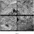
- SEM images displayed in Figure 2 show mineral deposition over time: after 2 days of immersion in SBF globular mineral deposits of up to 0.2 ⁇ m in size are observed: after 4 days of immersion in SBF globular mineral crystals are observed on the surface of the fibrin/alginate matrix, which grow with increasing immersion time; after 6 days of immersion in SBF globular mineral crystals (0.05-0,8 ⁇ m-larger aggregates) are observed on the entire surface for the biomaterial.
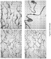
- FIG. 3 images show low magnification photos of CaP deposition over time, suggesting that the original micro-porosity found in Smart Matrix® is reduced upon immersion in SBF, as the reduction is noticeable after only 2 days of immersion in SBF ( Fig. 3 , top left):
- EDX analysis ( Fig. 4 ) of CaP-SM after 6 days of immersion in SBF shows that Ca and P are the main elements of the globular crystals, also at day 9. Before day 6 no Ca and P are detected in the EDAX spectra. Calculated Ca/P was 1.7 ⁇ 0.6 (average ⁇ standard deviation; Ca/P for hydroxyapatite is 1.67).
- Mg is also present: SBF contains Mg 2+ and Mg 2+ is among the reported substituting ions present in bone mineral (Wopenka and Pasteris 2005; LeGeros 2008).
- FIG 5 shows eosin staining of Smart Matrix® scaffold used (left) and a Von Kossa stained scaffold, counterstained with eosin (right).
- Smart Matrix® appears darker after the Von Kossa staining.
- the homogeneous open pore structure of the scaffold can also be seen as already observed in the SEM photos:
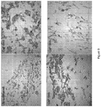
- Figure 6 shows the Von Kossa staining results of CaP-SM scaffolds after immersion in simulated body fluid (SBF) for up to 9 days. Samples were counterstained with eosin. CaP deposits are stained black/dark brown while Smart Matrix® stains pink/red.
- the scaffold fibres appear darkened with immersion in SBF. Black/brown staining seems to increase with longer immersion periods in SBF. It is also interesting to note that the CaP deposits are found throughout the scaffold structure, which appears homogeneously stained. These results suggest that even deposition of CaP deposits throughout Smart Matrix® can be achieved with a biomimetic procedure of immersion of scaffolds in SBF.
- Figure 7 shows that the in vitro degradation rate of the scaffolds decreases as the immersion time in SBF increases.
- the degradation rate is especially decreased in the first 6 h when compared to Smart Matrix®.
- Results displayed in Figure 8 show that cells are able to grow and proliferate on the CaP-SM scaffolds, suggesting that the CaP deposited by the biomimetic method is not toxic to the cells.
- Photographs in Figure 9 show that primary normal human dermal fibroblasts were able to migrate through the CaP-SM scaffolds and after 10 days of culture populated the scaffolds throughout.
- Table 2 describes the concentration of relevant ions present in the SBF solutions: Table 2: Concentration of relevant ions in SBF solution. Solution Ion Na + K + Mg 2+ Ca 2+ Cl - HCO 3 - SO 4 2- HPO 4 2- SBF-1 mM 766.3 35.9 10.9 18.8 740.9 71.5 3.6 7.1 SBF-2 mM 686.1 31.2 9.3 15.6 728.6 26.1 6.2 SBF-3 mM 660 12.4 - 15.6 691.3 - - 6.2
- Lyophilised scaffolds can be further immersed in SBF-3 for 48 hours with constant stirring at 80 rpm on an orbital shaker at 37°C. After the immersion time, scaffolds are removed from the SBF-3 solution, washed with dH 2 O in an ultrasonic water cleaner for 60 seconds, frozen at -80 °C and lyophilised.
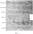
- Results in Figure 10 show CaP mineral deposits with a globular morphology distributed evenly throughout the fibrin matrix in CaP-SM1.A plate-like morphology is observed in CaP-SM2. SM1 and SM2 show the typical fibres seen in Smart Matrix®.
- EDX elemental analysis ( Fig. 11 ) showed that the main elements in the mineral deposits of CaP-SM1 and CaP-SM2 were Ca and P. Calculated Ca/P was 1.29 ⁇ 0.28 and 1.02 ⁇ 0.37 (average ⁇ standard deviation) for CaP-SM1 and CaP-SM2, respectively. Mg was also present in CaP-SM1 spectra.
- FTIR spectra displayed in Figure 12 show that differences exist between the controls (SM1 and SM2) and the CaP-SM scaffolds.
- PO 4 burst peaks can be seen for the CaP-SM samples at approximately 1050 cm -1 followed by a small shoulder peak at approximately 850 cm -1 which could be due to a HPO 4 group or a CO 3 group or both.
- a peak at approximately 600 cm -1 , specially visible for CaP-SM2, could be due to a PO 4 group.
- the small peak seen at approximately 1450 cm -1 for CaP-SM1 could be due to a CO 3 group.
- spectra show that PO 4 peaks grow in intensity in CaP-SM2 compared to CaP-SM1 suggesting deposition of a higher amount of a phosphate mineral phase in CaP-SM2 than in CaP-SM1 scaffolds.
- FTIR results suggest the presence of PO 4 , and possibly both HPO 4 and CO 3 groups, especially in CaP-SM1, in the CaP deposits.
- XRD spectra of CaP-SM1 did not show peaks as the CaP deposits in these scaffolds are very amorphous and XRD does not detect very amorphous mineral phases.
- XRD spectra revealed peaks that could correspond to hydroxyapatite (HA) and/or octacalcium phosphate (OCP). However, these peaks were broad indicating the CaP mineral phase/s present in CaP-SM2 were not very crystalline.
- Figure 13 shows Von Kossa stained scaffolds counterstained with eosin. Although it may not readily be visible from Figure 13 , CaP mineral deposits are stained black/dark brown while the fibrin fibres stain pink/red. As observed from Figure 13 , CaP mineral deposits are found throughout the scaffold structure suggesting an even deposition of CaP throughout the scaffolds. No CaP mineral deposits were observed on the SM1 and SM2 control samples.
- Rheology is a branch of engineering that studies the viscoelastic properties (both solid and fluid) of materials as well as biological tissues (Holt et al. 2008; Saitoh et al. 2010).
- a combined measurement including an 'amplitude sweep' and a 'frequency sweep' was carried out on each sample. The 'amplitude sweep' was performed by applying controlled stresses that were linearly increased from 0.05 to 5%.
- G' is related to elasticity and is an indication of how the material stores energy which can be re-used in the form of elastic deformation. Thus, G' relates to the solid characteristics of the material.
- Scaffolds (Smart Matrix®, demineralised bone matrix or DBM, CaP-SM 1 and CaP-SM 2) were cut into 5mm x 5mm square pieces prior to treatment with 1% trypsin at pH 7.2 at 37°C for up to 48h hours. Scaffolds were immersed in PBS alone as controls. Samples were imaged macroscopically using canon camera and microscopically using a stereomicroscope at 0h, 18h, 24h and 48h.
- Scaffolds (Smart Matrix® used as control, CaP-SM 1 and CaP-SM 2) were cut into 5mm x 5mm square pieces and sterilised with 70% IMS, washed three times with PBS and placed in flat bottomed-24 well ultra-low attachment plates.
- the scaffolds were seeded with 1x10 ⁇ MC3T3-E1 subclone mouse pre-osteoblasts (osteoprogenitor cells) in 20 ⁇ l medium. After seeding, the plates were incubated for 3 h at 37 ⁇ C with 5% CO 2 to allow cells to attach to the scaffolds.
- ⁇ MEM Minimum Essential Medium Eagle Alpha Modification with 1 % antibiotics and 10% fetal calf serum
- (+OM or without (-OM) osteogenic supplements
- 50 ⁇ g/ml ascorbic acid, 10 mM ⁇ -glycerophosphate and 100 nM dexamethasone was added per well and cultured over a 28 day period at 37 ⁇ C with 5% CO 2 .
- scaffolds were assessed for cell incorporation and viability using Live/Dead cell staining according to the manufacturer's guidelines (Sigma), wherein live cells fluoresce green and dead cells fluoresce red. Briefly, scaffolds were washed in PBS prior to staining with the live/dead staining solution and then the staining procedure was performed in the dark for 30 min at 37°C and 5% CO 2 . Live and dead cells were visualized by fluorescence imaging and confocal microscopy.
- seeded scaffolds were transferred to fresh 24 well plates and 1 ml of alamarBlue® working solution (diluted 10x from stock solution with phenol free Dubelcco's Modified Eagles Medium supplemented with 10% FCS and 1% antibiotics) was added per well and samples incubated at 37°C with 5% CO 2 for 3 h, after which each well content was transferred to a cuvette and absorbance measured at 570 nm in a uv/vis spectrophotometer.
- alamarBlue® working solution diluted 10x from stock solution with phenol free Dubelcco's Modified Eagles Medium supplemented with 10% FCS and 1% antibiotics
- Results showed that cells remained viable over the culture period and were able to proliferate.
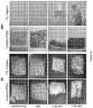
- the Live/Dead assay showed green fluorescent cells (viable) on all scaffolds both under standard and osteogenic conditions ( Fig. 15 ).
- MC3T3-E1 cells within Smart Matrix® , CaP-SM 1 and CaP-SM2 scaffolds was examined using DAPI staining of the nuclei after 28 days of culture under standard (-OM) and osteogenic (+OM) conditions. Histological slides were deparaffinised, rehydrated and washed in distilled water before FluoroshieldTM with DAPI mounting media (F6057 Sigma) was applied as per manufacturer's instructions. Briefly, the vial was brought to room temperature and 1-2 drops of the mounting media was applied directly on top of the sample and left to set for approximately 5 minutes at room temperature. The sections were then cover slipped carefully avoiding air bubbles and the edges were sealed using a nail varnish.
- the sections were then imaged using confocal scanning laser microscope using 405 nm wavelength laser for DAPI (Excitation 372, Emission 456).
- the sections were tile scanned for the entire XY plane of the scaffold and then merged using 5% overlap automated function of the confocal microscope.
- Results showed cells integrated with the CaP-SM scaffolds in both standard and osteogenic culture conditions ( Fig. 19 ). Cells can be seen forming a layer of interconnected cells which in some areas is embedded within the scaffolds. Some cytoplasmic processes are seen on all samples. These results demonstrate the good cellular properties of CaP-SM scaffolds.
- Scaffolds (Smart Matrix® used as control, CaP-SM 1 and CaP-SM 2) were cut into 5mm x 5mm square pieces, sterilised, seeded with 1x10 ⁇ MC3T3-E1 and cultured in both standard and osteogenic media as already explained.
- Non-collagenous matrix was assessed by osteopontin immunostaining of paraffin embedded sections. Briefly, antigen retrieval was performed by heat mediated antigen retrieval using sodium citrate buffer at pH 6. After antigen retrieval the sections were blocked using 5% bovine serum albumin at 35°C for 1 h and stained with primary anti-osteopontin antibody (1:50, abcam ab8448) at 4°C overnight. Following this, the sections were stained using the alexa fluor 546 secondary antibody for 2 h at room temperature. The sections were then washed and mounted with DAPI based mounting media, cover slipped and visualised using Leica DM IRE2 confocal microscope.
- Pro-angiogenic potential of the scaffolds was assessed using an ex ovo chorionic allantoic membrane (CAM) assay.
- Scaffolds Smart Matrix® used as control, CaP-SM 1 and CaP-SM 2 were cut into 5mm x 5mm square pieces, sterilised with 70 % IMS and washed three times with PBS.
- a negative and a positive control were run alongside the scaffolds: filter paper soaked in either PBS (negative) or 10ng/ml of vascular endothelial growth factor (VEGF) solution (positive). Fertile chicken eggs were incubated at 37.5°C, 3% CO 2 and humidity was maintained between 35%-45%.
- VEGF vascular endothelial growth factor
- the embryos were transferred to a shell-less culture system with 75-80% humidity and 37.5°C incubation temperature.
- scaffolds were applied onto the developing CAMs.
- Angiogenesis was examined in all the scaffolds macroscopically by taking photos using a stereomicroscope. ImageJ software was used to analyse the macroscopic photos and calculate the percentage of vascular area for each scaffold.
Landscapes
- Health & Medical Sciences (AREA)
- Chemical & Material Sciences (AREA)
- Medicinal Chemistry (AREA)
- Public Health (AREA)
- Oral & Maxillofacial Surgery (AREA)
- Transplantation (AREA)
- Epidemiology (AREA)
- Life Sciences & Earth Sciences (AREA)
- Animal Behavior & Ethology (AREA)
- General Health & Medical Sciences (AREA)
- Dermatology (AREA)
- Veterinary Medicine (AREA)
- Inorganic Chemistry (AREA)
- Dispersion Chemistry (AREA)
- Engineering & Computer Science (AREA)
- Composite Materials (AREA)
- Materials Engineering (AREA)
- Materials For Medical Uses (AREA)
- Chemical Kinetics & Catalysis (AREA)
- Polymers & Plastics (AREA)
- Organic Chemistry (AREA)
- Prostheses (AREA)
Claims (16)
- Matière de matrice extracellulaire, comprenant :un échafaudage réticulé comprenant de la fibrine ou du fibrinogène et un agent gonflant, dans laquelle la fibrine ou le fibrinogène et l'agent gonflant sont réticulés, et l'agent gonflant est réticulé avec la fibrine ou le fibrinogène, etune céramique déposée sur l'échafaudage, optionnellement dans laquelle la céramique est ou comprend une phase minérale de phosphate de calcium.
- Matière de matrice extracellulaire selon la revendication 1, dans laquelle l'agent gonflant est :a) un alginate, tel qu'un alginate de sodium ou un propylglycoalginate de sodium ; ou bienb) un glycosaminoglycane, tel qu'un 6-sulfate de chondroïtine, 4-sulfate de chondroïtine, héparine, sulfate d'héparine, sulfate de kératan, sulfate de dermatan, chitine, chitosan, sulfate de dextran ou hyaluronan ; ou bienc) de l'amidon hydroxyéthylique, de l'éthylcellulose, de la gomme de xanthane ou de l'agarose.
- Procédé de préparation d'une matière de matrice extracellulaire, comprenant :
déposer une céramique sur un échafaudage réticulé comprenant de la fibrine ou du fibrinogène et un agent gonflant, dans lequel la fibrine ou le fibrinogène et l'agent gonflant sont réticulés, et l'agent gonflant est réticulé avec la fibrine ou le fibrinogène, et optionnellement dans lequel la céramique est une phase minérale de phosphate de calcium. - Procédé selon la revendication 3, dans lequel la phase minérale de phosphate de calcium est déposée sur l'échafaudage :a) par dépôt biomimétique ; et/oub) en immergeant l'échafaudage dans un fluide comprenant du Ca2+ et du HPO4 2, comprenant optionnellement en outre : i) du Na+, K+, Mg2+, Ca2+, Cl-, HCO3 -, HCO4 2- et SO4 2- ; ii) du Na+, K+, Mg2+, Ca2+, Cl-, HCO3 - et HPO4 2- ; ou iii) du Na+, K+, Ca2+ ; et/ouc) en mettant l'échafaudage en contact avec, ou en immergeant l'échafaudage dans, un fluide comprenant de 1 à 30 mM de Ca2+, de préférence de 1 à 20 mM de Ca2+; et/oud) en mettant l'échafaudage en contact avec, ou en immergeant l'échafaudage dans, un fluide comprenant de 0,5 à 10 mM de HPO4 2-; et/oue) en mettant l'échafaudage en contact avec, ou en immergeant l'échafaudage dans, un fluide comprenant de 100 à 800 mM de Na+ ; et/ouf) en mettant l'échafaudage en contact avec, ou en immergeant l'échafaudage dans, un fluide comprenant de 1 à 50 mM de K+ ; et/oug) en mettant l'échafaudage en contact avec, ou en immergeant l'échafaudage dans, un fluide comprenant de 0,5 à 20 mM de Mg2+ ; et/ouh) en mettant l'échafaudage en contact avec, ou en immergeant l'échafaudage dans, un fluide comprenant de 100 à 800 mM de Cl- ; et/oui) en mettant l'échafaudage en contact avec, ou en immergeant l'échafaudage dans, un fluide comprenant de 1 à 100 mM de HCO3 - ; et/ouj) en mettant l'échafaudage en contact avec, ou en immergeant l'échafaudage dans, un fluide comprenant de 1 à 10 mM de SO4 2- ; et/ouk) en mettant l'échafaudage en contact avec, ou en immergeant l'échafaudage dans, un fluide comprenant de 1 à 5 mM de Ca2+, de préférence de 2 à 3 mM de Ca2+, le plus préférentiellement environ 2,5 mM de Ca2+ ; et/oul) en mettant l'échafaudage en contact avec, ou en immergeant l'échafaudage dans, un fluide comprenant de 0,5 à 3 mM de HPO4 2-, de préférence de 0,5 à 2 mM de HPO4 2-, le plus préférentiellement environ 1 mM de HPO4 2- ; et/oum) en mettant l'échafaudage en contact avec, ou en immergeant l'échafaudage dans, un fluide comprenant de 100 à 200 mM de Na+, de préférence de 120 à 160 mM de Na+, encore plus préférablement de 140 à 150 mM de Na+, le préférentiellement environ 142 mM de Na+ ; et/oun) en mettant l'échafaudage en contact avec, ou en immergeant l'échafaudage dans, un fluide comprenant de 1 à 10 mM de K+, de préférence de 3 à 7 mM de K+, le plus préférentiellement environ 5 mM de K+ ; et/ouo) en mettant l'échafaudage en contact avec, ou en immergeant l'échafaudage dans, un fluide comprenant de 0,5, à 3 mM de Mg2+, de préférence de 1 à 2 mM de Mg2+, le plus préférentiellement environ 1,5 mM de Mg2+ ; et/oup) en mettant l'échafaudage en contact avec, ou en immergeant l'échafaudage dans, un fluide comprenant de 100 à 200 mM de Cl-, de préférence de 120 à 160 mM de Cl-, plus préférablement de 140 à 150 mM de Cl-, le plus préférentiellement environ 148,8 mM de Cl- ; et/ouq) en mettant l'échafaudage en contact avec, ou en immergeant l'échafaudage dans, un fluide comprenant de 1 à 10 mM de HCO3 -, de préférence de 2 à 8 mM de HCO3 -, plus préférablement de 4 à 5 mM de HCO3 -, le plus préférentiellement environ 4,2 mM de HCO3 - ; et/our) en mettant l'échafaudage en contact avec, ou en immergeant l'échafaudage dans, un fluide comprenant de 1 à 5 mM de Ca2+, de 0,5 à 3 mM de HPO4 2, de 100 à 200 mM de Na+, de 1 à 10 mM de K+, de 0,5 à 3 mM de Mg2+, de 100 à 200 mM de Cl- et de 1 à 10 mM de HCO3 - ; et/ous) en mettant l'échafaudage en contact avec, ou en immergeant l'échafaudage dans, un fluide comprenant de 2 à 3 mM de Ca2+, de 0,5 à 2 mM de HPO4 2, de 120 à 160 mM de Na+, de 3 à 7 mM de K+, de 1 à 2 mM de Mg2+, de 120 à 160 mM de Cl- et de 2 à 8 mM de HCO3 -.
- Procédé selon l'une quelconque des revendications 3 ou 4 a) à 4 i), dans lequel la phase minérale de phosphate de calcium est déposée sur l'échafaudage :a) en mettant l'échafaudage en contact avec, ou en immergeant l'échafaudage dans, un fluide comprenant de 10 à 30 mM de Ca2+; et/oub) en mettant l'échafaudage en contact avec, ou en immergeant l'échafaudage dans, un fluide comprenant de 5 à 10 mM de HPO4 2- ; et/ouc) en mettant l'échafaudage en contact avec, ou en immergeant l'échafaudage dans, un fluide comprenant de 500 à 800 mM de Na+ ; et/oud) en mettant l'échafaudage en contact avec, ou en immergeant l'échafaudage dans, un fluide comprenant de 10 à 40 mM de K+ ; et/oue) en mettant l'échafaudage en contact avec, ou en immergeant l'échafaudage dans, un fluide comprenant de 5 à 15 mM de Mg2+ ; et/ouf) en mettant l'échafaudage en contact avec, ou en immergeant l'échafaudage dans, un fluide comprenant de 500 à 800 mM de Cl- ; et/oug) en mettant l'échafaudage en contact avec, ou en immergeant l'échafaudage dans, un fluide comprenant de 10 à 100 mM de HCO3 - ; et/ouh) en mettant l'échafaudage en contact avec, ou en immergeant l'échafaudage dans, un fluide comprenant de 1 à 10 mM de SO4 2-.
- Procédé selon l'une quelconque des revendications 4 b) à 4 s), ou 5 a) à 5 h), dans lequel l'échafaudage est mis en contact avec, ou immergé dans le fluide pendant :a) au moins 12 heures, au moins 24 heures, au moins 2 jours, au moins 5 jours, au moins 6 jours ou au moins 9 jours ; et/oub) jusqu'à 20 jours, de préférence jusqu'à 10 j ours, le plus préférentiellement jusqu'à 9 jours.
- Procédé selon l'une quelconque des revendications 3 à 6, dans lequel :i) l'échafaudage a été obtenu, a été formé ou peut être obtenu par un procédé comprenant les étapes suivantes ; ou bien ii) le procédé comprend former l'échafaudage par un procédé comprenant les étapes suivantes consistant à :(a) mélanger une solution aqueuse de fibrinogène avec un agent coagulant et un agent gonflant ;(b) incuber le mélange obtenu à l'étape (a) avec un agent de réticulation ; et(c) laver la composition réticulée obtenue à l'étape (b) pour retirer l'agent de réticulation.
- Procédé selon la revendication 7, dans lequel :a) le fibrinogène est présent à un niveau de pureté plus grand qu'un de 75 %, 80 %, 85 %, 90%, 95%, 97 % ou 99% ; et/oub) le fibrinogène est présent comme des formes tronquées de fibrinogène, telles que la fibrine A, fibrine B, fibrine C, fibrine D, fibrine X et fibrine Y, optionnellement dans lequel la forme tronquée de fibrinogène est la fibrine E ; et/ouc) le fibrinogène est présent comme une solution aqueuse tamponnée à un pH d'entre 4 et 10 avec une solution saline à tampon phosphate (PBS) ou une solution saline à tampon HEPES ; et/oud) l'agent coagulant comprend un agent coagulant enzymatique ou non enzymatique, optionnellement dans lequel l'agent coagulant est de la thrombine, telle que de la thrombine humaine.
- Procédé selon l'une quelconque des revendications 7 à 8, dans lequel l'étape (a) comprend mélanger avec un agent moussant, optionnellement dans lequel l'agent moussant comprend un détergent non ionique, un tensioactif gélifiant thermosensible (par ex. Pluronic 127), un phospholipide du type diphosphatydylglycérol ou un mélange d'une phase non miscible avec la phase en solution aqueuse de fibrinogène, de préférence dans lequel l'agent moussant comprend un tensioactif Pluronic, par ex. Pluronic F68 et/ou Pluronic F127, plus préférablement Pluronic F68.
- Procédé selon la revendication 9, dans lequel l'agent moussant :a) est constitué de ou comprend un ou plusieurs agents tensioactifs de la classe des tensioactifs dérivés de sucres ; et/oub) est constitué de ou comprend un ou plusieurs agents tensioactifs de la classe des tensioactifs dérivés de sucres-acyle, optionnellement dans lequel l'agent moussant est de la classe des tensioactifs dérivés de sucres-acyle ayant une longueur de chaîne acyle de C8 à C12 ; et/ouc) comprend ou est constitué d'au moins deux, de préférence trois tensioactifs dérivés de sucres ; et/oud) est constitué de ou comprend un ou plusieurs agents tensioactifs de la classe des tensioactifs dérivés de sucres-acyle sélectionnés parmi la classe de tensioactifs dérivés de pyranoside (particulièrement de glucopyranoside), de maltoside et d'acyle-saccharose ; et/oue) est constitué de ou comprend un ou plusieurs agents tensioactifs de la classe des tensioactifs dérivés de sucres-acyle, sélectionnés parmi le groupe composé de OGP, ODM, DGP et DdGP, TGP, HGP, DMP, décyl-saccharose (nDS), dodécyl-saccharose nDdS), de préférence OGM, DMP et DdGP.
- Procédé selon l'une quelconque des revendications 7 à 10, dans lequel l'agent de réticulation utilisé à l'étape (b) est sélectionné parmi : des agents de couplage de carbodiimide tels que le N-(3-diméthylaminopropyl)-N'-éthylcarbodiiniide (EDC) ; N-hydroxysuccinimide (NHS), des agents de couplage d'azide ; des agents de réticulation de diisocyanate tels que le diisocyanate d'hexaméthylène ; des agents de réticulation d'époxyde tels que l'épichlorhydrine, des glycidyléthers et des glycidylamines ; et des agents de réticulation d'aldéhyde tels que le formaldéhyde, glutaraldéhyle et glyoxal, optionnellement dans lequel l'agent de réticulation comprend un agent de réticulation d'aldéhyde tel que le formaldéhyde, glutaraldéhyde et glyoxal, optionnellement le glutaraldéhyde.
- Procédé selon l'une quelconque des revendications 7 à 11 :a) qui comprend en plus l'addition d'un agent réducteur ou d'un agent réducteur de toxicité tel que le borohydrure de sodium ou la lysine ; et/oub) dans lequel l'étape (a) de mélange est réalisée par moussage, tel qu'en mélangeant avec aération ; et/ouc) dans lequel le mélange obtenu à l'étape (a) est coulé, congelé ou optionnellement lyophilisé préalablement à l'étape d'incubation (b) ; et/oud) qui comprend en plus l'addition d'un ion de métal divalent ou polyvalent tel que du calcium, tel que le chlorure de calcium ; et/oue) dans lequel le mélange pour l'étape (a) de mélange comprend en outre un sucre en tant que stabilisant de protéine, dans lequel le sucre est un petit polyol ou carbohydrate, tel que glycérol, sorbitol, saccharose ou tréhalose, optionnellement dans lequel le sucre est du tréhalose, de préférence en une quantité de 10-11 % en poids par rapport au fibrinogène, et de préférence en une quantité de 4-7,5 % en poids dans le mélange de l'étape (a) ; et/ouf) pour préparer une composition de matrice extracellulaire ayant une forme prédéterminée, dans lequel soit (i) le mélange de l'étape (a) est coulé dans un moule d'une forme prédéterminée, congelé et optionnellement lyophilisé préalablement à l'étape (c) d'incubation, ou bien (ii) le produit de l'étape (d) est produit dans un moule d'une forme prédéterminée, et le produit est alors congelé et optionnellement lyophilisé ; et/oug) dans lequel l'agent gonflant est un alginate, tel que de l'alginate de sodium ou du propylglycoalginate de sodium ; et/ouh) dans lequel l'agent gonflant est un glycosaminoglycane, tel qu'un 6-sulfate de chondroïtine, 4-sulfate de chondroïtine, héparine, sulfate d'héparine, sulfate de kératan, sulfate de dermatan, chitine, chitosan, sulfate de dextran ou hyaluronan ; et/oui) dans lequel l'agent gonflant est sélectionné parmi l'amidon hydroxyéthylique, l'éthylcellulose, la gomme de xanthane et l'agarose.
- Matière de matrice extracellulaire selon la revendication 1 ou la revendication 2, dans laquelle la phase minérale de phosphate de calcium représente au moins 10%, de préférence au moins 20 %, plus préférablement au moins 50 % en poids de la matière de matrice extracellulaire.
- Matière de matrice extracellulaire selon la revendication 1 ou la revendication 2, pour une utilisation dans la régénération osseuse in vitro, ex vivo ou in vivo ou dans le traitement d'un défaut osseux.
- Matière de matrice extracellulaire selon la revendication 1 ou la revendication 2, dans laquelle :a) la phase minérale de phosphate de calcium comprend des composés d'orthophosphate de calcium, de préférence de l'hydroxyapatite ; ou bienb) la phase minérale de phosphate de calcium comprend du phosphate d'octacalcium ; ou bienc) la phase minérale de phosphate de calcium comprend une phase minérale de phosphate de calcium amorphe et/ou cristallin, et optionnellement dans laquelle i) la phase minérale de phosphate de calcium comprend des composés d'orthophosphate de calcium, de préférence de l'hydroxyapatite, ou bien ii) la phase minérale de phosphate de calcium comprend du phosphate d'octacalcium.
- Procédé selon l'une quelconque des revendications 3 à 12, dans lequel :a) la phase minérale de phosphate de calcium comprend des composés d'orthophosphate de calcium, de préférence de l'hydroxyapatite ; ou bienb) la phase minérale de phosphate de calcium comprend du phosphate d'octacalcium ; ou bienc) la phase minérale de phosphate de calcium comprend une phase minérale de phosphate de calcium amorphe et/ou cristallin, et optionnellement dans laquelle i) la phase minérale de phosphate de calcium comprend des composés d'orthophosphate de calcium, de préférence de l'hydroxyapatite, ou bien ii) la phase minérale de phosphate de calcium comprend du phosphate d'octacalcium.
Applications Claiming Priority (2)
| Application Number | Priority Date | Filing Date | Title |
|---|---|---|---|
| GBGB1615972.5A GB201615972D0 (en) | 2016-09-20 | 2016-09-20 | Extracellular matrix material |
| PCT/GB2017/052796 WO2018055361A1 (fr) | 2016-09-20 | 2017-09-20 | Matériau de matrice extracellulaire |
Publications (2)
| Publication Number | Publication Date |
|---|---|
| EP3515513A1 EP3515513A1 (fr) | 2019-07-31 |
| EP3515513B1 true EP3515513B1 (fr) | 2021-09-08 |
Family
ID=57288775
Family Applications (1)
| Application Number | Title | Priority Date | Filing Date |
|---|---|---|---|
| EP17777321.5A Active EP3515513B1 (fr) | 2016-09-20 | 2017-09-20 | Matériau de matrice extracellulaire |
Country Status (8)
| Country | Link |
|---|---|
| US (1) | US12280174B2 (fr) |
| EP (1) | EP3515513B1 (fr) |
| JP (1) | JP7104425B2 (fr) |
| AU (1) | AU2017331503B2 (fr) |
| CA (1) | CA3037594A1 (fr) |
| ES (1) | ES2899026T3 (fr) |
| GB (1) | GB201615972D0 (fr) |
| WO (1) | WO2018055361A1 (fr) |
Families Citing this family (4)
| Publication number | Priority date | Publication date | Assignee | Title |
|---|---|---|---|---|
| US11839659B2 (en) | 2020-07-02 | 2023-12-12 | Northwestern University | Proteolysis-targeting chimeric molecules (PROTACs) that induce degradation of indoleamine 2,3-dioxygenase (IDO) protein |
| US11981678B2 (en) | 2021-04-29 | 2024-05-14 | Northwestern University | Substituted [1,2,4]triazolo[4,3-c]pyrimidin-5-amines and proteolysis-targeting chimeric derivatives thereof (PROTACs) that induce degradation of embryonic ectoderm development (EED) protein |
| US12042543B2 (en) | 2021-04-30 | 2024-07-23 | Northwestern University | Substituted 3-amino-5-phenylbenzamide compounds as covalent inhibitors of enhancer zeste homolog 2 (EZH2) and proteolysis-targeting chimeric derivatives thereof (PROTACs) that induce degradation of EZH2 |
| CN115645617B (zh) * | 2022-09-09 | 2023-07-25 | 北京鑫康辰医学科技发展有限公司 | 一种具有生物活性的骨基质凝胶的制备方法 |
Family Cites Families (11)
| Publication number | Priority date | Publication date | Assignee | Title |
|---|---|---|---|---|
| JPH0629126B2 (ja) | 1989-03-29 | 1994-04-20 | 京都大学長 | 生体活性水酸アパタイト膜のコーティング法 |
| JP2754348B2 (ja) | 1994-10-20 | 1998-05-20 | バイオマップ有限会社 | アパタイト被覆基材とその製造法 |
| AU2003218271A1 (en) * | 2002-04-18 | 2003-11-03 | Carnegie Mellon University | Method of manufacturing hydroxyapatite and uses therefor in delivery of nucleic acids |
| JP3916516B2 (ja) | 2002-06-10 | 2007-05-16 | 独立行政法人科学技術振興機構 | 硬組織−軟組織界面再生用足場材料 |
| IL154208A (en) * | 2003-01-30 | 2009-05-04 | Prochon Biotech Ltd | Plasma protein matrices and methods for preparation thereof |
| CA2514474C (fr) | 2003-01-30 | 2014-05-06 | Avner Yayon | Matrices de fibrine lyophilisees et leurs procedes de preparation |
| US8273372B2 (en) | 2006-06-16 | 2012-09-25 | Restoration Of Appearance & Function Trust | Extracellular matrix composition |
| GB201207781D0 (en) * | 2012-05-03 | 2012-06-13 | Restoration Of Appearance And Function Trust | Extracellular matrix - synthetic skin scaffold |
| BR102012011209A2 (pt) | 2012-05-11 | 2014-03-25 | Bioactive Biomateriais Ltda | Material tridimensional polimérico biodegradável e processo de preparo de material tridimensional polimérico biodegradável |
| KR101680270B1 (ko) | 2014-12-12 | 2016-11-30 | 한국원자력연구원 | 생분해성 고분자 및 하이드록시아파타이트 복합체의 제조방법 및 이를 통해 제조된 생분해성 고분자 및 하이드록시아파타이트 복합체 |
| PL226891B1 (pl) | 2015-05-05 | 2017-09-29 | Inst Wysokich Ciśnień Polskiej Akademii Nauk | Sposób wytwarzania implantu kostnego iimplant kostny |
-
2016
- 2016-09-20 GB GBGB1615972.5A patent/GB201615972D0/en not_active Ceased
-
2017
- 2017-09-20 WO PCT/GB2017/052796 patent/WO2018055361A1/fr not_active Ceased
- 2017-09-20 AU AU2017331503A patent/AU2017331503B2/en not_active Ceased
- 2017-09-20 JP JP2019536339A patent/JP7104425B2/ja not_active Expired - Fee Related
- 2017-09-20 CA CA3037594A patent/CA3037594A1/fr active Pending
- 2017-09-20 ES ES17777321T patent/ES2899026T3/es active Active
- 2017-09-20 EP EP17777321.5A patent/EP3515513B1/fr active Active
- 2017-09-20 US US16/333,558 patent/US12280174B2/en active Active
Also Published As
| Publication number | Publication date |
|---|---|
| CA3037594A1 (fr) | 2018-03-29 |
| US12280174B2 (en) | 2025-04-22 |
| ES2899026T3 (es) | 2022-03-09 |
| US20190262502A1 (en) | 2019-08-29 |
| JP2019529022A (ja) | 2019-10-17 |
| AU2017331503A1 (en) | 2019-04-11 |
| EP3515513A1 (fr) | 2019-07-31 |
| WO2018055361A1 (fr) | 2018-03-29 |
| AU2017331503B2 (en) | 2022-06-30 |
| GB201615972D0 (en) | 2016-11-02 |
| JP7104425B2 (ja) | 2022-07-21 |
Similar Documents
| Publication | Publication Date | Title |
|---|---|---|
| Zhang et al. | 3D gel-printed porous magnesium scaffold coated with dibasic calcium phosphate dihydrate for bone repair in vivo | |
| Bastami et al. | Fabrication of a three-dimensional β-tricalcium-phosphate/gelatin containing chitosan-based nanoparticles for sustained release of bone morphogenetic protein-2: Implication for bone tissue engineering | |
| Guo et al. | Hybrid nanostructured hydroxyapatite–chitosan composite scaffold: Bioinspired fabrication, mechanical properties and biological properties | |
| Li et al. | Hyaluronic acid oligosaccharides modified mineralized collagen and chitosan with enhanced osteoinductive properties for bone tissue engineering | |
| Chen et al. | Umbilical cord stem cells released from alginate–fibrin microbeads inside macroporous and biofunctionalized calcium phosphate cement for bone regeneration | |
| Meng et al. | Effects of adding resorbable chitosan microspheres to calcium phosphate cements for bone regeneration | |
| Jahan et al. | Composite biopolymers for bone regeneration enhancement in bony defects | |
| EP1799277B1 (fr) | Composite de matiere de charge biologique poreuse et procede de fabrication correspondant | |
| Matsuno et al. | Preparation of injectable 3D-formed β-tricalcium phosphate bead/alginate composite for bone tissue engineering | |
| Kerativitayanan et al. | Elastomeric and mechanically stiff nanocomposites from poly (glycerol sebacate) and bioactive nanosilicates | |
| JP3727059B2 (ja) | 多孔質複合材料の製造方法 | |
| Xu et al. | Hierarchically porous nagelschmidtite bioceramic–silk scaffolds for bone tissue engineering | |
| CA2721900C (fr) | Implant biocompatible | |
| EP3515513B1 (fr) | Matériau de matrice extracellulaire | |
| Van et al. | Injectable hydrogel composite based gelatin-PEG and biphasic calcium phosphate nanoparticles for bone regeneration | |
| Radha et al. | Nano-hydroxyapatite/natural polymer composite scaffolds for bone tissue engineering: a brief review of recent trend | |
| WO2013059089A1 (fr) | Pâtes à os comprenant des ciments de phosphate de calcium biofonctionnalisés ayant des fonctions cellulaires améliorées pour une réparation osseuse | |
| Moussi et al. | Injectable macromolecule-based calcium phosphate bone substitutes | |
| Zhang et al. | Incorporation of synthetic water-soluble curcumin polymeric drug within calcium phosphate cements for bone defect repairing | |
| Tang et al. | Hard tissue compatibility of natural hydroxyapatite/chitosan composite | |
| Vu et al. | Fabrication of in situ crosslinking hydrogels based on oxidized alginate/N, O-carboxymethyl chitosan/β-tricalcium phosphate for bone regeneration | |
| Ramakrishnan et al. | Effect of wheat gluten on improved thermal cross-linking and osteogenesis of hydroxyapatite-gelatin composite scaffolds | |
| KR100937736B1 (ko) | 유도 조직 재생을 위한 다공성 지지체 및 그의 제조방법 | |
| WO2003070290A1 (fr) | Biomateriau composite contenant de la phospholine | |
| Krishnamoorthy et al. | Productizing Nano-Bioactive Glass-Based Bilayer Scaffolds: A Graft for Reconstruction of Mandibular and Femoral Bone Defects |
Legal Events
| Date | Code | Title | Description |
|---|---|---|---|
| STAA | Information on the status of an ep patent application or granted ep patent |
Free format text: STATUS: UNKNOWN |
|
| STAA | Information on the status of an ep patent application or granted ep patent |
Free format text: STATUS: THE INTERNATIONAL PUBLICATION HAS BEEN MADE |
|
| PUAI | Public reference made under article 153(3) epc to a published international application that has entered the european phase |
Free format text: ORIGINAL CODE: 0009012 |
|
| STAA | Information on the status of an ep patent application or granted ep patent |
Free format text: STATUS: REQUEST FOR EXAMINATION WAS MADE |
|
| 17P | Request for examination filed |
Effective date: 20190417 |
|
| AK | Designated contracting states |
Kind code of ref document: A1 Designated state(s): AL AT BE BG CH CY CZ DE DK EE ES FI FR GB GR HR HU IE IS IT LI LT LU LV MC MK MT NL NO PL PT RO RS SE SI SK SM TR |
|
| AX | Request for extension of the european patent |
Extension state: BA ME |
|
| DAV | Request for validation of the european patent (deleted) | ||
| DAX | Request for extension of the european patent (deleted) | ||
| STAA | Information on the status of an ep patent application or granted ep patent |
Free format text: STATUS: EXAMINATION IS IN PROGRESS |
|
| 17Q | First examination report despatched |
Effective date: 20200423 |
|
| REG | Reference to a national code |
Ref country code: DE Ref legal event code: R079 Ref document number: 602017045721 Country of ref document: DE Free format text: PREVIOUS MAIN CLASS: A61L0027220000 Ipc: A61L0027460000 |
|
| RIC1 | Information provided on ipc code assigned before grant |
Ipc: A61L 27/46 20060101AFI20210217BHEP Ipc: A61L 27/32 20060101ALI20210217BHEP Ipc: A61L 27/26 20060101ALI20210217BHEP Ipc: A61L 27/56 20060101ALI20210217BHEP Ipc: A61L 27/22 20060101ALI20210217BHEP |
|
| GRAP | Despatch of communication of intention to grant a patent |
Free format text: ORIGINAL CODE: EPIDOSNIGR1 |
|
| STAA | Information on the status of an ep patent application or granted ep patent |
Free format text: STATUS: GRANT OF PATENT IS INTENDED |
|
| INTG | Intention to grant announced |
Effective date: 20210329 |
|
| RAP3 | Party data changed (applicant data changed or rights of an application transferred) |
Owner name: RAFT ENTERPRISES LTD. |
|
| GRAS | Grant fee paid |
Free format text: ORIGINAL CODE: EPIDOSNIGR3 |
|
| GRAA | (expected) grant |
Free format text: ORIGINAL CODE: 0009210 |
|
| STAA | Information on the status of an ep patent application or granted ep patent |
Free format text: STATUS: THE PATENT HAS BEEN GRANTED |
|
| AK | Designated contracting states |
Kind code of ref document: B1 Designated state(s): AL AT BE BG CH CY CZ DE DK EE ES FI FR GB GR HR HU IE IS IT LI LT LU LV MC MK MT NL NO PL PT RO RS SE SI SK SM TR |
|
| REG | Reference to a national code |
Ref country code: GB Ref legal event code: FG4D |
|
| REG | Reference to a national code |
Ref country code: AT Ref legal event code: REF Ref document number: 1427991 Country of ref document: AT Kind code of ref document: T Effective date: 20210915 Ref country code: CH Ref legal event code: EP |
|
| REG | Reference to a national code |
Ref country code: IE Ref legal event code: FG4D |
|
| REG | Reference to a national code |
Ref country code: DE Ref legal event code: R096 Ref document number: 602017045721 Country of ref document: DE |
|
| REG | Reference to a national code |
Ref country code: FI Ref legal event code: FGE |
|
| REG | Reference to a national code |
Ref country code: SE Ref legal event code: TRGR |
|
| REG | Reference to a national code |
Ref country code: LT Ref legal event code: MG9D |
|
| REG | Reference to a national code |
Ref country code: NL Ref legal event code: FP |
|
| PG25 | Lapsed in a contracting state [announced via postgrant information from national office to epo] |
Ref country code: HR Free format text: LAPSE BECAUSE OF FAILURE TO SUBMIT A TRANSLATION OF THE DESCRIPTION OR TO PAY THE FEE WITHIN THE PRESCRIBED TIME-LIMIT Effective date: 20210908 Ref country code: RS Free format text: LAPSE BECAUSE OF FAILURE TO SUBMIT A TRANSLATION OF THE DESCRIPTION OR TO PAY THE FEE WITHIN THE PRESCRIBED TIME-LIMIT Effective date: 20210908 Ref country code: LT Free format text: LAPSE BECAUSE OF FAILURE TO SUBMIT A TRANSLATION OF THE DESCRIPTION OR TO PAY THE FEE WITHIN THE PRESCRIBED TIME-LIMIT Effective date: 20210908 Ref country code: BG Free format text: LAPSE BECAUSE OF FAILURE TO SUBMIT A TRANSLATION OF THE DESCRIPTION OR TO PAY THE FEE WITHIN THE PRESCRIBED TIME-LIMIT Effective date: 20211208 Ref country code: NO Free format text: LAPSE BECAUSE OF FAILURE TO SUBMIT A TRANSLATION OF THE DESCRIPTION OR TO PAY THE FEE WITHIN THE PRESCRIBED TIME-LIMIT Effective date: 20211208 |
|
| REG | Reference to a national code |
Ref country code: AT Ref legal event code: MK05 Ref document number: 1427991 Country of ref document: AT Kind code of ref document: T Effective date: 20210908 |
|
| PG25 | Lapsed in a contracting state [announced via postgrant information from national office to epo] |
Ref country code: LV Free format text: LAPSE BECAUSE OF FAILURE TO SUBMIT A TRANSLATION OF THE DESCRIPTION OR TO PAY THE FEE WITHIN THE PRESCRIBED TIME-LIMIT Effective date: 20210908 Ref country code: GR Free format text: LAPSE BECAUSE OF FAILURE TO SUBMIT A TRANSLATION OF THE DESCRIPTION OR TO PAY THE FEE WITHIN THE PRESCRIBED TIME-LIMIT Effective date: 20211209 |
|
| REG | Reference to a national code |
Ref country code: ES Ref legal event code: FG2A Ref document number: 2899026 Country of ref document: ES Kind code of ref document: T3 Effective date: 20220309 |
|
| PG25 | Lapsed in a contracting state [announced via postgrant information from national office to epo] |
Ref country code: AT Free format text: LAPSE BECAUSE OF FAILURE TO SUBMIT A TRANSLATION OF THE DESCRIPTION OR TO PAY THE FEE WITHIN THE PRESCRIBED TIME-LIMIT Effective date: 20210908 |
|
| PG25 | Lapsed in a contracting state [announced via postgrant information from national office to epo] |
Ref country code: IS Free format text: LAPSE BECAUSE OF FAILURE TO SUBMIT A TRANSLATION OF THE DESCRIPTION OR TO PAY THE FEE WITHIN THE PRESCRIBED TIME-LIMIT Effective date: 20220108 Ref country code: SM Free format text: LAPSE BECAUSE OF FAILURE TO SUBMIT A TRANSLATION OF THE DESCRIPTION OR TO PAY THE FEE WITHIN THE PRESCRIBED TIME-LIMIT Effective date: 20210908 Ref country code: SK Free format text: LAPSE BECAUSE OF FAILURE TO SUBMIT A TRANSLATION OF THE DESCRIPTION OR TO PAY THE FEE WITHIN THE PRESCRIBED TIME-LIMIT Effective date: 20210908 Ref country code: RO Free format text: LAPSE BECAUSE OF FAILURE TO SUBMIT A TRANSLATION OF THE DESCRIPTION OR TO PAY THE FEE WITHIN THE PRESCRIBED TIME-LIMIT Effective date: 20210908 Ref country code: PT Free format text: LAPSE BECAUSE OF FAILURE TO SUBMIT A TRANSLATION OF THE DESCRIPTION OR TO PAY THE FEE WITHIN THE PRESCRIBED TIME-LIMIT Effective date: 20220110 Ref country code: PL Free format text: LAPSE BECAUSE OF FAILURE TO SUBMIT A TRANSLATION OF THE DESCRIPTION OR TO PAY THE FEE WITHIN THE PRESCRIBED TIME-LIMIT Effective date: 20210908 Ref country code: EE Free format text: LAPSE BECAUSE OF FAILURE TO SUBMIT A TRANSLATION OF THE DESCRIPTION OR TO PAY THE FEE WITHIN THE PRESCRIBED TIME-LIMIT Effective date: 20210908 Ref country code: CZ Free format text: LAPSE BECAUSE OF FAILURE TO SUBMIT A TRANSLATION OF THE DESCRIPTION OR TO PAY THE FEE WITHIN THE PRESCRIBED TIME-LIMIT Effective date: 20210908 Ref country code: AL Free format text: LAPSE BECAUSE OF FAILURE TO SUBMIT A TRANSLATION OF THE DESCRIPTION OR TO PAY THE FEE WITHIN THE PRESCRIBED TIME-LIMIT Effective date: 20210908 |
|
| REG | Reference to a national code |
Ref country code: DE Ref legal event code: R097 Ref document number: 602017045721 Country of ref document: DE |
|
| PG25 | Lapsed in a contracting state [announced via postgrant information from national office to epo] |
Ref country code: MC Free format text: LAPSE BECAUSE OF FAILURE TO SUBMIT A TRANSLATION OF THE DESCRIPTION OR TO PAY THE FEE WITHIN THE PRESCRIBED TIME-LIMIT Effective date: 20210908 |
|
| PLBE | No opposition filed within time limit |
Free format text: ORIGINAL CODE: 0009261 |
|
| STAA | Information on the status of an ep patent application or granted ep patent |
Free format text: STATUS: NO OPPOSITION FILED WITHIN TIME LIMIT |
|
| PG25 | Lapsed in a contracting state [announced via postgrant information from national office to epo] |
Ref country code: LU Free format text: LAPSE BECAUSE OF NON-PAYMENT OF DUE FEES Effective date: 20210920 Ref country code: IE Free format text: LAPSE BECAUSE OF NON-PAYMENT OF DUE FEES Effective date: 20210920 Ref country code: DK Free format text: LAPSE BECAUSE OF FAILURE TO SUBMIT A TRANSLATION OF THE DESCRIPTION OR TO PAY THE FEE WITHIN THE PRESCRIBED TIME-LIMIT Effective date: 20210908 |
|
| 26N | No opposition filed |
Effective date: 20220609 |
|
| PG25 | Lapsed in a contracting state [announced via postgrant information from national office to epo] |
Ref country code: SI Free format text: LAPSE BECAUSE OF FAILURE TO SUBMIT A TRANSLATION OF THE DESCRIPTION OR TO PAY THE FEE WITHIN THE PRESCRIBED TIME-LIMIT Effective date: 20210908 |
|
| P01 | Opt-out of the competence of the unified patent court (upc) registered |
Effective date: 20230510 |
|
| PG25 | Lapsed in a contracting state [announced via postgrant information from national office to epo] |
Ref country code: CY Free format text: LAPSE BECAUSE OF FAILURE TO SUBMIT A TRANSLATION OF THE DESCRIPTION OR TO PAY THE FEE WITHIN THE PRESCRIBED TIME-LIMIT Effective date: 20210908 |
|
| PG25 | Lapsed in a contracting state [announced via postgrant information from national office to epo] |
Ref country code: HU Free format text: LAPSE BECAUSE OF FAILURE TO SUBMIT A TRANSLATION OF THE DESCRIPTION OR TO PAY THE FEE WITHIN THE PRESCRIBED TIME-LIMIT; INVALID AB INITIO Effective date: 20170920 |
|
| PGFP | Annual fee paid to national office [announced via postgrant information from national office to epo] |
Ref country code: NL Payment date: 20230816 Year of fee payment: 7 |
|
| PGFP | Annual fee paid to national office [announced via postgrant information from national office to epo] |
Ref country code: IT Payment date: 20230810 Year of fee payment: 7 Ref country code: GB Payment date: 20230727 Year of fee payment: 7 Ref country code: FI Payment date: 20230912 Year of fee payment: 7 |
|
| PGFP | Annual fee paid to national office [announced via postgrant information from national office to epo] |
Ref country code: SE Payment date: 20230710 Year of fee payment: 7 Ref country code: FR Payment date: 20230808 Year of fee payment: 7 Ref country code: DE Payment date: 20230726 Year of fee payment: 7 Ref country code: BE Payment date: 20230818 Year of fee payment: 7 |
|
| PGFP | Annual fee paid to national office [announced via postgrant information from national office to epo] |
Ref country code: ES Payment date: 20231004 Year of fee payment: 7 |
|
| PGFP | Annual fee paid to national office [announced via postgrant information from national office to epo] |
Ref country code: CH Payment date: 20231001 Year of fee payment: 7 |
|
| PG25 | Lapsed in a contracting state [announced via postgrant information from national office to epo] |
Ref country code: MK Free format text: LAPSE BECAUSE OF FAILURE TO SUBMIT A TRANSLATION OF THE DESCRIPTION OR TO PAY THE FEE WITHIN THE PRESCRIBED TIME-LIMIT Effective date: 20210908 |
|
| PG25 | Lapsed in a contracting state [announced via postgrant information from national office to epo] |
Ref country code: MT Free format text: LAPSE BECAUSE OF FAILURE TO SUBMIT A TRANSLATION OF THE DESCRIPTION OR TO PAY THE FEE WITHIN THE PRESCRIBED TIME-LIMIT Effective date: 20210908 |
|
| REG | Reference to a national code |
Ref country code: DE Ref legal event code: R119 Ref document number: 602017045721 Country of ref document: DE |
|
| PG25 | Lapsed in a contracting state [announced via postgrant information from national office to epo] |
Ref country code: FI Free format text: LAPSE BECAUSE OF NON-PAYMENT OF DUE FEES Effective date: 20240920 |
|
| REG | Reference to a national code |
Ref country code: SE Ref legal event code: EUG |
|
| REG | Reference to a national code |
Ref country code: CH Ref legal event code: PL |
|
| REG | Reference to a national code |
Ref country code: NL Ref legal event code: MM Effective date: 20241001 |
|
| GBPC | Gb: european patent ceased through non-payment of renewal fee |
Effective date: 20240920 |
|
| PG25 | Lapsed in a contracting state [announced via postgrant information from national office to epo] |
Ref country code: NL Free format text: LAPSE BECAUSE OF NON-PAYMENT OF DUE FEES Effective date: 20241001 |
|
| PG25 | Lapsed in a contracting state [announced via postgrant information from national office to epo] |
Ref country code: DE Free format text: LAPSE BECAUSE OF NON-PAYMENT OF DUE FEES Effective date: 20250401 |
|
| PG25 | Lapsed in a contracting state [announced via postgrant information from national office to epo] |
Ref country code: GB Free format text: LAPSE BECAUSE OF NON-PAYMENT OF DUE FEES Effective date: 20240920 |
|
| REG | Reference to a national code |
Ref country code: BE Ref legal event code: MM Effective date: 20240930 |
|
| PG25 | Lapsed in a contracting state [announced via postgrant information from national office to epo] |
Ref country code: IT Free format text: LAPSE BECAUSE OF NON-PAYMENT OF DUE FEES Effective date: 20240920 Ref country code: BE Free format text: LAPSE BECAUSE OF NON-PAYMENT OF DUE FEES Effective date: 20240930 |
|
| PG25 | Lapsed in a contracting state [announced via postgrant information from national office to epo] |
Ref country code: FR Free format text: LAPSE BECAUSE OF NON-PAYMENT OF DUE FEES Effective date: 20240930 |
|
| PG25 | Lapsed in a contracting state [announced via postgrant information from national office to epo] |
Ref country code: CH Free format text: LAPSE BECAUSE OF NON-PAYMENT OF DUE FEES Effective date: 20240930 |
|
| PG25 | Lapsed in a contracting state [announced via postgrant information from national office to epo] |
Ref country code: SE Free format text: LAPSE BECAUSE OF NON-PAYMENT OF DUE FEES Effective date: 20240921 |
|
| REG | Reference to a national code |
Ref country code: ES Ref legal event code: FD2A Effective date: 20251031 |
|
| PG25 | Lapsed in a contracting state [announced via postgrant information from national office to epo] |
Ref country code: TR Free format text: LAPSE BECAUSE OF FAILURE TO SUBMIT A TRANSLATION OF THE DESCRIPTION OR TO PAY THE FEE WITHIN THE PRESCRIBED TIME-LIMIT Effective date: 20210908 |
|
| PG25 | Lapsed in a contracting state [announced via postgrant information from national office to epo] |
Ref country code: ES Free format text: LAPSE BECAUSE OF NON-PAYMENT OF DUE FEES Effective date: 20240921 |