EP3531426A1 - Nichtinvasive messung der myokardleistung - Google Patents
Nichtinvasive messung der myokardleistung Download PDFInfo
- Publication number
- EP3531426A1 EP3531426A1 EP18158410.3A EP18158410A EP3531426A1 EP 3531426 A1 EP3531426 A1 EP 3531426A1 EP 18158410 A EP18158410 A EP 18158410A EP 3531426 A1 EP3531426 A1 EP 3531426A1
- Authority
- EP
- European Patent Office
- Prior art keywords
- heart
- images
- myocardial
- wall
- time
- Prior art date
- Legal status (The legal status is an assumption and is not a legal conclusion. Google has not performed a legal analysis and makes no representation as to the accuracy of the status listed.)
- Withdrawn
Links
Images

Classifications
-
- A—HUMAN NECESSITIES
- A61—MEDICAL OR VETERINARY SCIENCE; HYGIENE
- A61B—DIAGNOSIS; SURGERY; IDENTIFICATION
- A61B5/00—Measuring for diagnostic purposes; Identification of persons
- A61B5/02—Detecting, measuring or recording for evaluating the cardiovascular system, e.g. pulse, heart rate, blood pressure or blood flow
- A61B5/02028—Determining haemodynamic parameters not otherwise provided for, e.g. cardiac contractility or left ventricular ejection fraction
-
- G—PHYSICS
- G16—INFORMATION AND COMMUNICATION TECHNOLOGY [ICT] SPECIALLY ADAPTED FOR SPECIFIC APPLICATION FIELDS
- G16H—HEALTHCARE INFORMATICS, i.e. INFORMATION AND COMMUNICATION TECHNOLOGY [ICT] SPECIALLY ADAPTED FOR THE HANDLING OR PROCESSING OF MEDICAL OR HEALTHCARE DATA
- G16H50/00—ICT specially adapted for medical diagnosis, medical simulation or medical data mining; ICT specially adapted for detecting, monitoring or modelling epidemics or pandemics
- G16H50/50—ICT specially adapted for medical diagnosis, medical simulation or medical data mining; ICT specially adapted for detecting, monitoring or modelling epidemics or pandemics for simulation or modelling of medical disorders
-
- A—HUMAN NECESSITIES
- A61—MEDICAL OR VETERINARY SCIENCE; HYGIENE
- A61B—DIAGNOSIS; SURGERY; IDENTIFICATION
- A61B5/00—Measuring for diagnostic purposes; Identification of persons
- A61B5/0033—Features or image-related aspects of imaging apparatus, e.g. for MRI, optical tomography or impedance tomography apparatus; Arrangements of imaging apparatus in a room
- A61B5/0035—Features or image-related aspects of imaging apparatus, e.g. for MRI, optical tomography or impedance tomography apparatus; Arrangements of imaging apparatus in a room adapted for acquisition of images from more than one imaging mode, e.g. combining MRI and optical tomography
-
- A—HUMAN NECESSITIES
- A61—MEDICAL OR VETERINARY SCIENCE; HYGIENE
- A61B—DIAGNOSIS; SURGERY; IDENTIFICATION
- A61B5/00—Measuring for diagnostic purposes; Identification of persons
- A61B5/0033—Features or image-related aspects of imaging apparatus, e.g. for MRI, optical tomography or impedance tomography apparatus; Arrangements of imaging apparatus in a room
- A61B5/004—Features or image-related aspects of imaging apparatus, e.g. for MRI, optical tomography or impedance tomography apparatus; Arrangements of imaging apparatus in a room adapted for image acquisition of a particular organ or body part
- A61B5/0044—Features or image-related aspects of imaging apparatus, e.g. for MRI, optical tomography or impedance tomography apparatus; Arrangements of imaging apparatus in a room adapted for image acquisition of a particular organ or body part for the heart
-
- A—HUMAN NECESSITIES
- A61—MEDICAL OR VETERINARY SCIENCE; HYGIENE
- A61B—DIAGNOSIS; SURGERY; IDENTIFICATION
- A61B5/00—Measuring for diagnostic purposes; Identification of persons
- A61B5/02—Detecting, measuring or recording for evaluating the cardiovascular system, e.g. pulse, heart rate, blood pressure or blood flow
- A61B5/021—Measuring pressure in heart or blood vessels
- A61B5/022—Measuring pressure in heart or blood vessels by applying pressure to close blood vessels, e.g. against the skin; Ophthalmodynamometers
- A61B5/02225—Measuring pressure in heart or blood vessels by applying pressure to close blood vessels, e.g. against the skin; Ophthalmodynamometers using the oscillometric method
-
- A—HUMAN NECESSITIES
- A61—MEDICAL OR VETERINARY SCIENCE; HYGIENE
- A61B—DIAGNOSIS; SURGERY; IDENTIFICATION
- A61B5/00—Measuring for diagnostic purposes; Identification of persons
- A61B5/05—Detecting, measuring or recording for diagnosis by means of electric currents or magnetic fields; Measuring using microwaves or radio waves
- A61B5/055—Detecting, measuring or recording for diagnosis by means of electric currents or magnetic fields; Measuring using microwaves or radio waves involving electronic [EMR] or nuclear [NMR] magnetic resonance, e.g. magnetic resonance imaging
-
- A—HUMAN NECESSITIES
- A61—MEDICAL OR VETERINARY SCIENCE; HYGIENE
- A61B—DIAGNOSIS; SURGERY; IDENTIFICATION
- A61B6/00—Apparatus or devices for radiation diagnosis; Apparatus or devices for radiation diagnosis combined with radiation therapy equipment
- A61B6/02—Arrangements for diagnosis sequentially in different planes; Stereoscopic radiation diagnosis
- A61B6/03—Computed tomography [CT]
- A61B6/032—Transmission computed tomography [CT]
-
- A—HUMAN NECESSITIES
- A61—MEDICAL OR VETERINARY SCIENCE; HYGIENE
- A61B—DIAGNOSIS; SURGERY; IDENTIFICATION
- A61B6/00—Apparatus or devices for radiation diagnosis; Apparatus or devices for radiation diagnosis combined with radiation therapy equipment
- A61B6/50—Apparatus or devices for radiation diagnosis; Apparatus or devices for radiation diagnosis combined with radiation therapy equipment specially adapted for specific body parts; specially adapted for specific clinical applications
- A61B6/503—Apparatus or devices for radiation diagnosis; Apparatus or devices for radiation diagnosis combined with radiation therapy equipment specially adapted for specific body parts; specially adapted for specific clinical applications for diagnosis of the heart
-
- A—HUMAN NECESSITIES
- A61—MEDICAL OR VETERINARY SCIENCE; HYGIENE
- A61B—DIAGNOSIS; SURGERY; IDENTIFICATION
- A61B8/00—Diagnosis using ultrasonic, sonic or infrasonic waves
- A61B8/06—Measuring blood flow
- A61B8/065—Measuring blood flow to determine blood output from the heart
-
- A—HUMAN NECESSITIES
- A61—MEDICAL OR VETERINARY SCIENCE; HYGIENE
- A61B—DIAGNOSIS; SURGERY; IDENTIFICATION
- A61B8/00—Diagnosis using ultrasonic, sonic or infrasonic waves
- A61B8/08—Clinical applications
- A61B8/0858—Clinical applications involving measuring tissue layers, e.g. skin, interfaces
-
- A—HUMAN NECESSITIES
- A61—MEDICAL OR VETERINARY SCIENCE; HYGIENE
- A61B—DIAGNOSIS; SURGERY; IDENTIFICATION
- A61B8/00—Diagnosis using ultrasonic, sonic or infrasonic waves
- A61B8/08—Clinical applications
- A61B8/0883—Clinical applications for diagnosis of the heart
-
- A—HUMAN NECESSITIES
- A61—MEDICAL OR VETERINARY SCIENCE; HYGIENE
- A61B—DIAGNOSIS; SURGERY; IDENTIFICATION
- A61B8/00—Diagnosis using ultrasonic, sonic or infrasonic waves
- A61B8/13—Tomography
- A61B8/14—Echo-tomography
-
- A—HUMAN NECESSITIES
- A61—MEDICAL OR VETERINARY SCIENCE; HYGIENE
- A61B—DIAGNOSIS; SURGERY; IDENTIFICATION
- A61B8/00—Diagnosis using ultrasonic, sonic or infrasonic waves
- A61B8/46—Ultrasonic, sonic or infrasonic diagnostic devices with special arrangements for interfacing with the operator or the patient
- A61B8/461—Displaying means of special interest
- A61B8/463—Displaying means of special interest characterised by displaying multiple images or images and diagnostic data on one display
-
- A—HUMAN NECESSITIES
- A61—MEDICAL OR VETERINARY SCIENCE; HYGIENE
- A61B—DIAGNOSIS; SURGERY; IDENTIFICATION
- A61B8/00—Diagnosis using ultrasonic, sonic or infrasonic waves
- A61B8/48—Diagnostic techniques
- A61B8/488—Diagnostic techniques involving Doppler signals
-
- G—PHYSICS
- G16—INFORMATION AND COMMUNICATION TECHNOLOGY [ICT] SPECIALLY ADAPTED FOR SPECIFIC APPLICATION FIELDS
- G16H—HEALTHCARE INFORMATICS, i.e. INFORMATION AND COMMUNICATION TECHNOLOGY [ICT] SPECIALLY ADAPTED FOR THE HANDLING OR PROCESSING OF MEDICAL OR HEALTHCARE DATA
- G16H30/00—ICT specially adapted for the handling or processing of medical images
- G16H30/40—ICT specially adapted for the handling or processing of medical images for processing medical images, e.g. editing
-
- A—HUMAN NECESSITIES
- A61—MEDICAL OR VETERINARY SCIENCE; HYGIENE
- A61B—DIAGNOSIS; SURGERY; IDENTIFICATION
- A61B5/00—Measuring for diagnostic purposes; Identification of persons
- A61B5/02—Detecting, measuring or recording for evaluating the cardiovascular system, e.g. pulse, heart rate, blood pressure or blood flow
- A61B5/024—Measuring pulse rate or heart rate
Definitions
- the present invention relates to methods and devices for measuring myocardial power.
- the present invention also relates to a computer-readable storage medium storing program code, the program code comprising instructions for carrying out such a method.
- Myocardial energy demand and efficiency are affected, amongst others, by adaptive changes in myocardial mass, LV chamber size and interstitial fibrosis in order to maintain myocardial wall stress according the Law of Laplace.
- the objective of the present invention is to provide a method and a device for measuring myocardial power, wherein the method and the device overcome one or more of the above-mentioned problems of the prior art.
- a first aspect of the present invention provides method for determining an internal myocardial power of a heart, the method comprising:
- the method of the first aspect allows determining an internal myocardial power of the heart without exposing the patient to radiation and without requiring invasive procedures such as placing a catheter. Experiments have shown that the method of the first aspect nonetheless provides clinically reliable accuracy.
- Non-invasively obtaining images may include obtaining images over a network that have previously been acquired using a non-invasive imaging method (e.g. MRI or CT). Due to the required injection of a tracer, PET shall herein be considered as an invasive imaging method.
- a non-invasive imaging method e.g. MRI or CT. Due to the required injection of a tracer, PET shall herein be considered as an invasive imaging method.
- the one or more mechanical parameters of the heart may be any kind of parameters of the heart or a surrounding of the heart that relate to any kind of mechanical properties, e.g. a size, a volume, a duration, a duration of a periodic movement, a contraction time of a vehicle, an inflow closing time, or any other parameters relating to measured times or velocities of elements of the heart.
- the first images may comprise one or more images of the heart at a first point in time and one or more images of the heart at a second point in time. Segmenting the first images may comprise performing a first segmentation of the images of the first point in time and performing a second segmentation of the images of the second point in time. These first, second (and potentially many further segmentations) may in the following just be referred to as "the segmentation”.
- the internal myocardial power may also refer to a circulatory power of the heart.
- the method further comprises during the non-invasive acquisition of the first and second images, acquiring a heart rate and/or a systemic blood pressure. This has the advantage that the myocardial power can be determined with higher accuracy.
- imaging modalities have proven particularly useful for implementing the method of the first aspect.
- the one or more mechanical parameters include a contraction time of the heart, wherein the contraction time is determined based on a sum of an inflow valve closing time, an ejection time, and an isovolumetric contraction time.
- the inflow valve closing time is determined as a time when a first and a second pressure curve intersect, wherein preferably the first pressure curve is modeled as a hyperbola and the second curve is modeled as a parabola, the ejection time is determined based on an outflow curve that is determined based on the second images, and/or the isovolumetric contraction time is determined based on a combined inflow and outflow curve.
- the one or more mechanical parameters include a ventricular radius, a length, a volume, a myocardial volume, and/or a myocardial wall thickness of the heart.
- This equation is easy to calculate, yet provides an accurate estimate of the internal myocardial power.
- the method further comprises determining a circulatory power based on a mean arterial pressure and an effective cardiac output, preferably based on a product of mean arterial pressure and effective cardiac output.
- the one or more mechanical parameters include a myocardial volume, a ventricular wall stress, a peak systolic pressure, a forward flow value and/or a backward flow volume.
- a second aspect of the present invention provides device for determining an internal myocardial power of a heart based on non-invasive imaging, the device comprising:
- the image obtaining unit of the device comprises a network interface configured to obtain the first images and/or the second images from a remote device in the network, in particular from an MRI, CT or echocardiography machine.
- the device further comprises a user interface for a visual verification of the segmentation by a physician.
- a third aspect of the present invention provides computer-readable storage medium storing program code, the program code comprising instructions that when executed by a processor carry out the method of the first aspect or one of its implementations.
- Embodiments of the invention relate to the non-invasive diagnostic procedure characterizing individually myocardial internal and external power as well as respective myocardial power efficiency.
- embodiments relate to assessing the reduced myocardial efficiency due to congenital or acquired heart diseases such as coarctation of the aorta, bicuspid aortic valves, aortic or mitral valve stenosis and aortic or mitral valve regurgitation. These diseases cause additional load for the heart that could be initially compensated by the heart. With a disease development, heart remodeling is initiated and the myocardial efficiency is decreasing thus reducing the ability of the heart to provide additional power due to additional load caused by diseases as well as additional power required during stress conditions.
- An assessment of the myocardial efficiency which requires an assessment of both internal and external myocardial power, allows assessing the individual response of the heart to the disease and thus could support time and type (interventional and/or pharmacological) of a treatment decision of a physician.
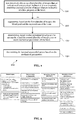
- FIG. 1 is a block diagram illustrating a method in accordance with an embodiment of the present invention.
- the method comprises a first step of non-invasively obtaining one or more first images that are indicative of a structure of the heart and non-invasively obtaining one or more second images that are indicative of a flow property of the heart.
- the obtained images may be images that have been previously acquired using non-invasive images and stored on a workstation.
- the blood pool and the myocardium of the heart are segmented. It is understood that the segmentation of the blood pool and the myocardium may be also be based on further information, e.g. from the one or more second images.
- a third step based on the segmented blood pool, the myocardium and the second images, one or more mechanical parameters of the heart are determined.
- the internal myocardial power is determined based on the one or more mechanical parameters.
- FIG. 2 shows a block chart for diagnostic measures including four steps: clinical imaging accompanied with an assessment of heart rate and systemic blood pressures, post-processing of imaging data for anatomy and flow, calculation of derived parameters based on post-processing and finally calculation of the myocardial power and efficiency for clinical measures.
- the steps shall be explained in more detail in the following.
- Imaging procedure allowing to acquire imaging data of the ventricle blood pool, myocardium as well as ventricular inflow and outflow conditions.
- MRI, CT and echocardiography or their combination are, among others, possible imaging modalities. 3D data are preferable. However, analysis could also be done based on 2D imaging data.
- Flow conditions can be assessed directly by methods like 4D MRI or Doppler echocardiography. Alternatively, they could be assessed indirectly by volumetric measurements of the ventricle (e.g. from 3D echocardiography or CINE MRT).
- Imaging acquisition is preferably accompanied by an assessment of the heart rate (HR) as well as the measurement of systemic blood pressure: 1) preferably using non-invasive methods (e.g. cuff-based measurements) or 2) can be performed based on invasive catheter-based measurements.
- HR heart rate
- systemic blood pressure 1) preferably using non-invasive methods (e.g. cuff-based measurements) or 2) can be performed based on invasive catheter-based measurements.
- Post-processing can be performed by various interchangeable software tools. Several products are regularly available for routinely-used imaging devices or independent solutions can be used.
- the segmentation of the blood pool and the myocardium needs to be performed. Based on segmentations geometrical parameters of the ventricle and myocardium necessary to calculate the ventricular wall stress are assessed: ventricular radii, lengths and volumes, myocardial volume, myocardial wall thickness.
- Flow analysis needs to be performed: If no direct ventricular pressure measurement is available, inflow and outflow curves should be generated in order to measure contraction time. Flow curves could be measured directly based on flow imaging or indirectly based on dynamic assessment of geometries. If no direct ventricular pressure measurement is available, maximal outflow valve velocity should be measured to allow estimation of the valve pressure drop.
- Myocardial volume is derived from myocardial wall segmentation or based on ventricular length together with myocardial wall thickness. If peak systolic ventricular pressure P sys is not measured directly, P sys should be derived indirectly.
- P sys is the calculated sum of the systolic pressure in the aorta and the pressure gradient across the outflow valve, whereas the systolic pressure in the aorta is derived from the peripherally (e.g. arm) measured systolic pressure by taking into account the increasing from the heart pulse pressure.
- the pulse pressure increase could be calculated using in the literature published equations.
- the valve pressure drop is calculated by using the Bernoulli's equation.
- the wall stress could also be calculated using other possible equations (e.g. thick wall Laplace law). These other equations may be based on the same kind of parameters, including S wall , P sys , and R BP .
- Contraction time ( t cs ) is the period between the start of ventricular contraction and peak systolic pressure and/or flow. If contraction time is not measured directly from ventricular pressure curves, tcs should be derived indirectly:
- the sum of inflow valve closing time t CT , isovolumetric contraction time t IVC and ejection time t ET define t cs .
- the ejection time t ET is derived from outflow curve.
- the isovolumetric contraction time t IVC is derived from combined inflow and outflow curves.
- the sum of the inflow valve closing time t CT and isovolumetric contraction time t IVC is derived from ejection time t ET together with systemic pressure values assuming ventricular pressure increase according to parabolic equation.
- t cs , t IVC , t ET may provide further simplification with adequate diagnostic accuracy.
- Forward and backward flow volumes are derived from outflow curves or based on dynamic assessment of ventricular geometry.
- the contraction time t cs is defined as the period between the start of ventricular (myocardial) contraction at the end-diastolic pressure and a peak systolic pressure which is correlating with a peak aortic flow.
- a known direct way of measuring the contraction time is based on catheter measured pressure curves that represent an invasive measurement associated with X-Ray radiation as well as a use of contrast agent. However, these are both associated with a risk for patients.
- the inflow valve closing time t CT is a period of the increasing ventricle pressure necessary to stop the blood inflowing from the atrium into the ventricle, which is owning a momentum.
- the inflow valve closing time t CT can be determined as a time at which two curves, a hyperbola and a parabola, are crossing.
- a hyperbola is a pressure curve p H as a function of time t, [ms], e.g.
- T dia 60 * 10 3 / HR ⁇ 805 * 0.18365317 + 0.54677315 * e ⁇ 0.015150054 * HR
- P SYS and P DIA are systolic and diastolic pressures.
- the ejection time t ET is directly measured from outflow curve.
- the parabola equation is fully defined by three parameters: two pressure values P SYS and P DIA of the parabola based on cuff based measurements and a period t ET between these two parabola points as measured from flow curve as well as an assigning of the point P SYS to the parabola vertex.
- the isovolumetric contraction time t IVC can be directly measured from combined inflow and outflow curves.
- the sum of the inflow valve closing time t CT and isovolumetric contraction time t IVC could be estimated from ejection time t ET together with systemic pressure values P SYS and P DIA , assuming ventricular pressure increase according to parabolic equation (4).
- FIG. 3 provides a further illustration of parameters used to estimate the contraction time
- FIG. 4 illustrates a method for estimating the inflow valve closing time.
- Internal myocardial power is defined as the power of the heart to perform myocardial contraction.
- Internal myocardial power is indexed to BSA allowing inter-individual comparison.
- MAP mean arterial pressure
- CO eff (forward flow volume - backward flow volume) * HR (heart rate) or from outflow curves, depending on the imaging modalities used.
- HR heart rate
- Eff CP is the level of efficiency of the myocardium to provide effective cardiac output in the arterial system taking into account power loss in the myocardium and due to diseased heart valves.
- External Myocardial Power is defined as hydrodynamic power at the outlet of the ventricle. It is the power needed to pump a given amount of blood against a given resistance and across a given gradient by a stenotic valve.
- AoG is the aortic pressure gradient and CO total (forward flow volume) multiplied by HR.
- the disclosed method can be applied in patients with different heart disease.
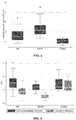
- Myocardial power was successfully shown to be able to quantify cardiac remodeling processes. Circulatory efficiency and internal myocardial power measurements gradually reflect disease response in patients with aortic valve stenosis (AS) and combine aortic valve stenosis and insufficiency (AS/AI), even when external myocardial efficiency and EF are still compensated.
- AS aortic valve stenosis
- AS/AI insufficiency
Landscapes
- Health & Medical Sciences (AREA)
- Life Sciences & Earth Sciences (AREA)
- Engineering & Computer Science (AREA)
- Medical Informatics (AREA)
- Nuclear Medicine, Radiotherapy & Molecular Imaging (AREA)
- Public Health (AREA)
- Physics & Mathematics (AREA)
- General Health & Medical Sciences (AREA)
- Biomedical Technology (AREA)
- Pathology (AREA)
- Biophysics (AREA)
- Heart & Thoracic Surgery (AREA)
- Molecular Biology (AREA)
- Surgery (AREA)
- Animal Behavior & Ethology (AREA)
- Veterinary Medicine (AREA)
- Radiology & Medical Imaging (AREA)
- Cardiology (AREA)
- High Energy & Nuclear Physics (AREA)
- Vascular Medicine (AREA)
- Physiology (AREA)
- Optics & Photonics (AREA)
- Primary Health Care (AREA)
- Epidemiology (AREA)
- Ophthalmology & Optometry (AREA)
- Hematology (AREA)
- Pulmonology (AREA)
- Oral & Maxillofacial Surgery (AREA)
- Dentistry (AREA)
- Theoretical Computer Science (AREA)
- Data Mining & Analysis (AREA)
- Databases & Information Systems (AREA)
- Magnetic Resonance Imaging Apparatus (AREA)
- Ultra Sonic Daignosis Equipment (AREA)
Priority Applications (4)
| Application Number | Priority Date | Filing Date | Title |
|---|---|---|---|
| EP18158410.3A EP3531426A1 (de) | 2018-02-23 | 2018-02-23 | Nichtinvasive messung der myokardleistung |
| DE112019000945.1T DE112019000945T5 (de) | 2018-02-23 | 2019-02-22 | Nicht-invasive messung der myokardleistung |
| PCT/EP2019/054480 WO2019162461A1 (en) | 2018-02-23 | 2019-02-22 | Non-invasive measurement of myocardial power |
| US16/971,052 US20200383585A1 (en) | 2018-02-23 | 2019-02-22 | Non-invasive measurement of myocardial power |
Applications Claiming Priority (1)
| Application Number | Priority Date | Filing Date | Title |
|---|---|---|---|
| EP18158410.3A EP3531426A1 (de) | 2018-02-23 | 2018-02-23 | Nichtinvasive messung der myokardleistung |
Publications (1)
| Publication Number | Publication Date |
|---|---|
| EP3531426A1 true EP3531426A1 (de) | 2019-08-28 |
Family
ID=61628107
Family Applications (1)
| Application Number | Title | Priority Date | Filing Date |
|---|---|---|---|
| EP18158410.3A Withdrawn EP3531426A1 (de) | 2018-02-23 | 2018-02-23 | Nichtinvasive messung der myokardleistung |
Country Status (4)
| Country | Link |
|---|---|
| US (1) | US20200383585A1 (de) |
| EP (1) | EP3531426A1 (de) |
| DE (1) | DE112019000945T5 (de) |
| WO (1) | WO2019162461A1 (de) |
Families Citing this family (1)
| Publication number | Priority date | Publication date | Assignee | Title |
|---|---|---|---|---|
| EP3910644A1 (de) * | 2020-05-12 | 2021-11-17 | Koninklijke Philips N.V. | Qualitätskontrolle in der medizinischen bildgebung |
Citations (2)
| Publication number | Priority date | Publication date | Assignee | Title |
|---|---|---|---|---|
| US20110144967A1 (en) * | 2008-08-12 | 2011-06-16 | Lev Adirovich | System and method for dynamic cardiac analysis, detection, prediction, and response using cardio-physiological mathematical modeling |
| US20160228190A1 (en) * | 2015-02-05 | 2016-08-11 | Siemens Aktiengesellschaft | Three-dementional quantitative heart hemodynamics in medical imaging |
-
2018
- 2018-02-23 EP EP18158410.3A patent/EP3531426A1/de not_active Withdrawn
-
2019
- 2019-02-22 WO PCT/EP2019/054480 patent/WO2019162461A1/en not_active Ceased
- 2019-02-22 DE DE112019000945.1T patent/DE112019000945T5/de not_active Withdrawn
- 2019-02-22 US US16/971,052 patent/US20200383585A1/en not_active Abandoned
Patent Citations (2)
| Publication number | Priority date | Publication date | Assignee | Title |
|---|---|---|---|---|
| US20110144967A1 (en) * | 2008-08-12 | 2011-06-16 | Lev Adirovich | System and method for dynamic cardiac analysis, detection, prediction, and response using cardio-physiological mathematical modeling |
| US20160228190A1 (en) * | 2015-02-05 | 2016-08-11 | Siemens Aktiengesellschaft | Three-dementional quantitative heart hemodynamics in medical imaging |
Also Published As
| Publication number | Publication date |
|---|---|
| WO2019162461A1 (en) | 2019-08-29 |
| US20200383585A1 (en) | 2020-12-10 |
| DE112019000945T5 (de) | 2020-11-19 |
Similar Documents
| Publication | Publication Date | Title |
|---|---|---|
| US10299862B2 (en) | Three-dimensional quantitative heart hemodynamics in medical imaging | |
| US10162932B2 (en) | Method and system for multi-scale anatomical and functional modeling of coronary circulation | |
| CN100377165C (zh) | 无创式左心室的容积测定 | |
| US10111635B2 (en) | Image processing apparatus, image processing method, and storage medium | |
| EP3537977B1 (de) | Verfahren und system zur modellierung eines menschlichen herzes und vorhöfen | |
| US10134127B2 (en) | Method for post-processing flow-sensitive phase contrast magnetic resonance images | |
| US9462952B2 (en) | System and method for estimating artery compliance and resistance from 4D cardiac images and pressure measurements | |
| Järvinen et al. | Right atrial MR imaging studies of cadaveric atrial casts and comparison with right and left atrial volumes and function in healthy subjects. | |
| CN107072531A (zh) | 用于心肌壁动力学的分析的方法和系统 | |
| JP5736462B2 (ja) | 超音波撮像装置及び方法 | |
| Iino et al. | Time course of reversed cardiac remodeling after pulmonary endarterectomy in patients with chronic pulmonary thromboembolism | |
| US10548494B2 (en) | Method for determining a personalized cardiac model using a magnetic resonance imaging sequence | |
| Lechartier et al. | Magnetic resonance imaging in pulmonary hypertension: an overview of current applications and future perspectives | |
| Forni et al. | Assessment of right ventricular function in patients with congestive heart failure by echocardiographic automated boundary detection | |
| EP3531426A1 (de) | Nichtinvasive messung der myokardleistung | |
| WO2006124919A2 (en) | Determining left ventricular end-diastolic pressure | |
| US20100135548A1 (en) | Medical Imaging System | |
| Ali et al. | Diagnostic accuracy of cardiovascular magnetic resonance imaging for assessment of right ventricular morphology and function in pulmonary artery hypertension | |
| Sorrell et al. | Role of echocardiography in the diagnostic assessment and etiology of heart failure in the elderly—opacify, quantify, and rectify | |
| Lee | Three-chamber function with cardiac CT | |
| EP4329590B1 (de) | System und verfahren zur schätzung des werts für einen zielherzparameter | |
| Sanfilippo et al. | Assessment of the left heart | |
| Le et al. | Intra-Left Ventricular Flow Velocity Distributions, Based on Color Doppler Echo Vector Flow Mapping of Normal Subjects and Heart Failure Patients | |
| Swan | Significance and Prognostic Value of Left | |
| Haykowsky et al. | Measurements of changes in left ventricular volume, strain, and twist during isovolumic relaxation using MRI 2 |
Legal Events
| Date | Code | Title | Description |
|---|---|---|---|
| PUAI | Public reference made under article 153(3) epc to a published international application that has entered the european phase |
Free format text: ORIGINAL CODE: 0009012 |
|
| AK | Designated contracting states |
Kind code of ref document: A1 Designated state(s): AL AT BE BG CH CY CZ DE DK EE ES FI FR GB GR HR HU IE IS IT LI LT LU LV MC MK MT NL NO PL PT RO RS SE SI SK SM TR |
|
| AX | Request for extension of the european patent |
Extension state: BA ME |
|
| STAA | Information on the status of an ep patent application or granted ep patent |
Free format text: STATUS: THE APPLICATION IS DEEMED TO BE WITHDRAWN |
|
| 18D | Application deemed to be withdrawn |
Effective date: 20200229 |