EP3548516B1 - Anticorps trimérique à chaîne unique spécifique à cd33, cd16 et cd123 - Google Patents
Anticorps trimérique à chaîne unique spécifique à cd33, cd16 et cd123 Download PDFInfo
- Publication number
- EP3548516B1 EP3548516B1 EP17822159.4A EP17822159A EP3548516B1 EP 3548516 B1 EP3548516 B1 EP 3548516B1 EP 17822159 A EP17822159 A EP 17822159A EP 3548516 B1 EP3548516 B1 EP 3548516B1
- Authority
- EP
- European Patent Office
- Prior art keywords
- cells
- triplebody
- single chain
- spm
- nucleic acid
- Prior art date
- Legal status (The legal status is an assumption and is not a legal conclusion. Google has not performed a legal analysis and makes no representation as to the accuracy of the status listed.)
- Active
Links
Images







Classifications
-
- C—CHEMISTRY; METALLURGY
- C07—ORGANIC CHEMISTRY
- C07K—PEPTIDES
- C07K16/00—Immunoglobulins [IG], e.g. monoclonal or polyclonal antibodies
- C07K16/46—Hybrid immunoglobulins
- C07K16/468—Immunoglobulins having two or more different antigen binding sites, e.g. multifunctional antibodies
-
- A—HUMAN NECESSITIES
- A61—MEDICAL OR VETERINARY SCIENCE; HYGIENE
- A61P—SPECIFIC THERAPEUTIC ACTIVITY OF CHEMICAL COMPOUNDS OR MEDICINAL PREPARATIONS
- A61P35/00—Antineoplastic agents
- A61P35/02—Antineoplastic agents specific for leukemia
-
- C—CHEMISTRY; METALLURGY
- C07—ORGANIC CHEMISTRY
- C07K—PEPTIDES
- C07K16/00—Immunoglobulins [IG], e.g. monoclonal or polyclonal antibodies
- C07K16/18—Immunoglobulins [IG], e.g. monoclonal or polyclonal antibodies against material from animals or humans
- C07K16/28—Immunoglobulins [IG], e.g. monoclonal or polyclonal antibodies against material from animals or humans against receptors, cell surface antigens or cell surface determinants
- C07K16/2803—Immunoglobulins [IG], e.g. monoclonal or polyclonal antibodies against material from animals or humans against receptors, cell surface antigens or cell surface determinants against the immunoglobulin superfamily
-
- C—CHEMISTRY; METALLURGY
- C07—ORGANIC CHEMISTRY
- C07K—PEPTIDES
- C07K16/00—Immunoglobulins [IG], e.g. monoclonal or polyclonal antibodies
- C07K16/18—Immunoglobulins [IG], e.g. monoclonal or polyclonal antibodies against material from animals or humans
- C07K16/28—Immunoglobulins [IG], e.g. monoclonal or polyclonal antibodies against material from animals or humans against receptors, cell surface antigens or cell surface determinants
- C07K16/2803—Immunoglobulins [IG], e.g. monoclonal or polyclonal antibodies against material from animals or humans against receptors, cell surface antigens or cell surface determinants against the immunoglobulin superfamily
- C07K16/283—Immunoglobulins [IG], e.g. monoclonal or polyclonal antibodies against material from animals or humans against receptors, cell surface antigens or cell surface determinants against the immunoglobulin superfamily against Fc-receptors, e.g. CD16, CD32, CD64
-
- C—CHEMISTRY; METALLURGY
- C07—ORGANIC CHEMISTRY
- C07K—PEPTIDES
- C07K16/00—Immunoglobulins [IG], e.g. monoclonal or polyclonal antibodies
- C07K16/18—Immunoglobulins [IG], e.g. monoclonal or polyclonal antibodies against material from animals or humans
- C07K16/28—Immunoglobulins [IG], e.g. monoclonal or polyclonal antibodies against material from animals or humans against receptors, cell surface antigens or cell surface determinants
- C07K16/2866—Immunoglobulins [IG], e.g. monoclonal or polyclonal antibodies against material from animals or humans against receptors, cell surface antigens or cell surface determinants against receptors for cytokines, lymphokines, interferons
-
- C—CHEMISTRY; METALLURGY
- C07—ORGANIC CHEMISTRY
- C07K—PEPTIDES
- C07K2317/00—Immunoglobulins specific features
- C07K2317/20—Immunoglobulins specific features characterized by taxonomic origin
- C07K2317/24—Immunoglobulins specific features characterized by taxonomic origin containing regions, domains or residues from different species, e.g. chimeric, humanized or veneered
-
- C—CHEMISTRY; METALLURGY
- C07—ORGANIC CHEMISTRY
- C07K—PEPTIDES
- C07K2317/00—Immunoglobulins specific features
- C07K2317/30—Immunoglobulins specific features characterized by aspects of specificity or valency
- C07K2317/31—Immunoglobulins specific features characterized by aspects of specificity or valency multispecific
-
- C—CHEMISTRY; METALLURGY
- C07—ORGANIC CHEMISTRY
- C07K—PEPTIDES
- C07K2317/00—Immunoglobulins specific features
- C07K2317/60—Immunoglobulins specific features characterized by non-natural combinations of immunoglobulin fragments
- C07K2317/62—Immunoglobulins specific features characterized by non-natural combinations of immunoglobulin fragments comprising only variable region components
- C07K2317/622—Single chain antibody (scFv)
-
- C—CHEMISTRY; METALLURGY
- C07—ORGANIC CHEMISTRY
- C07K—PEPTIDES
- C07K2317/00—Immunoglobulins specific features
- C07K2317/60—Immunoglobulins specific features characterized by non-natural combinations of immunoglobulin fragments
- C07K2317/62—Immunoglobulins specific features characterized by non-natural combinations of immunoglobulin fragments comprising only variable region components
- C07K2317/624—Disulfide-stabilized antibody (dsFv)
-
- C—CHEMISTRY; METALLURGY
- C07—ORGANIC CHEMISTRY
- C07K—PEPTIDES
- C07K2317/00—Immunoglobulins specific features
- C07K2317/70—Immunoglobulins specific features characterized by effect upon binding to a cell or to an antigen
- C07K2317/73—Inducing cell death, e.g. apoptosis, necrosis or inhibition of cell proliferation
- C07K2317/732—Antibody-dependent cellular cytotoxicity [ADCC]
-
- C—CHEMISTRY; METALLURGY
- C07—ORGANIC CHEMISTRY
- C07K—PEPTIDES
- C07K2317/00—Immunoglobulins specific features
- C07K2317/90—Immunoglobulins specific features characterized by (pharmaco)kinetic aspects or by stability of the immunoglobulin
- C07K2317/92—Affinity (KD), association rate (Ka), dissociation rate (Kd) or EC50 value
-
- C—CHEMISTRY; METALLURGY
- C07—ORGANIC CHEMISTRY
- C07K—PEPTIDES
- C07K2317/00—Immunoglobulins specific features
- C07K2317/90—Immunoglobulins specific features characterized by (pharmaco)kinetic aspects or by stability of the immunoglobulin
- C07K2317/94—Stability, e.g. half-life, pH, temperature or enzyme-resistance
Definitions
- the present invention relates to a nucleic acid molecule encoding a single chain triplebody capable of specifically binding to CD33, CD16 and CD123, wherein said nucleic acid molecule comprises: (a) a nucleic acid molecule encoding a protein represented by SEQ ID NO:1 or 3; (b) a nucleic acid molecule represented by SEQ ID NO:2 or 4; (c) the nucleic acid molecule of (b), wherein each thymine is replaced by uracil; or (d) a nucleic acid molecule that is degenerate with respect to the nucleic acid molecule of (b) or (c).
- the present invention further relates to a vector comprising the nucleic acid molecule of the invention, a host cell transformed or transfected with the nucleic acid molecule or the vector of the invention, as well as to a method for the production of a single chain triplebody capable of specifically binding to CD33, CD16 and CD123. Furthermore, the present invention also relates to a single chain triplebody capable of specifically binding to CD33, CD16 and CD123 encoded by the nucleic acid molecule of the invention, as well as to a pharmaceutical composition comprising the nucleic acid molecule, the vector, the host cell and/or the single chain triplebody of the invention. Also encompassed by the present invention are the nucleic acid molecule, the vector, the single chain triplebody and/or the composition of the invention for use in the treatment of acute myeloid leukemia and/or myelodysplastic syndrome.
- AML Acute myeloid leukemia
- BM bone marrow
- CR complete remission
- Prognosis for patients with relapsed disease is generally poor and, therefore, there is an unmet medical need for the development of new treatment options [1,2].
- MRD Minimal Residual Disease
- LSCs AML Leukemia Stem Cells
- This hypothesis also known as “the leukemia stem cell model”
- new treatment options are needed, which aim at the elimination of both the bulk leukemia blasts and the MRD cells/LSCs.
- it is desirable that such new therapies cause less systemic toxicity than current chemotherapeutic approaches.
- a promising choice for this purpose are immunotherapeutic approaches, because the molecular mechanisms conferring chemoresistance to LSCs do not protect these cells against lysis by immune effector cells.
- One mechanism to achieve immunotherapeutic effects is to recruit cytolytic effector cells of the immune system towards AML cells through the identification of suitable target antigens expressed on the leukemic cell surface and the development of antibodies or antibody-derived agents with specificity for both these tumor targets and trigger molecules on the effector cell surface. This approach is known as Antibody Dependent Cellular Cytotoxicity (ADCC) or Re-Directed Lysis (RDL) of AML cells.
- ADCC Antibody Dependent Cellular Cytotoxicity
- RDL Re-Directed Lysis
- Suitable target antigens for redirected lysis of both bulk AML blasts and MRD cells/LSCs include the cell surface proteins CD33 and CD123.
- Expression of CD33 is restricted to normal and malignant hematopoietic cells [8], including AML-LSCs [9-12]. Therefore, therapeutic approaches directed against CD33 can be expected to cause only low systemic toxicities, apart from haematotoxicity and toxicity for myeloid cells residing in other tissues such as the Kupffer cells in the liver.
- CD33 thus, represents a promising target for the therapy of AML [9,13].
- MDS Myelo-Dysplastic Syndrome
- AML blasts 25 % of MDS patients progress to refractory AML with poor prognosis [14,15]. Accordingly, new treatment options are urgently needed also for this disease.
- MDS cells carry many of the same surface antigens as AML blasts, including CD33 and CD123 and, therefore, a significant fraction of MDS patients are expected to respond to treatment with similar antibody-derived agents as AML patients [16-19].
- MDS cancer stem cells CSCs
- CSCs MDS cancer stem cells
- CD33 and CD123 are regularly expressed on the surface of MDS CSCs [16-18]. It has therefore been proposed that MDS-CSCs may be eliminated by similar antibody-derived agents, which target CD33 and cause RDL, as AML-LSCs [18].
- a CD33-directed Antibody-Drug Conjugate namely Gemtuzumab-Ozogamycin (GO, Mylotarg)
- ADC Antibody-Drug Conjugate
- This agent is a conjugate between the DNA-toxin calicheamycin and a CD33-reactive antibody. It has pro-ven clinical efficacy for certain subtypes of AM, but has been withdrawn from the market due to safety concerns [24].
- the reported side effects were due, in part, to an uncontrolled cleavage of the linker between the antibody and the toxin, leading to target-independent ("off-target”) systemic toxicity.
- CD33-directed ADC SGN-CD33A
- SGN-CD33A Another CD33-directed ADC, SGN-CD33A
- a bispecific T cell Engager (BiTE) targeting CD33 and recruiting T cells as cytolytic effectors for the elimination of AML cells is in early clinical development [27-31; see the world wide web under: clinicaltrials.gov/ct2/show/NCT02520427].
- a bi-specific two-component system with specificity for CD33 and CD3 [32] is in pre-clinical development.
- Tandabs i.e. antibody-derived fusion proteins in a new molecular format
- These proteins carry two copies of a binding site for an antigen on a cancer cell, such as CD33, and two for a trigger molecule on an effector cell, such as CD3 on cytolytic T-lymphocytes (T-CTL). They mediate lysis of leukemic cells by recruited effector cells (RDL), similar to Micromet/AMGEN's BiTE protein AMG 330.
- RDL cytolytic T-lymphocytes
- Tandabs are homo-dimers, built by self-assembly of two identical copies of a polypeptide chain, which spontaneously assemble in an anti-parallel orientation via the non-covalent attraction between the interfaces of the V H and V L domains of a single-chain Fv fragment.
- each antigen binding site is composed of a V H and a V L domain located on separate polypeptide-chains, held together only by the attractive forces between the complementary domain interfaces.
- the molecular architecture of Tandabs therefore, differs significantly from Micromet/AMGEN's BiTEs. In BiTE proteins, all four V H and V L domains composing the 2 antigen binding sites are carried in a single polypeptide chain. BiTEs are, therefore, also called "single chain tandem diabodies".
- Tandabs and BiTEs carry only one binding site for the cancer cell and one for the trigger on the effector cell and, therefore, are "bi-valent, mono-targeting and mono-triggering".
- Tandabs carry 2 binding sites for the same tumor antigen and 2 for the same trigger. They are, thus, "bi-specific, tetravalent, mono-targeting and mono-triggering".
- the companies Amphivena and Affimed have jointly developed a Tandab with specificities for CD33 and CD3, AMV-564, which is currently in early clinical development for the treatment of AML [33-35; see the world wide web under: affimed.com and under: biocentury.com/products/amv-564].
- This agent is expected to offer an advantage over Micromet/AMGEN's BiTE AMG 330, because it has a molecular mass of 110 kDA compared with 55 kDA for the BiTE agent.
- the BiTE agent needs to be delivered by continuous infusion, due to its short plasma half-life of less than 1 hour, whereas the Tandab can be delivered by bolus injections due to its larger mass and longer plasma half-life, which allows for a more convenient dosing schedule.
- Another variant of tandem diabodies are the so-called BiKEs, and a CD16-CD33 BiKE has been reported [19,36].
- CD123 the alpha chain of the interleukin-3 receptor
- CD34-positive leukemic blasts are a subset of AML cells, which comprises most of the LSCs as a further subset.
- CD123 is expressed with greater surface densities on AML-LSCs and leukemic progenitor cells than on normal hematopoietic stem cells (HSCs) [5,11,42,44].
- CD123 is also expressed on blasts from a variety of other hematologic malignancies including Acute Lymphoblastic Leukemia (ALL) [46,47], Chronic Myeloid Leukemia (CML), Myelo-Dysplastic Syndrome (MDS), Hodgkin Lymphoma (HL), Hairy Cell Leukemia (HCL) and others [17,46].
- ALL Acute Lymphoblastic Leukemia
- CML Chronic Myeloid Leukemia
- MDS Myelo-Dysplastic Syndrome
- HL Hodgkin Lymphoma
- HCL Hairy Cell Leukemia
- CD123 is therefore a potential target for the design of new immuno-therapies based on antibody-derived agents and the recruitment of cytolytic effectors for the elimination of AML cells by RDL/ADCC reactions.
- CD123 shows low expression on megakaryocytic progenitors [66] and, therefore, agents targeting CD123 are expected to produce fewer toxicities for the megakaryocytic-thrombocytic lineage than CD33-specific agents such as Mylotarg.
- DART Dual Antigen Re-Targeting
- CD33 and CD123 are co-expressed on blasts from patients with a broad range of AML-subtypes [7].
- the studies reported to date have mostly addressed the expression of CD33 and CD123 individually in patient samples, and the cohorts analyzed were small [6,43,45,67].
- Larger studies evaluating the co-expression of these antigens for a greater number of patients with many different genetic- and risk-subtypes of the disease would be needed in order to investigate whether new dual-targeting agents simultaneously addressing both antigens on the same AML cell [58,60], and combinations of corresponding mono-targeting agents, are promising for clinical applications for a broad group of AML patients.
- Bi- and tri-specific antibodies and related molecular agents are under development by a number of teams [68,69]. Dual-targeting agents in late preclinical or early clinical development have been produced by several companies, including Roche (Crossmabs, [70,71]), Genmab (Duomabs [72]), Abbvie (Dual Variable Region DVD IgGs [73,74]), Genentech (Two-in-one antibodies [75,76]), and Sanofi (tetravalent and multifunctional CODV IgGs [77]).
- Roche Cherossmabs, [70,71]
- Genmab Duomabs [72]
- Abbvie Dual Variable Region DVD IgGs [73,74]
- Genentech Tewo-in-one antibodies [75,76]
- Sanofi tetravalent and multifunctional CODV IgGs [77]
- a number of other new molecular formats are based on the common light chain principle [78-80], such as Novimmune'
- Additional new agents include Genentech's "Dual action antibodies” [81], Rinat-Pfizer's “Duobodies” [82], “Dutafabs” [ WO 2012 163520 ; 83], “Duetmabs” [84], and monovalent, bi-specific hetero-dimeric IgG antibodies generated with the help of guiding mutations [85,86].
- the most advanced members of this family are in clinical development, such as Merrimack's bi- and multi-specific antibody MM-141, which has completed a phase I study for hepatic cancer and is currently tested in a phase II study for pancreatic cancer [87].
- This agent acts by simultaneously blocking the IGF-1 receptor (IGF-1R) and the EGF-receptor ErbB3, and by thus depriving cancer cells simultaneously of two essential growth factor signals, which drives them into cell death (apoptosis).
- MM-111 is a bi-specific diabody with one scFv-binding module each for ErbB2 (HER2) and ErbB3 (HER3), linked to modified human serum albumin for half-life extension [88,89].
- Another class of new agents are Janssen Cilag's (Covagen's) bi- and tri-specific "Fynomabs” [91; see the world wide web under: covagen.com].
- the agents are fusion proteins between antibodies and "fynomers", robust small binding modules for certain targets, which are derived from an SH3 domain of the tyrosine kinase FYN.
- COVA 208 targets 2 different epitopes on HER2 and is scheduled for clinical development against metastatic breast cancer [91; see the world wide web under: bioworld.com/content/swiss-firm-covagen-moves-lead-fynomab-toward-clinic].
- COVA 322 targeting TNF ⁇ and IL-17, is in a phase 1 trial for psoriasis (see the world wide web under: clinicaltrials.gov/ct2/show/NCT02243787).
- the present invention relates to a nucleic acid molecule encoding a single chain triplebody capable of specifically binding to CD33, CD16 and CD123, wherein said nucleic acid molecule comprises: (a) a nucleic acid molecule encoding a protein represented by SEQ ID NO:1 or 3; (b) a nucleic acid molecule represented by SEQ ID NO:2 or 4; (c) the nucleic acid molecule of (b), wherein each thymine is replaced by uracil; or (d) a nucleic acid molecule that is degenerate with respect to the nucleic acid molecule of (b) or, (c).
- nucleic acid molecule also referred to as nucleic acid sequence or polynucleotide herein, includes DNA, such as cDNA, and RNA. It is understood that the term “RNA” as used herein comprises all forms of RNA including e.g. mRNA. Both single-strand as well as double-strand nucleic acid molecules are encompassed by this term.
- the nucleic acid molecules of the invention can e.g. be synthesized by standard chemical synthesis methods, can be produced semi-synthetically, i.e. by combining parts synthesized by chemical synthesis with parts that are isolated from natural sources, or can be produced recombinantly.
- Ligation of the coding sequences to transcriptional regulatory elements and/or to other amino acid encoding sequences can be carried out using established methods, such as restriction digests, ligations and molecular cloning.
- the nucleic acid molecule encodes a single chain triplebody.
- This triplebody of the invention is also referred to herein as SPM-2.
- the term "triplebody”, as used herein, relates to a protein that contains three different binding domains which each bind a different corresponding antigen.
- the three different antigens are CD33, CD16 and CD123.
- the triplebody of the present invention is capable of specifically binding to CD33, CD16 and CD123.
- binding domains of the triplebody bind their respective antigens CD33, CD16 and CD123, but do not, or essentially do not, cross-react with an epitope with a structure similar to that of the target antigen, or with an unrelated structure.
- Cross-reactivity of a panel of molecules under investigation may be tested, for example, by assessing binding of said panel of molecules under conventional conditions to an epitope of the antigen of interest as well as to a number of more or less (structurally and/or functionally) closely related epitopes. Only those molecules that bind to an epitope of the antigen of interest in its relevant context (e.g.
- said triplebody is a single chain triplebody, i.e. all three binding domains are located on a single polypeptide chain.
- said single chain triplebody is encoded by a nucleic acid molecule that comprises one of the nucleic acid molecules recited in options (a) to (d).
- nucleic acid molecule comprises (rather than consists of) the recited sequence
- additional nucleotides extend over the specific sequence either on the 5' end or the 3' end or both.
- at most 99 additional nucleotides extend over the specific sequence either on the 5' end or the 3' end or both.
- at most 90 additional nucleotides such as e.g. at most 81, such as e.g. at most 75, more preferably at most 69 and even more preferably at most 65 additional nucleotide(s) extend(s) over the specific sequence either on the 5' end or the 3' end or both.
- At most tinctive nucleotides relates to a number of nucleotides that includes any integer below and including the specifically recited number.
- a preferred embodiment of a nucleic acid molecule that comprises additional nucleotides that extend over the specific recited sequence is shown in SEQ ID NO: 6, which represents the nucleic acid molecule of SEQ ID NO:2, with an additional IgK leader sequence, as e.g. employed in the appended examples.
- nucleic acid molecule encoding the single chain triplebody capable of specifically binding to CD33, CD16 and CD123 consists of (rather than comprises) the recited nucleic acid molecule of options (a) to (e), or consists of the sequence shown in SEQ ID NO:6.
- the term "represented by SEQ ID NO:[...]” refers to a sequence (amino acid sequence or nucleic acid sequence) that consists of exactly the sequence of the recited SEQ ID number, i.e. without any additional residues (amino acids or nucleotides) present.
- the nucleic acid molecule of the invention comprises a nucleic acid molecule encoding a protein represented by SEQ ID NO:1 or 3.
- the protein shown in SEQ ID NO:1 corresponds to the mature protein SPM-2 described in the appended examples in more detail.
- the nucleic acid molecule of the invention comprises a nucleic acid molecule represented by SEQ ID NO:2 or 4.
- the nucleic acid sequence shown in SEQ ID NO:2 is the nucleic acid sequence encoding the protein shown in SEQ ID NO:1, which corresponds to the mature protein SPM-2 described in the appended examples in more detail.
- SEQ ID NO:6 represents a further nucleic acid sequence encoding the protein shown in SEQ ID NO:1, wherein said nucleic acid molecule comprises an additional IgK leader sequence, which is cleaved off during the production of the protein of SEQ ID NO:1 in HEK293 cells, as shown in the appended examples.
- An example of the immature amino acid sequence of the protein encoded by SEQ ID NO:6 is provided in SEQ ID NO:5.
- the nucleic acid molecule of the invention comprises the nucleic acid molecule of (b), wherein each thymine is replaced by uracil.
- the nucleic acid molecule is an RNA molecule.
- the protein of SEQ ID NO:3 corresponds to the protein of SEQ ID NO:1 with the exception of the most C-terminal 11 amino acids, which represent a linker (also referred to as "spacer” herein) and the 6xHis-tag, and which have been removed in the protein of SEQ ID NO:3.
- a corresponding nucleic acid molecule encoding said protein of SEQ ID NO:3 is represented in SEQ ID NO:4.
- nucleic acid molecule comprising additional nucleotides
- the protein of SEQ ID NO:3 can be encoded by a nucleic acid molecule that extends over the specific sequence (i.e. SEQ ID NO:4) either on the 5' end or the 3' end or both, but preferably only at the 5' end.
- SEQ ID NO:8 A particularly preferred nucleic acid sequence is provided in SEQ ID NO:8 and the immature protein encoded by said sequence is shown in SEQ ID NO:7.
- the mature protein encoded by SEQ ID NO:8 will be the protein shown in SEQ ID NO:3, due to the removal of the leader sequence within the cell.
- the nucleic acid molecule of the invention comprises a nucleic acid molecule that is degenerate with respect to the nucleic acid molecule of options (b) or (c).
- nucleic acid molecules having a different sequence than the nucleic acid sequence specified above, but still encoding the same polypeptide lie within the scope of the present invention.
- Such nucleic acid molecule are referred to herein as being degenerate with respect to another nucleic acid molecule.
- the prototype needed to be refined. This was achieved by introducing a series of mutations into its amino acid and cDNA coding sequences.
- the agent needed to be "humanized” to minimize immune responses of the patient to the therapeutic agent and to minimize the production of neutralizing antibodies, which would reduce its therapeutic efficacy.
- the building blocks of the inventive triplebody also referred to as SPM-2 herein
- the single chain antibody variable fragments scFvs
- scFvs single chain antibody variable fragments
- the most immunogenic portions of murine scFvs in human recipients are the framework regions embedded in the V H and V L subdomains, the backbone of the three-dimensional (3D) architecture of these domains, which build the "fundamental immunoglobulin fold".
- the hypervariable loops also called “complementarity-determining regions” (CDRs)
- CDRs complementarity-determining regions
- the objective of the present inventors was to exchange the murine frameworks of the original scFvs against the best-suited human frameworks, while keeping the sequence and 3D conformation of the murine CDRs fixed as far as possible, in order to maintain specificity and affinity of the scFvs.
- additional point mutations need to be introduced into the human frameworks to further improve stability and functional activity of the humanized scFvs [98-100].
- the second needed refinement was the "disulfide-stabilization" of scFvs.
- This set of mutations was helpful for the majority of scFvs contained in the triplebodies studied by the inventors to date.
- the reason is that the V L and V H subdomains of scFvs are not held together by naturally occurring disulfide bonds and are, therefore, free to assume flexible positions in space, even though they are covalently connected by a linker. This flexibility can lead to instability and a propensity to form aggregates, which are damaging for the therapeutic usefulness of an agent and should be eliminated.
- disulfide-stabilization In the process of "disulfide-stabilization", suitable amino acid residues were identified, which are located in a desired spatial distance on the surface of the V H and V L domains, respectively. These were then mutated by site-specific mutagenesis into cysteines with the intent that these cysteines will spontaneously form a disulfide-bridge between the V H and V L domains and, thus, fix their position in space and eliminate the tendencies to unfold and to form aggregates [101-104]. In the present case, an unusual combination of disulfide-bridges was required to provide a suitable clinical candidate, as is evidenced by the appended examples.
- the finding implies that the blasts from all patients with different disease subtypes could be lysed, as long as they carried these antigens on their surface, and that no disease subtype had subtype-specific resistance mechanisms, which prevented successful lysis of the malignant cells in spite of the presence of the target antigens on their surface.
- the occurence of resistance mechanisms might have been expected, because the CD33-directed immunotoxin Mylotarg is effective on average for only about 40 % of the patients treated with this agent, although all patients' leukemia cells carried CD33 on their surface, as only patients meeting this requirement fulfilled the criteria for enrollment in the respective clinical studies [22,24]. Specific resistance mechanisms against the immunotoxin and the mechanism of action of the toxin were therefore frequently encountered for Mylotarg in clinical use.
- the mechanism of action of the triplebody differs dramatically from the mechanism of cytolysis employed by the immunotoxin Mylotarg.
- Mylotarg acts by causing DNA brakes in the nucleus of the cancer cell, into which the toxin has been imported.
- the triplebody lyses the cell from the outside, and acts independently of the import of a toxin and the potential mechanisms of resistance of the target cell against import of the toxin into the cell nucleus, DNA damage by the toxin, or the cells ability to repair such damage. This important finding raises hope that the same broad efficacy of the triplebody across a wide range of disease subtypes will also be observed during future clinical use of the agent.
- the triplebody of the present invention is expected to help eliminate at least a significant fraction of the LSCs and, thus, to delay the occurrence of relapse and to extend the overall survival (OS) of many patients.
- OS overall survival
- the triplebody of the present invention has an inbuilt ability to discriminate between normal myeloid cells and AML cells, and to preferentially eliminate AML cells.
- AML cells express far greater surface antigen densities of CD33 and CD123 than normal myeloid cells; i.e. normal myeloid cells carry a few 100 copies per cell each of CD33 and CD123, whereas the AML blasts carry a few 1 000 copies per cell each [15; tables 10, 12, 15].
- the desired discrimination can be achieved by providing the triplebody in limiting concentrations, because under these conditions the probability of decorating a malignant cell with the triplebody is greater than of decorating a normal cell.
- the inventors expect that the triplebody of the present invention can discriminate between AML-LSCs and normal HSCs to a certain degree, because AML-LSCs, as far as they have been defined, carry approximately 10-fold greater densities of CD123 than normal myeloid cells, namely a few 1 000 up to 10 000 copies per cell compared with a few 100 copies per cell on normal HSCs [5,11,12,42,46].
- the dual-targeting triplebody of the present invention offers the additional benefit that it will still function for a patient whose cells have developed resistance to mono-targeting agents through the outgrowth of escape variants (antigen-loss variants), which frequently occur in leukemia patients under therapy with mono-targeting agents [47].
- escape variants antigen-loss variants
- the triplebody of the present invention offers significant advantages over other currently known molecular formats, including dual-targeting agents developed by others.
- the present invention further relates to a vector comprising the nucleic acid molecule of the invention.
- the vector is a plasmid, cosmid, virus, bacteriophage or another vector used conventionally e.g. in genetic engineering.
- Non-limiting examples of vectors include plasmid vectors, such as the pSecTag Hygro expression vector (Life Technologies, Darmstadt, Germany) as e.g. used in the appended examples, or pCRTOPO (Invitrogen), pJOE, the pBBR1-MCS series, pJB861, pBSMuL, pBC2, pUCPKS, pTACT1 and vectors compatible with expression in mammalian cells like E-027 pCAG Kosak-Cherry (L45a) vector system, pREP (Invitrogen), pCEP4 (Invitrogen), pMC1neo (Stratagene), pXT1 (Stratagene), pSG5 (Stratagene), EBO-pSV2neo, pBPV-1, pdBPVMMTneo, pRSVgpt, pRSVneo, pSV2-dhfr, plZD35,
- vectors can contain one or more origins of replication (ori) and inheritance systems for cloning or expression, one or more markers for selection in the host, e.g., antibiotic resistance, and one or more expression cassettes.
- origins of replication ori
- inheritance systems for cloning or expression
- markers for selection in the host e.g., antibiotic resistance
- expression cassettes e.g., antibiotic resistance
- the coding sequences comprised in the vector can be ligated to transcriptional regulatory elements and/or to other amino acid encoding sequences using established methods.
- regulatory sequences are well known to those skilled in the art and include, without being limiting, regulatory sequences ensuring the initiation of transcription, and optionally regulatory elements ensuring termination of transcription, and stabilization of the transcript.
- Non-limiting examples for such regulatory elements ensuring the initiation of transcription comprise promoters, enhancers, insulators and/or regulatory elements ensuring transcription termination, which are to be included downstream of the nucleic acid molecules of the invention.
- Further examples include regulatory elements for translational control, such as a translation initiation codon, internal ribosomal entry sites (IRES) [106], Kozak sequences and nucleotide sequences encoding secretion signals or, depending on the expression system used, signal sequences capable of directing the expressed protein to a cellular compartment or to the culture medium.
- the vectors may also contain an additional expressible polynucleotide sequence (open reading frame) coding for one or more chaperones to facilitate correct protein folding.
- RNA splicing may contain regulatory elements for RNA splicing, such as intervening sequences flanked by donor and acceptor sites for RNA splicing.
- Techniques for vector modification, PCR amplification and ligation have been adequately described by laboratory manuals such as Sambrook & Russel [107].
- Non-limiting examples of suitable origins of replication include, for example, the full length ColE1, the SV40 viral, and the M13 origins of replication.
- suitable promoters for eukaryotic expression include, without being limiting, the cytomegalovirus (CMV) promoter, SV40-promoter, RSV-promoter (Rous sarcoma virus), chicken ⁇ -actin promoter, CAG-promoter (a combination of chicken ⁇ -actin promoter and cytomegalovirus immediate-early enhancer), the gai10 promoter, human elongation factor 1 ⁇ -promoter, AOX1 promoter, or CaM-kinase promoter.
- CMV cytomegalovirus
- SV40-promoter RSV-promoter
- CAG-promoter a combination of chicken ⁇ -actin promoter and cytomegalovirus immediate-early enhancer
- the gai10 promoter human elongation factor 1 ⁇ -promoter
- Non-limiting examples for regulatory elements ensuring transcription termination include the SV40-poly-A site, the tk-poly-A site or the AcMNPV polyhedral polyadenylation signals.
- selectable markers include dhfr, gpt, neomycin, hygromycin, blasticidin or geneticin.
- the vector of the present invention is an expression vector.
- An expression vector according to this invention is capable of directing the replication and the expression of the nucleic acid molecule of the invention and, accordingly, of the triplebody of the present invention encoded thereby.
- the expression vector was the pSecTag Hygro expression vector (Life Technologies, Darmstadt, Germany).
- nucleic acid molecules and/or vectors of the invention as described herein above may be designed for introduction into cells by e.g. non-chemical methods (electroporation, sono- poration, optical transfection, gene electrotransfer, hydrodynamic delivery or naturally occurring transformation upon contacting cells with the nucleic acid molecule of the invention), chemical-based methods (calcium phosphate, liposomes, DEAE-dextrane, polyethylenimine, nucleofection, TranslT ® LT1 Transfection Reagent (Mirus Bio LLC, Madison, USA)), particle- based methods (gene gun, magnetofection, impalefection), phage vector-based methods and viral methods.
- non-chemical methods electroroporation, sono- poration, optical transfection, gene electrotransfer, hydrodynamic delivery or naturally occurring transformation upon contacting cells with the nucleic acid molecule of the invention
- chemical-based methods calcium phosphate, liposomes, DEAE-dextrane, polyethylenimine, nucleo
- expression vectors derived from viruses such as retroviruses, vaccinia virus, adeno-associated virus, herpes viruses, Semliki Forest Virus or bovine papilloma virus, may be used for delivery of the nucleic acid molecules into a targeted cell population.
- the nucleic acid molecules and/or vectors of the invention are designed for stable transfection of CHO or HEK 293 F cells, preferably CHO cells.
- the present invention further relates to a host cell transformed or transfected with the nucleic acid molecule or the vector of the invention.
- a host cell transformed or transfected with Colour in accordance with the present invention, relates to a host cell that comprises the nucleic acid molecule or the vector of the invention.
- Typical mammalian host cells include Chinese hamster ovary (CHO) cells, COS 1, COS 7, Per.C6, NS0. Other possibilities include HEK 293, HEK 293 F, Hela, H9, Jurkat cells, mouse NIH3T3, C127, CV1, mouse L cells, mouse sarcoma cells, and Bowes melanoma cells.
- CHO cell lines are often used as the mammalian cell line of choice, because they have particularly favorable properties for large scale industrial production, such as shear-resistance upon stirring in large scale batch production.
- the triplebody produced by the method of the present invention is active in far lower concentrations than conventional therapeutic antibodies in cytolysis assays in cell culture (see Example 10 below), and therefore, smaller quantities of this agent are expected to be needed. Accordingly, smaller production volumes will most likely be sufficient, which means that other cell lines, such as those recited above, can also be employed for commercial production.
- Preferred mammalian host cells in accordance with the present invention are CHO cells, and HEK 293 F cells, most preferably CHO cells. These host cells as well as suitable media and cell culture conditions have been described in the art, as well as in the appended examples (e.g. Example 2). Further appropriate culture media and conditions for the above described host cells are known in the art.
- the host cells in accordance with this embodiment may e.g. be employed to produce the triplebody of the present invention.
- the present invention also relates to a method for the production of a single chain triplebody capable of specifically binding to CD33, CD16 and CD123, the method comprising culturing the host cell of the invention under suitable conditions and isolating the single chain triplebody capable of specifically binding to CD33, CD16 and CD123 produced.
- the vector present in the host cell of the invention is either an expression vector, or the vector mediates the stable integration of the nucleic acid molecule encoding the triplebody of the present invention into the genome of the host cell in such a manner that expression of the protein is ensured.
- the nucleic acid molecule encoding the triplebody of the present invention is stably integrated into the genome of the host cell in such a manner that expression of the protein is ensured. Means and methods for selection of a host cell, in which the nucleic acid molecule encoding the triplebody of the present invention has been successfully introduced such that expression of the protein is ensured, are well known in the art and have been described in the art.
- Suitable conditions for culturing eukaryotic host cells are well known to the person skilled in the art.
- the medium can be buffered or supplemented with suitable additives known to enhance or facilitate both.
- an inducible promoter controls the nucleic acid molecule of the invention in the vector present in the host cell
- expression of the triplebody can be induced by addition of an appropriate inducing agent, such as e.g. anhydrotetracycline.
- an appropriate inducing agent such as e.g. anhydrotetracycline.
- mammalian cell culture can e.g. be carried out in RPMI, Williams' E or DMEM medium containing 10 % (v/v) FCS, 2 mM L-glutamine and 100 U/ml penicillin/streptomycin.
- the cells can be kept e.g. at 37 °C in a 5 % CO 2 , water-saturated atmosphere.
- Suitable expression protocols for eukaryotic or vertebrate cells are well known to the skilled person and can be retrieved e.g. from the methods handbook of Sambrook and Russel [107].
- Methods of isolation of the protein produced are well-known in the art and comprise without limitation steps such as ion exchange chromatography, gel filtration chromatography (size exclusion chromatography), affinity chromatography, high pressure liquid chromatography (HPLC), reversed phase HPLC, disc gel electrophoresis or immunoprecipitation, as described for example by Sambrook and Russel [107] .
- the triplebody of the present invention in preferred embodiments, carries a hexa-histidine tag at its C-terminus, and this tag binds to nickel, zinc and stannous-ions. Beads carrying such ions can be purchased as GMP-compliant reagents from commercial sources, and can be added to the culture supernatant. The triplebody then binds to these beads, which can also either be stirred into the culture supernatant, and then collected by low-speed centrifugation.
- IMAC metal ion affinity chromatography
- the slur of supernatant with beads can be filled into a column, and then the culture supernatant is allowed to run out of the column, and the column is washed with a wash buffer.
- the elution is then performed with a suitable elution buffer, for example as specified in the examples listed below, and from then on the next purification steps using ion exchange column chromatography are the same as after capture with protein A, as described below.
- the IMAC capture method is cheaper than capture with protein A, because the protein A itself must be produced under GMP conditions in order to be usable for a GMP-compliant capture reagent, and this is more expensive than the GMP-grade reagents needed for capture by IMAC.
- IMAC capture method has been perfected by commercial producers, including Micromet/AMGEN, who initially purified their agent Blinatumomab by IMAC chromatography with stannous ions, and the protocols are in the public domain.
- the term "isolating the single chain triplebody capable of specifically binding to CD33, CD16 and CD123 produced” refers to the isolation of the triplebody encoded by the nucleic acid molecule of the present invention (in other words the nucleic acid molecule that is present in the host cell of the invention due to the transformation or transfection of said host cell with the nucleic acid molecule or the vector of the invention).
- the isolation of the single chain triplebody comprises the steps of: (a) incubating a cell culture supernatant containing the single chain triplebody with protein A; (b) separating the protein A with the bound single chain triplebody from the cell culture supernatant; (c) eluting the single chain triplebody from the protein A at a pH of between 2.9 to 3.1 at 4 °C to 5 °C for 60 to 70 min; and (d) purifying the single chain triplebody by: (d-i) anion exchange chromatography; and (d-ii) cation exchange chromatography.
- the triplebody is isolated from host cells. More specifically, the triplebody is isolated from the supernatant obtained by culturing the host cells of the present invention under appropriate conditions.
- any method of obtaining cell culture supernatants known in the art can be employed, including e.g. centrifugation and filtration.
- the cell culture supernatants are obtained by centrifugation, for example by centrifugation at 400 x g for an appropriate length of time, such as e.g. 4 minutes. Residual cells and debris may then, optionally, be removed by one or more further centrifugation steps.
- a second centrifugation step at 600 x g is carried out for an appropriate length of time, such as e.g. for 10 minutes.
- step (a)) said cell culture supematant is incubated with protein A, in order to capture the single chain triplebody present in the supematant.
- protein A for the capture of target proteins is well known in the art and is described here for the triplebody of the present invention in the appended examples, e.g. in Example 3.2.
- the protein A capturing step is carried out in a batch approach, more preferably by using protein A beads.
- protein A beads are commercially available, such as for example the protein A sepharose 4 Fast Flow beads provided by GE Healthcare.
- the incubation is carried out for 15 to 18 hours, preferably at 8 °C.
- the incubation can e.g. be carried out in plastic tubes on a rotating wheel, as described in the appended examples.
- the protein A with the bound single chain triplebody is separated from the cell culture supematant.
- any means of separating e.g. beads from a cell culture supernatant known in the art can be employed.
- the beads are sedimented, e.g. by centrifugation at an appropriate centrifugal force, such as e.g. 500 x g for an appropriate time, such as e.g. 5 minutes.
- the supernatant can be removed from the beads and the beads (with the bound triplebody) can be further processed, such as e.g. washing steps with appropriate buffers.
- the beads are transferred to a gravity flow column for easier handling. Gravity flow columns are also commercially available, such as e.g. the "Polyprep columns" provided by BioRad (Munich, Germany).
- this step (b) is carried out at room temperature.
- step (c)) of eluting the single chain triplebody from the protein A is performed. This step is carried out at a pH between 2.9 to 3.1 at 4 °C to 5 °C for 60 to 70 min. Elution can be carried out according to the manufacturer's instructions but may also be adjusted by the skilled person if desired. Preferably, elution is carried out in an elution buffer comprising glycine, citric acid and NaCl, more preferably in an elution buffer consisting of 50 mM glycine, 50 mM citric acid, pH 3.0, and 300 mM NaCl.
- one approach is as follows: one column volume of elution buffer is pumped into the column and kept in the column for 60-70 min at 4-5 °C. During this time, the triplebody protein is split from the protein A, because the mildly acidic pH is sufficient to break the non-covalent bonds between the triplebody and the protein A. This elution volume is then pumped out of the column, and to remove remaining triplebody, 1-2 more column volumes of elution buffer are pumped through the column. The column is attached to a photometer equipped with a flow-through detection cell, and the absorbance at 280 nm is continuously recorded.
- This absorbance is a measure of the protein concentration in the eluate, and when the absorbance drops, the elution of the protein from the column is complete.
- the total time for elution is 60-70 min, 60 min on the column, and an extra 10 min approximately for pumping through the extra 1-2 column volumes of elution buffer. It is recommendable to keep the time of exposure to acidic conditions to the strict minimum needed for an efficient elution, because the protein will slowly denature at low pH, and denaturation progresses with time. Therefore, as soon as the elution is complete, the pH is to be adjusted to pH 6.0, preferably by addition of 1 M Tris-HCl buffer pH 9.0 and monitoring the rise of the pH until pH 6.0 is reached.
- protein A as a capture reagent offers the advantage that the elution from the reagent at the same time fulfills the requirements for a first virus inactivation step, so that no separate virus inactivation step needs to be performed.
- the incubation at low pH for 60 min at 4 °C fulfills the requirements of regulatory authorities in Europe for a first viral inactivation step.
- An additional benefit of this capture procedure is that protein A resins are commercially available in GMP-grade quality (for example from GE healthcare), which is a considerable advantage for the commercial production of a GMP-grade triplebody.
- the IMAC capture procedure described above would require an extra first virus inactivation step and, conceivably, the combined cost for these 2 steps would be greater than the costs for the single capture and elution step with protein A.
- Capture by protein A is more elegant and therefore possibly more economic, because it is also important to keep the time for the purification as short as possible, to remove the triplebody as fast as possible from contaminants, which may still be present in the early stages of the purification, and which can lead to losses by inactivation, degradation or aggregation.
- the eluted triplebody is further purified (step (d)).
- the eluted fractions obtained from step (c) containing the triplebody are adjusted to a conductivity of 5 mS/cm.
- This conductivity is particularly suitable to permit loading of the triplebody onto the anion exchange chromatography column according to step (d-i).
- any suitable method can be obtained.
- the eluted fractions containing the triplebody can be pooled and diluted with water to a conductivity of 5 mS/cm.
- anion exchange chromatography can be carried out as follows: The pooled fractions from the elution step (step (c)) are loaded onto an appropriate anion exchange column, such as e.g. the HiTrap Q Sepharose HP column provided by GE Healthcare Europe (Munich, Germany) and the flow-through (FT) containing the triplebody is collected. Additional measures such as e.g. equilibration of the column, appropriate flow rates as well as washing steps, are well known in the art and have been described in more detail in the appended examples (see e.g. Example 4.1).
- the anion exchange chromatography is carried out at 8 °C.
- the equilibration buffer as well as the washing buffer comprises Histidine-HCl and NaCl, preferably 20 mM Histidine-HCl pH 6.0 and 50 mM NaCl.
- cation exchange chromatography can be carried out as follows: The flow-through (FT) fractions from the anion exchange chromatography step are loaded onto an appropriate cation exchange column, such as e.g. the Source 15S resin or the HiTrap SP Sepharose HP column, both available from GE Healthcare Europe (Munich, Germany). The column is then washed and the bound triplebody is eluted. Suitable elution buffers depend on the column chosen. For example, when using the Source 15S column, a linear NaCl gradient ranging from 0 to 50 % of an elution buffer (20 mM Histidine-HCl, 1 M NaCl ) can be employed.
- a linear NaCl gradient ranging from or 0 to 60 % of an elution buffer (20 mM Histidine-HCl, 1 M NaCl ) can be employed. It is further preferred that the column is additionally subjected to a washing step after elution, preferably with 100 % buffer (20 mM Histidine-HCl, 1 M NaCl). Additional measures such as e.g. equilibration of the column, appropriate flow rates as well as washing steps, are well known in the art and have been described in more detail in the appended examples (see e.g. Example 4.2). Preferably, the cation exchange chromatography is carried out at 8 °C. It is further preferred that the equilibration buffer comprises Histidine-HCl and NaCl, preferably 20 mM Histidine-HCl pH 6.0 and 50 mM NaCl.
- steps (d-i) and (d-ii) can be carried out in principle in any order however, it is particularly preferred that the step (d-i) is carried out prior to step (d-ii).
- the pooled fractions can be concentrated by ultrafiltration.
- One suitable, non-limiting example of such a concentration via ultrafiltration is shown in example 4.2 by using Amicon Ultra filter units with a molecular weight cut-off of 30 kDa (Millipore, Billerica, USA) at 8 °C and 1700 x g.
- analytical procedures can be performed subsequently to the elution step of the protein from the cation exchange column. Such steps are well known in the art and have also been detailed in the appended examples (e.g. Example 4.2).
- the eluted fractions can be monitored by analytical polyacrylamide SDS gel electrophoresis (SDS PAGE) to permit an assessment of purity and potential aggregates and proteolytic breakdown-products.
- SDS PAGE analytical polyacrylamide SDS gel electrophoresis
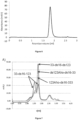
- all preparations are routinely monitored by size exclusion chromatography (SEC; also called gel filtration) for the presence of higher molecular mass aggregates and other contaminants.
- SEC size exclusion chromatography
- the liquid leaving the SEC column is monitored by absorbance at an appropriate wavelength, such as e.g. 280 nm, to identify the fractions containing the triplebody and other components still present in the sample at this stage.
- step (a) is followed by step (b), which is followed by step (c), which is followed by step (d).
- step (a) is followed by step (b)
- step (c) which is followed by step (d)
- further steps such as e.g. additional washing, equilibrations, re-buffering or concentration steps can be included, if desired.
- additional steps can be added at any stage of the method of the invention.
- the method of the present invention consists of the recited steps (a) to (d) and, optionally, the above detailed specific additional steps, i.e.
- the method of the present invention consists either of the recited steps (a) to (d) or of the steps (a) to (e), as detailed below. Most preferably, the method of the present invention consists of the recited steps (a) to (d).
- the method further comprises a step of re-buffering the single chain triplebody isolated to obtain a composition comprising the single chain triplebody present in a formulation buffer comprising 20 mM histidine-HCl, pH 6.0, 300 mM NaCl and 10 % weight/volume trehalose.
- the isolated triplebody is transferred into a different buffer (i.e. re-buffered) for formulation purposes.
- a buffer i.e. re-buffered
- said buffer is called a "formulation-buffer”.
- the purpose of this procedure is to use a buffer, which is simultaneously suited for the long-term storage of the protein in a concentrated state, and for subsequent dilution in a solution, such as buffered saline, which is compatible with the human body and which can be directly used for intravenous infusion, without the need for an additional re-buffering by the health-care provider at the site of the intervention, which is not feasible.
- the triplebody of the present invention is re-buffered into the formulation buffer at a concentration of up to 100 mg/ml. More preferably, the triplebody of the present invention is re-buffered into the formulation buffer at a concentration of 2-5 mg/ml. Storage at a concentration of 2-5 mg/ml is advantageous over storage at higher concentrations, because as a general rule, the amount of aggregates formed during long-term storage increases for proteins stored at higher concentrations.
- This step of re-buffering ensures optimum conditions for the storage of the triplebody, both short-as well as long-term storage.
- Example 5 numerous experiments were carried out to optimize the storage conditions for the triplebody of the present invention. To this end, the triplebody was examined for stability under various stress conditions and for its tendency to form aggregates in various formulation buffers. As is shown in Example 5 below, good stability for several months was obtained in the presence, but not in the absence of trehalose.
- composition in accordance with this preferred embodiment provides the triplebody isolated by the method of the present invention in an optimum formulation buffer that closely reflects industry standard conditions, as it consists exclusively of clinically validated industry standard components which are commercially available in "good manufacturing practice” (GMP)-compliant purity grade, and which, therefore, permit not only storage, but also direct administration to patients.
- GMP good manufacturing practice
- step (e) said step of re-buffering the single chain triplebody is referred to as step (e) and relates to a re-buffering of the single chain triplebody obtained in step (d) to obtain a composition comprising the single chain triplebody present in a formulation buffer comprising 20 mM histidine-HCl, pH 6.0, 300 mM NaCl and 10 % weight/volume trehalose.
- the formulation buffer in accordance with these preferred embodiments of the method of the invention can comprise further compounds.
- the formulation buffer can further comprise a surfactant, such as e.g. Tween, preferably Tween-80, or other well known compounds commonly employed in the formulation of therapeutic compounds.
- the formulation buffer does not comprise any further compounds other than the recited compounds, i.e. the formulation buffer consists of histidine-HCl, pH 6.0, NaCl and trehalose, and the single chain triplebody of the invention is dissolved in this buffer.
- the method further comprises an additional viral inactivation step.
- the use of protein A as a capturing agent provides the advantage that it combines the capture step with a first virus inactivation step, namely due to the use of a low pH incubation.
- a further viral inactivation step can be added to fulfill the regulatory requirements for therapeutic agents, which typically require a two-step virus inactivation protocol. Any of the established virus inactivation steps can be employed as the additional viral inactivation step.
- Such viral inactivation steps include, without being limiting, a) chemical inactivation through exposure to extreme pH conditions, e.g. either pH ⁇ 3.9 or pH > 13.; b) heat inactivation, e.g. through exposure to microwaves; c) photochemical inactivation, e.g.
- viruses include e.g. filtration (e.g. nano-filtration) and chromatographic separation.
- the present invention further relates to a single chain triplebody capable of specifically binding to CD33, CD16 and CD123 encoded by the nucleic acid molecule of the invention.
- the single chain triplebody of the present invention may be generated by molecular cloning techniques, for example recombinant expression can be accomplished using expression vectors and hosts as described above. Furthermore, the single chain triplebody of the invention may be produced semi-synthetically, for example by a combination of recombinant and synthetic production. It is preferred, however, that the single chain triplebody of the present invention is generated by using expression vectors and hosts as described above.
- nucleic acid molecule and the method of the invention apply mutatis mutandis to the single chain triplebody of the present invention.
- the single chain triplebody is present in a formulation buffer comprising 20 mM histidine-HCl, pH 6.0, 300 mM NaCl and 10 % weight/volume trehalose.
- This formulation buffer comprising 20 mM histidine-HCl, pH 6.0, 300 mM NaCl and 10 % weight/volume trehalose has been discussed in detail herein above with regard to the method of isolating the single chain triplebody of the present invention. All definitions and preferred embodiments provided above apply mutatis mutandis.
- this formulation buffer has been optimised to ensure superior stability during storage, while at the same time allowing for direct administration of the single chain triplebody of the present invention to patients.
- the present invention further relates to a single chain triplebody capable of specifically binding to CD33, CD16 and CD123 produced by the method of the present invention.
- the present invention further relates to a pharmaceutical composition
- a pharmaceutical composition comprising at least one of (i) the nucleic acid molecule of the invention; (ii) the vector of the invention; (iii) the host cell of the invention; and (iv) the single chain triplebody of the invention.
- the term "pharmaceutical composition” relates to a composition for administration to a human patient.
- the pharmaceutical composition of the invention comprises the compound recited above. It may, optionally, comprise further molecules capable of altering the characteristics of the compounds of the invention thereby, for example, stabilizing, modulating and/or activating their function.
- the pharmaceutical composition of the present invention may, optionally and additionally, comprise a pharmaceutically acceptable carrier.
- pharmaceutically acceptable carrier is meant a non-toxic solid, semisolid or liquid filler, diluent, encapsulating material or formulation auxiliary of any type.
- suitable pharmaceutical carriers include phosphate buffered saline solutions, water, emulsions, such as oil/water emulsions, various types of wetting agents, sterile solutions, organic solvents including DMSO etc.
- Compositions comprising such carriers can be formulated by well known conventional methods. Generally, the formulations are prepared by contacting the components of the pharmaceutical composition uniformly and intimately with liquid carriers or finely divided solid carriers or both.
- the pharmaceutically acceptable carrier is a carrier suitable for this mode of administration, i.e. in liquid form, i.e. as a solution. Preferably it is contained in the preferred buffer referred to herein above.
- These pharmaceutical compositions can also be in solid, or gaseous form and can be, inter alia, in the form of (a) powder(s), (a) tablet(s), or (an) aerosol(s).
- the pharmaceutical compositions can be administered to the subject at a suitable dose.
- the dosage regimen will be determined by the attending physician and clinical factors, as established through extensive clinical studies, which are performed prior to drug approval. As is well known in the medical arts, dosages for any one patient depend upon many factors, including the patient's size, body surface area, age, the particular compound to be administered, gender, time and route of administration, general health, and other drugs being administered concurrently.
- the therapeutically effective amount for a given situation will initially be determined by phase I- and II- , and possibly additional phase III clinical trials. Once these are completed, the recommended dosing will become part of the drug approval documents issued by the regulatory authorities, and from then on, routine use of the agent as an approved drug is within the skills and judgment of the clinician or physician administering the agent to the patient.
- the pharmaceutical composition may be for administration once or for a regular administration over a prolonged period of time.
- the actual tolerated and therapeutically effective doses will be determined through use of the agent in clinical trials.
- MABEL Minimum Active Biologically Effective Level
- MRSD Maximum Recommended Safe Dose
- MRSD doses would correspond to bolus injection doses of 6 ng/kg body weight for an adult patient with an average body weight of 70 kg and a blood volume of 5 L.
- concentrations needed will likely be 10- to 100-fold higher, because losses through pharmacokinetic and pharmacody-namic effects still need to be taken into consideration.
- the length of treatment needed to observe changes and the interval following treatment for responses to occur vary depending on the desired effect. The particular amounts may be determined by conventional tests which are well known to the person skilled in the art.
- compositions may be packaged as a kit with instructions for use.
- the pharmaceutical composition of the present invention is considered to be particularly useful for the treatment of acute myeloid leukemia and/or myelodysplastic syndrome, as discussed below.
- the present invention further relates to the nucleic acid molecule of the invention, the vector of the invention, the single chain triplebody of the invention or the composition of the invention for use in the treatment of acute myeloid leukemia and/or myelodysplastic syndrome.
- AML acute myeloid leukemia
- MDS myelodysplastic syndrome
- the single chain triplebody of the invention is capable of addressing these cancer cells through dual targeting of their surface markers CD33 and CD123 and is further capable of eliminating these cells by redirected lysis through recruited cytolytic effector cells. Accordingly, the single chain triplebody of the invention is a promising new agent for the treatment of acute myeloid leukemia and/or myelodysplastic syndrome.
- the therapeutic effect in connection with such treatment is achieved via recruiting effector cells against cells expressing the CD123 and CD33 antigens, especially AML leukemia stem cells (AML-LSCs).
- AML-LSCs AML leukemia stem cells
- the present invention further relates to the nucleic acid molecule of the invention, the vector of the invention, the single chain triplebody of the invention or the composition of the invention for use in the treatment of acute myeloid leukemia and/or myelodysplastic syndrome, wherein the nucleic acid molecule, vector, single chain triplebody or composition is to be administered in a partial or complete remission phase for acute myeloid leukemia or after diagnosis of myelodysplastic syndrome.
- the term "remission”, as used herein, relates to the state of absence of disease activity in patients with a chronic illness, when it may be expected that the illness will manifest again in the future. In accordance with the present invention, this term is used to refer to the absence of acute myeloid leukemia with manifest disease symptoms after induction chemotherapy.
- normal leukocytes are initially few in numbers, because most of the normal leukocytes have also been eliminated by the chemotherapy.
- HSCs normal myelopoiesis resumes and the first normal leukocytes to be reconstituted from the surviving normal hematopoietic stem cells (HSCs) are granulocytes (PMNs, polymorphonuclear granulocytes), followed by natural killer (NK)-cells, monocytes/ macrophages, B-lymphocytes, and the last to re-appear are T-lymphocytes.
- PMNs granulocytes
- NK-cells natural killer monocytes/ macrophages
- B-lymphocytes B-lymphocytes
- T-lymphocytes T-lymphocytes.
- AML-MRD i.e. AML-LSC
- each embodiment mentioned in a dependent claim is combined with each embodiment of each claim (independent or dependent) said dependent claim depends from.
- a dependent claim 2 reciting 3 alternatives D, E and F and a claim 3 depending from claims 1 and 2 and reciting 3 alternatives G, H and I
- the specification unambiguously discloses embodiments corresponding to combinations A, D, G; A, D, H; A, D, I; A, E, G; A, E, H; A, E, I; A, F, G; A, F, H; A, F, I; B, D, G; B, D, H; B, D, I; B, E, G; B, E, H; B, E, I; B, F, G; B, F, H; B, F, I; C, D, G; C, D, H; C, D, I; C,
- CD33-specific and 13 different CD123-specific scFv clones have been selected from several hundred initial isolates and characterized as summarized in [55] and in the European patent application EP2510008 A1 . From these candidates, one CD33-specific clone (K132) and one CD123-specific clone (K43) were selected for the construction of a prototype triplebody 123-16-33 [58].
- the CD33-specific clone K132 and the CD123-specific clone K43 were chosen because both the inventors' own data and independent work by other researchers, to whom these clones were provided, confirmed that they possessed the most favorable properties among the candidates tested.
- the linker (Gly4Ser) 3x with a length of 15 amino acids was used, whereas in fusion proteins with scFvs in the LH order, the linker (Gly4Ser) 4x with a length of 20 amino acids was used.
- Variant 1/3 showed the greatest expression yield, the best binding affinity (lowest K D value), as well as good stability in human serum at 37 °C and was, therefore, retained as the lead candidate for incorporation into the triplebody SPM-2. All 3 variants were disulfide-stabilized in addition and incorporated into triplebodies 19-16-19 carrying 2 humanized and disulfide-stabilized scFvs specific for tumor antigen CD19, present on B-cell leukemias and lymphomas. The corresponding triplebodies were purified and characterized. The triplebody carrying variant 1/3 of the CD16-specific scFv displayed the overall best properties: highest production yield, intermediate affinity and intermediate stability in human serum at 37 °C (Table 3). This variant was also used in the published triplebody SPM-1 (19-16-19) and displayed highly favorable properties also after insertion into this context [109].
- Disulfide-stabilized (ds) variants of 2 humanized versions (hu) of the CD123 specific scFv clone K43 were produced: "hu ds CD123 AHo” and “hu ds CD123 RK”, as well as a disulfide-stabilized version of the CD33 specific scFv, humanized as published [110] and described above.
- the corresponding cDNA sequences were custom synthesized by a commercial provider and integrated into the triplebody backbone.
- the results from working with the first 6 permutations (a-f) yielded insights that made it no longer necessary to study the last 6 variants (g-m).
- the first 6 combinations (a-f) were cloned and expressed in either HEK 293T or Freestyle HEK 293F cells. Expression constructs were transiently transfected and the proteins were enriched by affinity chromatography using the His-tag for capture. All 6 variants were expressed, and after enrichment by metal ion affinity chromatography (IMAC), measurable yields were obtained (Table 4).
- Triplebodies ds[123AHo-16-33] (option (c) above) and ds[33-16-123] (option (e) above) were tested for stability at 4 °C for 14 days.
- triplebodies carrying the hu ds CD123 RK variant had better affinities than the variants carrying the hu ds CD123 AHo variant, and that the affinities were best for the variants carrying the hu ds CD123 scFv RK in the C-terminal position.
- expression yields and aggregation behavior as analyzed by size exclusion chromatography (SEC) were also studied for these variants (see Example 6 below) and led to similar conclusions. Based on these results, the candidate triplebody hu [33-ds16-ds123RK] was selected for further refinement.
- Table 6 summarizes the above described improvements of the candidate triplebody hu[33-16-123], wherein all components are humanized, over various stages of disulfide-stabilization.
- the CD16-specific scFv was replaced with a disulfide-stabilized CD16-specific scFv, resulting in the 1st improved generation, hu[33-ds16-123] (table 6, first row). While this variant displayed good binding to CD16 extracellular domain, it showed an unfavorable aggregation behavior and a relatively low anti-leukemic activity (EC 50 value of 126 pM). Thus, the variant was further improved to result in the variant hu ds[33-16-123], in which all 3 scFv components were disulfide-stabilized.
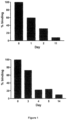
- This variant however, provided very low expression yields (table 6) and, in addition, the CD33 binding module had a reduced stability ( Fig. 1 ).
- This variant was thus further optimized, resulting in the improved variant of the 3rd generation, hu [33-ds16-ds123] (bottom row, table 6), wherein only the CD16- and CD123-specific scFvs are disulfide-stabilized.
- This latter variant showed superior properties in all categories: expression yield, absence of propensity to aggregate, stability in human serum at 37 °C, spontaneous formation of disulfide bridges, anti-leukemic bio-activity in cytolysis assays (RDL) with human leukemic target cells (see Figure 2 ); and good affinity of binding of each of the three scFv binding modules to their respective antigens on the surface of mammalian cells, as assessed separately.
- the candidate hu [33-ds16-ds123RK] still carried excess amino acids resulting from its construction from pre-fabricated components.
- these excess sequences were removed. Synthesis of the final DNA sequence was carried out by an external provider.
- Figure 3 shows a comparison between the previously published prototype ( Figure 3 A) and the improved clinical candidate SPM-2 ( Figure 3 B) .
- the sequences removed in the "polishing" process included: a Strep-tag intended for capture with the affinity matrix strep-tactin; a 2-amino acid spacer between the Strep tag and the N-terminal amino acid of the triplebody; a 2-amino acid spacer between the C-terminus of the CD123-specific scFv and the Myc-tag; a MYC tag; and a 2-amino acid spacer between the MYC-tag and the C-terminal HIS tag.
- This agent carried a C-terminal HIS tag and had been administered to several thousand patients in the course of several large Phase I and Phase II clinical trials. No immunogenicity nor any other adverse events linked to the presence of the HIS-tag had been reported and, therefore, the FDA withdrew this objection.
- other BiTE agents also carrying HIS tags are in clinical development, and a pipeline of BiTE agents carrying HIS tags are scheduled for clinical development by the company AMGEN [27-31; see also the world wide web under: http://www.amgenpipeline.com/pipeline/]. Accordingly, the C-terminal HIS-tag was retained in the clinical candidate, because the presence of the HIS-tag offers the desirable option to capture the protein from the culture supernatant of production cell lines by metal ion affinity chromatography (IMAC).
- IMAC metal ion affinity chromatography
- the cDNA coding sequence was subjected to an optimization of the codon usage by a commercial provider.
- the sequence changes introduced into the cDNA coding sequence were introduced solely for codon use optimization and did not alter the amino acid sequence of the clinical candidate.
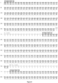
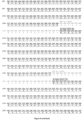
- the cDNA and amino acid sequence of the mature triplebody i.e. excluding the N-terminal leader sequence present in the coding sequence and in the immature protein is shown in Figure 9 .
- Example 2 Production of the triplebody by transfection and transient expression in HEK293 F cells
- the SPM-2 coding sequence was provided in a puc57 vector.
- the sequence was subcloned into the pSecTag Hygro expression vector (Life Technologies, Darmstadt, Germany) with the help of restriction endonucleases Nhel and EcoRV.
- the DNA sequence of the complete gene cassette was verified by DNA sequence analysis.
- a DNA stock solution with a concentration of 1 ⁇ g/ ⁇ l (in H 2 O) was prepared using a DNA Maxi-Prep kit (Qiagen, Hilden, Germany) according to the manufacturer's protocol.
- Transfection was performed at a cell density of ⁇ 1.0 x 10 6 cells/ml.
- TransIT ® LT1 Transfection Reagent (Mirus Bio LLC, Madison, USA) was used for transfection according to the manufacturer's protocol: TransIT LT1 Reagent was warmed to room temperature and vortexed gently before use. 3 ml of Opti-MEM I Reduced Serum Medium (Life Technologies, Darmstadt, Germany) were placed in a sterile tube. A total amount of 30 ⁇ g pSecTag SPM-2 plasmid DNA was added and gently mixed by pipetting. Then 60 ⁇ l of TranslT-LT1 Reagent were added and gently mixed. The mixture was incubated for 15 to 20 min. After the end of the incubation, the transfection mix was added drop wise to 30 ml cell culture.
- polyclonal cell pools were started 48 hrs after transfection. At this time, the growth medium was supplemented with 50 ⁇ g/ml Hygromycin B (InvivoGen, San Diego, USA) and anti-clumping factor (Life Technologies, Darmstadt, Germany) in a 1 : 1000 dilution. Cells were cultured and passaged under these selective conditions for 5 months. Then, a working cell bank was prepared from this polyclonal cell pool and stored in liquid nitrogen in FreeStyleTM 293 Expression Medium containing 10 % DMSO.
- Hygromycin B InvivoGen, San Diego, USA
- anti-clumping factor Life Technologies, Darmstadt, Germany
- the stably transfected polyclonal cell pool was used for production of SPM-2 protein. Frozen stocks from the working cell bank were thawed and passaged under Hygromycin selection (50 ⁇ g/ml) and addition of anti-clumping factor for 2 weeks before setting up expression cultures. To start expression cultures, the cells were diluted to a starting density of 0.5 x 10 6 cells/ml and cultured over 5 days. The culture supernatant containing SPM-2 was harvested by centrifugation for 4 min at 400 x g. Residual cells and debris were removed by a second centrifugation step for 10 min at 600 x g. Supernatants were either used directly for purifica tion or stored at -20 °C.
- SPM-2 was captured from cell culture supernatants via its C-terminal hexahistidine (HIS) tag by immobilized zinc ion affinity chromatography.
- High Density Zinc Agarose (Jena Bioscience, Germany) was used for this purpose, in which Zn 2+ ions are immobilized on an imino-di-acetic (IDA) acid matrix.
- IDA imino-di-acetic
- IDA imino-di-acetic
- the capture step was carried out in a batch mode.
- the Zn 2+ -IDA beads were washed with 5-10 column volumes (CV) of distilled water to eliminate the storage solution.
- 0.5 ml of solid zinc agarose beads were then added per 100 ml of cell culture supernatant containing SPM-2.
- 10 mM Imidazole-HCl pH 8 was added to avoid unspecific binding of host cell proteins (HCPs) to the resin.
- the supernatant was incubated with the resin in plastic tubes for 15-18 hrs at 8 °C on a rotating wheel. Subsequent steps were performed at room temperature.
- the Zn 2+ -IDA beads were then sedimented for 5 min at 500 x g in a desktop centrifuge.
- Elution was performed with 8 CV of elution buffer (200 mM Histidine-HCl pH 6.0, 150 mM NaCl). Fractions were collected in microcentrifuge tubes and the quantity and purity of SPM-2 was monitored by SDS-polyacrylamide gel electrophoresis (SDS-PAGE). Fractions containing SPM-2 were pooled and EDTA-NaOH pH 8.0 was added to a final concentration of 5 mM to neutralize divalent ions, in particular Zn 2+ , which also elutes from the column.
- elution buffer 200 mM Histidine-HCl pH 6.0, 150 mM NaCl
- SPM-2 can also be captured from cell culture supernatants via protein A, because some of the V-regions contained in SPM-2 carried the VH3 framework, which binds to protein A [111].
- the capture step was carried out in a batch mode. Protein A Sepharose 4 Fast Flow beads (GE Healthcare) were washed 8 times with 3 column volumes (CV) wash buffer (30 mM Na 2 HPO 4 -HCl pH 7.0, 250 mM NaCl) and subsequently added to cell culture supernatant containing SPM-2. The supernatant was incubated with the resin in plastic tubes for 15-18 hrs at 8 °C on a rotating wheel. The exact binding capacity of Protein A beads for SPM-2 has not yet been determined.
- CV column volumes
- the protein Prior to loading the captured and eluted protein pool onto the first ion exchange column (see Example 4 below), the protein was diluted to reduce the ionic strength of the buffer and thus to permit binding to the anion exchange (AEX) column described below. To this end, fractions containing SPM-2 were pooled and diluted with H 2 O to reach a conductivity of 5 milli Siemens/cm, which corresponds to the conductivity of the buffer carrying the pooled protein fractions after capture by IMAC and elution from said IMAC capture reagent.
- AEX anion exchange
- protein A Sepharose as a capture step enabled the combination of the capture with a first virus inactivation step (incubation at low pH). These conditions fulfill the requirements of regulatory authorities in Europe for a first viral inactivation step.
- An additional benefit of this modified capture procedure is that protein A resins are commercially available in GMP-grade quality (for example from GE healthcare), which is a considerable advantage of using this procedure for commercial production of GMP-grade SPM-2.
- Capture by IMAC requires a separate first virus inactivation step, which is expensive, and therefore, is less preferred than capture via protein A.
- the first purification step carried out was an anion exchange chromatography (AEX) using a 1 ml (bed volume) HiTrap Q Sepharose HP column (GE Healthcare Europe, Kunststoff, Germany) in flow through mode at 8 °C.
- the column was equilibrated with 10 CV of buffer A (20 mM Histidine-HCl pH 6.0, 50 mM NaCl) using either a peristaltic pump or an ⁇ kta liquid chromatography system (GE Healthcare Europe, Kunststoff, Germany).
- the default flow rate for all chromatography steps was 1 ml/min.
- the pooled fractions from the capture step were diluted 1 : 5 and loaded onto the column in buffer A.
- the flow-through (FT) containing SPM-2 was collected and the column was washed with 10 CV of buffer A. Bound proteins were then eluted using the ⁇ kta system with a linear gradient of 20 CV from 0-100 % of buffer B (20 mM Histidine-HCl pH 6.0, 1 M NaCl).
- the final purification step was a cation exchange (CEX) chromatography.
- a column was manually packed with 1 ml (bed volume) of Source 15S resin (GE Healthcare Europe, Kunststoff, Germany) and connected to an ⁇ kta liquid chromatography system at 8 °C.
- a 1 ml HiTrap SP Sepharose HP column (GE Healthcare Europe, Kunststoff, Germany) was used and produced similar results in terms of purity ( Fig. 5 ).
- the default flow rate for all steps was 1 ml/min.
- Eluted proteins were monitored by absorbance at 280 nm.
- the column was equilibrated with 10 CV of buffer A (20 mM Histidine-HCl pH 6.0, 50 mM NaCl).
- the flow-through (FT) fractions from the anion exchange chromatography step were then loaded onto the column in buffer A, followed by a 5 CV wash step with buffer A.
- Bound protein was then eluted with a linear NaCl gradient of 40 CV ranging from 0-50 % buffer B for the Source 15S column, or 0-60 % buffer B for the HiTrap SP-Sepharose HP column, followed by a wash step with 10 CV of 100 % buffer B (20 mM Histidine-HCl, 1 M NaCl).
- Monomeric SPM-2 eluted as a single peak in the linear NaCl gradient, followed by higher molecular weight species of SPM-2. Fractions of peak 1 were pooled and contained the purified SPM-2 protein.
- the conductivity of pooled fractions from the SP-Sepharose column was ⁇ 26 milliSiemens/cm, which corresponded to a buffer concentration of 20 mM Histidine-HCl pH 6.0 and a salt-ion concentration of approximately 300 mM NaCl.
- the pooled fractions from the CEX chromatography profile were concentrated by centrifugation and ultrafiltration using Amicon Ultra filter units with a molecular weight cut-off of 30 kDa (Millipore, Billerica, USA) at 8 °C and 1 700 x g.
- the protein concentration was determined by UV absorption at 280 nm using a NanoDrop spectrometer (PeqLab, Er Weg, Germany) and the theoretical extinction coefficient calculated from the primary amino acid sequence with the computer program ProtParam (see the world wide web under expasy.ch).
- SPM-2 Concentrations of SPM-2 up to 5 mg/ml in a buffer containing 20 mM Histidine-HCl pH 6.0 and 300 mM NaCl were achieved without any signs of aggregation detected by size exclusion chromatography (SEC) or visual inspection.
- SEC size exclusion chromatography
- a valuable property of the "upstream” and “downstream” processes described above are that they are “scalable”, i.e. they can be easily adjusted from the academic scale employed herein to large scale commercial production of therapeutic antibodies. This adjustment will be straightforward, because all steps described above are based on industry standard technology, which is thus scalable without the need for major changes, and without the risk for serious and costly setbacks. All capture materials, such as protein A- and IMAC affinity columns, anion- and cation exchange chromatography columns and all buffer components used can be purchased in industry-standard GMP-compliant quality from the same providers such as GE healthcare, which also supply the large commercial producers.
- the major species in the spectrum had a molecular mass of 82 614.6 Da, identical within less than 1 Da with the theoretical mass of 82 614.7 Da calculated from the primary amino acid sequence with the help of the program ProtParam (see the world wide web under expasy.ch).
- this major SPM-2 species did not carry any bulky post-translational modifications, such as N- or O-glycosylations, methylations, acetylations, ubiquitinylations or sumoylations, which are frequently found for mammalian antibodies and a variety of other recombinant mammalian proteins.
- post-translational modifications would have led to a greater difference between the observed molecular mass of the protein and its theoretical mass predicted from its primary cDNA and amino-acid sequence than the 1 Da difference actually measured.
- SPM-2 contains a total of 8 disulfide bonds (6 internal disulfide bonds from the IgV domains and 2 from disulfide engineering of CD16 and CD123), corresponding to a total of 16 thiol groups. Thus, more than 99 % of all thiol groups were present in an oxidized state in the tested batches of SPM-2. This result indicates that the overwhelming majority of all disulfide bonds in SPM-2 were spontaneously and correctly formed, which explains - at least in part - the extraordinary stability of this unusual protein.
- a first series of formulation screenings was performed with the aim to find a suitable formulation for long-term storage of SPM-2.
- the pH optimum for SPM-2 with regard to aggregation behavior was determined by screening of pH values in the range from pH 5-9 in steps of one pH unit.
- SPM-2 was enriched by nickel ion affinity chromatography and then transferred into one of the following different buffers, using a 5 ml HiTrap Desalting column (GE Healthcare, Kunststoff, Germany) connected to a syringe: Buffer 1: 100 mM NaH2PO4/Na2HPO4 pH 5.0, 150 mM NaCl Buffer 2: 100 mM NaH2PO4/Na2HPO4 pH 6.0, 150 mM NaCl Buffer 3: 100 mM NaH2PO4/Na2HPO4 pH 7.0, 150 mM NaCl Buffer 4: 100 mM NaH2PO4/Na2HPO4 pH 8.0, 150 mM NaCl Buffer 5: 100 mM Na2HPO4-HCl pH 9.0, 150 mM NaCl
- the different SPM-2 formulations were also analyzed by Differential Scanning Fluorimetry (DSF).
- DSF Differential Scanning Fluorimetry
- a comparison of DSF melting curves showed that the onset of fluorescence increase (the temperature at which a fraction of the molecules starts to unfold) was highest for pH 6.0 and pH 7.0. Based on this observation, a pH fine-screen in the range from pH 6.0 to 7.0 in steps of 0.2 pH units was performed, and samples were analyzed by SEC and DSF. The lowest ratio of higher Mol. Wt. species to monomeric SPM-2 was seen at pH 6.0.
- DSF melting profiles the onset of fluorescence increase and the Tm values for SPM-2 were highest in a buffer with pH 6.0.
- pH 6.0 was chosen as a basis for further formulation screenings. Subsequently, however, Histidine-HCl pH 6.0 has been used in the formulation buffers instead of NaH 2 PO 4 /Na 2 HPO 4 pH 6.0, because the pH of histidine buffer is more stable under temperature changes.
- trehalose was chosen as a supplement for long-term stability studies. However, a lower concentration (10 % w/v trehalose) was used to limit the viscosity of the formulation.
- the final formulation buffer was: 20 mM Histidine-HCl, pH 6.0; 300 mM NaCl; and 10 % trehalose. Long term stability at 4 °C was monitored in this buffer with and without trehalose, and good stability for up to 4 months was obtained in the presence, but not in the absence of trehalose.
- a formulation buffer has been identified which is very close to industry standard conditions, which consists exclusively of clinically validated industry standard components which are commercially available in GMP-compliant purity grade, and which is therefore expected to permit unproblematic storage and administration to patients.
- SPM-2 showed excellent stability in this buffer upon long-term storage at 4 °C, as detailed below.
- the candidate 33-ds16-123 showed aggregates as evidenced by the peak eluting to the left of the monomer peak when dissolved in SEC buffer (20 mM Histidine-HCl pH 6.0, 150 mM NaCl). Only 40 % of the total purified protein eluted as a monomer.
- the candidate 33-ds16-ds123 (corresponding to hu[33-ds16-ds123 RK]) showed the lowest degree of aggregate formation (72 % monomeric protein), but a small amount of higher molecular mass species (aggregates) were still present ( Fig. 7 , middle panel), when dissolved in the SEC buffer.
- Example 7 Long-term stability of SPM-2 in formulation buffer. Storage stability
- the anti-leukemic activity of SPM-2 purified by capture with protein A and stored for 5 months in formulation buffer at either 4 or 20 °C, was assayed by Re-Directed Lysis (RDL-) assays in cell culture as described below.
- the EC 50 value is the concentration of SPM-2, for which half-maximum specific lysis was reached, and characterizes the anti-leukemic activity of the agent. After storage at 20 °C for 5 months, the value was 22.7 pM. After storage at 4 °C for 5 months, it was 15.3 pM. Both values differ by less than a factor of 2 (a factor 2 is close to the precision limit of the assay).
- SPM-2 is an unusually robust and active protein.
- Example 8 Pre-clinical development of SPM-2: Development of a standardized bioassay to measure cytolytic activity of SPM-2.
- BM bone marrow
- PB peripheral blood
- ADCC Antibody-Dependent Cellular Cytotoxicity
- RDL Re-Directed Lysis
- NK cells Natural Killer cells from healthy donors were prepared. Initially, NK cells were routinely available only in limiting numbers, when they were prepared from 10 - 20 ml of freshly drawn PB. The numbers obtained in this way were sufficient to perform only one single experiment including the needed controls and with triplicate reactions for each measurement point for better statistical significance. NK cells from different healthy donors showed approximately ⁇ 10 % variability in their cytolytic activity with a standardized batch of SPM-2 and a standard AML-derived target cell-line, influenced by genetic variability in the genes controlling the cytolytic activity of NK cells, as well as age, gender and health status of the donor. Finally, fresh NK cells needed to be used within 24-48 hrs for optimum activity. These constraints were disadvantageous for the comparative study of several dozen or more cell samples from AML patients that needed to be performed.
- NK cells of constant quality sufficient for approx.100 experiments had to be produced, which could be frozen in aliquots and thawed, and which produced reproducible results in separate repeat runs of the same experiment, performed on separate days by different operators.
- PB peripheral Blood Mononuclear Cells
- MNCs Peripheral Blood Mononuclear Cells
- the PBMC fraction comprises lymphocytes and other MNCs, including NK cells, but excludes red blood cells, platelets and polymorph-nuclear cells (PMNs), such as neutrophilic granulocytes.
- RDL assays were performed with PBMCs from these donors, and the PBMCs from the donor producing the highest level of lysis of a standard AML line with a standard batch of SPM-2 were selected.
- IL-2 Interleukin-2
- IL-2 stimulates both the proliferation and the specific lytic activity of NK-cells.
- the expanded cells are also called "LAK cells” ("Lymphokine Activated Killer cells”).
- the fraction of specifically lysed target cells (% specific lysis, ordinate in Fig.10 ) showed only narrow variability in repeat runs of the experiment with separate thawed aliquots of LAK cells from the same frozen batch.
- the error bars in Fig.10 which reflect the variability between the different experiments, were acceptably small.
- the EC 50 value (concentration, for which half-maximum lysis was reached), derived from this data set was (24 ⁇ 8) pM and, therefore, the EC 50 values obtained with separate aliquots of frozen and thawed NK cells varied by no more than about ⁇ 25 %. Given that these EC 50 values were derived from plots such as the one shown in Fig.10 , where the concentration of the agent was plotted on an exponential scale, this variation was acceptable.
- this assay was suited for quantitative comparisons of many different samples from AML-patients, even when the assays were performed on separate days by separate operators with separate thawed aliquots of the standard batch of NK cells.
- a reference AML-derived cell line was carried along as a standard (positive control), such as the MOLM-13 line in Fig.10 , which is available from public depositories, including the DSMZ (German Collection of Microorganisms and Cell Lines, Braunschweig, Germany; see the world wide web under dsmz.de/catalogues/catalogue-human-and-animal-cell-lines.html and ref.
- MOLM-13 cells were lysed with half-maximum (EC 50 ) efficacy at approximately 100-fold lower concentrations of SPM-2 than THP-1 and OCI-AML3 cells ( Fig.11 ).
- EC 50 half-maximum
- normal MNCs from a healthy donor used as targets showed a nearly undetectable degree of lysis, although a fraction of these cells were myeloid cells expressing CD33, as shown in Fig.10 and Table 12, below.
- the density of CD33 on the surface of healthy myeloid cells was only in the range of a few hundred copies per cell, measured with a calibrated cytofluorimetric assay, whereas it was in the range of a few thousand, and often a few ten-thousand molecules per cell for the AML-derived cell-lines (Table 12, below).
- the density of the target antigens influences the susceptibility of AML cells for lysis in an RDL assay, although it is still debated in the scientific community to which extent.
- Both the healthy myeloid cells and the AML-derived cell lines in addition carry CD123 on their surface, which also influences the susceptibility for lysis by SPM-2.
- MOLM-13 cells were carried along as a reference standard, because these cells displayed the greatest surface antigen densities of CD33 and CD123 and showed the highest degree of lysis ( Fig.11 ). MOLM-13 cells also displayed a reasonably stable copy number of surface antigens per cell over many cell generations, and were fairly constant in their properties over time, and were the line best suited overall as a reference line among 6 AML-derived cell lines tested.
- target cells were labeled with calcein AM (aceto-methyl ester), a non-fluorescent hydrophobic molecule, which easily penetrates into intact living cells.
- Intracellular esterases convert it to calcein, a hydrophilic, polar, lipid-insoluble agent, retained in the cytoplasm, which is green fluorescent. It is retained inside cells with intact membranes and does not bind covalently to membranes or other intracellular structures and does not interfere with cell proliferation.
- Calcein is released from dying cells with damaged membranes into the extracellular space, where it can be quantitated as a measure of the extent of cell death.
- the measurement of the released fluorescence was performed with the help of an ELISA plate reader, equipped with a UV laser, and "black" ELISA plates permitting quantitative measurements of the intensity of fluorescent light [59]. The results are depicted in Figure 12 .
- the "% specific lysis” observed was the increment in lysis achieved by addition of the triplebody, the differential between the third and fourth bars, respectively, in Figure 12(A) , expressed in % as a fraction of the maximum lysis achieved by lysing all target cells with detergent.
- the NK cells are capable of recognizing and killing the target cells in their "Natural Killer” mode and of building a cytolytic synapse between the two cells.
- the enhancement caused by the triplebody viewed as an "antigen-specific glue” is measured by the "% specific lysis”.
- the RDL assays with primary cells from AML patients described below were performed with the following standard protocol.
- the reaction volume of 200 microliter in each well of a 96-well microtiter plate contained 10 000 target cells labeled with calcein AM.
- LAK cells were added to a LAK:target cell ratio of 10:1 or, alternatively, immuno-magnetically enriched NK cells were added to an NK:target cell ratio of 2:1. This amounted to 100 000 LAK cells per microtiter well.
- Triplebody or control proteins such as the control triplebody Her2-16-Her2, were added in various concentrations ranging from 0.1 to 1 000 pM (1 nM) final concentration.
- Triton X100 detergent was added to the wells intended for determination of maximum lysis. The reaction was allowed to proceed for 4 hrs at 37 °C in a 5 % CO 2 atmosphere. Cells were then collected by sedimentation in a centrifuge equipped with an adapter for microtiter plates. One hundred microliter of the supernatant were removed from each well and transferred to the wells of a black FluoroNunc TM F96 Microwell TM plate.
- Example 9 Quantitative measurement of surface antigen densities.
- the QIFI kit (DAKO, Hamburg, Germany; catalog # K0078; ref.113) was used for this purpose following manufacturer's instructions. Briefly, the kit contained a series of "calibration beads", and each subset of the series carried a known number of murine monoclonal antibodies covalently coupled to the beads.
- the cells to be analyzed (“test cells”) were labeled with a primary murine monoclonal antibody (mAb) directed against the antigen of interest, for example CD33.
- mAb primary murine monoclonal antibody
- test cells and in parallel "calibration beads", coated with the primary mAb were incubated with the fluorescein-conjugated secondary antibody.
- the primary antibody was used at saturating concentrations and therefore, the number of bound primary antibody molecules corresponded to the number of antigenic sites present on the "test cell” surface.
- MFI values were measured for the mixture of subsets of "calibration beads” and a calibration curve was generated by plotting the MFI values against the known numbers of bound antibodies for each subset of beads. Numbers of antibodies bound to the "calibration beads” have been pre-determined with precision by the manufacturer and were provided with the kit.
- the SABC "Specific Antibody Binding Capacity” was then measured for the "test cell” population. Unlabeled murine monoclonal antibodies for CD33 (BD Pharmingen, catalog # 555449) and CD123 (ebioscience, catalog # 14-1239-82) were used together with the QIFI kit's detection reagents.
- Antigen densities for the "test” cells were determined by measuring the fluorescence intensity obtained through staining with the primary antibody and the secondary fluorescent antibody contained in the kit.
- the detection method for the "standard” antigen on the beads and the "test” antigen on the test cells was the same. Therefore, this kit permits the most precise measurements of copy numbers per test cell possible to date.
- MFI values Mean Fluorescence Intensities
- CD33 and CD123 were also measured with the QIFI kit in the same manner for a few other AML cell lines. Table 12, top, gives raw data reported as MFI values; Table 12, bottom, gives the corresponding data after conversion to copy numbers per cell.
- surface antigen densities of CD33 and CD123 were also determined for PBMCs from healthy donors. PB was drawn from healthy donors 4, 5 and 6, and MNCs were prepared by density centrifugation in lymphoflot medium (Biotest, Dreieich, Germany). Surface antigen densities were then measured with the help of the QuiFi kit. The data entered in Table 12 (bottom) are copy numbers per cell after background correction.
- Example 10 Susceptibility of primary cells from AML patients with various subtypes of AML to lysis by SPM-2 plus NK cells
- Characteristics of the cells listed in Tables 13 and 14 including gender, age of the patients at diagnosis, the initial diagnosis, the source of material (BM or PB), blast count, cytogenetic data, characteristic mutations and genomic aberrations, and the genetic risk group assigned according to the current criteria of the European Leukemia Network [ELN; 114,115], were collected by the clinical diagnostic laboratories of these centers.
- the cohort included a number of patients with each of the major AML FAB subtypes M0 to M6, and this cohort should therefore permit to reach a first tentative answer to the question, whether cells from all major FAB subtypes of AML can be lysed by SPM-2 plus NK cells. Although the numbers of patients with AML of the different subtypes were still low, a first tentative conclusion and a trend was derived from this analysis.
- BMMCs Primary BM and PB cell samples enriched for MNCs by density gradient centrifugation were labeled with Calcein AM by published procedures [59,60].
- MNCs prepared by density gradient centrifugation in lymphoflot medium from BM are referred to as BMMCs
- PBMCs MNCs prepared by density gradient centrifugation in lymphoflot medium from BM.
- any potential therapeutic compound it is always important to assess the haematotoxicity of treatment with said compound, here SPM-2, for the remaining few normal haematopoietic cells of AML patients.
- This assessment is needed because previously developed potential treatments of AML were often accompanied by haematotoxicity.
- treatment with the antibody drug conjugate Mylotarg TM (Gemtuzumab Ozogamycin, GO; 22-24), frequently led to granulocytopenia and thrombocytopenia (platelet deficiency), due to effects of the treatment on the remaining healthy myeloid progenitor cells in the BM of AML patients.
- Non-AML donors BM samples from patients with other disorders were used, for whom drawing of BM samples was needed, but which did not suffer from leukemias and had a functionally normal haematopoietic system. These donors are referred to as "Non-AML donors”.
- RDL reactions were performed with BMMCs from 3 "Non-AML donors” (D1, D2, D3) and 1 representative AML patient (P5). Results are shown in Fig.13 .
- the BMMC populations from the 3 Non-AML donors D1-D3 responded far less strongly to the treatment by SPM-2 plus NK-cells than BMMC cells from AML patient P5.
- the result did not yet permit to distinguish whether the difference was due to the fact that the BMMCs from the AML patients had a greater intrinsic susceptibility to lysis by this treatment on a per cell basis than non-leukemic BM cells, or whether the result was simply due to the fact that myeloid cells bearing CD33 plus CD123 were more frequent in the BM sample from the AML patient than in the BM samples from the non-AML donors.
- myeloid cells were prepared from PBMCs of healthy donors instead and then the comparison was performed.
- PBMCs of healthy donors were CD11b-bearing myeloid cells and, therefore, to compare samples with similar frequencies of myeloid cells, myeloid cells from the PBMCs of healthy donors D4-D6 were enriched by immuno-magnetic procedures with beads carrying antibodies for the myeloid lineage antigen CD11b.
- CD33 and CD123 on PBMCs of donors D4-D6 had been determined before (Table 12) and the frequencies of the enriched cells and antigen densities on the cells immuno-magnetically enriched for CD11b are listed in Table 15.
- the PBMC samples from donors D4 and D6 carried therefore myeloid cells in roughly the same frequency range as the BMMC sample from patient 1: 90 % and 98 % of myeloid cells, respectively, for D4 and D6; 73 % of CD33-positive and 30% of CD123-positive cells for patient P1 (Table 10).
- the data suggest that the AML cells had no greater resistance to treatment with SPM-2 plus NK cells than healthy myeloid cells, as one may have expected, because AML cells resistant to certain chemotherapeutic agents have been observed.
- the impression is that the sensitivity to treatment of both normal and malignant AML cells with SPM-2 plus NK cells was driven more by the surface antigen density than by cell-internal genomic alterations and ensuing signal pathways controlling the expression of genes, which govern susceptibility to lysis.
- MABEL Minimum Active Biologically Effective Level
- MRSD Maximum Recommended Safe Dose
- SPM-2 first-in-human clinical use
- MRSD concentrations correspond to bolus injection doses of 6 ng/kg body weight for an adult patient with an average body weight of 70 kg and a blood volume of 5 L.
- These computed doses would be 100- to 1 000-fold lower than doses of therapeutic IgG antibodies currently used for the treatment of lymphomas such as Rituxan, which are on the order of 10 mg/kg body weight per injection.
- AML Leukemia Stem Cells should also be susceptible to lysis by SPM-2 plus NK cells. because they display high surface densities of CD33 and CD123 as reported e.g. in references 5, 10, 11, 17, 46, 67. As AML-LSCs may still be refractory to lysis by SPM-2 plus NK-cells, despite the high surface densities of these antigens, this prediction nonetheless needed to be tested directly.
- the current opinion is that the relapse-relevant AML-LSCs are only a subset within the Vergez compartment, and that their overall surface antigen composition may vary during clonal evolution of the disease (as discussed e.g. in references 10, 11, 17, 46, 67). However, although the global composition of the surface may vary, the core phenotype (CD34 + CD38- CD123 high ) is expected to remain constant. Therefore, a demonstration that cells from the Vergez-compartment can be lysed by SPM-2 plus NK cells would plausibly support the notion that the relapse-relevant AML-LSCs are susceptible to lysis by SPM-2 plus NK cells.
- CD34-bearing cells were enriched by immuno-magnetic sorting of MNC samples from patients 9 and 11 (Tables 14 and 16). Both MNC samples carried intermediate combined densities of CD33 plus CD123 (approx. 8 000 and 3 600 copies/cell, respectively; Table 14). Both MNC samples were lysed by SPM-2 plus NK cells in RDL reactions with EC 50 values of 166 and 1 078 pM, respectively (Tables 14, 16).
- CD33 and CD123 densities and susceptibility to lysis in RDL assays were determined again after the enrichment.
- the CD33 density was reduced for cells from patient 11 to 500 copies/cell, and the CD123 density was increased by about 6-fold to 14 500 copies/cell (Table 16).
- the EC 50 value was lowered to 681 pM, indicating an increased susceptibility for lysis to about twice the value measured for the non-enriched cells.
- the CD33 density on the enriched blasts rose by > 2-fold to 13 000 copies/cell, while the density of CD123 rose by > 3-fold to 9 100 copies/cell, and the combined density was increased by about 2.5-fold to 22 100 copies/cell.
- the EC 50 value for the enriched cells of this patient was reduced by about 4-fold to 43 pM (Table 16).
- the CD123 density was several-fold greater on the CD34-enriched cells than on the overall blasts, whereas the CD33 density was greater in one case and lower in the other.
- the combined CD33 plus CD123 density was increased by about 2.5 to 3.5-fold with correspondingly reduced EC 50 values, consistent with the expectation that cells closer to the AML-LSCs may indeed have higher susceptibilities to lysis by SPM-2 than the corresponding bulk AML blasts.
- Example 11 Susceptibility of primary cells from AML patients with various subtypes of AML to lysis by SPM-2 plus NK cells
- AML expands in the BM.
- different patients display different blast counts in the BM, and the degree of out-crowding of normal leukocytes from the BM by the malignant blasts varies with the blast count.
- blast counts vary between approximately 30 % and 95 % of all BMMCs. Therefore, at diagnosis, only between 5 % and 70 % of normal leukocytes remain, and these remaining normal cells are affected by the altered milieu in the BM.
- the pH in the BM has changed, and the BM becomes progressively more hypoxic the higher the blast count grows. Also the cytokine milieu in the BM has changed in response to these alterations.
- NCRs activating natural cytotoxicity receptors
- NKs The capacity of NKs to secrete IFN- ⁇ was highly impaired in AML patients and was correlated with suppressed immune responses against autologous leukemic cells [123-125]. Finally, in adult acute leukemia, impaired production of cytokines by NKs was associated with early relapse. However, there are reports showing that in first remission, the NK cell titers of many AML patients had partially recovered [120].
- NK cells in remission had not only recovered in numbers, but also in the cytolytic potential per NK cell, and whether this recovered potential was large enough to make SPM-2 a promising agent for the treatment of AML patients in a first remission, or in a state where the blast count was low, such as in a partial remission or a smoldering AML.
- the patient was a 21 year old woman diagnosed with an AML FAB M1 with the immuno-phenotype reported in reference 60.
- the patient had a BM blast titer of 89.7 % at diagnosis.
- the blasts were CD34 positive (89.6 %) and fewer than 1 % were CD33 positive according to the initial diagnosis (CD33-negative FAB M1 AML).
- the cells were later reanalyzed with more sensitive methods, and 130 copies per cell of CD33 and 230 copies of CD123, respectively, were detected on the BMMCs. These are very low densities, considering that routinely several 1 000 copies each of CD33 and CD123 were found herein, sometimes several 10 000 copies on blasts from other patients, dependent on the AML subtype.
- MNCs were prepared by density gradient centrifugation in lymphoflot medium as described above from a BM sample drawn at diagnosis.
- the patients BM blasts were lysed efficiently by SPM-2 in combination with ex vivo expanded MNCs from an unrelated healthy donor, presumably because even these low antigen densities were sufficient for an effective binding of SPM-2 to one copy each of CD33 and CD123 on the same cell, followed by NK cell-mediated lysis.
- PBMCs were also prepared from PB of her healthy twin sister, and PB was also available from the patient in first remission after induction chemotherapy. Using BMMCs from the patient drawn at diagnosis as targets, and PBMCs from the patient drawn at diagnosis and in remission and from her healthy sister as effectors, RDL assays were performed.
- the MNCs from the patient drawn at diagnosis were inefficient effectors, while PBMCs from the patient drawn in remission and from her healthy sister had very similar cytolytic efficacy in combination with SPM-2. Therefore, the patient's own NK cells had not only recovered to a large extent in numbers by the time the patient had reached a remission, but had also recovered in their specific cytolytic activity per cell and were capable of lysing the patient's autologous BM blasts in combination with SPM-2.
- NK-cells drawn in remission were not only of comparable quality as her healthy sisters as effectors for lysis of the patients autologous AML cells in combination with SPM-2, but also for lysis of a CD20-positive human lymphoma cell line (Raji) mediated by the reference antibody Mabthera (Rituximab).
- Cytokine- and mediator release from whole blood cells often accompanies immune- and inflammatory reactions that can potentially damage a patient if they exceed certain thresholds. Therefore, it was determined whether cytokines are released to a measurable extent after addition of SPM-2 to whole blood, drawn either from an AML patient or from a healthy donor.
- Concentrations of human IFN-y and TNF- ⁇ were measured with commercial ELISA kits (eBioscience) following manufacturer's instructions.
- the triplebody SPM-2 was added to peripheral blood samples at concentrations of 10, 1, 0.1 or 0 nM, and the reaction mixtures were then kept for 6 h at 37 °C in 96 well round bottom Nunc plates in 200 microliter volumes. Blood samples were frozen and stored at - 20 °C and were thawed only immediately before use in cytokine release assays. Quantities of released cytokines were evaluated by quantitating the intensity of the color reactions created with the help of the kit's reagents (antibodies specific for the cytokines under investigation).
- sctb yield [mg/l] hu[123Aho-ds16-33] 0.6 hu[ds123Aho-ds16-33] 0.6 huds[123Aho-16-33] 0.3 hu[33-ds16-123] 1.8 huds[33-16-123] 0.2 hu[33-ds16-ds123] 0.7
Landscapes
- Health & Medical Sciences (AREA)
- Chemical & Material Sciences (AREA)
- Immunology (AREA)
- Organic Chemistry (AREA)
- Life Sciences & Earth Sciences (AREA)
- General Health & Medical Sciences (AREA)
- Medicinal Chemistry (AREA)
- Biochemistry (AREA)
- Biophysics (AREA)
- Genetics & Genomics (AREA)
- Molecular Biology (AREA)
- Proteomics, Peptides & Aminoacids (AREA)
- Hematology (AREA)
- Oncology (AREA)
- Chemical Kinetics & Catalysis (AREA)
- General Chemical & Material Sciences (AREA)
- Nuclear Medicine, Radiotherapy & Molecular Imaging (AREA)
- Pharmacology & Pharmacy (AREA)
- Animal Behavior & Ethology (AREA)
- Public Health (AREA)
- Veterinary Medicine (AREA)
- Micro-Organisms Or Cultivation Processes Thereof (AREA)
- Medicines That Contain Protein Lipid Enzymes And Other Medicines (AREA)
- Pharmaceuticals Containing Other Organic And Inorganic Compounds (AREA)
- Peptides Or Proteins (AREA)
Claims (13)
- Molécule d'acide nucléique codant pour un triacorps monocaténaire capable de se lier spécifiquement à CD33, CD16 et CD123, laquelle molécule d'acide nucléique comprend :(a) une molécule d'acide nucléique codant pour une protéine représentée par la SEQ ID NO : 1 ou 3 ;(b) une molécule d'acide nucléique représentée par la SEQ ID NO : 2 ou 4(c) la molécule d'acide nucléique de (b), dans laquelle chaque thymine est remplacée par un uracile ; ou(d) une molécule d'acide nucléique qui est dégénérée par rapport à la molécule d'acide nucléique de (b) ou (c).
- Vecteur comprenant la molécule d'acide nucléique de la revendication 1.
- Cellule hôte transformée ou transfectée avec la molécule d'acide nucléique de la revendication 1 ou le vecteur de la revendication 2.
- Méthode pour la production d'un triacorps monocaténaire capable de se lier spécifiquement à CD33, CD16 et CD123, la méthode comprenant la culture de la cellule hôte de la revendication 3 dans des conditions adaptées et l'isolation du triacorps monocaténaire capable de se lier spécifiquement à CD33, CD16 et CD123 produit.
- Méthode selon la revendication 4, dans laquelle l'isolation du triacorps monocaténaire comprend les étapes de :(a) incubation d'un surnageant de culture cellulaire contenant le triacorps monocaténaire avec de la protéine A ;(b) séparation de la protéine A avec le triacorps monocaténaire lié à partir du surnageant de culture cellulaire ;(c) élution du triacorps monocaténaire à partir de la protéine A à un pH compris entre 2,9 et 3,1 à une température de 4 °C à 5 °C pendant 60 à 70 minutes ; et(d) purification du triacorps monocaténaire par :(d-i) chromatographie par échange d'ions ; et(d-ii) chromatographie par échange de cations.
- Méthode selon la revendication 4 ou 5, comprenant en outre une étape de re-tamponnage du triacorps monocaténaire isolé pour que soit obtenue une composition comprenant le triacorps monocaténaire présent dans un tampon de formulation comprenant de l'histidine-HCl 20 mM, pH 6,0, du NaCl 300 mM et 10 % en poids/volume de tréhalose.
- Méthode selon la revendication 5 ou 6, comprenant en outre une étape d'inactivation virale additionnelle.
- Triacorps monocaténaire capable de se lier spécifiquement à CD33, CD16 et CD123 codé par la molécule d'acide nucléique de la revendication 1.
- Triacorps monocaténaire selon la revendication 8, lequel triacorps monocaténaire est présent dans un tampon de formulation comprenant de l'histidine-HCl 20 mM, pH 6,0, du NaCl 300 mM et 10 % en poids/volume de tréhalose.
- Triacorps monocaténaire capable de se lier spécifiquement à CD33, CD16 et CD123, produit par la méthode de l'une quelconque des revendications 4 à 7.
- Composition pharmaceutique comprenant au moins l'un parmi(a) la molécule d'acide nucléique de la revendication 1 ;(b) le vecteur de la revendication 2 ;(c) la cellule hôte de la revendication 3 ; et(d) le triacorps monocaténaire de l'une quelconque des revendications 8 à 10.
- Molécule d'acide nucléique selon la revendication 1, vecteur selon la revendication 2, triacorps monocaténaire selon l'une quelconque des revendications 8 à 10 ou composition selon la revendication 11 pour une utilisation dans le traitement d'une leucémie myéloïde aiguë et/ou d'un syndrome myélodysplasique.
- Molécule d'acide nucléique selon la revendication 1, vecteur selon la revendication 2, triacorps monocaténaire selon l'une quelconque des revendications 8 à 10 ou composition selon la revendication 11 pour une utilisation dans le traitement d'une leucémie myéloïde aiguë et/ou d'un syndrome myélodysplasique, dans lequel la molécule d'acide nucléique, le vecteur, le triacorps monocaténaire ou la composition est destiné à être administré lors d'une phase de rémission de leucémie myéloïde aiguë ou après le diagnostic d'un syndrome myélodysplasique.
Applications Claiming Priority (2)
| Application Number | Priority Date | Filing Date | Title |
|---|---|---|---|
| EP16202026.7A EP3330289A1 (fr) | 2016-12-02 | 2016-12-02 | Anticorps trimérique à chaîne unique spécifique à cd33, cd16 et cd123 |
| PCT/EP2017/081150 WO2018100139A1 (fr) | 2016-12-02 | 2017-12-01 | Triplecorps à chaîne unique spécifique à cd33, cd16 et cd123 |
Publications (3)
| Publication Number | Publication Date |
|---|---|
| EP3548516A1 EP3548516A1 (fr) | 2019-10-09 |
| EP3548516B1 true EP3548516B1 (fr) | 2025-05-21 |
| EP3548516C0 EP3548516C0 (fr) | 2025-05-21 |
Family
ID=57544212
Family Applications (2)
| Application Number | Title | Priority Date | Filing Date |
|---|---|---|---|
| EP16202026.7A Withdrawn EP3330289A1 (fr) | 2016-12-02 | 2016-12-02 | Anticorps trimérique à chaîne unique spécifique à cd33, cd16 et cd123 |
| EP17822159.4A Active EP3548516B1 (fr) | 2016-12-02 | 2017-12-01 | Anticorps trimérique à chaîne unique spécifique à cd33, cd16 et cd123 |
Family Applications Before (1)
| Application Number | Title | Priority Date | Filing Date |
|---|---|---|---|
| EP16202026.7A Withdrawn EP3330289A1 (fr) | 2016-12-02 | 2016-12-02 | Anticorps trimérique à chaîne unique spécifique à cd33, cd16 et cd123 |
Country Status (5)
| Country | Link |
|---|---|
| US (1) | US11945863B2 (fr) |
| EP (2) | EP3330289A1 (fr) |
| CA (1) | CA3045385A1 (fr) |
| ES (1) | ES3036457T3 (fr) |
| WO (1) | WO2018100139A1 (fr) |
Families Citing this family (6)
| Publication number | Priority date | Publication date | Assignee | Title |
|---|---|---|---|---|
| EP3165536A1 (fr) * | 2015-11-09 | 2017-05-10 | Ludwig-Maximilians-Universität München | Molécule de combinaison de ciblage de tumeur spécifique et d'inhibition de point de contrôle immunitaire local |
| WO2023060180A1 (fr) * | 2021-10-06 | 2023-04-13 | The Wistar Institute Of Anatomy And Biology | Nouveaux agents de mise en contact de cellules immunitaires pour l'immunothérapie |
| US20260070986A1 (en) * | 2021-11-03 | 2026-03-12 | Affimed Gmbh | Bispecific cd16a binders |
| IL312515A (en) * | 2021-11-03 | 2024-07-01 | Affimed Gmbh | Bispecific cd16a binders |
| CN116948029A (zh) * | 2022-04-20 | 2023-10-27 | 南京融捷康生物科技有限公司 | 一种包含IgG类Fc区变体的抗体及其用途 |
| WO2026080803A1 (fr) * | 2024-10-11 | 2026-04-16 | Alexion Pharmaceuticals, Inc. | Procédés d'élimination de contaminants présents dans des préparations biologiques |
Family Cites Families (3)
| Publication number | Priority date | Publication date | Assignee | Title |
|---|---|---|---|---|
| CN102413839A (zh) * | 2009-03-06 | 2012-04-11 | 医学免疫有限责任公司 | 人源化抗cd19抗体制剂 |
| EP2332994A1 (fr) | 2009-12-09 | 2011-06-15 | Friedrich-Alexander-Universität Erlangen-Nürnberg | Thérapie trispécifique contre la leucémie myéloïde aiguë |
| US20140206846A1 (en) | 2011-05-27 | 2014-07-24 | Roland Beckmann | Dual Targeting |
-
2016
- 2016-12-02 EP EP16202026.7A patent/EP3330289A1/fr not_active Withdrawn
-
2017
- 2017-12-01 ES ES17822159T patent/ES3036457T3/es active Active
- 2017-12-01 CA CA3045385A patent/CA3045385A1/fr active Pending
- 2017-12-01 WO PCT/EP2017/081150 patent/WO2018100139A1/fr not_active Ceased
- 2017-12-01 EP EP17822159.4A patent/EP3548516B1/fr active Active
- 2017-12-01 US US16/466,018 patent/US11945863B2/en active Active
Also Published As
| Publication number | Publication date |
|---|---|
| WO2018100139A1 (fr) | 2018-06-07 |
| ES3036457T3 (en) | 2025-09-19 |
| EP3548516A1 (fr) | 2019-10-09 |
| EP3330289A1 (fr) | 2018-06-06 |
| CA3045385A1 (fr) | 2018-06-07 |
| US20190359710A1 (en) | 2019-11-28 |
| US11945863B2 (en) | 2024-04-02 |
| EP3548516C0 (fr) | 2025-05-21 |
Similar Documents
| Publication | Publication Date | Title |
|---|---|---|
| US12291561B2 (en) | Antigen binding receptors specific for mutated Fc domains | |
| EP3548516B1 (fr) | Anticorps trimérique à chaîne unique spécifique à cd33, cd16 et cd123 | |
| JP7082620B2 (ja) | 抗pd1モノクローナル抗体、その医薬組成物およびその使用 | |
| EP3245231B1 (fr) | Anticorps et récepteurs antigéniques chimériques spécifiques de ror1 | |
| US20200093861A1 (en) | Antigen binding receptor formats | |
| CN114901691A (zh) | Ilt3结合剂及其使用方法 | |
| CN116507640A (zh) | 改善的抗原结合受体 | |
| IL298449A (en) | Improved antigen binding receptors | |
| US20240424095A1 (en) | Antigen binding receptors | |
| WO2019086574A1 (fr) | Molécules de liaison à l'antigène cd47 et cd33 | |
| EP3744400A1 (fr) | Ciblage de cellules car-t il-1rap et leur utilisation dans la leucémie myéloïde aiguë (aml) | |
| CN120917051A (zh) | 多特异性抗原结合蛋白及其用途 | |
| WO2024085182A1 (fr) | Agent thérapeutique pour une tumeur à lymphocytes t | |
| JP2025529903A (ja) | デルタ様リガンド3(dll3)に特異的な抗体およびキメラ抗原受容体 | |
| WO2023180511A1 (fr) | Récepteurs chimériques améliorés | |
| WO2025012620A1 (fr) | Constructions de liaison | |
| HK40119868A (zh) | 经改善的嵌合受体 | |
| WO2024169985A1 (fr) | Anticorps p329g ciblant her2/p95her2, combinaison de celui-ci avec une cellule de récepteur antigénique chimérique et utilisation associée | |
| EP4692119A1 (fr) | Anticorps ou fragment de liaison à l'antigène de celui-ci ciblant cd3 et son utilisation | |
| KR20240101629A (ko) | Cd137을 표적화하는 이중특이적 항체 및 항암 면역요법을 위한 이의 용도 | |
| HK40042566A (en) | Antibodies and chimeric antigen receptors specific for ror1 | |
| HK1246314B (en) | Antibodies and chimeric antigen receptors specific for ror1 |
Legal Events
| Date | Code | Title | Description |
|---|---|---|---|
| STAA | Information on the status of an ep patent application or granted ep patent |
Free format text: STATUS: UNKNOWN |
|
| STAA | Information on the status of an ep patent application or granted ep patent |
Free format text: STATUS: THE INTERNATIONAL PUBLICATION HAS BEEN MADE |
|
| PUAI | Public reference made under article 153(3) epc to a published international application that has entered the european phase |
Free format text: ORIGINAL CODE: 0009012 |
|
| STAA | Information on the status of an ep patent application or granted ep patent |
Free format text: STATUS: REQUEST FOR EXAMINATION WAS MADE |
|
| 17P | Request for examination filed |
Effective date: 20190702 |
|
| AK | Designated contracting states |
Kind code of ref document: A1 Designated state(s): AL AT BE BG CH CY CZ DE DK EE ES FI FR GB GR HR HU IE IS IT LI LT LU LV MC MK MT NL NO PL PT RO RS SE SI SK SM TR |
|
| AX | Request for extension of the european patent |
Extension state: BA ME |
|
| DAV | Request for validation of the european patent (deleted) | ||
| DAX | Request for extension of the european patent (deleted) | ||
| STAA | Information on the status of an ep patent application or granted ep patent |
Free format text: STATUS: EXAMINATION IS IN PROGRESS |
|
| 17Q | First examination report despatched |
Effective date: 20200624 |
|
| GRAP | Despatch of communication of intention to grant a patent |
Free format text: ORIGINAL CODE: EPIDOSNIGR1 |
|
| STAA | Information on the status of an ep patent application or granted ep patent |
Free format text: STATUS: GRANT OF PATENT IS INTENDED |
|
| INTG | Intention to grant announced |
Effective date: 20241216 |
|
| GRAS | Grant fee paid |
Free format text: ORIGINAL CODE: EPIDOSNIGR3 |
|
| GRAA | (expected) grant |
Free format text: ORIGINAL CODE: 0009210 |
|
| STAA | Information on the status of an ep patent application or granted ep patent |
Free format text: STATUS: THE PATENT HAS BEEN GRANTED |
|
| AK | Designated contracting states |
Kind code of ref document: B1 Designated state(s): AL AT BE BG CH CY CZ DE DK EE ES FI FR GB GR HR HU IE IS IT LI LT LU LV MC MK MT NL NO PL PT RO RS SE SI SK SM TR |
|
| REG | Reference to a national code |
Ref country code: GB Ref legal event code: FG4D |
|
| REG | Reference to a national code |
Ref country code: CH Ref legal event code: EP |
|
| REG | Reference to a national code |
Ref country code: DE Ref legal event code: R096 Ref document number: 602017089581 Country of ref document: DE |
|
| REG | Reference to a national code |
Ref country code: IE Ref legal event code: FG4D |
|
| U01 | Request for unitary effect filed |
Effective date: 20250618 |
|
| U07 | Unitary effect registered |
Designated state(s): AT BE BG DE DK EE FI FR IT LT LU LV MT NL PT RO SE SI Effective date: 20250627 |
|
| REG | Reference to a national code |
Ref country code: ES Ref legal event code: FG2A Ref document number: 3036457 Country of ref document: ES Kind code of ref document: T3 Effective date: 20250919 |
|
| PG25 | Lapsed in a contracting state [announced via postgrant information from national office to epo] |
Ref country code: GR Free format text: LAPSE BECAUSE OF FAILURE TO SUBMIT A TRANSLATION OF THE DESCRIPTION OR TO PAY THE FEE WITHIN THE PRESCRIBED TIME-LIMIT Effective date: 20250822 Ref country code: NO Free format text: LAPSE BECAUSE OF FAILURE TO SUBMIT A TRANSLATION OF THE DESCRIPTION OR TO PAY THE FEE WITHIN THE PRESCRIBED TIME-LIMIT Effective date: 20250821 |
|
| PG25 | Lapsed in a contracting state [announced via postgrant information from national office to epo] |
Ref country code: PL Free format text: LAPSE BECAUSE OF FAILURE TO SUBMIT A TRANSLATION OF THE DESCRIPTION OR TO PAY THE FEE WITHIN THE PRESCRIBED TIME-LIMIT Effective date: 20250521 |
|
| PG25 | Lapsed in a contracting state [announced via postgrant information from national office to epo] |
Ref country code: HR Free format text: LAPSE BECAUSE OF FAILURE TO SUBMIT A TRANSLATION OF THE DESCRIPTION OR TO PAY THE FEE WITHIN THE PRESCRIBED TIME-LIMIT Effective date: 20250521 |
|
| PG25 | Lapsed in a contracting state [announced via postgrant information from national office to epo] |
Ref country code: RS Free format text: LAPSE BECAUSE OF FAILURE TO SUBMIT A TRANSLATION OF THE DESCRIPTION OR TO PAY THE FEE WITHIN THE PRESCRIBED TIME-LIMIT Effective date: 20250821 |
|
| PG25 | Lapsed in a contracting state [announced via postgrant information from national office to epo] |
Ref country code: IS Free format text: LAPSE BECAUSE OF FAILURE TO SUBMIT A TRANSLATION OF THE DESCRIPTION OR TO PAY THE FEE WITHIN THE PRESCRIBED TIME-LIMIT Effective date: 20250921 |
|
| REG | Reference to a national code |
Ref country code: CH Ref legal event code: U11 Free format text: ST27 STATUS EVENT CODE: U-0-0-U10-U11 (AS PROVIDED BY THE NATIONAL OFFICE) Effective date: 20260101 |
|
| PGFP | Annual fee paid to national office [announced via postgrant information from national office to epo] |
Ref country code: GB Payment date: 20251222 Year of fee payment: 9 |
|
| PG25 | Lapsed in a contracting state [announced via postgrant information from national office to epo] |
Ref country code: SM Free format text: LAPSE BECAUSE OF FAILURE TO SUBMIT A TRANSLATION OF THE DESCRIPTION OR TO PAY THE FEE WITHIN THE PRESCRIBED TIME-LIMIT Effective date: 20250521 |
|
| PG25 | Lapsed in a contracting state [announced via postgrant information from national office to epo] |
Ref country code: CZ Free format text: LAPSE BECAUSE OF FAILURE TO SUBMIT A TRANSLATION OF THE DESCRIPTION OR TO PAY THE FEE WITHIN THE PRESCRIBED TIME-LIMIT Effective date: 20250521 |
|
| PG25 | Lapsed in a contracting state [announced via postgrant information from national office to epo] |
Ref country code: SK Free format text: LAPSE BECAUSE OF FAILURE TO SUBMIT A TRANSLATION OF THE DESCRIPTION OR TO PAY THE FEE WITHIN THE PRESCRIBED TIME-LIMIT Effective date: 20250521 |
|
| U20 | Renewal fee for the european patent with unitary effect paid |
Year of fee payment: 9 Effective date: 20251229 |
|
| PLBE | No opposition filed within time limit |
Free format text: ORIGINAL CODE: 0009261 |
|
| STAA | Information on the status of an ep patent application or granted ep patent |
Free format text: STATUS: NO OPPOSITION FILED WITHIN TIME LIMIT |
|
| REG | Reference to a national code |
Ref country code: CH Ref legal event code: L10 Free format text: ST27 STATUS EVENT CODE: U-0-0-L10-L00 (AS PROVIDED BY THE NATIONAL OFFICE) Effective date: 20260402 |
|
| PGFP | Annual fee paid to national office [announced via postgrant information from national office to epo] |
Ref country code: ES Payment date: 20260122 Year of fee payment: 9 |
|
| PGFP | Annual fee paid to national office [announced via postgrant information from national office to epo] |
Ref country code: CH Payment date: 20260101 Year of fee payment: 9 |
|
| 26N | No opposition filed |
Effective date: 20260224 |