EP3565459B1 - System und verfahren zur unterscheidung adipösen gewebes von narbengewebe während einer elektrophysiologischen kartierung - Google Patents
System und verfahren zur unterscheidung adipösen gewebes von narbengewebe während einer elektrophysiologischen kartierung Download PDFInfo
- Publication number
- EP3565459B1 EP3565459B1 EP18712043.1A EP18712043A EP3565459B1 EP 3565459 B1 EP3565459 B1 EP 3565459B1 EP 18712043 A EP18712043 A EP 18712043A EP 3565459 B1 EP3565459 B1 EP 3565459B1
- Authority
- EP
- European Patent Office
- Prior art keywords
- tissue
- location
- scar
- probability
- dielectric properties
- Prior art date
- Legal status (The legal status is an assumption and is not a legal conclusion. Google has not performed a legal analysis and makes no representation as to the accuracy of the status listed.)
- Active
Links
Images



Classifications
-
- A—HUMAN NECESSITIES
- A61—MEDICAL OR VETERINARY SCIENCE; HYGIENE
- A61B—DIAGNOSIS; SURGERY; IDENTIFICATION
- A61B5/00—Measuring for diagnostic purposes; Identification of persons
- A61B5/05—Detecting, measuring or recording for diagnosis by means of electric currents or magnetic fields; Measuring using microwaves or radio waves
- A61B5/053—Measuring electrical impedance or conductance of a portion of the body
- A61B5/0537—Measuring body composition by impedance, e.g. tissue hydration or fat content
-
- A—HUMAN NECESSITIES
- A61—MEDICAL OR VETERINARY SCIENCE; HYGIENE
- A61B—DIAGNOSIS; SURGERY; IDENTIFICATION
- A61B34/00—Computer-aided surgery; Manipulators or robots specially adapted for use in surgery
- A61B34/10—Computer-aided planning, simulation or modelling of surgical operations
-
- A—HUMAN NECESSITIES
- A61—MEDICAL OR VETERINARY SCIENCE; HYGIENE
- A61B—DIAGNOSIS; SURGERY; IDENTIFICATION
- A61B5/00—Measuring for diagnostic purposes; Identification of persons
- A61B5/05—Detecting, measuring or recording for diagnosis by means of electric currents or magnetic fields; Measuring using microwaves or radio waves
- A61B5/053—Measuring electrical impedance or conductance of a portion of the body
- A61B5/0538—Measuring electrical impedance or conductance of a portion of the body invasively, e.g. using a catheter
-
- A—HUMAN NECESSITIES
- A61—MEDICAL OR VETERINARY SCIENCE; HYGIENE
- A61B—DIAGNOSIS; SURGERY; IDENTIFICATION
- A61B5/00—Measuring for diagnostic purposes; Identification of persons
- A61B5/24—Detecting, measuring or recording bioelectric or biomagnetic signals of the body or parts thereof
-
- A—HUMAN NECESSITIES
- A61—MEDICAL OR VETERINARY SCIENCE; HYGIENE
- A61B—DIAGNOSIS; SURGERY; IDENTIFICATION
- A61B5/00—Measuring for diagnostic purposes; Identification of persons
- A61B5/24—Detecting, measuring or recording bioelectric or biomagnetic signals of the body or parts thereof
- A61B5/25—Bioelectric electrodes therefor
- A61B5/279—Bioelectric electrodes therefor specially adapted for particular uses
- A61B5/28—Bioelectric electrodes therefor specially adapted for particular uses for electrocardiography [ECG]
- A61B5/283—Invasive
- A61B5/287—Holders for multiple electrodes, e.g. electrode catheters for electrophysiological study [EPS]
-
- A—HUMAN NECESSITIES
- A61—MEDICAL OR VETERINARY SCIENCE; HYGIENE
- A61B—DIAGNOSIS; SURGERY; IDENTIFICATION
- A61B5/00—Measuring for diagnostic purposes; Identification of persons
- A61B5/68—Arrangements of detecting, measuring or recording means, e.g. sensors, in relation to patient
- A61B5/6846—Arrangements of detecting, measuring or recording means, e.g. sensors, in relation to patient specially adapted to be brought in contact with an internal body part, i.e. invasive
- A61B5/6847—Arrangements of detecting, measuring or recording means, e.g. sensors, in relation to patient specially adapted to be brought in contact with an internal body part, i.e. invasive mounted on an invasive device
- A61B5/6852—Catheters
-
- A—HUMAN NECESSITIES
- A61—MEDICAL OR VETERINARY SCIENCE; HYGIENE
- A61B—DIAGNOSIS; SURGERY; IDENTIFICATION
- A61B5/00—Measuring for diagnostic purposes; Identification of persons
- A61B5/68—Arrangements of detecting, measuring or recording means, e.g. sensors, in relation to patient
- A61B5/6846—Arrangements of detecting, measuring or recording means, e.g. sensors, in relation to patient specially adapted to be brought in contact with an internal body part, i.e. invasive
- A61B5/6847—Arrangements of detecting, measuring or recording means, e.g. sensors, in relation to patient specially adapted to be brought in contact with an internal body part, i.e. invasive mounted on an invasive device
- A61B5/6852—Catheters
- A61B5/6858—Catheters with a distal basket, e.g. expandable basket
-
- A—HUMAN NECESSITIES
- A61—MEDICAL OR VETERINARY SCIENCE; HYGIENE
- A61B—DIAGNOSIS; SURGERY; IDENTIFICATION
- A61B5/00—Measuring for diagnostic purposes; Identification of persons
- A61B5/74—Details of notification to user or communication with user or patient; User input means
- A61B5/742—Details of notification to user or communication with user or patient; User input means using visual displays
- A61B5/743—Displaying an image simultaneously with additional graphical information, e.g. symbols, charts, function plots
-
- A—HUMAN NECESSITIES
- A61—MEDICAL OR VETERINARY SCIENCE; HYGIENE
- A61B—DIAGNOSIS; SURGERY; IDENTIFICATION
- A61B34/00—Computer-aided surgery; Manipulators or robots specially adapted for use in surgery
- A61B34/10—Computer-aided planning, simulation or modelling of surgical operations
- A61B2034/101—Computer-aided simulation of surgical operations
- A61B2034/105—Modelling of the patient, e.g. for ligaments or bones
-
- A—HUMAN NECESSITIES
- A61—MEDICAL OR VETERINARY SCIENCE; HYGIENE
- A61B—DIAGNOSIS; SURGERY; IDENTIFICATION
- A61B34/00—Computer-aided surgery; Manipulators or robots specially adapted for use in surgery
- A61B34/10—Computer-aided planning, simulation or modelling of surgical operations
- A61B2034/107—Visualisation of planned trajectories or target regions
-
- A—HUMAN NECESSITIES
- A61—MEDICAL OR VETERINARY SCIENCE; HYGIENE
- A61B—DIAGNOSIS; SURGERY; IDENTIFICATION
- A61B2562/00—Details of sensors; Constructional details of sensor housings or probes; Accessories for sensors
- A61B2562/02—Details of sensors specially adapted for in-vivo measurements
- A61B2562/0209—Special features of electrodes classified in A61B5/24, A61B5/25, A61B5/283, A61B5/291, A61B5/296, A61B5/053
-
- A—HUMAN NECESSITIES
- A61—MEDICAL OR VETERINARY SCIENCE; HYGIENE
- A61B—DIAGNOSIS; SURGERY; IDENTIFICATION
- A61B2562/00—Details of sensors; Constructional details of sensor housings or probes; Accessories for sensors
- A61B2562/04—Arrangements of multiple sensors of the same type
-
- A—HUMAN NECESSITIES
- A61—MEDICAL OR VETERINARY SCIENCE; HYGIENE
- A61B—DIAGNOSIS; SURGERY; IDENTIFICATION
- A61B2562/00—Details of sensors; Constructional details of sensor housings or probes; Accessories for sensors
- A61B2562/06—Arrangements of multiple sensors of different types
Definitions
- the present disclosure relates generally to electrophysiological mapping, such as may be performed in cardiac diagnostic and therapeutic procedures.
- the present disclosure relates to systems, apparatuses, and methods for differentiating adipose tissue (that is, high fat content tissue) from scar tissue during electrophysiological mapping.
- adipose tissue that is, high fat content tissue
- Epicardial mapping has become an important diagnostic tool for ablation of ventricular tachycardia ("VT") in the presence of structural cardiac arrhythmias.
- VT ventricular tachycardia
- Ventricular arrhythmias can often be caused by scar tissue regions with low-voltage electrograms located on the ventricular epicardial surface of the heart. Therefore, voltage mapping is often used to identify regions of scar tissue.
- Tissue with a high concentration of fat also exhibits low voltage markings.
- voltage mapping can erroneously detect adipose tissue as scar tissue.
- TUNG ET AL. "Distinguishing epicardial far from scar: Analysis of electrograms using high-density electroanatomic mapping in a novel porcine infarct model", HEART RHYTHM, vol. 7, 2010, pages 389-395 , discloses high density electroanatomic mapping by distinguishing epicardial fat from scar using gross and histopathologic analysis.
- the present disclosure relates to a method, and associated system, for generating a tissue map that differentiates between adipose tissue and scar tissue from an electrophysiology map including a plurality of electrophysiology data points, each electrophysiology data point including an electrophysiological signal and one or more dielectric properties associated with a tissue location.
- the invention is solely defined by the appended claims.
- the present disclosure provides methods, apparatuses, and systems to aid in differentiating between structural tissue (e.g ., veins), healthy tissue, scar tissue, and high fat content tissue (adipose tissue), in particular during electrophysiological mapping procedures.
- structural tissue e.g ., veins
- scar tissue e.g., scar tissue
- high fat content tissue e.g ., high fat content tissue
- electrophysiological mapping procedures e.g ., a ventricular mapping procedure carried out using an electrophysiology mapping system (e.g ., using a electroanatomical mapping system such as the EnSite Precision TM cardiac mapping system from Abbott Laboratories). It is contemplated, however, that the methods, apparatuses, and systems described herein can be utilized in other contexts, including, but not limited to atrial mapping and/or coronary sinus mapping.
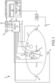
- Figure 1 shows a schematic diagram of an exemplary system 8 for conducting cardiac electrophysiology studies by navigating a cardiac catheter and measuring electrical activity occurring in a heart 10 of a patient 11 and three-dimensionally mapping the electrical activity and/or information related to or representative of the electrical activity so measured.
- System 8 can be used, for example, to create an anatomical model of the patient's heart 10 using one or more electrodes.
- System 8 can also be used to measure electrophysiology data at a plurality of points along a cardiac surface and store the measured data in association with location information for each measurement point at which the electrophysiology data was measured, for example to create a diagnostic data map of the patient's heart 10.
- the system 8 can facilitate differentiation between scar tissue and adipose tissue, by computing (1) a probability that a particular tissue location is scar tissue and/or (2) a probability that the particular tissue location is adipose tissue.
- system 8 determines the location, and in some aspects the orientation, of objects, typically within a three-dimensional space, and expresses those locations as position information determined relative to at least one reference.
- the patient 11 is depicted schematically as an oval.
- three sets of surface electrodes e.g., patch electrodes
- a surface of the patient 11 defining three generally orthogonal axes, referred to herein as an x-axis, a y-axis, and a z-axis.
- the electrodes could be positioned in other arrangements, for example multiple electrodes on a particular body surface.
- the electrodes do not need to be on the body surface, but could be positioned internally to the body.
- the x-axis surface electrodes 12, 14 are applied to the patient along a first axis, such as on the lateral sides of the thorax region of the patient ( e.g ., applied to the patient's skin underneath each arm) and may be referred to as the Left and Right electrodes.
- the y-axis electrodes 18, 19 are applied to the patient along a second axis generally orthogonal to the x-axis, such as along the inner thigh and neck regions of the patient, and may be referred to as the Left Leg and Neck electrodes.
- the z-axis electrodes 16, 22 are applied along a third axis generally orthogonal to both the x-axis and the y-axis, such as along the sternum and spine of the patient in the thorax region, and may be referred to as the Chest and Back electrodes.
- the heart 10 lies between these pairs of surface electrodes 12/14, 18/19, and 16/22.
- An additional surface reference electrode (e.g ., a "belly patch”) 21 provides a reference and/or ground electrode for the system 8.
- the belly patch electrode 21 may be an alternative to a fixed intra-cardiac electrode 31, described in further detail below.
- the patient 11 may have most or all of the conventional electrocardiogram ("ECG" or "EKG") system leads in place.
- ECG electrocardiogram
- a standard set of 12 ECG leads may be utilized for sensing electrocardiograms on the patient's heart 10. This ECG information is available to the system 8 ( e.g ., it can be provided as input to computer system 20).
- ECG leads are well understood, and for the sake of clarity in the figures, only a single lead 6 and its connection to computer 20 is illustrated in Figure 1 .
- a representative catheter 13 having at least one electrode 17 is also shown.
- This representative catheter electrode 17 is referred to as the "roving electrode,” “moving electrode,” or “measurement electrode” throughout the specification.
- multiple electrodes 17 on catheter 13, or on multiple such catheters will be used.
- the system 8 may comprise sixty-four electrodes on twelve catheters disposed within the heart and/or vasculature of the patient.
- this embodiment is merely exemplary, and any number of electrodes and catheters may be used.
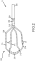
- HD grid catheter 13 includes a catheter body 200 coupled to a paddle 202.
- Catheter body 200 can further include first and second body electrodes 204, 206, respectively.
- Paddle 202 can include a first spline 208, a second spline 210, a third spline 212, and a fourth spline 214, which are coupled to catheter body 200 by a proximal coupler 216 and to each other by a distal coupler 218.
- first spline 208 and fourth spline 214 can be one continuous segment and second spline 210 and third spline 212 can be another continuous segment.
- the various splines 208, 210, 212, 214 can be separate segments coupled to each other ( e.g ., by proximal and distal couplers 216, 218, respectively).
- splines 208, 210, 212, 214 can include any number of electrodes 17; in Figure 2 , sixteen electrodes 17 are shown arranged in four-by-four array. It should also be understood that electrodes 17 can be evenly and/or unevenly spaced, as measured both along and between splines 208, 210, 212, 214.
- Catheter 13 (or multiple such catheters) are typically introduced into the heart and/or vasculature of the patient via one or more introducers using familiar procedures. Indeed, various approaches to introduce catheter 13 into the left ventricle of the patient's heart 10, such as transseptal approaches, will be familiar to those of ordinary skill in the art, and therefore need not be further described herein.
- each electrode 17 Since each electrode 17 lies within the patient, location data may be collected simultaneously for each electrode 17 by system 8. Similarly, each electrode 17 can be used to gather electrophysiological data from the cardiac surface.
- the ordinarily skilled artisan will be familiar with various modalities for the acquisition and processing of electrophysiology data points (including, for example, both contact and non-contact electrophysiological mapping), such that further discussion thereof is not necessary to the understanding of the techniques disclosed herein.
- various techniques familiar in the art can be used to generate a graphical representation from the plurality of electrophysiology data points. Insofar as the ordinarily skilled artisan will appreciate how to create electrophysiology maps from electrophysiology data points, the aspects thereof will only be described herein to the extent necessary to understand the instant disclosure.
- an optional fixed reference electrode 31 (e.g ., attached to a wall of the heart 10) is shown on a second catheter 29.
- this electrode 31 may be stationary (e.g ., attached to or near the wall of the heart) or disposed in a fixed spatial relationship with the roving electrodes ( e.g ., electrodes 17), and thus may be referred to as a "navigational reference” or "local reference.”
- the fixed reference electrode 31 may be used in addition or alternatively to the surface reference electrode 21 described above.
- a coronary sinus electrode or other fixed electrode in the heart 10 can be used as a reference for measuring voltages and displacements; that is, as described below, fixed reference electrode 31 may define the origin of a coordinate system.
- Each surface electrode is coupled to a multiplex switch 24, and the pairs of surface electrodes are selected by software running on a computer 20, which couples the surface electrodes to a signal generator 25.
- switch 24 may be eliminated and multiple ( e.g ., three) instances of signal generator 25 may be provided, one for each measurement axis (that is, each surface electrode pairing).
- the computer 20 may comprise, for example, a conventional general-purpose computer, a special-purpose computer, a distributed computer, or any other type of computer.
- the computer 20 may comprise one or more processors 28, such as a single central processing unit (“CPU"), or a plurality of processing units, commonly referred to as a parallel processing environment, which may execute instructions to practice the various aspects described herein.
- processors 28 such as a single central processing unit (“CPU"), or a plurality of processing units, commonly referred to as a parallel processing environment, which may execute instructions to practice the various aspects described herein.
- three nominally orthogonal electric fields are generated by a series of driven and sensed electric dipoles (e.g ., surface electrode pairs 12/14, 18/19, and 16/22) in order to realize catheter navigation in a biological conductor.
- these orthogonal fields can be decomposed and any pairs of surface electrodes can be driven as dipoles to provide effective electrode triangulation.
- the electrodes 12, 14, 18, 19, 16, and 22 (or any number of electrodes) could be positioned in any other effective arrangement for driving a current to or sensing a current from an electrode in the heart.
- multiple electrodes could be placed on the back, sides, and/or belly of patient 11. Additionally, such non-orthogonal methodologies add to the flexibility of the system.
- the potentials measured across the roving electrodes resulting from a predetermined set of drive (source-sink) configurations may be combined algebraically to yield the same effective potential as would be obtained by simply driving a uniform current along the orthogonal axes.
- any two of the surface electrodes 12, 14, 16, 18, 19, 22 may be selected as a dipole source and drain with respect to a ground reference, such as belly patch 21, while the unexcited electrodes measure voltage with respect to the ground reference.
- the roving electrodes 17 placed in the heart 10 are exposed to the field from a current pulse and are measured with respect to ground, such as belly patch 21.
- the catheters within the heart 10 may contain more or fewer electrodes than the sixteen shown, and each electrode potential may be measured.
- at least one electrode may be fixed to the interior surface of the heart to form a fixed reference electrode 31, which is also measured with respect to ground, such as belly patch 21, and which may be defined as the origin of the coordinate system relative to which system 8 measures positions. Data sets from each of the surface electrodes, the internal electrodes, and the virtual electrodes may all be used to determine the location of the roving electrodes 17 within heart 10.
- the measured voltages may be used by system 8 to determine the location in three-dimensional space of the electrodes inside the heart, such as roving electrodes 17 relative to a reference location, such as reference electrode 31. That is, the voltages measured at reference electrode 31 may be used to define the origin of a coordinate system, while the voltages measured at roving electrodes 17 may be used to express the location of roving electrodes 17 relative to the origin.
- the coordinate system is a three-dimensional (x, y, z) Cartesian coordinate system, although other coordinate systems, such as polar, spherical, and cylindrical coordinate systems, are contemplated.
- the data used to determine the location of the electrode(s) within the heart is measured while the surface electrode pairs impress an electric field on the heart.
- the electrode data may also be used to create a respiration compensation value used to improve the raw location data for the electrode locations as described, for example, in United States Patent No. 7,263,397 .
- the electrode data may also be used to compensate for changes in the impedance of the body of the patient as described, for example, in United States Patent No. 7,885,707 .
- system 8 first selects a set of surface electrodes and then drives them with current pulses. While the current pulses are being delivered, electrical activity, such as the voltages measured with at least one of the remaining surface electrodes and in vivo electrodes, is measured and stored. Compensation for artifacts, such as respiration and/or impedance shifting, may be performed as indicated above.
- system 8 is the EnSite TM Velocity TM cardiac mapping and visualization system of Abbott Laboratories, which generates electrical fields as described above, or another localization system that relies upon electrical fields.
- Other localization systems may be used in connection with the present teachings, including for example, systems that utilize magnetic fields instead of or in addition to electrical fields for localization. Examples of such systems include, without limitation, the CARTO navigation and location system of Biosense Webster, Inc., the AURORA ® system of Northern Digital Inc., Sterotaxis' NIOBE ® Magnetic Navigation System, as well as MediGuide TM Technology and the EnSite TM Precision TM system, both from Abbott Laboratories.
- the system 8 therefore also includes a tissue differentiation module 58 that can be used to determine (1) the probability that a given tissue location is scar tissue and/or (2) the probability that a given tissue location is adipose tissue.
- electrophysiology maps include a plurality of electrophysiology data points, and that each electrophysiology data point in turn includes both measured electrophysiology data (e.g ., an electrophysiological signal, such as a cardiac electrogram (“EGM”)) and location data (e.g ., information regarding the location of catheter 13 and/or electrodes 17 thereon), thus allowing the measured electrophysiology information to be associated with a particular location in space (that is, allowing the measured electrophysiology information to be interpreted as indicative of electrical activity at a particular tissue location).
- EMM cardiac electrogram
- location data e.g ., information regarding the location of catheter 13 and/or electrodes 17 thereon
- electrophysiology data points also include information about dielectric properties, such as permittivity, conductivity, and impedance, of the tissue location.
- flowchart 300 may represent several exemplary steps that can be carried out by the computer 20 of Figure 1 (e.g ., by processor 28, including tissue differentiation module 58). It should be understood that the representative steps described below can be either hardware- or software-implemented. For the sake of explanation, the term "signal processor" is used herein to describe both hardware- and software-based implementations of the teachings herein.
- An electrophysiology data point is collected in block 302.
- the collected electrophysiology data point includes an electrophysiological signal (e.g ., an EGM signal) associated with a tissue location, and can optionally further include dielectric properties of the tissue location.
- an electrophysiological signal e.g ., an EGM signal
- the electrophysiological signal (e.g ., the EGM signal) possesses one or more characteristics that are analyzed according to aspects of the instant disclosure to compute scar tissue and/or adipose tissue probabilities as described below.
- Suitable signal characteristics for analysis include, without limitation, signal duration, signal amplitude, signal fractionation, and late potentials. Each of these is discussed in detail below.
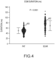
- Scar tissue generally exhibits longer-duration EGMs than adipose tissue.
- Figure 4 which appears in Tung et al., "Distinguishing epicardial fat from scar: Analysis of electrograms using high-density electroanatomic mapping in a novel porcine infarct model," Heart Rhythm 2010; 7:389-395 , plots the distribution of EGM duration for adipose tissue 400 and for scar tissue 402.
- adipose tissue EGM duration (distribution 400) is about 50.1 ms ⁇ 11.6 ms
- scar tissue EGM duration (distribution 402) is about 68.6 ms ⁇ 18.9 ms.
- EGM duration from healthy myocardial tissue typically ranges from about 50.0 ms ⁇ 6.7 ms.
- a cutoff point of 80 ms has a 99% specificity for scar tissue.
- Low amplitude signals are typically characteristic of adipose and scar tissue EGMs.
- Mean bipolar EGM amplitudes are typically low for adipose tissue (about 0.77 mV ⁇ 0.34 mv) and for scar tissue (about 0.75 mv ⁇ 0.38 mV) by comparison to healthy myocardial tissue (about 5.9 mV ⁇ 3.5 mV).
- Scar tissue EGMs typically present greater fractionation than adipose tissue EGMs, such as measured by the number of deflections in the EGM.
- Figure 5 which also appears in Tung et al., supra, plots the distribution of the number of deflections in adipose tissue 500 and for scar tissue 502.
- adipose tissue EGMs (distribution 500) exhibit about 4.7 ⁇ 1.8 deflections
- scar tissue EGMs (distribution 502) exhibit about 8.5 ⁇ 3.1 deflections.
- Late Potentials are rarely present in adipose tissue EGMs. Late potentials are present in about 39.1% of scar tissue EGMs. The presence of late potentials in an EGM has a specificity of about 99% for scar tissue.
- the collected electrophysiology data point also include one or more dielectric properties measured at the tissue location. These properties can also be analyzed when computing adipose and/or scar tissue probabilities according to the instant teachings. Suitable dielectric properties for such analysis include, without limitation, tissue impedance, tissue conductivity, and tissue permittivity.
- the dielectric properties can be measured between electrodes on a single spline (e.g., between electrodes 17a and 17b in Figure 2 ) and/or between electrodes across splines (e.g., between electrodes 17a and 17c in Figure 2 ).
- Figure 6 depicts the relationships between tissue permittivity and tissue conductivity at various stimulation frequencies for healthy cardiac muscle.
- Figure 7 depicts the same relationships for adipose tissue. When stimulated at the same frequency, Figures 6 and 7 illustrate that cardiac muscle and adipose tissue exhibit different combinations of tissue permittivity and conductivity, which differences can be used to distinguish between tissue types according to the teachings herein.
- Figures 6 and 7 appear in Gabriel et al., "The dielectric properties of biological tissues: II. Measurements in the frequency range 10 Hz to 20 GhZ," Phys. Med. Biol. 41, 1996, 2251-2269 .
- adipose tissue probability refers to the probability that the tissue location of the collected electrophysiology data point is adipose tissue.
- scar tissue probability is used herein to refer to the probability that the tissue location of the collected electrophysiology data point is scar tissue.
- the adipose tissue probability and the scar tissue probability are independent. That is, it is not required that, for a given electrophysiology data point, the sum of the adipose tissue probability and the scar tissue probability must equal 1.0.
- Both the adipose tissue probability and the scar tissue probability are computed as functions of one or more characteristics of the electrophysiological signal and/or one or more dielectric properties of the tissue location. Further, the adipose tissue probability and the scar tissue probability are computed as weighted functions of one or more characteristic of the electrophysiological signal and/or one or more dielectric properties of the tissue location.
- amplitude is the amplitude of the electrophysiological signal
- fractionation is a measure of the fractionation of the electrophysiological signal ( e.g ., the number of deflections in the EGM)
- impedance is the tissue impedance between electrodes 17 on catheter 13, and late potentials reflects the presence of late potentials in the signal.
- a xyz , b xyz , c xyz , and d xyz are weight factors that are spatially-dependent (e.g ., their relative values depend upon where mapping data is acquired). For example, amplitude may have a lower weight than fractionation or impedance when mapping in the coronary sinus ridge.
- a xyz equals 0.1
- b xyz equals 0.6
- c xyz equals 0.3
- d xyz equals 0.0.
- p adipose e xyz amplitude + f xyz fractionation + g xyz impedance + h xyz late potentials
- amplitude is the amplitude of the electrophysiological signal
- fractionation is a measure of the fractionation of the electrophysiological signal ( e.g ., the number of deflections in the EGM)
- impedance is the tissue impedance between electrodes 17 on catheter 13, and late potentials reflects the presence of late potentials in the signal.
- e xyz , f xyz , g xyz , and h xyz are weight factors that are spatially-dependent (e.g ., their relative values depend upon where mapping data is acquired). For example, amplitude may have a higher weight than fractionation or impedance when mapping in the coronary sinus ridge.
- e xyz equals 0.2
- f xyz equals 0.2
- g xyz equals 0.4
- h xyz equals 0.2.
- the variables utilized in the probability functions are user-selected.
- the weighting factors assigned to these user-selected variables are user-preset (that is, user-defined and -adjusted).
- the practitioner can manipulate the sensitivity and/or specificity of the probability calculations depending upon, for example, the particular substrate being mapped.
- the variables utilized in the probability functions and/or their relative weights may be pre-selected depending, for example, upon the type of electrophysiology study being conducted.
- the adipose and/or scar tissue probabilities are associated with the collected electrophysiology data point in block 306.
- Decision block 308 checks to see if additional electrophysiology data points remain to be processed.
- the teachings herein can be applied in real time (e.g ., electrophysiology data points can be analyzed for scar tissue and/or adipose tissue probabilities at the time they are collected) or during post-processing (e.g ., electrophysiology data points making up an electrophysiology map can be analyzed for scar tissue and/or adipose tissue probabilities after they have been collected).
- one or more tissue probability maps can be computed in block 310 from the plurality of adipose and/or scar tissue probabilities computed in block 304. More particularly, block 310 can include the computation of a scar tissue probability map, which describes the probability that tissue locations over a given tissue region are scar tissue, and/or an adipose tissue probability map, which describes the probability that tissue locations over a given tissue region have a high fat concentration.
- Graphical representations of the tissue probability maps can be output in block 312, for example by displaying the computed probabilities numerically and/or in colorscale or greyscale on a three-dimensional tissue geometry model (e.g ., a three-dimensional cardiac geometry model generated by system 8; a CT or MRI cardiac image; or the like).
- a three-dimensional tissue geometry model e.g ., a three-dimensional cardiac geometry model generated by system 8; a CT or MRI cardiac image; or the like.
- Such representations can aid a practitioner in differentiating between scar tissue and adipose tissue on the ventricular epicardial surface, which can help guide the practitioner in delivering ablation therapy in the treatment of VT.
- All directional references e.g., upper, lower, upward, downward, left, right, leftward, rightward, top, bottom, above, below, vertical, horizontal, clockwise, and counterclockwise
- Joinder references e.g., attached, coupled, connected, and the like
- Joinder references are to be construed broadly and may include intermediate members between a connection of elements and relative movement between elements. As such, joinder references do not necessarily infer that two elements are directly connected and in fixed relation to each other.
Landscapes
- Health & Medical Sciences (AREA)
- Life Sciences & Earth Sciences (AREA)
- Surgery (AREA)
- Engineering & Computer Science (AREA)
- Animal Behavior & Ethology (AREA)
- Public Health (AREA)
- Biomedical Technology (AREA)
- Heart & Thoracic Surgery (AREA)
- Medical Informatics (AREA)
- Molecular Biology (AREA)
- General Health & Medical Sciences (AREA)
- Veterinary Medicine (AREA)
- Pathology (AREA)
- Biophysics (AREA)
- Physics & Mathematics (AREA)
- Nuclear Medicine, Radiotherapy & Molecular Imaging (AREA)
- Radiology & Medical Imaging (AREA)
- Physiology (AREA)
- Cardiology (AREA)
- Robotics (AREA)
- Measurement And Recording Of Electrical Phenomena And Electrical Characteristics Of The Living Body (AREA)
Claims (6)
- Computerimplementiertes Verfahren zum Erzeugen einer Gewebekarte, die zwischen Fettgewebe und Narbengewebe unterscheidet, aus einer elektrophysiologischen Karte, die eine Mehrzahl von elektrophysiologischen Datenpunkten enthält, wobei jeder elektrophysiologische Datenpunkt ein elektrophysiologisches Signal und eine oder mehrere dielektrische Eigenschaften, die zu einem Gewebeort gehören, aufweist, wobei das Verfahren aufweist:für jeden elektrophysiologischen Datenpunkt von der Mehrzahl von elektrophysiologischen Datenpunkten, ein Berechnen:einer Fettgewebewahrscheinlichkeit für einen Gewebeort als erste Funktion der einen oder der mehreren Charakteristiken des elektrophysiologischen Signals und der einen oder mehreren dielektrischen Eigenschaften, die zu dem Gewebeort gehören, undeiner Narbengewebewahrscheinlichkeit für den Gewebeort als zweite Funktion der einen oder der mehreren Charakteristiken des elektrophysiologischen Signals und der einen oder der mehreren dielektrischen Eigenschaften, die zu dem Gewebeort gehören;wobei die erste Funktion einen ersten Satz von benutzervoreingestellten räumlich abhängigen Gewichtungsfaktoren für die eine oder die mehreren Charakteristiken des elektrophysiologischen Signals und die eine oder die mehreren dielektrischen Eigenschaften, die an dem Gewebeort gemessen werden, verwendet, undwobei die zweite Funktion einen zweiten Satz von benutzervoreingestellten räumlich abhängigen Gewichtungsfaktoren für die eine oder die mehreren Charakteristiken des elektrophysiologischen Signals und die eine oder die mehreren dielektrischen Eigenschaften, die an dem Gewebeort gemessen werden, verwendet, undErzeugen einer oder mehrerer von einer Fettgewebewahrscheinlichkeitskarte und einer Narbengewebewahrscheinlichkeitskarte.
- Verfahren nach Anspruch 1, bei dem die eine oder die mehreren Charakteristiken des elektrophysiologischen Signals aus der Gruppe bestehend aus Signaldauer, Signalamplitude, Signalaufteilung und Spätpotentialen ausgewählt werden, und
wobei die eine oder die mehreren dielektrischen Eigenschaften, die an dem Gewebeort gemessen werden, aus der Gruppe bestehend aus Gewebeimpedanz, Gewebeleitfähigkeit und Gewebedurchlässigkeit ausgewählt werden. - Verfahren nach Anspruch 1, ferner mit:mehrmaliges Wiederholen des Schritts der Verarbeitung eines elektrophysiologischen Signals, das zu einem Gewebeort gehört;Erzeugen einer Fettgewebewahrscheinlichkeitskarte; undErzeugen einer Narbengewebewahrscheinlichkeitskarte.
- Verfahren nach Anspruch 3, ferner mit einem Ausgeben einer grafischen Darstellung der Fettgewebewahrscheinlichkeitskarte und/oder der Narbengewebewahrscheinlichkeitskarte auf einem dreidimensionalen Gewebegeometriemodell.
- Verfahren nach Anspruch 1, bei dem die eine oder die mehreren dielektrischen Eigenschaften, die an dem Gewebeort gemessen werden, unter Verwendung einer ersten Elektrode auf einem ersten Arm eines Multielektrodenkatheters und einer zweiten Elektrode auf dem ersten Arm des Multielektrodenkatheters gemessen werden.
- System zum Unterscheiden von Fettgewebe und Narbengewebe, mit:
einem Gewebewahrscheinlichkeitsbestimmungsprozessor, der konfiguriert ist zum Durchführen der Verfahren nach Ansprüchen 1 bis 5.
Applications Claiming Priority (2)
| Application Number | Priority Date | Filing Date | Title |
|---|---|---|---|
| US201762466079P | 2017-03-02 | 2017-03-02 | |
| PCT/US2018/020116 WO2018160631A1 (en) | 2017-03-02 | 2018-02-28 | System and method for differentiation of adipose tissue from scar tissue during electrophysiological mapping |
Publications (2)
| Publication Number | Publication Date |
|---|---|
| EP3565459A1 EP3565459A1 (de) | 2019-11-13 |
| EP3565459B1 true EP3565459B1 (de) | 2024-03-27 |
Family
ID=61691567
Family Applications (1)
| Application Number | Title | Priority Date | Filing Date |
|---|---|---|---|
| EP18712043.1A Active EP3565459B1 (de) | 2017-03-02 | 2018-02-28 | System und verfahren zur unterscheidung adipösen gewebes von narbengewebe während einer elektrophysiologischen kartierung |
Country Status (5)
| Country | Link |
|---|---|
| US (1) | US20180249928A1 (de) |
| EP (1) | EP3565459B1 (de) |
| JP (1) | JP6872625B2 (de) |
| CN (1) | CN110381813B (de) |
| WO (1) | WO2018160631A1 (de) |
Families Citing this family (6)
| Publication number | Priority date | Publication date | Assignee | Title |
|---|---|---|---|---|
| US10905347B2 (en) * | 2018-02-06 | 2021-02-02 | Biosense Webster (Israel) Ltd. | Catheter with increased electrode density spine assembly having reinforced spine covers |
| US10945626B2 (en) * | 2018-02-06 | 2021-03-16 | Biosense Webster (Israel) Ltd. | Catheter with staggered electrodes spine assembly |
| US10842400B2 (en) * | 2018-11-08 | 2020-11-24 | Biosense Webster (Israel) Ltd. | Iterative coherent mapping of cardiac electrophysiological (EP) activation including scar effects |
| CN111281387B (zh) * | 2020-03-09 | 2024-03-26 | 中山大学 | 基于人工神经网络的左心房与心房瘢痕的分割方法及装置 |
| CN116491925B (zh) * | 2023-04-12 | 2026-04-03 | 华中科技大学 | 一种皮肤瘢痕状态检测方法及修复效果评估方法 |
| CN116746943B (zh) * | 2023-08-18 | 2023-11-07 | 四川锦江电子医疗器械科技股份有限公司 | 一种医用电极标测导管 |
Family Cites Families (14)
| Publication number | Priority date | Publication date | Assignee | Title |
|---|---|---|---|---|
| DE69315354T2 (de) | 1992-09-23 | 1998-03-19 | Endocardial Solutions Inc | Endokard-mapping system |
| US5662108A (en) | 1992-09-23 | 1997-09-02 | Endocardial Solutions, Inc. | Electrophysiology mapping system |
| US6947785B1 (en) | 1993-09-23 | 2005-09-20 | Endocardial Solutions, Inc. | Interface system for endocardial mapping catheter |
| US5697377A (en) | 1995-11-22 | 1997-12-16 | Medtronic, Inc. | Catheter mapping system and method |
| US7263397B2 (en) | 1998-06-30 | 2007-08-28 | St. Jude Medical, Atrial Fibrillation Division, Inc. | Method and apparatus for catheter navigation and location and mapping in the heart |
| US7499745B2 (en) * | 2000-02-28 | 2009-03-03 | Barbara Ann Karmanos Cancer Institute | Multidimensional bioelectrical tissue analyzer |
| WO2006066324A1 (en) * | 2004-12-21 | 2006-06-29 | Sydney West Area Health Service | Automated processing of electrophysiological data |
| US20060241708A1 (en) * | 2005-04-22 | 2006-10-26 | Willem Boute | Multiple sensors for sleep apnea with probability indication for sleep diagnosis and means for automatic activation of alert or therapy |
| US7885707B2 (en) | 2005-09-15 | 2011-02-08 | St. Jude Medical, Atrial Fibrillation Division, Inc. | Method of scaling navigation signals to account for impedance drift in tissue |
| US20110264000A1 (en) * | 2007-12-28 | 2011-10-27 | Saurav Paul | System and method for determining tissue type and mapping tissue morphology |
| US8644917B2 (en) * | 2011-09-20 | 2014-02-04 | Albert Einstein Healthcare Network | Cardio mapping system and method for cardio mapping |
| EP2890292B1 (de) * | 2012-08-31 | 2021-01-13 | Acutus Medical, Inc. | Kathetersystem für das herz |
| EP3021745B1 (de) * | 2013-10-30 | 2019-04-10 | St. Jude Medical, Cardiology Division, Inc. | Kardiokartierungssystem und verfahren zur spannungsbasierten auswertung von ekg-signalen |
| EP3073907B1 (de) * | 2014-02-25 | 2020-06-17 | St. Jude Medical, Cardiology Division, Inc. | System zur lokalen elektrophysiologischen charakterisierung eines kardialen substrats mit mehrfachelektrodenkathetern |
-
2018
- 2018-02-28 EP EP18712043.1A patent/EP3565459B1/de active Active
- 2018-02-28 WO PCT/US2018/020116 patent/WO2018160631A1/en not_active Ceased
- 2018-02-28 JP JP2019546289A patent/JP6872625B2/ja active Active
- 2018-02-28 CN CN201880014599.0A patent/CN110381813B/zh active Active
- 2018-02-28 US US15/907,512 patent/US20180249928A1/en active Pending
Also Published As
| Publication number | Publication date |
|---|---|
| JP2020508760A (ja) | 2020-03-26 |
| WO2018160631A1 (en) | 2018-09-07 |
| EP3565459A1 (de) | 2019-11-13 |
| US20180249928A1 (en) | 2018-09-06 |
| CN110381813A (zh) | 2019-10-25 |
| CN110381813B (zh) | 2022-10-21 |
| JP6872625B2 (ja) | 2021-05-19 |
Similar Documents
| Publication | Publication Date | Title |
|---|---|---|
| EP3498156B1 (de) | Koaxiale elektrodenkatheter zur extraktion elektrophysiologischer parameter | |
| EP3565459B1 (de) | System und verfahren zur unterscheidung adipösen gewebes von narbengewebe während einer elektrophysiologischen kartierung | |
| US9717429B2 (en) | System and method for analyzing biological signals and generating electrophyisology maps | |
| EP3021744B1 (de) | System zur erzeugung von elektrophysiologischen karten | |
| US11751794B2 (en) | System and method for mapping electrophysiological activation | |
| US12369835B2 (en) | System and method for electrophysiological mapping | |
| EP3344135B1 (de) | Verfahren und systeme zur abbildung der herzwiederherstellung | |
| EP3923797B1 (de) | System und verfahren zur herzkartierung | |
| US11969254B2 (en) | System and method for cardiac mapping | |
| US11380029B2 (en) | System and method for mapping local activation times | |
| EP3609400B1 (de) | System zur elektrophysiologischen kartierung | |
| US20240350100A1 (en) | Method and system for generating respiration signals for use in electrophysiology procedures | |
| US12138060B2 (en) | System and method for cardiac mapping | |
| US20220142545A1 (en) | High density electrode catheters |
Legal Events
| Date | Code | Title | Description |
|---|---|---|---|
| STAA | Information on the status of an ep patent application or granted ep patent |
Free format text: STATUS: UNKNOWN |
|
| STAA | Information on the status of an ep patent application or granted ep patent |
Free format text: STATUS: THE INTERNATIONAL PUBLICATION HAS BEEN MADE |
|
| PUAI | Public reference made under article 153(3) epc to a published international application that has entered the european phase |
Free format text: ORIGINAL CODE: 0009012 |
|
| STAA | Information on the status of an ep patent application or granted ep patent |
Free format text: STATUS: REQUEST FOR EXAMINATION WAS MADE |
|
| 17P | Request for examination filed |
Effective date: 20190808 |
|
| AK | Designated contracting states |
Kind code of ref document: A1 Designated state(s): AL AT BE BG CH CY CZ DE DK EE ES FI FR GB GR HR HU IE IS IT LI LT LU LV MC MK MT NL NO PL PT RO RS SE SI SK SM TR |
|
| AX | Request for extension of the european patent |
Extension state: BA ME |
|
| RIN1 | Information on inventor provided before grant (corrected) |
Inventor name: MANGUAL-SOTO, JAN O. Inventor name: JIANG, CHUNLAN Inventor name: MARKOVITZ, CRAIG Inventor name: RICHER, LOUIS-PHILIPPE Inventor name: CASSET, CYRILLE |
|
| DAV | Request for validation of the european patent (deleted) | ||
| DAX | Request for extension of the european patent (deleted) | ||
| STAA | Information on the status of an ep patent application or granted ep patent |
Free format text: STATUS: EXAMINATION IS IN PROGRESS |
|
| 17Q | First examination report despatched |
Effective date: 20210824 |
|
| P01 | Opt-out of the competence of the unified patent court (upc) registered |
Effective date: 20230616 |
|
| RIC1 | Information provided on ipc code assigned before grant |
Ipc: A61B 5/287 20210101ALI20230724BHEP Ipc: A61B 5/24 20210101ALI20230724BHEP Ipc: A61B 5/0537 20210101ALI20230724BHEP Ipc: A61B 5/0538 20210101ALI20230724BHEP Ipc: A61B 5/00 20060101AFI20230724BHEP |
|
| GRAP | Despatch of communication of intention to grant a patent |
Free format text: ORIGINAL CODE: EPIDOSNIGR1 |
|
| STAA | Information on the status of an ep patent application or granted ep patent |
Free format text: STATUS: GRANT OF PATENT IS INTENDED |
|
| INTG | Intention to grant announced |
Effective date: 20231117 |
|
| GRAS | Grant fee paid |
Free format text: ORIGINAL CODE: EPIDOSNIGR3 |
|
| GRAA | (expected) grant |
Free format text: ORIGINAL CODE: 0009210 |
|
| STAA | Information on the status of an ep patent application or granted ep patent |
Free format text: STATUS: THE PATENT HAS BEEN GRANTED |
|
| AK | Designated contracting states |
Kind code of ref document: B1 Designated state(s): AL AT BE BG CH CY CZ DE DK EE ES FI FR GB GR HR HU IE IS IT LI LT LU LV MC MK MT NL NO PL PT RO RS SE SI SK SM TR |
|
| REG | Reference to a national code |
Ref country code: GB Ref legal event code: FG4D |
|
| REG | Reference to a national code |
Ref country code: CH Ref legal event code: EP |
|
| REG | Reference to a national code |
Ref country code: DE Ref legal event code: R096 Ref document number: 602018067114 Country of ref document: DE |
|
| REG | Reference to a national code |
Ref country code: IE Ref legal event code: FG4D |
|
| PG25 | Lapsed in a contracting state [announced via postgrant information from national office to epo] |
Ref country code: LT Free format text: LAPSE BECAUSE OF FAILURE TO SUBMIT A TRANSLATION OF THE DESCRIPTION OR TO PAY THE FEE WITHIN THE PRESCRIBED TIME-LIMIT Effective date: 20240327 |
|
| REG | Reference to a national code |
Ref country code: LT Ref legal event code: MG9D |
|
| PG25 | Lapsed in a contracting state [announced via postgrant information from national office to epo] |
Ref country code: GR Free format text: LAPSE BECAUSE OF FAILURE TO SUBMIT A TRANSLATION OF THE DESCRIPTION OR TO PAY THE FEE WITHIN THE PRESCRIBED TIME-LIMIT Effective date: 20240628 |
|
| PG25 | Lapsed in a contracting state [announced via postgrant information from national office to epo] |
Ref country code: RS Free format text: LAPSE BECAUSE OF FAILURE TO SUBMIT A TRANSLATION OF THE DESCRIPTION OR TO PAY THE FEE WITHIN THE PRESCRIBED TIME-LIMIT Effective date: 20240627 Ref country code: HR Free format text: LAPSE BECAUSE OF FAILURE TO SUBMIT A TRANSLATION OF THE DESCRIPTION OR TO PAY THE FEE WITHIN THE PRESCRIBED TIME-LIMIT Effective date: 20240327 |
|
| PG25 | Lapsed in a contracting state [announced via postgrant information from national office to epo] |
Ref country code: RS Free format text: LAPSE BECAUSE OF FAILURE TO SUBMIT A TRANSLATION OF THE DESCRIPTION OR TO PAY THE FEE WITHIN THE PRESCRIBED TIME-LIMIT Effective date: 20240627 Ref country code: NO Free format text: LAPSE BECAUSE OF FAILURE TO SUBMIT A TRANSLATION OF THE DESCRIPTION OR TO PAY THE FEE WITHIN THE PRESCRIBED TIME-LIMIT Effective date: 20240627 Ref country code: LT Free format text: LAPSE BECAUSE OF FAILURE TO SUBMIT A TRANSLATION OF THE DESCRIPTION OR TO PAY THE FEE WITHIN THE PRESCRIBED TIME-LIMIT Effective date: 20240327 Ref country code: HR Free format text: LAPSE BECAUSE OF FAILURE TO SUBMIT A TRANSLATION OF THE DESCRIPTION OR TO PAY THE FEE WITHIN THE PRESCRIBED TIME-LIMIT Effective date: 20240327 Ref country code: GR Free format text: LAPSE BECAUSE OF FAILURE TO SUBMIT A TRANSLATION OF THE DESCRIPTION OR TO PAY THE FEE WITHIN THE PRESCRIBED TIME-LIMIT Effective date: 20240628 Ref country code: FI Free format text: LAPSE BECAUSE OF FAILURE TO SUBMIT A TRANSLATION OF THE DESCRIPTION OR TO PAY THE FEE WITHIN THE PRESCRIBED TIME-LIMIT Effective date: 20240327 Ref country code: BG Free format text: LAPSE BECAUSE OF FAILURE TO SUBMIT A TRANSLATION OF THE DESCRIPTION OR TO PAY THE FEE WITHIN THE PRESCRIBED TIME-LIMIT Effective date: 20240327 |
|
| REG | Reference to a national code |
Ref country code: NL Ref legal event code: MP Effective date: 20240327 |
|
| PG25 | Lapsed in a contracting state [announced via postgrant information from national office to epo] |
Ref country code: SE Free format text: LAPSE BECAUSE OF FAILURE TO SUBMIT A TRANSLATION OF THE DESCRIPTION OR TO PAY THE FEE WITHIN THE PRESCRIBED TIME-LIMIT Effective date: 20240327 Ref country code: LV Free format text: LAPSE BECAUSE OF FAILURE TO SUBMIT A TRANSLATION OF THE DESCRIPTION OR TO PAY THE FEE WITHIN THE PRESCRIBED TIME-LIMIT Effective date: 20240327 |
|
| PG25 | Lapsed in a contracting state [announced via postgrant information from national office to epo] |
Ref country code: NL Free format text: LAPSE BECAUSE OF FAILURE TO SUBMIT A TRANSLATION OF THE DESCRIPTION OR TO PAY THE FEE WITHIN THE PRESCRIBED TIME-LIMIT Effective date: 20240327 |
|
| REG | Reference to a national code |
Ref country code: AT Ref legal event code: MK05 Ref document number: 1669112 Country of ref document: AT Kind code of ref document: T Effective date: 20240327 |
|
| PG25 | Lapsed in a contracting state [announced via postgrant information from national office to epo] |
Ref country code: NL Free format text: LAPSE BECAUSE OF FAILURE TO SUBMIT A TRANSLATION OF THE DESCRIPTION OR TO PAY THE FEE WITHIN THE PRESCRIBED TIME-LIMIT Effective date: 20240327 |
|
| PG25 | Lapsed in a contracting state [announced via postgrant information from national office to epo] |
Ref country code: IS Free format text: LAPSE BECAUSE OF FAILURE TO SUBMIT A TRANSLATION OF THE DESCRIPTION OR TO PAY THE FEE WITHIN THE PRESCRIBED TIME-LIMIT Effective date: 20240727 |
|
| PG25 | Lapsed in a contracting state [announced via postgrant information from national office to epo] |
Ref country code: PT Free format text: LAPSE BECAUSE OF FAILURE TO SUBMIT A TRANSLATION OF THE DESCRIPTION OR TO PAY THE FEE WITHIN THE PRESCRIBED TIME-LIMIT Effective date: 20240729 Ref country code: SM Free format text: LAPSE BECAUSE OF FAILURE TO SUBMIT A TRANSLATION OF THE DESCRIPTION OR TO PAY THE FEE WITHIN THE PRESCRIBED TIME-LIMIT Effective date: 20240327 |
|
| PG25 | Lapsed in a contracting state [announced via postgrant information from national office to epo] |
Ref country code: ES Free format text: LAPSE BECAUSE OF FAILURE TO SUBMIT A TRANSLATION OF THE DESCRIPTION OR TO PAY THE FEE WITHIN THE PRESCRIBED TIME-LIMIT Effective date: 20240327 |
|
| PG25 | Lapsed in a contracting state [announced via postgrant information from national office to epo] |
Ref country code: EE Free format text: LAPSE BECAUSE OF FAILURE TO SUBMIT A TRANSLATION OF THE DESCRIPTION OR TO PAY THE FEE WITHIN THE PRESCRIBED TIME-LIMIT Effective date: 20240327 Ref country code: CZ Free format text: LAPSE BECAUSE OF FAILURE TO SUBMIT A TRANSLATION OF THE DESCRIPTION OR TO PAY THE FEE WITHIN THE PRESCRIBED TIME-LIMIT Effective date: 20240327 |
|
| PG25 | Lapsed in a contracting state [announced via postgrant information from national office to epo] |
Ref country code: AT Free format text: LAPSE BECAUSE OF FAILURE TO SUBMIT A TRANSLATION OF THE DESCRIPTION OR TO PAY THE FEE WITHIN THE PRESCRIBED TIME-LIMIT Effective date: 20240327 |
|
| PG25 | Lapsed in a contracting state [announced via postgrant information from national office to epo] |
Ref country code: PL Free format text: LAPSE BECAUSE OF FAILURE TO SUBMIT A TRANSLATION OF THE DESCRIPTION OR TO PAY THE FEE WITHIN THE PRESCRIBED TIME-LIMIT Effective date: 20240327 |
|
| PG25 | Lapsed in a contracting state [announced via postgrant information from national office to epo] |
Ref country code: SK Free format text: LAPSE BECAUSE OF FAILURE TO SUBMIT A TRANSLATION OF THE DESCRIPTION OR TO PAY THE FEE WITHIN THE PRESCRIBED TIME-LIMIT Effective date: 20240327 |
|
| PG25 | Lapsed in a contracting state [announced via postgrant information from national office to epo] |
Ref country code: SM Free format text: LAPSE BECAUSE OF FAILURE TO SUBMIT A TRANSLATION OF THE DESCRIPTION OR TO PAY THE FEE WITHIN THE PRESCRIBED TIME-LIMIT Effective date: 20240327 Ref country code: SK Free format text: LAPSE BECAUSE OF FAILURE TO SUBMIT A TRANSLATION OF THE DESCRIPTION OR TO PAY THE FEE WITHIN THE PRESCRIBED TIME-LIMIT Effective date: 20240327 Ref country code: RO Free format text: LAPSE BECAUSE OF FAILURE TO SUBMIT A TRANSLATION OF THE DESCRIPTION OR TO PAY THE FEE WITHIN THE PRESCRIBED TIME-LIMIT Effective date: 20240327 Ref country code: PT Free format text: LAPSE BECAUSE OF FAILURE TO SUBMIT A TRANSLATION OF THE DESCRIPTION OR TO PAY THE FEE WITHIN THE PRESCRIBED TIME-LIMIT Effective date: 20240729 Ref country code: PL Free format text: LAPSE BECAUSE OF FAILURE TO SUBMIT A TRANSLATION OF THE DESCRIPTION OR TO PAY THE FEE WITHIN THE PRESCRIBED TIME-LIMIT Effective date: 20240327 Ref country code: IS Free format text: LAPSE BECAUSE OF FAILURE TO SUBMIT A TRANSLATION OF THE DESCRIPTION OR TO PAY THE FEE WITHIN THE PRESCRIBED TIME-LIMIT Effective date: 20240727 Ref country code: ES Free format text: LAPSE BECAUSE OF FAILURE TO SUBMIT A TRANSLATION OF THE DESCRIPTION OR TO PAY THE FEE WITHIN THE PRESCRIBED TIME-LIMIT Effective date: 20240327 Ref country code: EE Free format text: LAPSE BECAUSE OF FAILURE TO SUBMIT A TRANSLATION OF THE DESCRIPTION OR TO PAY THE FEE WITHIN THE PRESCRIBED TIME-LIMIT Effective date: 20240327 Ref country code: CZ Free format text: LAPSE BECAUSE OF FAILURE TO SUBMIT A TRANSLATION OF THE DESCRIPTION OR TO PAY THE FEE WITHIN THE PRESCRIBED TIME-LIMIT Effective date: 20240327 Ref country code: AT Free format text: LAPSE BECAUSE OF FAILURE TO SUBMIT A TRANSLATION OF THE DESCRIPTION OR TO PAY THE FEE WITHIN THE PRESCRIBED TIME-LIMIT Effective date: 20240327 |
|
| REG | Reference to a national code |
Ref country code: DE Ref legal event code: R097 Ref document number: 602018067114 Country of ref document: DE |
|
| PG25 | Lapsed in a contracting state [announced via postgrant information from national office to epo] |
Ref country code: DK Free format text: LAPSE BECAUSE OF FAILURE TO SUBMIT A TRANSLATION OF THE DESCRIPTION OR TO PAY THE FEE WITHIN THE PRESCRIBED TIME-LIMIT Effective date: 20240327 |
|
| PG25 | Lapsed in a contracting state [announced via postgrant information from national office to epo] |
Ref country code: DK Free format text: LAPSE BECAUSE OF FAILURE TO SUBMIT A TRANSLATION OF THE DESCRIPTION OR TO PAY THE FEE WITHIN THE PRESCRIBED TIME-LIMIT Effective date: 20240327 |
|
| PLBE | No opposition filed within time limit |
Free format text: ORIGINAL CODE: 0009261 |
|
| STAA | Information on the status of an ep patent application or granted ep patent |
Free format text: STATUS: NO OPPOSITION FILED WITHIN TIME LIMIT |
|
| 26N | No opposition filed |
Effective date: 20250103 |
|
| PG25 | Lapsed in a contracting state [announced via postgrant information from national office to epo] |
Ref country code: SI Free format text: LAPSE BECAUSE OF FAILURE TO SUBMIT A TRANSLATION OF THE DESCRIPTION OR TO PAY THE FEE WITHIN THE PRESCRIBED TIME-LIMIT Effective date: 20240327 |
|
| PG25 | Lapsed in a contracting state [announced via postgrant information from national office to epo] |
Ref country code: MC Free format text: LAPSE BECAUSE OF FAILURE TO SUBMIT A TRANSLATION OF THE DESCRIPTION OR TO PAY THE FEE WITHIN THE PRESCRIBED TIME-LIMIT Effective date: 20240327 |
|
| REG | Reference to a national code |
Ref country code: CH Ref legal event code: PL |
|
| PG25 | Lapsed in a contracting state [announced via postgrant information from national office to epo] |
Ref country code: LU Free format text: LAPSE BECAUSE OF NON-PAYMENT OF DUE FEES Effective date: 20250228 |
|
| PG25 | Lapsed in a contracting state [announced via postgrant information from national office to epo] |
Ref country code: CH Free format text: LAPSE BECAUSE OF NON-PAYMENT OF DUE FEES Effective date: 20250228 |
|
| REG | Reference to a national code |
Ref country code: BE Ref legal event code: MM Effective date: 20250228 |
|
| PG25 | Lapsed in a contracting state [announced via postgrant information from national office to epo] |
Ref country code: BE Free format text: LAPSE BECAUSE OF NON-PAYMENT OF DUE FEES Effective date: 20250228 |
|
| PG25 | Lapsed in a contracting state [announced via postgrant information from national office to epo] |
Ref country code: IE Free format text: LAPSE BECAUSE OF NON-PAYMENT OF DUE FEES Effective date: 20250228 |
|
| PGFP | Annual fee paid to national office [announced via postgrant information from national office to epo] |
Ref country code: GB Payment date: 20260113 Year of fee payment: 9 |
|
| PGFP | Annual fee paid to national office [announced via postgrant information from national office to epo] |
Ref country code: DE Payment date: 20260115 Year of fee payment: 9 |
|
| PGFP | Annual fee paid to national office [announced via postgrant information from national office to epo] |
Ref country code: IT Payment date: 20260212 Year of fee payment: 9 |
|
| PGFP | Annual fee paid to national office [announced via postgrant information from national office to epo] |
Ref country code: FR Payment date: 20260109 Year of fee payment: 9 |