EP3613466A1 - Anticorps capable de se lier à un norovirus, à un composite, dispositif de détection et son procédé d'utilisation - Google Patents
Anticorps capable de se lier à un norovirus, à un composite, dispositif de détection et son procédé d'utilisation Download PDFInfo
- Publication number
- EP3613466A1 EP3613466A1 EP19192787.0A EP19192787A EP3613466A1 EP 3613466 A1 EP3613466 A1 EP 3613466A1 EP 19192787 A EP19192787 A EP 19192787A EP 3613466 A1 EP3613466 A1 EP 3613466A1
- Authority
- EP
- European Patent Office
- Prior art keywords
- seq
- amino acid
- acid sequence
- antibody
- represented
- Prior art date
- Legal status (The legal status is an assumption and is not a legal conclusion. Google has not performed a legal analysis and makes no representation as to the accuracy of the status listed.)
- Pending
Links
Images










Classifications
-
- G—PHYSICS
- G01—MEASURING; TESTING
- G01N—INVESTIGATING OR ANALYSING MATERIALS BY DETERMINING THEIR CHEMICAL OR PHYSICAL PROPERTIES
- G01N33/00—Investigating or analysing materials by specific methods not covered by groups G01N1/00 - G01N31/00
- G01N33/48—Biological material, e.g. blood, urine; Haemocytometers
- G01N33/50—Chemical analysis of biological material, e.g. blood, urine; Testing involving biospecific ligand binding methods; Immunological testing
- G01N33/53—Immunoassay; Biospecific binding assay; Materials therefor
- G01N33/569—Immunoassay; Biospecific binding assay; Materials therefor for microorganisms, e.g. protozoa, bacteria, viruses
- G01N33/56983—Viruses
-
- A—HUMAN NECESSITIES
- A61—MEDICAL OR VETERINARY SCIENCE; HYGIENE
- A61P—SPECIFIC THERAPEUTIC ACTIVITY OF CHEMICAL COMPOUNDS OR MEDICINAL PREPARATIONS
- A61P31/00—Antiinfectives, i.e. antibiotics, antiseptics, chemotherapeutics
- A61P31/12—Antivirals
-
- A—HUMAN NECESSITIES
- A61—MEDICAL OR VETERINARY SCIENCE; HYGIENE
- A61P—SPECIFIC THERAPEUTIC ACTIVITY OF CHEMICAL COMPOUNDS OR MEDICINAL PREPARATIONS
- A61P31/00—Antiinfectives, i.e. antibiotics, antiseptics, chemotherapeutics
- A61P31/12—Antivirals
- A61P31/14—Antivirals for RNA viruses
-
- B—PERFORMING OPERATIONS; TRANSPORTING
- B01—PHYSICAL OR CHEMICAL PROCESSES OR APPARATUS IN GENERAL
- B01L—CHEMICAL OR PHYSICAL LABORATORY APPARATUS FOR GENERAL USE
- B01L3/00—Containers or dishes for laboratory use, e.g. laboratory glassware; Droppers
- B01L3/50—Containers for the purpose of retaining a material to be analysed, e.g. test tubes
- B01L3/502—Containers for the purpose of retaining a material to be analysed, e.g. test tubes with fluid transport, e.g. in multi-compartment structures
- B01L3/5027—Containers for the purpose of retaining a material to be analysed, e.g. test tubes with fluid transport, e.g. in multi-compartment structures by integrated microfluidic structures, i.e. dimensions of channels and chambers are such that surface tension forces are important, e.g. lab-on-a-chip
- B01L3/502769—Containers for the purpose of retaining a material to be analysed, e.g. test tubes with fluid transport, e.g. in multi-compartment structures by integrated microfluidic structures, i.e. dimensions of channels and chambers are such that surface tension forces are important, e.g. lab-on-a-chip characterised by multiphase flow arrangements
-
- C—CHEMISTRY; METALLURGY
- C07—ORGANIC CHEMISTRY
- C07K—PEPTIDES
- C07K16/00—Immunoglobulins [IG], e.g. monoclonal or polyclonal antibodies
- C07K16/08—Immunoglobulins [IG], e.g. monoclonal or polyclonal antibodies against material from viruses
- C07K16/10—RNA viruses
-
- G—PHYSICS
- G01—MEASURING; TESTING
- G01N—INVESTIGATING OR ANALYSING MATERIALS BY DETERMINING THEIR CHEMICAL OR PHYSICAL PROPERTIES
- G01N21/00—Investigating or analysing materials by the use of optical means, i.e. using sub-millimetre waves, infrared, visible or ultraviolet light
-
- G—PHYSICS
- G01—MEASURING; TESTING
- G01N—INVESTIGATING OR ANALYSING MATERIALS BY DETERMINING THEIR CHEMICAL OR PHYSICAL PROPERTIES
- G01N21/00—Investigating or analysing materials by the use of optical means, i.e. using sub-millimetre waves, infrared, visible or ultraviolet light
- G01N21/17—Systems in which incident light is modified in accordance with the properties of the material investigated
- G01N21/25—Colour; Spectral properties, i.e. comparison of effect of material on the light at two or more different wavelengths or wavelength bands
- G01N21/251—Colorimeters; Construction thereof
-
- G—PHYSICS
- G01—MEASURING; TESTING
- G01N—INVESTIGATING OR ANALYSING MATERIALS BY DETERMINING THEIR CHEMICAL OR PHYSICAL PROPERTIES
- G01N21/00—Investigating or analysing materials by the use of optical means, i.e. using sub-millimetre waves, infrared, visible or ultraviolet light
- G01N21/17—Systems in which incident light is modified in accordance with the properties of the material investigated
- G01N21/25—Colour; Spectral properties, i.e. comparison of effect of material on the light at two or more different wavelengths or wavelength bands
- G01N21/31—Investigating relative effect of material at wavelengths characteristic of specific elements or molecules, e.g. atomic absorption spectrometry
-
- G—PHYSICS
- G01—MEASURING; TESTING
- G01N—INVESTIGATING OR ANALYSING MATERIALS BY DETERMINING THEIR CHEMICAL OR PHYSICAL PROPERTIES
- G01N21/00—Investigating or analysing materials by the use of optical means, i.e. using sub-millimetre waves, infrared, visible or ultraviolet light
- G01N21/17—Systems in which incident light is modified in accordance with the properties of the material investigated
- G01N21/59—Transmissivity
-
- G—PHYSICS
- G01—MEASURING; TESTING
- G01N—INVESTIGATING OR ANALYSING MATERIALS BY DETERMINING THEIR CHEMICAL OR PHYSICAL PROPERTIES
- G01N23/00—Investigating or analysing materials by the use of wave or particle radiation, e.g. X-rays or neutrons, not covered by groups G01N3/00 – G01N17/00, G01N21/00 or G01N22/00
-
- G—PHYSICS
- G01—MEASURING; TESTING
- G01N—INVESTIGATING OR ANALYSING MATERIALS BY DETERMINING THEIR CHEMICAL OR PHYSICAL PROPERTIES
- G01N33/00—Investigating or analysing materials by specific methods not covered by groups G01N1/00 - G01N31/00
- G01N33/48—Biological material, e.g. blood, urine; Haemocytometers
- G01N33/50—Chemical analysis of biological material, e.g. blood, urine; Testing involving biospecific ligand binding methods; Immunological testing
- G01N33/52—Use of compounds or compositions for colorimetric, spectrophotometric or fluorometric investigation, e.g. use of reagent paper and including single- and multilayer analytical elements
-
- G—PHYSICS
- G01—MEASURING; TESTING
- G01N—INVESTIGATING OR ANALYSING MATERIALS BY DETERMINING THEIR CHEMICAL OR PHYSICAL PROPERTIES
- G01N33/00—Investigating or analysing materials by specific methods not covered by groups G01N1/00 - G01N31/00
- G01N33/48—Biological material, e.g. blood, urine; Haemocytometers
- G01N33/50—Chemical analysis of biological material, e.g. blood, urine; Testing involving biospecific ligand binding methods; Immunological testing
- G01N33/53—Immunoassay; Biospecific binding assay; Materials therefor
- G01N33/543—Immunoassay; Biospecific binding assay; Materials therefor with an insoluble carrier for immobilising immunochemicals
- G01N33/54306—Solid-phase reaction mechanisms
-
- G—PHYSICS
- G01—MEASURING; TESTING
- G01N—INVESTIGATING OR ANALYSING MATERIALS BY DETERMINING THEIR CHEMICAL OR PHYSICAL PROPERTIES
- G01N33/00—Investigating or analysing materials by specific methods not covered by groups G01N1/00 - G01N31/00
- G01N33/48—Biological material, e.g. blood, urine; Haemocytometers
- G01N33/50—Chemical analysis of biological material, e.g. blood, urine; Testing involving biospecific ligand binding methods; Immunological testing
- G01N33/68—Chemical analysis of biological material, e.g. blood, urine; Testing involving biospecific ligand binding methods; Immunological testing involving proteins, peptides or amino acids
- G01N33/6854—Immunoglobulins
- G01N33/6857—Antibody fragments
-
- C—CHEMISTRY; METALLURGY
- C07—ORGANIC CHEMISTRY
- C07K—PEPTIDES
- C07K2317/00—Immunoglobulins specific features
- C07K2317/20—Immunoglobulins specific features characterized by taxonomic origin
- C07K2317/22—Immunoglobulins specific features characterized by taxonomic origin from camelids, e.g. camel, llama or dromedary
-
- C—CHEMISTRY; METALLURGY
- C07—ORGANIC CHEMISTRY
- C07K—PEPTIDES
- C07K2317/00—Immunoglobulins specific features
- C07K2317/30—Immunoglobulins specific features characterized by aspects of specificity or valency
- C07K2317/31—Immunoglobulins specific features characterized by aspects of specificity or valency multispecific
-
- C—CHEMISTRY; METALLURGY
- C07—ORGANIC CHEMISTRY
- C07K—PEPTIDES
- C07K2317/00—Immunoglobulins specific features
- C07K2317/30—Immunoglobulins specific features characterized by aspects of specificity or valency
- C07K2317/33—Crossreactivity, e.g. for species or epitope, or lack of said crossreactivity
-
- C—CHEMISTRY; METALLURGY
- C07—ORGANIC CHEMISTRY
- C07K—PEPTIDES
- C07K2317/00—Immunoglobulins specific features
- C07K2317/30—Immunoglobulins specific features characterized by aspects of specificity or valency
- C07K2317/34—Identification of a linear epitope shorter than 20 amino acid residues or of a conformational epitope defined by amino acid residues
-
- C—CHEMISTRY; METALLURGY
- C07—ORGANIC CHEMISTRY
- C07K—PEPTIDES
- C07K2317/00—Immunoglobulins specific features
- C07K2317/30—Immunoglobulins specific features characterized by aspects of specificity or valency
- C07K2317/35—Valency
-
- C—CHEMISTRY; METALLURGY
- C07—ORGANIC CHEMISTRY
- C07K—PEPTIDES
- C07K2317/00—Immunoglobulins specific features
- C07K2317/50—Immunoglobulins specific features characterized by immunoglobulin fragments
- C07K2317/52—Constant or Fc region; Isotype
- C07K2317/53—Hinge
-
- C—CHEMISTRY; METALLURGY
- C07—ORGANIC CHEMISTRY
- C07K—PEPTIDES
- C07K2317/00—Immunoglobulins specific features
- C07K2317/50—Immunoglobulins specific features characterized by immunoglobulin fragments
- C07K2317/56—Immunoglobulins specific features characterized by immunoglobulin fragments variable (Fv) region, i.e. VH and/or VL
- C07K2317/565—Complementarity determining region [CDR]
-
- C—CHEMISTRY; METALLURGY
- C07—ORGANIC CHEMISTRY
- C07K—PEPTIDES
- C07K2317/00—Immunoglobulins specific features
- C07K2317/50—Immunoglobulins specific features characterized by immunoglobulin fragments
- C07K2317/56—Immunoglobulins specific features characterized by immunoglobulin fragments variable (Fv) region, i.e. VH and/or VL
- C07K2317/567—Framework region [FR]
-
- C—CHEMISTRY; METALLURGY
- C07—ORGANIC CHEMISTRY
- C07K—PEPTIDES
- C07K2317/00—Immunoglobulins specific features
- C07K2317/50—Immunoglobulins specific features characterized by immunoglobulin fragments
- C07K2317/56—Immunoglobulins specific features characterized by immunoglobulin fragments variable (Fv) region, i.e. VH and/or VL
- C07K2317/569—Single domain, e.g. dAb, sdAb, VHH, VNAR or nanobody®
-
- C—CHEMISTRY; METALLURGY
- C07—ORGANIC CHEMISTRY
- C07K—PEPTIDES
- C07K2317/00—Immunoglobulins specific features
- C07K2317/70—Immunoglobulins specific features characterized by effect upon binding to a cell or to an antigen
- C07K2317/76—Antagonist effect on antigen, e.g. neutralization or inhibition of binding
-
- C—CHEMISTRY; METALLURGY
- C07—ORGANIC CHEMISTRY
- C07K—PEPTIDES
- C07K2317/00—Immunoglobulins specific features
- C07K2317/90—Immunoglobulins specific features characterized by (pharmaco)kinetic aspects or by stability of the immunoglobulin
- C07K2317/92—Affinity (KD), association rate (Ka), dissociation rate (Kd) or EC50 value
-
- C—CHEMISTRY; METALLURGY
- C07—ORGANIC CHEMISTRY
- C07K—PEPTIDES
- C07K2319/00—Fusion polypeptide
- C07K2319/60—Fusion polypeptide containing spectroscopic/fluorescent detection, e.g. green fluorescent protein [GFP]
-
- G—PHYSICS
- G01—MEASURING; TESTING
- G01N—INVESTIGATING OR ANALYSING MATERIALS BY DETERMINING THEIR CHEMICAL OR PHYSICAL PROPERTIES
- G01N2333/00—Assays involving biological materials from specific organisms or of a specific nature
- G01N2333/005—Assays involving biological materials from specific organisms or of a specific nature from viruses
- G01N2333/08—RNA viruses
Definitions
- the present invention relates to an antibody capable of binding to norovirus, a composite, a detection device and a method using the same.
- Patent Literature 1 discloses an antibody which is a divalent structure. At least a part of the antibodies disclosed in Patent Literature 1 is capable of binding to a norovirus. Patent Literature 1 is incorporated herein by reference.
- Patent Literature 1 United States Patent No. 9,193,780
- An object of the present invention is to provide a novel antibody capable of binding to a norovirus, a composite, a detection device and a method using the same.
- the present invention provides an antibody including a structural domain represented by the following amino acid sequence, in an N- to C-direction, N-FR1-CDR1-FR2-CDR2-FR3-CDR3-FR4-C wherein
- the present invention provides a novel antibody capable of binding to a norovirus, a composite, a detection device and a method using the same.
- antibody in the present specification includes, for example, an antibody, a single chain antibody, a heavy chain antibody, an antigen binding portion, and a VHH antibody.
- the antibody of the invention may be bound or fused to a peptide, an oligopeptide, or a protein.
- sequence identity in the present specification means a percentage of identical bases or amino acids at corresponding positions in two or more sequences which are aligned in consideration of gaps and insertions in such a way that the degree of the identity of the sequences is maximized.
- a method for determining the identity is designed to give the largest degree of accordance between the aligned sequences.
- a method for determining the identity between two sequences includes, but is not limited to, BLASTP, BLASTN, or FASTA. Alternatively, the determination can be made using DNASIS (manufactured by Hitachi Software Engineering Co., Ltd.) or GENTYYX (manufactured by Genetics Co., Ltd.). Alternatively, a short peptide can be determined simply by comparing the sequences thereof.
- One skilled in the art can determine the identity between sequences in the above-described way.
- the binding ability of an antibody to an antigen can be evaluated by a method publicly known to one skilled in the art.
- evaluation can be performed by determining a dissociation constant Kd by a method using the following noro antigen and surface plasmon resonance evaluation apparatus (i.e., SPR method).
- evaluation can be performed by a method using an ELISA method in which an antigen is immobilized.
- the present invention relates to an antibody capable of binding to the norovirus.
- the antibody according to the present invention is capable of binding to a GII/4 norovirus.
- the antibody according to the present disclosure recognizes an epitope included in the SEQ ID NO: 48.
- the antibody according to the present disclosure is capable of binding to a norovirus and includes an amino acid sequence including, in an N- to C-direction, the following structural domain: N-FR1-CDR1-FR2-CDR2-FR3-CDR3-FR4-C wherein FR denotes a framework region amino acid sequence and CDR denotes a complementary determining region amino acid sequence.
- the CDR1 of the structural domain according to the present disclosure includes or consists of any one of the amino acid sequences represented by SEQ ID NO: 1 - SEQ ID NO: 6.
- the CDR2 of the structural domain according to the present disclosure includes or consists of any one of the amino acid sequences represented by SEQ ID NO: 7 - SEQ ID NO: 12.
- the CDR3 of the structural domain according to the present disclosure includes or consists of any one of the amino acid sequences represented by SEQ ID NO: 13 - SEQ ID NO: 17.
- the CDR1, the CDR2, and the CDR3 of the structural domain according to the present disclosure includes a CDR sequence having an amino acid sequence having the sequence identity of not less than 60%, not less than 70%, not less than 75%, not less than 80%, not less than 85%, not less than 86%, not less than 87%, not less than 88%, not less than 89%, not less than 90%, not less than 91%, not less than 92%, not less than 93%, not less than 94%, not less than 95%, not less than 96%, not less than 97%, not less than 98%, or not less than 99%, respectively, with the CDR1 represented by any one of SEQ ID NO: 1 - SEQ ID NO: 6, the CDR2 represented by any one of SEQ ID NO: 7 - SEQ ID NO: 12, and the CDR3 represented by any one of SEQ ID NO: 13 - SEQ ID NO: 17.
- the CDR1, the CDR2, and the CDR3 of the structural domain according to the present disclosure includes the CDR1, the CDR2, and the CDR3, each having an amino acid sequence in which one or more (for example, two or three) amino acid(s) has/have been substituted in, deleted from, or added to the CDR1, the CDR2, and the CDR3, respectively.
- the FR1 includes or consists of any one of the amino acid sequences represented by SEQ ID NO: 18 - SEQ ID NO: 23.
- the FR2 includes or consists of any one of the amino acid sequences represented by SEQ ID NO: 24 - SEQ ID NO: 28.
- the FR3 includes or consists of any one of the amino acid sequences represented by SEQ ID NO: 29 - SEQ ID NO: 34.
- the FR4 includes or consists of any one of the amino acid sequences represented by SEQ ID NO: 35 - SEQ ID NO: 37.
- the FR1, the FR2, the FR4, and the FR4 of the structural domain according to the present disclosure includes a FR sequence having an amino acid sequence having the sequence identity of not less than 60%, not less than 70%, not less than 75%, not less than 80%, not less than 85%, not less than 86%, not less than 87%, not less than 88%, not less than 89%, not less than 90%, not less than 91%, not less than 92%, not less than 93%, not less than 94%, not less than 95%, not less than 96%, not less than 97%, not less than 98%, or not less than 99%, respectively, with the FR1 represented by any one of SEQ ID NO: 18 - SEQ ID NO: 23, the FR2 represented by any one of SEQ ID NO: 24 - SEQ ID NO: 28, the FR3 represented by any one of SEQ ID NO: 29 - SEQ ID NO: 34, and the FR4 represented by any one of SEQ ID NO: 35 - SEQ ID NO: 37
- the FR1, the FR2, the FR3, and the FR4 of the structural domain according to the present disclosure includes the FR1, the FR2, the FR3, and the FR4 each having an amino acid sequence in which one or more (for example, two or three) amino acid(s) has/have been substituted in, deleted from, or added to the FR1, the FR2, the FR3, and the FR4, respectively.
- the antibody according to the present disclosure includes a structural domain including or consisting of any one of the amino acids represented by SEQ ID NO: 38 - SEQ ID NO: 43.
- the antibody according to the present disclosure includes the structural domain including or consisting of an amino acid sequence having the sequence identity of not less than 60%, not less than 70%, not less than 75%, not less than 80%, not less than 85%, not less than 86%, not less than 87%, not less than 88%, not less than 89%, not less than 90%, not less than 91%, not less than 92%, not less than 93%, not less than 94%, not less than 95%, not less than 96%, not less than 97%, not less than 98%, or not less than 99%, respectively, with any one of the amino acid sequences represented by SEQ ID NO: 38 - SEQ ID NO: 43.
- the antibody according to the present disclosure includes a structural domain including or consisting of an amino acid sequence in which one or more (for example, two or three) amino acid has/have been substituted in, deleted from, or added to any one of the amino acid sequences represented by SEQ ID NO: 38 - SEQ ID NO: 43.
- An example of the antibody including the above structural domain is an antibody including or consisting of any one of the amino acid sequences represented by SEQ ID NO: 66 - SEQ ID NO: 71.
- the present disclosure provides an antibody bound to a protein.
- a protein molecule is connected to the structural domain.
- the protein molecule according to the present aspect has an amino acid sequence length of, for example, 100 - 300, 100 - 150, 115- 135.
- the protein molecule according to the present aspect is, for example, an antibody, a single-stranded antibody, a heave-chain antibody, a part bound to an antigen, or a VHH antibody.
- the protein molecule according to the present aspect does not bind to the norovirus.
- the structural domain and the protein molecule may be linked directly to each other.
- the structural domain and the protein molecule may be linked with a linker to each other.
- the linker has an amino acid length of 5 - 60.
- An example of a preferable amino acid length of the linker is 5 - 30, 5 - 20, 5 - 10, 10 - 30, or 10 - 20.
- the linker includes the sequence represented by GGGGS (SEQ ID NO: 91) or SGGGGS (SEQ ID NO: 92).
- a specific example of the linker is GGGGSGGGASGGGGS (SEQ ID NO: 93) or SGGGGSAGGGSGGGG (SEQ ID NO: 94).
- the antibody bound to the protein according to the present aspect has a stronger binding ability to the norovirus than a monomer antibody which does not include the protein molecule.
- the monomer antibody has a dissociation constant Kd of approximately 50 - 150 nM
- the antibody bound to the protein has a dissociation constant Kd of approximately 1 - 10 nM.
- the antibody bound to the protein according to the present aspect includes any one of the amino acid sequences represented by SEQ ID NO: 45 - SEQ ID NO: 47.
- the antibody bound to the protein according to the present aspect includes an amino acid sequence having the sequence identity of not less than 60%, not less than 70%, not less than 75%, not less than 80%, not less than 85%, not less than 86%, not less than 87%, not less than 88%, not less than 89%, not less than 90%, not less than 91%, not less than 92%, not less than 93%, not less than 94%, not less than 95%, not less than 96%, not less than 97%, not less than 98%, or not less than 99%, with any one of the amino acid sequences represented by SEQ ID NO: 45 - SEQ ID NO: 47.
- the antibody bound to the protein according to the present aspect includes an amino acid sequence in which one or more (for example, two or three) amino acid(s) has/have been substituted in, deleted from, or added to any one of the amino acid sequences represented by SEQ ID NO: 45 - SEQ ID NO: 47.
- an specific example of the antibody bound to the protein according to the present aspect is an antibody bound to the protein including or consisting of any one of the amino acid sequcences represented by SEQ ID NO: 100 - SEQ ID NO: 102.
- a production method of the antibody according to the present disclosure is not limited in particular.
- the antibody according to the present disclosure may be synthesized from amino acid, or produced by transfecting a host cell with a DNA coding for the antibody according to the present disclosure.
- a production method of the antibody bound to the protein according to the present disclosure is also not limited in particular.
- the antibody bound to the protein according to the present disclosure may be produced by linking the two antibodies with the linker or directly. If the two antibodies are linked with the liner, a DNA sequence coding for the structural domains linked to each other with the linker may be produced by a PCR method with primers.
- the antibody according to the present invention can be employed in a detection device or in a detection method for detecting the norovirus.
- the antibody according to the present invention may be used in a state of a composite bound to another material, for example, in a state of a composite in which the antibody according to the present invention has been bound to at least one selected from the group consisting of a solid phase support and a labeled substance.
- a shape and a material of the solid phase support is not limited.
- An example of the shape of the solid phase support is a plate, a bead, a disk, a tube, a filter, and a film.
- An example of a material of the solid phase support is a polymer such as polyethylene terephthalate, cellulose acetate, polycarbonate, polystyrene, or polymethylmethacrylate, a metal such as gold, silver, or aluminum, or glass.
- a known method such as a physical adsorption method, a covalent binding method, an ion bonding method, or a cross-linking method is employed as a method for binding the antibody to the solid phase support.
- a labeled substance such as a fluorescent substance, a luminescent substance, a dye, an enzyme, or a radioactive substance is used.
- a known method such as a physical adsorption method, a covalent binding method, an ion bonding method, or a cross-linking method is employed as a method for binding the antibody to the labeled substance.
- the composite including the antibody is brought into contact with an analyte. Then, detected is a change of a physical amount based on an antigen-antibody reaction of the norovirus contained in the analyte and the antibody included in the composite.
- An example of the physical amount is luminescence intensity, chromaticity, light transmission, turbidness, absorbance, or radiation dose.
- a known method such as an enzyme immunoassay method, an immunochromatography method, a latex agglutination method, a radioimmunoassay method, a fluorescence immunoassay method, or a surface plasmon resonance spectroscopy method is employed as an example of the detection method.
- the detection device in which the antibody according to the present invention is employed includes a detector for detecting any one of the physical amount which is changed on the basis of the antigen-antibody reaction.
- the detector is composed of a known device such as a photometer, a spectroscope, or a dosimeter.
- the antibody may be used not only as a composite bound to another material but also as a composition including the antibody according to the present invention or as a kit including the antibody according to the present invention.
- VHH antibodies i.e., a variable domain of a heavy chain of a heavy chain antibody
- VHH antibodies were prepared in accordance with the following procedures as a peptide capable of binding to a protein which exists on a surface of a GII/4 norovirus.
- an antigen derived from the GII/4 norovirus was prepared.
- a p-domain protein of the GII/4 norovirus (NSW-2012), which is a capsid protein existing on the surface of the norovirus, was converted into its recombinant.
- An alpaca was immunized using the recombinant p-domain protein as the antigen of the norovirus (SEQ ID NO: 48).
- the antigen of the norovirus is referred to as "noro antigen” .
- the noro antigen was prepared with an adjuvant before the immunization of the alpaca.
- SEQ ID NO: 48 which is a recombinant of the p-domain protein of the GII/4 norovirus (NSW-2012) used in the inventive example 1 was shown below.
- the noro antigen having a concentration of 100 micrograms/milliliter was administered to the alpaca. After one week, the noro antigen having the same concentration was administered to the alpaca, again. In this way, the alpaca was immunized with the noro antigen five times over five weeks. After another week, blood of the alpaca was extracted. Then, mononuclear cells were acquired from the blood as below.
- a blood cell separation solution (available from COSMO BIO Co., Ltd., trade name: Lymphoprep) was added to a lymphocyte separation tube (available from Greiner Bio-One Co., Ltd., trade name: Leucosep). Then, the solution was subjected to centrifugation at 1, 000xg at a temperature of 20 degrees Celsius for one minute.
- the blood extracted from the alpaca was treated with heparin. Then, an equivalent amount of phosphate buffered saline (hereinafter, referred to as "PBS") was added to the thus-treated blood to provide a sample solution. Then, the sample solution was added to the lymphocyte separation tube containing the blood cell separation solution.
- PBS phosphate buffered saline
- the lymphocyte separation tube was subjected to centrifugation at 800xg at a temperature of 20 degrees Celsius for thirty minutes.
- a fraction containing mononuclear cells was collected. Three times its volume of PBS was added. The fraction was subjected to centrifugation at 300xg at a temperature of 20 degrees Celsius for five minutes. The precipitate was suspended with PBS gently. After the suspending, 10 microliters of the suspension was separated in order for the count of the number of cells. The remaining suspension was subjected to centrifugation at 300xg at a temperature of 20 degrees Celsius for five minutes.
- RNA storage solution (trade name: RNAlater) having a volume of 2 milliliters was added to the precipitate. Then, the solution was suspended gently. The suspension was injected into two tubes each having a volume of 1. 5 milliliters. Each tube contained 1 milliliter of the suspension. The tube was stored at a temperature of - 20 degrees Celsius. The suspension (5 microliters) separated for the count of the number of cells was mixed with a Mitk' s solution (15 microliters), and the number of the mononuclear cells was counted with a counting chamber.
- RNA was extracted from the mononuclear cells, and a cDNA gene library of the VHH antibody was formed in accordance with the following procedure. In the following procedure, RNase-free-grade reagents and instruments were used.
- RNA isolation reagent (trade name: TRIzol Regent, 1 milliliter) was added to the mononuclear cell fraction. The reagent was mixed gently with the fraction, and left at rest at room temperature for five minutes. Chloroform (200 microliters) was added to the reagent, and the reagent was shaken strongly for fifteen seconds. The reagent was left at rest at room temperature for two - three minutes. The reagent was subjected to centrifugation at 12, 000xg or less at a temperature of 4 degrees Celsius for 15 minutes.
- the supernatant was moved to a new tube.
- RNase-free water and chloroform 200 microliters, each) were added to the tube.
- 500 milliliters of isopropanol was added to the tube.
- the liquid contained in the tube was stirred with a vortex mixer. The liquid was left at rest at room temperature for ten minutes. Then, the liquid was subjected to centrifugation at 12,000xg or less at a temperature of 4 degrees Celsius for fifteen minutes. The supernatant was removed, and the precipitate was rinsed with one milliliter of 75% ethanol. This solution was subjected to centrifugation at 7, 500xg or less at a temperature of four degrees Celsius for five minutes. The solution was dried to obtain total RNA. The obtained total RNA was dissolved in RNase-free water.
- a kit including a reverse transcriptase was employed.
- the kit was available from Takara Bio Inc., as a trade name of PrimeScript II 1 st strand cDNA Synthesis Kit.
- the Random 6 mer and Oligo dT primer included in the kit were used as primers.
- the cDNA was obtained in accordance with the standard protocol attached to the kit.
- the gene of the VHH antibody included in the alpaca was obtained from the cDNA by a PCR method.
- An enzyme for PCR was available from Takara Bio Inc., as a trade name of Ex-taq.
- the mixture solution was subjected to the following PCR method.
- the mixture solution was heated at a temperature of 95 degrees Celsius for two minutes.
- This cycle was repeated thirty times.
- the mixture solution was heated at a temperature of sixty eight degrees Celsius for four minutes and stored at a temperature of four degrees Celsius.
- the following primers were used in the present PCR method.
- a primer set A composed of the cDNA, Primer 1 and Primer 3 and a primer set B composed of the cDNA, Primer 1 and Primer 4 were used.
- a primer set C composed of the gene amplified with the primer set A, Primer 2, and Primer 3
- a primer set D composed of the gene amplified with the primer set B, Primer 2, and Primer 4 were used.
- a primer set E composed of the gene amplified with the primer set C, Primer 2, and Primer 5
- a primer set F composed of the gene amplified with the primer set D, Primer 2, and Primer 6 were used.
- the gene library of the VHH antibody was formed.
- the gene library of the VHH antibody included the genes amplified with the primer sets E and F.
- a phage library was formed from the gene library of the VHH antibody in accordance of the following procedures.
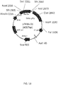
- a plasmid Vector 1 (4057bp, see FIG. 1A ) derived from a commercially available plasmid pUC119 (for example, available from Takara Bio Inc.) was treated with a restriction enzyme SfiI.
- the restriction enzyme site SfiI(a) consists of the gene sequence represented by GGCCCAGCCGGCC (SEQ ID NO: 55).
- the restriction enzyme site SfiI(b) consists of the gene sequence represented by GGCCTCTGCGGCC (SEQ ID NO: 56).
- FIG. 1B shows a detailed vector map of the plasmid Vector 1.
- the plasmid Vector 1 consists of the following gene sequence.
- VHH antibody gene fragments were obtained.
- the thus-treated plasmid Vector 1 was mixed with the VHH antibody gene fragments at a ratio of 1:2.
- An enzyme (available from Toyobo Co. Ltd., trade name: Ligation High ver. 2) was injected into the mixture solution.
- the mixture solution was left at rest at a temperature of 16 degrees Celsius for two hours. In this way, each of the VHH antibody gene fragments was ligated into the plasmid Vector 1.
- Coli bacteria available from Takara Bio Inc., trade name: HST02 were transfected with the thus-ligated plasmid Vector 1.
- the coli bacteria were incubated for fifteen hours on a 2YT plate culture medium containing ampicillin at a concentration of 100 micrograms/milliliter. In this way, obtained was a library of phages each of which displays a protein obtained from the gene fragment included in the gene library of the VHH antibody.
- the library of the phages had a concentration of 5E+7/milliliter.
- VHH antibodies capable of specifically binding to the noro antigen i.e., the recombinant of the p-domain protein of the GII/4 norovirus (NSW-2012), the recombinant being represented by SEQ ID NO: 48
- the noro antigen i.e., the recombinant of the p-domain protein of the GII/4 norovirus (NSW-2012), the recombinant being represented by SEQ ID NO: 48
- Coli bacteria (HST02) to which the VHH antibody gene fragment included in the gene library of the VHH antibody had been introduced were incubated at a temperature of 30 degrees Celsius in the 2YT AG culture medium containing 100 micrograms/milliliter of ampicillin and 1% glucose until a value OD 600 indicating absorbance reached 1.0.
- the 2YT AG culture medium had a volume of 100 milliliters. In this way, the coli bacteria were proliferated.
- Helper phages (available from Invitrogen company, trade name: M13KO7) were added to the coli bacteria culture medium in such a manner that the multiplicity of infection (hereinafter, referred to as "MOI") was approximately twenty.
- MOI multiplicity of infection
- the culture medium was warmed for about thirty minutes at a temperature of 37 degrees Celsius. Then, the culture medium was subjected to centrifugation at a rotation speed of 4000 rpm for ten minutes to collect the coli bacteria.
- the coli bacteria were incubated overnight at a temperature of 30 degrees Celsius in a 2YTAK culture medium containing 100 micrograms/milliliter of ampicillin and 50 micrograms/milliliter of kanamycin, while subjected to centrifugation at 213 rpm.
- the 2YTAK culture medium had a volume of 100 milliliters.
- the incubation liquid (100 milliliters) containing the thus-incubated coli bacteria was injected into two centrifugation tubes (volume: 50 milliliters, each).
- the two centrifugation tubes were subjected to centrifugation for ten minutes at a rotation speed of 4,000 rpm. Then, the supernatants (20 milliliters, each) were collected.
- the supernatants (40 milliliters) were added to a 20% polyethylene glycol solution (10 milliliters) containing NaCl (2.5 M). Then, the mixture solution was inverted and mixed. Subsequently, the mixture solution was cooled on ice for approximately one hour. The mixture was subjected to centrifugation for ten minutes at a rotation speed of 4,000 rpm. Then, the supernatant was removed. PBS containing 10% glycerol was injected toward the precipitate. Finally, the precipitate was loosened and dissolved. In this way, a library of phages each of which displays the VHH antibody was obtained.
- the noro antigen was mixed with PBS to prepare a norovirus solution.
- the concentration of norovirus was 2 micrograms/milliliter.
- the norovirus solution (2 milliliters) was injected into an immunotube (available from NUNC Co. Ltd.). The norovirus solution was left at rest overnight in the immunotube. In this way, norovirus was immobilized in the immunotube.
- the inside of the immunotube was filled with PBS which contained 3% skim milk (available from Wako Pure Chemical Industries, Ltd.). In this way, norovirus was blocked as an antigen in the immunotube.
- the immunotube was left at rest at room temperature for one hour. Subsequently, the inside of the immunotube was washed three times with PBS.
- the library of the phages each of which displays the VHH antibody (concentration: approximately 5E+11/milliliter) was mixed with 3 milliliters of PBS containing 3% skim milk to prepare a mixture solution.
- the mixture solution was injected into the immunotube in which the noro antigen was immobilized.
- the immunotube was provided with a lid formed of Parafilm. Then, the immunotube was rotated upside down in a rotator for ten minutes.
- the immunotube was left at rest at room temperature for one hour.
- PBST PBS containing 0. 05% Tween 20
- the inside of the immunotube was filled with PBST. Subsequently, the immunotube was left at rest for ten minutes. Then, the inside of the immunotube was washed ten times with PBST.
- the immunotube was provided with a lid formed of Parafilm. Then, the immunotube was rotated upside down in a rotator for ten minutes.
- the solution was moved to a tube containing 1 mL of 0.5 M Tris/HCl (pH: 6.8). Again, the extraction of the phage was repeated using a 100 mM trimethylamine solution (1 milliliter). In this way, 3 mL of an extraction liquid was obtained.
- the extraction liquid (1 mL) was mixed with 9 mL of coli bacteria HST02. The mixture solution was left at rest for one hour at a temperature of 30 degrees Celsius.
- Second panning was conducted identically to the procedure of the first panning. In other words, the panning was repeated. In this way, the monoclonal phages on which the VHH antibody was displayed were purified.
- a colony of the coli bacteria was picked up with a toothpick. The picked-up one colony was put onto one well of 96-flat-bottom plate. This was repeated. One well contained 200 microliters of a 2YTAG culture medium.
- the solutions contained in the wells were stirred at a rotation speed of 213 rpm at a temperature of 30 degrees Celsius.
- the solution (50 microliters) containing grown coli bacteria was collected.
- the collected solution was mixed with 50 microliters of a 2YTA culture medium contained in a plate.
- the 2YTA culture medium contained helper phages such that the multiplicity of infection (i.e., MOI) was set to be 20.
- the solution was left at rest at a temperature of 37 degrees Celsius for forty minutes.
- the plate containing the 2YTA culture medium was subjected to centrifugation at 1, 800 rpm for twenty minutes. The supernatant was removed. The precipitate contained the coli bacteria. The precipitate was mixed with 200 microliters of a 2YTAK culture medium. The mixture solution was left at rest overnight at a temperature of 30 degrees Celsius.
- the mixture solution was subjected to centrifugation at 1,800 rpm for twenty minutes.
- the supernatant containing the coli bacteria was collected.
- a solution containing the noro antigen i.e., the recombinant of the p-domain protein of the GII/4 norovirus (NSW-2012), the recombinant being represented by SEQ ID NO: 48
- SEQ ID NO: 48 a concentration of 2 micrograms/milliliter
- the volume of the solution containing the noro antigen in each well was 50 microliters.
- the 96-well plate was left at rest overnight at a temperature of 4 degrees Celsius. In this way, the noro antigen was immobilized in each well.
- each of the wells was washed with PBS three times. Then, PBS containing 3% skim milk (available from Wako Pure Chemical Industries, Ltd.) was injected into each well (200 microliters/well). The 96-well plate was left at rest at room temperature for one hour. In this way, the noro antigen was blocked in each well. Subsequently, each well was washed three times with PBS.
- the monoclonal phages each of which displays the VHH antibody were injected into each well (50 microliters/well). Then, the 96-well plate was left at rest for one hour. In this way, the phages reacted with the noro antigen.
- each well was washed three times with PBST. Then, an anti-M13 antibody (available from ABCAM company, trade name: ab50370, 10,000-fold dilution) was injected into each well (50 microliters/well). Then, each well was washed three times with PBST.
- an anti-M13 antibody available from ABCAM company, trade name: ab50370, 10,000-fold dilution
- a color-producing agent (available from Thermo Scientific, trade name: 1-step ultra TMB-ELISA) was injected into each well (50 microliters/well). The 96-well plate was left at rest for two minutes to cause the color-producing agent to react with the antibody.
- a sulfuric acid aqueous solution (normal, i.e., 1 N) was injected into each well at a concentration of 50 microliters/well to cease the reaction.
- the absorbance of the solution at a wavelength of 450 nanometers was measured.
- the proteins synthesized from the DNA sequences represented by SEQ ID NO: 58 - SEQ ID NO: 63 consist of the following amino acid sequences (SEQ ID NO: 66 - SEQ ID NO: 71), respectively.
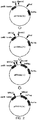
- a vector pET22b(+) was purchased from Merck Millipore Company. Using PrimeStar Mutagenesis Basal Kit (available from Takara Bio Inc.), a 3xFlag tag and two restriction enzyme sites SfiI(a) and SfiI(b) were added to the vector pET22b(+) by a PCR method. See FIG. 2 . The procedure shown in FIG. 2 will be described below in more detail.
- the restriction enzyme site SfiI(a) was add to the vector pET22b(+) by a PCR method using the following two primers and a restriction enzyme (available from Takara Bio Inc., trade name: PrimeSTAR Max DNA polymerase).
- 3xFlag tag DNA fragment having restriction enzyme sites BamhI and XhoI at 5' -terminal end and 3' -terminal end, respectively, was formed by a PCR method using the following two primers and restriction enzymes (available from Takara Bio Inc., trade name: PrimeSTAR Max DNA polymerase).
- This 3xFlag tag DNA fragment and the vector pET22b(+) were treated with two restriction enzymes BamhI and XhoI (available from Takara Bio Inc.)
- the 3xFlag tag DNA fragment was ligated into the vector pET22b(+) using Ligation Kit (available from Takara Bio Inc.). In this way, obtained was the vector pET22b(+) to which the 3xFlag tag and the restriction enzyme site SfiI(a) were added.
- this DNA fragment and the vector pET22b(+) were treated with two restriction enzymes Ncol and BamhI (available from Takara Bio Inc.)
- This DNA fragment was ligated into the vector pET22b(+) using Ligation Kit (available from Takara Bio Inc.). In this way, provided was the vector pET22b(+) to which the 3xFlag tag and the restriction enzyme sites SfiI(a) and SfiI(b) were added.
- the sequence of the vector pET22b (+) was analyzed by Greiner Company.
- a general T7 promotor primer set was used for the analysis of the sequence.
- Vectors pET22b(+) included in the liquid obtained by the PCR method were purified and collected in 50 microliters of diluted water using a DNA extraction kit (available from Promega KK). The thus-collected vectors pET22b (+) was treated with the SfiI restriction enzyme.
- the plasmid Vector 1 into which the VHH antibody gene fragment included in the gene library of the VHH antibody was ligated was treated with the SfiI restriction enzyme.
- SfiI restriction enzyme provided were the following six DNAs (SEQ ID NO: 79 - SEQ ID NO: 84) including the gene sequence coding for the amino acid sequences represented by SEQ ID NO: 38 - SEQ ID NO: 43.
- the ligation solution (2.5 microliters) and coli bacteria DH5 ⁇ (available from Nippon Gene, 25 microliters) were mixed on ice. The mixture solution was left at rest on the ice for six minutes. Then, the mixture solution was heated at a temperature of 42 degrees Celsius for forty five seconds. Finally, the mixture solution was left at rest on the ice for one minute. This procedure is known as a general heat shock method.
- the total amount of the mixture solution was distributed onto a LBA culture medium containing ampicillin at a concentration of 100 micrograms/milliliter.
- the LBA culture medium was left at rest overnight at a temperature of 37 degrees Celsius.
- the plasmids contained in the incubated coli bacteria were extracted from the LBA culture medium using a plasmid extraction kit (available from QIAGEN, trade name: QIAprepspin miniprep kit).
- a plasmid extraction kit available from QIAGEN, trade name: QIAprepspin miniprep kit.
- the sequence of the plasmid was analyzed by Greiner Company. For the analysis of the sequence, a general T7 promotor primer set was used.
- Coli bacteria (Competent Cell BL21 (DE3) pLysS, available from Life technologies Company) were transfected with the selected plasmids by a heat shock method.
- An SOC culture medium (50 microliters) was injected into the solution containing the transfected coli bacteria. Then, the coli bacteria were rescued at a temperature of 37 degrees Celsius for one hour, while shaken at 213 rpm.
- the collected coli bacteria solution was collected.
- the collected coli bacteria solution (5 milliliters) was distributed onto a LBA culture medium.
- the LBA culture medium was left at rest overnight at a temperature of 37 degrees Celsius.
- One colony was selected from among the colonies formed in the LBA culture medium.
- the selected colony was picked up with a toothpick.
- the picked-up colony was incubated in a LBA culture medium (3 milliliters) at a temperature of 37 degrees Celsius, while shaken at 213 rpm. In this way, a culture liquid was provided.
- the culture liquid 25 milliliters
- a LBA culture medium 500 milliliters
- IPTG solution an isopropylthiogalactoside solution
- the final concentration of the IPTG solution was 1 mM.
- the coli bacteria contained in the mixture solution were incubated at a temperature of 37 degrees Celsius for six hours. In order to collect the thus-incubated coli bacteria, the mixture solution was subjected to centrifugation at 6,000 rpm for ten minutes at a temperature of 4 degrees Celsius.
- the collected coli bacteria were mixed with ten times its volume of PBS.
- the mixture solution was stirred with a vortex mixer. In this way, the coli bacteria were washed. Then, the mixture solution was subjected to centrifugation at 6,000 rpm for ten minutes at a temperature of 4 degrees Celsius to collect coli bacteria.
- the collected coli bacteria were mixed again with ten times its volume of PBS.
- the coli bacteria contained in the mixture solution were disintegrated with an ultrasonic wave.
- the disintegration liquid containing coli bacteria was subjected to centrifugation at 10,000 rpm for fifteen minutes at a temperature of 4 degrees Celsius. The supernatant was collected. The collected supernatant was filtered through a 0.45-micrometer filter.
- the filtrate was purified with His-trap (available from GE Healthcare) in accordance with the recommended protocol.
- His-trap available from GE Healthcare
- an elution buffer having a total amount of 3 microliters was used for 1 milliliter of the filtrate.
- the buffer solution contained in the filtrate was substituted with PBS, using PD-10 (available from GE Healthcare).
- PBS having a total amount of 2.5 microliters was used for 1 milliliter of the filtrate. In this way, a solution containing the VHH antibody was provided.
- the VHH antibody contained in the thus-provided solution was quantified with an absorption spectrometer (available from Scrum Inc., trade name: nanodrop) on the basis of the absorption measurement value at a wavelength of 280 nanometers.
- the concentration of the VHH antibody was 4 milligrams/milliliter.
- the VHH antibody was evaluated as below using the noro antigen and a surface plasmon resonance evaluation device.
- the details of the surface plasmon resonance (hereinafter, referred to as "SPR") will be described below.
- the noro antigen was immobilized in accordance with the wizard included in the control software of the SPR evaluation device T200.
- the noro antigen was diluted with an acetic acid solution having a pH of 4.5 and was used at a concentration of 50 microgram/milliliter.
- the acetic acid solution had a concentration of 1 microgram/milliliter.
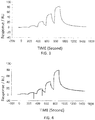
- the VHH antibodies including the amino acid sequences represented by SEQ ID NO: 38 - SEQ ID NO: 40 were used as analytes.
- the concentrations of the VHH antibodies contained in the running buffer were adjusted to 1.6 nM, 8 nM, 40 nM, 200 nM, and 1,000 nM. Then, the VHH antibodies were added serially.
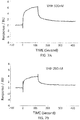
- FIG. 3 -FIG. 5 are graphs each showing evaluation result provided from the SPR evaluation device T200.
- the dissociation constant Kd was calculated using the evaluation software (available from GE Healthcare). As a result, the dissociation constants Kd were 1. 15 nM, 15. 8 nM, and 9.5 nM.
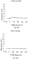
- the anti-noro antibodies including the amino acid sequence represented by SEQ ID NO: 41 - SEQ ID NO: 43 were used as analytes.
- the concentrations of the anti-noro antibodies contained in the running buffer were adjusted to 500nM, 250nM, 125nM, 62.5 nM, 31. 25nM, 15.63 nM, 7.81nM, and 3.91 nM.
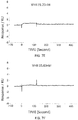
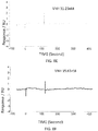
- FIG. 6A - FIG. 8H are graphs each showing evaluation result provided from the SPR evaluation device T200.
- the dissociation constant Kd was calculated using the evaluation software (available from GE Healthcare). As a result, the dissociation constants Kd were 81.6 nM, 63.8 nM, and 5.45 nM.
- the VHH antibodies (SEQ ID NO: 66 - SEQ ID NO: 71) were immobilized to evaluate the binding to the noro antigen.
- the VHH antibodies were immobilized in accordance with the wizard included in the control software of the SPR evaluation device T200.
- the VHH antibodies was diluted with an acetic acid solution having a pH of 5.5 and was used at a concentration of 50 microgram/milliliter.
- the acetic acid solution had a concentration of 1 microgram/milliliter.
- the noro antigen was used as an analyte.
- the concentrations of the noro antigen contained in the running buffers were adjusted to 10 nM, 31.
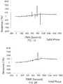
- FIG. 9 - FIG. 14 are graphs each showing evaluation result provided from the SPR evaluation device T200.
- the dissociation constant Kd was calculated using the evaluation software (available from GE Healthcare). As a result, the dissociation constants Kd were 4.15 nM, 15.9 nM, 9.57 nM, 4.98 nM, 13. 1 nM, and 10. 3 nM.
- restriction enzyme Sfil recognization portions were added at the 5'-end terminal of the base sequence represented by SEQ ID NO: 64 and at the 3' -end terminal of the base sequence represented by SEQ ID NO: 65.
- a DNA sequence in which the DNA sequence coding for the structural domain and the protein molecule were linked with a linker was provided through NheI digestion and ligation.
- the thus-provided DNA sequence was digested with SfiI, and then, incorporated into the plasmid vector shown in FIG. 15 .
- Coli bacteria were transfected with the thus-provided plasmid vector to produce an antibody bound to the protein.
- the antibody bound to the protein had the amino acid sequence represented by SEQ ID NO: 38 and the amino acid sequence represented by SEQ ID NO: 44.
- the amino acid sequence of the antibody bound to the protein is represented by SEQ ID NO: 100.
- the antibodies each bound to the protein were provided.
- the one is represented by SEQ ID NO: 101, whereas the other is represented by SEQ ID NO: 102.
- the protein molecule having the amino acid sequence represented by SEQ ID NO: 44 and the structural domain having the amino acid sequence represented by SEQ ID NO: 38 were connected with a linker.
- the protein molecule having the amino acid sequence represented by SEQ ID NO: 44 and the structural domain having the amino acid sequence represented by SEQ ID NO: 39 were connected with a linker.
- the antibody having the amino acid sequence represented by SEQ ID NO: 100 exhibited a higher binding ability to the norovirus than the VHH antibody having the amino acid sequence represented by SEQ ID NO: 66, which is a monomer antibody.
- the VHH antibody was evaluated as below using the noro antigen and the surface plasmon resonance evaluation device.
- the details of the surface plasmon resonance (namely, "SPR") will be described below.
- the VLP noro antigen was derived from the norovirus GII/4 composed by associating 90 dimers of VP1 each including the noro antigen (SEQ ID NO: 48).
- the VLP noro antigen was immobilized in accordance with the wizard included in the control software of the SPR evaluation device T200.
- the VLP noro antigen was diluted with an acetic acid solution having a pH of 4.0 and was used at a concentration of 25 microgram/milliliter.
- the acetic acid solution had a concentration of 1 microgram/milliliter.
- the antibody bound to the protein was used as an analytes.
- the antibody bound to the protein had the amino acid sequences represented by SEQ ID NO: 100.
- the concentration of the antibody bound to the protein contained in the running buffer was adjusted to 0. 16 nM, 0.8 nM, 4 nM, 20 nM, and 100 nM. Then, the antibody bound to the protein was added serially.
- FIG. 17 is a graph showing evaluation result provided from the SPR evaluation device T200.
- the dissociation constant Kd was calculated using the evaluation software (available from GE Healthcare).
- the antibody i.e., the antibody bound to the protein having the amino acid sequence represented by SEQ ID NO: 102 was also used as an analytes.
- the dissociation constant Kd was calculated similarly. The results thereof are shown in the following Table 1 and FIG. 17 .
- Table 1 data of the monomer antibodies represented by SEQ ID NO: 66 and SEQ ID NO: 67 are also shown.
- the monomer antibodies represented by SEQ ID NO: 66 and SEQ ID NO: 67 are included, as the structual domains thereof, in the antibodies (i.e., the antibodies each bound to the protein) having the amino acid sequences represented by SEQ ID NO: 100 and SEQ ID NO: 102, respectively.
- the antibody bound to the protein exhibited lower dissociation constant Kd than the monomer antibody. This means that the antibody bound to the protein has a stronger binding ability to the norovirus than the monomer antibody.
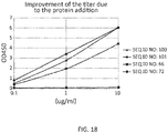
- the present inventors evaluated the binding ability of the monomer antibodies represented by SEQ ID NO: 66, SEQ ID NO: 72 and the antibodies (i.e., antibodies each bound to the protein) represented by SEQ ID NO: 100 and SEQ ID NO: 101 to the norovirus by an ELISA method in which GII/4 Frag 4 (2 micrograms / milliliter, in PBS) and skim milk (concentration: 3% in PBST) were used as a solid phase and a blocking solution, respectively.
- GII/4 Frag 4 (2 micrograms / milliliter, in PBS) and skim milk (concentration: 3% in PBST) were used as a solid phase and a blocking solution, respectively.
- FIG. 18 It was proved that the antibodies each bound to the protein had a higher binding ability to the norovirus than the monomer antibodies.
- the present invention provides an antibody capable of binding to norovirus, a composite, a detection device and a method using the same.
Landscapes
- Health & Medical Sciences (AREA)
- Life Sciences & Earth Sciences (AREA)
- Chemical & Material Sciences (AREA)
- Immunology (AREA)
- Engineering & Computer Science (AREA)
- General Health & Medical Sciences (AREA)
- Molecular Biology (AREA)
- Hematology (AREA)
- Biochemistry (AREA)
- Physics & Mathematics (AREA)
- Virology (AREA)
- Analytical Chemistry (AREA)
- Urology & Nephrology (AREA)
- Biomedical Technology (AREA)
- Medicinal Chemistry (AREA)
- General Physics & Mathematics (AREA)
- Pathology (AREA)
- Organic Chemistry (AREA)
- Food Science & Technology (AREA)
- Microbiology (AREA)
- Cell Biology (AREA)
- Biotechnology (AREA)
- Proteomics, Peptides & Aminoacids (AREA)
- Chemical Kinetics & Catalysis (AREA)
- Biophysics (AREA)
- Genetics & Genomics (AREA)
- Spectroscopy & Molecular Physics (AREA)
- Public Health (AREA)
- General Chemical & Material Sciences (AREA)
- Nuclear Medicine, Radiotherapy & Molecular Imaging (AREA)
- Veterinary Medicine (AREA)
- Animal Behavior & Ethology (AREA)
- Tropical Medicine & Parasitology (AREA)
- Oncology (AREA)
- Communicable Diseases (AREA)
- Pharmacology & Pharmacy (AREA)
- Clinical Laboratory Science (AREA)
- Dispersion Chemistry (AREA)
- Peptides Or Proteins (AREA)
- Apparatus Associated With Microorganisms And Enzymes (AREA)
Applications Claiming Priority (1)
| Application Number | Priority Date | Filing Date | Title |
|---|---|---|---|
| JP2018156399A JP7190675B2 (ja) | 2018-08-23 | 2018-08-23 | ノロウイルスに結合する抗体、複合体、それを用いた検出装置及び検出方法 |
Publications (1)
| Publication Number | Publication Date |
|---|---|
| EP3613466A1 true EP3613466A1 (fr) | 2020-02-26 |
Family
ID=67659648
Family Applications (1)
| Application Number | Title | Priority Date | Filing Date |
|---|---|---|---|
| EP19192787.0A Pending EP3613466A1 (fr) | 2018-08-23 | 2019-08-21 | Anticorps capable de se lier à un norovirus, à un composite, dispositif de détection et son procédé d'utilisation |
Country Status (4)
| Country | Link |
|---|---|
| US (1) | US11312759B2 (fr) |
| EP (1) | EP3613466A1 (fr) |
| JP (1) | JP7190675B2 (fr) |
| CN (1) | CN110857320A (fr) |
Families Citing this family (2)
| Publication number | Priority date | Publication date | Assignee | Title |
|---|---|---|---|---|
| JP7190674B2 (ja) * | 2018-08-23 | 2022-12-16 | パナソニックIpマネジメント株式会社 | ノロウイルスに結合する抗体、複合体、それを用いた検出装置及び検出方法 |
| JPWO2022244862A1 (fr) * | 2021-05-20 | 2022-11-24 |
Citations (4)
| Publication number | Priority date | Publication date | Assignee | Title |
|---|---|---|---|---|
| WO2014183052A1 (fr) * | 2013-05-09 | 2014-11-13 | The United States Of America, As Represented By The Secretary, Depart Of Health And Human Services | Anticorps vhh à domaine unique dirigé contre les norovirus gi.1 et gii.4 et leur utilisation |
| US9193780B2 (en) | 2008-06-05 | 2015-11-24 | Ablynx N.V. | Amino acid sequences directed against envelope proteins of a virus and polypeptides comprising the same for the treatment of viral diseases |
| WO2016059113A1 (fr) * | 2014-10-14 | 2016-04-21 | Deutsches Krebsforschungszentrum | Anticorps de norovirus |
| US20190002535A1 (en) * | 2017-06-30 | 2019-01-03 | Panasonic Intellectual Property Management Co., Ltd. | Antibody capable of binding to norovirus, composite, detection device and method using the same |
Family Cites Families (5)
| Publication number | Priority date | Publication date | Assignee | Title |
|---|---|---|---|---|
| WO2004041863A2 (fr) | 2002-11-08 | 2004-05-21 | Ablynx N.V. | Anticorps a domaine unique diriges contre un interferon gamma et leurs utilisations |
| WO2016156468A1 (fr) * | 2015-03-31 | 2016-10-06 | Vhsquared Limited | Polypeptides |
| JP6868763B2 (ja) | 2016-04-01 | 2021-05-12 | パナソニックIpマネジメント株式会社 | インフルエンザウィルスに結合する抗体 |
| JP7190674B2 (ja) * | 2018-08-23 | 2022-12-16 | パナソニックIpマネジメント株式会社 | ノロウイルスに結合する抗体、複合体、それを用いた検出装置及び検出方法 |
| CN114736292B (zh) * | 2022-06-10 | 2023-03-07 | 广州明药科技有限公司 | 靶向诺如病毒蛋白的纳米抗体及其应用 |
-
2018
- 2018-08-23 JP JP2018156399A patent/JP7190675B2/ja active Active
-
2019
- 2019-07-17 US US16/513,752 patent/US11312759B2/en active Active
- 2019-08-20 CN CN201910768014.0A patent/CN110857320A/zh active Pending
- 2019-08-21 EP EP19192787.0A patent/EP3613466A1/fr active Pending
Patent Citations (4)
| Publication number | Priority date | Publication date | Assignee | Title |
|---|---|---|---|---|
| US9193780B2 (en) | 2008-06-05 | 2015-11-24 | Ablynx N.V. | Amino acid sequences directed against envelope proteins of a virus and polypeptides comprising the same for the treatment of viral diseases |
| WO2014183052A1 (fr) * | 2013-05-09 | 2014-11-13 | The United States Of America, As Represented By The Secretary, Depart Of Health And Human Services | Anticorps vhh à domaine unique dirigé contre les norovirus gi.1 et gii.4 et leur utilisation |
| WO2016059113A1 (fr) * | 2014-10-14 | 2016-04-21 | Deutsches Krebsforschungszentrum | Anticorps de norovirus |
| US20190002535A1 (en) * | 2017-06-30 | 2019-01-03 | Panasonic Intellectual Property Management Co., Ltd. | Antibody capable of binding to norovirus, composite, detection device and method using the same |
Non-Patent Citations (4)
| Title |
|---|
| ALVARADO GABRIELA ET AL: "Human Monoclonal Antibodies That Neutralize Pandemic GII.4 Noroviruses", GASTROENTEROLOGY : OFFICIAL PUBLICATION OF THE AMERICAN GASTROENTEROLOGICAL ASSOCIATION, WILLIAMS & WILKINS, US, vol. 155, no. 6, 28 August 2018 (2018-08-28), pages 1898 - 1907, XP085546397, ISSN: 0016-5085, DOI: 10.1053/J.GASTRO.2018.08.039 * |
| ANNA D. KOROMYSLOVA ET AL: "Nanobody binding to a conserved epitope promotes norovirus particle disassembly", JOURNAL OF VIROLOGY., vol. 89, no. 5, 17 December 2014 (2014-12-17), US, pages 2718 - 2730, XP055438443, ISSN: 0022-538X, DOI: 10.1128/JVI.03176-14 * |
| BIOMED ENVIRON SCI., vol. 27, no. 2, 2012, pages 118 - 121 |
| LORENA GARAICOECHEA ET AL: "Llama Nanoantibodies with Therapeutic Potential against Human Norovirus Diarrhea", PLOS ONE, vol. 10, no. 8, 12 August 2015 (2015-08-12), pages 1 - 33, XP055612756, DOI: 10.1371/journal.pone.0133665 * |
Also Published As
| Publication number | Publication date |
|---|---|
| CN110857320A (zh) | 2020-03-03 |
| JP7190675B2 (ja) | 2022-12-16 |
| JP2020029427A (ja) | 2020-02-27 |
| US20200062829A1 (en) | 2020-02-27 |
| US11312759B2 (en) | 2022-04-26 |
Similar Documents
| Publication | Publication Date | Title |
|---|---|---|
| US10544206B2 (en) | Antibody capable of binding to norovirus, composite, detection device and method using the same | |
| CN1711476B (zh) | 小分析物的非竞争性免疫测定 | |
| CN113999308B (zh) | 靶向钙粘蛋白17的纳米抗体及其应用 | |
| CN111303279A (zh) | 一种针对新型冠状病毒的单域抗体及其应用 | |
| US20170037112A1 (en) | Antibody capable of binding to influenza virus | |
| JP2011524013A (ja) | 大麻の使用の検出 | |
| JP2018058812A (ja) | インフルエンザウィルス核内蛋白質に結合する抗体、複合体、それを用いた検出装置及び検出方法 | |
| JP6931765B2 (ja) | インフルエンザウィルス核内蛋白質に結合する抗体、複合体、それを用いた検出装置及び検出方法 | |
| US10280212B2 (en) | Composite comprising antibody capable of binding to intranuclear protein of influenza virus | |
| CN118638214A (zh) | 一种aav9特异性的纳米抗体及其应用 | |
| EP3613466A1 (fr) | Anticorps capable de se lier à un norovirus, à un composite, dispositif de détection et son procédé d'utilisation | |
| US10273289B2 (en) | Composite comprising antibody capable of binding to intranuclear protein of influenza virus | |
| US20190077836A1 (en) | Antibody capable of binding to intranuclear protein of influenza virus, composite, detection device and method using same | |
| US10906961B2 (en) | Antibody capable of binding to norovirus, composite, detection device and method using the same | |
| JP7178608B2 (ja) | インフルエンザウイルス核内蛋白質に結合する抗体、複合体、それを用いた検出装置及び検出方法 | |
| JP4566911B2 (ja) | コバラミン分析方法 | |
| JP2017186298A (ja) | インフルエンザウィルスに結合する抗体 | |
| CN120623329A (zh) | 抗埃博拉病毒vp40蛋白的纳米抗体3e07及在病毒检测中的应用 | |
| US11214611B2 (en) | Antibody capable of binding to influenza virus intranuclear protein, complex, detection apparatus and detection method using same | |
| JP2022142155A (ja) | 抗体、複合体、それを用いた検出装置及び検出方法 | |
| CN115594757B (zh) | 抗新型冠状病毒的中和抗体及其制备方法、制剂和用途 | |
| JP7308477B2 (ja) | 抗体、複合体、それを用いた検出装置及び検出方法 | |
| JP2020148511A (ja) | 検出方法 |
Legal Events
| Date | Code | Title | Description |
|---|---|---|---|
| PUAI | Public reference made under article 153(3) epc to a published international application that has entered the european phase |
Free format text: ORIGINAL CODE: 0009012 |
|
| STAA | Information on the status of an ep patent application or granted ep patent |
Free format text: STATUS: THE APPLICATION HAS BEEN PUBLISHED |
|
| AK | Designated contracting states |
Kind code of ref document: A1 Designated state(s): AL AT BE BG CH CY CZ DE DK EE ES FI FR GB GR HR HU IE IS IT LI LT LU LV MC MK MT NL NO PL PT RO RS SE SI SK SM TR |
|
| AX | Request for extension of the european patent |
Extension state: BA ME |
|
| STAA | Information on the status of an ep patent application or granted ep patent |
Free format text: STATUS: REQUEST FOR EXAMINATION WAS MADE |
|
| 17P | Request for examination filed |
Effective date: 20200819 |
|
| RBV | Designated contracting states (corrected) |
Designated state(s): AL AT BE BG CH CY CZ DE DK EE ES FI FR GB GR HR HU IE IS IT LI LT LU LV MC MK MT NL NO PL PT RO RS SE SI SK SM TR |
|
| STAA | Information on the status of an ep patent application or granted ep patent |
Free format text: STATUS: EXAMINATION IS IN PROGRESS |
|
| 17Q | First examination report despatched |
Effective date: 20230531 |