EP3614953B1 - Matériaux et procédés de protection contre les névromes - Google Patents
Matériaux et procédés de protection contre les névromes Download PDFInfo
- Publication number
- EP3614953B1 EP3614953B1 EP18791114.4A EP18791114A EP3614953B1 EP 3614953 B1 EP3614953 B1 EP 3614953B1 EP 18791114 A EP18791114 A EP 18791114A EP 3614953 B1 EP3614953 B1 EP 3614953B1
- Authority
- EP
- European Patent Office
- Prior art keywords
- neuroma
- cap
- biomaterial
- nerve
- external body
- Prior art date
- Legal status (The legal status is an assumption and is not a legal conclusion. Google has not performed a legal analysis and makes no representation as to the accuracy of the status listed.)
- Active
Links
Images




Classifications
-
- A—HUMAN NECESSITIES
- A61—MEDICAL OR VETERINARY SCIENCE; HYGIENE
- A61B—DIAGNOSIS; SURGERY; IDENTIFICATION
- A61B90/00—Instruments, implements or accessories specially adapted for surgery or diagnosis and not covered by any of the groups A61B1/00 - A61B50/00, e.g. for luxation treatment or for protecting wound edges
- A61B90/08—Accessories or related features not otherwise provided for
-
- A—HUMAN NECESSITIES
- A61—MEDICAL OR VETERINARY SCIENCE; HYGIENE
- A61B—DIAGNOSIS; SURGERY; IDENTIFICATION
- A61B90/00—Instruments, implements or accessories specially adapted for surgery or diagnosis and not covered by any of the groups A61B1/00 - A61B50/00, e.g. for luxation treatment or for protecting wound edges
-
- A—HUMAN NECESSITIES
- A61—MEDICAL OR VETERINARY SCIENCE; HYGIENE
- A61L—METHODS OR APPARATUS FOR STERILISING MATERIALS OR OBJECTS IN GENERAL; DISINFECTION, STERILISATION OR DEODORISATION OF AIR; CHEMICAL ASPECTS OF BANDAGES, DRESSINGS, ABSORBENT PADS OR SURGICAL ARTICLES; MATERIALS FOR BANDAGES, DRESSINGS, ABSORBENT PADS OR SURGICAL ARTICLES
- A61L27/00—Materials for grafts or prostheses or for coating grafts or prostheses
- A61L27/14—Macromolecular materials
- A61L27/16—Macromolecular materials obtained by reactions only involving carbon-to-carbon unsaturated bonds
-
- A—HUMAN NECESSITIES
- A61—MEDICAL OR VETERINARY SCIENCE; HYGIENE
- A61L—METHODS OR APPARATUS FOR STERILISING MATERIALS OR OBJECTS IN GENERAL; DISINFECTION, STERILISATION OR DEODORISATION OF AIR; CHEMICAL ASPECTS OF BANDAGES, DRESSINGS, ABSORBENT PADS OR SURGICAL ARTICLES; MATERIALS FOR BANDAGES, DRESSINGS, ABSORBENT PADS OR SURGICAL ARTICLES
- A61L27/00—Materials for grafts or prostheses or for coating grafts or prostheses
- A61L27/14—Macromolecular materials
- A61L27/18—Macromolecular materials obtained otherwise than by reactions only involving carbon-to-carbon unsaturated bonds
-
- A—HUMAN NECESSITIES
- A61—MEDICAL OR VETERINARY SCIENCE; HYGIENE
- A61L—METHODS OR APPARATUS FOR STERILISING MATERIALS OR OBJECTS IN GENERAL; DISINFECTION, STERILISATION OR DEODORISATION OF AIR; CHEMICAL ASPECTS OF BANDAGES, DRESSINGS, ABSORBENT PADS OR SURGICAL ARTICLES; MATERIALS FOR BANDAGES, DRESSINGS, ABSORBENT PADS OR SURGICAL ARTICLES
- A61L27/00—Materials for grafts or prostheses or for coating grafts or prostheses
- A61L27/36—Materials for grafts or prostheses or for coating grafts or prostheses containing ingredients of undetermined constitution or reaction products thereof, e.g. transplant tissue, natural bone, extracellular matrix
- A61L27/3604—Materials for grafts or prostheses or for coating grafts or prostheses containing ingredients of undetermined constitution or reaction products thereof, e.g. transplant tissue, natural bone, extracellular matrix characterised by the human or animal origin of the biological material, e.g. hair, fascia, fish scales, silk, shellac, pericardium, pleura, renal tissue, amniotic membrane, parenchymal tissue, fetal tissue, muscle tissue, fat tissue, enamel
-
- A—HUMAN NECESSITIES
- A61—MEDICAL OR VETERINARY SCIENCE; HYGIENE
- A61L—METHODS OR APPARATUS FOR STERILISING MATERIALS OR OBJECTS IN GENERAL; DISINFECTION, STERILISATION OR DEODORISATION OF AIR; CHEMICAL ASPECTS OF BANDAGES, DRESSINGS, ABSORBENT PADS OR SURGICAL ARTICLES; MATERIALS FOR BANDAGES, DRESSINGS, ABSORBENT PADS OR SURGICAL ARTICLES
- A61L27/00—Materials for grafts or prostheses or for coating grafts or prostheses
- A61L27/36—Materials for grafts or prostheses or for coating grafts or prostheses containing ingredients of undetermined constitution or reaction products thereof, e.g. transplant tissue, natural bone, extracellular matrix
- A61L27/3604—Materials for grafts or prostheses or for coating grafts or prostheses containing ingredients of undetermined constitution or reaction products thereof, e.g. transplant tissue, natural bone, extracellular matrix characterised by the human or animal origin of the biological material, e.g. hair, fascia, fish scales, silk, shellac, pericardium, pleura, renal tissue, amniotic membrane, parenchymal tissue, fetal tissue, muscle tissue, fat tissue, enamel
- A61L27/362—Skin, e.g. dermal papillae
-
- A—HUMAN NECESSITIES
- A61—MEDICAL OR VETERINARY SCIENCE; HYGIENE
- A61L—METHODS OR APPARATUS FOR STERILISING MATERIALS OR OBJECTS IN GENERAL; DISINFECTION, STERILISATION OR DEODORISATION OF AIR; CHEMICAL ASPECTS OF BANDAGES, DRESSINGS, ABSORBENT PADS OR SURGICAL ARTICLES; MATERIALS FOR BANDAGES, DRESSINGS, ABSORBENT PADS OR SURGICAL ARTICLES
- A61L27/00—Materials for grafts or prostheses or for coating grafts or prostheses
- A61L27/36—Materials for grafts or prostheses or for coating grafts or prostheses containing ingredients of undetermined constitution or reaction products thereof, e.g. transplant tissue, natural bone, extracellular matrix
- A61L27/3604—Materials for grafts or prostheses or for coating grafts or prostheses containing ingredients of undetermined constitution or reaction products thereof, e.g. transplant tissue, natural bone, extracellular matrix characterised by the human or animal origin of the biological material, e.g. hair, fascia, fish scales, silk, shellac, pericardium, pleura, renal tissue, amniotic membrane, parenchymal tissue, fetal tissue, muscle tissue, fat tissue, enamel
- A61L27/3629—Intestinal tissue, e.g. small intestinal submucosa
-
- A—HUMAN NECESSITIES
- A61—MEDICAL OR VETERINARY SCIENCE; HYGIENE
- A61L—METHODS OR APPARATUS FOR STERILISING MATERIALS OR OBJECTS IN GENERAL; DISINFECTION, STERILISATION OR DEODORISATION OF AIR; CHEMICAL ASPECTS OF BANDAGES, DRESSINGS, ABSORBENT PADS OR SURGICAL ARTICLES; MATERIALS FOR BANDAGES, DRESSINGS, ABSORBENT PADS OR SURGICAL ARTICLES
- A61L27/00—Materials for grafts or prostheses or for coating grafts or prostheses
- A61L27/36—Materials for grafts or prostheses or for coating grafts or prostheses containing ingredients of undetermined constitution or reaction products thereof, e.g. transplant tissue, natural bone, extracellular matrix
- A61L27/3641—Materials for grafts or prostheses or for coating grafts or prostheses containing ingredients of undetermined constitution or reaction products thereof, e.g. transplant tissue, natural bone, extracellular matrix characterised by the site of application in the body
- A61L27/3675—Nerve tissue, e.g. brain, spinal cord, nerves, dura mater
-
- A—HUMAN NECESSITIES
- A61—MEDICAL OR VETERINARY SCIENCE; HYGIENE
- A61L—METHODS OR APPARATUS FOR STERILISING MATERIALS OR OBJECTS IN GENERAL; DISINFECTION, STERILISATION OR DEODORISATION OF AIR; CHEMICAL ASPECTS OF BANDAGES, DRESSINGS, ABSORBENT PADS OR SURGICAL ARTICLES; MATERIALS FOR BANDAGES, DRESSINGS, ABSORBENT PADS OR SURGICAL ARTICLES
- A61L27/00—Materials for grafts or prostheses or for coating grafts or prostheses
- A61L27/50—Materials characterised by their function or physical properties, e.g. injectable or lubricating compositions, shape-memory materials, surface modified materials
- A61L27/58—Materials at least partially resorbable by the body
-
- C—CHEMISTRY; METALLURGY
- C08—ORGANIC MACROMOLECULAR COMPOUNDS; THEIR PREPARATION OR CHEMICAL WORKING-UP; COMPOSITIONS BASED THEREON
- C08L—COMPOSITIONS OF MACROMOLECULAR COMPOUNDS
- C08L23/00—Compositions of homopolymers or copolymers of unsaturated aliphatic hydrocarbons having only one carbon-to-carbon double bond; Compositions of derivatives of such polymers
- C08L23/02—Compositions of homopolymers or copolymers of unsaturated aliphatic hydrocarbons having only one carbon-to-carbon double bond; Compositions of derivatives of such polymers not modified by chemical after-treatment
- C08L23/04—Homopolymers or copolymers of ethene
- C08L23/06—Polyethylene
-
- C—CHEMISTRY; METALLURGY
- C08—ORGANIC MACROMOLECULAR COMPOUNDS; THEIR PREPARATION OR CHEMICAL WORKING-UP; COMPOSITIONS BASED THEREON
- C08L—COMPOSITIONS OF MACROMOLECULAR COMPOUNDS
- C08L71/00—Compositions of polyethers obtained by reactions forming an ether link in the main chain; Compositions of derivatives of such polymers
- C08L71/02—Polyalkylene oxides
-
- A—HUMAN NECESSITIES
- A61—MEDICAL OR VETERINARY SCIENCE; HYGIENE
- A61B—DIAGNOSIS; SURGERY; IDENTIFICATION
- A61B90/00—Instruments, implements or accessories specially adapted for surgery or diagnosis and not covered by any of the groups A61B1/00 - A61B50/00, e.g. for luxation treatment or for protecting wound edges
- A61B90/08—Accessories or related features not otherwise provided for
- A61B2090/0815—Implantable devices for insertion in between organs or other soft tissues
-
- A—HUMAN NECESSITIES
- A61—MEDICAL OR VETERINARY SCIENCE; HYGIENE
- A61L—METHODS OR APPARATUS FOR STERILISING MATERIALS OR OBJECTS IN GENERAL; DISINFECTION, STERILISATION OR DEODORISATION OF AIR; CHEMICAL ASPECTS OF BANDAGES, DRESSINGS, ABSORBENT PADS OR SURGICAL ARTICLES; MATERIALS FOR BANDAGES, DRESSINGS, ABSORBENT PADS OR SURGICAL ARTICLES
- A61L2430/00—Materials or treatment for tissue regeneration
- A61L2430/02—Materials or treatment for tissue regeneration for reconstruction of bones; weight-bearing implants
Definitions
- Neuromas develop as a part of a normal reparative process following peripheral nerve injury. They are formed when nerve recovery towards the distal nerve end or target organ fails and nerve fibers improperly and irregularly regenerate into the surrounding scar tissue. Neuromas consist of a deranged architecture of tangled axons, Schwann cells, endoneurial cells, and perineurial cells in a dense collagenous matrix with surrounding fibroblasts ( Mackinnon SE et al. 1985. Alteration of neuroma formation by manipulation of its microenvironment. Plast Reconstr Surg. 76:345-53 ). The up-regulation of certain channels and receptors during neuroma development can also cause abnormal sensitivity and spontaneous activity of injured axons ( Curtin C and Carroll I. 2009.
- Haphazardly arranged nerve fibers are known to produce abnormal activity that stimulates central neurons ( Wall PD and Gutnick M. 1974. Ongoing activity in peripheral nerves; physiology and pharmacology of impulses originating from neuroma. Exp Neurol. 43:580-593 ).
- This ongoing abnormal activity can be enhanced by mechanical stimulation, for example, from the constantly rebuilding scar at the injury site ( Nordin M et al. 1984. Ectopic sensory discharges and paresthesiae in patients with disorders of peripheral nerves, dorsal roots and dorsal columns. Pain. 20:231-245 ; Scadding JW. 1981. Development of ongoing activity, mechanosensitivity, and adrenaline sensitivity in severed peripheral nerve axons. Exp Neurol. 73:345-364 ).
- Neuromas of the nerve stump are unavoidable consequences of nerve injury when the nerve is not, or cannot be, repaired and can result in debilitating pain. It has been estimated that approximately 30% of neuromas become painful and problematic. This is particularly likely if the neuroma is present at or near the skin surface as physical stimulation induces signaling in the nerve resulting in a sensation of pain.
- Neuroma prevention and attenuation strategies have used various methods to limit neuroma size and protect the neuroma from external stimuli.
- Current prevention methods attempt to limit the size of the neuroma and so reduce or limit potential contact and "cross-talk" between axons within the injured nerve site and within the disorganized structure that characterizes neuromas. Due to a variety of factors, current methods of neuroma mitigation/prevention have an unacceptable level of efficacy.
- Another method is to cut the nerve stump back to leave a segment or sleeve of overhanging epineurium.
- This overhang can be ligated to cover the face of the nerve stump.
- a segment of epineurium can be acquired from other nerve tissue or a corresponding nerve stump can be cut back to create an epineurium sleeve that can be used to connect with and cover the other nerve stump.
- a suture ligation where a loop of suture is placed around the end of the nerve and tightened. This pressure is believed to mechanically block the exit of axons and causes the terminal end to eventually form scar tissue over the site. Clinical and pre-clinical evidence has shown, however, that this procedure can cause a painful neuroma to form behind the ligation. Furthermore, the ligated nerve is generally not positioned to minimize mechanical stimulation of the neuroma, since it is anticipated that the scar tissue will provide sufficient protection to the nerve end..
- US 2014/094932 A1 describes, among other things, materials and methods for protecting against neuromas.
- the subject invention provides devices for alleviating discomfort associated with neuroma formation as defined by the claims. Specifically, the subject invention provides biomedical devices for neuroma size limitation and neuroma protection and isolation.
- the subject device is a cylindrical cap with a single open end to be applied to nerves within a subject's body, wherein the internal chamber of the cylindrical cap has separators or dividers that can physically partition the nerve structures into distinct, and in some embodiments, separate channels to enable a more regular arrangement, rather than the haphazard arrangement often produced in neuromas.
- the device is sterile and has a single open end, such that the nerve end can be inserted into the internal chamber through the open end. There is a hollow indentation or retaining cavity between the open end and the separators or dividers in which the terminal end of the nerve can be situated and secured. As the nerve grows, the axons and other structures will extend and grow into the separate channels to limit contact between them.
- the partitioning of the internal chamber of the cap is in the form of a spiral-shaped wall that forms a longitudinal spiralled channel with one edge facing the open end. As nerve tissue grows, it can fill in the space between the spirals, so that the outer nerve tissue is substantially separated, subdivided, or partially subdivided from inner nerve tissue.
- the internal chamber is partitioned with at least one panel, directed longitudinally with the internal chamber, such that a portion of the internal chamber, opposite to the single open end, is divided into two or more distinct and separate channels.
- the division of the neuroma tissue into separate or subdivided channels, to form smaller, disconnected, neuroma masses, can mitigate the cascade effect caused when one area of the neuroma is stimulated and the effect is felt through the entire tissue mass.
- the dimensions of the cylindrical cap can be about 1 mm to about 25 mm in diameter and about 1 mm to about 100 mm in length.
- the material of the cap is a biomaterial that can remodel to generate a volume of protective connective tissue around a neuroma (rather than the current use of a biomaterial to contain a neuroma volumetrically).
- the material of the cap is a membrane biomaterial, such as, for example, small intestine submucosa (SIS), amnion, dermis, or decellularized fascia.
- a terminal nerve end is one that has been cut or transected leaving exposed axons and other internal nerve structures.
- either the outside of the cap and/or the epineurium around the nerve are held with a forceps so the nerve can be moved through the open end. Excessive pressure applied to the nerve can damage internal axons and structures. Likewise, excess force applied around the cap can distort the shape or the open end, inhibiting insertion of the nerve end.
- An open end in the cap that is too large can allow the neuroma to escape and grow outside the cap. If the open end is made smaller, the nerve can be damaged if forced into the open end.
- a cap can have one or more tabs arranged on and around the external surface 16 of the cap.
- a tab can be arranged at or near the open end. The tabs can be used to grasp the cap and manipulate it around the terminal nerve end. Grasping the tabs rather than the exterior surface can help to maintain the shape of the cap, in particular the open end.
- the subject invention provides devices for alleviating discomfort associated with neuromas as defined by the claims. More specifically, the subject invention pertains to devices for limiting neuroma size and physically surrounding the neuroma to inhibit stimulation that elicits neuropathic pain.
- the subject invention is directed to a tissue-engineered scaffold that provides: a barrier that limits the size of a neuroma, dividers that subdivide the neuroma volume or mass into smaller, disconnected neuromas masses to reduce axonal cross-talk or the "cascade effect," and mechanical isolation of the neuroma to inhibit stimulation.
- a device of the subject invention is designed to form a protective, connective tissue covering or cap surrounding the terminal nerve end 29, thereby surrounding any resulting neuroma formation.
- sub-dividing the neuroma volume as it forms can limit the amount of interaction between axons, thereby limiting signalling cascades being triggered inside the neuroma that can be interpreted as pain.
- the subject device limits stimulation of the neuroma.
- the ability of the material of the subject device to remodel into a native tissue cushion is another advantageous feature when combined with the physical partitioning of the neuroma.
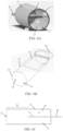
- FIG. 1A-1C Current methods for limiting the size and stimulation of a neuroma are shown in Figures 1A-1C .
- the methods include dissecting or cutting the terminal end to form a flap of epineurium that can be folded over the terminal nerve end 29, as shown in the first column of Figures 1A-1C .
- the nerve can also be dissected or cut back to form a sleeve of epineurium that can be ligated over the terminal nerve end, as seen in the middle column in Figures 1A-1C .
- a patch epineurium can be obtained from another location and attached to the terminal nerve end, as shown in the last column in Figures 1A-1C .
- the subject invention provides the benefits of an epineural flap (or ligation) to limit neuroma size and the advantages of isolating a nerve end by emplacement in another tissue combined in a single device.
- This device isolates and protects the neuroma, thereby inhibiting painful sensations without the need for any repositioning of the nerve.
- the device can be secured to the stump of the nerve prior to formation of a neuroma and as the neuroma forms the material of the device can remodel into a tissue cushion 55 and become integrated into the surrounding host tissues, which effectively isolates 30 the neuroma from undesirable contact and stimulation.
- Figures 2A-8B illustrate embodiments of the invention.
- the cap 10 of the subject invention has an external body 15 that comprises a proximal end 20 and a distal end 25.
- one embodiment of the external body 15 of the cap 10 is cylindrical in shape.
- the shape of the external body 15 of the cap 10 can vary depending upon the type, diameter and location of a nerve stump, as well as other factors known to those with skill in the art.
- the circumferential shape of the external body can be variable and be, by way of non-limiting examples, an ovoid, a circle, a square, a rectangle, a triangle, or any other polygonal shape.
- the circumferential shape of the external body is generally a circle.
- the dimensions of the external body can vary depending on the type, diameter and location of a nerve stump on which it will be used, as well as other factors known to those skilled in the art.
- the external body of the device has dimensions of approximately 1mm to approximately 100 mm in diameter and is approximately 1 mm to approximately 500 mm in length, between the proximal end 20 and the distal end 25.
- the external body of the device will be less than 100 mm in diameter and less than 500 mm in length.
- the diameter of the external body is between approximately 1 mm and approximately 25 mm and the length of the external body, between the proximal end and the distal end of the cap, is between approximately 1 mm and approximately 100 mm.
- the diameter of the distal end 25 of the external body 15 can be narrower than the diameter of the proximal end 20, as seen, for example, in Figures 2 and 3 . This can give the cap a bullet or conical shape.
- the distal end 25 of the external body is cone-shaped.
- the diameter of the distal end and the proximal end are the same or substantially the same, such that the distal end ofthe cap is flat, giving the cap a cylinder or barrel shape.
- the external body 15 of the cap 10 has no openings other than the open end 18 at the proximal end 20 of the cap through which a nerve can be inserted or placed.
- the proximal end 20 is open and the distal end 25 of the external body 15 is closed off entirely so that the external body forms a cap 10.
- Figure 3 also illustrates an embodiment in which an internal chamber 35 is provided in the external body 15.
- the interior surface 22 of the closed off 30 portion of external body 15 can be in the form of a bevel 40, as shown in Figure 3A .
- a closed off portion can be substantially flat, as shown in Figures 6C and 7A .
- the closed off portion or closed end 30 can be conical, such as shown, for example, in Figures 2A and 6E .
- An external body without excess openings can inhibit axon growth from extending outside the cap.
- the open end 18 of the cap can provide the only access into the internal chamber. Fitting a terminal nerve end 29 through the open end of a cap can be challenging and can potentially damage the nerve. If a cap with a larger diameter is selected, it can be easier to insert the terminal nerve end, but may result is excess space around the nerve perimeter. If not properly closed off, the openings around the nerve can allow axons to escape.
- flaps 90 around the open end 18 can provide a temporary enlargement of the open end, making nerve placement into the internal chamber easier, while the flap can be wrapped around and sutured to provide for a close fit around the terminal nerve end.
- the enlargement of the open end allows a terminal nerve end to be moved through a slot 92 formed in the open end.
- Figures 8A and 8E illustrate non-limiting examples of a cap having an open end 18 with one or more flaps.
- the slot provided by one or more flaps is large enough to allow a terminal nerve end to pass through the slot, with minimal compression of the nerve tissue, so as to minimize the number of sutures necessary to close the flap and the slot.
- the single flap can provide a slot of between approximately 25% to approximately 75% of the diameter of an opening 18.
- the dual flaps can open in opposite directions.
- the dual flaps can provide a slot of between approximately 25% to approximately 75% of the diameter of an opening 18.
- a slit 98 in the proximal end 20 of the cap that is integral with opening 18 and that advances towards the distal end 25 to form a flap with three corners part-way along the external body 15 of the cap.
- Figure 8E illustrates a non-limiting example of a three-corner flap.
- the nerve tissue in a neuroma mass 13 grows in a random, haphazard fashion and the axons in one section of a neuroma can be in contact with other surrounding axons.
- stimulation of a neuroma in one area can trigger a cascade effect wherein the signal received by axons in one area, because of contact, will trigger signals in nearby axons.
- Each step in the cascade can amplify the initial signal until finally most or all of the axons in the neuroma are "fired.”
- This massive signal cascade can also amplify pain.
- this cascade effect can be minimized, thereby limiting the amount of pain caused by stimulation of the neuroma.
- the internal chamber 35 can be configured with one or more dividers 70 that subdivide at least a portion of the internal chamber into two or more separate channels 36 into which the neuroma mass 13 can grow and become portioned into smaller, disconnected neuroma masses. This separation of the neuroma mass into the different channels creates a physical barrier between the axons of the smaller, disconnected portions in the different separate channels, thereby limiting the scope of a cascade effect.
- the dividers 70 used in an internal chamber can be configured in a myriad of ways to create different sizes and shapes of separate chambers. Likewise, the dividers can be individual or unconnected or they can be joined together. Variations in the configurations of the dividers that separate a neuroma mass into smaller, disconnected masses 17 or portions with different channels are within the scope of this invention.
- a nerve end can be positioned within the cap 10 so that the neuronal growth is directed towards the closed end 30 and the separate chambers. It can be beneficial if there is some overlap between the external body and the epineurium, so as to provide space or distance between the end of the nerve and the open end when the terminal nerve end 29 is secured in the cap. This can be achieved by having the dividers 70 terminate some distance from the open end, thereby forming a retaining cavity 60 between the open end 18 and a divider.
- Figures 6A, 6C , 7A , and 7D illustrate non-limiting examples of dividers that are shorter than the length, between the distal end and the proximal end of the cap, of the external body 15.
- the termination of the dividers before the open end forms an undivided retaining cavity in which the terminal nerve end can be disposed.
- the subsequently forming neuroma will be directed towards the separate channels and divided into separate, smaller, disconnected portions or volumes as it grows into the separate channels.
- a divider is shorter than length of the internal chamber, such that between approximately 25% to approximately 75% of the internal chamber is divided into separate channels. In a more particular embodiment, a divider is shorter than the length of the internal chamber, such that between approximately 40% and 60% of the internal chamber is divided into separate channels. In a specific embodiment, a divider is shorter than the length of the internal chamber, such that approximately 50% of the internal chamber is subdivided into separate chambers.
- the external body 15 can have an internal chamber 35 that includes spiral partitions 45.
- the spiral partitions enable subdivision and arrangement of axons from the nerve stump.
- the internal chamber of the external body comprises a tightly packed spiral of a solid sheet of biomaterial.
- the internal chamber of the external body comprises a tightly packed spiral of a solid sheet of biomaterial, there are no voids present that could lead to axonal escape from the device.
- the internal chamber can be subdivided by separate concentric tubes 46, such as shown, for example, in Figure 2C .
- the concentric tubes 46 can also partition or subdivide the internal chamber, enabling the formation of smaller, disconnected neuromas masses 17.
- the divider 70 is a panel that extends across and attaches to the internal surface 22 of the internal chamber 35. The panel divider can extend from the closed portion 30 at distal end 25 and towards the open end 18 proximal end 20 thereby dividing the closed off portion 30 of the internal chamber in a longitudinal direction 28 into two separate channels 36.
- a panel extends in a longitudinal direction 28 between approximately 1 ⁇ 4 to approximately 3 ⁇ 4 of the length, between the proximal end 20 and the distal end 25 of the internal chamber. In a more specific embodiment, a panel extends in a longitudinal direction between approximately 2/5 to approximately 3/5 of the length of the internal chamber. In a specific embodiment, a panel extends approximately 1 ⁇ 2 of the length of the internal chamber.
- the flaps 90 can provide a slot 92 that allows the terminal nerve end 29 to be placed in close proximity to the dividers 70.
- the flaps have a rear edge 96 that allows the flap to open onto the dividers, so that the slot is directly open to the dividers, allowing a terminal nerve end to be placed against the dividers, which is shown, for example, in Figure 8C .
- the flaps have a rear edge 96 that is proximal to the dividers, such that the flap opens more proximal 20 to the dividers, providing for at least a small retaining cavity 60 between the rear edge or the slot and the dividers, such as shown, for example, in Figure 8D .
- the flap is formed by a slit 98 in the external body of the cap, extending from the opening towards the distal end.
- Figure 8E illustrates a non-limiting example of a flap formed by a single slit 98 in the external body.
- the slit can extend part-way towards the distal end, such that it terminates at the dividers 70, such that the slot 92 opens onto the dividers.
- the slit 98 can terminate proximal to the dividers, such that the slot 92 opens more proximal or a distance in front of to the dividers, providing for at least small retaining cavity 60 between the dividers and the end of the distal end of the slit.
- the internal chamber 35 is an unchambered or undivided layer of biomaterial 50.
- the biomaterial preferably isolates the neuroma and prevents axons from escaping the external body 15 of the device.
- Figure 4A is a schematic drawing showing an untreated neuroma.
- the biomaterial is a homogenous material.
- biomaterials for use in manufacturing the subject invention include, but are not limited to, high density polyethylene (HDPE), polyethylene glycol (PEG) hydrogel, purified proteins from human or animal sources (e.g ., membrane of purified collagen or fibrin), and decellularized tissue constructs (e.g ., demineralized bone, amnion, SIS, dermis, or fascia).
- An HDPE or PEG device can comprise or consist of a cylinder of porous HDPE or PEG surrounded by a layer of non-porous HDPE or PEG.
- Biomaterials that can form a fluid material, such as soluble purified collagen or particulate SIS and dermis can be directly cast to form the device without a membrane as an intermediate.
- the external body 15 of the device can be made by rolling a sheet of biomaterial to form spiral partitions.
- the layers of the roll separate slightly to allow nerve regeneration to proceed a short distance into the device before encountering, and being stopped by, infiltrating non-nerve tissue (i.e ., the rolled version has longitudinal pores or characteristics).
- layers of the rolled biomaterial are situated such that a spiral channel is present on the face of the device facing the nerve stump, while the device face external to the nerve stump is solid.
- the external body of the device can be made of a porous biomaterial.
- the body includes a hollow central cavity to facilitate insertion of a nerve stump.
- a body is provided with a hollow cavity wherein layers of biomaterial scaffolding fill a portion of the hollow cavity to create a laminar or multi-laminar construct 60 (see Figure 2C ).
- the external body 15 of the device resembles a test tube, where the body is made of a thin layer of biomaterial (either a single layer or multiple layers) and there is a hollow central cavity 55 to facilitate insertion of a nerve stump.
- FIG. 5B Illustrated in Figure 5B is a method for producing the embodiment of the device shown in Figure 5A.
- Figure 5B illustrates a press formation apparatus 100 for manufacturing the device of Figure 5A .
- a thin layer of biomaterial 105 (formed from either a single layer or multiple layers of biomaterial) is mounted on a receiving portion 115 of the press formation apparatus 100.
- the receiving portion 115 has an opening 120 for receiving a punch 125, where the shape of the opening 120 corresponds with that of the punch 125.
- the receiving portion 115 and the punch 125 can be manufactured by die molding.
- the punch 125 is rod shaped; however, other shapes known to the skilled artisan can be used for the opening of the receiving portion and the punch.
- the thin layer of biomaterial 105 is arranged over the opening 120 of the receiving portion 115. Once the biomaterial 105 is positioned, the punch 125 is then driven downward and received in the opening 120. In this way, the body 15 of the device is formed.
- a cylindrical body having a hollow interior with open ends is provided.
- the distal end of the body may be "crimped" during the manufacturing process to present a more solid biomaterial as a barrier to axonal escape from the device.
- An example is the use of a crimped mold during vacuum pressing of a rolled cylinder, such that one end is of a smaller diameter.
- the body of the subject device comprises a cylinder of SIS.
- a hollow cavity is provided at the proximal end to allow insertion of a nerve stump.
- a deep spiral partition is present in the body to subdivide the neuroma that will form from the nerve stump, and a dense layer of biomaterial is provided on the entire exterior surface to mechanically isolate the neuroma and prevents axons from escaping the device.
- a nerve stump is secured in the internal chamber 35 by means of a suture, staple, clip, or surgical adhesive or sealant.
- the cap is remodeled into the body's own tissue and provides a tissue cushion 55 for the neuroma.
- fibroblasts and other cells supporting remodeling into a connective tissue 1) migrate and proliferate faster than Schwann cells/axons and 2) I infiltrate from multiple sides and 3) axonal regeneration stops when it encounters other tissues (such as muscle, connective tissue layers, etc); the device will result in a layer of connective tissue surrounding a small neuroma in a vascularized tissue capsule. This capsule provides the desired isolation and protection.
- the nerve end can be fully encapsulated within the cap, so that when the nerve begins to grow and form a neuroma, all neuronal growth is contained within the cap and, preferably, directed towards the separate channels at the closed end 30.
- Manipulation of a terminal nerve end 29 into a cap can be difficult. Nerve tissue is usually soft and susceptible to damage if too much force is applied.
- the cap is manipulated onto the terminal nerve end. The cap can be grasped or encircled by various instruments to ) manipulate it onto the terminal nerve end. This can distort the shape of the external body and make it difficult to manipulate onto the terminal nerve end.
- the external body 15 is configured with one or more tabs 80 extending out from the external body.
- the tabs can provide points for grasping the cap without contacting, or by making minimal contact, with the external body.
- a tab can be any size or shape and there can be more than one tab on an external body.
- the size and shape of a tab provides opposing surfaces 82 for grasping with a forceps or any other instrument with pincers that can be squeezed together to grasp or hold small structures.
- a tab can also be used as a connection point, wherein by the cap and a terminal nerve secured therein can be attached to another structure in the body using a tab.
- a tab comprises the same or similar material as the cap.
- a tab can be formed from the same biomaterial that the cap is formed from, allowing the tab to be remodeled with the cap, as described above.
- the tab comprises I a different material than the cap.
- a tab can comprise a polymer that dissolves or is absorbed by the body. After the tab has been used to manipulate the cap into position, the tab can be left in place on the cap. Alternatively, a tab can be removed from the cap, such as by cutting with a scissors, blade, or compression sealing.
- a tab extends out from the closed end 30 of the cap 10, such as shown, for example, in Figures 6B, 6C , and 6D .
- the tab is shown traversing the full diameter of the closed end. This is not required and the width of the tab can be less than the diameter of the cap, which is not shown, but is understood by a person with skill in the art.
- a tab can extend from the open end 18 of the cap 10, such that it forms a lip or shelf around the edge 19 of the open end.
- Figure 6E illustrates one example of a tab 80 extending from the edge 19 of the open end 18. This type of tab can be used to pull the cap onto a terminal nerve end.
- a tab extending from the edge of the open end can also support a terminal nerve end placed thereon and other tabs on the external body 15 can be used to push the cap onto the terminal nerve end.
- tabs can be located in and extend from other areas of the cap and used to twist, turn, rotate, or otherwise manipulate a cap 10 onto a terminal nerve end.
- at least one tab extends out from or away from a side of the external body.
- Figures 7A and 7B One example of this is shown in Figures 7A and 7B , where two tabs are shown extending out from each side of the ) external body.
- tabs can be arranged in any of a myriad of orientations on the external body.
- One embodiment, shown in Figure 7B has at least one tab arranged with an opposing surface parallel with, or approximately parallel with, the longitudinal direction 28 of the internal chamber 35.
- Figure 6E shows an alternative embodiment where the tab is arranged with an opposing surface perpendicular, or approximately perpendicular, to the longitudinal direction of the internal chamber.
- Tabs can also be arranged with an opposing surface, relative to the opening 18, at an oblique angle with the longitudinal direction of the internal chamber.
- a person with skill in the art will be able to determine, for one or more tabs, the appropriate location on the external body and orientation with respect to the longitudinal direction of the internal chamber. Such variations are within the scope of this invention.
- Neuroma formation occurs in approximately 30% of severed nerve ends. Without intervention, neuroma formation can form a randomly arranged conglomeration of axons. This haphazard arrangement of axons makes the neuroma susceptible to a cascade effect when the neuroma is stimulated, where stimulation in one area radiates and intensifies through the entire neuroma mass, resulting in intense pain.
- the embodiments of the subject invention can mitigate the effects of a cascade effect with caps that can be attached to terminal nerve ends.
- the caps can aid in reducing the size of a neuroma and can provide channels for mechanical separation of a neuroma mass, thereby limiting the effect of stimulation of the neuroma.
- the caps of the subject invention also have external tabs and flaps that can aid in fitting the cap over a terminal nerve end.
- the caps of the subject invention represent a significant improvement in amelioration of the pain caused by neuromas.
Landscapes
- Health & Medical Sciences (AREA)
- Life Sciences & Earth Sciences (AREA)
- Chemical & Material Sciences (AREA)
- Biomedical Technology (AREA)
- Engineering & Computer Science (AREA)
- Public Health (AREA)
- General Health & Medical Sciences (AREA)
- Veterinary Medicine (AREA)
- Animal Behavior & Ethology (AREA)
- Oral & Maxillofacial Surgery (AREA)
- Medicinal Chemistry (AREA)
- Epidemiology (AREA)
- Transplantation (AREA)
- Dermatology (AREA)
- Chemical Kinetics & Catalysis (AREA)
- Molecular Biology (AREA)
- Botany (AREA)
- Urology & Nephrology (AREA)
- Zoology (AREA)
- Surgery (AREA)
- Heart & Thoracic Surgery (AREA)
- Vascular Medicine (AREA)
- Nuclear Medicine, Radiotherapy & Molecular Imaging (AREA)
- Medical Informatics (AREA)
- Pathology (AREA)
- Neurology (AREA)
- Neurosurgery (AREA)
- Orthopedic Medicine & Surgery (AREA)
- Polymers & Plastics (AREA)
- Organic Chemistry (AREA)
- Prostheses (AREA)
- Materials For Medical Uses (AREA)
- Preparing Plates And Mask In Photomechanical Process (AREA)
- Surgical Instruments (AREA)
- Cardiology (AREA)
- Radiology & Medical Imaging (AREA)
- Knitting Of Fabric (AREA)
Claims (11)
- Capuchon stérile se prêtant à une ou plusieurs fonctions parmi la formation d'un entourage protecteur au niveau d'une terminaison nerveuse, la protection d'un névrome au niveau d'une terminaison nerveuse, le soulagement de l'inconfort associé à un névrome, la réduction de la taille d'un névrome, l'isolement mécanique d'un névrome, la protection d'un névrome contre des stimuli externes et/ou la limitation de la taille d'un névrome, le capuchon comprenant :un corps externe ayant une extrémité distale, une extrémité proximale comportant une seule ouverture et une chambre interne entre l'extrémité distale et l'extrémité proximale, le corps externe comprenant un biomatériau qui se remodèle autour d'une terminaison nerveuse pour former un coussin tissulaire ;au moins un séparateur à l'intérieur de la chambre interne qui subdivise au moins une partie de la chambre interne, à l'extrémité distale, l'au moins un séparateur comprenant un biomatériau qui forme un coussin tissulaire entre les parties subdivisées de la chambre interne ; de telle sorte que, après qu'une terminaison nerveuse a été positionnée à l'intérieur du capuchon par l'ouverture unique, toute masse de névrome résultante est formée en masses de névrome plus petites et séparées à l'intérieur des parties subdivisées séparées de la chambre interne pour isoler et protéger ces masses de névrome plus petites et séparées les unes des autres à l'intérieur du capuchon ;une cavité de rétention entre l'ouverture unique à l'extrémité proximale et l'au moins un séparateur ; etau moins une aile configurée autour de l'ouverture unique du corps externe, l'au moins une aile formant un espace pour augmenter la taille de l'ouverture unique et l'au moins une aile étant conçue pour être enroulée autour de la terminaison nerveuse afin de fermer l'espace et de réduire la taille de l'ouverture unique autour de la terminaison nerveuse.
- Capuchon selon la revendication 1, dans lequel le biomatériau est sélectionné dans le groupe constitué d'un polyéthylène de haute densité (HDPE), d'un hydrogel de polyéthylène glycol (PEG), et de protéines purifiées provenant de sources humaines ou animales.
- Capuchon selon la revendication 1, dans lequel le biomatériau est sélectionné dans le groupe constitué d'une sous-muqueuse d'intestin grêle (SIG), d'une membrane amniotique, d'un derme, d'un collagène ou d'un fascia décellularisé.
- Capuchon selon l'une quelconque des revendications précédentes, dans lequel l'au moins un séparateur comprend au moins l'un d'un panneau, d'une paroi en forme de spirale, et de tubes concentriques.
- Capuchon selon l'une quelconque des revendications précédentes, comprenant en outre au moins une languette s'étendant à partir du corps externe.
- Capuchon selon la revendication 5, dans lequel l'au moins une languette s'étend à partir d'un ou plusieurs d'un côté du corps externe, de l'extrémité distale du corps externe, et d'un bord de l'ouverture unique dans le corps externe.
- Capuchon selon l'une quelconque des revendications précédentes, le capuchon étant conçu pour protéger une masse de névrome au niveau d'une terminaison nerveuse et le biomatériau étant un biomatériau qui se remodèle autour de la masse de névrome au niveau de la terminaison nerveuse pour former le coussin tissulaire.
- Capuchon selon l'une quelconque des revendications précédentes, le capuchon étant configuré de telle sorte qu'une terminaison nerveuse puisse être insérée par l'ouverture unique dans la cavité de retenue.
- Capuchon selon l'une quelconque des revendications précédentes, le capuchon étant configuré pour être fixé à la terminaison nerveuse afin de contenir une masse de névrome si celle-ci se développe.
- Capuchon selon l'une quelconque des revendications 5 ou 6, dans lequel l'au moins une languette est configurée pour être utilisée pour manipuler le capuchon afin de l'appliquer sur la terminaison nerveuse, optionnellement dans lequel l'au moins une languette peut être utilisée pour une fixation à d'autres structures en bloquant le capuchon et la terminaison nerveuse en place à l'intérieur de la languette.
- Procédé de fabrication d'un capuchon stérile, de préférence selon l'une quelconque des revendications 1 à 10, comprenant :
le formage sous pression d'une couche de biomatériau, de préférence en forme de tige, optionnellement dans lequel le formage sous pression de la couche de biomatériau comprend :le montage d'une couche de biomatériau sur une partie réceptrice d'un appareil de formage sous pression, la partie réceptrice étant pourvue d'une ouverture pour recevoir un poinçon, la forme de l'ouverture correspondant à celle du poinçon ;la mise en place de la couche de biomatériau sur l'ouverture de la partie réceptrice ; etune fois que le biomatériau a été positionné, l'abaissement du poinçon pour l'introduire dans l'ouverture.
Priority Applications (2)
| Application Number | Priority Date | Filing Date | Title |
|---|---|---|---|
| EP24162643.1A EP4371585B1 (fr) | 2017-04-25 | 2018-04-25 | Matériaux et procédés de protection contre les névromes |
| DK24162643.1T DK4371585T3 (da) | 2017-04-25 | 2018-04-25 | Materialer og fremgangsmåder til beskyttelse mod neuromer |
Applications Claiming Priority (2)
| Application Number | Priority Date | Filing Date | Title |
|---|---|---|---|
| US15/496,578 US10952806B2 (en) | 2012-09-25 | 2017-04-25 | Materials and methods for protecting against neuromas |
| PCT/US2018/029305 WO2018200638A1 (fr) | 2017-04-25 | 2018-04-25 | Matériaux et procédés de protection contre les névromes |
Related Child Applications (2)
| Application Number | Title | Priority Date | Filing Date |
|---|---|---|---|
| EP24162643.1A Division EP4371585B1 (fr) | 2017-04-25 | 2018-04-25 | Matériaux et procédés de protection contre les névromes |
| EP24162643.1A Division-Into EP4371585B1 (fr) | 2017-04-25 | 2018-04-25 | Matériaux et procédés de protection contre les névromes |
Publications (3)
| Publication Number | Publication Date |
|---|---|
| EP3614953A1 EP3614953A1 (fr) | 2020-03-04 |
| EP3614953A4 EP3614953A4 (fr) | 2020-12-09 |
| EP3614953B1 true EP3614953B1 (fr) | 2024-12-11 |
Family
ID=63918691
Family Applications (2)
| Application Number | Title | Priority Date | Filing Date |
|---|---|---|---|
| EP24162643.1A Active EP4371585B1 (fr) | 2017-04-25 | 2018-04-25 | Matériaux et procédés de protection contre les névromes |
| EP18791114.4A Active EP3614953B1 (fr) | 2017-04-25 | 2018-04-25 | Matériaux et procédés de protection contre les névromes |
Family Applications Before (1)
| Application Number | Title | Priority Date | Filing Date |
|---|---|---|---|
| EP24162643.1A Active EP4371585B1 (fr) | 2017-04-25 | 2018-04-25 | Matériaux et procédés de protection contre les névromes |
Country Status (13)
| Country | Link |
|---|---|
| EP (2) | EP4371585B1 (fr) |
| KR (3) | KR102670925B1 (fr) |
| AU (2) | AU2018258064B2 (fr) |
| CA (3) | CA3061520C (fr) |
| CL (2) | CL2019003073A1 (fr) |
| DK (2) | DK4371585T3 (fr) |
| ES (1) | ES3008608T3 (fr) |
| IL (3) | IL305539B2 (fr) |
| MX (1) | MX2024002687A (fr) |
| NZ (2) | NZ799318A (fr) |
| PT (1) | PT3614953T (fr) |
| SG (1) | SG11201909867WA (fr) |
| WO (1) | WO2018200638A1 (fr) |
Families Citing this family (1)
| Publication number | Priority date | Publication date | Assignee | Title |
|---|---|---|---|---|
| CN109550083B (zh) * | 2018-11-21 | 2021-04-23 | 中山大学附属第一医院 | 一种预防创伤性神经瘤形成的封堵器及其制备方法 |
Family Cites Families (8)
| Publication number | Priority date | Publication date | Assignee | Title |
|---|---|---|---|---|
| US4778467A (en) * | 1984-04-25 | 1988-10-18 | The University Of Utah | Prostheses and methods for promoting nerve regeneration and for inhibiting the formation of neuromas |
| JP5416415B2 (ja) * | 2006-02-10 | 2014-02-12 | テンジオン, インコーポレイテッド | 臓器の再建および増強のための骨格 |
| US9061464B2 (en) * | 2008-09-03 | 2015-06-23 | Collagen Matrix, Inc. | Re-rollable wrapping implant |
| WO2012097297A2 (fr) * | 2011-01-14 | 2012-07-19 | The Regents Of The University Of Michigan | Interface régénérative du nerf périphérique |
| JP2014113170A (ja) * | 2011-03-30 | 2014-06-26 | Terumo Corp | 神経再生用医療デバイス |
| DK3782659T3 (da) * | 2012-09-25 | 2025-12-01 | Axogen Corp | Materialer og fremgangsmåder til beskyttelse mod neuromer |
| WO2014130419A1 (fr) * | 2013-02-19 | 2014-08-28 | Board Of Regents, The University Of Texas System | Dispositifs et procédés pour la prévention et le traitement de neuromes |
| EP3265142B1 (fr) * | 2015-03-06 | 2021-05-26 | Polyganics IP B.V. | Coiffe pulpaire et son procédé de production |
-
2018
- 2018-04-25 IL IL305539A patent/IL305539B2/en unknown
- 2018-04-25 KR KR1020237020367A patent/KR102670925B1/ko active Active
- 2018-04-25 CA CA3061520A patent/CA3061520C/fr active Active
- 2018-04-25 ES ES18791114T patent/ES3008608T3/es active Active
- 2018-04-25 KR KR1020197034819A patent/KR102546047B1/ko active Active
- 2018-04-25 DK DK24162643.1T patent/DK4371585T3/da active
- 2018-04-25 KR KR1020247017687A patent/KR102713709B1/ko active Active
- 2018-04-25 EP EP24162643.1A patent/EP4371585B1/fr active Active
- 2018-04-25 SG SG11201909867W patent/SG11201909867WA/en unknown
- 2018-04-25 DK DK18791114.4T patent/DK3614953T3/da active
- 2018-04-25 CA CA3254757A patent/CA3254757A1/en active Pending
- 2018-04-25 PT PT187911144T patent/PT3614953T/pt unknown
- 2018-04-25 AU AU2018258064A patent/AU2018258064B2/en active Active
- 2018-04-25 NZ NZ799318A patent/NZ799318A/en unknown
- 2018-04-25 NZ NZ799319A patent/NZ799319A/en unknown
- 2018-04-25 IL IL295969A patent/IL295969B2/en unknown
- 2018-04-25 CA CA3254751A patent/CA3254751A1/en active Pending
- 2018-04-25 WO PCT/US2018/029305 patent/WO2018200638A1/fr not_active Ceased
- 2018-04-25 EP EP18791114.4A patent/EP3614953B1/fr active Active
-
2019
- 2019-10-23 IL IL270107A patent/IL270107B2/en unknown
- 2019-10-23 MX MX2024002687A patent/MX2024002687A/es unknown
- 2019-10-25 CL CL2019003073A patent/CL2019003073A1/es unknown
-
2020
- 2020-09-07 CL CL2020002315A patent/CL2020002315A1/es unknown
-
2024
- 2024-03-28 AU AU2024202006A patent/AU2024202006B2/en active Active
Also Published As
Similar Documents
| Publication | Publication Date | Title |
|---|---|---|
| US9629997B2 (en) | Materials and methods for protecting against neuromas | |
| US12285299B2 (en) | Devices and methods for protecting against neuromas | |
| AU2024202006B2 (en) | Materials and methods for protecting against neuromas | |
| HK40112140A (en) | Materials and methods for protecting against neuromas | |
| HK40026508B (en) | Materials and methods for protecting against neuromas | |
| HK40026508A (en) | Materials and methods for protecting against neuromas | |
| NZ759113B2 (en) | Materials and methods for protecting against neuromas | |
| HK1211879B (zh) | 用於防止神经瘤的材料和方法 |
Legal Events
| Date | Code | Title | Description |
|---|---|---|---|
| STAA | Information on the status of an ep patent application or granted ep patent |
Free format text: STATUS: THE INTERNATIONAL PUBLICATION HAS BEEN MADE |
|
| PUAI | Public reference made under article 153(3) epc to a published international application that has entered the european phase |
Free format text: ORIGINAL CODE: 0009012 |
|
| STAA | Information on the status of an ep patent application or granted ep patent |
Free format text: STATUS: REQUEST FOR EXAMINATION WAS MADE |
|
| 17P | Request for examination filed |
Effective date: 20191125 |
|
| AK | Designated contracting states |
Kind code of ref document: A1 Designated state(s): AL AT BE BG CH CY CZ DE DK EE ES FI FR GB GR HR HU IE IS IT LI LT LU LV MC MK MT NL NO PL PT RO RS SE SI SK SM TR |
|
| AX | Request for extension of the european patent |
Extension state: BA ME |
|
| DAV | Request for validation of the european patent (deleted) | ||
| DAX | Request for extension of the european patent (deleted) | ||
| A4 | Supplementary search report drawn up and despatched |
Effective date: 20201109 |
|
| RIC1 | Information provided on ipc code assigned before grant |
Ipc: A61L 27/36 20060101ALI20201103BHEP Ipc: A61L 27/16 20060101ALI20201103BHEP Ipc: A61B 90/00 20160101AFI20201103BHEP Ipc: A61L 27/18 20060101ALI20201103BHEP |
|
| REG | Reference to a national code |
Ref country code: HK Ref legal event code: DE Ref document number: 40026508 Country of ref document: HK |
|
| STAA | Information on the status of an ep patent application or granted ep patent |
Free format text: STATUS: EXAMINATION IS IN PROGRESS |
|
| 17Q | First examination report despatched |
Effective date: 20230222 |
|
| GRAP | Despatch of communication of intention to grant a patent |
Free format text: ORIGINAL CODE: EPIDOSNIGR1 |
|
| STAA | Information on the status of an ep patent application or granted ep patent |
Free format text: STATUS: GRANT OF PATENT IS INTENDED |
|
| INTG | Intention to grant announced |
Effective date: 20240402 |
|
| P01 | Opt-out of the competence of the unified patent court (upc) registered |
Effective date: 20240501 |
|
| GRAJ | Information related to disapproval of communication of intention to grant by the applicant or resumption of examination proceedings by the epo deleted |
Free format text: ORIGINAL CODE: EPIDOSDIGR1 |
|
| STAA | Information on the status of an ep patent application or granted ep patent |
Free format text: STATUS: EXAMINATION IS IN PROGRESS |
|
| GRAP | Despatch of communication of intention to grant a patent |
Free format text: ORIGINAL CODE: EPIDOSNIGR1 |
|
| STAA | Information on the status of an ep patent application or granted ep patent |
Free format text: STATUS: GRANT OF PATENT IS INTENDED |
|
| INTC | Intention to grant announced (deleted) | ||
| INTG | Intention to grant announced |
Effective date: 20240708 |
|
| GRAS | Grant fee paid |
Free format text: ORIGINAL CODE: EPIDOSNIGR3 |
|
| GRAA | (expected) grant |
Free format text: ORIGINAL CODE: 0009210 |
|
| STAA | Information on the status of an ep patent application or granted ep patent |
Free format text: STATUS: THE PATENT HAS BEEN GRANTED |
|
| AK | Designated contracting states |
Kind code of ref document: B1 Designated state(s): AL AT BE BG CH CY CZ DE DK EE ES FI FR GB GR HR HU IE IS IT LI LT LU LV MC MK MT NL NO PL PT RO RS SE SI SK SM TR |
|
| REG | Reference to a national code |
Ref country code: GB Ref legal event code: FG4D |
|
| REG | Reference to a national code |
Ref country code: CH Ref legal event code: EP |
|
| REG | Reference to a national code |
Ref country code: DE Ref legal event code: R096 Ref document number: 602018077532 Country of ref document: DE |
|
| REG | Reference to a national code |
Ref country code: IE Ref legal event code: FG4D |
|
| REG | Reference to a national code |
Ref country code: PT Ref legal event code: SC4A Ref document number: 3614953 Country of ref document: PT Date of ref document: 20250107 Kind code of ref document: T Free format text: AVAILABILITY OF NATIONAL TRANSLATION Effective date: 20241230 |
|
| REG | Reference to a national code |
Ref country code: DK Ref legal event code: T3 Effective date: 20250213 |
|
| REG | Reference to a national code |
Ref country code: SE Ref legal event code: TRGR |
|
| REG | Reference to a national code |
Ref country code: NL Ref legal event code: FP |
|
| REG | Reference to a national code |
Ref country code: ES Ref legal event code: FG2A Ref document number: 3008608 Country of ref document: ES Kind code of ref document: T3 Effective date: 20250324 Ref country code: GR Ref legal event code: EP Ref document number: 20250400263 Country of ref document: GR Effective date: 20250314 |
|
| REG | Reference to a national code |
Ref country code: LT Ref legal event code: MG9D |
|
| PG25 | Lapsed in a contracting state [announced via postgrant information from national office to epo] |
Ref country code: HR Free format text: LAPSE BECAUSE OF FAILURE TO SUBMIT A TRANSLATION OF THE DESCRIPTION OR TO PAY THE FEE WITHIN THE PRESCRIBED TIME-LIMIT Effective date: 20241211 |
|
| PG25 | Lapsed in a contracting state [announced via postgrant information from national office to epo] |
Ref country code: FI Free format text: LAPSE BECAUSE OF FAILURE TO SUBMIT A TRANSLATION OF THE DESCRIPTION OR TO PAY THE FEE WITHIN THE PRESCRIBED TIME-LIMIT Effective date: 20241211 |
|
| PG25 | Lapsed in a contracting state [announced via postgrant information from national office to epo] |
Ref country code: BG Free format text: LAPSE BECAUSE OF FAILURE TO SUBMIT A TRANSLATION OF THE DESCRIPTION OR TO PAY THE FEE WITHIN THE PRESCRIBED TIME-LIMIT Effective date: 20241211 |
|
| PG25 | Lapsed in a contracting state [announced via postgrant information from national office to epo] |
Ref country code: LV Free format text: LAPSE BECAUSE OF FAILURE TO SUBMIT A TRANSLATION OF THE DESCRIPTION OR TO PAY THE FEE WITHIN THE PRESCRIBED TIME-LIMIT Effective date: 20241211 |
|
| PG25 | Lapsed in a contracting state [announced via postgrant information from national office to epo] |
Ref country code: RS Free format text: LAPSE BECAUSE OF FAILURE TO SUBMIT A TRANSLATION OF THE DESCRIPTION OR TO PAY THE FEE WITHIN THE PRESCRIBED TIME-LIMIT Effective date: 20250311 |
|
| PGFP | Annual fee paid to national office [announced via postgrant information from national office to epo] |
Ref country code: NL Payment date: 20250411 Year of fee payment: 8 |
|
| PG25 | Lapsed in a contracting state [announced via postgrant information from national office to epo] |
Ref country code: SM Free format text: LAPSE BECAUSE OF FAILURE TO SUBMIT A TRANSLATION OF THE DESCRIPTION OR TO PAY THE FEE WITHIN THE PRESCRIBED TIME-LIMIT Effective date: 20241211 |
|
| PG25 | Lapsed in a contracting state [announced via postgrant information from national office to epo] |
Ref country code: PL Free format text: LAPSE BECAUSE OF FAILURE TO SUBMIT A TRANSLATION OF THE DESCRIPTION OR TO PAY THE FEE WITHIN THE PRESCRIBED TIME-LIMIT Effective date: 20241211 |
|
| PGFP | Annual fee paid to national office [announced via postgrant information from national office to epo] |
Ref country code: ES Payment date: 20250514 Year of fee payment: 8 Ref country code: GB Payment date: 20250411 Year of fee payment: 8 Ref country code: DK Payment date: 20250422 Year of fee payment: 8 |
|
| PG25 | Lapsed in a contracting state [announced via postgrant information from national office to epo] |
Ref country code: IS Free format text: LAPSE BECAUSE OF FAILURE TO SUBMIT A TRANSLATION OF THE DESCRIPTION OR TO PAY THE FEE WITHIN THE PRESCRIBED TIME-LIMIT Effective date: 20250411 |
|
| PGFP | Annual fee paid to national office [announced via postgrant information from national office to epo] |
Ref country code: NO Payment date: 20250414 Year of fee payment: 8 |
|
| PG25 | Lapsed in a contracting state [announced via postgrant information from national office to epo] |
Ref country code: EE Free format text: LAPSE BECAUSE OF FAILURE TO SUBMIT A TRANSLATION OF THE DESCRIPTION OR TO PAY THE FEE WITHIN THE PRESCRIBED TIME-LIMIT Effective date: 20241211 |
|
| PGFP | Annual fee paid to national office [announced via postgrant information from national office to epo] |
Ref country code: FR Payment date: 20250411 Year of fee payment: 8 |
|
| PGFP | Annual fee paid to national office [announced via postgrant information from national office to epo] |
Ref country code: GR Payment date: 20250411 Year of fee payment: 8 |
|
| PGFP | Annual fee paid to national office [announced via postgrant information from national office to epo] |
Ref country code: CH Payment date: 20250501 Year of fee payment: 8 |
|
| PG25 | Lapsed in a contracting state [announced via postgrant information from national office to epo] |
Ref country code: RO Free format text: LAPSE BECAUSE OF FAILURE TO SUBMIT A TRANSLATION OF THE DESCRIPTION OR TO PAY THE FEE WITHIN THE PRESCRIBED TIME-LIMIT Effective date: 20241211 |
|
| PGFP | Annual fee paid to national office [announced via postgrant information from national office to epo] |
Ref country code: AT Payment date: 20250410 Year of fee payment: 8 |
|
| PG25 | Lapsed in a contracting state [announced via postgrant information from national office to epo] |
Ref country code: SK Free format text: LAPSE BECAUSE OF FAILURE TO SUBMIT A TRANSLATION OF THE DESCRIPTION OR TO PAY THE FEE WITHIN THE PRESCRIBED TIME-LIMIT Effective date: 20241211 |
|
| PG25 | Lapsed in a contracting state [announced via postgrant information from national office to epo] |
Ref country code: CZ Free format text: LAPSE BECAUSE OF FAILURE TO SUBMIT A TRANSLATION OF THE DESCRIPTION OR TO PAY THE FEE WITHIN THE PRESCRIBED TIME-LIMIT Effective date: 20241211 |
|
| PG25 | Lapsed in a contracting state [announced via postgrant information from national office to epo] |
Ref country code: IT Free format text: LAPSE BECAUSE OF FAILURE TO SUBMIT A TRANSLATION OF THE DESCRIPTION OR TO PAY THE FEE WITHIN THE PRESCRIBED TIME-LIMIT Effective date: 20241211 |
|
| PGFP | Annual fee paid to national office [announced via postgrant information from national office to epo] |
Ref country code: IE Payment date: 20250411 Year of fee payment: 8 |
|
| PGFP | Annual fee paid to national office [announced via postgrant information from national office to epo] |
Ref country code: SE Payment date: 20250410 Year of fee payment: 8 |
|
| REG | Reference to a national code |
Ref country code: DE Ref legal event code: R097 Ref document number: 602018077532 Country of ref document: DE |
|
| PGFP | Annual fee paid to national office [announced via postgrant information from national office to epo] |
Ref country code: PT Payment date: 20250915 Year of fee payment: 8 |
|
| PLBE | No opposition filed within time limit |
Free format text: ORIGINAL CODE: 0009261 |
|
| STAA | Information on the status of an ep patent application or granted ep patent |
Free format text: STATUS: NO OPPOSITION FILED WITHIN TIME LIMIT |
|
| REG | Reference to a national code |
Ref country code: CH Ref legal event code: L10 Free format text: ST27 STATUS EVENT CODE: U-0-0-L10-L00 (AS PROVIDED BY THE NATIONAL OFFICE) Effective date: 20251022 |
|
| 26N | No opposition filed |
Effective date: 20250912 |
|
| PG25 | Lapsed in a contracting state [announced via postgrant information from national office to epo] |
Ref country code: LU Free format text: LAPSE BECAUSE OF NON-PAYMENT OF DUE FEES Effective date: 20250425 |
|
| PG25 | Lapsed in a contracting state [announced via postgrant information from national office to epo] |
Ref country code: MC Free format text: LAPSE BECAUSE OF FAILURE TO SUBMIT A TRANSLATION OF THE DESCRIPTION OR TO PAY THE FEE WITHIN THE PRESCRIBED TIME-LIMIT Effective date: 20241211 |
|
| REG | Reference to a national code |
Ref country code: BE Ref legal event code: MM Effective date: 20250430 |
|
| PGFP | Annual fee paid to national office [announced via postgrant information from national office to epo] |
Ref country code: DE Payment date: 20251017 Year of fee payment: 8 |
|
| PG25 | Lapsed in a contracting state [announced via postgrant information from national office to epo] |
Ref country code: BE Free format text: LAPSE BECAUSE OF NON-PAYMENT OF DUE FEES Effective date: 20250430 |
|
| REG | Reference to a national code |
Ref country code: CH Ref legal event code: U11 Free format text: ST27 STATUS EVENT CODE: U-0-0-U10-U11 (AS PROVIDED BY THE NATIONAL OFFICE) Effective date: 20260501 |