EP3616620A1 - Spektrale zweischichtige ct-geführte eingriffe - Google Patents
Spektrale zweischichtige ct-geführte eingriffe Download PDFInfo
- Publication number
- EP3616620A1 EP3616620A1 EP18191114.0A EP18191114A EP3616620A1 EP 3616620 A1 EP3616620 A1 EP 3616620A1 EP 18191114 A EP18191114 A EP 18191114A EP 3616620 A1 EP3616620 A1 EP 3616620A1
- Authority
- EP
- European Patent Office
- Prior art keywords
- contrast
- image
- spectral
- modality
- organ
- Prior art date
- Legal status (The legal status is an assumption and is not a legal conclusion. Google has not performed a legal analysis and makes no representation as to the accuracy of the status listed.)
- Withdrawn
Links
Images

Classifications
-
- A—HUMAN NECESSITIES
- A61—MEDICAL OR VETERINARY SCIENCE; HYGIENE
- A61B—DIAGNOSIS; SURGERY; IDENTIFICATION
- A61B34/00—Computer-aided surgery; Manipulators or robots specially adapted for use in surgery
- A61B34/20—Surgical navigation systems; Devices for tracking or guiding surgical instruments, e.g. for frameless stereotaxis
-
- A—HUMAN NECESSITIES
- A61—MEDICAL OR VETERINARY SCIENCE; HYGIENE
- A61B—DIAGNOSIS; SURGERY; IDENTIFICATION
- A61B6/00—Apparatus or devices for radiation diagnosis; Apparatus or devices for radiation diagnosis combined with radiation therapy equipment
- A61B6/50—Apparatus or devices for radiation diagnosis; Apparatus or devices for radiation diagnosis combined with radiation therapy equipment specially adapted for specific body parts; specially adapted for specific clinical applications
- A61B6/507—Apparatus or devices for radiation diagnosis; Apparatus or devices for radiation diagnosis combined with radiation therapy equipment specially adapted for specific body parts; specially adapted for specific clinical applications for determination of haemodynamic parameters, e.g. perfusion CT
-
- A—HUMAN NECESSITIES
- A61—MEDICAL OR VETERINARY SCIENCE; HYGIENE
- A61B—DIAGNOSIS; SURGERY; IDENTIFICATION
- A61B6/00—Apparatus or devices for radiation diagnosis; Apparatus or devices for radiation diagnosis combined with radiation therapy equipment
- A61B6/48—Diagnostic techniques
- A61B6/481—Diagnostic techniques involving the use of contrast agents
-
- A—HUMAN NECESSITIES
- A61—MEDICAL OR VETERINARY SCIENCE; HYGIENE
- A61B—DIAGNOSIS; SURGERY; IDENTIFICATION
- A61B6/00—Apparatus or devices for radiation diagnosis; Apparatus or devices for radiation diagnosis combined with radiation therapy equipment
- A61B6/50—Apparatus or devices for radiation diagnosis; Apparatus or devices for radiation diagnosis combined with radiation therapy equipment specially adapted for specific body parts; specially adapted for specific clinical applications
-
- A—HUMAN NECESSITIES
- A61—MEDICAL OR VETERINARY SCIENCE; HYGIENE
- A61B—DIAGNOSIS; SURGERY; IDENTIFICATION
- A61B6/00—Apparatus or devices for radiation diagnosis; Apparatus or devices for radiation diagnosis combined with radiation therapy equipment
- A61B6/54—Control of apparatus or devices for radiation diagnosis
-
- A—HUMAN NECESSITIES
- A61—MEDICAL OR VETERINARY SCIENCE; HYGIENE
- A61B—DIAGNOSIS; SURGERY; IDENTIFICATION
- A61B6/00—Apparatus or devices for radiation diagnosis; Apparatus or devices for radiation diagnosis combined with radiation therapy equipment
- A61B6/02—Arrangements for diagnosis sequentially in different planes; Stereoscopic radiation diagnosis
- A61B6/03—Computed tomography [CT]
- A61B6/032—Transmission computed tomography [CT]
-
- A—HUMAN NECESSITIES
- A61—MEDICAL OR VETERINARY SCIENCE; HYGIENE
- A61B—DIAGNOSIS; SURGERY; IDENTIFICATION
- A61B6/00—Apparatus or devices for radiation diagnosis; Apparatus or devices for radiation diagnosis combined with radiation therapy equipment
- A61B6/46—Arrangements for interfacing with the operator or the patient
- A61B6/461—Displaying means of special interest
-
- A—HUMAN NECESSITIES
- A61—MEDICAL OR VETERINARY SCIENCE; HYGIENE
- A61B—DIAGNOSIS; SURGERY; IDENTIFICATION
- A61B6/00—Apparatus or devices for radiation diagnosis; Apparatus or devices for radiation diagnosis combined with radiation therapy equipment
- A61B6/48—Diagnostic techniques
- A61B6/482—Diagnostic techniques involving multiple energy imaging
Definitions
- the present invention relates to a spectral imaging method generally and to spectral imaging by dual-layer CT for guidance more particularly.
- Contrast-enhanced CT mitigates this problem partially, but, in turn, faces the problems of declining concentrations of injected contrast agents due to renal excretion, complex contrast agent dynamics during different uptake conditions, as well as washout of the contrast agent in a delayed phase after injection.
- a visual contrast may be vanishing quickly or may become inappropriate under a changing uptake condition at a moment of time, which may cause the failure of interventions which target the contrast-enhanced lesion and use CT for guidance, in particular liver or kidney interventions. Therefore, there is still need for good imaging methods of organs like the liver which yield improved contrast and maintain it over longer periods of time.
- a spectral imaging method of the present invention is useful for guidance of a physician performing an intervention for lesions in organs with complex contrast agent dynamics, such as the liver or the kidneys, because it dynamically adapts a displayed image modality, thereby improving visual contrast of a liver lesion over extended periods of time, which makes planned liver interventions safer and their success rates more likely.
- the present invention relates to a method for spectrally imaging an organ during an image-guided intervention.
- the method comprises displaying a first image modality of a contrast-enhanced organ based on at least one first spectral scan of the contrast-enhanced organ.
- the first image modality may be obtained by first performing a first spectral scan of the contrast-enhanced organ and by subsequently performing a reconstruction for deriving the first image modality from the spectral projection data. Alternatively, it may be obtained as data from a data source, such as, for example, a memory.
- the method also comprises selecting a region of interest within the displayed first image modality.
- the method then further comprises obtaining at least three second image modalities of the contrast-enhanced organ based on an at least one second spectral scan of the contrast-enhanced organ and determining a contrast value over the region of interest for each of the at least three second image modalities, using image processing means.
- the method also comprises selecting the one of the at least three second image modalities with the largest contrast value and displaying it, thereby providing a dynamic adaptation of the displayed image modality such that a time interval available for reliably guiding the intervention, before washout of the contrast agent, is prolonged.
- This spectral imaging method has the advantage that an intervening physician or radiologist is, at every moment of the intervention, provided visual feedback with improved visual contrast, which increases reliability and success of such intervention.
- Changing conditions in the uptake of the contrast agent due to the complex dynamics of contrast agents in some organs are actively compensated for.
- Processing means are typically programmed to analyze and to select largest contrast in a fast and automated way, thereby achieving real-time updates of displayed image modalities with contrast adjustment over time, which eases the intervention.
- a displayed image contrast may be boosted or enhanced for better visual perception if the determined contrast value over a region of interest is falling below a pre-determined threshold value.
- Image-guided interventions may comprise spectral CT-guided, e.g. dual-layer CT, liver biopsies for which the lesion from which a test sample is taken is well distinguishable from the surrounding liver parenchyma.
- Selecting a region of interest may advantageously reduce the processing resources spent on analyzing contrast in image modalities and/or reconstructing image modalities.
- a selected image modality may, in consequence, displayed faster or in shorter intervals.
- Spectral scans the display of a first and further image modalities or the start of the image-guided method may be triggered by a suitable triggering means, which may include voice command or foot pedals. Therefore, the intervening physician or radiologist can decide on the best moment to start the intervention and also enjoys the benefit of having his or her hands free to manipulate additional devices, such as aspiration needles and the like, which also presents a gain of time in a time-limited intervention.
- the at least one second spectral scan may be triggered automatically after performing the at least one first spectral scan with a predetermined time delay.
- Image modalities selected for display may include spectral image modalities like virtual non-contrast, effective atomic number, iodine, monoenergetic at a pre-determined energy level. This provides a plurality of image modalities which cover a wide range of uptake conditions of contrast agents in different organs and for different lesion types and elemental compositions. Monoenergetic energy levels may range between 40 keV and 120 keV. These advanced imaging modalities may be applied to 2D or 3D image data for better visualization of the organ or lesion. A peculiar feature of these advanced techniques is presented by the fact that irrelevant or distracting elements may be removed or subtracted from the displayed image, for instance capillary blood vessels supplying the lesion or organ.
- the present invention relates to an alternative method for spectrally imaging an organ during an image-guided intervention.
- the alternative method comprises displaying different image modalities of a contrast-enhanced organ over time, but a lesion type is pre-detected for the imaged organ.
- the image modalities are based on a timestamped spectral scan of the contrast-enhanced organ and are selected according to a generated imaging protocol which is corresponding with the pre-detected lesion type and the given timestamp.
- the alternative method also comprises obtaining, for each type of lesion in a pre-determined set of lesion types, a plurality of sets of at least three image modalities.
- Each set of at least three image modalities is based on a spectral sample scan of a contrast-enhanced sample organ using a contrast agent. Each spectral sample scan is also timestamped with respect to a reference time. A contrast value in a region of interest around the lesion is determined for each spectral sample scan and for each of the corresponding at least three image modalities. An imaging protocol is then generated for each lesion type by associating the timestamp of a spectral sample scan with a display modality, the display modality being selected, among the at least three image modalities obtained for that particular spectral sample scan, as the modality with the largest contrast value in the region of interest.
- This method facilitates the sharing and portability of standard imaging protocols for a large variety of lesions encountered in image-guided interventions. It also benefits from the expertise input of experienced radiologists selecting the region of interests for each lesion type and assessing appropriate contrast values. Furthermore, the imaging protocols are easily referenced during an intervention and work with only one reconstructed image modality, as compared to the more time-consuming process of obtaining at least three different image modalities according to the method of the first aspect.
- a pre-determined set of lesion types may comprise at least the set of metastasis, hepatocellular carcinoma of type I, hepatocellular carcinoma of type II, Hemangioma, cyst.
- a reference time for timestamping spectral scans may be determined as the moment in time at which the contrast agent is injected.
- the present invention relates to a controller configured to carry out at least the steps of determining a contrast value over a selected region of interest in an image modality and selecting the image modality with the largest contrast value for display, and a spectral scanning system comprising the same.
- the spectral scanning system may further comprise an image reconstruction unit for accelerated reconstruction of image modalities.
- the spectral scanning system may comprise a spectral CT scanner comprising an energy-resolving dual-layer radiation detector.
- the present invention relates to a controller adapted for performing steps of a method as described above.
- the present invention also relates to a spectral scanning system comprising such a controller.
- a patient may be a human or an animal.
- An imaging method according to the present invention is suitable for guiding a physician during an intervention, e.g. a liver biopsy or aspiration.
- an imaging method according to the present invention can be performed concurrently with a surgical intervention it is typically part of, the present imaging method remains a self-contained imaging method.
- administering a CT contrast agent may typically be performed for performing the imaging method
- the step of administering the CT contrast agent typically may be outside the imaging method and therefore may not form a step of the imaging method.
- the present invention relates to a method for spectrally imaging an organ during an image-guided intervention.
- the method comprises displaying a first image modality of a contrast-enhanced organ based on at least one first spectral scan of the contrast-enhanced organ.
- the first image modality may be obtained by first performing a first spectral scan of the contrast-enhanced organ and by subsequently performing a reconstruction for deriving the first image modality. Alternatively, it may be obtained as data from a data source, such as, for example, a memory.
- the method also comprises selecting a region of interest within the displayed first image modality.
- the method then further comprises obtaining at least three second image modalities of the contrast-enhanced organ based on an at least one second spectral scan of the contrast-enhanced organ and determining a contrast value over the region of interest for each of the at least three second image modalities, using image processing means.
- the method also comprises selecting the one of the at least three second image modalities with the largest contrast value and displaying it, thereby providing a dynamic adaptation of the displayed image modality such that a time interval available for reliably guiding the intervention, before washout of the contrast agent, is prolonged.
- a contrast agent may have been administered 100 to the patient orally or intravenously in a soluble form, e.g. as a prepared solution containing a pre-determined concentration of a CT contrast agent, such as iodine or gadolinium.
- a CT contrast agent such as iodine or gadolinium.
- Administrating CT contrast agents 100 is considered a safe clinical routine procedure, which is often carried out by trained health care professionals. In consequence, there are only little and limited health risks and accompanying side effects involved.
- Kidneys are another non-limiting example for organs, in particular non-mammal kidneys with a renal portal venous system.
- the blood circulatory system of the liver is special in many ways, which affects the uptake of intravenous contrast. It is important to note, for example, the fact that the liver has a portal venous system.
- the hepatic portal system receives the partially deoxygenated blood of the gastrointestinal tract, in large parts via the portal vein, which is subsequently drained to the hepatic vein.
- the liver also receives oxygen-rich blood through the hepatic artery.
- normal liver parenchyma is supplied to about 80 % by blood of the portal vein and only to about 20 % by artery blood.
- the liver parenchyma enhances maximally in the hepatic phase or portal venous phase about 70 seconds to 80 seconds past injection and only little in the late arterial phase about 35 seconds to 40 seconds past injection.
- hypervascular lesions such as https://en.wikipedia.org/wiki/Hepatocellular_carcinoma (HCC), focal nodular hyperplasia, adenoma and hemangioma, which are primarily supplied by oxygen-rich blood of the hepatic vein and therefore optimally enhance in the later arterial phase during which the liver background, i.e. the parenchyma, only shows little enhancement.
- HCC Hepatocellular_carcinoma
- hypovascular lesions being only poorly supplied with blood such as cysts, abscesses and metastases
- Fibrotic lesions may be best differentiated in a delayed phase (e.g. 6 to 10 minutes past injection) during a contrast-enhanced CT scan because they are characterized by a poor washout of the contrast agent.
- contrast enhancement is significantly reduced or washed out during the excretory and delayed phase due to a declining concentration of the contrast agent being excreted and/or an equalization of the contrast agent throughout the different tissues.
- a spectral CT imaging method which is capable of dynamically adapting the displayed image of the liver lesion so as to provide improved visual contrast in real-time for guiding a surgeon has the potential of increasing the success rate of liver intervention.
- An additional advantage of such spectral CT imaging method is that the time window available to carry out such a liver intervention before the contrast agent washes or declines to a level where the targeted lesion is not visible any longer, may be increased by artificially boosting the image contrast or by providing an image modality that has improved visual contrast.
- a contrast-enhanced liver is spectrally scanned at a first time 101. Therefore, a patient or animal may rest on a support structure which is moved through a spectral CT scanner, e.g. in a helical scanner.
- the first spectral scan 101 may be initiated by a health care professional or the radiologist, e.g. manually, via voice command or by means of a foot pedal.
- Intravenous injection of the contrast agent 100 may be initiated in an analogous manner. Referencing time in respect of the moment at which intravenous injection of the contrast agent starts, a first time for performing a spectral CT scan may correspond to a late arterial phase for hypervascular lesions, e.g.
- first time may be selected for performing the first spectral CT scan, depending on factors like the CT scanner, the concentration and injection speed of the contrast agent, the CT scan protocol, the lesion type, etc. It is also possible to perform two first spectral scans, e.g. one in the late arterial phase and one in the hepatic phase, or one prior to injection of the contrast agent and one in the hepatic phase. This may be advantageous for first coarsely locating the liver or abdomen and then perform a finer liver scan, or for coarsely placing and orienting a needle before approaching safely the lesion which is optimally enhanced at a later time.
- Reconstruction algorithms may include (multi-) material decomposition combined with image reconstruction algorithms, e.g. based on the backprojection filtration framework (FBP), algebraic reconstruction technique (ART), model-based iterative reconstruction, or statistical algorithms, e.g. penalized likelihood.
- FBP backprojection filtration framework
- ART algebraic reconstruction technique
- model-based iterative reconstruction or statistical algorithms, e.g. penalized likelihood.
- the first image modality may be a conventional CT image, e.g. displaying Hounsfield units (HU) or CT numbers as grayscales.
- the first image modality may also be provided as one of the group of mono-energetic image at a pre-determined X-ray energy level, effective atomic number (Z-effective) image, virtual non-contrast (VNC) image, material specific image, e.g. fat or iodine. If mono-energetic image modalities are obtained, energy levels may be ranging from 40 keV to 120 keV.
- a physician or radialogist in charge of the liver intervention selects a region of interest 103 based on the reconstructed set of image modalities.
- the region of interest maybe box-shaped, circular, or of any other shape, and encompasses the lesion under examination. If volumetric image information is reconstructed, the region of interest is selected correspondingly as a 3D box, sphere, etc. It is advantageous to only select a region of interest around the lesion as this reduces the number of exposures and a radiation dose received by the patient. It also accelerates the reconstruction of subsequent image modalities restricted to that smaller area or volume. Defining an appropriate region of interest 103 affects the later selection of displayed image modalities because improved visual contrast is determined with respect to the region of interest. This guarantees that the lesion is optimally differentiated from the surrounding liver tissue.
- At least one second spectral CT scan of the contrast-enhanced liver is performed at a second time 104.
- a plurality of second spectral CT scans are performed sequentially at regular intervals or in a pseudo-continuous manner, whereby a constantly updated image modality of the liver, and the lesion in particular, facilitates and guides the physician or radiologist during the intervention, e.g. by having continuous visual feedback of the lesion and a placed needle at improved contrast.
- the second spectral CT scan may be initiated automatically with a given adjustable delay with respect to the first scan, or may be initiated by the physician or radiologist when he or she is ready to carry out or proceed with the intervention.
- the second time may fall into the late arterial phase, the hepatic phase, or even later phases.
- the at least one second spectral CT scan gathers energy-resolved second projection data from which at least three different image modalities are reconstructed 105.
- Reconstruction algorithms available for reconstructing the at least three different image modalities may include the algorithms listed in relation to the reconstruction of the first image modality of the first spectral scan.
- the at least three different image modalities may be selected from the non-exhaustive, but pre-determined group of mono-energetic image at a pre-determined X-ray energy level, effective atomic number (Z-effective) image, virtual non-contrast (VNC) image, material specific image, e.g. fat or iodine.
- a contrast value is determined 106 inside the previously defined region of interest.
- a contrast value may be obtained as a contrast ratio of maximum signal to minimum signal within the region of interest, as contrast ratio of maximum signal inside the lesion to average signal outside the lesion, but within the region of interest, as contrast ratio of average signal inside the lesion to average signal outside the lesion, but within the region of interest, an average signal-to-noise ratio, or any other signal ratios derivable form the reconstructed image modality.
- the contrast value of each of the at least three image modalities may also take into account statistical information of the reconstructed image, e.g.
- contrast-to-noise ratio CNR HU_lesion ⁇ HU_parenchyma / 0.5 * stddevHU_lesion + stddevHU ⁇ parenchyma .
- the contrast value may only be determined after an image filter or a sequence of filters 105a has been applied to the reconstructed at least three image modalities, for instance a noise-removal/denoising filter, smoothing filter, moving-average filter, median filter, edge enhancement filter, etc., or any combination thereof.
- a noise-removal/denoising filter for instance a noise-removal/denoising filter, smoothing filter, moving-average filter, median filter, edge enhancement filter, etc., or any combination thereof.
- Reconstruction of image modalities and the calculation of contrast values is typically carried out by image processing means, but which may be different processing means. For instance, it may be of advantage to run the computationally intensive reconstruction algorithms on dedicated acceleration hardware, e.g. GPUs or GPU arrays, special purpose signal processors, etc. Calculation of the contrast values may also be achieved by a controller.
- a controller is programmed to select the image modality with the largest contrast value 107 among the at least three reconstructed image modalities for display.
- the so selected image modality 107 is sent to a display device, e.g. screen or monitor, for dynamically updating a displayed image modality 108 of the liver, and in particular of the region of interest comprising the contrast-enhanced lesion.
- a display device e.g. screen or monitor
- This ensures the timely (e.g. real-time or at short intervals) adaption of the displayed image modality such that an intervening physician or radiologist looking at it is always guaranteed to have improved visual contrast, even in the case of a declining concentration of a contrast agent and/or washout.
- This threshold level may be adjusted to the needs and visual acuity or human contrast sensitivity function of the intervening physician or radiologist such that a vanishingly small visual contrast of the displayed image modality is artificially increased, thereby providing a longer lasting time window during which the liver intervention, e.g. biopsy, may be successfully performed.
- the contrast boosting step 107a may furthermore include the step of applying additional visual guiding aids, e.g. colored overlays delimiting the lesion and/or the needle, arrows indicating an orientation correction of the needle or a shortest path between the tip of the needle and the lesion.
- the controller may, depending on a decision of the physician or radiologist, or on a current time, repeat another second spectral scan 104, reconstruction of image modalities 105, determination of contrast values 106, and selection of the image modality with the largest contrast value for display 107, it may delay or stall such a repetition (e.g. depending on the imaging protocol of the CT scanner), or it may end the spectral imaging method of the liver, e.g. at the end of the liver intervention or in case of abortion.
- a signal representative of the hypo- or hypervascular nature of a lesion may be derived, based on the determined contrast value of the displayed image modality, but not limited thereto. For example, shape information and/or uniformity may also be taken into account when deriving said signal.
- a patient has been administered orally or intravenously a contrast agent 200, e.g. iodine or gadolinium.
- the administering step 200 is not integral part of the present embodiment, only its resulting condition, a contrast-enhanced organ, e.g. liver, is a feature of the invention.
- a plurality of timestamped spectral CT scans is performed 201, repeatedly or at regular intervals, of a contrast-enhanced organ (e.g. liver) with a pre-determined lesion type and time is measured in respect of a reference time, e.g. the start of injection of the contrast agent.
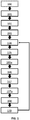
- spectral scans are analogous to the spectral scan 201 performed in Fig. 1 , except for the fact that the lesion type is known beforehand for the scanned organ.
- an image modality for display is reconstructed 202 and displayed 203, e.g. displayed to an intervening physician or radiologist on a screen or monitor, in a subsequent step.
- the contrast of the reconstructed image modality may be artificially boosted 202a if it falls below a visually perceptible threshold and/or may add further visual aids for guidance to the image modality for display.
- a controller may be programmed to take one of the following actions 204, with or without the input of an intervening physician or radiologist: initiate another timestamped spectral CT scan of the contrast-enhanced organ (e.g. liver) 201, followed by another reconstruction of an image modality for display 202 and updating/displaying the next image modality 203, stall or delay such a repetition, abort or interrupt the imaging method.
- initiate another timestamped spectral CT scan of the contrast-enhanced organ (e.g. liver) 201 followed by another reconstruction of an image modality for display 202 and updating/displaying the next image modality 203, stall or delay such a repetition, abort or interrupt the imaging method.
- the specific image modality (e.g. Z-effective, VNC or mono-energetic) leading to improved visual contrast in the displayed, reconstructed image modality is determined in a different way.
- an imaging protocol has been established 217 before the spectral imaging of the contrast-enhanced organ starts.
- the imaging protocol communicates the specific modality for reconstruction to a reconstruction unit comprising processing means.
- a specific reconstruction modality is unambiguously determined by the imaging protocol if the timestamp of the current spectral CT scan and the lesion type are known inputs.
- an imaging protocol is thus generated for a pre-determined set of lesion types, wherein the set may include, but is not limited to the following indications: HCC with various appearances, metastasis, hemangioma, cyst.
- Generating an imaging protocol 217 may comprise the following steps. Patients for which one of the above listed lesions has been found or is expected to be found are administered a contrast agent 210. A plurality of timed spectral sample CT scans is performed 211 for each patient, and therefore for each lesion type, under reproducible conditions. A reference time for the timestamps may be the start of the injection of the contrast agent. A trained radiologist then defines a region of interest 213 around the contrast-enhanced lesion in each reconstructed sample image modality 212. It is pointed out that for scanned each patient/lesion type all the pre-determined modalities of reconstruction (e.g. VNC, Z-effective, and mono-energetic) are exhausted at every timestamp of that particular scan.
- VNC variable modalities of reconstruction
- image processing means calculate a contrast value 216 over the defined region of interest for each reconstructed image modality, which includes each timestamp and each lesion type.
- a statistical ensemble of reconstructed image modalities is available, which is categorized by lesion type and timestamp.
- the present invention relates to a controller which is configured to perform the steps of selecting an image modality having a largest calculated contrast value, and/or calculating the contrast values for a given number of reconstructed image modalities and region of interest, and/or deriving a reconstruction modality from an imaging protocol based on a pre-detected lesion type and a current time stamp.
- a controller 301 is shown in FIG. 3 .
- the controller 301 maybe of a larger spectral scanning system 300 which also comprises a spectral CT scanner 302, a reconstruction unit 303, and a display 304.
- the spectral CT scanner 302 may comprise a dual-layer energy-discriminating radiation detector or detector array. Alternatively a spectral scan gathering spectral, e.g. energy-resolved, projection data may be achieved through voltage switching of an X-ray source, through the interleaved or sequential use of two or more X-ray sources operating at different voltages, or through photon-counting detectors.
- the reconstruction unit 303 may comprise dedicated image processing means to run the computationally intensive reconstruction algorithms, e.g. acceleration hardware, GPUs or GPU arrays, special purpose signal processors, etc.
Landscapes
- Health & Medical Sciences (AREA)
- Life Sciences & Earth Sciences (AREA)
- Engineering & Computer Science (AREA)
- Medical Informatics (AREA)
- Surgery (AREA)
- Heart & Thoracic Surgery (AREA)
- General Health & Medical Sciences (AREA)
- Nuclear Medicine, Radiotherapy & Molecular Imaging (AREA)
- Veterinary Medicine (AREA)
- Public Health (AREA)
- Animal Behavior & Ethology (AREA)
- Biomedical Technology (AREA)
- Molecular Biology (AREA)
- Physics & Mathematics (AREA)
- Biophysics (AREA)
- Radiology & Medical Imaging (AREA)
- High Energy & Nuclear Physics (AREA)
- Pathology (AREA)
- Optics & Photonics (AREA)
- Dentistry (AREA)
- Oral & Maxillofacial Surgery (AREA)
- Human Computer Interaction (AREA)
- Pulmonology (AREA)
- Theoretical Computer Science (AREA)
- Robotics (AREA)
- Apparatus For Radiation Diagnosis (AREA)
Priority Applications (4)
| Application Number | Priority Date | Filing Date | Title |
|---|---|---|---|
| EP18191114.0A EP3616620A1 (de) | 2018-08-28 | 2018-08-28 | Spektrale zweischichtige ct-geführte eingriffe |
| US17/270,461 US12329464B2 (en) | 2018-08-28 | 2019-08-28 | Spectral dual-layer CT-guided interventions |
| EP19758756.1A EP3843634B1 (de) | 2018-08-28 | 2019-08-28 | Spektrale zweischichtige ct-geführte eingriffe |
| PCT/EP2019/072993 WO2020043789A1 (en) | 2018-08-28 | 2019-08-28 | Spectral dual-layer ct-guided interventions |
Applications Claiming Priority (1)
| Application Number | Priority Date | Filing Date | Title |
|---|---|---|---|
| EP18191114.0A EP3616620A1 (de) | 2018-08-28 | 2018-08-28 | Spektrale zweischichtige ct-geführte eingriffe |
Publications (1)
| Publication Number | Publication Date |
|---|---|
| EP3616620A1 true EP3616620A1 (de) | 2020-03-04 |
Family
ID=63442439
Family Applications (2)
| Application Number | Title | Priority Date | Filing Date |
|---|---|---|---|
| EP18191114.0A Withdrawn EP3616620A1 (de) | 2018-08-28 | 2018-08-28 | Spektrale zweischichtige ct-geführte eingriffe |
| EP19758756.1A Active EP3843634B1 (de) | 2018-08-28 | 2019-08-28 | Spektrale zweischichtige ct-geführte eingriffe |
Family Applications After (1)
| Application Number | Title | Priority Date | Filing Date |
|---|---|---|---|
| EP19758756.1A Active EP3843634B1 (de) | 2018-08-28 | 2019-08-28 | Spektrale zweischichtige ct-geführte eingriffe |
Country Status (3)
| Country | Link |
|---|---|
| US (1) | US12329464B2 (de) |
| EP (2) | EP3616620A1 (de) |
| WO (1) | WO2020043789A1 (de) |
Cited By (1)
| Publication number | Priority date | Publication date | Assignee | Title |
|---|---|---|---|---|
| WO2021197996A1 (de) * | 2020-04-03 | 2021-10-07 | Bayer Aktiengesellschaft | Erzeugen von radiologischen aufnahmen |
Citations (4)
| Publication number | Priority date | Publication date | Assignee | Title |
|---|---|---|---|---|
| DE102007024450A1 (de) * | 2007-05-25 | 2008-11-27 | Siemens Ag | Ermittlungsverfahren für ein farbcodiertes erstes Auswertungsbild und hiermit korrespondierende Gegenstände |
| WO2013011418A2 (en) * | 2011-07-15 | 2013-01-24 | Koninklijke Philips Electronics N.V. | Spectral ct |
| WO2014128595A1 (en) * | 2013-02-21 | 2014-08-28 | Koninklijke Philips N.V. | Structure propagation restoration for spectral ct |
| US20170148156A1 (en) * | 2015-11-25 | 2017-05-25 | Zebra Medical Vision Ltd. | Systems and methods for detecting a fatty liver from a computed tomography (ct) scan |
Family Cites Families (9)
| Publication number | Priority date | Publication date | Assignee | Title |
|---|---|---|---|---|
| US6650928B1 (en) * | 2000-11-27 | 2003-11-18 | Ge Medical Systems Global Technology Company, Llc | Color parametric and composite maps for CT perfusion |
| SE524380C2 (sv) * | 2002-03-12 | 2004-08-03 | Xcounter Ab | Exponeringsstyrning i scannerbaserad detektering av joniserande strålning |
| US20110282194A1 (en) * | 2010-05-06 | 2011-11-17 | Bruce Reiner | Method and apparatus of quantitative analysis and data mining of medical imaging agent administration |
| DE102010043709B4 (de) * | 2010-11-10 | 2018-12-27 | Siemens Healthcare Gmbh | Verfahren zur Ermittlung des Wertes einer Röhrenspannung, Röntgeneinrichtung, Rechenprogramm und Datenträger |
| JP6030071B2 (ja) * | 2011-01-27 | 2016-11-24 | コーニンクレッカ フィリップス エヌ ヴェKoninklijke Philips N.V. | スペクトル撮像 |
| EP2879580B1 (de) * | 2012-08-06 | 2022-03-02 | Koninklijke Philips N.V. | Durchlässigkeitsmetrik auf basis dynamischer kontrastverstärkter bildgebung |
| US10398397B2 (en) | 2014-12-31 | 2019-09-03 | General Electric Company | Contrast-enhanced X-ray image guided biopsy system and method |
| CA2976516C (en) | 2015-07-27 | 2022-11-22 | Synaptive Medical (Barbados) Inc. | Navigational feedback for intraoperative waypoint |
| DE102016224717B4 (de) * | 2016-12-12 | 2018-12-20 | Siemens Healthcare Gmbh | Verfahren zum Ermitteln von Gewebeeigenschaften von Tumoren, sowie Bildanalyseeinrichtung, Computertomographiesystem, Computerprogrammprodukt und computerlesbares Medium |
-
2018
- 2018-08-28 EP EP18191114.0A patent/EP3616620A1/de not_active Withdrawn
-
2019
- 2019-08-28 WO PCT/EP2019/072993 patent/WO2020043789A1/en not_active Ceased
- 2019-08-28 US US17/270,461 patent/US12329464B2/en active Active
- 2019-08-28 EP EP19758756.1A patent/EP3843634B1/de active Active
Patent Citations (4)
| Publication number | Priority date | Publication date | Assignee | Title |
|---|---|---|---|---|
| DE102007024450A1 (de) * | 2007-05-25 | 2008-11-27 | Siemens Ag | Ermittlungsverfahren für ein farbcodiertes erstes Auswertungsbild und hiermit korrespondierende Gegenstände |
| WO2013011418A2 (en) * | 2011-07-15 | 2013-01-24 | Koninklijke Philips Electronics N.V. | Spectral ct |
| WO2014128595A1 (en) * | 2013-02-21 | 2014-08-28 | Koninklijke Philips N.V. | Structure propagation restoration for spectral ct |
| US20170148156A1 (en) * | 2015-11-25 | 2017-05-25 | Zebra Medical Vision Ltd. | Systems and methods for detecting a fatty liver from a computed tomography (ct) scan |
Non-Patent Citations (1)
| Title |
|---|
| DANIELA MUENZEL ET AL: "Photon counting CT of the liver with dual-contrast enhancement", PROCEEDINGS OF SPIE, vol. 9783, 22 March 2016 (2016-03-22), 1000 20th St. Bellingham WA 98225-6705 USA, pages 97835N, XP055558801, ISSN: 0277-786X, ISBN: 978-1-5106-2099-5, DOI: 10.1117/12.2216391 * |
Cited By (3)
| Publication number | Priority date | Publication date | Assignee | Title |
|---|---|---|---|---|
| WO2021197996A1 (de) * | 2020-04-03 | 2021-10-07 | Bayer Aktiengesellschaft | Erzeugen von radiologischen aufnahmen |
| JP2023521640A (ja) * | 2020-04-03 | 2023-05-25 | バイエル、アクチエンゲゼルシャフト | 放射線画像の生成 |
| JP7729836B2 (ja) | 2020-04-03 | 2025-08-26 | バイエル、アクチエンゲゼルシャフト | 放射線画像の生成 |
Also Published As
| Publication number | Publication date |
|---|---|
| EP3843634B1 (de) | 2022-04-13 |
| US20210251699A1 (en) | 2021-08-19 |
| US12329464B2 (en) | 2025-06-17 |
| EP3843634A1 (de) | 2021-07-07 |
| WO2020043789A1 (en) | 2020-03-05 |
Similar Documents
| Publication | Publication Date | Title |
|---|---|---|
| JP5921132B2 (ja) | 医用画像処理システム | |
| US8731262B2 (en) | Medical image and vessel characteristic data processing system | |
| US7433507B2 (en) | Imaging chain for digital tomosynthesis on a flat panel detector | |
| CN101529475B (zh) | 3d图像结合2d投影图像的呈现 | |
| CN101779961B (zh) | X射线图像诊断装置及x射线图像诊断装置的控制方法 | |
| EP3250128B1 (de) | Systeme und verfahren zur registrierung medizinischer bilder | |
| JP6021420B2 (ja) | 画像処理装置、画像処理方法およびプログラム | |
| US20130064343A1 (en) | Method and Apparatus for Determining Three-Dimensional Reconstruction of an Object | |
| CN113962949A (zh) | 脑部医学图像处理系统、方法及计算机可读存储介质 | |
| CN1681438A (zh) | 血流动态分析设备及方法和图像诊断设备 | |
| JP2019208903A (ja) | 医用画像処理装置、医用画像処理方法、及び医用画像処理プログラム | |
| CN113940698B (zh) | 基于超声造影的处理方法、超声装置及计算机存储介质 | |
| CN101039622B (zh) | 产生高质量x射线投影的成像系统 | |
| CN110876627A (zh) | X射线摄影装置和x射线图像处理方法 | |
| EP3843634B1 (de) | Spektrale zweischichtige ct-geführte eingriffe | |
| JP6716197B2 (ja) | 画像処理装置およびx線診断装置 | |
| Ozkurt et al. | Split bolus method in computerized tomography | |
| US20150164450A1 (en) | System and Method for Real Time 4D Quantification | |
| JP5591555B2 (ja) | X線診断装置、画像処理装置及びx線診断システム | |
| JP2016189831A (ja) | X線画像撮影装置 | |
| EP4312772B1 (de) | Subtraktionsbildgebung | |
| RU2633949C1 (ru) | Способ формирования составного изображения из серии ангиографических цифровых субтракционных кадров | |
| RU2644928C1 (ru) | Способ определения кадров, соответствующих границам фаз кровообращения, при проведении ангиографического исследования (варианты) | |
| JP2009225979A (ja) | 医用画像作成装置及び方法 | |
| Nikolau | Development of a prototype 2D dual-energy subtraction angiography system for image-guided interventions |
Legal Events
| Date | Code | Title | Description |
|---|---|---|---|
| PUAI | Public reference made under article 153(3) epc to a published international application that has entered the european phase |
Free format text: ORIGINAL CODE: 0009012 |
|
| AK | Designated contracting states |
Kind code of ref document: A1 Designated state(s): AL AT BE BG CH CY CZ DE DK EE ES FI FR GB GR HR HU IE IS IT LI LT LU LV MC MK MT NL NO PL PT RO RS SE SI SK SM TR |
|
| AX | Request for extension of the european patent |
Extension state: BA ME |
|
| RAP1 | Party data changed (applicant data changed or rights of an application transferred) |
Owner name: KONINKLIJKE PHILIPS N.V. |
|
| STAA | Information on the status of an ep patent application or granted ep patent |
Free format text: STATUS: THE APPLICATION IS DEEMED TO BE WITHDRAWN |
|
| 18D | Application deemed to be withdrawn |
Effective date: 20200905 |