EP3628330A1 - Imipramin zur verwendung als inhibitor der fascin1-überexpression - Google Patents
Imipramin zur verwendung als inhibitor der fascin1-überexpression Download PDFInfo
- Publication number
- EP3628330A1 EP3628330A1 EP18382696.5A EP18382696A EP3628330A1 EP 3628330 A1 EP3628330 A1 EP 3628330A1 EP 18382696 A EP18382696 A EP 18382696A EP 3628330 A1 EP3628330 A1 EP 3628330A1
- Authority
- EP
- European Patent Office
- Prior art keywords
- fascin1
- imipramine
- cancer
- colorectal cancer
- cells
- Prior art date
- Legal status (The legal status is an assumption and is not a legal conclusion. Google has not performed a legal analysis and makes no representation as to the accuracy of the status listed.)
- Ceased
Links
- 0 *C[n]1nc(**)c2c1C=CC(*)C=C2 Chemical compound *C[n]1nc(**)c2c1C=CC(*)C=C2 0.000 description 1
Images




Classifications
-
- A—HUMAN NECESSITIES
- A61—MEDICAL OR VETERINARY SCIENCE; HYGIENE
- A61K—PREPARATIONS FOR MEDICAL, DENTAL OR TOILETRY PURPOSES
- A61K31/00—Medicinal preparations containing organic active ingredients
- A61K31/33—Heterocyclic compounds
- A61K31/395—Heterocyclic compounds having nitrogen as a ring hetero atom, e.g. guanethidine or rifamycins
- A61K31/55—Heterocyclic compounds having nitrogen as a ring hetero atom, e.g. guanethidine or rifamycins having seven-membered rings, e.g. azelastine, pentylenetetrazole
-
- A—HUMAN NECESSITIES
- A61—MEDICAL OR VETERINARY SCIENCE; HYGIENE
- A61P—SPECIFIC THERAPEUTIC ACTIVITY OF CHEMICAL COMPOUNDS OR MEDICINAL PREPARATIONS
- A61P35/00—Antineoplastic agents
Definitions
- the present invention relates to the field of medicinal products, particularly to therapeutic products against cancer.
- Fascin1 is a key protein in filopodia and invadopodia formation as it possesses actin-binding and actin-bundling activity by cross-linking filamentous actin into tightly packed parallel bundles. Fascin1 is absent in most normal epithelia but expressed in many human carcinomas (Hashimoto, Kim, & Adams, 2011) and several previous studies have nominated this protein as a candidate biomarker for aggressive carcinomas. In fact, several studies including a meta-analysis have demonstrated that fascin1 expression is associated with increased lymph-node and distant-metastasis, disease progression and mortality in colorectal and breast cancer (Tan, Lewis, Adams, & Martin, 2013).
- Colorectal cancer is the third leading cause of cancer-related deaths in men and women in the United States.
- CRC Colorectal cancer
- SAC Serrated adenocarcinoma
- fascin1 was found to be the most discriminatory marker in distinguishing SAC from CC with an immunohistochemical overexpression in the former of 88.6% compared to 14.3% in CC. Even tumors showing histological and molecular features of microsatellite instability displayed lower fascin1 expression than SAC (50%) and qPCR assays analyzing the expression of fascin1 in these CRC supbtypes also confirmed these findings (Conesa-Zamora et al., 2013).
- fascin1 Given the causative role of fascin1 in the invading phenotype of tumor cells and its overexpression associated with worse survival in a wide variety of cancer types (Tan et al., 2013) including many with no tailored molecular therapy as triple-negative breast carcinoma, it is mandatory to obtain efficient fascin1 blockers.
- migrastatin and its macroketone analogues are considered typical fascin1 inhibitors thus decreasing metastatic tumor cell migration, invasion and tumor metastasis (Chen et al., 2010) despite the fact that their complex structure hinders the process of synthesis.
- Some authors have reported an anti-fascin1, anti-migratory and anti-invasive activity for compounds derived from indazol-furan-carboxamides (Han et al., 2016).
- WO2015127125 (A1 ) provides compounds of the general formula. compositions and methods for inhibiting fascin1 activity or treating a condition or disorder mediated by fascin1 activity in a subject in need thereof.
- SACs show a higher frequency of KRAS and BRAF mutations compared to conventional colorectal adenocarcinoma (Garcia-Solano et al., 2012; Stefanius et al., 2011) thus making SAC more resistant to anti-EGFR therapy in which effectiveness is obtained when KRAS and BRAF are non-mutated.
- SACs are mostly microsatellite stable and therefore cannot benefit from immune checkpoint inhibitors such as pembrolizumab. Given the high expression of fascin1 in SAC and the causative role of fascin1 in invasion and metastasis it has been found that imipramine is useful for treating SAC and other tumors overexpressing fascin1.
- Imipramine is an antidepressant and FDA-approved compound for the treatment of depression.
- imipramine is a compound that is effective in the treatment of CRC; and preferably in the treatment of serrated adenocarcinoma.
- Imipramine is a compound that is effective in the treatment of tumors overexpressing fascin1.
- the present invention refers to imipramine for use as inhibitor of fascin1 overexpression.
- Imipramine is the compound named 3-(10,11-dihydro-5H-dibenzo[b,f]azepin-5-yl)-N,N-dimethylpropan-1-amine, of chemical formula of C 19 H 2 N 2 .
- the overexpression of fascin1 is associated to the presence of cancer.
- the cancer is colorectal cancer.
- the colorectal cancer is serrated adenocarcinoma.
- the colorectal cancer is metastatic colorectal cancer.
- the present invention also refers to a composition comprising imipramine for use as inhibitor of fascin1 overexpression.
- the present invention also refers to a method for treating cancer mediated by fascin1 comprising administering to a subject in need therof an effective amount of imipramine.
- treating means one or more of preventing, curing, managing, reversing, attenuating, alleviating, minimizing, suppressing, or halting the deleterious effects of the above-described diseases.
- the fascin1 is overexpressed.
- the cancer is colorectal cancer.
- the colorectal cancer is serrated adenocarcinoma.
- the colorectal cancer is metastatic colorectal cancer
- the colorectal cancer can also be selected from gastrointestinal stromal tumors, leiomyosarcomas or melanomas.
- a ligand based pharmacophore model was derived from MGS_CORE using the Ligand Scout (LS) program (Gerhard Wolber, 2005). This model was screened against a subset of the DrugBank library (version 5.0; of 9,591 compounds including 2,037 approved by the American Food and Drug Administration (FDA), 96 nutraceuticals and 6000 experimental) after the fine tunning on a High Performance Computing (HPC) cluster of all related necessary programs from the LS suite. From resulting calculations and after careful visual inspection of the results and taking into account previous insights from literature, molecule imipramine was selected for posterior experimental validation.
- FIG 1A the pharmacophore model for imipramine is depicted.
- a 2D diagram of matching pharmacophoric features between MGS and imipramine is shown. 3D alignment of MGS (dark grey) and imipramine (light grey) is shown in figure 1.B .
- Imipramine was prepared at 10 mM in 100% DMSO and added to each well to a final concentration of 1 mM and 10% DMSO. Three replicates with six internal controls, containing only free protein in 10% DMSO, were included in the 96-well plates. The PCR plates were covered and subsequently shaken, centrifuged, incubated for 2 min at 20 °C inside the RT-PCR machine and heated from 20 to 100 °C at a 1°C/min scan-rate. Fascin1 thermal denaturation profiles were obtained recording the fluorescence intensity for the FAM, HEX and T-Red predefined filters. The derivative of the fluorescence curve was used to determine the Tm. Changes in Tm associated to ligand binding were estimated taking the average Tm value derived from the free protein internal controls.
- Fascin1 was extensively dialized against the appropriate buffer prior to each titration experiment. Fascin1 concentration was determined by measuring absorbance at 280 nm using an extinction coefficient of 67840 cm-1 ⁇ M-1. Fluorescence titration experiments were performed in a Cary Eclipse spectrofluorimeter (Varian Inc.). A 15 ⁇ M (14.7 ⁇ M) fascin1 solution was titrated with each compound by adding increasing volumes of concentrated solutions. Emission spectra were recorded between 307 and 500 nm at 25 °C in 10% DMSO, 100 mM NaCl, 20 mM Hepes, pH 7.4, with the excitation wavelength fixed at 280 nm. Binding isotherms were generated using either the changes in spectral area or in spectral centre of mass (CM), depending whether the change was only in fluorescence intensity or was also in the maximum emission wavelength.
- CM spectral centre of mass
- Ff and Fb are the fluorescence signal (area or CM) of free and bound fascin1 and PT and LT are the total protein and ligand concentration, respectively, at each addition point.
- HCT116 Three human colorectal adenocarcinoma cell lines were obtained from the American Type Culture Collection (ATCC, Rockville, MD): HCT116, SW480 and DLD1.
- the cell lines were cultivated in high glucose Dulbecco's Modified Eagle's Medium (DMEM) containing 10% heat-inactivated fetal bovine serum (FBS), 50 U/ml penicillin and 50 ⁇ g/ml streptomycin (Sigma Aldrich Chemical Co., USA) in an atmosphere of 5% CO 2 and 95% humidified air at 37°C. Subculture was performed when 90% confluence was obtained. Fascin1 gene expression was measured following the protocol previously described [Conesa-Zamora IJC].
- DMEM Dulbecco's Modified Eagle's Medium
- FBS heat-inactivated fetal bovine serum
- streptomycin Sigma Aldrich Chemical Co., USA
- RNA was extracted by treating cell line pellets around 200,000 cells with 700 ⁇ l of Qiazol (Qiagen ref: 1023537) and by adding 140 ⁇ l of chloroform and centrifuging at 12,000g for 15 min at 4°C. The aqueous phase containing 350 ⁇ l was then subjected to automatic total RNA extraction using the Qiacube equipment and the miRNeasy Mini Kit (ref: 217004), both provided by Qiagen.
- cDNA was obtained using the Maxima First Strand cDNA Synthesis Kit for RT-qPCR by Thermo Fisher (cat. No. K1671) following the manufacturer information.
- qPCR was performed on a 7500F real time PCR system by Applied Biosystems (Foster City, CA, USA) according to the instruction manual and following the standard protocol: 50°C 2 min, 95°C 10 min, 40 cycles of 95°C 15 sec, 60°C 1min and a melt curve stage consisting in 95°C 15 sec, 60°C min, 95°C 30 sec and 60°C 30 sec.
- the relative quantitation was done by the 2- ⁇ Ct method using ⁇ -actin as housekeeping gene.
- the amounts of mRNA are given as number of copies per million of copies of ⁇ -actin.
- Primers of ⁇ -actin used for FSCN quantitation were 115F and 116R whereas those for HPCA and DAG1 were b-a-blgF and b-a-blgR.
- Table 2 Primer sequences used for quantitative PCR validation (Gene, Primer name, Primer sequence (5'-3'), fragment size (bp).
- FIG. 3 illustrates how HCT-116 and SW-480 shows the highest fascin1 expression whilst LOVO, DLD-1 and HT-29 show the lowest. Given the ease for cell culture and the suitable morphology with prominent cytoplasm for immunofluorescence, migration and invasion assessment we selected DLD-1, SW-480 and HCT-116 cell lines in the subsequent assays.
- Exponentially growing cells were plated in triplicate in flat-bottomed 96-well plates (Nunc, Roskilde, Denmark) at 1200 cells/well. The day after, drugs were added in serial dilution from 5100 nM to 3100 ⁇ M. Control wells contained medium without drug plus 0.1% DMSO. Plates were incubated for 3 days in a humidified 5% CO 2 incubator and assayed for cell viability. Tetrazolium (MTT) dissolved in phosphate-buffered saline (PBS), pH 7.2, at 1.9 mg/mLl was added to the cells (30 ⁇ l/well). After incubation at 37°C for 4 h, the medium was aspirated.
- PBS phosphate-buffered saline
- Coverslips were then placed in 6-well plate with 2 mL serum free DMEM and 100 ⁇ M migrastatin, 20 ⁇ M imipramine, Epidermal Growth Factor (EGF) 10 ng/ml and MEK inhibitor PD98059 50 ⁇ M (MEKi) (both from Sigma-Aldrich, St Louis, MO, USA) for 24h.
- Cells were then fixed with Bouin (for fascin1 protein) or 4% formaldehyde (for actin protein) and subsequently permeabilized in a 0.3% Triton X-100/PBS solution, and then exposed to blocking buffer for 30 min.
- Appropriate fluorescent-labelled primary antibodies were incubated later with Alexa fluor 488- conjugated anti-mouse IgG (from donkey) (1/400) or Alexa fluor 594-labelled phalloidin, respectively (1/1000) (Molecular Probes, Thermo Fisher Scientific, Waltham, MA USA) and Hoechst 33258 (Fluka, Biochemika, Sigma-Aldrich, St Louis, MO, USA) for 30 min at room temperature and darkness. Samples were examined and representative images were taken with a confocal microscope (LSM 510 META from ZEISS, Jena, Germany).
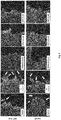
- FIG. 5 shows the immunofluorescence of fascin1 in two cell lines; the cell line expressing higher fascin1 level, HCT-116 and a non tumoral cell line derived keratinocyte cell line HaCaT which was used as a control of the immunofluorescence stainin.
- Lamellipodia protrusion number was calculated in the different conditions, being significantly lower after migrastatin, and imipramine treatments (data not shown).
- FIG. 5 the cell morphology of HCT-116 and HaCaT (as an example of cells that form cytoplasmic protrusions) of upon inhibition of filopodia formation is shown.
- Inset shows immunofluorescence analysis of the filopodia marker fascin (green protrusions indicated in arrows) upon control condition, 100 uM migrastatin, 20 ⁇ M imipramine, and 100 ng/ml Epidermal Growth Factor (migration stimulator). Images were captured with a LSM 510 META confocal fluorescence microscope with 63X oil objective. Migrastatin and imipramine inhibit lamellipodia protrusion (arrows) and consequently fascin1 localization.
- fascin1 inhibitors on filopodia lamellipodium formation protrusion has a quantifiable effect on cell migration
- MOCK basal condition, and migrastatin or imipramine-treated cells were investigated for their migration activity after 4-7 hours in vitro scratch.
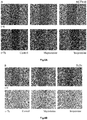
- Figure 6 shows the cell migration assay for the three cell lines A) HCT-116, B) DLD-1, C) SW-480, D) Migration was calculated with respect to the control conditions for a slope between 4 and 7h (lineal phase). * p ⁇ 0.05, ** p ⁇ 0.01. As shown in Figure 6 , imipramine produces a remarkable inhibition of migration in all cell lines assayed (p ⁇ 0.05). Except for SW-480, imipramine effect was more pronounced than migrastatin, especially for DLD-1. Cell migration was studied with the three cell lines by performing the wound healing assay in the presence of 5% FBS. Cancer colorectal cells (50,000 cells) were plated in low 35-mm-dishes with culture inserts (Ibidi, Martinsried, Germany).
- inserts were then removed with sterile forceps to create a wound field of approximately 500 ⁇ m, according to the manufacturer protocol.
- Detached cells were gently removed with phosphate buffered saline (PBS, Dubelco) before the addition of drugs.
- Confluent cells were incubated in one of the following treatments: 100 ⁇ M migrastatin (AnalytiCon Discovery, cat. No. NP-006108), and 20 ⁇ M imipramine ((purchased via Molport, Letonia) and, control dishes that were incubated in the presence of 0.1% DMSO. Cells were then allowed to migrate in a cell culture incubator .for 24h.
- the migration speed of the wound closure is given as the percentage of the recovered area at each time point, relative to the initially covered area (0 h).
- Table 3 shows the reduction in migration upon migrastatin and imipramine treatment on DLD1, SW-480 and HCT-116 colorectal cancer cell lines.
- HCT116 cells were determined using Cytoselect TM 24 Well Cell Invasion Assay (Basement Membrane Colorimetric Format) with coated Transwell chambers (8 ⁇ m pore size). Briefly, cells were resuspended with serum-free medium and treated with corresponding inhibitors (migrastatin 100 ⁇ M and, imipramine 20 ⁇ M). Cells (9.5 ⁇ 10 4 ) were seeded into the upper chamber, and in the lower chamber 500 ⁇ L of DMEM containing 10% FBS were added.
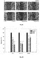
- FIG. 7 shows the transwell invasion assay performed to evaluate HCT116 cells invasive ability.
- A) Invasiveness of cells after treatment with different drugs. Pictures were taken under an inverted phase contrast microscope. The magnification was ⁇ 200 and scale bars 50 ⁇ m.
- B) Quantification of the invasive cells D.O. by a spectrophotometer at ⁇ 560 nm.
- Myoma disks were pre-incubated in 100 ⁇ M MGS, 10 ⁇ M and 20 ⁇ M imipramine or 0.1% DMSO-DMEM treatments at +4°C for 48 h.
- the myoma disks were placed into Transwell inserts (diameter 6.5 mm; Corning Incorporated, Corning, NY) and 700,000 cells in 50 ⁇ l of media were added on top of each myoma disk.
- the cells were allowed to attach overnight and the myoma disks were transferred onto uncoated nylon disks resting on curved steel grids in 12-well plates with 12 well plates with 1 ml of media plus migrastatin and imipramine in each well.
- the cancer cells were left to invade the myoma disks for 14 days, while changing the treatment media every 3 days. Subsequently the myoma discs were fixed with 4% neutral buffered formalin at room temperature for 24 h, and processed for histology. Six- ⁇ m sections were cut and stained with cytokeratin AE1/AE3 (M3515, Dako). Sections were documented at x10 magnification, using the Leica DMRB microscope DFC 480 camera with the Leica application suite v3.8 (Leica Microsystems, Wetzlar, Germany). Image J v1.46o (National Institute of Health, Bethesda, MD, USA) was used to measure invasion areas and depths. Each treatment was performed in triplicate.
- Figure 8 shows the myoma organotypic invasion model showing the effect of imipramine on HCT-116 colorectal cell line (A) on depth (B) and area (C) of invasion.
- imipramine inhibits both depth and area of invasion of HCT-116 compared to control in a comparable extent to migrastatin.
- CM-Dil Vibrant, Invitrogen
- HCT-116 two colorectal cancer cell lines
- Figure 9 shows the migration of colorectal cancer cell lines that can be visualized in the larvae of zebra fish. Injection and migration of different colorectal cancer cell lines in zebra fish model (A). Two colorectal cancer cell lines (DLD-1 and HCT-166) were treated with migrastatin and imipramine and injected in zebra fish larvae. The effect of imipramine (IMP) in diminishing cell invasion is evident compared to control treated with migrastatin (B).
- IMP imipramine
- Data are expressed as mean ⁇ standard deviation (SD). Data were analyzed for statistical differences by the Student's t-test for paired and unpaired data after testing for normal distribution of the data. For in vitro experiments, one-way analysis of variance (ANOVA) was performed followed by a Tukey poshoc test to compare each group. Differences were considered significant at an error probability of Pp ⁇ 0.05. SPSS 18.0 software was used for the rest of statistical analyses (SPSS, Inc, Chicago, Illinois, USA).
Landscapes
- Health & Medical Sciences (AREA)
- Life Sciences & Earth Sciences (AREA)
- Public Health (AREA)
- Veterinary Medicine (AREA)
- Medicinal Chemistry (AREA)
- Pharmacology & Pharmacy (AREA)
- Chemical & Material Sciences (AREA)
- Animal Behavior & Ethology (AREA)
- General Health & Medical Sciences (AREA)
- Epidemiology (AREA)
- Chemical Kinetics & Catalysis (AREA)
- Organic Chemistry (AREA)
- Nuclear Medicine, Radiotherapy & Molecular Imaging (AREA)
- General Chemical & Material Sciences (AREA)
- Pharmaceuticals Containing Other Organic And Inorganic Compounds (AREA)
- Peptides Or Proteins (AREA)
- Medicines That Contain Protein Lipid Enzymes And Other Medicines (AREA)
Priority Applications (6)
| Application Number | Priority Date | Filing Date | Title |
|---|---|---|---|
| EP18382696.5A EP3628330A1 (de) | 2018-09-28 | 2018-09-28 | Imipramin zur verwendung als inhibitor der fascin1-überexpression |
| AU2019346057A AU2019346057A1 (en) | 2018-09-28 | 2019-09-22 | Imipramine for use as an inhibitor of fascin1 overexpression |
| CA3114554A CA3114554A1 (en) | 2018-09-28 | 2019-09-22 | Imipramine for use as an inhibitor of fascin1 overexpression |
| US17/281,084 US12343349B2 (en) | 2018-09-28 | 2019-09-22 | Imipramine for use as an inhibitor of Fascin1 overexpression |
| JP2021542276A JP2022513325A (ja) | 2018-09-28 | 2019-09-22 | ファスシン1の過剰発現の阻害剤としての使用のためのイミプラミン |
| PCT/ES2019/070631 WO2020065113A1 (es) | 2018-09-28 | 2019-09-22 | Imipramina para uso como inhibidor de la sobrexpresión de la fascina 1 |
Applications Claiming Priority (1)
| Application Number | Priority Date | Filing Date | Title |
|---|---|---|---|
| EP18382696.5A EP3628330A1 (de) | 2018-09-28 | 2018-09-28 | Imipramin zur verwendung als inhibitor der fascin1-überexpression |
Publications (1)
| Publication Number | Publication Date |
|---|---|
| EP3628330A1 true EP3628330A1 (de) | 2020-04-01 |
Family
ID=63722332
Family Applications (1)
| Application Number | Title | Priority Date | Filing Date |
|---|---|---|---|
| EP18382696.5A Ceased EP3628330A1 (de) | 2018-09-28 | 2018-09-28 | Imipramin zur verwendung als inhibitor der fascin1-überexpression |
Country Status (6)
| Country | Link |
|---|---|
| US (1) | US12343349B2 (de) |
| EP (1) | EP3628330A1 (de) |
| JP (1) | JP2022513325A (de) |
| AU (1) | AU2019346057A1 (de) |
| CA (1) | CA3114554A1 (de) |
| WO (1) | WO2020065113A1 (de) |
Cited By (2)
| Publication number | Priority date | Publication date | Assignee | Title |
|---|---|---|---|---|
| ES2919724A1 (es) * | 2021-01-26 | 2022-07-27 | Fundacion Univ San Antonio | Raltegravir para uso como inhibidor de la fascina1 en cancer |
| EP4686471A1 (de) | 2024-08-01 | 2026-02-04 | Fundacion Universitaria San Antonio (Ucam) | Verbindung zur verwendung als fascin1-hemmer bei krebs |
Citations (3)
| Publication number | Priority date | Publication date | Assignee | Title |
|---|---|---|---|---|
| US3326896A (en) * | 1964-01-24 | 1967-06-20 | Geigy Chem Corp | Embonic acid addition salts of 10, 11-dihydro-5h-dibenz[b, f]azepines |
| WO2012129082A1 (en) * | 2011-03-18 | 2012-09-27 | Georgetown University | Treatments targeting gamma-synuclein expression levels |
| WO2015127125A1 (en) | 2014-02-20 | 2015-08-27 | Cornell University | Compounds and methods for inhibiting fascin |
-
2018
- 2018-09-28 EP EP18382696.5A patent/EP3628330A1/de not_active Ceased
-
2019
- 2019-09-22 JP JP2021542276A patent/JP2022513325A/ja not_active Withdrawn
- 2019-09-22 US US17/281,084 patent/US12343349B2/en active Active
- 2019-09-22 WO PCT/ES2019/070631 patent/WO2020065113A1/es not_active Ceased
- 2019-09-22 CA CA3114554A patent/CA3114554A1/en active Pending
- 2019-09-22 AU AU2019346057A patent/AU2019346057A1/en not_active Abandoned
Patent Citations (3)
| Publication number | Priority date | Publication date | Assignee | Title |
|---|---|---|---|---|
| US3326896A (en) * | 1964-01-24 | 1967-06-20 | Geigy Chem Corp | Embonic acid addition salts of 10, 11-dihydro-5h-dibenz[b, f]azepines |
| WO2012129082A1 (en) * | 2011-03-18 | 2012-09-27 | Georgetown University | Treatments targeting gamma-synuclein expression levels |
| WO2015127125A1 (en) | 2014-02-20 | 2015-08-27 | Cornell University | Compounds and methods for inhibiting fascin |
Non-Patent Citations (20)
| Title |
|---|
| ARIMOCHI H ET AL: "Characterization of cytotoxic actions of tricyclic antidepressants on human HT29 colon carcinoma cells", EUROPEAN JOURNAL OF PHARMACOLOGY, ELSEVIER SCIENCE, NL, vol. 541, no. 1-2, 10 July 2006 (2006-07-10), pages 17 - 23, XP028029098, ISSN: 0014-2999, [retrieved on 20060710], DOI: 10.1016/J.EJPHAR.2006.04.053 * |
| ASTROM, P.; HELJASVAARA, R.; NYBERG, P.; AL-SAMADI, A.; SALO, T.: "Human Tumor Tissue-Based 3D In Vitro Invasion Assays", METHODS IN MOLECULAR BIOLOGY (CLIFTON, N.J.), vol. 1731, 2018, pages 213 - 221 |
| BEGOÑA ALBURQUERQUE-GONZÁLEZ ET AL: "New role of the antidepressant imipramine as a Fascin1 inhibitor in colorectal cancer cells", EXPERIMENTAL AND MOLECULAR MEDICINE, vol. 52, no. 2, 1 February 2020 (2020-02-01), KR, pages 281 - 292, XP055758336, ISSN: 1226-3613, DOI: 10.1038/s12276-020-0389-x * |
| CHEN, L.; YANG, S.; JAKONCIC, J.; ZHANG, J. J.; HUANG, X. Y.: "Migrastatin analogues target fascin to block tumour metastasis", NATURE, 2010, Retrieved from the Internet <URL:https://doi.org/10.1038/nature08978> |
| CONESA-ZAMORA, P.; GARCIA-SOLANO, J.; GARCIA-GARCIA, F.; TURPIN, M. D. C.; TRUJILLO-SANTOS, J.; TORRES-MORENO, D.; PEREZ-GUILLERMO: "Expression profiling shows differential molecular pathways and provides potential new diagnostic biomarkers for colorectal serrated adenocarcinoma", INTERNATIONAL JOURNAL OF CANCER, vol. 132, no. 2, 2013, pages 297 - 307, Retrieved from the Internet <URL:https://doi.org/10.1002/ijc.27674> |
| D AHMED ET AL: "Epigenetic and genetic features of 24 colon cancer cell lines", ONCOGENESIS, vol. 2, no. 9, 1 September 2013 (2013-09-01), pages e71 - e71, XP055607246, DOI: 10.1038/oncsis.2013.35 * |
| GARCIA-SOLANO, J.; CONESA-ZAMORA, P.; CARBONELL, P.; TRUJILLO-SANTOS, J.; TORRES-MORENO D, D.; PAGAN-GOMEZ, I.; PEREZ-GUILLERMO, M: "Colorectal serrated adenocarcinoma shows a different profile of oncogene mutations, MSI status and DNA repair protein expression compared to conventional and sporadic MSI-H carcinomas", INTERNATIONAL JOURNAL OF CANCER, 2012, Retrieved from the Internet <URL:https://doi.org/10.1002/ijc.27454> |
| GERHARD WOLBER, T. L.: "LigandScout: 3-D Pharmacophores Derived from Protein-Bound Ligands and Their Use as Virtual Screening Filters", JOURNAL OF CHEMICAL INFORMATION AND MODELING, vol. 45, no. 1, 2005, pages 160 - 169 |
| HAMILTON, S. R. ET AL.: "WHO Classification of Tumours, Volume 3 IARC WHO Classification of Tumours", vol. 3, 2010, IARC, article "Carcinoma of the colon and rectum. In, WHO classification of tumours of the digestive system", pages: 134 |
| HAN, S.; HUANG, J.; LIU, B.; XING, B.; BORDELEAU, F.; REINHART-KING, C. A.; HUANG, X.-Y.: "Improving fascin inhibitors to block tumor cell migration and metastasis", MOLECULAR ONCOLOGY, vol. 10, 2016, pages 966 - 980, Retrieved from the Internet <URL:https://doi.org/10.1016/j.moionc.2016.03.006> |
| HASHIMOTO, Y.; KIM, D. J.; ADAMS, J. C.: "The roles of fascins in health and disease", JOURNAL OF PATHOLOGY, 2011, Retrieved from the Internet <URL:https://doi.org/10.1002/path.2894> |
| LAIHO, P.; KOKKO, A.; VANHARANTA, S.; SALOVAARA, R.; SAMMALKORPI, H.; JARVINEN, H.; AALTONEN, L. A.: "Serrated carcinomas form a subclass of colorectal cancer with distinct molecular basis", ONCOGENE, vol. 26, no. 2, 2007, pages 312 - 320, Retrieved from the Internet <URL:https://doi.org/10.1038/sj.onc.1209778> |
| MACHESKY, L. M.; LIA., A.: "Fascin: Invasive filopodia promoting metastasis", COMMUNICATIVE AND INTEGRATIVE BIOLOGY, 2010, Retrieved from the Internet <URL:https://doi.org/10.4161/cib.3.3.11556> |
| R. KRAFT ET AL: "A cell-based fascin bioassay identifies compounds with potential anti-metastasis or cognition-enhancing functions", DISEASE MODELS & MECHANISMS, vol. 6, no. 1, 23 August 2012 (2012-08-23), GB, pages 217 - 235, XP055562913, ISSN: 1754-8403, DOI: 10.1242/dmm.008243 * |
| STEFANIUS, K.; YLITALO, L.; TUOMISTO, A.; KUIVILA, R.; KANTOLA, T.; SIRNIO, P.; MAKINEN, M. J.: "Frequent mutations of KRAS in addition to BRAF in colorectal serrated adenocarcinoma", HISTOPATHOLOGY, 2011, Retrieved from the Internet <URL:https://doi.org/10.1111/j.1365-2559.2011.03821.x> |
| TAN, V. Y.; LEWIS, S. J.; ADAMS, J. C.; MARTIN, R. M.: "Association of fascin-1 with mortality, disease progression and metastasis in carcinomas: A systematic review and meta-analysis", BMC MEDICINE, vol. 11, no. 1, 2013, pages 1 - 17, XP021145635, Retrieved from the Internet <URL:https://doi.org/10.1 186/1741-7015-11-52> DOI: doi:10.1186/1741-7015-11-52 |
| THOMAS SAUER ET AL: "Is Imipramine Helpful in the Treatment of Cancer? A Case Presentation and Discussion of Possible Clinical Implications Case Report Open Access", BIOMED J SCI &TECH RES BJSTR. MS.ID, 29 May 2018 (2018-05-29), XP055562965, Retrieved from the Internet <URL:https://biomedres.us/pdfs/BJSTR.MS.ID.001118.pdf> [retrieved on 20190228], DOI: 10.26717/BJSTR.2018.04.001118 * |
| TUOMISTO, A.; GARCÃ-A-SOLANO, J.; SIMIÃ, P.; VäYRYNEN, J.; PéREZ-GUILLERMO, M.; MäKINEN, M. J.; CONESA-ZAMORA, P.: "HIF-1α expression and high microvessel density are characteristic features in serrated colorectal cancer", VIRCHOWS ARCHIV., 2016, Retrieved from the Internet <URL:https://doi.org/10.1007/s00428-016-1988-8> |
| VANESSA Y TAN ET AL: "Association of fascin-1 with mortality, disease progression and metastasis in carcinomas: a systematic review and meta-analysis", BMC MEDICINE, BIOMED CENTRAL LTD., LONDON, GB, vol. 11, no. 1, 26 February 2013 (2013-02-26), pages 52, XP021145635, ISSN: 1741-7015, DOI: 10.1186/1741-7015-11-52 * |
| YOSUKE HASHIMOTO ET AL: "Dual Actin-bundling and Protein Kinase C-binding Activities of Fascin Regulate Carcinoma Cell Migration Downstream of Rac and Contribute to Metastasis - D - V", MOLECULAR BIOLOGY OF THE CELL, 1 November 2007 (2007-11-01), pages 4591 - 4602, XP055563675, Retrieved from the Internet <URL:https://www.ncbi.nlm.nih.gov/pmc/articles/PMC2043557/pdf/zmk4591.pdf> DOI: 10.1091/mbc.E07- * |
Cited By (2)
| Publication number | Priority date | Publication date | Assignee | Title |
|---|---|---|---|---|
| ES2919724A1 (es) * | 2021-01-26 | 2022-07-27 | Fundacion Univ San Antonio | Raltegravir para uso como inhibidor de la fascina1 en cancer |
| EP4686471A1 (de) | 2024-08-01 | 2026-02-04 | Fundacion Universitaria San Antonio (Ucam) | Verbindung zur verwendung als fascin1-hemmer bei krebs |
Also Published As
| Publication number | Publication date |
|---|---|
| AU2019346057A1 (en) | 2021-05-20 |
| US20230330096A1 (en) | 2023-10-19 |
| CA3114554A1 (en) | 2020-04-02 |
| WO2020065113A1 (es) | 2020-04-02 |
| JP2022513325A (ja) | 2022-02-07 |
| US12343349B2 (en) | 2025-07-01 |
Similar Documents
| Publication | Publication Date | Title |
|---|---|---|
| Dong et al. | Exosome-mediated transfer of lncRNA‑SNHG14 promotes trastuzumab chemoresistance in breast cancer Retraction in/10.3892/ijo. 2022.5382 | |
| Gammazza et al. | Doxorubicin anti-tumor mechanisms include Hsp60 post-translational modifications leading to the Hsp60/p53 complex dissociation and instauration of replicative senescence | |
| Van Ginkel et al. | Expression of the receptor tyrosine kinase Axl promotes ocular melanoma cell survival | |
| Nie et al. | Overexpression of stathmin 1 is a poor prognostic biomarker in non-small cell lung cancer | |
| Douchi et al. | Silencing of LRRFIP1 reverses the epithelial–mesenchymal transition via inhibition of the Wnt/β-catenin signaling pathway | |
| Li et al. | Tripartite motif‐containing 37 (TRIM37) promotes the aggressiveness of non‐small‐cell lung cancer cells by activating the NF‐κB pathway | |
| Wang et al. | NLRC5 mediates cell proliferation, migration, and invasion by regulating the Wnt/β-catenin signalling pathway in clear cell renal cell carcinoma | |
| Wang et al. | AGBL2 promotes cancer cell growth through IRGM-regulated autophagy and enhanced Aurora A activity in hepatocellular carcinoma | |
| He et al. | TRAF7 enhances ubiquitin-degradation of KLF4 to promote hepatocellular carcinoma progression | |
| Tseng et al. | Deregulation of SLIT2-mediated Cdc42 activity is associated with esophageal cancer metastasis and poor prognosis | |
| Dantas-Barbosa et al. | Expression and role of TYRO3 and AXL as potential therapeutical targets in leiomyosarcoma | |
| Kumarasamy et al. | Discrete vulnerability to pharmacological CDK2 inhibition is governed by heterogeneity of the cancer cell cycle | |
| Gao et al. | The membrane protein sortilin can be targeted to inhibit pancreatic cancer cell invasion | |
| Yang et al. | Transcription factor Snai1-1 induces osteosarcoma invasion and metastasis by inhibiting E-cadherin expression | |
| BELIZZI et al. | The scaffolding protein NHERF1 sensitizes EGFR-dependent tumor growth, motility and invadopodia function to gefitinib treatment in breast cancer cells | |
| Wang et al. | MTMR3 is upregulated in patients with breast cancer and regulates proliferation, cell cycle progression and autophagy in breast cancer cells | |
| Xue et al. | IL-21/IL-21R inhibit tumor growth and invasion in non-small cell lung cancer cells via suppressing Wnt/β-catenin signaling and PD-L1 expression | |
| Ishimoto et al. | Role of endothelin receptor signalling in squamous cell carcinoma | |
| Strietz et al. | ERN1 and ALPK1 inhibit differentiation of bi-potential tumor-initiating cells in human breast cancer | |
| Wen et al. | Eya2 overexpression promotes the invasion of human astrocytoma through the regulation of ERK/MMP9 signaling | |
| Qu et al. | Role of AXL expression in non-small cell lung cancer | |
| Tao et al. | TRIM37 promotes tumor cell proliferation and drug resistance in pediatric osteosarcoma | |
| Wang et al. | Src promotes metastasis of human non-small cell lung cancer cells through Fn14-mediated NF-κB signaling | |
| Li et al. | CPNE1 silencing inhibits cell proliferation and accelerates apoptosis in human gastric cancer | |
| US12343349B2 (en) | Imipramine for use as an inhibitor of Fascin1 overexpression |
Legal Events
| Date | Code | Title | Description |
|---|---|---|---|
| PUAI | Public reference made under article 153(3) epc to a published international application that has entered the european phase |
Free format text: ORIGINAL CODE: 0009012 |
|
| STAA | Information on the status of an ep patent application or granted ep patent |
Free format text: STATUS: THE APPLICATION HAS BEEN PUBLISHED |
|
| AK | Designated contracting states |
Kind code of ref document: A1 Designated state(s): AL AT BE BG CH CY CZ DE DK EE ES FI FR GB GR HR HU IE IS IT LI LT LU LV MC MK MT NL NO PL PT RO RS SE SI SK SM TR |
|
| AX | Request for extension of the european patent |
Extension state: BA ME |
|
| STAA | Information on the status of an ep patent application or granted ep patent |
Free format text: STATUS: REQUEST FOR EXAMINATION WAS MADE |
|
| 17P | Request for examination filed |
Effective date: 20201001 |
|
| RBV | Designated contracting states (corrected) |
Designated state(s): AL AT BE BG CH CY CZ DE DK EE ES FI FR GB GR HR HU IE IS IT LI LT LU LV MC MK MT NL NO PL PT RO RS SE SI SK SM TR |
|
| STAA | Information on the status of an ep patent application or granted ep patent |
Free format text: STATUS: EXAMINATION IS IN PROGRESS |
|
| 17Q | First examination report despatched |
Effective date: 20201215 |
|
| P01 | Opt-out of the competence of the unified patent court (upc) registered |
Effective date: 20230515 |
|
| STAA | Information on the status of an ep patent application or granted ep patent |
Free format text: STATUS: THE APPLICATION HAS BEEN REFUSED |
|
| 18R | Application refused |
Effective date: 20231009 |