EP3629283A1 - Génération d'un ensemble de données d'image mixte - Google Patents
Génération d'un ensemble de données d'image mixte Download PDFInfo
- Publication number
- EP3629283A1 EP3629283A1 EP18197539.2A EP18197539A EP3629283A1 EP 3629283 A1 EP3629283 A1 EP 3629283A1 EP 18197539 A EP18197539 A EP 18197539A EP 3629283 A1 EP3629283 A1 EP 3629283A1
- Authority
- EP
- European Patent Office
- Prior art keywords
- image
- data set
- image data
- value
- mixed
- Prior art date
- Legal status (The legal status is an assumption and is not a legal conclusion. Google has not performed a legal analysis and makes no representation as to the accuracy of the status listed.)
- Withdrawn
Links
Images
Classifications
-
- G—PHYSICS
- G06—COMPUTING OR CALCULATING; COUNTING
- G06T—IMAGE DATA PROCESSING OR GENERATION, IN GENERAL
- G06T5/00—Image enhancement or restoration
- G06T5/50—Image enhancement or restoration using two or more images, e.g. averaging or subtraction
-
- A—HUMAN NECESSITIES
- A61—MEDICAL OR VETERINARY SCIENCE; HYGIENE
- A61B—DIAGNOSIS; SURGERY; IDENTIFICATION
- A61B6/00—Apparatus or devices for radiation diagnosis; Apparatus or devices for radiation diagnosis combined with radiation therapy equipment
- A61B6/48—Diagnostic techniques
- A61B6/482—Diagnostic techniques involving multiple energy imaging
-
- G—PHYSICS
- G06—COMPUTING OR CALCULATING; COUNTING
- G06T—IMAGE DATA PROCESSING OR GENERATION, IN GENERAL
- G06T5/00—Image enhancement or restoration
- G06T5/20—Image enhancement or restoration using local operators
-
- G—PHYSICS
- G06—COMPUTING OR CALCULATING; COUNTING
- G06T—IMAGE DATA PROCESSING OR GENERATION, IN GENERAL
- G06T5/00—Image enhancement or restoration
- G06T5/73—Deblurring; Sharpening
-
- G—PHYSICS
- G06—COMPUTING OR CALCULATING; COUNTING
- G06T—IMAGE DATA PROCESSING OR GENERATION, IN GENERAL
- G06T7/00—Image analysis
- G06T7/97—Determining parameters from multiple pictures
-
- G—PHYSICS
- G16—INFORMATION AND COMMUNICATION TECHNOLOGY [ICT] SPECIALLY ADAPTED FOR SPECIFIC APPLICATION FIELDS
- G16H—HEALTHCARE INFORMATICS, i.e. INFORMATION AND COMMUNICATION TECHNOLOGY [ICT] SPECIALLY ADAPTED FOR THE HANDLING OR PROCESSING OF MEDICAL OR HEALTHCARE DATA
- G16H40/00—ICT specially adapted for the management or administration of healthcare resources or facilities; ICT specially adapted for the management or operation of medical equipment or devices
- G16H40/60—ICT specially adapted for the management or administration of healthcare resources or facilities; ICT specially adapted for the management or operation of medical equipment or devices for the operation of medical equipment or devices
- G16H40/67—ICT specially adapted for the management or administration of healthcare resources or facilities; ICT specially adapted for the management or operation of medical equipment or devices for the operation of medical equipment or devices for remote operation
-
- G—PHYSICS
- G06—COMPUTING OR CALCULATING; COUNTING
- G06T—IMAGE DATA PROCESSING OR GENERATION, IN GENERAL
- G06T2207/00—Indexing scheme for image analysis or image enhancement
- G06T2207/10—Image acquisition modality
- G06T2207/10116—X-ray image
-
- G—PHYSICS
- G06—COMPUTING OR CALCULATING; COUNTING
- G06T—IMAGE DATA PROCESSING OR GENERATION, IN GENERAL
- G06T2207/00—Indexing scheme for image analysis or image enhancement
- G06T2207/20—Special algorithmic details
- G06T2207/20212—Image combination
- G06T2207/20221—Image fusion; Image merging
Definitions
- the invention relates to a method for generating a mixed image data set based on an energy-resolved image of an energy-resolving x-ray detector and an image generation unit therefor.
- energy-dissolving in particular counting, direct-converting, X-ray detectors or integrating, in particular indirectly converting, X-ray detectors can be used.
- the X-rays or the photons can be converted into electrical impulses in a directly converting X-ray detector using a suitable converter material.
- a suitable converter material For example, CdTe, CdZnTe (CZT), CdZnTeSe, CdTeSe, CdMnTe, InP, TlBr2, HgI2, GaAs or others can be used as converter material.
- the electrical impulses are evaluated by evaluation electronics, for example an integrated circuit (Application Specific Integrated Circuit, ASIC).
- ASIC Application Specific Integrated Circuit
- the level of the electrical pulse is, in particular after a preamplifier and a shaper, generally proportional to the energy of the absorbed X-ray photon.
- spectral or energy-resolving information can be extracted by comparing the level of the electrical pulse with an energy threshold.
- the incident photons are counted in each detector element of the X-ray detector in the corresponding energy range above an energy threshold.
- These reference signals or these energy threshold values are usually set globally during a method for energy calibration by specifying a single digital value (DAC value) per energy threshold for the entire X-ray detector.
- Each detector element can have several individually adjustable energy thresholds, each with an energy threshold value.
- the energy threshold values can be set at least partially the same or at least partially differently for all detector elements, in particular after an energy calibration.
- indirectly converting, energy-resolving X-ray detectors for example so-called sandwich X-ray detectors, can also be used.
- the spatial resolution or spatial resolution of the X-ray detector is usually given by the so-called pixel or / and sensor design, that is to say the structuring of the converter element or the evaluation unit in detector elements.
- the spatial resolution during the development of the X-ray detector can be increased by, for example, smaller detector elements.
- the spatial resolution is thus essentially determined by the structuring of the converter element or the evaluation unit in a matrix comprising a plurality of detector elements.
- the areal size of the detector elements also influences other properties of the detector element. For example, smaller detector elements generally lead to higher pixel noise.
- Another disadvantage of smaller detector elements can be lower frame rates, for example due to bandwidth limitations for larger matrices with a large number of detector elements. The data of the detector elements can therefore be read out less frequently due to the large amounts of data.
- the spatial resolution of an X-ray detector is described by its MTF (Modulation Transfer Function). This size is defined for known energy-integrating X-ray detectors and is given by the pixel and sensor design. Influential parameters regarding spatial resolution include the size of the detector elements, the sensitivity, the size and shape of the diode or the sensitivity of the converter material and the thickness of the sensor or converter material, for example CsI, CdTe or Se. This applies to both direct-converting and indirectly converting X-ray detectors.
- MTF Modulation Transfer Function
- the inventors have recognized that for some clinical applications it would be desirable to be retrospective, i.e. After the data acquisition, to be able to influence the MTF or the spatial resolution, for example in order to be able to recognize more details in fine structures such as stents.
- the object is achieved according to the invention by a method according to claim 1, an image generation unit according to claim 8, a computer program product according to claim 9, a computer-readable medium according to claim 10 and a medical device according to claim 11.
- the invention relates to a method for generating a mixed image data set based on an energy-resolved image of an energy-resolving X-ray detector, comprising the steps of generating and mixing.
- the step of generating the first image value of a first image element of a first image data set is generated in a first energy range and the second image value of the first image element of a second image data set is generated in a second energy range.
- the mixing step the first image value and the second image value are mixed as a function of an image parameter and the mixed image data set is generated with a mixed image value of the first image element.
- the energy-resolving x-ray detector can in particular be designed as a counting x-ray detector, preferably with a directly converting converter element.
- a radiation or particle detector can be used.
- the radiation or particle detector can be used as an X-ray detector.
- the energy-resolving x-ray detector can be designed as a sandwich detector, for example having at least two converter layers in the converter element.
- the converter layers can be designed to be direct or indirect converting.
- the x-ray detector can have a plurality of detector elements.
- An image data set can be generated from the measured values of the detector elements.
- the image data set comprises image values.
- the image value can, for example, essentially correspond to the measured value of the x-ray detector.
- a slice image can be reconstructed from a plurality of images and their measured values, the slice image being able to form the image data set.
- the first image value and the second image value comprise the image information for an essentially identical location in the image, but in different energy areas.
- the first and the second energy range can be limited by means of at least one energy threshold.
- the first energy range can be limited to higher energy values by means of a first energy threshold, for example.
- a lower energy threshold can be set above the noise limit and limit the first energy range to lower energy values.
- measured values with energy values between the lower energy threshold and the first energy threshold can be registered.
- measured values with energy values above the first energy threshold and can be registered.
- Energy areas can be discriminated one-sided or two-sided.
- the image parameter can be assigned to the first image element or to a plurality of image elements of the X-ray detector.
- the image parameter can be assigned to the first energy range and / or the second energy range.
- the method according to the invention can be expanded to more than two energy ranges.
- the method according to the invention can be carried out simultaneously or in succession for several picture elements.
- the steps of the method according to the invention can be repeated for a second picture element.
- the spatial resolution of a (quantum) counting X-ray detector with small detector elements can strongly depend on the set energy threshold. At a very low energy threshold value, photons that strike just outside the detector element can already lead to a counted event or a measured value.
- the effective aperture of the detector element can thus be larger than the geometric aperture of the detector element. This means that the effective aperture of the picture element can also be larger than the geometric aperture of the picture element.
- the energy threshold is set higher, for example only just below the energy of the X-ray photons, only events that hit the detector element near the center of the detector element can be counted, since, due to the size of the charge cloud, the whole only for the centrally incident X-ray photons Energy can be deposited in the detector element.
- the effective aperture can be smaller than the geometric aperture.
- Quantum counting X-ray detectors typically evaluate several energy thresholds at the same time. The inventors have recognized that this provides an image data set with a higher spatial resolution retrospectively. However, since the quantum efficiency of an energy-resolving, in particular counting, X-ray detector can be lower at higher energy threshold values, the energy threshold value can be increased lead that the signal-to-noise ratio in areas of the image data set, in particular the image, is reduced with a few registered events.
- the inventors therefore propose a method in which the image data sets of the different energy ranges can be mixed locally, for example as a function of the signal and the movement in the image as image parameters.
- the image data sets of the different energy ranges can be mixed locally, for example as a function of the signal and the movement in the image as image parameters.
- more information from the higher energy range or more higher energy ranges can be mixed into the final image or the mixed image data set.
- the weighting of the location information from the image data set with a higher energy range can thus be increased.
- the first image data record and the second image data record can be linearly weighted.
- the image data sets can be broken down into different image frequency ranges.
- filtering and / or adaptation for example based on a look-up table
- filtering and / or adaptation for example based on a look-up table
- already known image algorithms for example for optimizing or reconstructing moving images, can be expanded and advantageously improved using the method according to the invention.
- the primary interaction point of an X-ray photon or event within the detector element or the X-ray detector can be inferred.
- the registered signal can be broken down into different parts by means of several energy threshold values, each with differently wide probability distributions for the location of the primary interaction.
- the mixing ratio between the first image data set and the second image data set be local, i.e. to vary from picture element to picture element or from picture area to picture area.
- the mixing ratio can depend on the image content or an image parameter.
- the higher spatial resolution of the image data set with the higher energy range can advantageously be used to improve the spatial resolution in the mixed image data set.
- the method further comprises a step of determining, the image parameter being determined based at least on at least a partial area of the first image data set and / or the second image data set.
- a different image parameter for example with regard to the value or type of the image parameter, can be determined for different image elements.
- the partial area can comprise at least one picture element.
- the partial area can in particular comprise a plurality of adjacent picture elements.
- the image parameter can be determined based on the image value of the first image element or the image values of a plurality of image elements comprising the first image element of the first image data set and / or the image value of the first image element or the image values of a plurality of image elements comprising the first image element of the second image data set.
- the spatial resolution can advantageously be improved in a targeted manner.
- the image parameter is based on an image property, a spatial frequency band property or a time-varying image property compared to a previous picture.
- the image property can be determined in the spatial space or in the frequency space.
- the image property can comprise, for example, a contrast property, an intensity property or information about the signal-to-noise ratio.
- the spatial frequency band property can include information about edges between, for example, different materials in the examination object in the image (data record) or the modulation transfer function.
- the temporally variable image property can be determined from two first or / and second image data records, that is to say an image sequence, recorded at different times from one another.
- a difference between the image values between the first image and the second image can be formed for an image element of the first or / and second image data set, so that, for example, a movement in the examination object or in the image can be determined.
- external signals can be used as image parameters, for example an EKG signal or information from an optical camera recording. If an image parameter exceeds a suitable threshold value, the mixing ratio can be adapted to the image property.
- the spatial resolution can advantageously be adapted to the image property. The spatial resolution in diagnostically particularly relevant partial areas of the mixed image can advantageously be improved.
- the method further comprises a step of filtering by means of a filter function of the first image data set and / or the second image data set.
- the first or / and the second image data set can be transferred into the spatial frequency space, for example, by means of a Fourier transformation and filtered there.
- the filtering step may be performed before or after a determining step.
- the first or / and the second image data record can be filtered.
- the inverse of the MTF noise suppression, for example by frequency band decomposition, or edge preservation in spatial space, for example using gradients from the image data set of the lower energy range, can be used.
- the image quality or the image impression of the mixed image data set can advantageously be improved.
- the method also has a step of optimizing the filter function based on an examination type.
- the filter function can be selected depending on the materials in the examination object in the recording area, for example contrast media such as iodine or tissue (in particular water) or bone (in particular calcium).
- contrast media such as iodine or tissue (in particular water) or bone (in particular calcium).
- the first and / or the second image data record can advantageously be adapted to the medical question.
- the filter function can be optimized for a material, an examination type, an examination region, an examination program or an X-ray spectrum.
- the material can be iodine or calcium, for example.
- the material can be typical of an object in the examination object, for example a wire or a stent.
- the type of examination can be, for example, fluoroscopy, mammography or radiography. Optimal noise reduction and contrast optimization can advantageously be achieved in the mixed image data set.
- the mixed image data set has a mixed image value of the first image element in at least the first image element and the respective first image value or the respective second image value in at least one other image element.
- the mixed image value corresponds to the mixed image value from the first image value and the second image value.
- a partial area with mixed picture values surrounded by first picture values and / or second picture values can be formed in the mixed picture.
- the mixed image data record can comprise several image elements.
- the image or the mixed image data set can advantageously be improved in one partial area, the first or second image data being retained in the other partial area.
- the mixing is carried out by detector element in the local space.
- the image values of neighboring Detector elements can be used, for example, in the steps of determining, filtering and / or optimizing.
- the mixing step preferably only the first and second image values of the first detector element are mixed.
- a look-up table, a linear weighting, a (non-linear) function, a gamma function or logarithm function can be used to mix or weight the first image value and the second image value.
- the mixture or its underlying function can be based on measurements, calculations or machine learning. Mixing of the image values of adjacent detector elements can advantageously be avoided.
- the image information of the first detector element can advantageously be essentially obtained.
- the invention further relates to an image generation unit having means for performing the method according to the invention.
- the image generation unit has a generation unit for generating the first image data set and the second image data set, a mixing unit for mixing the first image values and the second image values as a function of an image parameter and for generating the mixed image data set with a mixed image value of the first image element.
- the image generation unit can furthermore have a filter unit for filtering by means of a filter function of the first image data set and / or the second image data set.
- the image generation unit can furthermore have a determination unit for determining, the image parameter being determined at least based on at least a partial area of the first image data set and / or the second image data set.
- the image generation unit can furthermore have an optimization unit for optimizing the filter function based on an examination type.
- the image generation unit can advantageously carry out all steps of the method according to the invention.
- the imaging unit can be a distributed system.
- the invention further relates to a computer program product with a computer program, which can be loaded directly into a storage device of a control device of a medical device, with program sections in order to carry out all steps of the method according to the invention when the computer program is executed in the control device of the medical device.
- the invention further relates to a computer-readable medium on which program sections readable and executable by a computer unit are stored in order to carry out all steps of the method according to the invention when the program sections are executed by the computer unit.
- the invention further relates to a medical device having an image generation unit according to the invention.
- the advantages of the method according to the invention can advantageously be transferred to the medical device.
- the medical device can be a radiography, fluoroscopy, mammography or computed tomography system.
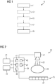
- the Fig. 1 shows an exemplary embodiment of the method 10 according to the invention.
- the method 10 for generating a mixed image data set based on an energy-resolved image of an energy-resolving X-ray detector 25 has the steps of generating 11 and mixing 19.
- a first image value of a first image element of a first image data set in a first energy range and a second image value of the first image element of a second image data set are in a second Energy area generated.
- the first image value and the second image value are mixed as a function of an image parameter and a mixed image data set is generated with a mixed image value of the first image element.
- the mixed image data record has a mixed image value of the first image element in at least the first image element. In other picture elements, the mixed picture can have the respective first picture value or the respective second picture value.
- Mixing 19 is carried out on a detector element basis in the local area.
- the method 10 can also have a step of determining 13, the image parameter being determined at least based on at least a partial area of the first image data set and / or the second image data set.
- the image parameter is based on an image property, a spatial frequency band property or a time-variable image property compared to a previous picture.
- the temporally variable image property can be determined by means of external sensors.
- the method 10 can further comprise a step of filtering 17 by means of a filter function of the first image data set and / or the second image data set.
- the method 10 can further comprise a step of optimizing 15 the filter function based on an examination type.
- the Fig. 2 shows an exemplary embodiment of the medical device 20 according to the invention.
- the medical device 20 comprises an x-ray source 21 and an opposing energy-resolving x-ray detector 25.
- the x-ray source 21 emits x-rays 35.
- the examination object 23 is arranged between the x-ray source 21 and the x-ray detector 25.
- the x-ray detector 25 has a plurality of detector elements 27. An image value can be determined based on the measured value of the detector element 27. In the simplest case, the image value can essentially correspond to the measured value.
- the medical device 20 further comprises an image generation unit 29.
- the image generation unit 29 is connected to a display unit 31 and an input unit 33.
- the image generation unit 29 comprises a generation unit 41 for generating the first image data set and the second image data set and a mixing unit 43 for mixing the first image values and the second image values as a function of an image parameter and thereby generating the mixed image data set with a mixed image value of the first image element.
- the image generation unit 29 can further comprise a filter unit 45, a determination unit 47 and an optimization unit 49.
Landscapes
- Engineering & Computer Science (AREA)
- Health & Medical Sciences (AREA)
- Physics & Mathematics (AREA)
- General Physics & Mathematics (AREA)
- Theoretical Computer Science (AREA)
- Medical Informatics (AREA)
- Biomedical Technology (AREA)
- Life Sciences & Earth Sciences (AREA)
- Public Health (AREA)
- General Health & Medical Sciences (AREA)
- Business, Economics & Management (AREA)
- Radiology & Medical Imaging (AREA)
- Epidemiology (AREA)
- General Business, Economics & Management (AREA)
- Biophysics (AREA)
- High Energy & Nuclear Physics (AREA)
- Nuclear Medicine, Radiotherapy & Molecular Imaging (AREA)
- Optics & Photonics (AREA)
- Pathology (AREA)
- Primary Health Care (AREA)
- Heart & Thoracic Surgery (AREA)
- Molecular Biology (AREA)
- Surgery (AREA)
- Animal Behavior & Ethology (AREA)
- Veterinary Medicine (AREA)
- Computer Vision & Pattern Recognition (AREA)
- Apparatus For Radiation Diagnosis (AREA)
Priority Applications (2)
| Application Number | Priority Date | Filing Date | Title |
|---|---|---|---|
| EP18197539.2A EP3629283A1 (fr) | 2018-09-28 | 2018-09-28 | Génération d'un ensemble de données d'image mixte |
| US16/580,100 US11205255B2 (en) | 2018-09-28 | 2019-09-24 | Generating a mixed image data record |
Applications Claiming Priority (1)
| Application Number | Priority Date | Filing Date | Title |
|---|---|---|---|
| EP18197539.2A EP3629283A1 (fr) | 2018-09-28 | 2018-09-28 | Génération d'un ensemble de données d'image mixte |
Publications (1)
| Publication Number | Publication Date |
|---|---|
| EP3629283A1 true EP3629283A1 (fr) | 2020-04-01 |
Family
ID=63708253
Family Applications (1)
| Application Number | Title | Priority Date | Filing Date |
|---|---|---|---|
| EP18197539.2A Withdrawn EP3629283A1 (fr) | 2018-09-28 | 2018-09-28 | Génération d'un ensemble de données d'image mixte |
Country Status (2)
| Country | Link |
|---|---|
| US (1) | US11205255B2 (fr) |
| EP (1) | EP3629283A1 (fr) |
Citations (4)
| Publication number | Priority date | Publication date | Assignee | Title |
|---|---|---|---|---|
| WO2002028154A1 (fr) * | 2000-09-29 | 2002-04-04 | Analogic Corporation | Procede et systeme d'amelioration des caracteristiques signal/bruit d'images provenant d'un detecteur numerique de rayons x |
| DE102009032441A1 (de) * | 2009-07-09 | 2011-01-27 | Siemens Aktiengesellschaft | Verfahren zur automatischen Bearbeitung eines Röntgenbildes, Röntgensystem und Computerprogrammprodukt |
| WO2012015285A2 (fr) * | 2010-07-29 | 2012-02-02 | Samsung Electronics Co., Ltd. | Procédé et appareil de traitement de l'image et système médical utilisant ledit appareil |
| US20160350910A1 (en) * | 2015-05-27 | 2016-12-01 | Samsung Electronics Co., Ltd. | X-ray apparatus and control method for the same |
Family Cites Families (1)
| Publication number | Priority date | Publication date | Assignee | Title |
|---|---|---|---|---|
| KR102153345B1 (ko) * | 2013-05-30 | 2020-09-09 | 삼성전자주식회사 | 방사선 촬영 장치 및 방사선 촬영 장치의 제어 방법 |
-
2018
- 2018-09-28 EP EP18197539.2A patent/EP3629283A1/fr not_active Withdrawn
-
2019
- 2019-09-24 US US16/580,100 patent/US11205255B2/en active Active
Patent Citations (4)
| Publication number | Priority date | Publication date | Assignee | Title |
|---|---|---|---|---|
| WO2002028154A1 (fr) * | 2000-09-29 | 2002-04-04 | Analogic Corporation | Procede et systeme d'amelioration des caracteristiques signal/bruit d'images provenant d'un detecteur numerique de rayons x |
| DE102009032441A1 (de) * | 2009-07-09 | 2011-01-27 | Siemens Aktiengesellschaft | Verfahren zur automatischen Bearbeitung eines Röntgenbildes, Röntgensystem und Computerprogrammprodukt |
| WO2012015285A2 (fr) * | 2010-07-29 | 2012-02-02 | Samsung Electronics Co., Ltd. | Procédé et appareil de traitement de l'image et système médical utilisant ledit appareil |
| US20160350910A1 (en) * | 2015-05-27 | 2016-12-01 | Samsung Electronics Co., Ltd. | X-ray apparatus and control method for the same |
Also Published As
| Publication number | Publication date |
|---|---|
| US11205255B2 (en) | 2021-12-21 |
| US20200104985A1 (en) | 2020-04-02 |
Similar Documents
| Publication | Publication Date | Title |
|---|---|---|
| DE102013200021B4 (de) | Verfahren zur Kalibrierung eines zählenden digitalen Röntgendetektors, Röntgensysteme zur Durchführung eines solchen Verfahrens und Verfahren zur Aufnahme eines Röntgenbildes | |
| EP3839577B1 (fr) | Procédé de génération d'un ensemble de données de radiographie | |
| DE102011076346B4 (de) | Verfahren und Computertomographiesystem zur Erzeugung tomographischer Bilddatensätze | |
| DE69711124T2 (de) | Verfahren und Vorrichtung zur Aufnahme von Röntgen- und Gammastrahlen-Bildern mit Optimierung der Belichtungszeit | |
| DE102010024626B4 (de) | Zählender Detektor und Computertomographiesystem | |
| DE102011076781B4 (de) | Verfahren zur Korrektur einer Zählratendrift bei einem quantenzählenden Detektor, Röntgen-System mit quantenzählendem Detektor und Schaltungsanordnung für einen quantenzählenden Detektor | |
| DE102011077859B4 (de) | Quantenzählender Strahlungsdetektor | |
| DE102010060989A1 (de) | System und Verfahren zur Kompensation von Daten schwacher Signale für Dualenergie-CT | |
| EP3798684B1 (fr) | Correction de données dans l'imagerie par rayons x | |
| DE102015218928B4 (de) | Verfahren zur Erzeugung von Röntgenbilddaten eines Untersuchungsobjektes mit unterdrücktem Calcium-Signal | |
| DE10106221A1 (de) | Röntgendetektor mit großem Dynamikbereich | |
| DE102015207107A1 (de) | Verfahren zur Erzeugung einer virtuellen Röntgenprojektion anhand eines mittels Röntgenbildaufnahmevorrichtung erhaltenen Bilddatensatzes, Computerprogramm, Datenträger sowie Röntgenbildaufnahmevorrichtung | |
| DE102005027436B4 (de) | Verfahren zur Berechnung von absorberspezifischen Gewichtungskoeffizienten und Verfahren zur Verbesserung eines von einem Absorber abhängigen Kontrast-zu-Rausch-Verhältnisses in einem von einer Röntgeneinrichtung erzeugten Röntgenbild eines zu untersuchenden Objektes | |
| DE102011076351A1 (de) | Verfahren und Computertomographiesystem zur Erzeugung tomographischer Bilddatensätze | |
| DE102011005539A1 (de) | Verfahren zur Detektion von Röntgenstrahlung und Detektorsystem mit direktkonvertierenden Detektoren | |
| DE102017213479A1 (de) | Computertomographische Aufnahme mit verschiedenen Energieschwellensätzen | |
| EP3327673B1 (fr) | Génération d'images ct à haute résolution comportant des informations spectrales | |
| EP3795082B1 (fr) | Procédé et dispositif de génération d'un ensemble de données d'image de tomodensitométrie | |
| DE69004308T2 (de) | Verfahren zur Elimination von gestreuten Gammastrahlen und zur Rekonstruktion eines Bildes und Gamma-Kameravorrichtung. | |
| DE602005004410T2 (de) | System und verfahren zur korrektur zeitlicher artefakte in tomographischen bildern | |
| DE102015212155A1 (de) | Verfahren zur Aufnahme von energieselektiven Bilddaten, Röntgendetektor und Röntgensystem | |
| DE102015217617A1 (de) | Verfahren zum Korrigieren von Röntgenbilddaten umfassend Information bezüglich eines Zerfallsprozesses eines radioaktiven Materials | |
| DE102014225399B4 (de) | Rauschunterdrückung bei der Korrektur von Artefakten von CT-Bildern | |
| DE10361397A1 (de) | Bildaufnahmevorrichtung | |
| DE69323257T2 (de) | Nuklearmedizinische Bildwiedergabe - Einrichtung |
Legal Events
| Date | Code | Title | Description |
|---|---|---|---|
| PUAI | Public reference made under article 153(3) epc to a published international application that has entered the european phase |
Free format text: ORIGINAL CODE: 0009012 |
|
| STAA | Information on the status of an ep patent application or granted ep patent |
Free format text: STATUS: THE APPLICATION HAS BEEN PUBLISHED |
|
| AK | Designated contracting states |
Kind code of ref document: A1 Designated state(s): AL AT BE BG CH CY CZ DE DK EE ES FI FR GB GR HR HU IE IS IT LI LT LU LV MC MK MT NL NO PL PT RO RS SE SI SK SM TR |
|
| AX | Request for extension of the european patent |
Extension state: BA ME |
|
| STAA | Information on the status of an ep patent application or granted ep patent |
Free format text: STATUS: THE APPLICATION IS DEEMED TO BE WITHDRAWN |
|
| 18D | Application deemed to be withdrawn |
Effective date: 20201002 |