EP3632485A1 - Traitement de l'insuffisance cardiaque congestive - Google Patents
Traitement de l'insuffisance cardiaque congestive Download PDFInfo
- Publication number
- EP3632485A1 EP3632485A1 EP19197851.9A EP19197851A EP3632485A1 EP 3632485 A1 EP3632485 A1 EP 3632485A1 EP 19197851 A EP19197851 A EP 19197851A EP 3632485 A1 EP3632485 A1 EP 3632485A1
- Authority
- EP
- European Patent Office
- Prior art keywords
- pump
- assist device
- conduit
- cases
- aorta
- Prior art date
- Legal status (The legal status is an assumption and is not a legal conclusion. Google has not performed a legal analysis and makes no representation as to the accuracy of the status listed.)
- Granted
Links
Images


Classifications
-
- A—HUMAN NECESSITIES
- A61—MEDICAL OR VETERINARY SCIENCE; HYGIENE
- A61N—ELECTROTHERAPY; MAGNETOTHERAPY; RADIATION THERAPY; ULTRASOUND THERAPY
- A61N1/00—Electrotherapy; Circuits therefor
- A61N1/18—Applying electric currents by contact electrodes
- A61N1/32—Applying electric currents by contact electrodes alternating or intermittent currents
- A61N1/38—Applying electric currents by contact electrodes alternating or intermittent currents for producing shock effects
- A61N1/39—Heart defibrillators
- A61N1/3956—Implantable devices for applying electric shocks to the heart, e.g. for cardioversion
- A61N1/3962—Implantable devices for applying electric shocks to the heart, e.g. for cardioversion in combination with another heart therapy
-
- A—HUMAN NECESSITIES
- A61—MEDICAL OR VETERINARY SCIENCE; HYGIENE
- A61M—DEVICES FOR INTRODUCING MEDIA INTO, OR ONTO, THE BODY; DEVICES FOR TRANSDUCING BODY MEDIA OR FOR TAKING MEDIA FROM THE BODY; DEVICES FOR PRODUCING OR ENDING SLEEP OR STUPOR
- A61M60/00—Blood pumps; Devices for mechanical circulatory actuation; Balloon pumps for circulatory assistance
- A61M60/10—Location thereof with respect to the patient's body
- A61M60/122—Implantable pumps or pumping devices, i.e. the blood being pumped inside the patient's body
- A61M60/165—Implantable pumps or pumping devices, i.e. the blood being pumped inside the patient's body implantable in, on, or around the heart
-
- A—HUMAN NECESSITIES
- A61—MEDICAL OR VETERINARY SCIENCE; HYGIENE
- A61M—DEVICES FOR INTRODUCING MEDIA INTO, OR ONTO, THE BODY; DEVICES FOR TRANSDUCING BODY MEDIA OR FOR TAKING MEDIA FROM THE BODY; DEVICES FOR PRODUCING OR ENDING SLEEP OR STUPOR
- A61M60/00—Blood pumps; Devices for mechanical circulatory actuation; Balloon pumps for circulatory assistance
- A61M60/20—Type thereof
- A61M60/247—Positive displacement blood pumps
- A61M60/253—Positive displacement blood pumps including a displacement member directly acting on the blood
- A61M60/268—Positive displacement blood pumps including a displacement member directly acting on the blood the displacement member being flexible, e.g. membranes, diaphragms or bladders
- A61M60/279—Peristaltic pumps, e.g. roller pumps
-
- A—HUMAN NECESSITIES
- A61—MEDICAL OR VETERINARY SCIENCE; HYGIENE
- A61M—DEVICES FOR INTRODUCING MEDIA INTO, OR ONTO, THE BODY; DEVICES FOR TRANSDUCING BODY MEDIA OR FOR TAKING MEDIA FROM THE BODY; DEVICES FOR PRODUCING OR ENDING SLEEP OR STUPOR
- A61M60/00—Blood pumps; Devices for mechanical circulatory actuation; Balloon pumps for circulatory assistance
- A61M60/50—Details relating to control
- A61M60/508—Electronic control means, e.g. for feedback regulation
- A61M60/515—Regulation using real-time patient data
-
- A—HUMAN NECESSITIES
- A61—MEDICAL OR VETERINARY SCIENCE; HYGIENE
- A61M—DEVICES FOR INTRODUCING MEDIA INTO, OR ONTO, THE BODY; DEVICES FOR TRANSDUCING BODY MEDIA OR FOR TAKING MEDIA FROM THE BODY; DEVICES FOR PRODUCING OR ENDING SLEEP OR STUPOR
- A61M60/00—Blood pumps; Devices for mechanical circulatory actuation; Balloon pumps for circulatory assistance
- A61M60/80—Constructional details other than related to driving
- A61M60/855—Constructional details other than related to driving of implantable pumps or pumping devices
- A61M60/865—Devices for guiding or inserting pumps or pumping devices into the patient's body
- A61M60/867—Devices for guiding or inserting pumps or pumping devices into the patient's body using position detection during deployment, e.g. for blood pumps mounted on and driven through a catheter
-
- A—HUMAN NECESSITIES
- A61—MEDICAL OR VETERINARY SCIENCE; HYGIENE
- A61M—DEVICES FOR INTRODUCING MEDIA INTO, OR ONTO, THE BODY; DEVICES FOR TRANSDUCING BODY MEDIA OR FOR TAKING MEDIA FROM THE BODY; DEVICES FOR PRODUCING OR ENDING SLEEP OR STUPOR
- A61M2205/00—General characteristics of the apparatus
- A61M2205/33—Controlling, regulating or measuring
- A61M2205/3303—Using a biosensor
-
- A—HUMAN NECESSITIES
- A61—MEDICAL OR VETERINARY SCIENCE; HYGIENE
- A61M—DEVICES FOR INTRODUCING MEDIA INTO, OR ONTO, THE BODY; DEVICES FOR TRANSDUCING BODY MEDIA OR FOR TAKING MEDIA FROM THE BODY; DEVICES FOR PRODUCING OR ENDING SLEEP OR STUPOR
- A61M60/00—Blood pumps; Devices for mechanical circulatory actuation; Balloon pumps for circulatory assistance
- A61M60/10—Location thereof with respect to the patient's body
- A61M60/122—Implantable pumps or pumping devices, i.e. the blood being pumped inside the patient's body
- A61M60/126—Implantable pumps or pumping devices, i.e. the blood being pumped inside the patient's body implantable via, into, inside, in line, branching on, or around a blood vessel
- A61M60/148—Implantable pumps or pumping devices, i.e. the blood being pumped inside the patient's body implantable via, into, inside, in line, branching on, or around a blood vessel in line with a blood vessel using resection or like techniques, e.g. permanent endovascular heart assist devices
-
- A—HUMAN NECESSITIES
- A61—MEDICAL OR VETERINARY SCIENCE; HYGIENE
- A61M—DEVICES FOR INTRODUCING MEDIA INTO, OR ONTO, THE BODY; DEVICES FOR TRANSDUCING BODY MEDIA OR FOR TAKING MEDIA FROM THE BODY; DEVICES FOR PRODUCING OR ENDING SLEEP OR STUPOR
- A61M60/00—Blood pumps; Devices for mechanical circulatory actuation; Balloon pumps for circulatory assistance
- A61M60/40—Details relating to driving
- A61M60/403—Details relating to driving for non-positive displacement blood pumps
- A61M60/408—Details relating to driving for non-positive displacement blood pumps the force acting on the blood contacting member being mechanical, e.g. transmitted by a shaft or cable
- A61M60/411—Details relating to driving for non-positive displacement blood pumps the force acting on the blood contacting member being mechanical, e.g. transmitted by a shaft or cable generated by an electromotor
- A61M60/414—Details relating to driving for non-positive displacement blood pumps the force acting on the blood contacting member being mechanical, e.g. transmitted by a shaft or cable generated by an electromotor transmitted by a rotating cable, e.g. for blood pumps mounted on a catheter
-
- A—HUMAN NECESSITIES
- A61—MEDICAL OR VETERINARY SCIENCE; HYGIENE
- A61N—ELECTROTHERAPY; MAGNETOTHERAPY; RADIATION THERAPY; ULTRASOUND THERAPY
- A61N1/00—Electrotherapy; Circuits therefor
- A61N1/18—Applying electric currents by contact electrodes
- A61N1/32—Applying electric currents by contact electrodes alternating or intermittent currents
- A61N1/36—Applying electric currents by contact electrodes alternating or intermittent currents for stimulation
- A61N1/362—Heart stimulators
- A61N1/3627—Heart stimulators for treating a mechanical deficiency of the heart, e.g. congestive heart failure or cardiomyopathy
Definitions
- This document relates to materials and methods for circulatory bypass of a ventricle in the heart of a mammal.
- materials and methods for bypassing a permanently or temporarily impaired left ventricle in the heart of a mammal e.g ., a human
- a mammal e.g ., a human
- Congestive heart failure affects the ability of the heart to provide sufficient blood flow to the organs of the body.
- Conditions such as coronary artery disease, scar tissue from a myocardial infarction, high blood pressure, and heart valve disease can contribute to congestive heart failure.
- Pharmaceutical intervention can expand blood vessels and/or or lower blood pressure to allow blood to flow more easily and the heart to pump more efficiently. If the damage to the heart is extensive, surgical intervention can be required.
- This document relates to materials and methods for circulatory bypass of a ventricle in the heart of a mammal.
- materials and methods for bypassing a permanently or temporarily impaired left ventricle in the heart of a mammal are provided.
- the devices provided herein can be used as percutaneous assist devices for enhancing blood flow from a failing heart (e.g., a heart with a failing left ventricle or valvular disease).
- the methods provided herein can be used to position an assist device within the heart of a mammal (e.g ., within the shared wall of the aorta and left atrium of a human heart).
- Assist devices provided herein can be configured to reduce the risk of thrombosis and conform to the anatomy of a recipient without causing damage to the heart itself.
- assist devices can be used to support the function of a heart to treat congestive heart failure (e.g., left, right, and bilateral failure), cardiac arrhythmias (e.g., tachycardia and fibrillations), diastolic dysfunction, or valve disease (e.g., aortic and/or mitral valve stenosis).
- an assist device can be configured to provide phasic flow to coordinate device activity with the natural rhythm of a heart.
- sensing and/or pacing electrodes can be implanted with an assist device to time blood flow.
- An assist device provided herein can be used to replace or support other cardiac technologies (e.g ., defibrillators or pacemakers).
- assist devices can be combined to provide a complete heart system (e.g., left ventricular and right ventricular assist devices), which can be used to replace a failing heart.
- assist devices provided herein can be used to bypass an impaired heart and to sustain circulation in a patient with end-stage heart failure until a donor heart or an artificial heart can be implanted ( e.g ., as a bridge to transplant).
- the ventricular assist device comprises, or consists essentially of, a conduit and a pump.
- the conduit can include a proximal region, a distal region and an intermediate region located between the proximal and distal regions.
- the proximal region can be adapted to be positioned within a first compartment in the cardiovascular system of the mammal, and the distal region can be adapted to be positioned within a second compartment in the cardiovascular system.
- the intermediate region can define a lumen through a wall between the first and second compartments.
- the pump can be adapted to be positioned within the conduit.
- the mammal can be a human.
- the conduit can be constructed of expandable or malleable material.
- the malleable material can be nitinol.
- the proximal region and the distal region can be independently adjustable.
- the intermediate region can comprise a tubular core.
- the tubular core can comprise a septum or valve.
- the first compartment can be the aorta and the second compartment can be the left atrium.
- the ventricular assist device can comprise electrodes.
- this document features a method for treating heart disease in a mammal.
- the method comprises, or consists essentially of, implanting an assist device within the cardiovascular system of the mammal, under conditions wherein blood flow in the mammal is enhanced, thereby treating the heart disease.
- the assist device can comprise, or consist essentially of, a conduit, and a pump.
- the conduit can comprise a proximal region, a distal region and an intermediate region located between the proximal and distal regions.
- the proximal region can be adapted to be positioned within a first compartment in the cardiovascular system of the mammal, and the distal region can be adapted to be positioned within a second compartment in the cardiovascular system.
- the intermediate region can define a lumen through a wall between the first and second compartments.
- the pump can be adapted to be positioned within the conduit.
- the method can include identifying the mammal as having a heart disease selected from the group consisting of congestive heart failure, valvular disease, and malignant cardiac arrhythmia.
- the mammal can be a human.
- the implanting step can comprise inserting the conduit into a shared wall between the aorta and the left atrium of the mammal.
- the conduit can be constructed of expandable or malleable material.
- the malleable material can be nitinol.
- the proximal region and the distal region can be independently adjustable.
- the intermediate region can comprise a tubular core.
- the tubular core can comprise a septum or valve.
- the first compartment can be the aorta and the second compartment can be the left atrium.
- the ventricular assist device can include electrodes.
- this document features a method for implanting an assist device into a human.
- the method comprises, or consists essentially of, inserting a conduit having a proximal region, a distal region, and an intermediate region located between the proximal and distal regions into a position wherein the proximal region is located within the aorta of the human, the distal region is located within the left atrium of the human, and the intermediate region defines a lumen through the shared wall between the aorta and left atrium, and inserting a pump into the conduit such that blood is pumped through the conduit from the left atrium to the aorta.
- a heart is a muscular organ responsible for pumping blood in the body of a mammal.
- a healthy heart functions by collecting de-oxygenated blood from the body in the right atrium and pumping it, via the right ventricle outlet (LVO), into the lungs to be re-oxygenated.
- the left side of the heart collects oxygenated blood from the lungs into the left atrium (LA).
- LV left ventricle
- Ao aorta
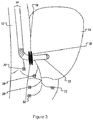
- an assist device 10 can be positioned within two compartments of the cardiovascular system.
- an assist device can enhance the flow of blood to a first compartment, such as the aorta 12, from a second compartment, such as the left atrium 14, via a puncture in a shared wall 16 of the two compartments.
- a percutaneous assist device can include a conduit 20.
- a conduit can facilitate placement of a pump 22 to enhance blood flow.
- a pump can transport blood from the left atrium to the aorta via a pump inflow 24 located in the left atrium and a pump outflow 26 located in the aorta.
- the pump can be manipulated into position in the cardiovascular system via a delivery element 28 that functions to place and then secure the pump in position between the left atrium and ascending aorta, for example.
- An assist device described herein can enhance the flow of blood between any first compartment and second compartment in the cardiovascular system of a mammal.
- a compartment can be any chamber of the heart or any blood vessel in the heart or the peripheral circulation.
- a first and second compartment can be in close apposition.
- a first and second compartment can share a common wall (e.g., the aorta and the left atrium).
- a pump of an assist device can be any type of blood pump for insertion into the cardiovascular system of a mammal including, without limitation, a positive displacement pump, a roller pump, a centrifugal pump, an impeller pump, an axial flow pump, a pulsatile pump, a magnetic flux pump, and a continuous-flow pump.
- a positive displacement pump e.g., a positive displacement pump, a roller pump, a centrifugal pump, an impeller pump, an axial flow pump, a pulsatile pump, a magnetic flux pump, and a continuous-flow pump.
- Suitable pumps can be inserted and secured into position and later removed or replaced, as necessary.
- pumps can be pre-fitted on a delivery element and configured for specific anatomic locations.
- a pump can be designed with any length required for an ultimate anatomic destination.
- a pump for placement between the left atrium and ascending aorta can have a relatively short length, for example.
- an assist device can have more than one pump (e.g ., multiple pumps connected in series).
- a first pump can be positioned between the left atrium and ascending aorta, and include a second pump placed between the right atrial appendage and the pulmonary trunk that is connected to the first pump.
- a pump can be designed to pass through a conduit.
- a pump can be adapted so that one end of the pump (e.g., the inflow) can be positioned near one end of a conduit ( e.g., near the distal end) and the other end of the pump ( e.g., the outflow) can be positioned near the other end of a conduit ( e.g ., the proximal end).
- the distance between a pump's inflow and outflow can be relatively short ( e.g., the thickness of the shared wall between the left atrium and the ascending aorta).
- a pump can be designed so that the pump motor and/or power supply can be located outside the conduit ( e.g ., for implantation subcutaneously in a mammal's infraclavicular, pectoral, or abdominal region).
- a pump can be adapted to reduce the risk of thrombus formation.
- a pump can be designed to minimize contact between the blood and the pump components, thereby presenting a limited risk of hemolysis and thrombus formation ( e.g ., by shortening the length of components).
- a pump can feature a diverting element (e.g., a funnel-like shelf) that directs blood flow backwards onto the pump itself.
- an assist device can be adapted to minimize ventricular stasis and the risk of thrombus formation by providing side ports or irrigation ports to the pump at the atrial end of the pump ( i.e., inflow). Side ports or irrigation ports can divert blood flow to irrigate the pump itself, thereby preventing stasis and thrombus formation.
- a pump can feature a combination of side ports, irrigation ports, and diverting elements.
- side ports or irrigation ports in a pump can depend on the anatomical destination of the pump or the surgical approach used to position the pump.
- sections of the pump that are in contact with a left-sided circulation can feature multiple irrigation ports (e.g., a pump that will bridge the Fossa Ovalis to the aorta or a transaortic pump).
- a pump that can be implanted using a retrograde approach can have a lengthy portion featuring numerous side ports or irrigation ports.
- Inflow at side ports facing the mitral valve can create a circulation-like effect.
- a side port can be extended to the left atrial appendage or in the direction of the left atrial appendage to prevent stasis in the left atrial appendage.
- An extended side port can be placed across the atrio-ventricular valve, or just atrial to the atrio-ventricular valve, to function as an inflow or intermittent outflow port.
- a pump can be configured to reverse the direction of flow from a side port on regular intervals ( e.g ., every about 5 or 10 beats).
- An intermittent inflow and outflow to side ports can be used to create an agitation-type effect and decrease the risk of thrombus formation.
- a pump can require continuous irrigation.
- a direct current charge can be used to prevent thrombus formation on an implanted assist device.
- the device can be coated with a dielectric and can be configured to distribute a charge as described elsewhere (e.g ., WIPO Patent Application WO/2008/02471 ).
- the battery source that powers the pump can power side ports to produce a very low flow circulation on the surface of the tubing, wiring, and pump itself.
- a second pump can be used to circulate blood along the left sided course of a primary assist device to reduce the risk of thrombus formation.
- a pump can be powered by any appropriate power supply used with implanted devices (e.g ., batteries/power source in the pump, or batteries positioned in the peripheral venous system, the infra-pectoral region, or other regions utilized in percutaneous techniques).
- a pump can share a power supply with another implantable device.
- a pump can be configured to alter blood flow in response to external stimuli.
- a pump can be adapted to receive signals from sensing electrodes 30 capable of detecting atrial and ventricular function.
- a pump can be adapted to receive input from a micro electro-mechanical sensor (e.g., a multi-axis accelerometer) or series of single-axis sensors that measure changes in body position. For example, rapid changes in body position (e.g ., syncope) would activate the pump, or increase pump activity, to maintain perfusion during changes in body position.
- a pump can be adapted to coordinate with cardiac function using pacing electrodes 32 and timing algorithms.
- a pump can be capable of receiving signals from sensing electrodes, and/or be capable of responding to a cardiac event. For example, a pump can deliver an electrical shock in response to an arrhythmia ( e.g ., to the left ventricle).
- sensing electrodes can be connected to an assist device to record changes in the electric potentials of specific cardiac loci.
- a pressure sensor can be connected to an assist device.
- Sensing and pressure electrodes can permit real-time, online calculation of variations in heart rates and relative systolic and diastolic filling times to determine an algorithm that incorporates the timing of atrial signals at two different sites ( e.g., to provide inter-atrial and intra-ventricular conduction times) and the pressure signals to assess electromechanical translation times. For example, the relative times between systole and diastole and heart rate can be predicted about 10 to 15 beats in advance.
- a pump can be combined with pacing electrodes, to pace the heart according to timing algorithms determined by real-time calculations.
- the pump can be combined with pressure and sensing electrodes, and a QT sensor, impendence sensor, or accelerometer.
- an assist device can include a conduit 20 to provide access to two compartments of the cardiovascular system.
- a conduit can have a proximal region 34, a distal region 36, and an intermediate region 38 located between the proximal and distal regions. When viewed from either the proximal end or the distal end, the intermediate region of a conduit can define a lumen.
- the proximal and distal regions can be configured to secure a conduit within the cardiovascular system, for example, as shown in Figure 4A .
- the proximal and distal regions can include a covering 40, for example, as shown in Figure 4B .
- the lumen defined by the intermediate region of the conduit can be supported by a tubular core 42, for example, as shown in Figure 4C , which can include a septum or valve 44 as shown in Figure 4D , for example.
- the dimensions of a conduit can be adapted to allow a needle or dilator 46 to pass through the intermediate region via the lumen, and then allow the conduit and needle to be inserted into a delivery sheath 48 for placement in the cardiovascular system, as shown in Figure 4E , for example.
- a conduit can be constructed as one piece or multiple pieces ( e.g ., with separate proximal and distal regions).
- a conduit can be constructed from compressible, expandable, or malleable materials (e.g ., a balloon or shape-memory alloy (nickel titanium (nitinol)).
- compressible, expandable, or malleable materials e.g ., a balloon or shape-memory alloy (nickel titanium (nitinol)
- construction materials can permit the proximal and distal regions of a conduit to be deformed or compressed within the delivery sheath, but regain their original shape (e.g. lip, rim, or disk) when the sheath is retracted.
- the expandable region can be filled with any suitable material for providing long-term stability (e.g ., a polymer capable of crosslinking, thermosetting, or hardening).
- a conduit can be constructed from magnetic or paramagnetic materials (e.g ., to secure two close, but separate compartments of the cardiovascular system). Regions that are constructed from woven nitinol can be covered in a material for an atraumatic, fluid-tight seal ( e.g ., biocompatible polymers or fabrics).
- a tubular core can be constructed from any material that will support the intermediate region ( e.g ., polymer and/or metal).
- a septum or valve within a tubular core can be configured to prevent or control blood flow. In some cases, a septum can be adapted to be punctured to permit blood flow ( e.g., upon pump placement).
- the conduit size and shape can be adapted for the implantation site, method, or the anatomy of the recipient.
- a conduit can be configured to be positioned between two chambers of a mammal's heart, between a chamber of the heart and a blood vessel, or within a blood vessel ( e.g. to increase perfusion in the peripheral circulatory system).
- a conduit can be configured for insertion in the shared wall between the aorta and the left atrium of a recipient.
- a conduit can be configured to puncture the shared wall.
- the proximal and distal regions of a conduit can be adapted to fit securely against the site of implantation, while not fitting so tightly as to cause necrosis or other tissue damage.
- proximal and distal regions of a conduit can be independently adjustable to be adapted to a specific recipient's anatomy as shown in Figure 4A .
- a conduit can be adapted to be secured to a tissue in a mammal (e.g ., myocardium).
- the proximal and distal regions of a conduit can feature teeth or tines that can be twisted into the aortic valve, thereby securing the conduit, for example.
- a conduit can be adapted to be secured using sutures.
- an assist device can include more than one conduit 20 to control blood flow.
- an assist device can include a second conduit to act as a valve or balloon-like device to be positioned in the aorta 12.
- Activation of balloon expansion or valve closure can be controlled by timing algorithms as described herein and/or by direct timing. This configuration can provide accentuated phasic systolic flow and greater diastolic filling into the coronary arteries.
- the position of the expandable conduit or valve can be variable. For example, a longer length can be used for recipients with a bypass graft. In some cases, the timing can be different from the timing of the native aortic valve. No prospective algorithms or pump design changes are required with this configuration because the flow rate is consistent.
- An assist device can be inserted into the cardiovascular system of a mammal using cardiac catheterization techniques (e.g ., percutaneously). For example, a doctor can insert a delivery sheath into an artery or vein in a limb of a mammal. The sheath can be advanced into the chambers of the heart.
- a mammal can be any type of mammal including, without limitation, a mouse, rat, dog, cat, horse, sheep, goat, cow, pig, monkey, or human.
- a mammal can be identified as having a cardiovascular disease, such as congestive heart failure, valvular disease, or malignant arrhythmia using standard diagnostic techniques.
- a mammal can have an artificial pacemaker or an implanted cardioverter-defibrillator.
- a needle or dilator can be used to puncture a shared wall in the cardiovascular system to move a sheath into place across the shared wall (e.g., the wall between the non-coronary cusp of the aortic valve and the left atrium) as shown in Figure 6A , for example.
- puncturing can be accomplished using a radiofrequency energy/cautery or mechanical drill at the tip of the needle or dilator.
- a conduit can be advanced out of the sheath by moving the distal region out of the sheath for placement on the distal side of the wall ( e.g., as shown in Figure 6B ), and then the sheath can be pulled back into the proximal side of the wall exposing the intermediate region of the conduit ( e.g ., as shown in Figure 6C ).
- the proximal region of the conduit can be advanced out of a sheath against the proximal wall (e.g., as shown in Figure 6D ).
- a pump 22 can be advanced through a sheath 48 and through a conduit 20 in the shared wall 16 of the aorta 12 and left atrium 14 such that blood can be pumped from the left atrium into the aorta, for example, as shown in Figure 6E .
- the left atrium is accessed in a retrograde fashion.
- a needle can access the descending aorta and puncture the left atrium wall.
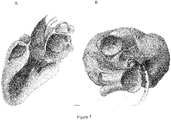
- a pump can be placed in the ascending aorta with the inflow in the left atrium (e.g ., as shown in Figures 2 and 7 ).
- any aortic cusp can be used to access the left atrium (e.g., the left coronary cusp or non-coronary cusp).
- a conduit can be placed to permit communication between the posterior part of the left coronary cusp and the left atrium.
- an electrogram guidance or other modality can be used.
- a puncturing device e.g., a needle or wire
- electrodes can be positioned at the tip of the needle (and/or wire) and on the shaft.
- a large near-field ventricular electrogram can be obtained.
- the electrogram can show that the needle or wire is above the valve plane, indicating that the needle or wire is pointing to the right coronary cusp; the surgeon would not advance that direction.
- the surgeon can turn the needle or wire (if coming from the femoral) clockwise after withdrawing from the depths of the cusp until a large atrial electrogram and a far-field ventricular electrogram can be seen.
- the surgeon can advance the needle or wire into the noncoronary cusp.
- the timing of the local electrogram relative to the P wave (assuming sinus rhythm) can be checked. For example, an electrogram in the first 20 to 40% of the P wave can signify neighboring right atrium, and the needle or wire can puncture between the noncoronary cusp and the right atrium. If a local electrogram is in the terminal 40% of the P wave, the electrogram can indicate that the needle or wire is at the wall of the left atrium.
- a very large atrial electrogram which is not in the terminal 40%, can indicate that the needle or wire is adjacent to the interatrial septum.
- a surgeon can use the electrogram to turn the delivery catheter further counterclockwise, until the atrial electrogram becomes smaller (while remaining larger than the ventricular electrograms), indicating that the left atrium is neighboring.
- a relatively equal atrial and ventricular electrogram with neither particularly near field can indicate that the needle or wire is in the left coronary cusp. From this position, a surgeon can withdraw the delivery catheter slightly and then turn or torque the delivery catheter clockwise to enter the noncoronary cusp. A surgeon can remain within the left coronary cusp, while avoiding the left main coronary artery, turn the needle or wire clockwise within the cusp, and then puncture from the left coronary cusp to the left atrium.
- the ascending aorta posterior can be accessed through the right atrium 50 using a transseptal approach.
- the right atrium can be accessed using a venous approach
- the left atrium 14 can be accessed by a transseptal puncture.
- the transseptal needle can be directed anteriorly to puncture from the left atrium into the ascending aorta 12.
- the portion of the pump that bridges the Fossa Ovalis to the aorta can be adapted to prevent thrombus formation.
- a left ventricular assist device can be positioned using an epicardial method.
- the ascending aorta is the immediate anterior neighbor of the left atrium in the left part of the floor of the transverse sinus.
- the transverse sinus can be cannulated.
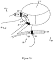
- a first conduit 20 can be positioned in the wall of left atrium 14
- a second conduit can be positioned in the wall of the ascending aorta 12
- a pump 22 can be positioned through the first and second conduit to provide blood flow from the left atrium ( e.g., via pump inflow 24 ) to the ascending aorta ( e.g., via pump outflow 26 ).
- a pump can be adapted to allow access to the left atrium and the ascending aorta (e.g., a bowed, Y-shaped, or V-shaped configuration).
- a pump can be connected to a power source 56 by wire 58 ( e.g., at the stem of the pump where pump inflow and outflow diverge).
- a power source 56 can be located in the abdomen or pericardial space, for example.
- an epicardial placement of an assist device can be accomplished using the oblique sinus to connect left atrium to the descending aorta.
- the non-coronary cusp of the ascending aorta can be accessed directly from the right atrium.
- the aorta 12 can be punctured from the right atrium 50 into the non-coronary cusp.
- the left atrium 14 can be punctured from the aorta 12.
- a pump 22 used for this approach can feature a generally circular shape ( e.g., any configuration that permits perfusion from the right atrium, through the aortic valve, back to the left atrium, and then back to the right atrium across the septum).
- pump 22 can be electrically synchronized with atria and ventricles.
- a combined approach can be used.
- a retrograde aortic approach can be used to place the device, as described herein, and a transseptal route can be used to facilitate device placement.
- the aorta can be punctured with a small needle that takes a small wire (e.g., 0.027 wire) to provide radiographic visualization of the site of puncture in the left atrium.
- the wire can be snared from the left atrium and pulled into a sheath. In some cases, the wire can be pulled into a smaller sheath, and then the smaller sheath can be moved into a larger sheath.
- the sheath can be adapted with a needle or a bevel to puncture the left atrium wall to gain access to the aorta.
- the sheath can be used to place the conduit into the aorta.
- the sheath can be pulled back into the left atrium to place the other end of the conduit.
- a pump can be positioned in the heart through the sheath and inserted into a conduit.
- the wire can enter the infrapectoral region (or other location), to secure an implanted assist device.
- assist devices can be implanted into a mammal.
- a complete heart system can be achieved by implanting several assist devices. See, e.g., U.S. Pat. No. 6,926,662 .
- one device can be placed in the aorta 12 and left atrium 14 using the transseptal approach described herein, and a second device can be placed into the pulmonary artery from the right atrial appendage 52.
- the assist devices can be attached in series.
- an assist device can be used in lieu of a Fontan procedure (e.g ., to divert venous blood from the right atrium to the pulmonary arteries).
- an assist device can be placed between the right atrial appendage (or other right atrial site) into the right ventricular outflow tract 54 or pulmonary artery.
- an assist device can be positioned between the right atrial appendage and right ventricular outflow tract, the superior vena cava and pulmonary artery, or the right atrium and the pulmonary artery.
- a right assist device and left assist device can include one or more electrodes.
- electrodes on both the left-sided and right-sided pump can be electrically paired to allow simultaneous support of the right and left-sided circulation.
- a space can exist between the left atrium and coronary cusp ( e.g., in distal portions to the ascending aorta).
- the distal region of a conduit can be positioned in the aorta, and the proximal region can be placed in the left atrium in close apposition.
- the conduit ends and the pump can be secured or stabilized by magnets.
- tines or clasp devices can be used to bring separate walls into close apposition.
- a percutaneous assist device can be used to treat congestive heart failure, ventricular fibrillation, aortic or mitral valve stenosis, syncope (as seen in structural heart disease) and other heart diseases or conditions that impair blood flow ( e.g., as shown in Figure 11 ).
- this document provides methods of treating cardiovascular diseases using assist devices in series.
- an assist device positioned between the left atrium and the ascending aorta can be connected in series to a second assist device placed between the right atrial appendage and the right ventricular outflow tract, the superior vena cava and the pulmonary artery, or the right atrium and the pulmonary artery.
- assist devices in series can be used to treat patients with severe right ventricular failure concomitant with left ventricular failure, patients with congenital heart disease including tricuspid atresia, pulmonary atresia, or right ventricular hypoplasia, or patients with severe right ventricular arrhythmogenic cardiomyopathy, Uhl's anomaly or similar disease states.
- a pump can be placed across the aortic valve through a transseptal, prograde, or antegrade route to enhance blood flow from the left ventricle to the aorta.
- An expandable balloon which can be configured to prevent aortic regurgitation due to the presence of the pump across the aorta, can be positioned above the aortic valve.
- An expandable conduit can be filled with any appropriate substance to prevent regurgitation above the aortic valve (e.g ., polymers that can be cross-linked using thermal or radio frequency energy, heat-denatured polymers, fibrinogen-based polymers, or thermosetting epoxide polymers).
- the pump can be configured to permit coronary perfusion via side holes that allow blood to pass between the aorta valve and the occlusion balloon.
- a balloon can be placed above the aortic valve (e.g ., in patients who have underlying aortic regurgitation (as shown in Figure 7 , for example)).
- the balloon can be secured to the wall of the aorta itself.
- the balloon can be inflated.
- An expandable conduit with a tubular core can be inflated intermittently to serve as a counter pulsation device.
- an assist device can be used to treat ventricular tachycardia, atrial fibrillation, and symptomatic patients (e.g ., patients with severe diastolic dysfunction) who do not have congestive heart failure or critical valvular disease.
- the pump can be configured to begin pumping when an arrhythmia, ventricular fibrillation, or a rapid ventricular tachycardia is detected.
- a right ventricular assist device and a left ventricular assist device can be implanted. When an arrhythmia is detected, the left ventricular assist device can be turned on. If the patient remains symptomatic, the activity of the right ventricular assist device can be triggered.
- the power of the second pump can be controlled externally (e.g ., with a magnet or other external applicator).
- the pump can be configured to respond to electrodes positioned to detect ventricular fibrillation or rapid ventricular tachycardia (e.g ., as shown in Figure 3 )).
- an electrode can be extended from the assist device to the left ventricle.
- an electrode can extend from the connection to the battery source into the right ventricle.
- a far-field electrode can be included.
- one electrode can be placed at the tip of the pump ( i.e., above the aortic valve) and another electrode can be in the power supply of the pump.
- a second electrode can be prolapsed into the right ventricle or left ventricle.
- An assist device can provide support to a patient with an implanted cardioverter-defibrillator (ICD).
- ICD implanted cardioverter-defibrillator
- the sensing components of an ICD and/or the control/power unit of the ICD
- an assist device can be implanted in an ICD patient to provide support during an arrhythmia.
- symptoms of an arrhythmia can be minimized with a pump output of approximately 3 liters/minute and provide time for the patient to proceed to a hospital for sedation, defibrillation, or more invasive procedures.
- an assist device can be used to off-load the left atrium and improve left atrial pressures, pulmonary venous pressures, pulmonary capillary wedge pressures, and diastolic ventricular function.
- Configurations to off-load the left atrium to the right atrial circulation can augment the ability of an assist device to prevent pulmonary edema and improve diastolic function.
- incorporating sinus rhythm into the timing algorithm can cause left atrial to left ventricular (or aortic) filling to occur prior to the natural ventricular outflow. This period can be about equal to the atrioventricular interval.
- the atrial contribution to cardiac output can be directed to the aorta and can be synchronized with a pump.
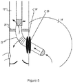
- An assist device can be used as a valve through the left atrial wall and the non-coronary cusp to enhance forward flow ( e.g., as shown in Figure 5 ).
- Critical mitral valve disease, tricuspid disease, or pulmonary valve disease can also be treated.
- off loading of the left atrium can improve the hemodynamics of mitral regurgitation.
- the regurgitant jet can be directed into the aorta to enhance the natural forward flow by positioning a pump inflow at the mitral valve ( e.g ., as shown in Figure 7 ).
- the pump inflow can avoid blocking the mitral annulus to maintain diastolic filling.
- An assist device can be combined with a valve-like structure.
- an assist device can be deployed using a transseptal or trans-aortic approach, as described herein.
- An inflow port can be placed on the mitral annulus such that the mitral regurgitant jet takes blood to the aorta.
- a valve can be placed on the superior-convexity-leftward portion such that when mitral regurgitation occurs the valve closes and the blood flows to the aorta. In the absence of mitral regurgitation, inflow proceeds to the ventricle. The pump can accentuate inflow to the ventricle and outflow to the aorta, as needed.
- assist devices described herein can be used for perfusion or for treatment of small vessel coronary disease (e.g., as a "stent-pump").
- an outflow port can be placed in the proximal aortic root ( e.g., as shown in Figure 5 ).
- An inflatable conduit can be positioned at the supravalvar cusp to increase pressure in the proximal aorta and perfuse the coronary artery.
- a cannula can be used to engage in coronary arteries such that left atrial blood is "pumped" into the coronary artery.
- Direct cannulation of the coronary arteries can be achieved using an assist device as a stand-alone pump.
- a battery source can be contained within the pump, in the infra-pectoral region, or other regions utilized in percutaneous techniques.
- the pump can be implanted in the coronary arteries as described for a stent.
- the power can be supplied with a battery placed within or near the stent, via a wire and battery source, or through an external source.
- peripheral vascular disease can be achieved with an assist device with an active pump or as a stand-alone device (a "pump-stent") in any peripheral vascular bed (e.g., the vascular bed of the brain, lower extremities, or upper extremities.).
- a pump stent can cannulate a vessel such as a carotid artery or femoral artery.
- the stand-alone pump-stent can be used to increase perfusion pressure for the treatment of vascular and vascular bed disease (e.g., small vessel coronary disease, small vessel peripheral arterial disease, or small vessel central nervous system disease).
- a pump-stent can be powered by a battery located within the stent.
- a battery source can be contained outside of the vessel in which the assist device is implanted (e.g ., a subcutaneously implanted power supply).
- a distal vessel or a vessel close to the implant can be a point of entry for connecting a battery source to a "pump-stent" (e.g ., femoral artery or subclavian artery).
- an assist device can be used in combination with other cardiac technology.
- an assist device can function as a defibrillator, or can have wires that can be used for pacing of the cardiac chambers.
- the device can serve an adjunctive role.
- a pump can be triggered by a ventricular arrhythmia, as described herein.
- anti-tachycardia pacing can be attempted repeatedly without a risk syncope or sudden death. Prolonged anti-tachycardia pacing attempts can allow for conversion to normal rhythm.
- an assist device can be activated to deliver a shock after the patient has been sedated and evaluated by a health care provider.
- a shock can be delivered via coils that are screwed in or imbedded into myocardium and serve as a multi-coil defibrillator. Coils can serve as electrodes to help time the pump ( e.g., by multi-site ventricular stimulation and multi-site sensing).
- Example 1 Continuity of septum between left atrium and ascending aorta
- the anatomies of 40 autopsied human hearts were studied. A continuity of the septum between the left atrium and the ascending aorta posterior (in the region of the non-coronary cusp) was present in all 40 hearts. The average area of this continuity was 9.6 mm x 5.6 mm (length x width). Thus, this region of the heart can be used to position an assist device to enhance blood flow from the left atrium to the aorta.
Landscapes
- Health & Medical Sciences (AREA)
- Engineering & Computer Science (AREA)
- Heart & Thoracic Surgery (AREA)
- Cardiology (AREA)
- Animal Behavior & Ethology (AREA)
- Veterinary Medicine (AREA)
- Public Health (AREA)
- Life Sciences & Earth Sciences (AREA)
- Biomedical Technology (AREA)
- General Health & Medical Sciences (AREA)
- Mechanical Engineering (AREA)
- Anesthesiology (AREA)
- Hematology (AREA)
- Radiology & Medical Imaging (AREA)
- Nuclear Medicine, Radiotherapy & Molecular Imaging (AREA)
- Medical Informatics (AREA)
- External Artificial Organs (AREA)
- Prostheses (AREA)
- Medicines Containing Material From Animals Or Micro-Organisms (AREA)
Applications Claiming Priority (3)
| Application Number | Priority Date | Filing Date | Title |
|---|---|---|---|
| US17707509P | 2009-05-11 | 2009-05-11 | |
| EP10775405.3A EP2429603B1 (fr) | 2009-05-11 | 2010-05-11 | Traitement d'une insuffisance cardiaque congestive |
| PCT/US2010/034389 WO2010132451A2 (fr) | 2009-05-11 | 2010-05-11 | Traitement d'une insuffisance cardiaque congestive |
Related Parent Applications (1)
| Application Number | Title | Priority Date | Filing Date |
|---|---|---|---|
| EP10775405.3A Division EP2429603B1 (fr) | 2009-05-11 | 2010-05-11 | Traitement d'une insuffisance cardiaque congestive |
Publications (2)
| Publication Number | Publication Date |
|---|---|
| EP3632485A1 true EP3632485A1 (fr) | 2020-04-08 |
| EP3632485B1 EP3632485B1 (fr) | 2024-01-03 |
Family
ID=43085535
Family Applications (2)
| Application Number | Title | Priority Date | Filing Date |
|---|---|---|---|
| EP10775405.3A Active EP2429603B1 (fr) | 2009-05-11 | 2010-05-11 | Traitement d'une insuffisance cardiaque congestive |
| EP19197851.9A Active EP3632485B1 (fr) | 2009-05-11 | 2010-05-11 | Traitement de l'insuffisance cardiaque congestive |
Family Applications Before (1)
| Application Number | Title | Priority Date | Filing Date |
|---|---|---|---|
| EP10775405.3A Active EP2429603B1 (fr) | 2009-05-11 | 2010-05-11 | Traitement d'une insuffisance cardiaque congestive |
Country Status (3)
| Country | Link |
|---|---|
| US (3) | US10137229B2 (fr) |
| EP (2) | EP2429603B1 (fr) |
| WO (1) | WO2010132451A2 (fr) |
Families Citing this family (38)
| Publication number | Priority date | Publication date | Assignee | Title |
|---|---|---|---|---|
| EP2349097B1 (fr) * | 2008-10-10 | 2024-07-03 | MedicalTree Patent Ltd. | Valve artificielle améliorée |
| US10137229B2 (en) | 2009-05-11 | 2018-11-27 | Mayo Foundation For Medical Education And Research | Treating congestive heart failure |
| US9517348B2 (en) | 2011-09-14 | 2016-12-13 | Biotronik Se & Co. Kg | Implantable cardiac therapy device |
| EP2570142A1 (fr) * | 2011-09-14 | 2013-03-20 | BIOTRONIK SE & Co. KG | Appareil thérapeutique cardiaque implantable |
| CA2858067C (fr) | 2011-12-03 | 2020-07-21 | Indiana University Research And Technology Corporation | Dispositif et procede d'assistance de propulseur de liquide visqueux cavopulmonaire |
| GB2527075A (en) | 2014-03-17 | 2015-12-16 | Daassist As | Percutaneous system, devices and methods |
| US10893847B2 (en) | 2015-12-30 | 2021-01-19 | Nuheart As | Transcatheter insertion system |
| US10537672B2 (en) | 2016-10-07 | 2020-01-21 | Nuheart As | Transcatheter device and system for the delivery of intracorporeal devices |
| EP3366332A1 (fr) * | 2017-02-22 | 2018-08-29 | Berlin Heart GmbH | Dispositif et procédé de liaison de deux parties de vaisseau sanguin |
| US10888646B2 (en) * | 2017-04-28 | 2021-01-12 | Nuheart As | Ventricular assist device and method |
| US10537670B2 (en) | 2017-04-28 | 2020-01-21 | Nuheart As | Ventricular assist device and method |
| CA3061644A1 (fr) * | 2017-04-28 | 2018-11-01 | Nuheart As | Procede et dispositif d'assistance ventriculaire |
| EP4732889A2 (fr) | 2017-06-07 | 2026-04-29 | Supira Medical, Inc. | Dispositifs de déplacement de fluide intravasculaire, systèmes et procédés d'utilisation |
| JP7319266B2 (ja) | 2017-11-13 | 2023-08-01 | シファメド・ホールディングス・エルエルシー | 血管内流体移動デバイス、システム、および使用方法 |
| EP4085965A1 (fr) | 2018-02-01 | 2022-11-09 | Shifamed Holdings, LLC | Pompes à sang intravasculaires et procédés d'utilisation et de fabrication |
| WO2019173739A1 (fr) * | 2018-03-09 | 2019-09-12 | Medtronic, Inc. | Dispositif d'assistance ventriculaire et système de stimulation électrique cardiaque pour commande de thérapie |
| AU2019262094A1 (en) * | 2018-05-03 | 2020-11-19 | Northern Development AS | Implantable device and delivery method |
| CN112566689A (zh) * | 2018-06-13 | 2021-03-26 | 耶鲁大学 | 心内装置 |
| US12161857B2 (en) | 2018-07-31 | 2024-12-10 | Shifamed Holdings, Llc | Intravascular blood pumps and methods of use |
| WO2020073047A1 (fr) | 2018-10-05 | 2020-04-09 | Shifamed Holdings, Llc | Pompes à sang intravasculaires et procédés d'utilisation |
| WO2020091910A1 (fr) * | 2018-11-02 | 2020-05-07 | W. L. Gore & Associates, Inc. | Dispositifs et procédés d'assistance ventriculaire implantables |
| US10888644B2 (en) | 2019-02-06 | 2021-01-12 | inQB8 Medical Technologies, LLC | Intra-cardiac left atrial and dual support systems |
| EP3921010A4 (fr) | 2019-02-08 | 2023-03-08 | NXT Biomedical, LLC | Réduction de stase dans l'auricule gauche |
| EP3996797A4 (fr) | 2019-07-12 | 2023-08-02 | Shifamed Holdings, LLC | Pompes à sang intravasculaires et méthode d'utilisation et procédé de fabrication |
| US11654275B2 (en) | 2019-07-22 | 2023-05-23 | Shifamed Holdings, Llc | Intravascular blood pumps with struts and methods of use and manufacture |
| EP4010046A4 (fr) | 2019-08-07 | 2023-08-30 | Calomeni, Michael | Pompes sanguines à cathéter et boîtiers de pompe pliants |
| US12102815B2 (en) | 2019-09-25 | 2024-10-01 | Shifamed Holdings, Llc | Catheter blood pumps and collapsible pump housings |
| EP4034192B1 (fr) | 2019-09-25 | 2025-12-24 | Supira Medical, Inc. | Dispositifs et systèmes de pompes à sang intravasculaires et leurs procédés d'utilisation et de commande |
| US12121713B2 (en) | 2019-09-25 | 2024-10-22 | Shifamed Holdings, Llc | Catheter blood pumps and collapsible blood conduits |
| WO2021101622A1 (fr) * | 2019-11-21 | 2021-05-27 | W. L. Gore & Associates, Inc. | Systèmes et procédés de pose pour dispositifs d'assistance cardiaque implantables |
| EP4072650A4 (fr) | 2019-12-11 | 2024-01-10 | Shifamed Holdings, LLC | Pompes à sang d'aorte descendante et de veine cave |
| WO2021127503A1 (fr) | 2019-12-19 | 2021-06-24 | Shifamed Holdings, Llc | Pompes à sang intravasculaires, moteurs et commande de fluide |
| US12082792B2 (en) * | 2020-02-25 | 2024-09-10 | Boston Scientific Medical Device Limited | Systems and methods for creating a puncture between aorta and the left atrium |
| US11559690B2 (en) | 2020-04-30 | 2023-01-24 | Medtronic, Inc. | Ventricular assist system and method |
| CN116407754B (zh) * | 2022-03-23 | 2026-01-02 | 上海魅丽纬叶医疗科技有限公司 | 一种主动脉循环泵及血液泵系统 |
| JP2025523622A (ja) * | 2022-07-01 | 2025-07-23 | ブライトフロウ | 膜横断型右心補助デバイス |
| US20240299728A1 (en) * | 2023-03-10 | 2024-09-12 | Star Bp, Inc. | System and method of treating cardiovascular impairment |
| WO2025149655A1 (fr) * | 2024-01-12 | 2025-07-17 | Brightflow | Dispositif d'assistance cardiaque axiale |
Citations (10)
| Publication number | Priority date | Publication date | Assignee | Title |
|---|---|---|---|---|
| US4753221A (en) | 1986-10-22 | 1988-06-28 | Intravascular Surgical Instruments, Inc. | Blood pumping catheter and method of use |
| US4919647A (en) | 1988-10-13 | 1990-04-24 | Kensey Nash Corporation | Aortically located blood pumping catheter and method of use |
| EP0445782A1 (fr) * | 1990-03-08 | 1991-09-11 | Sun Medical Technology Research Corporation | Coeur artificiel auxiliaire implanté in-vivo |
| US6136025A (en) * | 1999-07-27 | 2000-10-24 | Barbut; Denise R. | Endoscopic arterial pumps for treatment of cardiac insufficiency and venous pumps for right-sided cardiac support |
| US6926662B1 (en) | 1998-12-23 | 2005-08-09 | A-Med Systems, Inc. | Left and right side heart support |
| US20050187425A1 (en) | 2004-01-14 | 2005-08-25 | Scout Medical Technologies, Llc | Left ventricular function assist system and method |
| US20060181843A1 (en) | 2005-02-14 | 2006-08-17 | Kabushiki Kaisha Toshiba | Portable microcomputer and display unit |
| US20070156010A1 (en) | 1997-07-11 | 2007-07-05 | Aboul-Hosn Walid N | Single port cardiac support apparatus related applications |
| WO2008002471A2 (fr) | 2006-06-23 | 2008-01-03 | Spherics, Inc. | Polymères bioadhésifs stabilisés contre l'érosion fonctionnalisés ou mélangés avec du cathecol et ses dérivés |
| WO2008027869A2 (fr) * | 2006-08-30 | 2008-03-06 | Circulite, Inc. | Dispositifs, methodes et systemes permettant d'etablir un flux sanguin supplementaire dans le systeme circulatoire |
Family Cites Families (17)
| Publication number | Priority date | Publication date | Assignee | Title |
|---|---|---|---|---|
| US4302854A (en) * | 1980-06-04 | 1981-12-01 | Runge Thomas M | Electrically activated ferromagnetic/diamagnetic vascular shunt for left ventricular assist |
| US5676651A (en) * | 1992-08-06 | 1997-10-14 | Electric Boat Corporation | Surgically implantable pump arrangement and method for pumping body fluids |
| US6395026B1 (en) * | 1998-05-15 | 2002-05-28 | A-Med Systems, Inc. | Apparatus and methods for beating heart bypass surgery |
| US6532964B2 (en) * | 1997-07-11 | 2003-03-18 | A-Med Systems, Inc. | Pulmonary and circulatory blood flow support devices and methods for heart surgery procedures |
| DE29804046U1 (de) * | 1998-03-07 | 1998-04-30 | Günther, Rolf W., Prof. Dr.med., 52074 Aachen | Perkutan implantierbare selbstentfaltbare Axialpumpe zur temporären Herzunterstützung |
| US6123724A (en) * | 1999-04-14 | 2000-09-26 | Denker; Stephen | Heart assist method and apparatus employing magnetic repulsion force |
| US7022100B1 (en) | 1999-09-03 | 2006-04-04 | A-Med Systems, Inc. | Guidable intravascular blood pump and related methods |
| US7229402B2 (en) * | 2001-02-09 | 2007-06-12 | Cardiac Output Technologies, Inc. | Minimally invasive ventricular assist technology and method |
| US20060155158A1 (en) * | 2002-06-11 | 2006-07-13 | Aboul-Hosn Walid N | Percutaneously introduced blood pump and related methods |
| AR036548A1 (es) * | 2002-09-18 | 2004-09-15 | Domingo Santo Liotta | Dispositivo de implante corporal para asistencia circulatoria sanguinea y ventricular cardiaca |
| WO2005037345A2 (fr) * | 2003-10-17 | 2005-04-28 | Vanderbilt University | Dispositifs d'assistance ventriculaire introduits par voie cutanee et procedes associes |
| DE102005052628B4 (de) | 2005-11-04 | 2014-06-05 | Jenavalve Technology Inc. | Selbstexpandierendes, flexibles Drahtgeflecht mit integrierter Klappenprothese für den transvaskulären Herzklappenersatz und ein System mit einer solchen Vorrichtung und einem Einführkatheter |
| PT2026856E (pt) | 2006-05-31 | 2015-11-24 | Vadovations Inc | Dispositivo de assistência cardíaca |
| EP2195043B1 (fr) | 2007-10-01 | 2014-12-03 | Minvasc Devices, LLC | Dispositif d'assistance ventriculaire intraauriculaire |
| US10137229B2 (en) | 2009-05-11 | 2018-11-27 | Mayo Foundation For Medical Education And Research | Treating congestive heart failure |
| US20140228733A1 (en) * | 2013-02-14 | 2014-08-14 | Cook Medical Technologies Llc | Left Atrial Appendage Shunt |
| GB2527075A (en) | 2014-03-17 | 2015-12-16 | Daassist As | Percutaneous system, devices and methods |
-
2010
- 2010-05-11 US US13/319,672 patent/US10137229B2/en active Active
- 2010-05-11 WO PCT/US2010/034389 patent/WO2010132451A2/fr not_active Ceased
- 2010-05-11 EP EP10775405.3A patent/EP2429603B1/fr active Active
- 2010-05-11 EP EP19197851.9A patent/EP3632485B1/fr active Active
-
2018
- 2018-09-21 US US16/138,382 patent/US10869957B2/en active Active
-
2020
- 2020-11-25 US US17/105,365 patent/US20210106807A1/en not_active Abandoned
Patent Citations (10)
| Publication number | Priority date | Publication date | Assignee | Title |
|---|---|---|---|---|
| US4753221A (en) | 1986-10-22 | 1988-06-28 | Intravascular Surgical Instruments, Inc. | Blood pumping catheter and method of use |
| US4919647A (en) | 1988-10-13 | 1990-04-24 | Kensey Nash Corporation | Aortically located blood pumping catheter and method of use |
| EP0445782A1 (fr) * | 1990-03-08 | 1991-09-11 | Sun Medical Technology Research Corporation | Coeur artificiel auxiliaire implanté in-vivo |
| US20070156010A1 (en) | 1997-07-11 | 2007-07-05 | Aboul-Hosn Walid N | Single port cardiac support apparatus related applications |
| US6926662B1 (en) | 1998-12-23 | 2005-08-09 | A-Med Systems, Inc. | Left and right side heart support |
| US6136025A (en) * | 1999-07-27 | 2000-10-24 | Barbut; Denise R. | Endoscopic arterial pumps for treatment of cardiac insufficiency and venous pumps for right-sided cardiac support |
| US20050187425A1 (en) | 2004-01-14 | 2005-08-25 | Scout Medical Technologies, Llc | Left ventricular function assist system and method |
| US20060181843A1 (en) | 2005-02-14 | 2006-08-17 | Kabushiki Kaisha Toshiba | Portable microcomputer and display unit |
| WO2008002471A2 (fr) | 2006-06-23 | 2008-01-03 | Spherics, Inc. | Polymères bioadhésifs stabilisés contre l'érosion fonctionnalisés ou mélangés avec du cathecol et ses dérivés |
| WO2008027869A2 (fr) * | 2006-08-30 | 2008-03-06 | Circulite, Inc. | Dispositifs, methodes et systemes permettant d'etablir un flux sanguin supplementaire dans le systeme circulatoire |
Also Published As
| Publication number | Publication date |
|---|---|
| WO2010132451A3 (fr) | 2011-02-24 |
| EP3632485B1 (fr) | 2024-01-03 |
| WO2010132451A2 (fr) | 2010-11-18 |
| US20210106807A1 (en) | 2021-04-15 |
| EP2429603B1 (fr) | 2019-09-18 |
| US10137229B2 (en) | 2018-11-27 |
| US20190105435A1 (en) | 2019-04-11 |
| EP2429603A2 (fr) | 2012-03-21 |
| US20120059459A1 (en) | 2012-03-08 |
| US10869957B2 (en) | 2020-12-22 |
| EP2429603A4 (fr) | 2017-07-19 |
Similar Documents
| Publication | Publication Date | Title |
|---|---|---|
| US10869957B2 (en) | Treating congestive heart failure | |
| US10888423B2 (en) | Left heart assist device and method | |
| JP6902617B2 (ja) | リードレスペーシング装置およびその位置決めシステム | |
| JP6888112B2 (ja) | 心不整脈を治療するためのリードレスペーシング装置 | |
| KR100544944B1 (ko) | 이식가능한 심장 보조 기관 | |
| US8123668B2 (en) | Signal transmitting and lesion excluding heart implants for pacing defibrillating and/or sensing of heart beat | |
| US8868194B2 (en) | Aortic pacemaker | |
| US20180207336A1 (en) | Device And A Method For Augmenting Heart Function | |
| US20110224655A1 (en) | Central core multifunctional cardiac devices | |
| US20100137927A1 (en) | Multifunctional cardiac pacemaker system | |
| Hawkins | Epicardial Wireless Pacemaker for Improved Left Ventricular Resynchronization (Conceptual Design) | |
| HK1181293B (en) | A device and a method to controllably assist movement of a mitral valve | |
| HK1212268B (zh) | 可控制地辅助二尖瓣运动的装置和方法 |
Legal Events
| Date | Code | Title | Description |
|---|---|---|---|
| PUAI | Public reference made under article 153(3) epc to a published international application that has entered the european phase |
Free format text: ORIGINAL CODE: 0009012 |
|
| STAA | Information on the status of an ep patent application or granted ep patent |
Free format text: STATUS: THE APPLICATION HAS BEEN PUBLISHED |
|
| AC | Divisional application: reference to earlier application |
Ref document number: 2429603 Country of ref document: EP Kind code of ref document: P |
|
| AK | Designated contracting states |
Kind code of ref document: A1 Designated state(s): AL AT BE BG CH CY CZ DE DK EE ES FI FR GB GR HR HU IE IS IT LI LT LU LV MC MK MT NL NO PL PT RO SE SI SK SM TR |
|
| STAA | Information on the status of an ep patent application or granted ep patent |
Free format text: STATUS: REQUEST FOR EXAMINATION WAS MADE |
|
| 17P | Request for examination filed |
Effective date: 20201006 |
|
| RBV | Designated contracting states (corrected) |
Designated state(s): AL AT BE BG CH CY CZ DE DK EE ES FI FR GB GR HR HU IE IS IT LI LT LU LV MC MK MT NL NO PL PT RO SE SI SK SM TR |
|
| STAA | Information on the status of an ep patent application or granted ep patent |
Free format text: STATUS: EXAMINATION IS IN PROGRESS |
|
| 17Q | First examination report despatched |
Effective date: 20210304 |
|
| REG | Reference to a national code |
Ref country code: DE Ref legal event code: R079 Free format text: PREVIOUS MAIN CLASS: A61M0001120000 Ipc: A61M0060148000 Ref country code: DE Ref legal event code: R079 Ref document number: 602010069201 Country of ref document: DE Free format text: PREVIOUS MAIN CLASS: A61M0001120000 Ipc: A61M0060148000 |
|
| GRAP | Despatch of communication of intention to grant a patent |
Free format text: ORIGINAL CODE: EPIDOSNIGR1 |
|
| STAA | Information on the status of an ep patent application or granted ep patent |
Free format text: STATUS: GRANT OF PATENT IS INTENDED |
|
| RIC1 | Information provided on ipc code assigned before grant |
Ipc: A61M 29/02 20060101ALI20230626BHEP Ipc: A61M 60/867 20210101ALI20230626BHEP Ipc: A61M 60/515 20210101ALI20230626BHEP Ipc: A61M 60/20 20210101ALI20230626BHEP Ipc: A61M 60/165 20210101ALI20230626BHEP Ipc: A61M 60/414 20210101ALI20230626BHEP Ipc: A61M 60/279 20210101ALI20230626BHEP Ipc: A61M 60/148 20210101AFI20230626BHEP |
|
| INTG | Intention to grant announced |
Effective date: 20230724 |
|
| RIN1 | Information on inventor provided before grant (corrected) |
Inventor name: KUSHWAHA, SUDHIR, S. Inventor name: BRUCE, CHARLES, J. Inventor name: FRIEDMAN, PAUL, A. Inventor name: PARK, SOON, J. Inventor name: ASIRVATHAM, SAMUEL, J. |
|
| GRAS | Grant fee paid |
Free format text: ORIGINAL CODE: EPIDOSNIGR3 |
|
| GRAA | (expected) grant |
Free format text: ORIGINAL CODE: 0009210 |
|
| STAA | Information on the status of an ep patent application or granted ep patent |
Free format text: STATUS: THE PATENT HAS BEEN GRANTED |
|
| AC | Divisional application: reference to earlier application |
Ref document number: 2429603 Country of ref document: EP Kind code of ref document: P |
|
| AK | Designated contracting states |
Kind code of ref document: B1 Designated state(s): AL AT BE BG CH CY CZ DE DK EE ES FI FR GB GR HR HU IE IS IT LI LT LU LV MC MK MT NL NO PL PT RO SE SI SK SM TR |
|
| REG | Reference to a national code |
Ref country code: GB Ref legal event code: FG4D |
|
| P01 | Opt-out of the competence of the unified patent court (upc) registered |
Effective date: 20231206 |
|
| REG | Reference to a national code |
Ref country code: DE Ref legal event code: R096 Ref document number: 602010069201 Country of ref document: DE |
|
| REG | Reference to a national code |
Ref country code: CH Ref legal event code: EP |
|
| REG | Reference to a national code |
Ref country code: IE Ref legal event code: FG4D |
|
| REG | Reference to a national code |
Ref country code: LT Ref legal event code: MG9D |
|
| PG25 | Lapsed in a contracting state [announced via postgrant information from national office to epo] |
Ref country code: ES Free format text: LAPSE BECAUSE OF FAILURE TO SUBMIT A TRANSLATION OF THE DESCRIPTION OR TO PAY THE FEE WITHIN THE PRESCRIBED TIME-LIMIT Effective date: 20240103 |
|
| PG25 | Lapsed in a contracting state [announced via postgrant information from national office to epo] |
Ref country code: ES Free format text: LAPSE BECAUSE OF FAILURE TO SUBMIT A TRANSLATION OF THE DESCRIPTION OR TO PAY THE FEE WITHIN THE PRESCRIBED TIME-LIMIT Effective date: 20240103 |
|
| REG | Reference to a national code |
Ref country code: NL Ref legal event code: MP Effective date: 20240103 |
|
| REG | Reference to a national code |
Ref country code: AT Ref legal event code: MK05 Ref document number: 1646192 Country of ref document: AT Kind code of ref document: T Effective date: 20240103 |
|
| PG25 | Lapsed in a contracting state [announced via postgrant information from national office to epo] |
Ref country code: NL Free format text: LAPSE BECAUSE OF FAILURE TO SUBMIT A TRANSLATION OF THE DESCRIPTION OR TO PAY THE FEE WITHIN THE PRESCRIBED TIME-LIMIT Effective date: 20240103 |
|
| PG25 | Lapsed in a contracting state [announced via postgrant information from national office to epo] |
Ref country code: NL Free format text: LAPSE BECAUSE OF FAILURE TO SUBMIT A TRANSLATION OF THE DESCRIPTION OR TO PAY THE FEE WITHIN THE PRESCRIBED TIME-LIMIT Effective date: 20240103 |
|
| PG25 | Lapsed in a contracting state [announced via postgrant information from national office to epo] |
Ref country code: IS Free format text: LAPSE BECAUSE OF FAILURE TO SUBMIT A TRANSLATION OF THE DESCRIPTION OR TO PAY THE FEE WITHIN THE PRESCRIBED TIME-LIMIT Effective date: 20240503 |
|
| PG25 | Lapsed in a contracting state [announced via postgrant information from national office to epo] |
Ref country code: LT Free format text: LAPSE BECAUSE OF FAILURE TO SUBMIT A TRANSLATION OF THE DESCRIPTION OR TO PAY THE FEE WITHIN THE PRESCRIBED TIME-LIMIT Effective date: 20240103 |
|
| PG25 | Lapsed in a contracting state [announced via postgrant information from national office to epo] |
Ref country code: GR Free format text: LAPSE BECAUSE OF FAILURE TO SUBMIT A TRANSLATION OF THE DESCRIPTION OR TO PAY THE FEE WITHIN THE PRESCRIBED TIME-LIMIT Effective date: 20240404 |
|
| PG25 | Lapsed in a contracting state [announced via postgrant information from national office to epo] |
Ref country code: HR Free format text: LAPSE BECAUSE OF FAILURE TO SUBMIT A TRANSLATION OF THE DESCRIPTION OR TO PAY THE FEE WITHIN THE PRESCRIBED TIME-LIMIT Effective date: 20240103 |
|
| PG25 | Lapsed in a contracting state [announced via postgrant information from national office to epo] |
Ref country code: AT Free format text: LAPSE BECAUSE OF FAILURE TO SUBMIT A TRANSLATION OF THE DESCRIPTION OR TO PAY THE FEE WITHIN THE PRESCRIBED TIME-LIMIT Effective date: 20240103 Ref country code: CZ Free format text: LAPSE BECAUSE OF FAILURE TO SUBMIT A TRANSLATION OF THE DESCRIPTION OR TO PAY THE FEE WITHIN THE PRESCRIBED TIME-LIMIT Effective date: 20240103 |
|
| PG25 | Lapsed in a contracting state [announced via postgrant information from national office to epo] |
Ref country code: NO Free format text: LAPSE BECAUSE OF FAILURE TO SUBMIT A TRANSLATION OF THE DESCRIPTION OR TO PAY THE FEE WITHIN THE PRESCRIBED TIME-LIMIT Effective date: 20240403 Ref country code: LT Free format text: LAPSE BECAUSE OF FAILURE TO SUBMIT A TRANSLATION OF THE DESCRIPTION OR TO PAY THE FEE WITHIN THE PRESCRIBED TIME-LIMIT Effective date: 20240103 Ref country code: IS Free format text: LAPSE BECAUSE OF FAILURE TO SUBMIT A TRANSLATION OF THE DESCRIPTION OR TO PAY THE FEE WITHIN THE PRESCRIBED TIME-LIMIT Effective date: 20240503 Ref country code: HR Free format text: LAPSE BECAUSE OF FAILURE TO SUBMIT A TRANSLATION OF THE DESCRIPTION OR TO PAY THE FEE WITHIN THE PRESCRIBED TIME-LIMIT Effective date: 20240103 Ref country code: GR Free format text: LAPSE BECAUSE OF FAILURE TO SUBMIT A TRANSLATION OF THE DESCRIPTION OR TO PAY THE FEE WITHIN THE PRESCRIBED TIME-LIMIT Effective date: 20240404 Ref country code: CZ Free format text: LAPSE BECAUSE OF FAILURE TO SUBMIT A TRANSLATION OF THE DESCRIPTION OR TO PAY THE FEE WITHIN THE PRESCRIBED TIME-LIMIT Effective date: 20240103 Ref country code: BG Free format text: LAPSE BECAUSE OF FAILURE TO SUBMIT A TRANSLATION OF THE DESCRIPTION OR TO PAY THE FEE WITHIN THE PRESCRIBED TIME-LIMIT Effective date: 20240103 Ref country code: AT Free format text: LAPSE BECAUSE OF FAILURE TO SUBMIT A TRANSLATION OF THE DESCRIPTION OR TO PAY THE FEE WITHIN THE PRESCRIBED TIME-LIMIT Effective date: 20240103 |
|
| PG25 | Lapsed in a contracting state [announced via postgrant information from national office to epo] |
Ref country code: PL Free format text: LAPSE BECAUSE OF FAILURE TO SUBMIT A TRANSLATION OF THE DESCRIPTION OR TO PAY THE FEE WITHIN THE PRESCRIBED TIME-LIMIT Effective date: 20240103 Ref country code: PT Free format text: LAPSE BECAUSE OF FAILURE TO SUBMIT A TRANSLATION OF THE DESCRIPTION OR TO PAY THE FEE WITHIN THE PRESCRIBED TIME-LIMIT Effective date: 20240503 |
|
| PG25 | Lapsed in a contracting state [announced via postgrant information from national office to epo] |
Ref country code: SE Free format text: LAPSE BECAUSE OF FAILURE TO SUBMIT A TRANSLATION OF THE DESCRIPTION OR TO PAY THE FEE WITHIN THE PRESCRIBED TIME-LIMIT Effective date: 20240103 Ref country code: PT Free format text: LAPSE BECAUSE OF FAILURE TO SUBMIT A TRANSLATION OF THE DESCRIPTION OR TO PAY THE FEE WITHIN THE PRESCRIBED TIME-LIMIT Effective date: 20240503 Ref country code: PL Free format text: LAPSE BECAUSE OF FAILURE TO SUBMIT A TRANSLATION OF THE DESCRIPTION OR TO PAY THE FEE WITHIN THE PRESCRIBED TIME-LIMIT Effective date: 20240103 Ref country code: LV Free format text: LAPSE BECAUSE OF FAILURE TO SUBMIT A TRANSLATION OF THE DESCRIPTION OR TO PAY THE FEE WITHIN THE PRESCRIBED TIME-LIMIT Effective date: 20240103 |
|
| REG | Reference to a national code |
Ref country code: DE Ref legal event code: R097 Ref document number: 602010069201 Country of ref document: DE |
|
| PG25 | Lapsed in a contracting state [announced via postgrant information from national office to epo] |
Ref country code: DK Free format text: LAPSE BECAUSE OF FAILURE TO SUBMIT A TRANSLATION OF THE DESCRIPTION OR TO PAY THE FEE WITHIN THE PRESCRIBED TIME-LIMIT Effective date: 20240103 |
|
| PG25 | Lapsed in a contracting state [announced via postgrant information from national office to epo] |
Ref country code: SM Free format text: LAPSE BECAUSE OF FAILURE TO SUBMIT A TRANSLATION OF THE DESCRIPTION OR TO PAY THE FEE WITHIN THE PRESCRIBED TIME-LIMIT Effective date: 20240103 |
|
| PG25 | Lapsed in a contracting state [announced via postgrant information from national office to epo] |
Ref country code: EE Free format text: LAPSE BECAUSE OF FAILURE TO SUBMIT A TRANSLATION OF THE DESCRIPTION OR TO PAY THE FEE WITHIN THE PRESCRIBED TIME-LIMIT Effective date: 20240103 |
|
| PG25 | Lapsed in a contracting state [announced via postgrant information from national office to epo] |
Ref country code: SK Free format text: LAPSE BECAUSE OF FAILURE TO SUBMIT A TRANSLATION OF THE DESCRIPTION OR TO PAY THE FEE WITHIN THE PRESCRIBED TIME-LIMIT Effective date: 20240103 |
|
| PG25 | Lapsed in a contracting state [announced via postgrant information from national office to epo] |
Ref country code: SM Free format text: LAPSE BECAUSE OF FAILURE TO SUBMIT A TRANSLATION OF THE DESCRIPTION OR TO PAY THE FEE WITHIN THE PRESCRIBED TIME-LIMIT Effective date: 20240103 Ref country code: SK Free format text: LAPSE BECAUSE OF FAILURE TO SUBMIT A TRANSLATION OF THE DESCRIPTION OR TO PAY THE FEE WITHIN THE PRESCRIBED TIME-LIMIT Effective date: 20240103 Ref country code: RO Free format text: LAPSE BECAUSE OF FAILURE TO SUBMIT A TRANSLATION OF THE DESCRIPTION OR TO PAY THE FEE WITHIN THE PRESCRIBED TIME-LIMIT Effective date: 20240103 Ref country code: EE Free format text: LAPSE BECAUSE OF FAILURE TO SUBMIT A TRANSLATION OF THE DESCRIPTION OR TO PAY THE FEE WITHIN THE PRESCRIBED TIME-LIMIT Effective date: 20240103 Ref country code: DK Free format text: LAPSE BECAUSE OF FAILURE TO SUBMIT A TRANSLATION OF THE DESCRIPTION OR TO PAY THE FEE WITHIN THE PRESCRIBED TIME-LIMIT Effective date: 20240103 |
|
| PLBE | No opposition filed within time limit |
Free format text: ORIGINAL CODE: 0009261 |
|
| STAA | Information on the status of an ep patent application or granted ep patent |
Free format text: STATUS: NO OPPOSITION FILED WITHIN TIME LIMIT |
|
| PG25 | Lapsed in a contracting state [announced via postgrant information from national office to epo] |
Ref country code: IT Free format text: LAPSE BECAUSE OF FAILURE TO SUBMIT A TRANSLATION OF THE DESCRIPTION OR TO PAY THE FEE WITHIN THE PRESCRIBED TIME-LIMIT Effective date: 20240103 |
|
| 26N | No opposition filed |
Effective date: 20241007 |
|
| REG | Reference to a national code |
Ref country code: CH Ref legal event code: PL |
|
| PG25 | Lapsed in a contracting state [announced via postgrant information from national office to epo] |
Ref country code: IT Free format text: LAPSE BECAUSE OF FAILURE TO SUBMIT A TRANSLATION OF THE DESCRIPTION OR TO PAY THE FEE WITHIN THE PRESCRIBED TIME-LIMIT Effective date: 20240103 |
|
| PG25 | Lapsed in a contracting state [announced via postgrant information from national office to epo] |
Ref country code: MC Free format text: LAPSE BECAUSE OF FAILURE TO SUBMIT A TRANSLATION OF THE DESCRIPTION OR TO PAY THE FEE WITHIN THE PRESCRIBED TIME-LIMIT Effective date: 20240103 |
|
| PG25 | Lapsed in a contracting state [announced via postgrant information from national office to epo] |
Ref country code: LU Free format text: LAPSE BECAUSE OF NON-PAYMENT OF DUE FEES Effective date: 20240511 |
|
| PG25 | Lapsed in a contracting state [announced via postgrant information from national office to epo] |
Ref country code: MC Free format text: LAPSE BECAUSE OF FAILURE TO SUBMIT A TRANSLATION OF THE DESCRIPTION OR TO PAY THE FEE WITHIN THE PRESCRIBED TIME-LIMIT Effective date: 20240103 Ref country code: LU Free format text: LAPSE BECAUSE OF NON-PAYMENT OF DUE FEES Effective date: 20240511 Ref country code: CH Free format text: LAPSE BECAUSE OF NON-PAYMENT OF DUE FEES Effective date: 20240531 |
|
| REG | Reference to a national code |
Ref country code: BE Ref legal event code: MM Effective date: 20240531 |
|
| PG25 | Lapsed in a contracting state [announced via postgrant information from national office to epo] |
Ref country code: IE Free format text: LAPSE BECAUSE OF NON-PAYMENT OF DUE FEES Effective date: 20240511 |
|
| PG25 | Lapsed in a contracting state [announced via postgrant information from national office to epo] |
Ref country code: SI Free format text: LAPSE BECAUSE OF FAILURE TO SUBMIT A TRANSLATION OF THE DESCRIPTION OR TO PAY THE FEE WITHIN THE PRESCRIBED TIME-LIMIT Effective date: 20240103 Ref country code: BE Free format text: LAPSE BECAUSE OF NON-PAYMENT OF DUE FEES Effective date: 20240531 |
|
| PGFP | Annual fee paid to national office [announced via postgrant information from national office to epo] |
Ref country code: DE Payment date: 20250507 Year of fee payment: 16 |
|
| PGFP | Annual fee paid to national office [announced via postgrant information from national office to epo] |
Ref country code: GB Payment date: 20250501 Year of fee payment: 16 |
|
| PGFP | Annual fee paid to national office [announced via postgrant information from national office to epo] |
Ref country code: FR Payment date: 20250508 Year of fee payment: 16 |
|
| PG25 | Lapsed in a contracting state [announced via postgrant information from national office to epo] |
Ref country code: CY Free format text: LAPSE BECAUSE OF FAILURE TO SUBMIT A TRANSLATION OF THE DESCRIPTION OR TO PAY THE FEE WITHIN THE PRESCRIBED TIME-LIMIT; INVALID AB INITIO Effective date: 20100511 |
|
| PG25 | Lapsed in a contracting state [announced via postgrant information from national office to epo] |
Ref country code: HU Free format text: LAPSE BECAUSE OF FAILURE TO SUBMIT A TRANSLATION OF THE DESCRIPTION OR TO PAY THE FEE WITHIN THE PRESCRIBED TIME-LIMIT; INVALID AB INITIO Effective date: 20100511 |
|
| PG25 | Lapsed in a contracting state [announced via postgrant information from national office to epo] |
Ref country code: FI Free format text: LAPSE BECAUSE OF FAILURE TO SUBMIT A TRANSLATION OF THE DESCRIPTION OR TO PAY THE FEE WITHIN THE PRESCRIBED TIME-LIMIT Effective date: 20240103 |