EP3634442B1 - Verfahren zur diagnose der parkinson-krankheit - Google Patents
Verfahren zur diagnose der parkinson-krankheit Download PDFInfo
- Publication number
- EP3634442B1 EP3634442B1 EP18805942.2A EP18805942A EP3634442B1 EP 3634442 B1 EP3634442 B1 EP 3634442B1 EP 18805942 A EP18805942 A EP 18805942A EP 3634442 B1 EP3634442 B1 EP 3634442B1
- Authority
- EP
- European Patent Office
- Prior art keywords
- phage
- protein
- mycobacterium
- bacteriophages
- virus
- Prior art date
- Legal status (The legal status is an assumption and is not a legal conclusion. Google has not performed a legal analysis and makes no representation as to the accuracy of the status listed.)
- Active
Links
Images




































Classifications
-
- C—CHEMISTRY; METALLURGY
- C12—BIOCHEMISTRY; BEER; SPIRITS; WINE; VINEGAR; MICROBIOLOGY; ENZYMOLOGY; MUTATION OR GENETIC ENGINEERING
- C12Q—MEASURING OR TESTING PROCESSES INVOLVING ENZYMES, NUCLEIC ACIDS OR MICROORGANISMS; COMPOSITIONS OR TEST PAPERS THEREFOR; PROCESSES OF PREPARING SUCH COMPOSITIONS; CONDITION-RESPONSIVE CONTROL IN MICROBIOLOGICAL OR ENZYMOLOGICAL PROCESSES
- C12Q1/00—Measuring or testing processes involving enzymes, nucleic acids or microorganisms; Compositions therefor; Processes of preparing such compositions
- C12Q1/70—Measuring or testing processes involving enzymes, nucleic acids or microorganisms; Compositions therefor; Processes of preparing such compositions involving virus or bacteriophage
- C12Q1/701—Specific hybridization probes
-
- A—HUMAN NECESSITIES
- A61—MEDICAL OR VETERINARY SCIENCE; HYGIENE
- A61K—PREPARATIONS FOR MEDICAL, DENTAL OR TOILETRY PURPOSES
- A61K31/00—Medicinal preparations containing organic active ingredients
- A61K31/33—Heterocyclic compounds
- A61K31/395—Heterocyclic compounds having nitrogen as a ring hetero atom, e.g. guanethidine or rifamycins
- A61K31/495—Heterocyclic compounds having nitrogen as a ring hetero atom, e.g. guanethidine or rifamycins having six-membered rings with two or more nitrogen atoms as the only ring heteroatoms, e.g. piperazine or tetrazines
- A61K31/496—Non-condensed piperazines containing further heterocyclic rings, e.g. rifampin, thiothixene or sparfloxacin
-
- A—HUMAN NECESSITIES
- A61—MEDICAL OR VETERINARY SCIENCE; HYGIENE
- A61K—PREPARATIONS FOR MEDICAL, DENTAL OR TOILETRY PURPOSES
- A61K31/00—Medicinal preparations containing organic active ingredients
- A61K31/70—Carbohydrates; Sugars; Derivatives thereof
- A61K31/7042—Compounds having saccharide radicals and heterocyclic rings
- A61K31/7052—Compounds having saccharide radicals and heterocyclic rings having nitrogen as a ring hetero atom, e.g. nucleosides, nucleotides
- A61K31/7056—Compounds having saccharide radicals and heterocyclic rings having nitrogen as a ring hetero atom, e.g. nucleosides, nucleotides containing five-membered rings with nitrogen as a ring hetero atom
-
- A—HUMAN NECESSITIES
- A61—MEDICAL OR VETERINARY SCIENCE; HYGIENE
- A61K—PREPARATIONS FOR MEDICAL, DENTAL OR TOILETRY PURPOSES
- A61K31/00—Medicinal preparations containing organic active ingredients
- A61K31/74—Synthetic polymeric materials
- A61K31/785—Polymers containing nitrogen
-
- A—HUMAN NECESSITIES
- A61—MEDICAL OR VETERINARY SCIENCE; HYGIENE
- A61K—PREPARATIONS FOR MEDICAL, DENTAL OR TOILETRY PURPOSES
- A61K35/00—Medicinal preparations containing materials or reaction products thereof with undetermined constitution
- A61K35/66—Microorganisms or materials therefrom
- A61K35/74—Bacteria
- A61K35/741—Probiotics
-
- A—HUMAN NECESSITIES
- A61—MEDICAL OR VETERINARY SCIENCE; HYGIENE
- A61K—PREPARATIONS FOR MEDICAL, DENTAL OR TOILETRY PURPOSES
- A61K35/00—Medicinal preparations containing materials or reaction products thereof with undetermined constitution
- A61K35/66—Microorganisms or materials therefrom
- A61K35/74—Bacteria
- A61K35/741—Probiotics
- A61K35/744—Lactic acid bacteria, e.g. enterococci, pediococci, lactococci, streptococci or leuconostocs
- A61K35/745—Bifidobacteria
-
- A—HUMAN NECESSITIES
- A61—MEDICAL OR VETERINARY SCIENCE; HYGIENE
- A61K—PREPARATIONS FOR MEDICAL, DENTAL OR TOILETRY PURPOSES
- A61K35/00—Medicinal preparations containing materials or reaction products thereof with undetermined constitution
- A61K35/66—Microorganisms or materials therefrom
- A61K35/74—Bacteria
- A61K35/741—Probiotics
- A61K35/744—Lactic acid bacteria, e.g. enterococci, pediococci, lactococci, streptococci or leuconostocs
- A61K35/747—Lactobacilli, e.g. L. acidophilus or L. brevis
-
- A—HUMAN NECESSITIES
- A61—MEDICAL OR VETERINARY SCIENCE; HYGIENE
- A61K—PREPARATIONS FOR MEDICAL, DENTAL OR TOILETRY PURPOSES
- A61K35/00—Medicinal preparations containing materials or reaction products thereof with undetermined constitution
- A61K35/66—Microorganisms or materials therefrom
- A61K35/76—Viruses; Subviral particles; Bacteriophages
-
- A—HUMAN NECESSITIES
- A61—MEDICAL OR VETERINARY SCIENCE; HYGIENE
- A61K—PREPARATIONS FOR MEDICAL, DENTAL OR TOILETRY PURPOSES
- A61K38/00—Medicinal preparations containing peptides
- A61K38/16—Peptides having more than 20 amino acids; Gastrins; Somatostatins; Melanotropins; Derivatives thereof
- A61K38/43—Enzymes; Proenzymes; Derivatives thereof
- A61K38/46—Hydrolases (3)
- A61K38/465—Hydrolases (3) acting on ester bonds (3.1), e.g. lipases, ribonucleases
-
- A—HUMAN NECESSITIES
- A61—MEDICAL OR VETERINARY SCIENCE; HYGIENE
- A61K—PREPARATIONS FOR MEDICAL, DENTAL OR TOILETRY PURPOSES
- A61K38/00—Medicinal preparations containing peptides
- A61K38/16—Peptides having more than 20 amino acids; Gastrins; Somatostatins; Melanotropins; Derivatives thereof
- A61K38/43—Enzymes; Proenzymes; Derivatives thereof
- A61K38/46—Hydrolases (3)
- A61K38/48—Hydrolases (3) acting on peptide bonds (3.4)
- A61K38/482—Serine endopeptidases (3.4.21)
-
- A—HUMAN NECESSITIES
- A61—MEDICAL OR VETERINARY SCIENCE; HYGIENE
- A61K—PREPARATIONS FOR MEDICAL, DENTAL OR TOILETRY PURPOSES
- A61K8/00—Cosmetics or similar toiletry preparations
- A61K8/18—Cosmetics or similar toiletry preparations characterised by the composition
- A61K8/96—Cosmetics or similar toiletry preparations characterised by the composition containing materials, or derivatives thereof of undetermined constitution
- A61K8/965—Cosmetics or similar toiletry preparations characterised by the composition containing materials, or derivatives thereof of undetermined constitution of inanimate origin
-
- A—HUMAN NECESSITIES
- A61—MEDICAL OR VETERINARY SCIENCE; HYGIENE
- A61K—PREPARATIONS FOR MEDICAL, DENTAL OR TOILETRY PURPOSES
- A61K9/00—Medicinal preparations characterised by special physical form
- A61K9/0012—Galenical forms characterised by the site of application
- A61K9/0014—Skin, i.e. galenical aspects of topical compositions
-
- A—HUMAN NECESSITIES
- A61—MEDICAL OR VETERINARY SCIENCE; HYGIENE
- A61K—PREPARATIONS FOR MEDICAL, DENTAL OR TOILETRY PURPOSES
- A61K9/00—Medicinal preparations characterised by special physical form
- A61K9/0012—Galenical forms characterised by the site of application
- A61K9/0019—Injectable compositions; Intramuscular, intravenous, arterial, subcutaneous administration; Compositions to be administered through the skin in an invasive manner
- A61K9/0021—Intradermal administration, e.g. through microneedle arrays or needleless injectors
-
- A—HUMAN NECESSITIES
- A61—MEDICAL OR VETERINARY SCIENCE; HYGIENE
- A61K—PREPARATIONS FOR MEDICAL, DENTAL OR TOILETRY PURPOSES
- A61K9/00—Medicinal preparations characterised by special physical form
- A61K9/0012—Galenical forms characterised by the site of application
- A61K9/0031—Rectum, anus
-
- A—HUMAN NECESSITIES
- A61—MEDICAL OR VETERINARY SCIENCE; HYGIENE
- A61K—PREPARATIONS FOR MEDICAL, DENTAL OR TOILETRY PURPOSES
- A61K9/00—Medicinal preparations characterised by special physical form
- A61K9/0012—Galenical forms characterised by the site of application
- A61K9/0034—Urogenital system, e.g. vagina, uterus, cervix, penis, scrotum, urethra, bladder; Personal lubricants
-
- A—HUMAN NECESSITIES
- A61—MEDICAL OR VETERINARY SCIENCE; HYGIENE
- A61K—PREPARATIONS FOR MEDICAL, DENTAL OR TOILETRY PURPOSES
- A61K9/00—Medicinal preparations characterised by special physical form
- A61K9/0012—Galenical forms characterised by the site of application
- A61K9/0043—Nose
-
- A—HUMAN NECESSITIES
- A61—MEDICAL OR VETERINARY SCIENCE; HYGIENE
- A61K—PREPARATIONS FOR MEDICAL, DENTAL OR TOILETRY PURPOSES
- A61K9/00—Medicinal preparations characterised by special physical form
- A61K9/0012—Galenical forms characterised by the site of application
- A61K9/0046—Ear
-
- A—HUMAN NECESSITIES
- A61—MEDICAL OR VETERINARY SCIENCE; HYGIENE
- A61K—PREPARATIONS FOR MEDICAL, DENTAL OR TOILETRY PURPOSES
- A61K9/00—Medicinal preparations characterised by special physical form
- A61K9/0012—Galenical forms characterised by the site of application
- A61K9/0048—Eye, e.g. artificial tears
-
- A—HUMAN NECESSITIES
- A61—MEDICAL OR VETERINARY SCIENCE; HYGIENE
- A61K—PREPARATIONS FOR MEDICAL, DENTAL OR TOILETRY PURPOSES
- A61K9/00—Medicinal preparations characterised by special physical form
- A61K9/0012—Galenical forms characterised by the site of application
- A61K9/0053—Mouth and digestive tract, i.e. intraoral and peroral administration
-
- A—HUMAN NECESSITIES
- A61—MEDICAL OR VETERINARY SCIENCE; HYGIENE
- A61K—PREPARATIONS FOR MEDICAL, DENTAL OR TOILETRY PURPOSES
- A61K9/00—Medicinal preparations characterised by special physical form
- A61K9/0012—Galenical forms characterised by the site of application
- A61K9/0053—Mouth and digestive tract, i.e. intraoral and peroral administration
- A61K9/0056—Mouth soluble or dispersible forms; Suckable, eatable, chewable coherent forms; Forms rapidly disintegrating in the mouth; Lozenges; Lollipops; Bite capsules; Baked products; Baits or other oral forms for animals
-
- A—HUMAN NECESSITIES
- A61—MEDICAL OR VETERINARY SCIENCE; HYGIENE
- A61K—PREPARATIONS FOR MEDICAL, DENTAL OR TOILETRY PURPOSES
- A61K9/00—Medicinal preparations characterised by special physical form
- A61K9/0012—Galenical forms characterised by the site of application
- A61K9/0053—Mouth and digestive tract, i.e. intraoral and peroral administration
- A61K9/0056—Mouth soluble or dispersible forms; Suckable, eatable, chewable coherent forms; Forms rapidly disintegrating in the mouth; Lozenges; Lollipops; Bite capsules; Baked products; Baits or other oral forms for animals
- A61K9/0058—Chewing gums
-
- A—HUMAN NECESSITIES
- A61—MEDICAL OR VETERINARY SCIENCE; HYGIENE
- A61K—PREPARATIONS FOR MEDICAL, DENTAL OR TOILETRY PURPOSES
- A61K9/00—Medicinal preparations characterised by special physical form
- A61K9/0012—Galenical forms characterised by the site of application
- A61K9/0053—Mouth and digestive tract, i.e. intraoral and peroral administration
- A61K9/006—Oral mucosa, e.g. mucoadhesive forms, sublingual droplets; Buccal patches or films; Buccal sprays
-
- A—HUMAN NECESSITIES
- A61—MEDICAL OR VETERINARY SCIENCE; HYGIENE
- A61K—PREPARATIONS FOR MEDICAL, DENTAL OR TOILETRY PURPOSES
- A61K9/00—Medicinal preparations characterised by special physical form
- A61K9/0012—Galenical forms characterised by the site of application
- A61K9/007—Pulmonary tract; Aromatherapy
- A61K9/0073—Sprays or powders for inhalation; Aerolised or nebulised preparations generated by other means than thermal energy
-
- A—HUMAN NECESSITIES
- A61—MEDICAL OR VETERINARY SCIENCE; HYGIENE
- A61K—PREPARATIONS FOR MEDICAL, DENTAL OR TOILETRY PURPOSES
- A61K9/00—Medicinal preparations characterised by special physical form
- A61K9/02—Suppositories; Bougies; Bases therefor; Ovules
-
- A—HUMAN NECESSITIES
- A61—MEDICAL OR VETERINARY SCIENCE; HYGIENE
- A61K—PREPARATIONS FOR MEDICAL, DENTAL OR TOILETRY PURPOSES
- A61K9/00—Medicinal preparations characterised by special physical form
- A61K9/06—Ointments; Bases therefor; Other semi-solid forms, e.g. creams, sticks, gels
-
- A—HUMAN NECESSITIES
- A61—MEDICAL OR VETERINARY SCIENCE; HYGIENE
- A61K—PREPARATIONS FOR MEDICAL, DENTAL OR TOILETRY PURPOSES
- A61K9/00—Medicinal preparations characterised by special physical form
- A61K9/20—Pills, tablets, discs, rods
- A61K9/2004—Excipients; Inactive ingredients
-
- A—HUMAN NECESSITIES
- A61—MEDICAL OR VETERINARY SCIENCE; HYGIENE
- A61K—PREPARATIONS FOR MEDICAL, DENTAL OR TOILETRY PURPOSES
- A61K9/00—Medicinal preparations characterised by special physical form
- A61K9/48—Preparations in capsules, e.g. of gelatin, of chocolate
-
- A—HUMAN NECESSITIES
- A61—MEDICAL OR VETERINARY SCIENCE; HYGIENE
- A61P—SPECIFIC THERAPEUTIC ACTIVITY OF CHEMICAL COMPOUNDS OR MEDICINAL PREPARATIONS
- A61P1/00—Drugs for disorders of the alimentary tract or the digestive system
-
- A—HUMAN NECESSITIES
- A61—MEDICAL OR VETERINARY SCIENCE; HYGIENE
- A61P—SPECIFIC THERAPEUTIC ACTIVITY OF CHEMICAL COMPOUNDS OR MEDICINAL PREPARATIONS
- A61P25/00—Drugs for disorders of the nervous system
- A61P25/28—Drugs for disorders of the nervous system for treating neurodegenerative disorders of the central nervous system, e.g. nootropic agents, cognition enhancers, drugs for treating Alzheimer's disease or other forms of dementia
-
- A—HUMAN NECESSITIES
- A61—MEDICAL OR VETERINARY SCIENCE; HYGIENE
- A61P—SPECIFIC THERAPEUTIC ACTIVITY OF CHEMICAL COMPOUNDS OR MEDICINAL PREPARATIONS
- A61P31/00—Antiinfectives, i.e. antibiotics, antiseptics, chemotherapeutics
- A61P31/12—Antivirals
-
- A—HUMAN NECESSITIES
- A61—MEDICAL OR VETERINARY SCIENCE; HYGIENE
- A61Q—SPECIFIC USE OF COSMETICS OR SIMILAR TOILETRY PREPARATIONS
- A61Q19/00—Preparations for care of the skin
- A61Q19/08—Anti-ageing preparations
-
- C—CHEMISTRY; METALLURGY
- C07—ORGANIC CHEMISTRY
- C07K—PEPTIDES
- C07K16/00—Immunoglobulins [IG], e.g. monoclonal or polyclonal antibodies
- C07K16/08—Immunoglobulins [IG], e.g. monoclonal or polyclonal antibodies against material from viruses
-
- C—CHEMISTRY; METALLURGY
- C12—BIOCHEMISTRY; BEER; SPIRITS; WINE; VINEGAR; MICROBIOLOGY; ENZYMOLOGY; MUTATION OR GENETIC ENGINEERING
- C12N—MICROORGANISMS OR ENZYMES; COMPOSITIONS THEREOF; PROPAGATING, PRESERVING, OR MAINTAINING MICROORGANISMS; MUTATION OR GENETIC ENGINEERING; CULTURE MEDIA
- C12N7/00—Viruses; Bacteriophages; Compositions thereof; Preparation or purification thereof
-
- C—CHEMISTRY; METALLURGY
- C12—BIOCHEMISTRY; BEER; SPIRITS; WINE; VINEGAR; MICROBIOLOGY; ENZYMOLOGY; MUTATION OR GENETIC ENGINEERING
- C12Q—MEASURING OR TESTING PROCESSES INVOLVING ENZYMES, NUCLEIC ACIDS OR MICROORGANISMS; COMPOSITIONS OR TEST PAPERS THEREFOR; PROCESSES OF PREPARING SUCH COMPOSITIONS; CONDITION-RESPONSIVE CONTROL IN MICROBIOLOGICAL OR ENZYMOLOGICAL PROCESSES
- C12Q1/00—Measuring or testing processes involving enzymes, nucleic acids or microorganisms; Compositions therefor; Processes of preparing such compositions
- C12Q1/68—Measuring or testing processes involving enzymes, nucleic acids or microorganisms; Compositions therefor; Processes of preparing such compositions involving nucleic acids
- C12Q1/6876—Nucleic acid products used in the analysis of nucleic acids, e.g. primers or probes
- C12Q1/6883—Nucleic acid products used in the analysis of nucleic acids, e.g. primers or probes for diseases caused by alterations of genetic material
-
- C—CHEMISTRY; METALLURGY
- C12—BIOCHEMISTRY; BEER; SPIRITS; WINE; VINEGAR; MICROBIOLOGY; ENZYMOLOGY; MUTATION OR GENETIC ENGINEERING
- C12Q—MEASURING OR TESTING PROCESSES INVOLVING ENZYMES, NUCLEIC ACIDS OR MICROORGANISMS; COMPOSITIONS OR TEST PAPERS THEREFOR; PROCESSES OF PREPARING SUCH COMPOSITIONS; CONDITION-RESPONSIVE CONTROL IN MICROBIOLOGICAL OR ENZYMOLOGICAL PROCESSES
- C12Q1/00—Measuring or testing processes involving enzymes, nucleic acids or microorganisms; Compositions therefor; Processes of preparing such compositions
- C12Q1/68—Measuring or testing processes involving enzymes, nucleic acids or microorganisms; Compositions therefor; Processes of preparing such compositions involving nucleic acids
- C12Q1/6876—Nucleic acid products used in the analysis of nucleic acids, e.g. primers or probes
- C12Q1/6883—Nucleic acid products used in the analysis of nucleic acids, e.g. primers or probes for diseases caused by alterations of genetic material
- C12Q1/6886—Nucleic acid products used in the analysis of nucleic acids, e.g. primers or probes for diseases caused by alterations of genetic material for cancer
-
- C—CHEMISTRY; METALLURGY
- C12—BIOCHEMISTRY; BEER; SPIRITS; WINE; VINEGAR; MICROBIOLOGY; ENZYMOLOGY; MUTATION OR GENETIC ENGINEERING
- C12Y—ENZYMES
- C12Y301/00—Hydrolases acting on ester bonds (3.1)
- C12Y301/21—Endodeoxyribonucleases producing 5'-phosphomonoesters (3.1.21)
- C12Y301/21001—Deoxyribonuclease I (3.1.21.1)
-
- C—CHEMISTRY; METALLURGY
- C12—BIOCHEMISTRY; BEER; SPIRITS; WINE; VINEGAR; MICROBIOLOGY; ENZYMOLOGY; MUTATION OR GENETIC ENGINEERING
- C12Y—ENZYMES
- C12Y304/00—Hydrolases acting on peptide bonds, i.e. peptidases (3.4)
- C12Y304/21—Serine endopeptidases (3.4.21)
- C12Y304/21064—Peptidase K (3.4.21.64)
-
- A—HUMAN NECESSITIES
- A01—AGRICULTURE; FORESTRY; ANIMAL HUSBANDRY; HUNTING; TRAPPING; FISHING
- A01K—ANIMAL HUSBANDRY; AVICULTURE; APICULTURE; PISCICULTURE; FISHING; REARING OR BREEDING ANIMALS, NOT OTHERWISE PROVIDED FOR; NEW BREEDS OF ANIMALS
- A01K2207/00—Modified animals
-
- A—HUMAN NECESSITIES
- A01—AGRICULTURE; FORESTRY; ANIMAL HUSBANDRY; HUNTING; TRAPPING; FISHING
- A01K—ANIMAL HUSBANDRY; AVICULTURE; APICULTURE; PISCICULTURE; FISHING; REARING OR BREEDING ANIMALS, NOT OTHERWISE PROVIDED FOR; NEW BREEDS OF ANIMALS
- A01K2207/00—Modified animals
- A01K2207/25—Animals on a special diet
-
- A—HUMAN NECESSITIES
- A01—AGRICULTURE; FORESTRY; ANIMAL HUSBANDRY; HUNTING; TRAPPING; FISHING
- A01K—ANIMAL HUSBANDRY; AVICULTURE; APICULTURE; PISCICULTURE; FISHING; REARING OR BREEDING ANIMALS, NOT OTHERWISE PROVIDED FOR; NEW BREEDS OF ANIMALS
- A01K2227/00—Animals characterised by species
- A01K2227/10—Mammal
- A01K2227/105—Murine
-
- A—HUMAN NECESSITIES
- A01—AGRICULTURE; FORESTRY; ANIMAL HUSBANDRY; HUNTING; TRAPPING; FISHING
- A01K—ANIMAL HUSBANDRY; AVICULTURE; APICULTURE; PISCICULTURE; FISHING; REARING OR BREEDING ANIMALS, NOT OTHERWISE PROVIDED FOR; NEW BREEDS OF ANIMALS
- A01K2267/00—Animals characterised by purpose
- A01K2267/03—Animal model, e.g. for test or diseases
- A01K2267/035—Animal model for multifactorial diseases
-
- A—HUMAN NECESSITIES
- A61—MEDICAL OR VETERINARY SCIENCE; HYGIENE
- A61K—PREPARATIONS FOR MEDICAL, DENTAL OR TOILETRY PURPOSES
- A61K39/00—Medicinal preparations containing antigens or antibodies
- A61K2039/505—Medicinal preparations containing antigens or antibodies comprising antibodies
-
- A—HUMAN NECESSITIES
- A61—MEDICAL OR VETERINARY SCIENCE; HYGIENE
- A61K—PREPARATIONS FOR MEDICAL, DENTAL OR TOILETRY PURPOSES
- A61K39/00—Medicinal preparations containing antigens or antibodies
- A61K2039/54—Medicinal preparations containing antigens or antibodies characterised by the route of administration
-
- C—CHEMISTRY; METALLURGY
- C07—ORGANIC CHEMISTRY
- C07K—PEPTIDES
- C07K2317/00—Immunoglobulins specific features
- C07K2317/70—Immunoglobulins specific features characterized by effect upon binding to a cell or to an antigen
-
- C—CHEMISTRY; METALLURGY
- C07—ORGANIC CHEMISTRY
- C07K—PEPTIDES
- C07K2317/00—Immunoglobulins specific features
- C07K2317/70—Immunoglobulins specific features characterized by effect upon binding to a cell or to an antigen
- C07K2317/76—Antagonist effect on antigen, e.g. neutralization or inhibition of binding
-
- C—CHEMISTRY; METALLURGY
- C12—BIOCHEMISTRY; BEER; SPIRITS; WINE; VINEGAR; MICROBIOLOGY; ENZYMOLOGY; MUTATION OR GENETIC ENGINEERING
- C12N—MICROORGANISMS OR ENZYMES; COMPOSITIONS THEREOF; PROPAGATING, PRESERVING, OR MAINTAINING MICROORGANISMS; MUTATION OR GENETIC ENGINEERING; CULTURE MEDIA
- C12N2795/00—Bacteriophages
- C12N2795/00011—Details
- C12N2795/00031—Uses of virus other than therapeutic or vaccine, e.g. disinfectant
-
- C—CHEMISTRY; METALLURGY
- C12—BIOCHEMISTRY; BEER; SPIRITS; WINE; VINEGAR; MICROBIOLOGY; ENZYMOLOGY; MUTATION OR GENETIC ENGINEERING
- C12N—MICROORGANISMS OR ENZYMES; COMPOSITIONS THEREOF; PROPAGATING, PRESERVING, OR MAINTAINING MICROORGANISMS; MUTATION OR GENETIC ENGINEERING; CULTURE MEDIA
- C12N2795/00—Bacteriophages
- C12N2795/00011—Details
- C12N2795/00034—Use of virus or viral component as vaccine, e.g. live-attenuated or inactivated virus, VLP, viral protein
-
- Y—GENERAL TAGGING OF NEW TECHNOLOGICAL DEVELOPMENTS; GENERAL TAGGING OF CROSS-SECTIONAL TECHNOLOGIES SPANNING OVER SEVERAL SECTIONS OF THE IPC; TECHNICAL SUBJECTS COVERED BY FORMER USPC CROSS-REFERENCE ART COLLECTIONS [XRACs] AND DIGESTS
- Y02—TECHNOLOGIES OR APPLICATIONS FOR MITIGATION OR ADAPTATION AGAINST CLIMATE CHANGE
- Y02A—TECHNOLOGIES FOR ADAPTATION TO CLIMATE CHANGE
- Y02A50/00—TECHNOLOGIES FOR ADAPTATION TO CLIMATE CHANGE in human health protection, e.g. against extreme weather
- Y02A50/30—Against vector-borne diseases, e.g. mosquito-borne, fly-borne, tick-borne or waterborne diseases whose impact is exacerbated by climate change
Definitions
- a method for determining a likelihood of Parkinson's disease in a host mammal comprising determination of a bacteriophage/ bacterial host ratio according to claim 1.
- methods for preventing or treating diseases comprising (i) inhibiting entry of bacteriophages and/or component(s) thereof into microbiota, bodily fluid(s) or tissue(s) of the mammals and/or (ii) inactivating or modifying bacteriophages and/or component(s) thereof present in microbiota, bodily fluid(s) or tissue(s) of the mammals and/or (iii) inactivating or modifying bacteriophages and/or component(s) thereof in one or more of food, drinking water, water for washing, water for air humidification, air, or habitat object of the mammals.
- the microbiota of the human intestinal tract is comprised of bacteria, fungi, and viruses, including bacteriophages. This highly diverse and complex ecosystem is characterised by dynamic stability of each of its components in the context of the host organism.
- the human gut contains approximately 10 15 bacteriophages, which >10 times of the number of bacterial cells and 100 times of the number of human cells (Dalmasso, M. et al., 2014).
- Intestinal barrier dysfunction or disruption is characterized by the translocation of macromolecules, bacteria or their toxins to the lamina intestinal, which is implicated in the pathogenesis of numerous diseases (Maes M. et al., 2012).
- Abnormally permeable mucosal barrier is associated with various pathologies including inflammatory bowel disease, Crohn's disease, neurodegenerative diseases, diabetes type 1, some types of cancers, cardiovascular disorders, rheumatoid arthritis, etc. (Tlaskalova-Hogenová H. et al., 2011; Berk, M. et al., 2013; Anderson, G. et al., 2015).
- the altered microbiota composition and dysfunctional intestinal barrier have emerged as potential triggers of the growing incidence of chronic diseases (Natividad, J. et al., 2013).
- bacteriophages have been considered not to be harmful to humans since they selectively interact with bacteria do not affect eukaryotic cells. Therefore, bacteriophages were used in a number of experimental and clinical therapeutic studies (Sulakvelidze, A. et al., 2001; Wittebole, X. et al., 2013).
- Prions are molecules characterized by self-propagation, which can undergo a conformational switch leading to the creation of new prions.
- Prion proteins have originally been associated with the development of mammalian pathologies; however, recently they have been shown to contribute to the environmental adaptation in a variety of prokaryotic and eukaryotic organisms.
- Bacteriophages are widespread and represent the important regulators of microbiota homeostasis and have been shown to be diverse across various bacterial families.
- the inventors examined whether bacteriophages contain prion-like proteins and whether these prion-like protein domains are involved in the regulation of homeostasis.
- PrPs were shown to play important roles in molecular transport, secretion, cell wall development, and other processes (Blanco et al., 2012; Yuan et al., 2014).
- Parkinson's disease is the second most common neurodegenerative disease and is characterized by motor disturbances such as resting tremor, rigidity, postural instability, gait problems, and gastrointestinal dysfunction (Lee, A. & Gilbert, R., 2016; Edwards, L. et al., 1991; Jankovic, J. et al., 2012) These motor symptoms are mainly related to the depletion of dopamine in the striatum as a result of a complicated multifactorial process (Agid, Y. et al., 1991).
- One of the pathways implicated in PD is a loss of dopaminergic neurons in the substantia nigra pars compacta due to accumulation of fibrils of insoluble misfolded ⁇ -synuclein (Furukawa, Y. et al., 1998, Cookson, M. et al., 2009 and Olanow, C. et al., 2013).
- ⁇ -synuclein plays a role in the regulation of vesicular release and is highly expressed in presynaptic neuronal terminals. The reasons why this protein adopts a ⁇ -sheet structure and forms aggregates are not completely understood.
- the insoluble synuclein fibrils referred to as Lewy bodies are a hallmark of PD and are toxic for neurons (Volpicelli-Daley, L. et al., 2011). In the Western world, the incidence of the disease is on the rise, with a higher prevalence in white men (Klingelhoefer, L. et al., 2015).
- GI gastrointestinal microbiota
- phagobiota are important regulators of microbial community composition in the GI and, as such, can influence the gut-brain axis
- phagobiota are important regulators of microbial community composition in the GI and, as such, can influence the gut-brain axis
- the causal relationship between the microbiota changes and PD pathogenesis has never been addressed.
- the study of bacteriophages in humans has been limited by the lack of systematic approaches and insufficient research on phage diversity.
- Da Paepe et al. (2014), Frontiers in Cell and Infect Microbiol vol 4 reviews the role of bacteriophages in human and animal health.
- the invention relates to a method for determining a likelihood of Parkinson's disease in a host mammal comprising determination of a bacteriophage/bacterial host ratio according to claim 1.
- a method for determining a likelihood of Parkinson's disease in a host mammal comprising determination of a bacteriophage/bacterial host ratio according to claim 1.
- a method for preventing or treating a microbiota disease or consequences thereof in a mammal in need thereof comprising (i) inhibiting entry of bacteriophages and/or component(s) thereof (e.g., bacteriophage protein(s) and/or nucleic acid(s)) into microbiota of the mammal and/or (ii) inactivating or modifying bacteriophages and/or component(s) thereof present in microbiota of the mammal and/or (iii) inactivating or modifying bacteriophages and/or component(s) thereof in one or more of food, probiotic compositions, prebiotic compositions, drinking water, water for washing, water for air humidification, air, or habitat object of the mammal.
- bacteriophages and/or component(s) thereof e.g., bacteriophage protein(s) and/or nucleic acid(s)
- a method for preventing or treating an increase in a barrier permeability e.g., mucosal permeability, intestinal permeability, blood-spinal barrier permeability, placenta permeability, cord barrier permeability, or blood-brain barrier permeability
- a barrier permeability e.g., mucosal permeability, intestinal permeability, blood-spinal barrier permeability, placenta permeability, cord barrier permeability, or blood-brain barrier permeability
- said increase in the barrier permeability is caused by entry of a bacteriophage or a component thereof into microbiota, bodily fluid(s) and/or tissue(s) of the mammal, said method comprising (i) inhibiting entry of bacteriophages and/or component(s) thereof (e.g., bacteriophage protein(s) and/or nucleic acid(s)) into microbiota, bodily fluid(s) and/or tissue(s) of the mam
- a method for preventing or treating a disease in a mammal in need thereof comprising (i) inhibiting entry of bacteriophages and/or component(s) thereof (e.g., bacteriophage protein(s) and/or nucleic acid(s)) into microbiota, bodily fluid(s) and/or tissue(s) of the mammal and/or (ii) inactivating or modifying bacteriophages and/or component(s) thereof present in microbiota, bodily fluid(s) and/or tissue(s) of the mammal and/or (iii) inactivating or modifying bacteriophages and/or component(s) thereof in one or more of food, probiotic compositions, prebiotic compositions, drinking water, water for washing, water for air humidification, air, or habitat object of the mammal.
- bacteriophages and/or component(s) thereof e.g., bacteriophage protein(s) and/or nucleic acid(s)
- the microbiota comprise eukaryotic cells and the bacteriophages are prevented from entering the eukaryotic cells.
- the eukaryotic cells are present in one or more of food, probiotic compositions, prebiotic compositions, drinking water, water for washing, water for air humidification, air, or habitat object of the mammal and the bacteriophages are prevented from entering the eukaryotic cells.
- the disease is endotoxemia, oncological diseases, obesity, irritable bowel syndrome (IBS), non-specific ulcerative colitis, celiac disease, diabetes, rheumatoid arthritis, multiple sclerosis, CADASIL Syndrome, stroke, psoriasis, age-related changes of skin, vaginosis, Sudden arrhythmic death syndrome, Crohn's disease, atopic dermatitis, ankylosing spondylitis, systemic lupus erythematosus (SLE), scleroderma, liver failure, liver cirrhosis, aseptic inflammation, unstable angina, chronic heart failure, atherosclerosis, myocardial infarction, thrombosis, gout, cancer cachexia, acute respiratory distress syndrome, graft-versus-host reactions, rhythm and conduction disturbances, primary biliary cirrhosis, primary sclerosing cholangitis, Hashimoto's thyroiditis, Dentatoru
- the oncological disease is a cancer. In one embodiment of the above aspect, the oncological disease is a malignancy.
- the disease is an amyloidosis.
- the disease is a disease associated with the formation of a misfolded protein.
- a method for increasing longevity and/or decreasing aging in a mammal in need thereof comprising (i) inhibiting entry of bacteriophages and/or component(s) thereof (e.g., bacteriophage protein(s) and/or nucleic acid(s)) into microbiota, bodily fluid(s) and/or tissue(s) of the mammal and/or (ii) inactivating or modifying bacteriophages and/or component(s) thereof present in microbiota, bodily fluid(s) and/or tissue(s) of the mammal and/or (iii) inactivating or modifying bacteriophages and/or component(s) thereof in one or more of food, probiotic compositions, prebiotic compositions, drinking water, water for washing, water for air humidification, air, or habitat object of the mammal.
- the aging is skin aging.
- the method of (ii) inactivating or modifying bacteriophages and/or component(s) thereof present in microbiota, bodily fluid(s) and/or tissue(s) of the mammal may comprise treating said microbiota, bodily fluid(s) and/or tissue(s) with an antibacteriophagal agent, an antifungal agent or a gene-editing nuclease.
- the antibacteriophagal agent is effective to inhibit replication of bacteriophage. In one embodiment of the above methods, the antifungal agent is effective to inhibit replication of bacteriophage.
- Non-limiting examples of an antibacteriophagal agent include reverse-transcriptase inhibitors, such as analog reverse-transcriptase inhibitors; Nucleotide analog reverse-transcriptase inhibitors; Non-nucleoside reverse-transcriptase inhibitors; Portmanteau inhibitors (Zidovudine, Didanosine, Zalcitabine, Stavudine, Lamivudine, Abacavir, Emtricitabine, Entecavir, Tenofovir, Adefovir, Hepsera,Thymidine analogues: zidovudine and stavudine;Cytidine analogues: zalcitabine (ddC), lamivudine (3TC), and emtricitabine (FTC) Guanosine analogues: abacavir (ABC) and entecavir (ETV) Adenosine analogues: didanosine (ddl), tenofovir (TD
- ganciclovir Valganciclovir , Valaciclovir Penciclovir Famciclovir,) 2,8-dithioxo-1H-pyrano[2,3d 6,5-d']dipyrimidyne and 10-aza-analogue; (2,6-dichlorophenyl)amide salt of carbopentoxysulfanilic acid.
- Bacteriophage entry inhibitors Maraviroc and enfuvirtide
- Penetration inhibitors Amantadine and rimantadine, Pleconaril
- integrase inhibitors raltegravir , elvitegravir and dolutegravir
- protease inhibitors lopinavir, indinavir, nelfinavir, amprenavir, ritonavir, darunavir, bevirimat and becon,
- translation and transcription inhibitors protease inhibitors, assembly inhibitors (rifampicin), release inhibitors ( zanamivir and o
- the antibacteriophagal agent is selected from the group consisting of phlepmycin, glycopeptide antibiotics, esterified milk proteins, Rifampcin, and polymerase inhibitors. In one embodiment of the above methods, the antifungal agent is selected from the group consisting of phlepmycin, glycopeptide antibiotics, esterified milk proteins, Rifampcin, and polymerase inhibitors.
- the glycopeptide antibiotic is selected from the group consisting of vancomycin, teicoplanin, telavancin, ramoplanin, decaplanin, and bleomycin.
- the polymerase inhibitor is selected from the group consisting of ribavirin, ribamidil, favipiravir, azidothymidine, and umifenovirum.
- the gene editing nuclease is capable of cleaving sequence from the bacteriophage present in the genome of the bacteria, and optionally wherein the bacteriophage is a temperate bacteriophage.
- the antibody is specific to bacteria comprising a temperate bacteriophage.
- the antibody is an anti-phage neutralizing antibody.
- the class of antibody is IgG or IgM.
- the antibody is prepared by a process comprising use of phage display to select for an antibody that is specific to the bacteriophage and/or component thereof.
- the agent is effective to stimulate a humoral immune response to the bacteriophage or to stimulate phagocytosis of the bacteriophage.
- the inhibition of entry of bacteriophages and/or component(s) thereof comprises treating said microbiota, bodily fluid(s) and/or tissue(s) with an agent selected from the group consisting of an antibody, an antibacteriophagal agent, an inactivated bacteriophage, an agent inactivating a bacteriophage receptor within said microbiota, and a bacteriophage receptor or a derivative thereof, wherein said agent is able to interact with bacteria in said microbiota, bodily fluid(s) and/or tissue(s) in a way to prevent the entry of bacteriophages and/or component(s) thereof (but in the case of microbiota does not inhibit growth and activity of bacteria within said microbiota).
- an agent selected from the group consisting of an antibody, an antibacteriophagal agent, an inactivated bacteriophage, an agent inactivating a bacteriophage receptor within said microbiota, and a bacteriophage receptor or a derivative thereof, wherein said agent
- the inhibition of entry of bacteriophages and/or component(s) thereof into the microbiota of the mammal comprises administering a probiotic composition or a microbiota transplant, wherein said administration results in populating said microbiota with one or more bacterial variants not susceptible to bacteriophage infection.
- said one or more bacterial variants lack bacteriophage receptor(s).
- said probiotic composition comprises one or more components selected from the group consisting of live bacterial cells, spores, conditionally lethal bacterial cells, and recombinant carrier strains.
- the inhibition of entry of bacteriophages and/or component(s) thereof into the microbiota, bodily fluid(s) and/or tissue(s) of the mammal comprises treating said bacteriophages and/or component(s) thereof with an antibody or an antibacteriophagal agent.
- the antibody is a synthetic antibody.
- the antibody is a single domain antibody.
- the antibody is a mini-antibody.
- the inhibition of entry of bacteriophages and/or component(s) thereof into the microbiota, bodily fluid(s) and/or tissue(s) of the mammal comprises administering to the mammal one or more components of a bacterial or fungal biofilm matrix.
- Non-limiting examples of components of biofilm matrices which can be used in the methods include, e.g., polysaccharides (e.g., sucrose-derived glucans, cellulose, curdlan, dextran, alginate, emulsan, gellan, xanthan, ⁇ -1,3 glucan), extracellular DNA, extracellular RNA, proteins (e.g., an amyloid [including, e.g., a bacterial amyloid, a human amyloid, a synthetic amyloid], lectins, enzymes), lipids (e.g., lysophospholipids, cardiolipin, glycerolipids), etc. See, e.g., Flemming, H.
- polysaccharides e.g., sucrose-derived glucans, cellulose, curdlan, dextran, alginate, emulsan, gellan, xanthan, ⁇ -1,3 glucan
- the bacteriophage inactivation in said microbiota, bodily fluid(s) and/or tissue(s) comprises subjecting said microbiota, bodily fluid(s) and/or tissue(s) to a treatment selected from the group consisting of filtration, pascalization, heat treatment, gamma radiation, UV radiation, electron flow, microwave radiation, capsid-targeted viral inactivation, photocatalytic inactivation, differential exposure, and centrifugation.
- a method for decreasing negative side-effects of a microbiota transplant in a mammal comprising treating said microbiota transplant prior to its administration to the mammal to inactivate, remove or modify bacteriophages and/or component(s) thereof contained in the transplant.
- the microbiota transplant is a fecal transplant.
- the microbiota transplant is a non-fecal transplant.
- the microbiota transplant is treated with an agent selected from the group consisting of "polyhexamethylene guanidine derivatives", e.g., those prepared by condensation polymerization of hexamethylenediamine with guanidine salt and hydrazine hydrate, as described in U.S. Provisional Pat. Appl. No. 62/510,446 , U.S. Patent No. 8,993,712 , U.S. Pat. Appl. Pub. No. 2017/0013838 and Int. Pat. Appl. Pub. No. WO2016/118043 , ozone, a peroxide (e.g., H 2 O 2 ), a metal, an antibody, and an antibacteriophagal agent.
- the microbiota transplant is subjected to a treatment selected from the group consisting of filtration, pascalization, heat treatment, gamma radiation, UV radiation, electron flow, microwave radiation, capsid-targeted viral inactivation, photocatalytic inactivation, differential exposure, and centrifugation.
- a treatment selected from the group consisting of filtration, pascalization, heat treatment, gamma radiation, UV radiation, electron flow, microwave radiation, capsid-targeted viral inactivation, photocatalytic inactivation, differential exposure, and centrifugation.
- a method for decreasing negative side-effects of an organ transplant or blood transfusion in a mammal comprising treating said organ transplant or blood transfusion prior to its administration to the mammal to inactivate, remove or modify bacteriophages and/or component(s) thereof contained in the transplant.
- the bacteriophage inactivation comprises subjecting said transplant to a treatment selected from the group consisting of filtration, pascalization, heat treatment, gamma radiation, UV radiation, electron flow, microwave radiation, capsid-targeted viral inactivation, photocatalytic inactivation, differential exposure, and centrifugation.
- the method further comprises detecting the presence of a mammalian host protein or a bacterial host protein in the blood, plasma, or serum of a donor and/or a recipient before, during or after the organ transplant or the blood transfusion, wherein the mammalian host protein or the bacterial host protein appears as a result of the presence of a bacteriophage in the blood, the plasma, or the serum of the donor and/or the recipient during the organ transplant or the blood transfusion.
- the habitat object is selected from the group consisting of furniture, dishes, bath, sink, toilet bowl, and container for packaging and/or storage of food products and/or water.
- the habitat object is selected from the group consisting of a room, a motor vehicle, a train, an airplane, a surface vessel, a submarine vessel, and a spacecraft.
- the agent or the component of biofilm matrix is in a form selected from the group consisting of a liquid, a tablet, a capsule, drops, a lozenge, a gel, an ointment, a suppository, a chewing gum, and a candy.
- the agent or the component of biofilm matrix is administered in combination with at least one other compound that increases the activity of said agent or component.
- the agent or the component of biofilm matrix is contained in a composition further comprising a pharmaceutically acceptable carrier or excipient.
- the method further comprises determining quantitative and/or qualitative bacteriophage composition of the microbiota of the transplant.
- a method for determining a likelihood of an increase in a barrier permeability e.g., mucosal permeability, intestinal permeability, blood-spinal barrier permeability, placenta permeability, cord barrier permeability, or blood-brain barrier permeability
- a barrier permeability e.g., mucosal permeability, intestinal permeability, blood-spinal barrier permeability, placenta permeability, cord barrier permeability, or blood-brain barrier permeability
- said increase in the barrier permeability is caused by entry of a bacteriophage and/or component(s) thereof into microbiota, bodily fluid(s) and/or tissue(s) of the mammal
- said method comprising quantitative and/or qualitative analysis of (i) bacteriophages and/or component(s) thereof (e.g., bacteriophage protein(s) and/or nucleic acid(s)) present in microbiota, bodily fluid(s) and
- the method comprises isolating the phage using a method comprising heating, filtering a probe, or the use of a chemical agent to isolate the bacteriophage. Heating may be conducted at a temperature from 80 °C to 300 °C and from 1 minute to 48 hours.
- the probe may be filtered mechanically through a 0.22 or 0.17 nm Millipore filter.
- the chemical agent may be chloroform.
- bacteriophages and/or component(s) thereof e.g., bacteriophage protein(s) and/or nucleic acid(s) present in microbiota, bodily fluid(s) and/or tissue(s) of the mammal and/or (ii) bacteriophages and/or component(s) thereof present in one or more of food, probiotic compositions, prebiotic compositions, drinking water, water for washing, water for air humidification, air, habitat object of the mammal, and/or (iii) bacteriophages and/or component(s) thereof present in microbiota transplant, organ transplant or blood before administering it to the mammal.
- bacteriophages and/or component(s) thereof e.g., bacteriophage protein(s) and/or nucleic acid(s)
- bodily fluid(s) and/or tissue(s) of the mammal and/or
- the method comprises isolating the phage using a method comprising heating or filtering a probe used to isolate the bacteriophage.
- bacteriophages and/or component(s) thereof e.g., bacteriophage protein(s) and/or nucleic acid(s) present in microbiota, bodily fluid(s) and/or tissue(s) of the mammal and/or (ii) bacteriophages and/or component(s) thereof present in one or more of food, probiotic compositions, prebiotic compositions, drinking water, water for washing, water for air humidification, air, habitat object of the mammal, and/or (iii) bacteriophages and/or component(s) thereof present in microbiota transplant, organ transplant or blood before administering it to the mammal.
- component(s) thereof e.g., bacteriophage protein(s) and/or nucleic acid(s)
- the microbiota is selected from the group consisting of gastrointestinal (GI) microbiota, mucosal microbiota, skin microbiota, microbiota of respiratory system, microbiota of otorhinolaryngology, microbiota of urinary tract.
- GI gastrointestinal
- the quantitative and/or qualitative analysis of bacteriophages is performed by assaying for alterations in the host organism.
- exemplary alterations may indicate the presence of, or a likelihood of, endotoxemia, oncological diseases, obesity, irritable bowel syndrome (IBS), non-specific ulcerative colitis, celiac disease, diabetes, rheumatoid arthritis, multiple sclerosis, CADASIL Syndrome, stroke, psoriasis, age-related changes of skin, vaginosis, Sudden arrhythmic death syndrome, Crohn's disease, atopic dermatitis, ankylosing spondylitis, systemic lupus erythematosus (SLE), scleroderma, liver failure, liver cirrhosis, aseptic inflammation, unstable angina, chronic heart failure, atherosclerosis, myocardial infarction, thrombosis, gout, cancer cachexia, acute respiratory distress syndrome, graft-versus-
- the assay is selected from an assay for detecting the appearance of a protein or a nucleic acid that is present in response to entry of a bacteriophage into the host organism or bacteriophagemediated lysis of infected bacteria.
- exemplary proteins include core proteins and surface proteins.
- the bacteriophage comprises a protein with a prion-like domain and the assay comprises analyzing the folding of a human protein or a bacterial protein.
- the protein present in response to entry of a bacteriophage into the host organism is a receptor on the bacterial surface, an antibody specific to an epitope of the bacteriophage, or a phage-specific immunoglobulin.
- the epitope is present on a portion of the bacteriophage viron required to infect a host cell.
- the analysis of bacteriophages and/or component(s) thereof is combined with the bacterial abundance analysis (e.g., as the analysis of bacteriophage/bacterial host ratio) and/or the analysis of the genetic susceptibility of the host to the disease.
- Non-limiting examples of the methods which can be used for the quantitative and/or qualitative analysis of bacteriophages and/or component(s) thereof in any of the above methods for determining likelihood include, e.g., cultural microbiology methods (including those used for isolation and cultivation phages), Western blotting, ELISA, liquid biopsy methods, liquid chromatography and mass spectrometry (LC/MS) analysis, genetic methods (e.g., DNA or RNA sequencing, including high-throughput methods such as, e.g., Sanger sequencing, single-molecule real-time sequencing, ion semiconductor sequencing, sequencing by synthesis, sequencing by ligation, nanopore sequencing, pyrosequencing, large-scale sequencing, whole genome sequencing, DNA nanoball sequencing, Heliscope single molecule sequencing, single molecule real time (SMRT) sequencing, Tunnelling currents DNA sequencing, sequencing by hybridization, sequencing with mass spectrometry, microfluidic Sanger sequencing, microscopy-based techniques, RNAP sequencing, in vitro virus high-throughput sequencing), proteomic methods
- the bodily fluid is blood (e.g., whole blood, serum, or plasma), cerebrospinal fluid (CSF) or amniotic fluid.
- blood e.g., whole blood, serum, or plasma
- CSF cerebrospinal fluid
- the tissue is a nervous tissue, a liver tissue or placenta.
- the bacteriophage are prevented from entering a human cell, modified in a human cell, or are inactivated in a human cell.
- the methods specifically target bacteriophages comprising prion-like domains (PrDs) (e.g., using antibodies targeting bacteriophage proteins comprising PrDs).
- the methods specifically target a PrD of a bacteriophage.
- proteins comprising PrDs comprise glutamine/asparagine (Q/N) enriched PrDs.
- PrDs are determined using protein analysis (e.g., Western blot, ELISA) and/or algorithms (e.g., PLAAC algorithm or PrionW).
- bacteriophage proteins comprising PrDs targeted by the methods include, e.g., proteins involved in interactions between bacteriophages and host cells such as, e.g., proteins associated with attachment and/or penetration (e.g., Tail protein, Baseplate wedge protein, Putative tail lysin, Collagen triple helix repeat protein, Tape measure protein, Central tail fiber, PblA-like tail protein, Gp36, Gp17, Gp22, Gp7, Gp25, Gp54, Gp4, TmpC) and proteins associated with release (e.g., D-ala-D-ala carboxypeptidase, Putative endolysin, Hydrolase, Amidase). Additional non-limiting examples of such proteins are provided in Examples 14-16 and the accompanying tables and figures.
- the methods specifically target bacteriophages using antibodies or antibodies targeting synthetic bacteriophages or genetically-modified bacteriophages.
- the methods specifically target bacteriophages contained in food products and/or water.
- such bacteriophages comprise prion-like domains (PrDs).
- such bacteriophages do not comprise PrDs.
- the bacteriopage(s) are active against E . coli, Salmonella spp., Shigella spp., Staphylococcus spp., and / or Listeria spp.
- the bacteriophage or the component thereof comprises a polypeptide with a prion-like domain.
- a method for preventing the growth of bacteria in a product comprising contacting said product with an effective amount of a composition comprising bacteriophages not comprising prion-like domains (PrDs).
- a method for packaging a product comprising packaging the product with a packaging material comprising an effective amount of a composition comprising bacteriophages not comprising PrDs.
- such bacteriophage compositions do not comprise any bacteriophages with PrDs.
- a method for preventing the growth of bacteria in a product comprising contacting said product with an effective amount of a composition comprising bacteriophages comprising PrDs.
- a method for packaging a product comprising packaging the product with a packaging material comprising an effective amount of a composition comprising bacteriophages comprising PrDs.
- Non-limiting examples of such bacteriopage(s) include Caudovirales and Ligamenvirales as well as currently unclassified bacteriophages of Ampullaviridae, Bicaudaviridae, Clavaviridae, Corticoviridae, Cystoviridae, Fuselloviridae, Globuloviridae, Guttaviridae, Inoviridae, Leviviridae, Microviridae, Plasmaviridae, or Tectiviridae families.
- the bacteriophages may be from another family besides the Ampullaviridae, Bicaudaviridae, Clavaviridae, Corticoviridae, Cystoviridae, Fuselloviridae, Globuloviridae, Guttaviridae, Inoviridae, Leviviridae, Microviridae, Plasmaviridae, and Tectiviridae families.
- LAB lactic acid bacteria
- VRE vancomycin-resistant enterococci
- MDRSA multi-drug resistant Staphylococcus aureus
- Bacteriophages may be used for food and agriculture sanitation (including meats, fruits and vegetable sanitation), hospital sanitation, home sanitation, military sanitation (including anti-bioterrorism applications and military vehicle and equipment sanitation), industrial sanitation, etc.
- compositions which can be used, e.g., in the above methods of preventing bacterial growth in a product.
- Such compositions can comprise, for example, a single bacteriophage, multiple bacteriophages, such as a bacteriophage cocktail, and mixtures of a bacteriophage(s) with an additional anti-microbial agent, such as, e.g., an antibiotic, a disinfectant, a detergent, a surfactant, etc.
- an additional anti-microbial agent such as, e.g., an antibiotic, a disinfectant, a detergent, a surfactant, etc.
- such compositions do not comprise any bacteriophages with PrDs.
- such compositions do not comprise any bacteriophages with PrDs and the product is food or water.
- LAB lactic acid bacteria
- VRE vancomycin-resistant enterococci
- MDRSA multi-drug resistant Staphylococcus aureus
- bacteriophages include, e.g., Caudovirales and Ligamenvirales and currently unclassified bacteriophages of Ampullaviridae, Bicaudaviridae, Clavaviridae, Corticoviridae, Cystoviridae, Fuselloviridae, Globuloviridae, Guttaviridae, Inoviridae, Leviviridae, Microviridae, Plasmaviridae, or Tectiviridae families.
- the bacteriophages may be from another family besides the Ampullaviridae, Bicaudaviridae, Clavaviridae, Corticoviridae, Cystoviridae, Fuselloviridae, Globuloviridae, Guttaviridae, Inoviridae, Leviviridae, Microviridae, Plasmaviridae, and Tectiviridae families.
- the bacteriophages may be one or more of the following listed bacteriophages that comprise at least one PrD: Acholeplasma_phage_MV-L1, Achromobacter_phage_JWAlpha, Achromobacter_phage_JWDelta, Achromobacter_phage_JWF, Achromobacter_phage_phiAxp-1, Achromobacter_phage_phiAxp-2, Achromobacter_phage_phiAxp-3, Acidianus_bottle-shaped_virus, Acidianus_bottle-shaped_virus_2, Acidianus_filamentous_virus_2, Acidianus_filamentous_virus_7, Acidianus_rod-shaped_virus_1, Acidianus_rod-shaped_virus_2, Acidianus_spindle-shaped_virus_1, Acidianus_tailed_spindle_virus, Acidianus_two-tailed_virus, Acinetobacter_bacteriophage_AP22, Acinetobacter
- bacteriophages comprising PrDs comprise glutamine/asparagine (Q/N) enriched PrDs.
- PrDs are determined using protein analysis (e.g., Western blot, ELISA) and/or algorithms (e.g., PLAAC algorithm or PrionW).
- Non-limiting examples of bacteriophage proteins comprising PrDs include, e.g., proteins involved in interactions between bacteriophages and host cells such as, e.g., proteins associated with attachment and/or penetration (e.g., Tail protein, Baseplate wedge protein, Putative tail lysin, Collagen triple helix repeat protein, Tape measure protein, Central tail fiber, PblA-like tail protein, Gp36, Gp17, Gp22, Gp7, Gp25, Gp54, Gp4, TmpC) and proteins associated with release (e.g., D-ala-D-ala carboxypeptidase, Putative endolysin, Hydrolase, Amidase). Additional non-limiting examples of such proteins are provided in Examples 14-16 and the accompanying tables and figures.
- PrD contained in the bacteriophage compositions are inactivated (e.g., using PrD-specific antibodies).
- the bacterial strain is from Actinomycetalis, Bacillales, Bacteroidales, Bifidobacteriales, Burkholderiales, Campylobacteriales, Clostridiales, Enterobacteriales, Flavobacteriales, Fusobacteriales, Lactobacillales, Neiserriales, Pasteuralles, Pseudomonadales, Phodobacteriales, Rhodospirillales, Spirochaetalles, Verrucomicrobiales, Synergistales, Halanaerobiales, Mycoplasmatales, Xanthomonadales, Sphingobacteriales, Caulobacterales, Desulfobacterales, Legionellales, Oceanospirillales, Deinococcales, Methanobacteriales, Myxococcales, Anaerolineales, Methylophilales, Chromatiales, Thermales, Bdellovibrionales, Desulfuromonadales, Solirub
- such strain(s) is from one or more of Streptococcaceae, Staphylococcaceae, Prevotellaceae, Lactobacillaceae, Lachnospiraceae, Erysipelotrichaceae, Coriobacteriaceae, Clostridiaceae, Bacillaceae, and Bifidobacteriaceae families.
- said composition comprises one or more strains of bacteria from one or more of Clostridium, Eubacterium, Staphylococcus, Coprococcus, Lactobacillus, Faecalibacterium, Catenibacterium, Collinsella, Blautia, Bifidobacterium, Dorea, and Prevotella genera.
- a method for preventing or treating an increase in intestinal permeability in a mammal in need thereof wherein said increase in intestinal permeability is secondary to, associated with, or caused by entry of a bacteriophage and/or component(s) thereof (e.g., bacteriophage protein(s) and/or nucleic acid(s)) into the gastrointestinal (GI) microbiota of the mammal, said method comprising administering to the mammal an effective amount of a probiotic or a prebiotic composition or a combination thereof, wherein said composition(s) stimulates growth and/or activity in the GI microbiota of the mammal of one or more strains of bacteria which strain(s) decreased in abundance as a direct or indirect result of bacteriophage entry into GI microbiota of the mammal.
- a bacteriophage and/or component(s) thereof e.g., bacteriophage protein(s) and/or nucleic acid(s)
- GI gastrointestinal
- Non-limiting examples of the families in which said strain(s) are associated include, but are not limited to, Staphylococcaceae, Prevotellaceae, Lactobacillaceae, Lachnospiraceae, Erysipelotrichaceae, Coriobacteriaceae, Clostridiaceae, Bacillaceae, and Bifidobacteriaceae families.
- said composition comprises one or more strains of bacteria from one or more of Staphylococcaceae, Prevotellaceae, Lactobacillaceae, Lachnospiraceae, Erysipelotrichaceae, Coriobacteriaceae, Clostridiaceae, Bacillaceae, and Bifidobacteriaceae families.
- said strain(s) is from one or more of Clostridium, Eubacterium, Staphylococcus, Coprococcus, Lactobacillus, Faecalibacterium, Catenibacterium, Collinsella, Blautia, Bifidobacterium, Dorea, and Prevotella genera.
- said composition comprises one or more strains of bacteria from one or more of Clostridium, Eubacterium, Staphylococcus, Coprococcus, Lactobacillus, Faecalibacterium, Catenibacterium, Collinsella, Blautia, Bifidobacterium, Dorea, and Prevotella genera.
- composition(s) is administered by a route selected from the group consisting of oral, rectal, sublingual, and via naso/oro-gastric gavage.
- a method for preventing or treating a disease of skin microbiota or consequences thereof in a mammal in need thereof wherein said disease of skin microbiota is secondary to, associated with, or caused by, entry of a bacteriophage into skin microbiota of the mammal, said method comprising administering to the mammal an effective amount of a probiotic or a prebiotic composition or a combination thereof, wherein said composition(s) stimulates growth and/or activity in the skin microbiota of the mammal of one or more strains of bacteria which strain(s) decreased in abundance as a direct or indirect result of bacteriophage entry into skin microbiota of the mammal.
- said composition comprises one or more strains of bacteria from one or more of Clostridium, Eubacterium, Staphylococcus, Coprococcus, Lactobacillus, Faecalibacterium, Catenibacterium, Collinsella, Blautia, Bifidobacterium, Dorea, and Prevotella genera.
- a method for preventing or treating a disease of mucosal microbiota or consequences thereof in a mammal in need thereof wherein said disease of mucosal microbiota is secondary to, associated with, or caused by, entry of a bacteriophage and/or component(s) thereof (e.g., bacteriophage protein(s) and/or nucleic acid(s)) into mucosal microbiota of the mammal, said method comprising administering to the mammal an effective amount of a probiotic or a prebiotic composition or a combination thereof, wherein said composition(s) stimulates growth and/or activity in the mucosal microbiota of the mammal of one or more strains of bacteria which strain(s) decreased in abundance as a direct or indirect result of bacteriophage entry into mucosal microbiota of the mammal.
- a bacteriophage and/or component(s) thereof e.g., bacteriophage protein(s) and/or
- such strain(s) is from one or more of Streptococcaceae, Staphylococcaceae, Prevotellaceae, Lactobacillaceae, Lachnospiraceae, Erysipelotrichaceae, Coriobacteriaceae, Clostridiaceae, Bacillaceae, and Bifidobacteriaceae families.
- said composition comprises one or more strains of bacteria from one or more of Staphylococcaceae, Prevotellaceae, Lactobacillaceae, Lachnospiraceae, Erysipelotrichaceae, Coriobacteriaceae, Clostridiaceae, Bacillaceae, and Bifidobacteriaceae families.
- such strain(s) is from one or more of Staphylococcaceae, Prevotellaceae, Lactobacillaceae, Lachnospiraceae, Erysipelotrichaceae, Coriobacteriaceae, Clostridiaceae, Bacillaceae, and Bifidobacteriaceae families.
- said composition comprises one or more strains of bacteria from one or more of Staphylococcaceae, Prevotellaceae, Lactobacillaceae, Lachnospiraceae, Erysipelotrichaceae, Coriobacteriaceae, Clostridiaceae, Bacillaceae, and Bifidobacteriaceae families.
- such strain(s) is from one or more of Clostridium, Eubacterium, Staphylococcus, Coprococcus, Lactobacillus, Faecalibacterium, Catenibacterium, Collinsella, Blautia, Bifidobacterium, Dorea, and Prevotella genera.
- said composition comprises one or more strains of bacteria from one or more of Clostridium, Eubacterium, Staphylococcus, Coprococcus, Lactobacillus, Faecalibacterium, Catenibacterium, Collinsella, Blautia, Bifidobacterium, Dorea, and Prevotella genera.
- the aging is skin aging.
- the mammal is human.
- the mammalian population has a reduced number of lytic and temperate bacteriophages as compared to a corresponding healthy human or animal population.
- the mammalian population has a known genetic predisposition to a disease
- the bacteriophage enters the body through the gut or a biological fluid.
- a method for inhibiting entry of bacteriophages or components thereof into a body fluid during blood transfusion to a mammal comprising treating said blood prior to its administration to the mammal to inactivate or modify bacteriophages and/or component(s) thereof contained in the body fluid.
- the agent or the component of the biofilm matrix is in a form selected from the group consisting of a liquid, a tablet, a capsule, drops, a lozenge, a gel, an ointment, a suppository, a chewing gum, and a candy.
- the agent or the component of the biofilm matrix is administered in combination with at least one other compound that increases the activity of said agent or component.
- the method further comprises detecting the presence of a bacteriophage or a component thereof in the blood, plasma, or serum of a donor and/or a recipient before, during or after the blood transfusion.
- a method for detecting a bacteriophage polypeptide comprising a prion-like domain comprising performing an assay selected from the group consisting of protein misfolding cyclic amplification and staining.
- the staining comprises applying Congo red or thioflavin.
- a method for detecting an alteration in a bacteriophage protein comprising a prion-like domain comprises performing one or more of a protein misfolding cyclic amplification assay.
- the method further comprises selecting the patient for the clinical trial if the bacteriophage or the component thereof is not present in the blood, plasma, or serum of the patient.
- the method further comprises selecting the patient for the clinical trial if the bacteriophage or the component thereof is present in the blood, plasma, or serum of the patient.
- the bacteriophage is a temperate bacteriophage. In one embodiment of the above aspect relating to a composition comprising a probiotic, the bacteriophage is a lytic bacteriophage. In one embodiment of the above aspect relating to a composition comprising a probiotic, the bacteriophage comprises a polypeptide with a prion-like domain. In one embodiment of the above aspect relating to a composition comprising a probiotic, the composition may be used to prevent a disease.
- a humanized microbiota composition for use in an animal model, where the composition comprises a microbiota from a human and bacteriophages.
- the bacteriophages are temperate bacteriophages.
- the bacteriophages are lytic bacteriophages.
- the bacteriophages comprise a polypeptide with a prion-like domain.
- the bacteriophages do not comprise a polypeptide with a prion-like domain.
- a method for diagnosing the safety of the deposition of microbiota into a mammal comprising identifying whether a disease-promoting bacteriophage is present in the microbiota.
- the disease-promoting bacteriophage is a lytic bacteriophage.
- the disease-promoting bacteriophage comprises a polypeptide with a prion-like domain.
- a method for inactivating bacteriophages in a microbiota for deposition in a mammal comprises treating the microbiota to inactivate or modify bacteriophages contained in the microbiota for deposition.
- the microbiota are treated prior to deposition in the mammal.
- the microbiota are treated during deposition in the mammal.
- the microbiota are treated after deposition in the mammal.
- the mammal is a human.
- the mammal is a human
- the mammalian population is a human population
- the patient is a human.
- bacteriophage is listed in Table 18 or Table 19.
- the method comprises contacting the bacterium with a gene editing nuclease configured to introduce a mutation into the genome of the bacterium, where the mutation is effective to reduce the expression or the function of a gene, and in which the gene encodes a protein whose function is required for a bacteriophage to enter the bacterium or to replicate within the bacterium.
- the gene editing nuclease is CRISPR, Cas9, CasX, or CasY.
- the gene editing nuclease introduces a palindromic repeat sequence into the genome.
- the disease is endotoxemia, an oncological disease, obesity, irritable bowel syndrome (IBS), non-specific ulcerative colitis, celiac disease, diabetes, rheumatoid arthritis, multiple sclerosis, CADASIL Syndrome, stroke, psoriasis, age-related changes of skin, vaginosis, Sudden arrhythmic death syndrome, Crohn's disease, atopic dermatitis, ankylosing spondylitis, systemic lupus erythematosus (SLE), scleroderma, liver failure, liver cirrhosis, aseptic inflammation, unstable angina, chronic heart failure, atherosclerosis, myocardial infarction, thrombosis, gout, cancer cachexia, acute respiratory distress syndrome
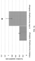
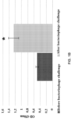
- the present inventors examined the effects of exposure to a bacteriophage cocktail on intestinal permeability and relative abundance of taxonomic units in the gut bacterial community. There was an increase in markers of impaired gut permeability, such as the lactulose/mannitol ratio, plasma endotoxin concentrations, and serum levels of inflammation-related cytokines, following the bacteriophage challenge. The present inventors observed significant differences in the alpha diversity of faecal bacterial species and found that richness and diversity index values increased following the bacteriophage challenge.
- Inactivation can occur by various means, including chemical treatment (e.g., with "polyhexamethylene guanidine derivatives", e.g., those prepared by condensation polymerization of hexamethylenediamine with guanidine salt and hydrazine hydrate, as described in U.S. Provisional Pat. Appl. No. 62/510,446 , U.S. Patent No. 8,993,712 , U.S. Pat. Appl. Pub. No. 2017/0013838 and Int. Pat. Appl. Pub. No. WO2016/118043 ), ozone, gamma irradiation, heat treatment, electron flow treatment, gamma irradiation, capsid-targeted viral inactivation, microwave radiation, pascalization and filtration.
- chemical treatment e.g., with "polyhexamethylene guanidine derivatives”, e.g., those prepared by condensation polymerization of hexamethylenediamine with guanidine salt
- bacteria encompasses both prokaryotic organisms and archaea present in mammalian microbiota.
- microbiota is used herein to refer to microorganisms (e.g., bacteria, archaea, fungi, protozoa) and viruses (e.g., phages and eukaryotic viruses) present in a host animal or human (e.g., in the gastrointestinal tract, skin, oral cavity, vagina, etc.). Microbiota exerts a significant influence on health and well-being of the host. Viruses present in microbiota are separately described as “virobiota”.
- microbiome refers to the collective genes of all organisms comprising the microbiota.
- microbiota can be detected using various methods, including without limitation quantitative PCR (qPCR) or high-throughput sequencing methods which detect over- and under-represented genes in the total bacterial population (e.g., 454-sequencing for community analysis; screening of microbial 16S ribosomal RNAs (16S rRNA), etc.), or transcriptomic or proteomic studies that identify lost or gained microbial transcripts or proteins within total bacterial populations. See, e.g., U.S. Patent Publication No.
- microbiota diseases in the GI tract have been reported to be associated with a wide variety of illnesses, such as, e.g., irritable bowel syndrome (IBS), inflammatory bowel disease (IBD), chronic fatigue syndrome, obesity, rheumatoid arthritis, ankylosing spondylitis, colitis, small intestinal cancer, colorectal cancer, metabolic syndrome, cardiovascular disease, Crohn's disease, gastroenteritis, food allergy, Celiac disease, gastrointestinal graft versus host disease, pouchitis, intestinal failure, short bowel syndrome, diarrhea, etc.
- IBS irritable bowel syndrome
- IBD inflammatory bowel disease
- chronic fatigue syndrome obesity
- rheumatoid arthritis ankylosing spondylitis
- colitis small intestinal cancer
- colorectal cancer metabolic syndrome
- cardiovascular disease Crohn's disease
- gastroenteritis food allergy
- Celiac disease gastrointestinal graft versus host disease
- pouchitis intestinal failure
- short bowel syndrome diarrhea
- PrD Prion-like domain
- PrPs are characterized by self-propagation, undergoing a conformational switch from one conformational state to another, which leads to the creation of new prions (Bolton et al., 1982; Telling et al., 1995).
- PrPSc infectious form of prion
- the term "therapeutically effective amount” refers to the amount of a compound, composition, particle, organism (e.g., a probiotic or a microbiota transplant), etc. that, when administered to a subject for treating (e.g., preventing or ameliorating) a state, disorder or condition, is sufficient to effect such treatment.
- the “therapeutically effective amount” will vary depending, e.g., on the agent being administered as well as the disease severity, age, weight, and physical conditions and responsiveness of the subject to be treated.
- patient refers to mammals, including, without limitation, human and veterinary animals (e.g., cats, dogs, cows, horses, sheep, pigs, etc.) and experimental animal models.
- subject is a human.
- Non-limiting examples of diseases and conditions treatable by bacteriophage inhibition or inactivation include, without limitation, diseases caused by changes in properties of microbiota (e.g., skin, mucosal, or GI microbiota); endotoxemia; oncological diseases; obesity; age-related changes of skin; vaginosis, neurodegenerative diseases (e.g., Alzheimer's disease, Parkinson's disease); Chronic Fatigue Syndrome, Obsessive-Compulsive Disorder, generalized anxiety disorder (GAD), major depressive disorder (MDD), social anxiety disorder (SAD), attentiondeficit/hyperactivity disorder (ADHD); diseases and conditions accompanied by increased intestinal permeability (e.g., irritable bowel syndrome [IBS], non-specific ulcerative colitis, celiac disease, diabetes, rheumatoid arthritis, multiple sclerosis, Amyotrophic Lateral Sclerosis [ALS], CADASIL Syndrome, Hashimoto's thyroiditis, Dentatorubro-pallido-Luysian at
- the term "about” or “approximately” means within a statistically meaningful range of a value. Such a range can be within an order of magnitude, preferably within 50%, more preferably within 20%, still more preferably within 10%, and even more preferably within 5% of a given value or range.
- the allowable variation encompassed by the term “about” or “approximately” depends on the particular system under study, and can be readily appreciated by one of ordinary skill in the art.
- John Wiley and Sons, Inc. Hoboken, NJ ; Coligan et al. eds. (2005) Current Protocols in Immunology, John Wiley and Sons, Inc.: Hoboken, NJ ; Coico et al. eds. (2005) Current Protocols in Microbiology, John Wiley and Sons, Inc.: Hoboken, NJ ; Coligan et al. eds. (2005) Current Protocols in Protein Science, John Wiley and Sons, Inc.: Hoboken, NJ ; and Enna et al. eds. (2005) Current Protocols in Pharmacology, John Wiley and Sons, Inc.: Hoboken, NJ . Additional techniques are explained, e.g., in U.S. Patent No. 7,912,698 and U.S. Patent Appl. Pub. Nos. 2011/0202322 and 2011/0307437 .
- disease is prevented and treated by preventing entry of bacteriophages into the body (e.g., into microbiota, bodily fluid(s) or tissue(s)) by modifying bacteriophages.
- bacteriophages e.g., into microbiota, bodily fluid(s) or tissue(s)
- DNase, RNase, proteases, or amyloid-like proteins can be applied to the bacteriophage in an amount that affects the bacteriophage surface without inactivating the bacteriophage.
- the bacteriophages are inactivated in a food product by ozone.
- capsid-targeted viral inactivation is used.
- photocatalytic inactivation is used.
- WO2016/118043 are used to inactivate bacteriophages in a food product.
- filtration is used to inactivate bacteriophages in a food product.
- pascalization is used to inactivate bacteriophages in a food product.
- heat treatment is used to inactivate bacteriophages in a food product.
- gamma radiation is used to inactivate bacteriophages in a food product.
- an electron flow is used to inactivate bacteriophages in a food product.
- microwave radiation is used to inactivate bacteriophages in a food product.
- the food product is treated by pascalization to inactivate bacteriophages in the food product.
- the food product is heat treated to inactivate bacteriophages in the food product.
- the food product is gamma irradiated to inactivate bacteriophages in the food product.
- the food product is treated with electron flow to inactivate bacteriophages in the food product.
- the food product is treated with microwave radiation to inactivate bacteriophages in the food product.
- the food product is treated with capsid-targeted viral inactivation to inactivate bacteriophages in the food product.
- the food product is treated with photocatalytic inactivation to inactivate phages in the food product.
- the food product is treated with ozone to inactivate phages in the food product.
- the bacteriophages in drinking water are inactivated by filtration. In one embodiment, the bacteriophages in drinking water are inactivated by photocatalysis. In one embodiment, the bacteriophages in drinking water are inactivated by ozone. In one embodiment, the bacteriophages in drinking water are inactivated by a peroxide (e.g., H 2 O 2 ). In one embodiment, the bacteriophages in drinking water are inactivated by a metal. In one embodiment, pascalization is used to inactivate the bacteriophages in drinking water. In one embodiment, heat treatment is used to inactivate the bacteriophages in drinking water.
- the drinking water is treated with photocatalysis to inactivate the bacteriophages in the drinking water.
- the drinking water is treated with ozone to inactivate the bacteriophages in the drinking water.
- the drinking water is treated with a peroxide (e.g., H 2 O 2 ), to inactivate the bacteriophages in the drinking water.
- the drinking water is treated with a metal to inactivate the bacteriophages in the drinking water.
- the drinking water is treated with pascalization to inactivate the bacteriophages in the drinking water.
- the drinking water is treated with heat sufficient to inactivate the bacteriophages in the drinking water.
- the bacteriophages in the water for washing are inactivated by filtration. In one embodiment, the bacteriophages in the water for washing are inactivated by photocatalysis. In one embodiment, the bacteriophages in the water for washing are inactivated by ozone. In one embodiment, the bacteriophages in the water for washing are inactivated by a peroxide (e.g., H 2 O 2 ).In one embodiment, the bacteriophages in the water for washing are inactivated by a metal. In one embodiment, pascalization is used to inactivate the bacteriophages in the water for washing.
- a peroxide e.g., H 2 O 2
- pascalization is used to inactivate the bacteriophages in the water for washing.
- the bacteriophages are inactivated in water for washing.
- the water for washing undergoes filtration sufficient to inactivate the bacteriophages.
- the water for washing undergoes photocatalysis sufficient to inactivate the bacteriophages.
- the water for washing is treated with sufficient ozone to inactivate the bacteriophages.
- the water for washing is treated with sufficient peroxide (e.g., H 2 O 2 ) to inactivate the bacteriophages.
- the water for air humidification is treated by pascalization sufficient to inactivate the bacteriophages. In one specific embodiment, the water for air humidification is treated with heat sufficient to inactivate the bacteriophages. In one specific embodiment, the water for air humidification is gamma irradiated to inactivate the bacteriophages. In one specific embodiment, the water for air humidification is treated with electron flow sufficient to inactivate the bacteriophages. In one specific embodiment, the water for air humidification is treated with microwave radiation sufficient to inactivate the bacteriophages. In one specific embodiment, the water for air humidification is treated by capsid-targeted viral inactivation sufficient to inactivate the bacteriophages. In one specific embodiment, the water for air humidification undergoes photocatalytic inactivation sufficient to inactivate the bacteriophages.
- the phages are inactivated in the environment.
- the phages are inactivated in the environment by treating items of habitat (e.g., furniture, a room, dishes, a bath, a sink, a toilet bowl, a container for packaging and storage of food products or water) with "polyhexamethylene guanidine derivatives", e.g., those prepared by condensation polymerization of hexamethylenediamine with guanidine salt and hydrazine hydrate, as described in U.S. Provisional Pat. Appl. No.
- the phages are inactivated in the environment by treating items of habitat (e.g., furniture, a room, dishes, a bath, a sink, a toilet bowl, a container for packaging and storage of food products or water) with an electron flow.
- the phages are inactivated in the environment by treating items of habitat (e.g., furniture, a room, dishes, a bath, a sink, a toilet bowl, a container for packaging and storage of food products or water) with ultraviolet.
- the phages are inactivated in the environment by treating items of habitat (e.g., furniture, a room, dishes, a bath, a sink, a toilet bowl, a container for packaging and storage of food products or water) with ozone.
- items of habitat e.g., furniture, a room, dishes, a bath, a sink, a toilet bowl, a container for packaging and storage of food products or water
- the phages are inactivated in the environment by treating items of habitat (e.g., furniture, a room, dishes, a bath, a sink, a toilet bowl, a container for packaging and storage of food products or water) with a peroxide (e.g., H 2 O 2 ).
- a peroxide e.g., H 2 O 2
- the phages are inactivated in the environment by treating items of habitat (e.g., furniture, a room, dishes, a bath, a sink, a toilet bowl, a container for packaging and storage of food products or water) with a method of capsid-targeted viral inactivation.
- the phages are inactivated in the environment by treating items of habitat (e.g., furniture, a room, dishes, a bath, a sink, a toilet bowl, a container for packaging and storage of food products or water) with a method of photocatalytic inactivation.
- items of habitat e.g., furniture, a room, dishes, a bath, a sink, a toilet bowl, a container for packaging and storage of food products or water
- a method to prevent entry of bacteriophages into the body comprising inactivating bacteriophages in the environment by preventing their distribution via an air conditioning and humidification system.
- the bacteriophages are inactivated in an air conditioning and humidification system in a room, in a hospital environment, a motor vehicle, a train, an airplane, a surface vessel, a submarine vessel and a spacecraft.
- the bacteriophages are inactivated in an air conditioning and humidification system by filtration of air and a liquid used for the humidification.
- the bacteriophages are inactivated in an air conditioning and humidification system by ultraviolet treatment of air and a liquid used for the humidification.
- the bacteriophages are inactivated in an air conditioning and humidification system by treating air and a liquid used for the humidification with "polyhexamethylene guanidine derivatives", e.g., those prepared by condensation polymerization of hexamethylenediamine with guanidine salt and hydrazine hydrate, as described in U.S. Provisional Pat. Appl. No. 62/510,446 , U.S. Patent No. 8,993,712 , U.S. Pat. Appl. Pub. No. 2017/0013838 and Int. Pat. Appl. Pub.
- bacteria having their bacteriophage receptors inactivated are transplanted to microbiota to interfere with an interaction between bacteriophages with the bacteria of microbiota.
- the bacteriophage receptors are inactivated by a method of capsid-targeted viral inactivation.
- antibodies are used, or administered to the mammal.
- phage receptors on bacteria are blocked by phage receptors for attaching to bacteria not able to cause death of target bacteria.
- the phage receptors are obtained as recombinant proteins.
- the methods are used for increasing longevity and/or decreasing aging in a mammal.
- the aging is skin aging.
- the method further comprises administering a compound in combination with at least one compound that potentiates activity of the compound.
- the compound is used with pharmaceutically acceptable carrier or excipient.
- the compound is used with other drugs for increasing the effect.
- the method is effective to prevent or treat an increase in intestinal permeability.
- the increase in intestinal permeability is caused by entry of a bacteriophage into gastrointestinal tract of the body.
- the method further comprises preventing changes in the normal microbiota leading to the development of increased intestinal permeability, by limiting exposure to bacteriophages from the objects of the environment, including products of daily use.
- the method further comprises prevention of changes in the normal microbiota leading to obesity, by exposure to bacteriophages from the environment.
- Another way to interfere with an interaction between bacteriophages with the bacteria of microbiota is to block receptors on bacteria by adding phage receptor proteins that attach to bacteria, but are not able to cause death of target bacteria.
- microbiota discussed herein can be detected using various methods, including without limitation quantitative PCR or highthroughput sequencing methods which detect over- and under-represented genes in the total bacterial population (e.g., 454-sequencing for community analysis; screening of microbial 16S ribosomal RNAs (16S rRNA), etc.), or transcriptomic or proteomic studies that identify lost or gained microbial transcripts or proteins within total bacterial populations. See, e.g., U.S. Patent Publication No.
- Phages may be implicated in protein misfolding, altered intestinal permeability, and chronic inflammation in mammals. Phages may play a critical role in the development of neurodegenerative diseases, such as PD. Such multifactorial diseases may have increased intestinal permeability as a triggering or aggravating factor.
- a method of vaccinating a mammal against a bacteriophage comprising administering a composition to the mammal comprising a polypeptide or epitope of the bacteriophage.
- the bacteriophage may be a temperate bacteriophage.
- the bacteriophage may be a lytic bacteriophage.
- vaccination against lytic bacteriophages, particularly those that target Lactobacillus bacteria may be beneficial to prevent the neurodegenerative disease, inhibit the triggering of the neurodegenerative disease or to reduce the progression of the neurodegenerative disease.
- Such course of treatment may help preserve the Lactococcus bacteria in the mammal, allowing for production of adequate amounts of dopamine and other neurotransmitters.
- Yet another way of targeting bacteriophages is to administer a gene editing nuclease that is configured to edit the genome of bacteriophages that target Lactococcus bacteria, or any other bacteria of interest.
- An exemplary, but non-limiting, gene editing nuclease is CRISPR.
- the nuclease may be configured to specifically target a sequence in one bacteriophage, such as a lytic bacteriophage targeting a bacterium, while not being effective to target other temperate bacteriophages that target the same bacterium.
- the gene editing nuclease can specifically target Lactococcus bacteria that are in group 936 and/or are c2-like.
- the various methods of targeting bacteriophages described herein may be adapted to target lytic bacteriophages specific to bacteria that are beneficial for the production of neurotransmitters. For instance, lytic bacteriophages that target Lactococcus bacteria may be selectively eliminated from food, water, and blood for transfusions.
- a method to treat a disease comprising detecting the presence of a mutation or gene in a bacteriophage, and then targeting the bacteriophage.
- the gene or mutation may increase the ability of the bacteriophage to target a specific bacterium in a microbiome.
- the gene or mutation may allow the bacteriophage to overcome bacterial antiphage systems.
- a non-limiting example of a bacterial antiphage system is the Abortive infection (Abi) systems.
- the bacteriophage with the detected mutation or gene may be inactivated by any of the methods described herein.
- a method for diagnosing the potential for a phage to cause a disease by detecting the presence of certain mutations in genes that or detecting the presence of genes within bacteriophages or a bacterium in the microbiota.
- the bacteriophages may be acquired by a mammalian patient in a hospital.
- the disease may be caused by a bacteriophage or a prophage present in a hospital.
- the detected genes or detected mutations within bacteriophages may be effective to increase the ability of bacteriophage to target bacteria in the microbiome that prevent or reduce the progression of the disease.
- the detected genes or detected mutations within the bacterium in the microbiota may increase the ability of the bacterium cause progression of the disease.
- the gene or mutation in the bacteriophage may increase the ability of a bacteriophage to overcome bacterial antiphage systems.
- the gene or mutation in the bacterium may increase the ability of a bacteriophage to overcome the bacterium's antiphage systems.
- a non-limiting example of a bacterial antiphage system is the Abortive infection (Abi) systems.
- Table 1 A summary of how bacteriophages may cause various infections or diseases is provided in Table 1 below: Table 1.
- Table 1 Main factors that may cause bacteriophage infections
- Comments Infection by lytic phages Primary infection by a bacteriophage in the environment
- Infection by bacteria carrying prophages in their DNA Primary infection by a bacterium in the environment that harbor prophages
- Induction of prophages Can be due to a variety of different internal and external triggers that lead to progeny release and an increase in the number of free phages
- Increased translocation of phages to biological fluids Can be due to increased intestinal permeability or increased blood-brain barrier permeability
- Alteration of the microbiota Alteration can shift the number of, or relative amount of, prophages or lytic phages Mutations in prophages leading to Abiescape phage mutants
- the mutated phages are able to overcome bacterial protective systems Changes in macroorganism's sensitivity to bacterioph
- Example 1 Bacteriophages introduced to rats increase intestinal permeability.
- a lactulose-mannitol permeability test was performed to determine whether bacteriophages may cause microbiota diseases resulting in alterations in the host organism in the form of increased intestinal permeability.
- the lactulose:mannitol ratio was measured as a marker of intestinal permeability 2 days before and 10 days after phage challenge as described by Meddings et al. (Meddings, J.B. et al., 2000).
- Lactulose (L7877, Sigma-Aldrich) and mannitol (M8429, Sigma-Aldrich) were utilized for all arms of the study.
- a rat model was used to measure markers of intestinal permeability and endotoxemia.
- faecal material was collected from the same animals before and after the phage challenge.
- Daily phage challenge involved administering a bacteriophage cocktail comprised of commercially available and characterised phages active against the Enterobacteriaceae, Staphylococcaceae, Streptococcaceae, and Pseudomonadaceae families.
- Salmonella bacteriophage cocktail from Microgen (Moscow, Russia; product batch number H20), containing bacteriophages against Salmonella enterica serotypes: S . Paratyphi, S. Typhimurium, S. Heidelberg, S. Newport, S. Choleraesuis, S. Oranienburg, S. Infans, S. Dublin, S. Enteritidis, S. Anatum, and S. Newlands.
- Pyobacteriophage another polyvalent commercial phage cocktail from Microgen (product batch number 4), contained phages against seven bacterial species, Staphylococcus aureus, Streptococcus pyogenes, Proteus mirabilis, Proteus vulgaris, Pseudomonas aeruginosa, Klebsiella pneumoniae, and Escherichia coli. The cocktails were mixed. Phages (1.5 mL, 1 ⁇ 10 6 plaque-forming units/mL of each phage cocktail) were added to drinking water according to the manufacturer's instruction and administered orally for 10 days. Each animal was used as its own control before the bacteriophage challenge.
- Gut permeability function was examined in animals before (at baseline) and 10 days after the daily challenge with a bacteriophage cocktail using each animal as its own control. Without wishing to be bound by theory, because bacteriophages are known to selectively interact with bacteria and not affect eukaryotic cells, any impaired gut permeability can only be a consequence of altered microbiota.
- the animals were monitored daily for any changes of their activity, behavior, and general health status, including the weight loss, ruffled coat, wheezing or abnormal respiration, and the presence of unformed faeces. Body weights of all animals were measured using a digital balance (Mettler-Toledo, Inc., Columbus, OH, USA). Urine was collected for 24 h.
- intestinal permeability was measured using a clinical test based on saccharide permeability, which measures the ratio of lactulose to mannitol after oral ingestion of the sugars.
- the lactulose/mannitol ratio was measured using gas chromatography as described previously.
- the animals showed higher gut permeability following the bacteriophage challenge, with an average post-treatment lactulose/mannitol ratio of 0.99 ⁇ 0.08 compared with that of 0.31 ⁇ 0.05 pre-bacteriophage treatment (p ⁇ 0.05) ( Fig. 2A ).
- An increased lactulose/mannitol ratio was found compared with that at baseline, which was determined prior to administration of the bacteriophages, indicating increased intestinal permeability following oral administration of bacteriophages.
- Rats were assayed to determine whether bacteriophage treatment and altered intestinal permeability could cause endotoxemia. As shown in Figs. 2A-2E , the rats exhibited dramatically elevated serum levels of endotoxemia markers after the phage challenge, as compared to the pre-treatment baseline data. The inventors found that the animals had higher levels of plasma lipopolysaccharide (LPS) after the bacteriophage challenge, with an average of 0.28 ⁇ 0.07 endotoxin units (EU)/mL as compared with an average of 0.08 ⁇ 0.02 EU/mL (p ⁇ 0.05) before the treatment ( Fig. 2B ). Accordingly, the levels of serum inflammatory cytokines were also elevated after the bacteriophage challenge.
- LPS plasma lipopolysaccharide
- EU endotoxin units
- Treatment with phages resulted in a significant increase in the serum concentrations of tumour necrosis factor-alpha (TNF- ⁇ ), interleukin (IL)-1 ⁇ , and IL-6 (p ⁇ 0.05 compared with the baseline pre-treatment data; Fig. 2C-E ).
- TNF- ⁇ tumour necrosis factor-alpha
- IL-1 ⁇ interleukin-1 ⁇
- IL-6 p ⁇ 0.05 compared with the baseline pre-treatment data; Fig. 2C-E .
- gut microbiota composition was analyzed in the rats before and after the bacteriophage challenge using Illumina-based 16s rRNA sequencing.
- V3 and V4 regions of the 16S bacterial rRNA gene were amplified using a two-step polymerase chain reaction (PCR) protocol with V3 and V4 region primers (forward: 5'-TCGTCGGCAGCGTCAGATGTGTATAAGAGACAGCCTACGGGNGGCWGCAG-3' (SEQ ID NO: 1); reverse: 5'-GTCTCGTGGGCTCGGAGATGTGTATAAGAGACAGGACTACHVGGGTATCTAATCC-3') (SEQ ID NO: 2) for the first PCR and Nextera XT index primers for the second PCR. Amplicons were cleaned using AMPure XP magnetic beads, and then, Illumina sequencing adapters and dual-index barcodes were added to each amplicon.
- PCR polymerase chain reaction
- FASTQ files generated by Illumina sequencing were qualitatively evaluated using FASTQC. Adaptor contamination and low-quality reads were detected with Trimmomatic. Raw data were quality-filtered to remove sequences with ⁇ 200 nucleotides or containing ambiguous bases, uncorrectable barcodes, and homopolymer runs of more than six bases. Sequences were grouped into OTUs with a 97% threshold of pairwise identity. Operational taxonomic units (OTUs) were defined as a set of sequence reads with the similarity cutoff of 97%. For clarity and visualisation purposes, the most abundant sequences with a relative abundance of ⁇ 0.5% within each taxa were designated as 'representative sequences'.
- the 16s rRNA data were analyzed at the species level with OTUs detected at the level of abundance of > 0.001% ( Fig. 5D ).
- the highest depletion at the genus level, >two-fold (i.e. log 2 1), was registered post-bacteriophage treatment for the genera Clostridium, Eubacterium, Staphylococcus, Coprococcus, Lactobacillus, Collinsella, Blautia, Bifidobacterium, Dorea, and Prevotella.
- certain members of the genera Ruminococcus and Prevotella were significantly enriched ( Fig. 5E ).
- Microwave radiation products with the addition of bacteriophages, treated in the microwave oven.
- Group "Prototype” products with the addition of bacteriophages, treated with potassium nitrate.
- Example 5 Inactivating bacteriophages in external environment prevents increase in intestinal permeability
- the resulting filtrate was incubated at 60 °C for 60 minutes to inactivate bacteria and was added to 100 ⁇ L of water.
- the resulting solutions were given to animals with water, and the development of an increased intestinal permeability was evaluated as described in Example 4, above.
- the animals were divided into the following treatment groups:
- Ozone water with the addition of phages obtained from objects and surfaces treated with ozone.
- Peroxide water with the addition of phages obtained from objects and surfaces treated with H 2 O 2 .
- Group "Prototype” water with the addition of phages obtained from objects and surfaces treated with potassium nitrate.
- mice in Group #1 were isolated from the feces of Group #1 mice in which obesity was modeled.
- the obtained phages were added to drinking water of mice (age 12 weeks) from Group #2 for 21 weeks.
- the diet of the animals remained standard, i.e., not a high fat diet.
- the mice in Group #2 were divided into the following treatment groups:
- Example 7 Slowing skin aging by limiting bacteriophage entry from the environment.
- Group "Prototype” water treated with potassium nitrate.
- Antibodies to bacteriophages against P . aeruginosa were obtained after hyperimmunization of a rabbit by the method according to "The guidelines for the production of antibodies in laboratory animals" See acuc.berkeley.edu/guidelines/antibodies.pdf. After treating bacteriophages with these antibodies, the bacteriophages lost the ability to cause infection in those microbes as detected by titration according to the Gratia's method ( Kropinski et al. "Enumeration of bacteriophages by double agar overlay plaque assay.” Bacteriophages: Methods and Protocols, Volume 1: Isolation, Characterization, and Interactions, Humana Press, 2009, vol. 501, 69-76 ).
- Example 10 Blocking bacteriophage receptors of bacteria by inactivated bacteriophages
- Bacteriophages of E . coli were inactivated by irradiation with UV light for 30 min.
- the protective effect of inactivated bacteriophages is shown in Table 8, which demonstrates that UV treatment of bacteriophages reduces the titer to undetectable levels (as determined by Gratia's method, see Kropinski et al. "Enumeration of bacteriophages by double agar overlay plaque assay.”
- Bacteriophages Methods and Protocols, Volume 1: Isolation, Characterization, and Interactions, Humana Press, 2009, vol. 501, 69-76 ). As a control, when the original phage is added back, the titer increases.
- a mixture of two commercial bacteriophage cocktails was used (1.5 ml [1 ⁇ 10 6 plaque-forming units/ml] of each phage according to the manufacturer's instruction): (i) Salmonella bacteriophage cocktail (Microgen, Russia) containing phages infecting Salmonella enterica serotypes: Paratyphi, Typhimurium, Heidelberg, Newport, Choleraesuis, Oranienburg, Infans, Dublin, Enteritidis, Anatum, and Newlands, and (ii) Pyobacteriophage Polyvalent, another commercial phage cocktail (Microgen, Russia) containing phages infecting Staphylococcus aureus, Streptococcus pyogenes, Proteus mirabilis and P .
- Fecal samples were collected at baseline, after 2 days and after 30 days of the bacteriophage challenge into a sterile container and immediately stored at -80°C until further processing.
- DNA extraction was performed from faecal samples and control sample of the milk enriched with phages using a QIAamp stool DNA mini kit (Qiagen, Germany) according to the manufacturer's instructions.
- Shotgun whole genome sequencing was performed according to the Illumina system instructions. Data were analyzed with the software Metagenomics workflow (Illumina). The taxonomic identity of the reads was analysed using available annotation source databases (NCBI database). Data of the shotgun sequencing are shown in Table 10. Table 10. Bacteriophage composition analysis.
- Phage description Identified phages in animals Milk artificially enriched with phages Baseline metagenomic faecal probes Day 2 after bacteriophage challenge Day 30 after bacteriophage challenge 1 2 3 4 5 1 2 3 4 5 1 2 3 4 5 Enterobacteria phages + - - - - + + + + + + + + - (including, among others, Caudovirales; Myoviridae; Tevenvirinae) Staphylococcus phages + - - - - - + + + + + + + + + + + + + + + + + + + + + + + + + + + + + + + + + + + + + + + + + + + + + + + + + + + + + + + + + + + + + + + + + + + + + + + + (Caudovirales; Siphoviridae) Other phages present in mammalian gut flora - + + + + + + +
- Example 13 Analysis of bacteriophage composition of donor fecal transplant and of recipient microbiota following fecal transplant.
- a composition of bacteriophage virome in gut microbiota of three subjects with Clostridium difficile infection was analyzed before and after faecal transplant using shotgun sequencing technology (Illumina) and computational predisposition method.
- Fecal samples of the donor were analyzed from the same lot as was used for the fecal microbiota transplant (FMT). Fecal samples of the FMT recipients were collected at baseline (pre-FMT) and after FMT (post-FMT). DNA extraction was performed using a QIAamp stool DNA mini kit (Qiagen, Germany) according to the manufacturer's instructions. Shotgun whole genome sequencing was performed according to the Illumina system instructions. Data was analyzed with the software Metagenomics workflow (Illumina). The taxonomic identity of the reads was analyzed using available annotation source databases. Results are shown in Table 11 (pre-FMT samples), Table 12 and Figure 6 (pre- and post-FMT samples). Table 11.
- the data demonstrate qualitative alterations of the bacteriophage composition of the recipients' gut microbiota and transfer of the donor's bacteriophages to recipients ( Microviridae and Inoviridae ), confirmed by their appearance in recipients' post-FMT fecal samples.
- Experimental data obtained confirmed data obtained by the mathematical predication of virome alterations following FMT
- Example 14 Identification of prionogenic domains (PrD) in bacteriophage proteomes.
- the inventors used a computational algorithm, prion-like amino acid composition (PLAAC), to detect prion-like domains in 370,617 publicly available bacteriophage protein sequences, which resulted in the identification of 5040 putative prions.
- PKAAC prion-like amino acid composition
- a set of these prion-like proteins was analyzed, regularities in their distribution across different phage families, associated with their interactions with the bacterial host cells, was observed.
- the inventors found that prion-like domains could be found across all phages of various groups of bacteria and archeae.
- bacteriophage prion-like proteins are predominantly involved in the interactions between bacteriophages and bacterial cell, such as those associated with the attachment and penetration of bacteriophage in the cell, and the release of the phage progeny. These data allow the identification of phage prion-like proteins as novel regulators of the interactions between bacteriophages and bacterial cells.
- PrDs putative prion like domains
- An HHM algorithm was employed for the analysis of bacteriophage proteomes, using the UniProtKB database to identify candidate PrD sequences among bacteriophages.
- the inventors analyzed the trends in the distribution of PrDs in different protein families and the correlations between bacteriophage families and the host bacterial host.
- the present predictive approach uncovers a large set of putative prionogenic proteins.
- PrDs in the interacting proteins in different bacteriophage families infecting a variety of bacterial hosts implies that these PrDs are conserved across species.
- PrD-containing proteins may be beneficial, but not obligatory since PrDs in the surface proteins were not identified in all analyzed bacteriophages, and in those where they were identified, PrDs were found in some, but not all surface proteins of the given phage.
- the attachment of bacteriophages is a complex process, accompanied by a number of bacteriophage-resistance responses initiated in a bacterial cell (Labrie et al., 2010).
- the inventors' findings may indicate that the proteins with the PrDs can alter their conformational states, adding further complexity and specificity in the phage-bacterial interactions, and this may serve as a mechanism to overcome some bacterial resistance mechanisms.
- administration of PrDs or bacteriophages with PrDs can be "used against" other bacteriophages or to target bacteriophages.
- An exemplary, but non-limiting mechanism could involve the PrD as a decoy that blocks an otherwise unwanted interaction between bacteria and the target bacteriophages.
- PrDs can also be associated with the singlestrand binding proteins, known as essential components of bacteriophage DNA synthesis. Therefore, this indicates that these PrDs can be easily misfolded into PrPSc through the interactions with the DNA, which was shown to be an important component inducing this misfolding (Cordeiro et al., 2001). Without wishing to be bound by theory, this type of interaction with the host cell may represent an additional process through which the bacteriophages control bacterial intracellular functions (Wagner and Waldor, 2002; Davies and Davies, 2010).
- the protein functions were manually curated using the information from the UniProt database, the National Center for Biotechnology Information (NCBI) protein sequence database (www.ncbi.nlm.nih.gov/), and the literature.
- NCBI National Center for Biotechnology Information
- PrDs The presence of PrDs was analyzed in all bacteriophage proteins using the PLAAC prion prediction program based on the HMM and trained using the known PrDs, by identifying the compositional bias towards N and Q residues.
- 2111 bacteriophage proteomes comprising a total of 370,617 proteins retrieved from the UniProtKB database, were analyzed.
- 5040 PrDs were identified (i.e. 1.35% of the total bacteriophage protein dataset) (Lancaster et al., 2014).
- the enrichment values obtained for the proteins detected in the bacteriophage family subsets were compared with the total number of analyzed proteins.
- Table 13 the trends in the PrD distribution across different bacteriophage families are presented. PrD frequency ranged from 1.16% to 4.49% when analyzing bacteriophage families with at least 400 sequenced proteins. Due to the low representation of archaeal phages, it was not possible to compare them with the bacteriophage proteins with sufficient reliability. Therefore, the inventors focused on the analysis of the PrDs in bacteriophages.
- PrDs were more frequently found among archaeal phages, and the analysis of the three best-known Caudovirales families showed that these domains are more abundant among Podoviridae than Myoviridae or Siphoviridae.
- Myoviridae members have larger proteomes than the members of Siphoviridae family, which may explain the observed differences in the PrD enrichment ( Sandaa, R. A. (2009). Viruses, Environmental, 553-567 ). Further analyses indicated a direct correlation between the proteome size and the PrD enrichment, and therefore, the inventors further explored whether the proteome size of bacteriophages in the Myoviridae family is related to the PrD enrichment. However, the inventors have not observed a linear correlation between these parameters, suggesting that the PrD enrichment does not depend only on the size of a Myoviridae proteome.
- the inventors aimed to select bacteriophage proteins in bacteria that may have a greater prion-forming potential.
- the correlation between the LLR score of the PrD proteins and the host bacteria was analyzed.
- the average LLR score varied significantly between the bacteriophages of different bacterial species.
- the lowest mean LLR values were detected among Cellulophaga and Escherichia bacteriophages (3.49 and 3.98, respectively) and the highest LLR score was obtained for the Bacillus bacteriophages, with the average LLR of 8.96 (p ⁇ 0.001).
- Table 15 Source DF Type III SS Mean Square F Value Pr> F Family 2 1920.410170 960.205085 16.84 ⁇ .0001
- Table 16 95% Confidence interval of bacteriophage families Family LLR Score LSMEAN 95% Confidence Limits 1 Myoviridae 5.908475 5.608805 6.208146 2 Podoviridae 7.780928 7.212436 8.349420 3 Siphoviridae 6.080153 5.717382 6.442923
- Table 17 Least squares means for effect of bacteriophage family on LLR score Least Squares Means for effect Family Pr >
- for H0: LSMean(i) LSMean(j) Dependent Variable: LLR Score i/j Myoviridae Podoviridae Siphoviridae 1 ⁇ .0001 0.7545 2 ⁇ .0001 ⁇ .0001 3 0.7545 ⁇ .0001
- the LLRs were compared between the remaining bacteriophage families. Myoviridae were shown to have the highest number of PrDs, with the LLR score over 30. 114 PrDs were identified with the medium LLR score over 20 within Myoviridae family, yielding 3.29% of the total number of PrDs identified in this bacteriophage family ( Figure 7B ).
- the inventors analyzed the proteome enrichment with PrDs in the bacteriophages of different bacteria.
- the highest enrichment rate, with at least five PrDs per proteome, was found among the bacteriophages of Bacillus, Cronobacter, Lactobacillus, Synechococcus, Staphylococcus, and other bacteria ( Figure 10 ; Table 18).
- Example 16 Analysis of structural domains associated with to bacteriophage PrDs
- PrPs The most abundant (2046 PrPs; medium LLR score 4.86) functional group was shown to be the attachment and penetration group, which included the proteins associated with the host cell binding and genome injection.
- the inventors identified receptor binding proteins, tail fiber proteins, peptidase_M23 baseplate wedge components, lysins, and other proteins known to have crucial functions in the various aspects of phage infection, including adsorption, peptidoglycan hydrolysis, cell wall penetration, and DNA ejection.
- PrDs were predominantly found among tail component, tail fiber, and baseplate proteins (Rodriguez-Rubio et al., 2012, Stockdale et al., 2013).
- PrD was identified in gp98/99 proteins that form the cell-puncturing device of Mycobacterium phages. Some of the identified proteins associated with surface structures, such as tail tube cap, are not involved in the direct contact with the bacterial cell, but they represent important structural components allowing the injection of DNA (Arisaka et al., 2003; Orlova 2012).
- the results of the heatmap analysis in Figure 9 showed that Podoviridae belonging to the Mycobacteriaceae family and Myoviridae of Listeriaceae have the highest LLR score.
- proteins with PrDs that are associated with the bacteriophage assembly in the host cells (170 proteins; medium LLR score, 6.16) were analyzed. These proteins were identified in all major families of Caudovirales. The highest average LLR score was obtained for the proteins of Myoviridae family of Moraxellaceae ( Figure 9 ), which are also associated with the organization and maturation of bacteriophages, and those involved in the tail assembly and DNA packaging (Linderoth et al., 1994; Boudko et al., 2002; Aksyuk and Rossmann, 2011).
- PrDs were identified in proteins that are involved in cell wall lysis and the release of bacteriophages from the host cells.
- This group contained only 105 proteins (medium LLR score, 5.13) and predominantly comprised lysins, including those with the amidase activity and proteins with peptidoglycan binding functions (hydrolases, D-ala-D-ala carboxypeptidase, endopeptidase CHAP domain protein, and others), which are involved in bacterial cell wall degradation, resulting in cell lysis and release of progeny viruses. They were predominantly detected among Siphoviridae infecting Streptococcaceae and Mycobacteriaceae. The highest average LLR score was obtained in Myoviridae family of Moraxellaceae ( Figure 9 ).
- Example 17 Bacterial community composition in PD
- HiSeq-mediated sequencing produced a total of 1,792,621,232 reads with an average of 30,383,411 reads per sample.
- Operational taxonomic units were defined as a set of sequence reads with a similarity cut-off of 97% (Schloss, P. et al., 2009).
- the QIIME pipeline was used for quality filtering of bacterial and bacteriophage DNA sequences, chimera removal (by the USEARCH software), taxonomic assignment, and calculation of ⁇ -diversity, as previously described (Caporaso, J. et al., 2010; Cox, L. et al., 2014). Downstream data analysis and calculation of diversity metrics were performed in R3.3.2 using ggplot2 and phyloseq libraries; DESeq2 was used to calculate logarithm of fold change (Love, M. et al., 2014).
- PCoA Principal Coordinate Analysis
- PD is associated with lower bacterial diversity and richness (Scheperjans, F. et al., 2014).
- the analysis described herein revealed significant differences in the microbiota structure between PD patients and control individuals. Some previously overlooked alterations in the bacterial community at the family and genus levels were detected using the MetaPhlAn tool, which provides accurate microbial profiling and estimates relative abundance of microbial cells by mapping reads against a set of clade-specific marker sequences (McMurdie, P. J. et al., 2013).
- Siphoviridae was the most abundant family in both PC and control groups.
- Example 19 Composition of phagobiota at the species and analysis of phage/bacteria ratio
- compositional changes in the bacteriophage community were examined at the species level.
- the abundance of phage species were analysed individually for each sample.
- the data is shown in the form of heat maps presented in Figures 12A-12I .
- the sampling methods used in extracting total metagenomic DNA data included sampling phages which cover the bulk of the phagobiota (Waller, A. et al., 2014).
- Species detectable only in one group were evaluated to assess the appearance or disappearance of individual bacteriophage species in comparison between one group versus the other.
- the data is shown in Figures 13A and 13B , as well as in Table 28.
- Significant changes include total disappearance of certain Bacillus, Enterobacteria, Lactococcus Streptococcus, and Salmonella phages, which belonged to the Siphoviridae family or were unclassified Caudovirales, as well as Lactobacillus phages of the Myoviridae family.
- the ratio between phages and their bacterial hosts in the gut microbiome was analysed by calculating the phage/bacteria ratio in a manner defined as the 'lytic potential' (Waller, A. et al., 2014). Phages were clustered according to their bacterial hosts. The phage abundance was normalized to that of the respective hosts in each group. The data is shown in Figures 14A-14D . The obtained data were consistent with the trend of reduction in ⁇ - and ⁇ -diversity in both bacteriophages and bacteria among PD patients, which indicated stability of the phage/bacteria equilibrium across PD and control samples. However, some significant alterations in the phage/bacteria ratio in the PD group were identified, suggesting their association with PD.
- a phage/bacteria ratio equal to one indicates that a prophage is stably integrated within the host bacterial genome, which was observed in this study for Bacillus and Vibrio phages and the corresponding bacterial host in the PD group (Waller, A. et al., 2014).
- the ratio less than one indicates that a prophage is most likely absent in the genomes of a part of the corresponding bacterial host population.
- Low phage/bacteria ratios were observed for Bacteroides, Klebsiella, Clostridium, and Streptococcus, which reflect low numbers of the corresponding phages and a high abundance of the host bacteria across both PD and control groups.
- Example 20 Analysis of Lytic and Temperate Lactococcus phages
- Lactococcus bacteria are considered as an important source of microbiota-derived neurochemicals, including dopamine which they produce in appreciable physiological amounts (Kuley, E. et al., 2011; Asano, Y. et al., 2012).
- a decrease in the production of intestinal dopamine may be associated with early gastrointestinal symptoms of PD and may be involved in triggering the neurodegenerative cascade of the disease (Houser, M. et al., 2017; Scheperjans, F., 2016).
- Lactococcus phages may promote or trigger the onset of PD, as well as its gastrointestinal symptoms, associated with a lack of dopamine (Pfeiffer, R., 2003).
- Two possible scenarios can lead to the accumulation of lytic Lactococcus phages in the gut and depletion of their bacterial hosts (Mirzaei, M. et al., 2017). The first is a symptom of dysbiosis and the second is a result of environmental introduction of lytic Lactococcus phages, which are widely used in the food industry and can be found in a variety of dairy products, including milk, cheese, and yogurt (Mills, S. et al., 2013; Lyte, M.
- Example 21 Bacteripohages circulation in human biologic fluids
- Example 23 Method for diagnosing diseases based on particularities of phages abundances in the gut
- the identified phages were clustered according to their bacterial hosts.
- samples of the microbiota from each patient were diluted in PBS and filtered through 0.22 mcm Millipore filter (Millipore Corp., Bedford, MA) and both fractions obtained following filtration were subjected to sequencing and processing.
- DNA sequencing data were generated by Illumina Hiseq4000 paired-end shotgun sequencing. Short sequence reads were retrieved from NCBI SRA (see www.ncbi.nlm.nih.gov/bioproject/382085; rather., J. et al., 2017).
- Sequences were qualityfiltered to remove adaptor contamination, low-quality reads with minimum quality score cut-off of 20, sequences with ⁇ 45 nucleotides, and human DNA. In the analysis of bacteria, sequences were grouped into OTUs with a 97% threshold of pairwise identity (Edgar, R. et al., 2010).
- Example 24 Method for diagnosing diseases based on particularities of phages circulation in CSF
- DNA from patients with different diseases was extracted from 200 ⁇ l of cerebrospinal fluid (CSF) using the Qiagen blood DNA mini kit, according to manufacturer's instructions.
- CSF cerebrospinal fluid
- Nextera XT Illumina
- 2 ng of DNA was tagmented with 6 ⁇ l ATM for 5 min at 56°C.
- the reaction was terminated with 5 ⁇ l of NT buffer with the following incubation at 22C for 5 minutes.
- products were PCR amplified in the presence of a barcode for 24 cycles.
- data from metagenomics were analyzed with custom database.
- Example 25 Method for treatment of diseases by inactivating phages circulating in biological fluids
- T1D type 1 diabetes
- Example 28 Bacteriophage can enter the bloodstream and promote release of pathogen-associated molecular patterns (PAMPs) to systemic circulation
- PAMPs pathogen-associated molecular patterns
- Bacteriophages were chosen from different bacteriophage families including Myoviridae, Podoviridae, and Siphoviridae, Ligamenvirales and unclassified phages. Assuming phages frequently adhere to blood cells, erythrocytes obtained following separation of plasma were resuspended in sterile water and lyzed by osmotic shock freezing. The earliest recovery from mouse blood was for the Klebsiella phage with 3 log10 PFU/ml was determined already at day 3, while all the other phages were recognized in circulation only at day 4. The highest phage titers on day four were for Pseudomonas phage with 5 log10 PFU/ml at day 4.
- phages on PAMPs and DNA levels in systemic circulation was studied at day 1. Then, an increase of circulating DNA, and particularly cell free bacterial DNA following bacteriophages treatment, was assayed by measuring the A260 value (SmartSpec Plus, BioRad) and by real-time PCR amplification (Thermofisher) of the 16S small-subunit rRNA gene. To evaluate extracullar DNA level, mouse plasma was filtered to remove bacterial contamination, followed by total DNA extraction from plasma using the Kit (Qiagen, Germany). The quantity of total DNA was measured at 260 nm and purity was assessed by the OD260/OD280 ratio. The extracted DNA was normalized to 50 ng/ ⁇ l.
- the amplification process included initial denaturation at 95°C for 2 min, 25 cycles of denaturation at 95°C for 30 s, annealing at 55°C for 30 s, elongation at 72°C for 30 s, and final extension at 72°C for 5 min.
- FIG. 17 shows the data in the form of a graph, where the Y axis shows the concentration of total cell-free circulating DNA in the plasma and, on the X axis, "1" indicates before bacteriophage administration and "2" indicates 24 hours after phage challenge.
- LPS lipopolysaccharide
- a Pyrochrome Limulus Amebocyte lysate kit (Associates of Cape Cod, Inc., East Falmouth, MA, USA) was used according to the manufacturer's instructions. Great care was taken to ensure aseptic collection and to avoid contamination with environmental LPS. Blood specimens were collected under pyrogen-free conditions and stored in LPS-free vials (Eppendorf, Germany). It was found that the animals had higher levels of plasma LPS after the bacteriophage challenge, with an average of 0.32 ⁇ 0.05 endotoxin units (EU)/mL as compared with an average of 0.06 ⁇ 0.03 EU/mL (p ⁇ 0.05) before the treatment.
- EU endotoxin units
- phage treatment unexpectedly increased plasma levels of PAMPs, including DNA, such as both bacterial-derived DNA and extracellular biofilm-derived DNA.
Landscapes
- Health & Medical Sciences (AREA)
- Life Sciences & Earth Sciences (AREA)
- Chemical & Material Sciences (AREA)
- General Health & Medical Sciences (AREA)
- Veterinary Medicine (AREA)
- Public Health (AREA)
- Animal Behavior & Ethology (AREA)
- Medicinal Chemistry (AREA)
- Pharmacology & Pharmacy (AREA)
- Epidemiology (AREA)
- Organic Chemistry (AREA)
- Engineering & Computer Science (AREA)
- Proteomics, Peptides & Aminoacids (AREA)
- Zoology (AREA)
- Bioinformatics & Cheminformatics (AREA)
- Microbiology (AREA)
- Genetics & Genomics (AREA)
- Wood Science & Technology (AREA)
- Immunology (AREA)
- Molecular Biology (AREA)
- Biochemistry (AREA)
- Mycology (AREA)
- Analytical Chemistry (AREA)
- General Engineering & Computer Science (AREA)
- Virology (AREA)
- Biophysics (AREA)
- Biotechnology (AREA)
- Pathology (AREA)
- Physics & Mathematics (AREA)
- Nutrition Science (AREA)
- Dermatology (AREA)
- Physiology (AREA)
- Chemical Kinetics & Catalysis (AREA)
- General Chemical & Material Sciences (AREA)
- Nuclear Medicine, Radiotherapy & Molecular Imaging (AREA)
- Biomedical Technology (AREA)
- Oncology (AREA)
- Gastroenterology & Hepatology (AREA)
- Neurosurgery (AREA)
- Otolaryngology (AREA)
Claims (5)
- Ein Verfahren zur Bestimmung der Wahrscheinlichkeit einer Krankheit in einem Wirtssäugetier, wobei das Verfahren umfasst: i) quantitative Analyse von Bakteriophagen und/oder Komponente(n) davon (z.B. Bakteriophagenprotein(e) und/oder Nukleinsäure(n)), die in der gastrointestinalen (GI) Mikroflora des Säugetiers vorhanden ist (sind), und (ii) Bestimmung des Bakteriophagen/Bakterienwirts-Verhältnisses, wobei die Krankheit eine Parkinson-Krankheit ist und wobei der Bakteriophage ein lytischer Lactococcus-Phage ist, der zur 936-Gruppe gehört.
- Das Verfahren nach Anspruch 1, wobei die Bakteriophagen ein Protein mit einer prionähnlichen Domäne umfassen.
- Das Verfahren nach Anspruch 1 oder 2, wobei die gastrointestinale (GI) Mikroflora eine zökale, ileale, colonale oder fäkale Mikroflora ist.
- Das Verfahren nach einem der Ansprüche 1 bis 3, wobei es sich bei der gastrointestinalen (GI) Mikroflora um eine fäkale Mikroflora handelt.
- Das Verfahren nach einem der Ansprüche 1 bis 4, wobei die quantitative und/oder qualitative Analyse von Bakteriophagen unter Verwendung eines oder mehrerer der folgenden Verfahren durchgeführt wird: Western Blot, ELISA, Flüssigbiopsieverfahren, Flüssigchromatographie- und Massenspektrometrieanalyse (LC/MS), kulturelle Mikrobiologieverfahren, genetische Verfahren, proteomische Verfahren, metagenomische Verfahren, computergestützte Modellierungs- und Simulationsverfahren und Datenanalyse.
Applications Claiming Priority (4)
| Application Number | Priority Date | Filing Date | Title |
|---|---|---|---|
| US201762510549P | 2017-05-24 | 2017-05-24 | |
| US201762537316P | 2017-07-26 | 2017-07-26 | |
| US201762581552P | 2017-11-03 | 2017-11-03 | |
| PCT/US2018/028640 WO2018217351A1 (en) | 2017-05-24 | 2018-04-20 | Methods for treating and preventing diseases |
Publications (3)
| Publication Number | Publication Date |
|---|---|
| EP3634442A1 EP3634442A1 (de) | 2020-04-15 |
| EP3634442A4 EP3634442A4 (de) | 2021-05-26 |
| EP3634442B1 true EP3634442B1 (de) | 2025-04-09 |
Family
ID=64396786
Family Applications (1)
| Application Number | Title | Priority Date | Filing Date |
|---|---|---|---|
| EP18805942.2A Active EP3634442B1 (de) | 2017-05-24 | 2018-04-20 | Verfahren zur diagnose der parkinson-krankheit |
Country Status (3)
| Country | Link |
|---|---|
| US (2) | US12274720B2 (de) |
| EP (1) | EP3634442B1 (de) |
| WO (1) | WO2018217351A1 (de) |
Families Citing this family (19)
| Publication number | Priority date | Publication date | Assignee | Title |
|---|---|---|---|---|
| EP3711488A1 (de) | 2015-05-06 | 2020-09-23 | Snipr Technologies Limited | Veränderung mikrobieller populationen und modifizierung von mikrobiota |
| GB201609811D0 (en) | 2016-06-05 | 2016-07-20 | Snipr Technologies Ltd | Methods, cells, systems, arrays, RNA and kits |
| GB201712733D0 (en) | 2017-08-08 | 2017-09-20 | Snipr Tech Ltd | Methods & cells |
| US10786448B2 (en) | 2018-01-18 | 2020-09-29 | Christian Arnold | Chewing gum composition comprising polyhexanide |
| US10760075B2 (en) | 2018-04-30 | 2020-09-01 | Snipr Biome Aps | Treating and preventing microbial infections |
| US11851663B2 (en) | 2018-10-14 | 2023-12-26 | Snipr Biome Aps | Single-vector type I vectors |
| CN110373345B (zh) * | 2019-05-08 | 2021-08-13 | 华东理工大学 | Dehp水解酶和基因及其在邻苯二甲酸酯类塑化剂降解中的应用 |
| JP2022548735A (ja) * | 2019-09-18 | 2022-11-21 | アンシリア, インコーポレイテッド | マイクロバイオーム調節のための組成物及び方法 |
| WO2021250183A1 (en) * | 2020-06-11 | 2021-12-16 | Chr. Hansen A/S | Method of increasing nisin production in lactococcus lactis |
| WO2022050082A1 (ja) * | 2020-09-01 | 2022-03-10 | 国立大学法人東北大学 | 乳汁中のIgA抗体含有量の増加剤 |
| US20240264176A1 (en) * | 2021-06-07 | 2024-08-08 | Regents Of The University Of Minnesota | Methods and materials for detecting prion diseases |
| CN113637606A (zh) * | 2021-08-10 | 2021-11-12 | 南京北极光生物科技有限公司 | 一种后生元和噬菌体复配的微生物组合制剂、制备方法及其应用 |
| CN114574450B (zh) * | 2021-11-24 | 2023-08-29 | 菲吉乐科(南京)生物科技有限公司 | 一种宽裂解谱耐紫外的奇异变形杆菌噬菌体和其组合物、试剂盒及其应用 |
| CN114921419B (zh) * | 2022-02-21 | 2024-05-17 | 武汉格瑞农生物科技有限公司 | 一种鸭疫里氏杆菌噬菌体 |
| CN114807104B (zh) * | 2022-04-11 | 2024-02-20 | 西南大学 | 肺炎克雷伯菌噬菌体裂解酶及其制备方法和应用 |
| GB202209518D0 (en) | 2022-06-29 | 2022-08-10 | Snipr Biome Aps | Treating & preventing E coli infections |
| CN115299438B (zh) * | 2022-08-04 | 2023-07-21 | 云南民族大学 | 多巴胺在抗噬菌体中的应用 |
| CN116555197B (zh) * | 2023-06-25 | 2023-09-01 | 中国科学院深圳先进技术研究院 | 一种沙门氏菌工程穿膜噬菌体及其构建方法与应用 |
| CN118048318A (zh) * | 2024-02-01 | 2024-05-17 | 瑞科盟(青岛)生物工程有限公司 | 一株对胍类消毒剂具有耐受性的溶藻弧菌噬菌体及其应用 |
Family Cites Families (20)
| Publication number | Priority date | Publication date | Assignee | Title |
|---|---|---|---|---|
| US6417178B1 (en) | 1994-07-19 | 2002-07-09 | University Of Pittsburgh | Amyloid binding nitrogen-linked compounds for the antemortem diagnosis of alzheimer's disease, in vivo imaging and prevention of amyloid deposits |
| US5955287A (en) | 1995-12-29 | 1999-09-21 | Fernandez-Pol; Jose Alberto | Method of determining level of biological substances elevated in the presence of cancer and other neoplasms |
| US6528269B1 (en) | 1998-06-22 | 2003-03-04 | Case Western Reserve University | Immunological agents specific for prion protein (PRP) |
| US6201104B1 (en) | 1998-12-04 | 2001-03-13 | Entremed, Inc. | Angiogenesis—inhibiting protein binding peptides and proteins and methods of use |
| WO2001068804A2 (en) | 2000-03-10 | 2001-09-20 | Lifespan Biosciences, Inc. | Nucleic acid sequences associated with aging, particularly skin aging |
| AUPQ899700A0 (en) | 2000-07-25 | 2000-08-17 | Borody, Thomas Julius | Probiotic recolonisation therapy |
| EP1575525A4 (de) | 2002-03-15 | 2008-04-02 | Attenuon Llc | Zelloberflächen-tropomyosin als target für die hemmung der angiogenese |
| RU2269356C2 (ru) | 2003-07-14 | 2006-02-10 | Дмитрий Дмитриевич Генкин | Способ лечения онкологических заболеваний |
| US8431123B2 (en) | 2003-07-14 | 2013-04-30 | Cls Therapeutics Limited | Method for treating systemic bacterial, fungal and protozoan infection |
| EP1882188A2 (de) | 2005-04-20 | 2008-01-30 | Board of Regents, The University of Texas System | Ultraempfindlicher nachweis von prionen durch zyklische amplifikation mit automatischer proteinfehlfaltung |
| ATE463512T1 (de) | 2005-11-09 | 2010-04-15 | Morphosys Ag | Identifizierung und charakterisierung von funktionsblockierenden anti-ed-b-fibronektin antikörpern |
| TWM306139U (en) | 2006-03-27 | 2007-02-11 | Wei-Renn Wang | Antisepsis filtering vase for water purifier |
| IL225247A (en) | 2010-09-14 | 2017-10-31 | Pimi Agro Cleantech Ltd | Compositions and methods of treating edible matter and substrates therefor |
| EP2527431A1 (de) | 2011-05-26 | 2012-11-28 | bioMérieux S.A. | Neuartiges Listeria-Bakteriophagen-Tailspike-Protein und dessen Verwendungen |
| US20140271701A1 (en) | 2011-10-26 | 2014-09-18 | Universita' Degli Studi Di Sassari | Use of mycobacterium avium paratuberculosis peptides to diagnose type 1 diabetes |
| GB201402139D0 (en) | 2014-02-07 | 2014-03-26 | Fixed Phage Ltd | Treatment of topical and systemic bacterial infections |
| ES2799515T3 (es) | 2015-05-22 | 2020-12-18 | Dmitry Dmitrievich Genkin | ADN extracelular como una diana terapéutica en la neurodegeneración |
| WO2017042831A2 (en) | 2015-09-07 | 2017-03-16 | Isense Solution Private Limited | A system and method of measuring productivity and longevity of human beings based on analysis of their genes in a biological and functional context |
| ITUB20169847A1 (it) | 2016-01-05 | 2017-07-05 | Wellmicro S R L | Metodo per valutare lo stato di salute di un individuo |
| JP2020529432A (ja) | 2017-08-04 | 2020-10-08 | アキシャル バイオセラピューティクス, インク.Axial Biotherapeutics, Inc. | 微生物により誘発されるアミロイドの阻害剤 |
-
2018
- 2018-04-20 EP EP18805942.2A patent/EP3634442B1/de active Active
- 2018-04-20 WO PCT/US2018/028640 patent/WO2018217351A1/en not_active Ceased
- 2018-04-20 US US16/616,231 patent/US12274720B2/en active Active
-
2025
- 2025-03-07 US US19/074,310 patent/US20260091068A1/en active Pending
Also Published As
| Publication number | Publication date |
|---|---|
| US20260091068A1 (en) | 2026-04-02 |
| WO2018217351A1 (en) | 2018-11-29 |
| EP3634442A4 (de) | 2021-05-26 |
| EP3634442A1 (de) | 2020-04-15 |
| US20200215131A1 (en) | 2020-07-09 |
| US12274720B2 (en) | 2025-04-15 |
Similar Documents
| Publication | Publication Date | Title |
|---|---|---|
| US20260091068A1 (en) | Methods for treating and preventing diseases | |
| Federici et al. | Phages and their potential to modulate the microbiome and immunity | |
| Dalmasso et al. | Three new Escherichia coli phages from the human gut show promising potential for phage therapy | |
| Abdelsattar et al. | Bacteriophages: From isolation to application | |
| Green et al. | Bacteriophages from ExPEC reservoirs kill pandemic multidrug-resistant strains of clonal group ST131 in animal models of bacteremia | |
| Galtier et al. | Bacteriophages to reduce gut carriage of antibiotic resistant uropathogens with low impact on microbiota composition | |
| Wu et al. | Epidemiology, environmental risks, virulence, and resistance determinants of Klebsiella pneumoniae from dairy cows in Hubei, China | |
| Nicolas et al. | Isolation and characterization of a novel phage collection against avian-pathogenic Escherichia coli | |
| Dufour et al. | Treatment of highly virulent extraintestinal pathogenic Escherichia coli pneumonia with bacteriophages | |
| Martinez-Soto et al. | Multireceptor phage cocktail against Salmonella enterica to circumvent phage resistance | |
| Xue et al. | The Yersinia phage X1 administered orally efficiently protects a murine chronic enteritis model against Yersinia enterocolitica infection | |
| Li et al. | Characterization, genome analysis, and therapeutic evaluation of a novel Salmonella phage vB_SalS_JNS02: a candidate bacteriophage for phage therapy | |
| Zhao et al. | Bacteriophages of Yersinia pestis | |
| Casey et al. | A tail of two phages: Genomic and functional analysis of Listeria monocytogenes phages vB_LmoS_188 and vB_LmoS_293 reveal the receptor-binding proteins involved in host specificity | |
| Juliet et al. | Characterization of Salmonella phage of the genus Kayfunavirus isolated from sewage infecting clinical strains of Salmonella enterica | |
| Filippov et al. | Bacteriophages against biothreat bacteria: diagnostic, environmental and therapeutic applications. | |
| EP3852774A1 (de) | Verfahren zur diagnose und behandlung von typ-1-diabetes | |
| Wei et al. | Bacteriophages: a double-edged sword in the gastrointestinal tract | |
| Chen et al. | Isolation and characterization of Salmonella enteritidis bacteriophage Salmp-p7 isolated from slaughterhouse effluent and its application in food | |
| Franklin et al. | A bacteriophage-conditional mouse model reveals the impact of phages within a conventionally colonized gut microbiota | |
| Guy | Investigation of antimicrobial sensitivity in bacteriophage-insensitive mutants of Salmonella enterica | |
| Alrashid | Interaction of phages with outer membrane vesicles: Immunological aspect | |
| El-Dougdoug et al. | Control of antibiotic-resistant Salmonella enterica serovar Typhi in water and milk using phage cocktail | |
| Niu et al. | Molecular Characterization and Prevalence of Hypervirulent and Multidrug-Resistant Klebsiella pneumoniae in Mastitis of Bactrian Camels from Xinjiang, China | |
| Duan | Identification and characterization of novel anti-phage compounds using a high throughput approach |
Legal Events
| Date | Code | Title | Description |
|---|---|---|---|
| STAA | Information on the status of an ep patent application or granted ep patent |
Free format text: STATUS: THE INTERNATIONAL PUBLICATION HAS BEEN MADE |
|
| PUAI | Public reference made under article 153(3) epc to a published international application that has entered the european phase |
Free format text: ORIGINAL CODE: 0009012 |
|
| STAA | Information on the status of an ep patent application or granted ep patent |
Free format text: STATUS: REQUEST FOR EXAMINATION WAS MADE |
|
| 17P | Request for examination filed |
Effective date: 20191204 |
|
| AK | Designated contracting states |
Kind code of ref document: A1 Designated state(s): AL AT BE BG CH CY CZ DE DK EE ES FI FR GB GR HR HU IE IS IT LI LT LU LV MC MK MT NL NO PL PT RO RS SE SI SK SM TR |
|
| AX | Request for extension of the european patent |
Extension state: BA ME |
|
| DAV | Request for validation of the european patent (deleted) | ||
| DAX | Request for extension of the european patent (deleted) | ||
| A4 | Supplementary search report drawn up and despatched |
Effective date: 20210422 |
|
| STAA | Information on the status of an ep patent application or granted ep patent |
Free format text: STATUS: EXAMINATION IS IN PROGRESS |
|
| 17Q | First examination report despatched |
Effective date: 20220328 |
|
| P01 | Opt-out of the competence of the unified patent court (upc) registered |
Effective date: 20230523 |
|
| REG | Reference to a national code |
Ref country code: DE Ref legal event code: R079 Free format text: PREVIOUS MAIN CLASS: A61K0035741000 Ipc: C12Q0001688300 Ref document number: 602018080994 Country of ref document: DE |
|
| GRAP | Despatch of communication of intention to grant a patent |
Free format text: ORIGINAL CODE: EPIDOSNIGR1 |
|
| STAA | Information on the status of an ep patent application or granted ep patent |
Free format text: STATUS: GRANT OF PATENT IS INTENDED |
|
| RIC1 | Information provided on ipc code assigned before grant |
Ipc: C12Q 1/70 20060101ALI20241023BHEP Ipc: C12Q 1/6883 20180101AFI20241023BHEP |
|
| INTG | Intention to grant announced |
Effective date: 20241104 |
|
| GRAS | Grant fee paid |
Free format text: ORIGINAL CODE: EPIDOSNIGR3 |
|
| GRAA | (expected) grant |
Free format text: ORIGINAL CODE: 0009210 |
|
| STAA | Information on the status of an ep patent application or granted ep patent |
Free format text: STATUS: THE PATENT HAS BEEN GRANTED |
|
| AK | Designated contracting states |
Kind code of ref document: B1 Designated state(s): AL AT BE BG CH CY CZ DE DK EE ES FI FR GB GR HR HU IE IS IT LI LT LU LV MC MK MT NL NO PL PT RO RS SE SI SK SM TR |
|
| REG | Reference to a national code |
Ref country code: GB Ref legal event code: FG4D |
|
| REG | Reference to a national code |
Ref country code: CH Ref legal event code: EP |
|
| REG | Reference to a national code |
Ref country code: DE Ref legal event code: R096 Ref document number: 602018080994 Country of ref document: DE |
|
| REG | Reference to a national code |
Ref country code: IE Ref legal event code: FG4D |
|
| REG | Reference to a national code |
Ref country code: NL Ref legal event code: MP Effective date: 20250409 |
|
| PG25 | Lapsed in a contracting state [announced via postgrant information from national office to epo] |
Ref country code: NL Free format text: LAPSE BECAUSE OF FAILURE TO SUBMIT A TRANSLATION OF THE DESCRIPTION OR TO PAY THE FEE WITHIN THE PRESCRIBED TIME-LIMIT Effective date: 20250409 |
|
| REG | Reference to a national code |
Ref country code: AT Ref legal event code: MK05 Ref document number: 1783573 Country of ref document: AT Kind code of ref document: T Effective date: 20250409 |
|
| PG25 | Lapsed in a contracting state [announced via postgrant information from national office to epo] |
Ref country code: ES Free format text: LAPSE BECAUSE OF FAILURE TO SUBMIT A TRANSLATION OF THE DESCRIPTION OR TO PAY THE FEE WITHIN THE PRESCRIBED TIME-LIMIT Effective date: 20250409 Ref country code: FI Free format text: LAPSE BECAUSE OF FAILURE TO SUBMIT A TRANSLATION OF THE DESCRIPTION OR TO PAY THE FEE WITHIN THE PRESCRIBED TIME-LIMIT Effective date: 20250409 Ref country code: PT Free format text: LAPSE BECAUSE OF FAILURE TO SUBMIT A TRANSLATION OF THE DESCRIPTION OR TO PAY THE FEE WITHIN THE PRESCRIBED TIME-LIMIT Effective date: 20250811 |
|
| PGFP | Annual fee paid to national office [announced via postgrant information from national office to epo] |
Ref country code: DE Payment date: 20250827 Year of fee payment: 8 |
|
| REG | Reference to a national code |
Ref country code: LT Ref legal event code: MG9D |
|
| PG25 | Lapsed in a contracting state [announced via postgrant information from national office to epo] |
Ref country code: GR Free format text: LAPSE BECAUSE OF FAILURE TO SUBMIT A TRANSLATION OF THE DESCRIPTION OR TO PAY THE FEE WITHIN THE PRESCRIBED TIME-LIMIT Effective date: 20250710 Ref country code: NO Free format text: LAPSE BECAUSE OF FAILURE TO SUBMIT A TRANSLATION OF THE DESCRIPTION OR TO PAY THE FEE WITHIN THE PRESCRIBED TIME-LIMIT Effective date: 20250709 |
|
| PG25 | Lapsed in a contracting state [announced via postgrant information from national office to epo] |
Ref country code: PL Free format text: LAPSE BECAUSE OF FAILURE TO SUBMIT A TRANSLATION OF THE DESCRIPTION OR TO PAY THE FEE WITHIN THE PRESCRIBED TIME-LIMIT Effective date: 20250409 |
|
| PG25 | Lapsed in a contracting state [announced via postgrant information from national office to epo] |
Ref country code: BG Free format text: LAPSE BECAUSE OF FAILURE TO SUBMIT A TRANSLATION OF THE DESCRIPTION OR TO PAY THE FEE WITHIN THE PRESCRIBED TIME-LIMIT Effective date: 20250409 |
|
| PG25 | Lapsed in a contracting state [announced via postgrant information from national office to epo] |
Ref country code: HR Free format text: LAPSE BECAUSE OF FAILURE TO SUBMIT A TRANSLATION OF THE DESCRIPTION OR TO PAY THE FEE WITHIN THE PRESCRIBED TIME-LIMIT Effective date: 20250409 |
|
| PG25 | Lapsed in a contracting state [announced via postgrant information from national office to epo] |
Ref country code: AT Free format text: LAPSE BECAUSE OF FAILURE TO SUBMIT A TRANSLATION OF THE DESCRIPTION OR TO PAY THE FEE WITHIN THE PRESCRIBED TIME-LIMIT Effective date: 20250409 |
|
| PG25 | Lapsed in a contracting state [announced via postgrant information from national office to epo] |
Ref country code: RS Free format text: LAPSE BECAUSE OF FAILURE TO SUBMIT A TRANSLATION OF THE DESCRIPTION OR TO PAY THE FEE WITHIN THE PRESCRIBED TIME-LIMIT Effective date: 20250709 |
|
| PG25 | Lapsed in a contracting state [announced via postgrant information from national office to epo] |
Ref country code: IS Free format text: LAPSE BECAUSE OF FAILURE TO SUBMIT A TRANSLATION OF THE DESCRIPTION OR TO PAY THE FEE WITHIN THE PRESCRIBED TIME-LIMIT Effective date: 20250809 |
|
| PG25 | Lapsed in a contracting state [announced via postgrant information from national office to epo] |
Ref country code: LV Free format text: LAPSE BECAUSE OF FAILURE TO SUBMIT A TRANSLATION OF THE DESCRIPTION OR TO PAY THE FEE WITHIN THE PRESCRIBED TIME-LIMIT Effective date: 20250409 |
|
| REG | Reference to a national code |
Ref country code: CH Ref legal event code: H13 Free format text: ST27 STATUS EVENT CODE: U-0-0-H10-H13 (AS PROVIDED BY THE NATIONAL OFFICE) Effective date: 20251125 |
|
| PG25 | Lapsed in a contracting state [announced via postgrant information from national office to epo] |
Ref country code: LU Free format text: LAPSE BECAUSE OF NON-PAYMENT OF DUE FEES Effective date: 20250420 |
|
| REG | Reference to a national code |
Ref country code: BE Ref legal event code: MM Effective date: 20250430 |
|
| REG | Reference to a national code |
Ref country code: DE Ref legal event code: R097 Ref document number: 602018080994 Country of ref document: DE |
|
| PG25 | Lapsed in a contracting state [announced via postgrant information from national office to epo] |
Ref country code: SM Free format text: LAPSE BECAUSE OF FAILURE TO SUBMIT A TRANSLATION OF THE DESCRIPTION OR TO PAY THE FEE WITHIN THE PRESCRIBED TIME-LIMIT Effective date: 20250409 Ref country code: DK Free format text: LAPSE BECAUSE OF FAILURE TO SUBMIT A TRANSLATION OF THE DESCRIPTION OR TO PAY THE FEE WITHIN THE PRESCRIBED TIME-LIMIT Effective date: 20250409 |
|
| PG25 | Lapsed in a contracting state [announced via postgrant information from national office to epo] |
Ref country code: BE Free format text: LAPSE BECAUSE OF NON-PAYMENT OF DUE FEES Effective date: 20250430 |
|
| PG25 | Lapsed in a contracting state [announced via postgrant information from national office to epo] |
Ref country code: CH Free format text: LAPSE BECAUSE OF NON-PAYMENT OF DUE FEES Effective date: 20250430 |
|
| PG25 | Lapsed in a contracting state [announced via postgrant information from national office to epo] |
Ref country code: CZ Free format text: LAPSE BECAUSE OF FAILURE TO SUBMIT A TRANSLATION OF THE DESCRIPTION OR TO PAY THE FEE WITHIN THE PRESCRIBED TIME-LIMIT Effective date: 20250409 |
|
| PG25 | Lapsed in a contracting state [announced via postgrant information from national office to epo] |
Ref country code: EE Free format text: LAPSE BECAUSE OF FAILURE TO SUBMIT A TRANSLATION OF THE DESCRIPTION OR TO PAY THE FEE WITHIN THE PRESCRIBED TIME-LIMIT Effective date: 20250409 |
|
| PG25 | Lapsed in a contracting state [announced via postgrant information from national office to epo] |
Ref country code: SK Free format text: LAPSE BECAUSE OF FAILURE TO SUBMIT A TRANSLATION OF THE DESCRIPTION OR TO PAY THE FEE WITHIN THE PRESCRIBED TIME-LIMIT Effective date: 20250409 Ref country code: RO Free format text: LAPSE BECAUSE OF FAILURE TO SUBMIT A TRANSLATION OF THE DESCRIPTION OR TO PAY THE FEE WITHIN THE PRESCRIBED TIME-LIMIT Effective date: 20250409 |
|
| PG25 | Lapsed in a contracting state [announced via postgrant information from national office to epo] |
Ref country code: IT Free format text: LAPSE BECAUSE OF FAILURE TO SUBMIT A TRANSLATION OF THE DESCRIPTION OR TO PAY THE FEE WITHIN THE PRESCRIBED TIME-LIMIT Effective date: 20250409 |
|
| PG25 | Lapsed in a contracting state [announced via postgrant information from national office to epo] |
Ref country code: MC Free format text: LAPSE BECAUSE OF FAILURE TO SUBMIT A TRANSLATION OF THE DESCRIPTION OR TO PAY THE FEE WITHIN THE PRESCRIBED TIME-LIMIT Effective date: 20250409 |
|
| PLBE | No opposition filed within time limit |
Free format text: ORIGINAL CODE: 0009261 |
|
| STAA | Information on the status of an ep patent application or granted ep patent |
Free format text: STATUS: NO OPPOSITION FILED WITHIN TIME LIMIT |
|
| REG | Reference to a national code |
Ref country code: CH Ref legal event code: L10 Free format text: ST27 STATUS EVENT CODE: U-0-0-L10-L00 (AS PROVIDED BY THE NATIONAL OFFICE) Effective date: 20260218 |
|
| 26N | No opposition filed |
Effective date: 20260112 |
|
| PGFP | Annual fee paid to national office [announced via postgrant information from national office to epo] |
Ref country code: GB Payment date: 20260304 Year of fee payment: 9 |
|
| PG25 | Lapsed in a contracting state [announced via postgrant information from national office to epo] |
Ref country code: IE Free format text: LAPSE BECAUSE OF NON-PAYMENT OF DUE FEES Effective date: 20250420 |
|
| PGFP | Annual fee paid to national office [announced via postgrant information from national office to epo] |
Ref country code: FR Payment date: 20260309 Year of fee payment: 9 |