EP3636773B1 - N-cadhérine et fgfr1 et/ou fgfr4 destinés à être utilisés dans la prédiction de la réponse des patients à un traitement du cancer du poumon et procédé et kit basés sur ladite utilisation - Google Patents
N-cadhérine et fgfr1 et/ou fgfr4 destinés à être utilisés dans la prédiction de la réponse des patients à un traitement du cancer du poumon et procédé et kit basés sur ladite utilisation Download PDFInfo
- Publication number
- EP3636773B1 EP3636773B1 EP18836033.3A EP18836033A EP3636773B1 EP 3636773 B1 EP3636773 B1 EP 3636773B1 EP 18836033 A EP18836033 A EP 18836033A EP 3636773 B1 EP3636773 B1 EP 3636773B1
- Authority
- EP
- European Patent Office
- Prior art keywords
- expression
- fgfr1
- level
- fgfr4
- cadherin
- Prior art date
- Legal status (The legal status is an assumption and is not a legal conclusion. Google has not performed a legal analysis and makes no representation as to the accuracy of the status listed.)
- Active
Links
Images












Classifications
-
- C—CHEMISTRY; METALLURGY
- C12—BIOCHEMISTRY; BEER; SPIRITS; WINE; VINEGAR; MICROBIOLOGY; ENZYMOLOGY; MUTATION OR GENETIC ENGINEERING
- C12Q—MEASURING OR TESTING PROCESSES INVOLVING ENZYMES, NUCLEIC ACIDS OR MICROORGANISMS; COMPOSITIONS OR TEST PAPERS THEREFOR; PROCESSES OF PREPARING SUCH COMPOSITIONS; CONDITION-RESPONSIVE CONTROL IN MICROBIOLOGICAL OR ENZYMOLOGICAL PROCESSES
- C12Q1/00—Measuring or testing processes involving enzymes, nucleic acids or microorganisms; Compositions therefor; Processes of preparing such compositions
- C12Q1/68—Measuring or testing processes involving enzymes, nucleic acids or microorganisms; Compositions therefor; Processes of preparing such compositions involving nucleic acids
- C12Q1/6876—Nucleic acid products used in the analysis of nucleic acids, e.g. primers or probes
- C12Q1/6883—Nucleic acid products used in the analysis of nucleic acids, e.g. primers or probes for diseases caused by alterations of genetic material
- C12Q1/6886—Nucleic acid products used in the analysis of nucleic acids, e.g. primers or probes for diseases caused by alterations of genetic material for cancer
-
- G—PHYSICS
- G01—MEASURING; TESTING
- G01N—INVESTIGATING OR ANALYSING MATERIALS BY DETERMINING THEIR CHEMICAL OR PHYSICAL PROPERTIES
- G01N33/00—Investigating or analysing materials by specific methods not covered by groups G01N1/00 - G01N31/00
- G01N33/48—Biological material, e.g. blood, urine; Haemocytometers
- G01N33/50—Chemical analysis of biological material, e.g. blood, urine; Testing involving biospecific ligand binding methods; Immunological testing
- G01N33/53—Immunoassay; Biospecific binding assay; Materials therefor
- G01N33/575—Immunoassay; Biospecific binding assay; Materials therefor for cancer
- G01N33/5752—Immunoassay; Biospecific binding assay; Materials therefor for cancer of the lungs
-
- G—PHYSICS
- G01—MEASURING; TESTING
- G01N—INVESTIGATING OR ANALYSING MATERIALS BY DETERMINING THEIR CHEMICAL OR PHYSICAL PROPERTIES
- G01N33/00—Investigating or analysing materials by specific methods not covered by groups G01N1/00 - G01N31/00
- G01N33/48—Biological material, e.g. blood, urine; Haemocytometers
- G01N33/50—Chemical analysis of biological material, e.g. blood, urine; Testing involving biospecific ligand binding methods; Immunological testing
- G01N33/53—Immunoassay; Biospecific binding assay; Materials therefor
- G01N33/575—Immunoassay; Biospecific binding assay; Materials therefor for cancer
- G01N33/5758—Immunoassay; Biospecific binding assay; Materials therefor for cancer involving compounds serving as markers for tumours, cancers or neoplasias, e.g. cellular determinants, receptors, heat shock/stress proteins, A-protein, oligosaccharides or metabolites
- G01N33/57585—Immunoassay; Biospecific binding assay; Materials therefor for cancer involving compounds serving as markers for tumours, cancers or neoplasias, e.g. cellular determinants, receptors, heat shock/stress proteins, A-protein, oligosaccharides or metabolites involving compounds identifiable in body fluids
-
- G—PHYSICS
- G01—MEASURING; TESTING
- G01N—INVESTIGATING OR ANALYSING MATERIALS BY DETERMINING THEIR CHEMICAL OR PHYSICAL PROPERTIES
- G01N33/00—Investigating or analysing materials by specific methods not covered by groups G01N1/00 - G01N31/00
- G01N33/48—Biological material, e.g. blood, urine; Haemocytometers
- G01N33/50—Chemical analysis of biological material, e.g. blood, urine; Testing involving biospecific ligand binding methods; Immunological testing
- G01N33/53—Immunoassay; Biospecific binding assay; Materials therefor
- G01N33/575—Immunoassay; Biospecific binding assay; Materials therefor for cancer
- G01N33/5758—Immunoassay; Biospecific binding assay; Materials therefor for cancer involving compounds serving as markers for tumours, cancers or neoplasias, e.g. cellular determinants, receptors, heat shock/stress proteins, A-protein, oligosaccharides or metabolites
- G01N33/5759—Immunoassay; Biospecific binding assay; Materials therefor for cancer involving compounds serving as markers for tumours, cancers or neoplasias, e.g. cellular determinants, receptors, heat shock/stress proteins, A-protein, oligosaccharides or metabolites involving compounds localised on the membrane of tumour or cancer cells
-
- A—HUMAN NECESSITIES
- A61—MEDICAL OR VETERINARY SCIENCE; HYGIENE
- A61K—PREPARATIONS FOR MEDICAL, DENTAL OR TOILETRY PURPOSES
- A61K31/00—Medicinal preparations containing organic active ingredients
-
- C—CHEMISTRY; METALLURGY
- C07—ORGANIC CHEMISTRY
- C07K—PEPTIDES
- C07K14/00—Peptides having more than 20 amino acids; Gastrins; Somatostatins; Melanotropins; Derivatives thereof
- C07K14/435—Peptides having more than 20 amino acids; Gastrins; Somatostatins; Melanotropins; Derivatives thereof from animals; from humans
- C07K14/705—Receptors; Cell surface antigens; Cell surface determinants
- C07K14/71—Receptors; Cell surface antigens; Cell surface determinants for growth factors; for growth regulators
-
- C—CHEMISTRY; METALLURGY
- C12—BIOCHEMISTRY; BEER; SPIRITS; WINE; VINEGAR; MICROBIOLOGY; ENZYMOLOGY; MUTATION OR GENETIC ENGINEERING
- C12Q—MEASURING OR TESTING PROCESSES INVOLVING ENZYMES, NUCLEIC ACIDS OR MICROORGANISMS; COMPOSITIONS OR TEST PAPERS THEREFOR; PROCESSES OF PREPARING SUCH COMPOSITIONS; CONDITION-RESPONSIVE CONTROL IN MICROBIOLOGICAL OR ENZYMOLOGICAL PROCESSES
- C12Q2600/00—Oligonucleotides characterized by their use
- C12Q2600/106—Pharmacogenomics, i.e. genetic variability in individual responses to drugs and drug metabolism
-
- C—CHEMISTRY; METALLURGY
- C12—BIOCHEMISTRY; BEER; SPIRITS; WINE; VINEGAR; MICROBIOLOGY; ENZYMOLOGY; MUTATION OR GENETIC ENGINEERING
- C12Q—MEASURING OR TESTING PROCESSES INVOLVING ENZYMES, NUCLEIC ACIDS OR MICROORGANISMS; COMPOSITIONS OR TEST PAPERS THEREFOR; PROCESSES OF PREPARING SUCH COMPOSITIONS; CONDITION-RESPONSIVE CONTROL IN MICROBIOLOGICAL OR ENZYMOLOGICAL PROCESSES
- C12Q2600/00—Oligonucleotides characterized by their use
- C12Q2600/158—Expression markers
Definitions
- the present invention pertains to the clinical medicine field and, more specifically, to treatment of lung cancer with anti-FGFR drugs.
- the principal object of the present invention is the use of N-cadherin and FGFR1 and/or FGFR4 biomarkers in the prognosis and prediction of the response of a subject suffering lung cancer to treatment with FGFR inhibitors.
- Lung cancer is a group of diseases resulting from the malignant growth of cells of the respiratory tract, in particular in lung tissue, and is one of the most common types of cancer on a worldwide level. Due to the fact that most patients are diagnosed at advanced stages of the disease, the survival rate is very low. Lung cancer usually originates from epithelial cells and can lead to metastasis and infiltration to other tissues of the body. Although in clinical trials FGFR inhibitors have been used in the treatment of lung cancer, many patients do not respond to these therapies.
- Fibroblast growth factor receptors are membrane receptors with tyrosine kinase activity that bind to members of the fibroblast growth factor family.
- the activation of FGFRs is related to the regulation of cell survival, proliferation, migration and differentiation. It has been disclosed that the dysregulation of the FGFR signalling pathways is associated with cancer in humans (Wesche 2011); they therefore represent a significant therapeutic target in cancer.
- FGFR1 Five members of the FGFR receptor family have been identified (FGFR1, FGFR2, FGFR3, FGFR4 and FGFRL1).
- FGFR inhibitors have been used successfully in preclinical models of different types of cancer. However, at a clinical level these inhibitors do not present efficacy in all patients, even though the latter were selected beforehand in accordance with the current predictive criteria regarding sensitivity to these inhibitors.
- the patient selection criteria for participation in clinical trials with FGFR inhibitors have generally been directed toward the detection of the amplification of certain FGFRs, but unfortunately these criteria have revealed that their capacity of prediction of response to treatment is not totally reliable, representing a problem in the treatment of patients with cancer.
- the object of the present invention is to determine whether a patient suffering from lung cancer will respond to treatment with FGFR inhibitors.
- US2015335643A1 discloses a method of predicting the response of a patient to FGFR1 inhibitors comprising determining the presence or absence of FGFR1 gene amplification or gene overexpression and the status of MYC gene amplification or MYC gene expression, in tumor cells or tissue of a patient having cancer, such as lung cancer.
- EP2695950A1 discloses a method for predicting the responsiveness of a tumor cell or cancer cell to an inhibitor of FGFR comprising evaluating the status of FGFR and the status of c-myc in said cell.
- NCAD Neural cadherin
- CDH2 Cadherin-2
- NCAD Neural cadherin
- CNS cells those of the cardiac muscle, but also in the metastasis of cancer. It also plays an important role in the mediation of neural signals.
- N-cadherin positively regulates FGFR activation in tumour cells, increasing the invasive activity of the tumour cells and enhancing the progression of metastasis in preclinical models of some types of cancer (Qian 2014; Nguyen 2016).
- the present invention relates to an in vitro method for predicting the response of a subject suffering from lung cancer to a treatment with at least one FGFR inhibitor, comprising:
- the determination of the level of expression of the biomarkers in the in vitro method is performed by measuring the amount of N-cadherin protein and the amount of FGFR1 and/or FGFR4 protein in the sample, and/or by measuring the amount of mRNA of the CDH2 gene and the amount of mRNA of the FGFR1 and/or FGFR4 genes in the sample.
- the lung cancer is non-small cell lung cancer (NSCLC).
- NSCLC non-small cell lung cancer
- the NSCLC is selected from adenocarcinoma and epidermoid or squamous cell carcinoma.
- the biological sample analysed in step (a) of the in vitro method is selected from the group comprising: whole blood, serum, plasma, sputum, sweat, urine, bronchoalveolar lavage, or biopsy of the primary or metastatic tumoural tissue.
- the biological sample is a biopsy of the primary tumour of the subject.
- the detection of the amount of the biomarkers is performed by means of at least one of the methods selected from: HPLC (high performance liquid chromatography), LC/MS (liquid chromatography coupled to mass spectrometry), ELISA, DAS ELISA, protein immunoprecipitation, immunoelectrophoresis, Western Blot, protein immunostaining, Northern Blot, reverse transcription PCR (RT-PCR), quantitative PCR (q-PCR), RIA (radioimmunoassay), in situ hybridisation, nuclease protection assay, massive sequencing, immunocytochemical or immunohistochemical techniques, genomic DNA microarrays, protein microarrays, messenger RNA microarrays, cDNA microarrays, peptide microarrays, tissue microarrays, cellular or transfection microarrays, antibody microarrays, lysate or serum microarrays, reverse phase protein microarrays, peptide microarrays or genotyp
- high levels of the biomarkers detected correspond to a level at least two times higher than the level of the same biomarker in a reference sample.
- the at least one FGFR inhibitor is selected from the group consisting of: BGJ398, AZD4547, Debio-1347, Dovitinib, BLU9931, FIIN-2, JNJ-42756493, LY2874455, Ponatinib, BIBF1120, PD173074, PD166866, BLU554, S49076, NSC12, PHA-739358, TSU-68, BMS-540215, TKI-258, MK-2461, BMS-582664, AG 1296, SSR128129E, LY2874455 and SU5402.
- Another embodiment of the invention relates to the use of the combination of the level of expression of the biomarker N-cadherin (CDH2) and the level of expression of at least one biomarkers FGFR1 and/or FGFR4, in the in vitro prognosis and/or in vitro prediction of the response of a subject suffering from lung cancer to a treatment with at least one FGFR inhibitor, wherein the level of expression of N-cadherin (CDH2) biomarker in combination with the level of expression of at least one of the biomarkers FGFR1 and/or FGFR4 is determined in a biological sample which has been obtained from said subject, and wherein the level of expression of each biomarker determined previously is compared with a reference sample having low or no expression of the same biomarker, wherein the presence of high level of expression of the biomarkers detected indicates that the subject will respond to the treatment, wherein high level of expression of each biomarker corresponds to a level at least two times higher than the level of expression of the same biomarker in the reference sample
- the level of expression of the biomarkers is determined by measuring the level of mRNA of the genes CDH2 and FGFR1 and/or FGFR4, and/or the level of N-cadherin and FGFR1 and/or FGFR4 protein.
- the lung cancer is a non-small cell lung cancer selected from adenocarcinoma or squamous cell carcinoma.
- the at least one FGFR inhibitor of the use of the combination of the level of expression of the biomarkers of the invention in accordance with any of the above embodiments is selected from the group consisting of: BGJ398, AZD4547, Debio-1347, Dovitinib, BLU9931, FIIN-2, JNJ-42756493, LY2874455, Ponatinib, BIBF1120, PD173074, PD166866, BLU554, S49076, NSC12, PHA-739358, TSU-68, BMS-540215, TKI-258, MK-2461, BMS-582664, AG 1296, SSR128129E, LY2874455 and SU5402.
- kits for in vitro prognosis and/or prediction of the response of a subject suffering from lung cancer to a treatment with at least one FGFR inhibitor comprising:
- the determination of the levels of expression of the biomarkers in the use of the kit is carried out by measuring the amount of protein of the biomarker N-cadherin and the amount of protein of at least one of the biomarkers FGFR1 or FGFR4 in the sample, and/or by measuring the amount of mRNA of the CDH2 gene and the amount of mRNA of at least one of the genes FGFR1 or FGFR4 in the sample.
- the lung cancer sample to be analysed in the use of the kit is a non-small cell lung cancer sample.
- the NSCLC is selected from adenocarcinoma and epidermoid or squamous cell carcinoma.
- the means for detecting the level of expression of the biomarkers comprise antibodies that specifically recognize the N-cadherin proteins and antibodies that specifically recognize the FGFR1 and/or FGFR4 proteins, or primers and/or probes that specifically detect the presence of CDH2 mRNA and also primers and/or probes that specifically detect the presence of FGFR1 and/or FGFR4 mRNA.
- the biological sample is selected from the group comprising: whole blood, serum, plasma, sputum, sweat, urine, bronchoalveolar lavage, or biopsy of primary or metastatic tumour tissue.
- the detection of the level of expression of the biomarkers is performed by means of at least one of the methods selected from: HPLC (high performance liquid chromatography), LC/MS (liquid chromatography coupled to mass spectrometry), ELISA, DAS ELISA, protein immunoprecipitation, immunoelectrophoresis, Western Blot, protein immunostaining, Northern Blot, reverse transcription PCR (RT-PCR), quantitative PCR (q-PCR), RIA (radioimmunoassay), in situ hybridization, nuclease protection assay, massive sequencing, immunocytochemical or immunohistochemical techniques, genomic DNA microarrays, protein microarrays, messenger RNA microarrays, cDNA microarrays, peptide microarrays, tissue microarrays, cellular or transfection microarrays, antibody microarrays, lysate or serum microarrays, reverse phase protein microarrays, peptide micro
- the present invention shows that the determination of the level of expression of N-cadherin (CDH2) and the determination of the level of expression of at least one of the biomarkers FGFR1 and/or FGFR4 improves the selection of patients for an anti-FGFR therapy in the treatment of lung cancer.
- the invention shows how this model predicts the response to the treatment with FGFR inhibitors in the patients, in accordance with a high expression of N-cadherin and also a high expression of at least one of the FGFRs selected from FGFR1 and/or FGFR4.
- the high expression of the biomarkers FGFR1 and/or FGFR4 may be due to different events occurring in the cell such as, for example, but not limited to: amplification, induction of expression at a transcriptional or translational level, gene translocations, etc.
- FGFR1 amplification has been the most widespread criterion for the inclusion of lung cancer patients in clinical trials with FGFR inhibitors. In spite of the initial description of the prevalence of this disruption being approximately 20% of epidermoid carcinomas, in practice only 5-10% of these patients present this disruption, reducing the number of patients to be included in clinical trials with these inhibitors.
- results disclosed in the present invention reveal that although a tumour may have amplification and/or high expression of FGFR1 and/or FGFR4, if it does not have high expression of N-cadherin it will not respond to FGFR inhibitors.
- determining the expression of N-cadherin in patients with high expression of FGFR1 and/or FGFR4 will define the group of patients that will benefit from a therapy with FGFR inhibitors, thus avoiding the unnecessary treatment of patients who will not respond to the therapy, identifying the group of patients that will respond to the therapy and which, with the current criteria of patient selection (FGFR1 amplification) are not candidates for the treatment.
- Biomarker Any biological parameter that enables the objective measurement of the presence of a biological activity. For example, it enables the determination of whether a pharmacological treatment will be effective, or the evaluation of tolerance to a drug.
- Lung cancer A group of diseases resulting from the malignant growth of cells of the respiratory tract, in particular lung tissue. Lung cancer usually originates from epithelial cells and can lead to metastasis and infiltration to other tissues of the body. The types of lung cancer are divided into two types:
- Expression is understood to be the presence, detectable by standard techniques known in the state of the art, of a protein or a messenger RNA.
- High expression is understood to be the presence, detectable by standard techniques known in the state of the art, of a protein or a messenger RNA, above a reference value compared with a reference sample or in relation to the median of a reference population. To be considered that there is high expression of a biomarker, this must be at least 2 times higher than the expression of said marker in the reference sample.
- the reference sample with which the amount of protein or messenger RNA of a biomarker under analysis is compared is that which has low or no expression of said biomarker.
- the reference sample of a particular biomarker consists of the arithmetic mean of the quantification of the expression of said biomarker in at least three cell lines previously described in the state of the art because they do not express said biomarker, or because they do so at a level that has no effect on the cell.
- the reference sample can also be obtained from samples from one or more individuals with non-small cell lung cancer and who do not respond to treatment with FGFR inhibitors, these may be cell lines, a cell pool, may be obtained commercially from a biobank of tissues or blood, from clinical studies published for a cohort of individuals or from reference samples recognized in the field, such as those of the TCGA cohort ( The Cancer Genome Atlas of the National Cancer Institute and National Human Genome Research Institute ). In this case, instead of the arithmetic mean, the median would be used.
- the "reference value" is considered to be the level of expression of a biomarker of the invention in the reference sample.
- Overexpression is understood to be the induced expression of a gene in a cell line by means of transfection with a plasmid expressing the gene of interest. Overexpression can be determined as an increase in the mRNA that encodes a given protein or an increase in the amount of protein.
- Activation of a receptor For the purposes of this invention, it is understood that a receptor is activated when it is capable of exerting its tyrosine kinase activity, triggering the specific cell signalling cascades. Activation occurs under physiological conditions when a ligand or agonist binds to the receptor. Conditions such as mutations in the receptor or the overexpression thereof, but not limited to these, can lead to a ligand-independent constitutive activation, resulting in the activation of cell signalling cascades downstream of the receptor, increasing the effect of the pathways in the cell.
- the constitutive activation of receptors is related to the appearance of diseases.
- the inhibitors of a receptor reduce or eliminate its activation, so they are commonly used as drugs in the treatment of diseases. Activating mutations have been described in the FGFR1 and FGFR4 genes, which might be involved in the tumourigenesis of lung cancer. However, it has been verified that patients who carry said mutations do not necessarily respond to a treatment with FGFR inhibitors.
- anti-FGFR and "FGFR inhibitor” are considered equivalent due to their concerning therapies whose effect is to reduce or eliminate the activation of FGFR receptors.
- the terms “individuals” and “subjects” are considered synonymous and refer to any animal living being.
- the "individuals” or “subjects” in the present invention relate to human beings.
- the level of expression of the N-cadherin and FGFR1 and/or FGFR4 biomarkers is determined by measuring the amount of protein of each, or by measuring the amount of mRNA of the genes respectively encoding said proteins in the sample.
- the sequences of the biomarkers can be found in public databases with which a person skilled in the art is familiar.
- the sequence of the N-cadherin protein also known as Cadherin-2, corresponds to UniProtKB code P19022 or NCBI reference sequence NP_001783.2 ( National Centre for Biotechnology Information, US National Library of Medicine ).
- the sequence of the messenger RNA of N-cadherin corresponds to the NCBI reference sequence NM_001792.4, the sequence SEQ ID NO:1 or any other that has a degree of identity therewith of at least 80%, preferably of at least 85%, more preferably of at least 90%, 95%, 96%, 97%, 98% or 99% relative to said sequence.
- the sequence of the FGFR1 protein corresponds to UniProtKB code P11362 or the NCBI reference sequence NP_075598.2.
- the sequence of the messenger RNA of FGFR1 corresponds to the NCBI reference sequence NM_023110.2, the sequence SEQ ID NO:2 or any other that has a degree of identity therewith of at least 80%, preferably of at least 85%, more preferably of at least 90%, 95%, 96%, 97%, 98% or 99% relative to said sequence.
- the sequence of the FGFR4 protein corresponds to UniProtKB code P22455 or to the NCBI reference sequence NP_002002.3.
- the sequence of the messenger RNA of FGFR4 corresponds to the NCBI reference sequence NM_002011.3, the sequence SEQ ID NO:3 or any other that has a degree of identity therewith of at least 80%, preferably of at least 85%, more preferably of at least 90%, 95%, 96%, 97%, 98% or 99% relative to said sequence.
- the degree of identity between two sequences may be determined by conventional methods, such as, for example, BLAST (Altschul SF 1999).
- the lung cancer is a non-small cell lung carcinoma.
- the non-small cell lung carcinoma is selected from adenocarcinoma and epidermoid or squamous cell carcinoma.
- the sample analysed is selected from whole blood, plasma, serum, urine, sputum, sweat, bronchoalveolar lavage, or biopsy of primary or metastatic tumor tissue.
- the biological sample is a biopsy of the primary tumour of the subject.
- whole blood is defined as that which contains all its components, i.e., blood plasma and all formed elements (red blood cells, white blood cells and platelets).
- Plasma is defined as the liquid component of blood, without the cellular fraction.
- Serum is defined as the fluid obtained after coagulation of the blood and elimination of the clot. It differs from plasma by the absence of coagulation factors.
- Urine is a yellow, liquid secretion that is secreted by the kidneys as a result of the purification and filtration of blood; it accumulates in the bladder and is eliminated via the urethra.
- Sputum is the secretion from the nose, throat or bronchi that is ejected from the mouth in an expectoration.
- Sweat is the transparent fluid that the sweat glands in the skins of mammals expel through the pores.
- Bronchoalveolar lavage (BAL) is defined as the instillation and subsequent aspiration of fluid in one or more lung segments or subsegments.
- a sample of about one million alveoli 1% of the pulmonary surface is taken, obtaining approximately 1 ml of real pulmonary secretions in the total of the fluid recovered.
- the biopsy is a piece of tissue or a part of organic liquid that is extracted from a living being, for diagnostic or prognostic purposes.
- a primary tumour tissue biopsy is a biopsy of the tumour in the place where the cancer originates, in this case the lung.
- a biopsy of metastatic tumour tissue refers to a lymph node biopsy obtained by a needle, or the puncture-aspiration of a tissue sample from a lymph node, or a tumour biopsy in a part of the body other than that where a cancer originally formed.
- the sample can be used fresh (directly obtained from the subject) or cryopreserved or fixed in formalin or preserved in paraffin.
- the detection of the biomarkers may be performed by means of any method that reflects their presence, such as the detection of the biomarker in its protein form, in the case of pEGFR, FGFR1 and FGFR4, or the detection of the messenger RNA encoding said protein (or fragments thereof). Methods for the detection of this type of molecules are widely known in the state of the art.
- the detection of the biomarkers may be carried out by means of any of the following methods: HPLC (high performance liquid chromatography), LC/MS (liquid chromatography coupled to mass spectrometry, ELISA, DAS ELISA (sandwich ELISA with double antibody), protein immunoprecipitation, immunoelectrophoresis, Western Blot, protein immunostaining, Northern Blot, reverse transcription PCR (RT-PCR), quantitative PCR (q-PCR), RIA (radioimmunoassay), in situ hybridization or nuclease protection assay, immunocytochemical or immunohistochemical techniques or any "big data” technique (massive analysis of data based on biochips or microarrays), such as genomic DNA microarrays, protein microarrays, messenger RNA microarrays, cDNA microarrays, peptide microarrays, tissue microarrays, cellular or transfection microarrays, antibody microarrays,
- the detection of the messenger RNA of the biomarkers is carried out by reverse transcription PCR (RT-PCR) or quantitative PCR (q-PCR).
- RT-PCR reverse transcription PCR
- q-PCR quantitative PCR
- the subject suffering from lung cancer is a human being.
- the FGFR inhibitor employed in the treatment of lung cancer is selected from the group consisting of, but without thereby limiting the scope of the invention: BGJ398, AZD4547, Debio-1347, Dovitinib, BLU9931, FIIN-2, JNJ-42756493, LY2874455, Ponatinib, BIBF1120, PD173074, PD166866, BLU554, S49076, NSC12, PHA-739358, TSU-68, BMS-540215, TKI-258, MK-2461, BMS-582664, AG 1296, SSR128129E, LY2874455 and SU5402.
- the present invention also discloses the use of the combination of the level of expression of the N-cadherin biomarker and at least one of the biomarkers FGFR1 and/or FGFR4, in in vitro predicting the response of a subject suffering from lung cancer to a treatment with at least one FGFR inhibitor, wherein the level of expression of N-cadherin (CDH2) biomarker in combination with the level of expression of at least one of the biomarkers FGFR1 and/or FGFR4 is determined in a biological sample which has been obtained from said subject, and wherein the level of expression of each biomarker determined previously is compared with a reference sample having low or no expression of the same biomarker, wherein the presence of high level of expression of the biomarkers detected indicates that the subject will respond to the treatment, wherein high level of expression of each biomarker corresponds to a level at least two times higher than the level of expression of the same biomarker in the reference sample.
- the level of expression of the biomarkers is selected from the level of
- the lung cancer is selected from adenocarcinoma and epidermoid or squamous cell carcinoma.
- the FGFR inhibitors are selected from the group consisting of, but without thereby limiting the scope of the invention: BGJ398, AZD4547, Debio-1347, Dovitinib, BLU9931, FIIN-2, JNJ-42756493, LY2874455, Ponatinib, BIBF1120, PD173074, PD166866, BLU554, S49076, NSC12, PHA-739358, TSU-68, BMS-540215, TKI-258, MK-2461, BMS-582664, AG 1296, SSR128129E, LY2874455 and SU5402.
- the present invention relates to the use of a kit for in vitro prognosing and/or in vitro predicting the response of a subject suffering from lung cancer to a treatment with at least one FGFR inhibitor, wherein the kit comprises:
- the means for detecting the expression of the biomarkers are selected from the group comprising antibodies for the detection of the proteins, or specific probes for detecting the messenger RNA of N-cadherin and FGFR1 and/or FGFR4, as well as reagents for carrying out said detection, such as, for instance, probes and primers that specifically recognize said messenger RNA.
- the probes for the detection of the messenger RNA of the CDH2, FGFR1 and/or FGFR4 genes are selected from any sequence that hybridizes specifically with the RNA of these genes.
- assays of the specific TaqMan type such as those described in Table 1 (Thermo Fisher) may be used.
- the kit comprises primers for the PCR amplification of at least part of the messenger RNA sequence encoding the N-cadherin protein, as well as primers for the PCR amplification of at least part of the messenger RNA sequences encoding the FGFR1 and/or FGFR4 proteins.
- the sequences of the biomarkers of the invention are known in the state of the art and any average expert could design primers, probes and antibodies for their detection.
- the antibodies used for the detection of the N-cadherin, FGFR1 and FGFR4 proteins are selected from any monoclonal or polyclonal antibody that specifically recognizes these proteins.
- the antibodies that recognize the biomarkers are those described in Table 2 of the present specification.
- the kit comprises monoclonal or polyclonal antibodies that specifically recognize the N-cadherin protein and also monoclonal or polyclonal antibodies that specifically recognize the FGFR1 and/or FGFR4 proteins. Said antibodies may or may not be marked with radioactive isotopes, enzymes, fluorophores, chemiluminescent reagents, enzyme substrates or cofactors, enzymatic inhibitors, particles, dyes, etc.
- the means for comparing the level of expression of the specific biomarkers with a reference sample are used for the quantification of the levels of expression of the biomarkers detected previously in the biological sample and in the reference sample.
- the levels of expression may be quantified by any conventional method in the state of the art.
- the levels of the biomarkers may be quantified, for example, by specific quantification programs linked to the aforementioned detection systems.
- the instructions for a medical professional to administer the treatment with FGFR inhibitors relate to a document or computer program which, starting from a table of values and/or data processing, indicates, on the basis thereof, if said treatment should be administered to a particular patient whose biological sample, analysed with said kit, has provided the aforementioned values and/or data.
- the present document discloses a device for diagnosing and/or predicting the response of a subject suffering from lung cancer to a treatment with FGFR inhibitors, comprising the elements required for the analysis of:
- the kit may contain oligonucleotides designed from a known sequence or an mRNA and/or capable of hybridising with the sequence of the CDH2 gene and also the FGFR1 and/or FGFR4 genes for subsequent PCR amplification.
- the kit comprises at least:
- the antibody recognizes the human protein and may be humanized or non-humanized, produced in mice, rabbits or in any other species, or synthetic. In another more preferred embodiment, the antibody is monoclonal. In another more preferred embodiment, the antibody is marked with a fluorochrome. More preferably, the fluorochrome is selected from the list comprising Fluorescein (FITC), Tetramethylrhodamine and derivatives, Phycoerythrin (PE), PerCP, Cy5, Texas, allophycocyanin or any combination thereof.
- FITC Fluorescein
- PE Phycoerythrin
- PerCP PerCP
- Cy5 Texas
- allophycocyanin or any combination thereof.
- the kit comprise means necessary to compare the expression level detected with a reference sample.
- the kit may also include, without any type of limitation, buffers, agents to prevent contamination, protein degradation inhibitors, etc.
- the kit may include all the supports and containers required for its start-up and optimization.
- the kit also comprises the instructions for carrying out any of the methods described above.
- the present invention relates to an in vitro method for diagnosing and/or predicting the response of a subject suffering from lung cancer to a treatment with at least one FGFR inhibitor, comprising determining in a sample which has been obtained from the subject the level of expression of the biomarker N-cadherin (CDH2) and also determining the level of expression of at least one of the FGFR1 and/or FGFR4 biomarkers, comparing said expression with a reference sample having low or no expression of the same biomarker, wherein the presence of high levels of expression of the biomarkers detected indicates that the subject will respond to the treatment, wherein high level of expression of each biomarker corresponds to a level at least two times higher than the level of expression of the same biomarker in the reference sample, wherein the high level of expression of N-cadherin and also the high level of expression of FGFR1 and/or FGFR4 indicate that the subject suffering from lung cancer will respond to the treatment, wherein high level of expression of each biomarker corresponds to
- the present invention relates to the use of the level of expression of the biomarkers N-cadherin, FGFR1 and FGFR4 to predict whether a subject suffering from lung cancer will respond to treatment with FGFR inhibitors.
- Another aspect of the disclosure relates to a computer-readable storage medium comprising program instructions capable of causing a computer to carry out the steps of any of the methods of the invention.
- Another aspect of the disclosure relates to a transmissible signal comprising program instructions capable of causing a computer to perform the steps of any of the methods of the invention.
- This invention demonstrates for the first time that the pro-oncogenic function of the FGFR1 and FGFR4 genes in lung cancer depends on the expression of N-cadherin protein. In the absence thereof, both genes generally present a tumour-suppressive role.
- a high expression of FGFR1 in models of xenografts derived from lung cancer patients reveals that the efficacy of a selective FGFR inhibitor is only evident in patients with high expression of N-cadherin.
- RNA was precipitated with isopropanol, washed with 75% ethanol and resuspended in DEPC water (water treated with diethylpyrocarbonate).
- RNA was extracted using the RecoverAll extraction kit (Life Technologies), following the manufacturer's instructions for the extraction of RNA from biological tissue.
- RNA samples were retrotranscribed using the "TaqMan Reverse Transcription" retrotranscription kit (Life Technologies), following the manufacturer's instructions.
- 1000 ng of RNA was used in 10 ⁇ L, with 10 ⁇ L of master mix, in a final volume of 20 ⁇ L.
- the reactions were subjected to the thermocycling protocol for 10 minutes at 25°C, 120 minutes at 37°C and 5 minutes at 85°C.
- RNA extracted from the waxed tissue was pre-amplified using the "TaqMan Preamp Master Mix” kit (Applied Biosystems), following the manufacturer's instructions.
- the final volume of the amplification reaction was 10 ⁇ L and 100 ng of total cDNA was added per reaction.
- Each real-time PCR reaction was performed in triplicate, according to the protocol indicated by the manufacturer for the Taqman probes (ThermoFisher) and the TaqMan master mix for qPCR (ThermoFisher).
- the final volume of each reaction was 10 ⁇ L, containing 5 ⁇ L of the master mix, 2.5 ⁇ L of the TaqMan probe of the gene under study and 2.5 ⁇ L of the cDNA sample.
- the concentration of the cDNA samples was 25 ng/ ⁇ L in the case of non-pre-amplified samples. In previously pre-amplified samples, 2.5 ⁇ L of the 1:20 dilution of the pre-amplification reaction was used.
- thermocycling protocol 10 minutes at 95°C, 40 cycles of: 15 seconds at 95°C and 1 minute at 60°C).
- the relative quantification of mRNA expression is determined from the Ct values obtained in the reaction, defining Ct as the number of the cycle in which an amplification signal of the target gene above a predetermined threshold is detected, which makes it possible to discern between a real amplification signal and noise.
- Ct the number of the cycle in which an amplification signal of the target gene above a predetermined threshold is detected, which makes it possible to discern between a real amplification signal and noise.
- the average of three technical replicates for each gene was calculated, and this mean Ct value was standardised with the Ct of the endogenous load control gene (glyceraldehyde-3-phosphate dehydrogenase, GAPDH, for cell line extracts and Beta-2-microglubin, B2M, for tumour extracts), obtaining the ⁇ Ct value.
- the expression levels were represented in the 2 - ⁇ Ct form, which is more intuitive because higher 2 - ⁇ Ct values indicate higher expression of the gene.
- the total proteins were first extracted.
- the cells of the cell lines were washed twice with PBS, and lysis buffer (RIPA (Sigma) was added, supplemented with a cocktail of protease inhibitors (cOmplete Mini EDTA-free, Roche) and a cocktail of phosphatase inhibitors (PhosSTOP EASYpack, Roche) at the concentration recommended by the manufacturer.
- the cell lysates were collected by scraping on ice and were added to a tube.
- the cell remnants were eliminated by centrifuging at 15,000 x g for 10 minutes at 4 °C and the supernatant (protein extract) was stored in aliquots at -80 °C.
- the quantification of protein extracts was performed using the modified Bradford method (BioRad), according to the manufacturer's instructions, using known concentrations of serum albumin (BSA) as a control.
- the total proteins of the tumours from xenografts were obtained by adding the lysis buffer to the previously pulverized tissue fragment in a mortar pre-cooled with liquid nitrogen, to avoid thawing the fragment; it was then incubated on ice for 2 hours, stirring every 10 minutes, and the volume was collected in 2 mL tubes that were centrifuged at 15,000 xg for 10 minutes at 4°C to eliminate the remnants of undissolved tissue, and the supernatant was stored at -80°C.
- the extraction buffer and the quantification method used were the same as in the case of protein extraction from cell lines.
- 5X loading buffer (62.5 mM Tris-HCl pH 6.8, 10% glycerol, SDS 1%, 2-mercaptoethanol 5%, bromophenol blue 0.0025% (Sigma)) was added to the samples and these were denatured for 5 minutes at 95 °C. They were then loaded in the electrophoresis gel to perform the western blot, or stored at -20 °C.
- Protein immunodetection was performed according to a standard protocol on PDVF membranes (GE Healthcare). The proteins were separated on SDS polyacrylamide gels using a 1X Tris HCl 0.13 M electrophoresis buffer, 0.95 M glycine, 0.5% SDS, and were transferred to PDVF membranes, using a Trans-Blot Turbo kit (BioRad). The transfer was performed at 400 mA for 3 hours in transfer buffer (0.025 M TrisHCl, 0.2 M glycine, 20% methanol). The membranes were blocked for 1 hour under stirring with the blocking buffer (TBS, 0.1% Tween20 and 1% BSA). Subsequently, the membranes were incubated with the appropriate dilution of primary antibody for 16 hours at 4°C.
- the membranes were washed with washing buffer (TBS 0.1 % Tween-20) and incubated with a 1:5,000 dilution of the appropriate peroxidase-conjugated secondary antibody, for one hour at ambient temperature.
- the detection was performed using a chemiluminescent reaction (Clarity ECL, Biorad) and visualization using luminescence detection equipment (ChemiDoc, BioRad).
- the software included in the chemiluminescence detection equipment was used.
- the volume of the band of the protein of interest was quantified and was standardised with the volume of the band corresponding to the gene of constitutive expression, which served as a load control (alpha tubulin, also called tubulin).
- the reference value of a biomarker in a reference sample is calculated from the value of the quantification of its expression in three cell lines that do not express, or have an expression level that has no effect on the cell.
- the values of this quantification are standardised as stated above and the arithmetic mean of the standardised expression of the biomarker in the three cell lines is calculated.
- the expression of the reference sample was calculated from the cell lines H2009, H358 and H1650.
- the reference sample was calculated from the expression of the biomarker in cell lines A459, H460 and H2009.
- Table 2 Antibodies used to determine protein expression of proteins of interest Target protein Antibody type Molecular weight Ref.
- the cell line panel used contains two immortalised lung cell lines, four epidermoid lung carcinoma lines and thirteen adenocarcinoma lines, the characteristics whereof are shown in Table 3.
- Table 3 Lung cell lines Cell line Histological Type Driver mutation described Ref. Culture medium A549 ADC KRAS p.G12S Helfrich 2006 DMEM supplemented with sodium pyruvate, HEPES and non-essential amino acids H460 ADC KRAS p.Q61H Helfrich 2006 RPMI 1640 H2009 ADC KRAS p.G12A COSMIC RPMI 1640 H358 ADC KRAS p.G12C Helfrich 2006 RPMI 1640 H1650 ADC EGFR E746-E750 del White 2009 RPMI 1640 H1975 ADC EGFR L858R/T790M cosmic RPMI 1640 HCC827 ADC EGFR E746-E750 of Helfrich 2006 RPMI 1640 H3122 ADC EML4-ALK
- the cell lines were cultured following the directions from the ATCC, supplementing all the media with 40 u/mL of penicillin (Sigma), 40 ⁇ g/mL of streptomycin (Sigma) and 1 ⁇ g/mL of amphotericin B (Sigma) and with glutamine (Sigma) in the case of the media that did not include stable glutamine in their composition.
- A549 was cultured in DMEM medium supplemented with HEPES 0.01M (Sigma), sodium pyruvate 1 mM (Sigma), and non-essential amino acids 1X (Sigma), in addition to the aforementioned supplements.
- the cells were cultured on 10 cm plates and subcultured at 1:4 every 2-3 days by means of trypsin treatment.
- the manipulation of the cell lines was carried out in a laminar flow booth of the II-Bio-II-A type (biosafety level 2) and their culture was carried out in incubators (ThermoScientific, Series 8000 Water-Jacketed CO 2 Incubators) at 95% relative humidity and 5% CO 2 .
- the cell lines were transfected with the plasmids described in Table 4. To this end, TransIT-X2 transfection reagent (Mirus) was used, following the manufacturer's instructions. 24 hours before transfection, the cells were seeded on 6-well plates, in search of approximately 40% confluence in each well for the day of transfection. Before transfection, the cell medium was changed by adding 2.5 mL of medium. Next, the transfection particles were added to the DNA and the cells were incubated therewith for 72 hours. After this time, the cells were transferred to 10-cm plates and, once adhered to the plate (after about 24 hours), fresh medium was added with the selected antibiotic at the appropriate concentration.
- a plate was used with the untransfected cell line also exposed to the selection medium.
- the antibiotic culture medium was renewed 3 times per week. After this period, it was verified that in the negative control no cells survived.
- the generated colonies were trypsinized and reseeded on a new plate, establishing a pool of the different transfected clones, with which a stable transfected cell line was constituted.
- the overexpression or silencing performed according to the examples described below was confirmed, and cryopreservation was performed. During the cultivation of the lines generated, these were maintained in the presence of selection antibiotic at half the concentration of that used during the selection.
- the appropriate antibiotic was used according to the plasmid transfected at the appropriate concentration according to the cell line (Table 5): puromycin (Calbiochem), blasticidin (Sigma) or G418 (Santa Cruz).
- the cells were amplified and subsequently those transfected were separated by means of a cell separator by flow cytometry using the BD LSRFortessa (BD) equipment.
- shRNAs interference RNAs
- pRS puromycin
- pB-RS pB-RS
- the transfections were performed independently and subsequently.
- the selection antibiotics of the transfected plasmids were maintained in the medium at the maintenance concentration in order to maintain a selective pressure, combining two or three antibiotics.
- the only time that the selection antibiotics were not present was during the incubation period of the cells with the transfection reagent (72 hours), since these could interfere with the transfection, reducing its efficacy.
- Table 4 Plasmids used in the tests Name Vector Selection method Ref.
- the control vectors correspond to the empty vectors.
- Table 5 Concentrations of antibiotics used.
- Cell line G418 Puromycin Blasticidin A549 2 ug/mL H460 2 mg/mL H2009 1 mg/mL 3 ug/mL H1650 1 mg/mL H1975 1 mg/mL HCC827 1 mg/mL H3122 2 mg/mL 3 ug/mL Calu-3 2 ug/mL Calu-1 1 mg/mL H520 2 ug/mL 2 ug/mL H226 1 mg/mL NL20 1 mg/mL 1 ug/mL 2 ug/mL
- the tumorigenicity assays were repeated a minimum of three times in order to confirm the results.
- each condition was seeded in triplicate.
- a number of cells of between 1,000 and 5,000 cells were seeded, according to the cell line, on 10-cm plates.
- the medium was renewed once per week for 2 or 3 weeks, depending on the cell line.
- the cells were fixed with a solution of glutaraldehyde in 0.5% PBS for 20-30 minutes and stained with a 1% solution of crystal violet in water. After washing and drying the plates, the number of colonies was quantified.
- the result is represented in a graph showing the relative number of colonies (with respect to time 0) of the cells that overexpress or inhibit the expression of a gene compared with the control cells, transfected with the empty vector. It was determined whether the difference in the relative number of colonies with respect to the control is statistically significant (* p-values less than 0.05, ** p-values less than 0.01, and *** p-values less than 0.001) .
- the cells were seeded on sterile coverslips and fixed with a 4% paraformaldehyde solution in PBS for 15 minutes. After two washes with PBS, the cells are waterproofed with a 0.1% solution of Triton X-100 in PBS for 5 minutes. They are then incubated for one hour in blocking solution (PBS, 0.1% Triton X-100, 1% SAB) at ambient temperature. Subsequently, they are incubated at the appropriate dilution in primary antibody blocking solution, for 3 hours at ambient temperature. After three washes with 0.1% Triton X-100 PBS of 5 minutes duration and under stirring, the cells were incubated with the secondary antibody at the appropriate dilution, in blocking solution, for 1 hour at ambient temperature. In a confocal microscope (SP5-WLL), photos are taken of at least 20 cells per experimental condition in different replicas of the experiment to analyse the co-localization of the proteins under study.
- SP5-WLL confocal microscope
- Total proteins were extracted as described above, but using a 50 mM HEPES extraction buffer, 150 mM NaCl and 1% n-octylglucoside, supplemented with a cocktail of protease inhibitors (cOmplete Mini EDTA-free, Roche) and phosphatases (PhosSTOP EASYpack, Roche).
- the total protein concentration of the extracts was quantified as previously indicated and aliquots of 2 mg were prepared. In these aliquots, protein pre-clearance was performed by incubation with 10 ⁇ L of EZ View Red Protein G Affinity Gel (Sigma) resin, which was used as an immunoprecipitation substrate for 2 hours at 4°C under gentle stirring.
- the resin was then removed by centrifuging at 6000 g at 4°C for 1 minute.
- the resin was conjugated with the primary antibody against the protein to be immunoprecipitated (anti-N-cadherin, described in Table 2), incubating 10 ⁇ L of resin with 2 ⁇ g of antibody for each mg of protein in the sample in lysis buffer at 3% of bovine serum albumin (BSA), for 2 hours under gentle stirring at 4°C.
- BSA bovine serum albumin
- an aliquot of the protein sample incubated with particles conjugated to a non-specific antibody of the same isotype as the antibody against the protein of interest was used, at the same concentration.
- the supernatant was used to perform a western blot as described above, to detect by means of specific anti-N-cadherin, anti-FGFR1 and anti-FGFR4 antibodies, the presence of biomarkers in each stage of the immunoprecipitation. Thus, it may be detected whether there are biomarkers that co-immunoprecipitate with N-cadherin, indicating a binding in the cell membrane.
- immunoprecipitation controls two aliquots of the protein extract were preserved, one prior to and one subsequent to the immunoprecipitation.
- the cells of the cell line to be xenografted were counted and a final concentration of 2 million
- Tumours from patients with non-small cell lung cancer, covered with Matrigel are implanted in the flank of immunocompromised nude mice of 5-6 weeks of age, under anaesthesia by isofluorane inhalation and under analgesia by buprenorphine.
- BD # 356234 is implanted in the flank of immunocompromised nude mice of 5-6 weeks of age, under anaesthesia by isofluorane inhalation and under analgesia by buprenorphine.
- a small cut is made in the flank of the mouse under anaesthesia and analgesia and the tumour fragment is inserted, about 100-150 mm 3 in volume.
- the size of the tumours generated by the xenografts was measured weekly with a measuring gauge.
- the shortest and longest length of each tumour was measured and the tumour volume was calculated with the formula: 0.5 ⁇ (longest dimension) ⁇ (shortest dimension) 2 .
- the median of the standardised tumour volumes was plotted against time. This was calculated by standardising the tumour volume of each measurement to the first measurement made (7 days after implantation). The median error was calculated with the formula: standard deviation / n , where n is the number of tumours in each group.
- tumour samples were sliced and quickly frozen in cryotubes, for preservation at -80°C and subsequent extraction of RNA or protein.
- Treatments were performed with the selective FGFR inhibitor AZD4547 to verify its effect on the growth of the tumours (PDXs).
- the model of interest was implanted in 4-6 mice per treatment group as stated above, and the growth of the tumours generated was monitored.
- the tumours had reached a volume of between 100 and 150 mm 3
- the mice were distributed in the different treatment groups, including in each group those whose tumour size had a similar median and standard deviation, and leaving a control group treated with the vehicle as a reference.
- the treatments were commenced, lasting between 3 and 5 weeks, depending on the growth rate of each model and on the drug or drugs employed.
- the concentration of AZD4547 employed was 7.5 mg/kg/day, administered from Monday to Friday.
- the route of administration of the drug was oral.
- the mice were sacrificed and the tumours processed and stored as stated above.
- tumour growth is shown in graphs of tumour growth against time, where each measurement of tumour volume is relativised with the initial measurement after commencing the treatment, and the median and mean error are shown for each treatment group.
- variation in tumour volume is represented with bar graphs which show the increase (or decrease), expressed as a percentage, of the volume of each tumour with regard to the initial volume in each treatment group.
- the Kaplan Meier method was used for the survival analysis, using the Log-Rank and the Cox models of proportional risks to adjust the explanatory variables and obtain the p-values.
- Overall survival was defined as the time period between diagnosis and the last clinical review or death and progression-free survival was defined as the period between the initial diagnosis and the diagnosis of recurrence.
- FGFR1 and FGFR4 were analysed in the 18 different cell lines described in Table 3.
- 16 cell lines are of non-small cell lung cancer from the two most representative histologies of this pathology, adenocarcinoma and squamous cell or epidermoid carcinoma.
- Cell lines were selected with the different molecular disruptions of greatest therapeutic relevance in lung cancer (mutation in KRAS, mutation in EGFR, EML4-ALK translocation or lines without any of the aforementioned disruptions, which we will call "triple negative”), and two immortalized lung cell lines.
- FGFR1 and FGFR4 The expression of FGFR1 and FGFR4 at protein level was determined. To this end, the total proteins were extracted from the cell lines, denatured and the presence of FGFR1 and FGFR4 was detected by western blot using the anti-FGFR1 and anti-FGFR4 monoclonal antibodies described in Table 2. Tubulin was used as load control.
- the protein expression of FGFR1 in the adenocarcinoma (ADC) cell lines is low, in comparison with the epidermoid (SCC) carcinoma lines.
- the protein expression of FGFR4 is less frequent and is not representative of any of the histological types under study.
- the bands detected in the western blot assay were quantified, as described in the materials and methods herein.
- the average of the expression of these receptors standardised with tubulin was used in the H2009, H358 and H1650 cell lines.
- the reference sample was the average of the biomarker expression standardised with tubulin in cell lines A459, H460 and H2009. Table 6.
- the epidermoid carcinoma H226 (triple negative) and Calu-1 (mutated KRAS) cell lines were selected for the overexpression therein of the FGFR4 gene.
- the overexpression of the FGFR4 gene was carried out by transfecting the cell lines with the pCMV6-FGFR4 plasmid described in Table 4. Next, the cells were cultured and the tumourigenic characteristics of the samples were analysed by means of growth curves, soft agar assays and analysis of the expression of the pro-oncogenic signalling pathways.
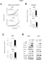
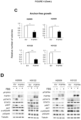
- Figure 2 shows the relative growth of the H226 epidermoid lung carcinoma (above) and Calu-1 (below) cell lines over time. It may be observed how the samples overexpressing FGFR4 have a greater cell growth ( Figure 2A ), clonability ( Figure 2B ) and capability of generating anchor-free growth colonies ( Figure 2C ) than the control lines (transfected with the empty vector (EV)). The overexpression of this gene induces its own overactivation, in addition to that of different pro-oncogenic signalling pathways, as revealed by the expression of the pSTAT3, pAKT and p-p42/p44 proteins ( Figure 2D ). All the tests were performed at least in triplicate. Representative growth curves are shown.
- the mean and standard deviation of the replica colonies are shown, after standardisation with the control condition, which takes the value of 1.
- the mean and standard deviation of the size of the colonies is also shown.
- the p-values were obtained by means of Student's T-test with a confidence interval of 95%; the p-values below 0.05 are represented as *, the p-values below 0.01 as **, and the p-values below 0.001 as ***.
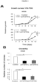
- Figure 3A shows the relative growth of the H520 line with the FGFR1 gene silenced (above) and with the FGFR4 gene silenced (below) over time, in medium with 10% FBS. It may be observed that the growth of the cell lines with one of the FGFRs silenced is less than that of the control cells. This effect is particularly apparent in the case of FGFR4 silencing.
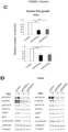
- Figure 3C anchor-free growth
- Figure 3B clonability
- the mean and standard deviation of the size of the colonies is also shown.
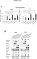
- the p-values were obtained by means of Student's T-test with a confidence interval of 95%; the p-values below 0.05 are represented as *, the p-values below 0.01 as **, and the p-values below 0.001 as ***.
- cell lines with different tumourigenic molecular disruptions were selected in order to verify whether their effect is dependent on the molecular context.
- cell lines with adenocarcinoma-relevant disruptions and low expression of both receptors were selected, in order to express them independently.
- the procedure was the same as that described in Example 1.
- the H2009 (with mutation in KRAS) and H3122 (carrier of the EML4-ALK translocation) cell lines were selected and the receptors were independently overexpressed in each by transfecting the lines with the appropriate plasmid, in accordance with Table 4.
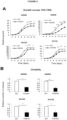
- each of the receptors caused a reduction in the tumourigenic characteristics of the two samples, as observed in the cell proliferation ( Figure 4A ) and clonability ( Figure 4B ) assays and the anchor-free growth assay ( Figure 4C ) with regard to their respective control lines transfected with the empty vector (EV).
- shRNA interference RNA
- Example 3 Identification of N-cadherin as responsible for the different behaviour of FGFR1 and FGFR4 in lung adenocarcinoma and squamous cell carcinoma
- N-cadherin mesenchymal marker
- E-cadherin epithelial marker
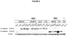
- Figure 6 shows how both proteins are expressed differentially in the two histological types analysed (adenocarcinoma (ADC) and squamous cell or epidermoid carcinoma (SCC)).
- ADC adenocarcinoma
- SCC epidermoid carcinoma
- N-cadherin was overexpressed in two adenocarcinoma lines (H2009 and H3122), transfecting them with the N-cadherin overexpression plasmid (Table 4) in combination or not with FGFR1 or FGFR4 overexpression plasmids.
- the mean and standard deviation of the replicas are shown, after standardising the control condition, which takes the value of 1.
- the mean and standard deviation of the size of the colonies is also shown.
- EV1 empty vector 1
- EV2 empty vector 2
- CDH2 Overexpression of N-cadherin
- FGFR1 Overexpression of FGFR1
- FGFR4 Overexpression of FGFR4
- FBS Foetal bovine serum.
- the p-values are represented by asterisks (*, p ⁇ 0.05; **, p ⁇ 0.01; ***, p ⁇ 0,001).
- N-cadherin is responsible for the oncogenic effect of FGFR1 and FGFR4, which would explain the differences in the behaviour of both genes between adenocarcinoma and epidermoid carcinoma.
- FGFR1 or FGFR4 has a tumour-suppressive effect in the absence of, or in the presence of low levels of, N-cadherin (shCDH2-EV1-FGFR1 and shCDH2-EV1-FGFR4), and a pro-tumourigenic effect in the presence of a high expression of N-cadherin (EV1-EV2-FGFR1 and EV1-EV2-FGFR4).
- Figure 8 shows the study of the activation of the oncogenic signalling pathways. The obtained results were concordant, revealing an increase in tumourigenicity and in the activation of the pathways under study when either of the FGFRs is overexpressed in the presence of endogenous N-cadherin, and the opposite effect when the FGFRs are overexpressed in the presence of low levels of N-cadherin.
- N-cadherin is responsible for the tumourigenic effect of FGFR1 and FGFR4 in non-small cell lung cancer (NSSLC).
- xenografts of the aforementioned cell lines were performed in immunocompromised nude mice and the growth of the tumours generated from the xenografts was determined.
- Figure 8D shows that the growth of the tumours generated by the different lines correlates with the results obtained in vitro, observing that the tumours co-expressing N-cadherin and FGFR1 or FGFR4 have a significantly greater growth than those expressing N-cadherin alone, or which do not express any of these genes, while the tumours expressing FGFR1 or FGFR4 alone display a slower tumour growth, as proven in the examples above.
- the NL20 cell line was used; this has high endogenous expression of N-cadherin with exogenous overexpression of FGFR1 or FGFR4.
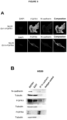
- Figure 9A shows that there is partial co-localisation of N-cadherin with FGFR1 and FGFR4 in some regions of the cell membrane. These results suggest that the interaction between the proteins may be physical. To test this hypothesis, co-immunoprecipitation experiments were performed. A cell line with high endogenous expression of the three proteins (H520) was selected. The immunoprecipitation of N-cadherin was performed, and the presence of FGFR1 or FGFR4 in the immunoprecipitate was determined, as described in the materials and methods herein.
- Figure 9B shows that both FGFR1 and FGFR4 co-immunoprecipitate with N-cadherin, supporting the existence of a physical interaction between both types of receptors.
- Example 4 Function of N-cadherin in the prognostic role of FGFR1 and FGFR4 in patients with non-small cell lung cancer
- the samples of tumour tissue, set in paraffin, of all the cases were analysed subsequent to the performance of the anatomopathological diagnosis.
- the mRNA was extracted from these samples as described above, although it is also possible to detect and quantify the expression of the genes of interest by means of direct immunohistochemistry in the biopsies.
- the overall survival (OS) and progression-free survival (PFS) were analysed; that is, the time elapsing during and subsequent to the treatment during which the cancer neither grows nor spreads further, against time (months).
- the p-values were calculated by means of Log Rank analysis.
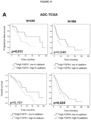
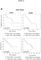
- Figure 10A shows the Kaplan-Meier curves of survival in accordance with the gene expression of the study cohort. It was observed that the group with high expression of N-cadherin and FGFR1 yielded the worst progression-free survival (PFS) and overall survival (OS) data, while the group with high expression of FGFR1 and low expression of N-cadherin yielded the best data for both types of survival. It is noteworthy that of the group with low expression of N-cadherin and high expression of FGFR1, over 70% of the patients are still alive, unlike the 30-40% of the other groups. Analogously, the samples with low N-cadherin and high expression of FGFR4 have better survival (both PFS and OS) than the samples with high expression of both genes. Table 7.
- Figure 10B shows the survival curves of patients with high expression of FGFR1 and/or FGFR4 with regard to their expression of N-cadherin.
- These survival curves clearly show the potential of N-cadherin as a modulator of the role of FGFR1 and FGFR4 in lung cancer oncogenesis.
- the categorical variables are represented as number of patients (percentage of the total) and the quantitative variables as a mean [interquartile range].
- the patients were divided with regard to the expression of FGFR1 and N-cadherin mRNA or FGFR4 and N-cadherin mRNA and their survival was analysed.
- the cut-off point between high and low expression in the case of the adenocarcinoma patient cohort was defined as the median in the case of FGFR1 and FGFR4, and as the first quartile in the case of FGFR4.
- the cut-off point was the median of expression for each of the genes.
- Example 5 Relationship between the expression of N-cadherin, FGFR1 and FGFR4 and the use of FGFR inhibitors in lung cancer treatment
- N-cadherin as a predictive factor in in-vitro anti-FGFR therapy
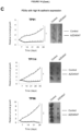
- Figure 13A shows the result of the cell proliferation (relative growth with regard to time) of the cell lines, untreated or treated with BGJ398 0.5 ⁇ M, BGJ398 1 ⁇ M, AZD4547 0.5 ⁇ M or AZD4547 1 ⁇ M. It may be observed that both inhibitors reduce the growth solely in the lines with high expression of N-cadherin, but do not exert a significant effect on those lines with low or no expression of N-cadherin at the concentrations tested. These results support the predictive role of N-cadherin with regard to FGFR-inhibiting therapies.
- Figure 13B shows the effect of two selective FGFR inhibitors on the proliferation of cell lines with exogenous expression of FGFR1 or FGFR4 in combination, or not, with exogenous expression of N-cadherin (CDH2). It may be observed that the inhibitors have no effect on the growth of the cell lines with no expression of exogenous N-cadherin, but do so when this is expressed (lower row).
- N-cadherin as a predictive factor in in-vivo anti-FGFR therapy
- adenocarcinoma models with similar expression of FGFR1 were selected, one with high expression of N-cadherin (TP91) and the other with low expression of this protein (TP60).
- Two epidermoid carcinoma models with comparable expression of FGFR1 were also selected, these having high and low expression of N-cadherin (TP114 and TP13, respectively).
- an epidermoid carcinoma model with high expression of FGFR1, FGFR4 and N-cadherin (TP96) was also selected ( Figure 14A ).
- Figure 14B shows how the treatment with AZD4547 had no effect on the tumour growth of the models with low N-cadherin expression (TP60 and TP13), regardless of their histology.
- the treatment with this selective FGFR inhibitor caused a considerable reduction in growth, with an average reduction in volume of 14.4%, 25.5% and 1.4% with regard to the control condition, respectively.
- Blanco R et al. A gene-alteration profile of human lung cancer cell lines. Human mutation (2009); 30:1199-206 .
Landscapes
- Health & Medical Sciences (AREA)
- Life Sciences & Earth Sciences (AREA)
- Chemical & Material Sciences (AREA)
- Immunology (AREA)
- Engineering & Computer Science (AREA)
- Molecular Biology (AREA)
- Biomedical Technology (AREA)
- Hematology (AREA)
- Urology & Nephrology (AREA)
- Pathology (AREA)
- Analytical Chemistry (AREA)
- Biochemistry (AREA)
- Physics & Mathematics (AREA)
- Microbiology (AREA)
- General Health & Medical Sciences (AREA)
- Biotechnology (AREA)
- Organic Chemistry (AREA)
- Proteomics, Peptides & Aminoacids (AREA)
- Food Science & Technology (AREA)
- Medicinal Chemistry (AREA)
- Cell Biology (AREA)
- General Physics & Mathematics (AREA)
- Genetics & Genomics (AREA)
- Wood Science & Technology (AREA)
- Zoology (AREA)
- Oncology (AREA)
- Biophysics (AREA)
- Hospice & Palliative Care (AREA)
- Bioinformatics & Cheminformatics (AREA)
- General Engineering & Computer Science (AREA)
- Measuring Or Testing Involving Enzymes Or Micro-Organisms (AREA)
- Medicines That Contain Protein Lipid Enzymes And Other Medicines (AREA)
- Pharmaceuticals Containing Other Organic And Inorganic Compounds (AREA)
Claims (16)
- Procédé de prédiction in vitro de la réponse d'un sujet souffrant d'un cancer du poumon à un traitement par au moins un inhibiteur de FGFR, comprenant :(a) détection dans un échantillon biologique qui a été obtenu à partir dudit sujet du niveau d'expression du biomarqueur N-cadhérine (CDH2) en combinaison avec le niveau d'expression d'au moins l'un des biomarqueurs FGFR1 et/ou FGFR4,(b) comparer le niveau d'expression de chaque biomarqueur déterminé en (a) avec un échantillon de référence ayant une expression faible ou aucune expression du même biomarqueur, dans lequel la présence de niveaux élevés d'expression des biomarqueurs détectés indique que le sujet répondra au traitement, dans lequel un niveau élevé d'expression de chaque biomarqueur correspond à un niveau au moins deux fois supérieur au niveau d'expression du même biomarqueur dans l'échantillon de référence.
- Procédé in vitro selon la revendication 1, dans lequel la détermination du niveau d'expression des biomarqueurs est effectuée en mesurant la quantité de protéine N-cadhérine et la quantité de protéine FGFR1 et/ou FGFR4 dans l'échantillon, et/ou en mesurant la quantité d'ARNm du gène CDH2 et la quantité d'ARNm des gènes FGFR1 et/ou FGFR4 dans l'échantillon.
- Utilisation d'une combinaison du niveau d'expression du biomarqueur de N-cadhérine (CDH2) et du niveau d'expression d'au moins un des biomarqueurs FGFR1 et/ou FGFR4 dans le pronostic in vitro et/ou la prédiction in vitro de la réponse à un traitement avec au moins un inhibiteur de FGFR d'un sujet souffrant d'un cancer du poumon, dans laquelle le niveau d'expression du biomarqueur de N-cadhérine (CDH2) en combinaison avec le niveau d'expression d'au moins un des biomarqueurs FGFR1 et/ou ou FGFR4 est déterminé dans un échantillon biologique qui a été obtenu à partir dudit sujet, et dans lequel le niveau d'expression de chaque biomarqueur déterminé précédemment est comparé à un échantillon de référence ayant une expression faible ou aucune expression du même biomarqueur, dans lequel la présence d'un niveau élevé d'expression des biomarqueurs détectés indique que le sujet répondra au traitement, dans lequel un niveau élevé d'expression de chaque biomarqueur correspond à un niveau au moins deux fois supérieur au niveau d'expression de celui-ci biomarqueur dans l'échantillon de référence.
- Utilisation de la combinaison des biomarqueurs selon la revendication 3, dans laquelle le niveau d'expression des biomarqueurs est déterminé en mesurant le niveau d'ARNm des gènes CDH2 et FGFR1 et/ou FGFR4, et/ou le niveau de protéine N-cadhérine et de protéine FGFR1 et/ou de protéine FGFR4.
- Utilisation de la combinaison de biomarqueurs selon la revendication 3 ou 4, dans laquelle le cancer du poumon est un cancer du poumon non à petites cellules (CPNPC).
- Utilisation de la combinaison de biomarqueurs selon la revendication 5, dans laquelle le cancer du poumon non à petites cellules est choisi parmi : un adénocarcinome et un carcinome épidermoïde ou à cellules squameuses.
- Procédé in vitro selon la revendication 1 ou 2, ou l'utilisation de la combinaison du niveau d'expression de biomarqueurs selon l'une quelconque des revendications 3 à 6, dans lequel l'au moins un inhibiteur de FGFR est choisi dans le groupe constitué de : BGJ398, AZD4547, Debio-1347, Dovitinib, BLU9931, FIIN-2, JNJ-42756493, LY2874455, Ponatinib, BIBF1120, PD173074, PD166866, BLU554, S49076, NSC12, PHA-739358, TSU-68, BMS-540215, TKI-258, MK-2461, BMS-582664, AG 1296, SSR128129E, LY2874455 et SU5402.
- Utilisation d'un kit pour le pronostic in vitro et/ou la prédiction in vitro de la réponse d'un sujet souffrant d'un cancer du poumon à un traitement avec au moins un inhibiteur de FGFR, dans laquelle le kit comprend :(a) des moyens pour détecter, dans un échantillon biologique qui a été obtenu à partir du sujet, le niveau d'expression du biomarqueur N-cadhérine (CDH2), et le niveau d'expression d'au moins l'un des biomarqueurs FGFR1 et/ou FGFR4,(b) des moyens pour comparer le niveau d'expression de chaque biomarqueur déterminé en (a) avec un échantillon de référence ayant une expression faible ou aucune expression du même biomarqueur, dans lequel la présence de niveaux élevés d'expression des biomarqueurs détectés indique que le sujet répondra au traitement, dans lequel un niveau élevé d'expression de chaque biomarqueur correspond à un niveau au moins deux fois supérieur au niveau d'expression du même biomarqueur dans l'échantillon de référence,
- Utilisation du kit selon la revendication 8, dans lequel la détermination du niveau d'expression des biomarqueurs est effectuée en mesurant la quantité de protéine du biomarqueur N- cadhérine et la quantité de protéine d'au moins l'un des biomarqueurs FGFR1 et/ou FGFR4 dans l'échantillon, et/ou en mesurant la quantité d'ARNm du gène CDH2 et la quantité d'ARNm d'au moins l'un des gènes FGFR1 ou FGFR4 dans l'échantillon.
- Procédé in vitro selon la revendication 1 ou 2, ou utilisation du kit selon la revendication 8 ou 9, dans lequel le cancer du poumon est un cancer du poumon non à petites cellules (CBNPC).
- Procédé in vitro selon la revendication 10 ou utilisation du kit selon la revendication 10, dans lequel le cancer du poumon non à petites cellules est choisi parmi : un adénocarcinome et un carcinome épidermoïde ou épidermoïde.
- Utilisation du kit selon l'une quelconque des revendications 8 à 11, dans laquelle les moyens pour détecter le niveau d'expression des biomarqueurs comprennent des anticorps qui reconnaissent spécifiquement la protéine N-cadhérine et des anticorps qui reconnaissent spécifiquement les protéines FGFR1 et/ou FGFR4, et/ou des amorces et/ou des sondes qui détectent spécifiquement la présence de l'ARNm du gène CDH2 et également des amorces et/ou des sondes qui détectent spécifiquement la présence de l'ARNm des gènes FGFR1 et/ou FGFR4.
- Procédé in vitro selon la revendication 1 ou 2, 10 ou 11, ou utilisation du kit selon l'une quelconque des revendications 8 à 12, dans lequel l'échantillon biologique est choisi dans le groupe constitué de : sang total, sérum, plasma, expectoration, sueur, urine, lavage bronchoalvéolaire et biopsie de tissu tumoral primaire ou métastatique.
- Procédé in vitro selon la revendication 1 ou 2, 10 ou 11, ou utilisation du kit selon l'une quelconque des revendications 8 à 13, dans lequel la détection du niveau d'expression des biomarqueurs est effectuée au moyen d'au moins l'un des procédés choisis dans le groupe constitué de : HPLC (chromatographie liquide haute performance), LC/MS (chromatographie liquide couplée à la spectrométrie de masse), ELISA, DAS ELISA, immunoprécipitation protéique, immunoélectrophorèse, Western Blot, immunocoloration protéique, Northern Blot, PCR par transcription inverse (RT-PCR), PCR quantitative (q-PCR), RIA (radioimmunoessai), hybridation in situ, dosage de protection par nucléase, séquençage massif, techniques immunocytochimiques ou immunohistochimiques, microéchantillons d'ADN génomique, microéchantillons de protéines, microéchantillons d'ARN messager, microéchantillons d'ADNc, microéchantillons de peptides, microéchantillons de tissus, microéchantillons de cellules ou de transfection, microéchantillons d'anticorps, microéchantillons de lysat ou de sérum, microéchantillons de protéines en phase inverse, microéchantillons de peptides et microéchantillons de génotypage.
- Le procédé in vitro selon la revendication 1 ou 2, 7, 10, 11, 13 ou 14, ou l'utilisation du kit selon l'une quelconque des revendications 8 à 14, dans lequel le niveau d'expression des trois biomarqueurs N-cadhérine, FGFR1 et FGFR4 est détecté.
- Utilisation de la combinaison de biomarqueurs selon l'une quelconque des revendications 3 à 7, dans laquelle le niveau d'expression des trois biomarqueurs N-cadhérine, FGFR1 et FGFR4 est combiné.
Applications Claiming Priority (2)
| Application Number | Priority Date | Filing Date | Title |
|---|---|---|---|
| ES201730929A ES2696798B2 (es) | 2017-07-13 | 2017-07-13 | N-cadherina y fgfr1 y/o fgfr4 para uso en la prediccion de la respuesta de los pacientes a un tratamiento del cancer de pulmon y metodo y kit basados en dicho uso |
| PCT/ES2018/070501 WO2019016422A1 (fr) | 2017-07-13 | 2018-07-13 | N-cadhérine et fgfr1 et/ou fgfr4 destinés à être utilisés dans la prédiction de la réponse des patients à un traitement du cancer du poumon et procédé et kit basés sur ladite utilisation |
Publications (3)
| Publication Number | Publication Date |
|---|---|
| EP3636773A1 EP3636773A1 (fr) | 2020-04-15 |
| EP3636773A4 EP3636773A4 (fr) | 2021-03-03 |
| EP3636773B1 true EP3636773B1 (fr) | 2023-08-23 |
Family
ID=65009864
Family Applications (1)
| Application Number | Title | Priority Date | Filing Date |
|---|---|---|---|
| EP18836033.3A Active EP3636773B1 (fr) | 2017-07-13 | 2018-07-13 | N-cadhérine et fgfr1 et/ou fgfr4 destinés à être utilisés dans la prédiction de la réponse des patients à un traitement du cancer du poumon et procédé et kit basés sur ladite utilisation |
Country Status (4)
| Country | Link |
|---|---|
| US (1) | US11408038B2 (fr) |
| EP (1) | EP3636773B1 (fr) |
| ES (2) | ES2696798B2 (fr) |
| WO (1) | WO2019016422A1 (fr) |
Families Citing this family (2)
| Publication number | Priority date | Publication date | Assignee | Title |
|---|---|---|---|---|
| CN110488014A (zh) * | 2019-08-27 | 2019-11-22 | 成都和同易创生物科技有限公司 | 一种检测结直肠癌转移潜能的试剂盒及其使用方法 |
| KR102315706B1 (ko) * | 2020-03-30 | 2021-10-21 | 한국원자력의학원 | 미세플라스틱 노출에 의해 유도되는 암 악성화 예후 예측용 바이오마커 조성물 |
Family Cites Families (4)
| Publication number | Priority date | Publication date | Assignee | Title |
|---|---|---|---|---|
| JP5240739B2 (ja) * | 2007-04-13 | 2013-07-17 | オーエスアイ・フアーマシユーテイカルズ・エル・エル・シー | キナーゼ阻害剤に対する抗癌応答を予測する生物学的マーカー |
| US8709718B1 (en) * | 2011-02-12 | 2014-04-29 | Max-Planck-Gesellschaft Zur Forderung Der Wissenschaften E.V. | Method of treating lung cancer |
| EP2695950A1 (fr) * | 2012-08-10 | 2014-02-12 | Blackfield AG | Marqueurs pour la réactivité à un inhibiteur du récepteur du facteur de croissance des fibroblastes |
| CA2862530A1 (fr) * | 2014-05-20 | 2015-11-20 | Momentous Ip Ventures Llc | Traitement personnalise du cancer a l'aide d'inhibiteurs fgfr |
-
2017
- 2017-07-13 ES ES201730929A patent/ES2696798B2/es not_active Expired - Fee Related
-
2018
- 2018-07-13 WO PCT/ES2018/070501 patent/WO2019016422A1/fr not_active Ceased
- 2018-07-13 EP EP18836033.3A patent/EP3636773B1/fr active Active
- 2018-07-13 US US16/630,021 patent/US11408038B2/en active Active
- 2018-07-13 ES ES18836033T patent/ES2966540T3/es active Active
Also Published As
| Publication number | Publication date |
|---|---|
| ES2696798A1 (es) | 2019-01-17 |
| EP3636773A4 (fr) | 2021-03-03 |
| WO2019016422A1 (fr) | 2019-01-24 |
| ES2696798B2 (es) | 2019-07-08 |
| EP3636773A1 (fr) | 2020-04-15 |
| US20210164053A1 (en) | 2021-06-03 |
| US11408038B2 (en) | 2022-08-09 |
| ES2966540T3 (es) | 2024-04-22 |
Similar Documents
| Publication | Publication Date | Title |
|---|---|---|
| Wang et al. | MEX3A mediates p53 degradation to suppress ferroptosis and facilitate ovarian cancer tumorigenesis | |
| Hong et al. | AKR1C1 activates STAT3 to promote the metastasis of non-small cell lung cancer | |
| Denysenko et al. | WNT/β-catenin signaling pathway and downstream modulators in low-and high-grade glioma | |
| US20130217721A1 (en) | Compositions and methods for detection and treatment of b-raf inhibitor-resistant melanomas | |
| Lin et al. | Overexpression of muscarinic receptor 3 promotes metastasis and predicts poor prognosis in non–small-cell lung cancer | |
| Xiao et al. | MEX3C-mediated decay of SOCS3 mRNA promotes JAK2/STAT3 signaling to facilitate metastasis in hepatocellular carcinoma | |
| MX2014010265A (es) | Seleccion de pacientes con cancer para administracion de inhibidores de señalizacion de wnt utilizando estado de mutacion de rnf43. | |
| EP2681330B1 (fr) | Utilisation de la protéine olfactomédine-4 (olfm4) dans le diagnostic du cancer colorectal | |
| Song et al. | USP18 promotes tumor metastasis in esophageal squamous cell carcinomas via deubiquitinating ZEB1 | |
| Pal et al. | Synaptophysin expression on circulating tumor cells in patients with castration resistant prostate cancer undergoing treatment with abiraterone acetate or enzalutamide | |
| TWI567391B (zh) | 辨識早期肺腺癌病患之次群組之生物標記 | |
| Chen et al. | CD146 promotes malignant progression of breast phyllodes tumor through suppressing DCBLD2 degradation and activating the AKT pathway | |
| Sawhney et al. | Cytoplasmic accumulation of activated leukocyte cell adhesion molecule is a predictor of disease progression and reduced survival in oral cancer patients | |
| WO2017223344A1 (fr) | Transdifférenciation servant de mécanisme de résistance au traitement pour le cancer de la prostate résistant à la castration | |
| Cheng et al. | Investigating the association between COMMD3 expression and the prognosis of hepatocellular carcinoma | |
| US11408038B2 (en) | N-cadherin and FGFR1 and/or FGFR4 for use in predicting the response of patients to a lung cancer treatment and method and kit based on said use | |
| McColl et al. | Identification of HEPACAM2 as a novel and specific marker of small cell carcinoma | |
| Zhang et al. | NTSR1 promotes epithelial-mesenchymal transition and metastasis in lung adenocarcinoma through the Wnt/β-catenin pathway | |
| Lee et al. | Expression of female sex hormone receptors and its relation to clinicopathological characteristics and prognosis of lung adenocarcinoma | |
| Hu et al. | TRIM22 inhibits the metastasis of colorectal cancer through facilitating β-Catenin degradation | |
| KR20140092422A (ko) | S100a9 및 egfr에 대한 저해제와 시스플라틴을 유효성분으로 포함하는 근육 침윤성 방광암의 치료용 약학적 조성물 | |
| Dechaphunkul et al. | Frequency of PIK3CA mutations in different subsites of head and neck squamous cell carcinoma in southern Thailand | |
| Yoshii et al. | The expression of SIRT6 is associated with treatment outcome in elder patients with oral cancer | |
| Qin et al. | HOXC8 Derived from Cancer-Associated Fibroblasts Regulates Lung Cancer Cell Malignant Metastasis and Ferroptosis by Mediating the Transcription of GCH1 | |
| US20130136755A1 (en) | Mlk4 gene, a new diagnostic and prognostic marker in cancers |
Legal Events
| Date | Code | Title | Description |
|---|---|---|---|
| STAA | Information on the status of an ep patent application or granted ep patent |
Free format text: STATUS: THE INTERNATIONAL PUBLICATION HAS BEEN MADE |
|
| PUAI | Public reference made under article 153(3) epc to a published international application that has entered the european phase |
Free format text: ORIGINAL CODE: 0009012 |
|
| STAA | Information on the status of an ep patent application or granted ep patent |
Free format text: STATUS: REQUEST FOR EXAMINATION WAS MADE |
|
| 17P | Request for examination filed |
Effective date: 20200109 |
|
| AK | Designated contracting states |
Kind code of ref document: A1 Designated state(s): AL AT BE BG CH CY CZ DE DK EE ES FI FR GB GR HR HU IE IS IT LI LT LU LV MC MK MT NL NO PL PT RO RS SE SI SK SM TR |
|
| AX | Request for extension of the european patent |
Extension state: BA ME |
|
| DAV | Request for validation of the european patent (deleted) | ||
| DAX | Request for extension of the european patent (deleted) | ||
| A4 | Supplementary search report drawn up and despatched |
Effective date: 20210129 |
|
| RIC1 | Information provided on ipc code assigned before grant |
Ipc: A61K 31/00 20060101ALI20210125BHEP Ipc: C12Q 1/6886 20180101AFI20210125BHEP Ipc: G01N 33/574 20060101ALI20210125BHEP Ipc: C07K 14/71 20060101ALI20210125BHEP Ipc: A61P 35/00 20060101ALI20210125BHEP |
|
| STAA | Information on the status of an ep patent application or granted ep patent |
Free format text: STATUS: EXAMINATION IS IN PROGRESS |
|
| 17Q | First examination report despatched |
Effective date: 20211018 |
|
| REG | Reference to a national code |
Ref country code: DE Ref legal event code: R079 Free format text: PREVIOUS MAIN CLASS: C12Q0001680000 Ipc: C12Q0001688600 Ref country code: DE Ref legal event code: R079 Ref document number: 602018056091 Country of ref document: DE Free format text: PREVIOUS MAIN CLASS: C12Q0001680000 Ipc: C12Q0001688600 |
|
| RIC1 | Information provided on ipc code assigned before grant |
Ipc: A61P 35/00 20060101ALI20220316BHEP Ipc: C12Q 1/6886 20180101AFI20220316BHEP |
|
| GRAJ | Information related to disapproval of communication of intention to grant by the applicant or resumption of examination proceedings by the epo deleted |
Free format text: ORIGINAL CODE: EPIDOSDIGR1 |
|
| STAA | Information on the status of an ep patent application or granted ep patent |
Free format text: STATUS: GRANT OF PATENT IS INTENDED |
|
| GRAP | Despatch of communication of intention to grant a patent |
Free format text: ORIGINAL CODE: EPIDOSNIGR1 |
|
| INTG | Intention to grant announced |
Effective date: 20230207 |
|
| GRAS | Grant fee paid |
Free format text: ORIGINAL CODE: EPIDOSNIGR3 |
|
| GRAA | (expected) grant |
Free format text: ORIGINAL CODE: 0009210 |
|
| STAA | Information on the status of an ep patent application or granted ep patent |
Free format text: STATUS: THE PATENT HAS BEEN GRANTED |
|
| AK | Designated contracting states |
Kind code of ref document: B1 Designated state(s): AL AT BE BG CH CY CZ DE DK EE ES FI FR GB GR HR HU IE IS IT LI LT LU LV MC MK MT NL NO PL PT RO RS SE SI SK SM TR |
|
| REG | Reference to a national code |
Ref country code: GB Ref legal event code: FG4D |
|
| REG | Reference to a national code |
Ref country code: CH Ref legal event code: EP |
|
| REG | Reference to a national code |
Ref country code: DE Ref legal event code: R096 Ref document number: 602018056091 Country of ref document: DE |
|
| REG | Reference to a national code |
Ref country code: IE Ref legal event code: FG4D |
|
| REG | Reference to a national code |
Ref country code: LT Ref legal event code: MG9D |
|
| REG | Reference to a national code |
Ref country code: NL Ref legal event code: MP Effective date: 20230823 |
|
| RAP2 | Party data changed (patent owner data changed or rights of a patent transferred) |
Owner name: FUNDACION DE INVESTIGACION HOSPITAL 12 DE OCTUBRE |
|
| REG | Reference to a national code |
Ref country code: AT Ref legal event code: MK05 Ref document number: 1602658 Country of ref document: AT Kind code of ref document: T Effective date: 20230823 |
|
| PG25 | Lapsed in a contracting state [announced via postgrant information from national office to epo] |
Ref country code: GR Free format text: LAPSE BECAUSE OF FAILURE TO SUBMIT A TRANSLATION OF THE DESCRIPTION OR TO PAY THE FEE WITHIN THE PRESCRIBED TIME-LIMIT Effective date: 20231124 |
|
| PG25 | Lapsed in a contracting state [announced via postgrant information from national office to epo] |
Ref country code: IS Free format text: LAPSE BECAUSE OF FAILURE TO SUBMIT A TRANSLATION OF THE DESCRIPTION OR TO PAY THE FEE WITHIN THE PRESCRIBED TIME-LIMIT Effective date: 20231223 |
|
| PG25 | Lapsed in a contracting state [announced via postgrant information from national office to epo] |
Ref country code: SE Free format text: LAPSE BECAUSE OF FAILURE TO SUBMIT A TRANSLATION OF THE DESCRIPTION OR TO PAY THE FEE WITHIN THE PRESCRIBED TIME-LIMIT Effective date: 20230823 Ref country code: RS Free format text: LAPSE BECAUSE OF FAILURE TO SUBMIT A TRANSLATION OF THE DESCRIPTION OR TO PAY THE FEE WITHIN THE PRESCRIBED TIME-LIMIT Effective date: 20230823 Ref country code: PT Free format text: LAPSE BECAUSE OF FAILURE TO SUBMIT A TRANSLATION OF THE DESCRIPTION OR TO PAY THE FEE WITHIN THE PRESCRIBED TIME-LIMIT Effective date: 20231226 Ref country code: NO Free format text: LAPSE BECAUSE OF FAILURE TO SUBMIT A TRANSLATION OF THE DESCRIPTION OR TO PAY THE FEE WITHIN THE PRESCRIBED TIME-LIMIT Effective date: 20231123 Ref country code: NL Free format text: LAPSE BECAUSE OF FAILURE TO SUBMIT A TRANSLATION OF THE DESCRIPTION OR TO PAY THE FEE WITHIN THE PRESCRIBED TIME-LIMIT Effective date: 20230823 Ref country code: LV Free format text: LAPSE BECAUSE OF FAILURE TO SUBMIT A TRANSLATION OF THE DESCRIPTION OR TO PAY THE FEE WITHIN THE PRESCRIBED TIME-LIMIT Effective date: 20230823 Ref country code: LT Free format text: LAPSE BECAUSE OF FAILURE TO SUBMIT A TRANSLATION OF THE DESCRIPTION OR TO PAY THE FEE WITHIN THE PRESCRIBED TIME-LIMIT Effective date: 20230823 Ref country code: IS Free format text: LAPSE BECAUSE OF FAILURE TO SUBMIT A TRANSLATION OF THE DESCRIPTION OR TO PAY THE FEE WITHIN THE PRESCRIBED TIME-LIMIT Effective date: 20231223 Ref country code: HR Free format text: LAPSE BECAUSE OF FAILURE TO SUBMIT A TRANSLATION OF THE DESCRIPTION OR TO PAY THE FEE WITHIN THE PRESCRIBED TIME-LIMIT Effective date: 20230823 Ref country code: GR Free format text: LAPSE BECAUSE OF FAILURE TO SUBMIT A TRANSLATION OF THE DESCRIPTION OR TO PAY THE FEE WITHIN THE PRESCRIBED TIME-LIMIT Effective date: 20231124 Ref country code: FI Free format text: LAPSE BECAUSE OF FAILURE TO SUBMIT A TRANSLATION OF THE DESCRIPTION OR TO PAY THE FEE WITHIN THE PRESCRIBED TIME-LIMIT Effective date: 20230823 Ref country code: AT Free format text: LAPSE BECAUSE OF FAILURE TO SUBMIT A TRANSLATION OF THE DESCRIPTION OR TO PAY THE FEE WITHIN THE PRESCRIBED TIME-LIMIT Effective date: 20230823 |
|
| REG | Reference to a national code |
Ref country code: DE Ref legal event code: R081 Ref document number: 602018056091 Country of ref document: DE Owner name: FUNDACION DE INVESTIGACION HOSPITAL 12 DE OCTU, ES Free format text: FORMER OWNERS: CONSEJO SUPERIOR DE INVESTIGACIONES CIENTIFICAS (CSIC), SEVILLA, ES; FUNDACION DE INVESTIGACION HOSPITAL 12 DE OCTUBRE, MADRID, ES; SERVICIO ANDALUZ DE SALUD, SEVILLA, ES |
|
| PG25 | Lapsed in a contracting state [announced via postgrant information from national office to epo] |
Ref country code: PL Free format text: LAPSE BECAUSE OF FAILURE TO SUBMIT A TRANSLATION OF THE DESCRIPTION OR TO PAY THE FEE WITHIN THE PRESCRIBED TIME-LIMIT Effective date: 20230823 |
|
| REG | Reference to a national code |
Ref country code: GB Ref legal event code: 732E Free format text: REGISTERED BETWEEN 20240222 AND 20240228 |
|
| REG | Reference to a national code |
Ref country code: ES Ref legal event code: FG2A Ref document number: 2966540 Country of ref document: ES Kind code of ref document: T3 Effective date: 20240422 |
|
| PG25 | Lapsed in a contracting state [announced via postgrant information from national office to epo] |
Ref country code: SM Free format text: LAPSE BECAUSE OF FAILURE TO SUBMIT A TRANSLATION OF THE DESCRIPTION OR TO PAY THE FEE WITHIN THE PRESCRIBED TIME-LIMIT Effective date: 20230823 Ref country code: RO Free format text: LAPSE BECAUSE OF FAILURE TO SUBMIT A TRANSLATION OF THE DESCRIPTION OR TO PAY THE FEE WITHIN THE PRESCRIBED TIME-LIMIT Effective date: 20230823 Ref country code: EE Free format text: LAPSE BECAUSE OF FAILURE TO SUBMIT A TRANSLATION OF THE DESCRIPTION OR TO PAY THE FEE WITHIN THE PRESCRIBED TIME-LIMIT Effective date: 20230823 Ref country code: DK Free format text: LAPSE BECAUSE OF FAILURE TO SUBMIT A TRANSLATION OF THE DESCRIPTION OR TO PAY THE FEE WITHIN THE PRESCRIBED TIME-LIMIT Effective date: 20230823 Ref country code: CZ Free format text: LAPSE BECAUSE OF FAILURE TO SUBMIT A TRANSLATION OF THE DESCRIPTION OR TO PAY THE FEE WITHIN THE PRESCRIBED TIME-LIMIT Effective date: 20230823 Ref country code: SK Free format text: LAPSE BECAUSE OF FAILURE TO SUBMIT A TRANSLATION OF THE DESCRIPTION OR TO PAY THE FEE WITHIN THE PRESCRIBED TIME-LIMIT Effective date: 20230823 |
|
| REG | Reference to a national code |
Ref country code: DE Ref legal event code: R097 Ref document number: 602018056091 Country of ref document: DE |
|
| PG25 | Lapsed in a contracting state [announced via postgrant information from national office to epo] |
Ref country code: IT Free format text: LAPSE BECAUSE OF FAILURE TO SUBMIT A TRANSLATION OF THE DESCRIPTION OR TO PAY THE FEE WITHIN THE PRESCRIBED TIME-LIMIT Effective date: 20230823 |
|
| PLBE | No opposition filed within time limit |
Free format text: ORIGINAL CODE: 0009261 |
|
| STAA | Information on the status of an ep patent application or granted ep patent |
Free format text: STATUS: NO OPPOSITION FILED WITHIN TIME LIMIT |
|
| 26N | No opposition filed |
Effective date: 20240524 |
|
| PG25 | Lapsed in a contracting state [announced via postgrant information from national office to epo] |
Ref country code: SI Free format text: LAPSE BECAUSE OF FAILURE TO SUBMIT A TRANSLATION OF THE DESCRIPTION OR TO PAY THE FEE WITHIN THE PRESCRIBED TIME-LIMIT Effective date: 20230823 |
|
| PG25 | Lapsed in a contracting state [announced via postgrant information from national office to epo] |
Ref country code: BG Free format text: LAPSE BECAUSE OF FAILURE TO SUBMIT A TRANSLATION OF THE DESCRIPTION OR TO PAY THE FEE WITHIN THE PRESCRIBED TIME-LIMIT Effective date: 20230823 |
|
| PG25 | Lapsed in a contracting state [announced via postgrant information from national office to epo] |
Ref country code: BG Free format text: LAPSE BECAUSE OF FAILURE TO SUBMIT A TRANSLATION OF THE DESCRIPTION OR TO PAY THE FEE WITHIN THE PRESCRIBED TIME-LIMIT Effective date: 20230823 |
|
| PG25 | Lapsed in a contracting state [announced via postgrant information from national office to epo] |
Ref country code: MC Free format text: LAPSE BECAUSE OF FAILURE TO SUBMIT A TRANSLATION OF THE DESCRIPTION OR TO PAY THE FEE WITHIN THE PRESCRIBED TIME-LIMIT Effective date: 20230823 |
|
| REG | Reference to a national code |
Ref country code: CH Ref legal event code: PL |
|
| PG25 | Lapsed in a contracting state [announced via postgrant information from national office to epo] |
Ref country code: LU Free format text: LAPSE BECAUSE OF NON-PAYMENT OF DUE FEES Effective date: 20240713 |
|
| PG25 | Lapsed in a contracting state [announced via postgrant information from national office to epo] |
Ref country code: LU Free format text: LAPSE BECAUSE OF NON-PAYMENT OF DUE FEES Effective date: 20240713 |
|
| PG25 | Lapsed in a contracting state [announced via postgrant information from national office to epo] |
Ref country code: CH Free format text: LAPSE BECAUSE OF NON-PAYMENT OF DUE FEES Effective date: 20240731 Ref country code: BE Free format text: LAPSE BECAUSE OF NON-PAYMENT OF DUE FEES Effective date: 20240731 |
|
| REG | Reference to a national code |
Ref country code: BE Ref legal event code: MM Effective date: 20240731 |
|
| PG25 | Lapsed in a contracting state [announced via postgrant information from national office to epo] |
Ref country code: IE Free format text: LAPSE BECAUSE OF NON-PAYMENT OF DUE FEES Effective date: 20240713 |
|
| PGFP | Annual fee paid to national office [announced via postgrant information from national office to epo] |
Ref country code: ES Payment date: 20250804 Year of fee payment: 8 |
|
| PGFP | Annual fee paid to national office [announced via postgrant information from national office to epo] |
Ref country code: DE Payment date: 20250728 Year of fee payment: 8 |
|
| PGFP | Annual fee paid to national office [announced via postgrant information from national office to epo] |
Ref country code: GB Payment date: 20250722 Year of fee payment: 8 |
|
| PGFP | Annual fee paid to national office [announced via postgrant information from national office to epo] |
Ref country code: FR Payment date: 20250723 Year of fee payment: 8 |
|
| PG25 | Lapsed in a contracting state [announced via postgrant information from national office to epo] |
Ref country code: CY Free format text: LAPSE BECAUSE OF FAILURE TO SUBMIT A TRANSLATION OF THE DESCRIPTION OR TO PAY THE FEE WITHIN THE PRESCRIBED TIME-LIMIT; INVALID AB INITIO Effective date: 20180713 |
|
| PG25 | Lapsed in a contracting state [announced via postgrant information from national office to epo] |
Ref country code: HU Free format text: LAPSE BECAUSE OF FAILURE TO SUBMIT A TRANSLATION OF THE DESCRIPTION OR TO PAY THE FEE WITHIN THE PRESCRIBED TIME-LIMIT; INVALID AB INITIO Effective date: 20180713 |