EP3637106A1 - Procédé de détection du cancer du poumon - Google Patents
Procédé de détection du cancer du poumon Download PDFInfo
- Publication number
- EP3637106A1 EP3637106A1 EP18813561.0A EP18813561A EP3637106A1 EP 3637106 A1 EP3637106 A1 EP 3637106A1 EP 18813561 A EP18813561 A EP 18813561A EP 3637106 A1 EP3637106 A1 EP 3637106A1
- Authority
- EP
- European Patent Office
- Prior art keywords
- kininogen
- lung cancer
- protein
- cases
- detection
- Prior art date
- Legal status (The legal status is an assumption and is not a legal conclusion. Google has not performed a legal analysis and makes no representation as to the accuracy of the status listed.)
- Granted
Links
Images


Classifications
-
- G—PHYSICS
- G01—MEASURING; TESTING
- G01N—INVESTIGATING OR ANALYSING MATERIALS BY DETERMINING THEIR CHEMICAL OR PHYSICAL PROPERTIES
- G01N33/00—Investigating or analysing materials by specific methods not covered by groups G01N1/00 - G01N31/00
- G01N33/48—Biological material, e.g. blood, urine; Haemocytometers
- G01N33/50—Chemical analysis of biological material, e.g. blood, urine; Testing involving biospecific ligand binding methods; Immunological testing
- G01N33/53—Immunoassay; Biospecific binding assay; Materials therefor
- G01N33/575—Immunoassay; Biospecific binding assay; Materials therefor for cancer
- G01N33/5752—Immunoassay; Biospecific binding assay; Materials therefor for cancer of the lungs
-
- C—CHEMISTRY; METALLURGY
- C12—BIOCHEMISTRY; BEER; SPIRITS; WINE; VINEGAR; MICROBIOLOGY; ENZYMOLOGY; MUTATION OR GENETIC ENGINEERING
- C12Q—MEASURING OR TESTING PROCESSES INVOLVING ENZYMES, NUCLEIC ACIDS OR MICROORGANISMS; COMPOSITIONS OR TEST PAPERS THEREFOR; PROCESSES OF PREPARING SUCH COMPOSITIONS; CONDITION-RESPONSIVE CONTROL IN MICROBIOLOGICAL OR ENZYMOLOGICAL PROCESSES
- C12Q1/00—Measuring or testing processes involving enzymes, nucleic acids or microorganisms; Compositions therefor; Processes of preparing such compositions
- C12Q1/56—Measuring or testing processes involving enzymes, nucleic acids or microorganisms; Compositions therefor; Processes of preparing such compositions involving blood clotting factors, e.g. involving thrombin, thromboplastin, fibrinogen
-
- G—PHYSICS
- G01—MEASURING; TESTING
- G01N—INVESTIGATING OR ANALYSING MATERIALS BY DETERMINING THEIR CHEMICAL OR PHYSICAL PROPERTIES
- G01N33/00—Investigating or analysing materials by specific methods not covered by groups G01N1/00 - G01N31/00
- G01N33/48—Biological material, e.g. blood, urine; Haemocytometers
- G01N33/50—Chemical analysis of biological material, e.g. blood, urine; Testing involving biospecific ligand binding methods; Immunological testing
- G01N33/53—Immunoassay; Biospecific binding assay; Materials therefor
- G01N33/575—Immunoassay; Biospecific binding assay; Materials therefor for cancer
- G01N33/5758—Immunoassay; Biospecific binding assay; Materials therefor for cancer involving compounds serving as markers for tumours, cancers or neoplasias, e.g. cellular determinants, receptors, heat shock/stress proteins, A-protein, oligosaccharides or metabolites
-
- G—PHYSICS
- G01—MEASURING; TESTING
- G01N—INVESTIGATING OR ANALYSING MATERIALS BY DETERMINING THEIR CHEMICAL OR PHYSICAL PROPERTIES
- G01N33/00—Investigating or analysing materials by specific methods not covered by groups G01N1/00 - G01N31/00
- G01N33/48—Biological material, e.g. blood, urine; Haemocytometers
- G01N33/50—Chemical analysis of biological material, e.g. blood, urine; Testing involving biospecific ligand binding methods; Immunological testing
- G01N33/68—Chemical analysis of biological material, e.g. blood, urine; Testing involving biospecific ligand binding methods; Immunological testing involving proteins, peptides or amino acids
- G01N33/6803—General methods of protein analysis not limited to specific proteins or families of proteins
- G01N33/6848—Methods of protein analysis involving mass spectrometry
-
- G—PHYSICS
- G01—MEASURING; TESTING
- G01N—INVESTIGATING OR ANALYSING MATERIALS BY DETERMINING THEIR CHEMICAL OR PHYSICAL PROPERTIES
- G01N2333/00—Assays involving biological materials from specific organisms or of a specific nature
- G01N2333/435—Assays involving biological materials from specific organisms or of a specific nature from animals; from humans
- G01N2333/745—Assays involving non-enzymic blood coagulation factors
Definitions
- the present invention relates to a method for detection of lung cancer.
- Lung cancer accounts for the largest number of yearly cancer deaths in the world. It is estimated that in 2012, 1.6 million people died of lung cancer worldwide. In Japan, 74,000 people each year die of lung cancer, which is the leading disease causing the death of cancer patients. Lung cancer is classified into small cell lung cancer and non-small cell lung cancer; the former accounts for 15% of all lung cancer patients and the latter accounts for the remaining 85%. Furthermore, non-small cell lung cancer can be pathologically classified into several types of cancer, such as adenocarcinoma, squamous cell carcinoma, large cell carcinoma, adenosquamous carcinoma, pleomorphic cancer, and sarcoma-like cancers such as carcinosarcoma.
- adenocarcinoma squamous cell carcinoma
- large cell carcinoma adenosquamous carcinoma
- pleomorphic cancer pleomorphic cancer
- sarcoma-like cancers such as carcinosarcoma.
- Non-Patent Document 1 patients with non-small cell lung cancer at clinical stages IA, IB, IIA, IIB, and IIIA, on whom surgery can be performed, have a five-year survival rate of 82.0%, 66.1%, 54.5%, 46.1%, and 42.8%, respectively
- Non-Patent Document 2 patients at clinical stages IIIB and IV, to whom surgery is not applied, have a median survival time of 22.4 months
- Non-Patent Document 3 10.3 months
- Non-Patent Documents 4 and 5 As existing tumor markers for lung cancers, for example, serum Carcino embryonic antigen (CEA) for lung adenocarcinoma, a cytokeratin 19 fragment (CYFRA 21-1) for squamous cell lung cancer, and Neuron-specific enolase (NSE) for small cell lung cancer are employed for clinical diagnosis.
- CEA serum Carcino embryonic antigen
- CYFRA 21-1 cytokeratin 19 fragment
- NSE Neuron-specific enolase
- the rate at which in cases of lung adenocarcinoma, CEA is positive for lung adenocarcinoma is only from 36.6 to 56.5% (Non-Patent Documents 4 and 5); especially cases of early lung adenocarcinoma at stage I give a decreased positive rate of 27% (Non-Patent Document 5).
- Non-Patent Document 6 The rate at which in cases of squamous cell lung cancer, CYFRA 21-1 is positive for squamous cell lung cancer is 57% (Non-Patent Document 6), and the rate at which in cases of small cell lung cancer, NSE is positive for small cell lung cancer is 45% (Non-Patent Document 7). Therefore, there is a need to develop a marker capable of detection of lung cancer with a higher sensitivity than that of the existing tumor markers.
- Tumor markers that are being developed for clinical applications include, for example, serum CYBP (Patent Document 1) and UBE2L3 (Patent Document 2). These markers involve drawing blood from patients, and thus are invasive. Therefore, examinations using such markers are required to be less invasive.
- the present invention aims to provide a method for detection of lung cancer.
- the present inventors have found an increase in the fragmentation of kininogen I and a decrease of a protein(s) having the normal structure derived therefrom in body fluids, and exposed portions due to nicks at abnormal sites or deletions at internal sites in a protein derived therefrom, which can be observed characteristically in patients with lung cancer.
- the present inventors have conducted diligent investigations to establish a simple, minimally invasive or non-invasive method for detection of lung cancer, with the result that the present invention has been completed.
- the present invention can be provided in the following items [1] to [12].
- a method for detection of lung cancer comprising a step of detecting in vitro the presence or absence of abnormal cleavage of kininogen I in a subject-derived sample.
- the method for detection of lung cancer according to item 1 or 2, wherein the step of detecting in vitro the presence or absence of abnormal cleavage of kininogen I in the subject-derived sample comprises at least one selected from the group consisting of:
- the method for detection of lung cancer according to item 1, wherein the step of detecting in vitro the presence or absence of abnormal cleavage of kininogen I comprises detecting the amount or presence of at least any one selected from the group consisting of:
- the method for detection of lung cancer according to any one of items 1 to 4, wherein the step of detecting in vitro the abnormal cleavage comprises detecting a decrease in the amount of a protein having the normal structure derived from kininogen I which is present in the subject's sample, and/or the amount or presence of a protein fragment derived from kininogen I.
- the method for detection of lung cancer according to any one of items 1 to 5, wherein the subject-derived sample is selected from urine or blood.
- the method for detection of lung cancer comprising a step of using at least one method selected from the group consisting of a mass spectrometric method, an immunochemical method, and a chromatographic method.
- lung cancer is a non-small cell cancer selected from the group consisting of adenocarcinoma, squamous cell carcinoma, large cell carcinoma, adenosquamous carcinoma, pleomorphic cancer, and carcinosarcoma, or is small cell lung cancer.
- the method for detection of lung cancer according to any one of items 1 to 9, wherein the method is directed to any one or more selected from the group consisting of a detection for assisting the diagnosis of early lung cancer, a detection for the diagnosis of lung cancer, a detection of advanced lung cancer, a detection for predicting the presence or absence of recurred lung cancer, and a detection on the presence or absence of therapeutic effects against lung cancer.
- a program for detection of lung cancer comprising a step of comparison in which data on the level of expression of a protein fragment derived from kininogen I that results from an abnormal cleavage thereof in a subject-derived sample are compared to those of kininogen I detected as normal cleavage, wherein the lung cancer is detected on the basis that by the comparison, the amount of a peptide resulting from an abnormal cleavage of a protein from kininogen I derived from a subject-derived sample is higher than a given normal value.
- a program for detection of lung cancer comprising a step of comparison in which data on the level of expression of one or more proteolytic enzyme-digested peptides derived from kininogen I in a subject-derived sample are compared to a given normal value, wherein the lung cancer is detected on the basis that the amount of the peptides is lower relative to the given normal value.
- lung cancer can be detected by a minimally invasive method using a body fluid such as urine and blood.
- a method for detection of lung cancer according to the present invention is based on a new discovery that was made by the present inventors.
- the present inventors have found that in the body of patients with lung cancer, kininogen I may be cleaved at a site or sites where the cleavage does not take place in healthy subjects, resulting in an increase of various types of deleted or nicked proteins derived therefrom.
- the present inventors have found that in a specific protein, there is observed a phenomenon of abnormal cleavages leading to, for example, a deletion of a C-terminal or N-terminal portion, and nicks into the peptide bond at an abnormal position or a deletion of an amino acid(s) at the nicks associated therewith.
- Abnormal cleavage of kininogen I may also be due to the presence of proteases specific for lung cancer.
- the present invention makes it possible that patients with lung cancer are distinguished from healthy subjects and evaluated using in vitro, as an indicator, nicks or deletion of kininogen I due to such abnormal cleavage. It is also possible, on the other hand, that patients with lung cancer are distinguished from healthy subjects and evaluated using, as an indicator, an increased abnormal cleavage of kininogen I or a decreased amount of a normal protein (including the relative ratio of the amount thereof).
- the present invention is directed to a method for detection of lung cancer, comprising a step of detecting in vitro the presence or absence of abnormal cleavage with respect to kininogen I in a subject-derived sample.
- an "abnormal cleavage" of kininogen I leads to, without limitation to, a primary, secondary, or tertiary structure that is different from the normal structure of kininogen I.
- an abnormal cleavage includes a cleavage that results in one or more nicks at peptide bonds in kininogen I, or a cleavage that results in a deletion of one or more than one amino acid residue at one or more sites in kininogen I.
- An "abnormal cleavage” can generate, without limitation to, a protein having a deletion of an amino acid residue or residues on the C-terminal side of kininogen I, or a protein having a deletion of an amino acid residue or residues on the N-terminal side of kininogen I.
- an "abnormal cleavage” brings about nicks at the peptide bond between amino acid residues or a deletion of an amino acid(s) at any internal position in the protein.
- the peptide bond at an abnormal cleavage site is cleaved, whereby a new amino acid residue is exposed at the cleavage site, but the amino acid(s) that has/have been cleaved may not be away from the parent protein, for example, due to disulfide bonding.
- the deletion or fragmentation of the kininogen I may not be observed in a raw sample taken from the living body.
- the abnormal cleavage in the present invention includes this type of cleavage.
- the parent protein maintains the binding structure, for example, by disulfide bonding and others, the peptide bond has been cleaved at an abnormal cleavage site, at which one or more than one amino acid residue may be further lost, thereby resulting in a deletion of an amino acid residue(s) within the protein at any internal position.
- kininogen I the intrinsic, normal post-translational processing is not encompassed by the abnormal cleavage described in the present invention.
- a "subject” refers to a mammal of interest for detection of cancer, and is preferably, without limitation, a dog, a cat, a mouse, or a human.
- lung cancer refers to a cancer that occurs in any tissue of the lung.
- the lung cancer can be a non-small cell cancer selected from the group consisting of adenocarcinoma, squamous cell carcinoma, large cell carcinoma, adenosquamous carcinoma, pleomorphic cancer, and carcinosarcoma, or can be small cell lung cancer.
- lung adenocarcinoma refers to a cancer that occurs in secretory gland tissues of the lung.
- the lung cancer may be at any stage of clinical stages IA, IB, IIA, IIB, and IIIA, and clinical stages IIIB and IV.
- early lung cancer is lung cancer at any of clinical stages IA, IB, IIA, and IIB.
- the “detection of lung cancer” may be a detection for assisting the diagnosis of the presence or absence of the affection of lung cancer.
- the “detection of lung cancer” encompasses any of a detection of the presence or absence of or the degree of advanced lung cancer, a detection of the presence or absence of or the degree of recurred lung cancer, or a detection of the presence or absence of therapeutic effects or the degree of effects exerted by lung cancer surgery, anticancer drug treatment against lung cancer, and others.
- the recurrence includes recurrence after surgery and after anticancer drug treatment.
- the presence or absence of therapeutic effects and the degree of treatment exerted are typically ones found, for example, in cancer reduction.
- an "abnormal cleavage" of kininogen I underlying the present invention can be found in association with the affection of lung cancer. Furthermore, the ratio of abnormal cleavage of kininogen I may increase with the progression of lung cancer. Furthermore, abnormal cleavage of kininogen I may be found or increased with the recurrence of lung cancer. Furthermore, abnormal cleavage of kininogen I may decrease with effects exerted by lung cancer surgery and anticancer drug treatment.
- a "subject-derived sample” refers to a body fluid derived from a subject, and represents, besides a body fluid itself, a concentration or dilution thereof, or other fluids treated as appropriate.
- the body fluid refers to, but is not limited to, for example, urine, blood (whole blood, plasma, and serum), sputum, sweat, cerebrospinal fluid, digestive juice, and ascites.
- the body fluid is urine or blood, and particularly preferably urine.
- the urine can be any of early morning midstream urine, pooled urine, and spot urine.
- the volume of a body fluid, such as urine and blood, to be collected is from 10 ⁇ l to 200 ml, preferably from 100 ⁇ l to 100 ml, further preferably from 1 ml to 100 ml.
- a treatment of a body fluid refers more specifically to pretreatments such as concentration, dilution, fractionation, and desalting, and addition of preservatives such as glycerin, stabilizers such as protease inhibitors, antiseptics, and others. Also included are refrigerating or freezing a body fluid, followed by returning the body fluid to room temperature, and subjecting a body fluid to an appropriate treatment either before or after the body fluid is refrigerated or frozen. Furthermore, for example, in cases where the body fluid is blood, an anticoagulant treatment can be performed as an appropriate treatment. These treatments can be combined.
- a sample of urine is preferably subjected to procedures which comprise concentrating the sample as a pretreatment prior to measurement.
- Methods for the concentration are not particularly limited, and include, for example, a method using an ultrafiltration membrane having a given molecular weight cutoff, freeze concentration, concentration under reduced pressure or in vacuum, and heating.
- the value of molecular weight cutoff is not limited, and can employ any value such as 3 kD, 10 kD, 30 kD, and 50 kD.
- the concentration of the total amount of proteins in raw urine samples is in the order of from 0.001g/dL to 0.6g/dL, and thus concentrating the samples is a useful pretreatment.
- Raw urine samples can be concentrated 200-250 times and used for analysis.
- the concentrated samples can be directly subjected to measurement, although they can also be diluted to adjust the total amount of proteins prior to measurement.
- the concentrated samples can be adjusted to have a total protein amount in the order of from 0.1 ⁇ g to 10 ⁇ g/ ⁇ l prior to measurement.
- Vivaspin® manufactured by Sartorius Japan K.K.
- Amicon Ultra manufactured by Merck Millipore
- the like can be used according to the manufacturer's instructions.
- the dilution can be performed using distilled water or a buffer solution, and can also be used to prepare concentrated urine samples.
- the deletion of kininogen I is not limited particularly in size and length, as long as the deletion product has a deletion of an amino acid residue(s) in a C-terminal portion, in an N-terminal portion, or at an internal position of the full-length protein from which kininogen I is derived, upon comparison of their primary structures.
- the deletion product having a deletion in a C-terminal portion, in an N-terminal portion, or at an internal position is meant that the deletion product has a shorter C-terminal portion or N-terminal portion than the unit protein that usually provides normal functioning of the protein, or has lost an amino acid residue(s) to be located at the internal position.
- each of the notation symbols used to describe the sequence means a usual letter used as a one-letter code of an amino acid residue.
- the step of detecting in vitro the presence or absence of abnormal cleavage with respect to kininogen I in a subject-derived sample is not limited, and can preferably be a step comprising at least one selected from the group consisting of the following:
- the method detects a decrease in the amount of one or more than one protein having the normal structure from kininogen I, in a subject-derived sample.
- the amount of kininogen I having the normal structure is determined by the presence or absence of the amino acid sequence of a C-terminal portion and/or of an N-terminal portion of the kininogen I, whereby a detection can be made as to whether or not the amount of a protein having the normal structure is decreased relatively, compared with that in the case of healthy subjects.
- the method comprises detecting a protein fragment from kininogen I in vitro using a subject-derived sample.
- the detecting of the protein fragment can be to detect a protein fragment that exists in a fragmented state in the subject-derived sample and/or a protein fragment that is generated, for example, after the subject-derived sample is subjected to reduction treatment or the like.
- the protein fragment from kininogen I is preferably a fragment derived from a protein having a deletion on the C-terminal side compared to the full length of the protein, and/or a fragment derived from a protein having nicks and/or a deletion of an amino acid(s) at an internal site.
- a C-terminal sequence of a protein fragment from kininogen I when a C-terminal sequence of a protein fragment from kininogen I is described, the fragment is expressed, for example, as a "protein fragment having ESNEELTESCET as a C-terminal amino acid sequence," a “protein fragment having IYPTVNCQPLG as a C-terminal amino acid sequence,” and a “protein fragment having TEHLASSSEDSTTPSA as a C-terminal amino acid sequence.”
- a "protein fragment with ESNEELTESCET as a C-terminal amino acid sequence” describes that the protein fragment has a deletion of (is devoid of) the amino acid residues that are originally located in the full-length protein on the C-terminal side from the residue T indicated at the right end, and means that the protein fragment may or may not have the amino acid residues that are located on the N-terminal side from the first residue E.
- the protein fragment from kininogen I is preferably at least one or more selected from the group consisting of:
- the method allows detection of early lung cancer, such as early lung adenocarcinoma, by determining in vitro at least one or more selected from the group consisting of these protein fragments from kininogen I.
- the number of amino acid residues in a protein fragment from kininogen I is not particularly limited, and is preferably at least 10 residues or more, and more preferably at least 50 residues or more.
- the upper limit of the size of the protein fragment is not particularly limited, and is less than the size of the full-length protein from which each protein fragment is derived.
- the protein fragment is one having a structure in which the amino acid residues on the C-terminal side are deleted relative to the full-length protein from which each protein fragment is derived.
- the protein fragment is not particularly limited, as long as the protein fragment has a size less than the full length, and in some cases, can be present as a short fragment having such a size as 200 residues or less, preferably 100 residues or less.
- the method also makes it possible to detect the presence of or an increase in the relative amount of a protein having a deletion of amino acid residues on the N-terminal side of kininogen I, in a subject-derived sample; or to detect a released protein fragment on the C-terminal side having a deletion of the N-terminal-side portion, in order to detect the presence of or an increase in the relative amount of a cleavage or deletion at any internal site in the amino acid sequence, with respect to kininogen I in a subject-derived sample.
- the detecting of such a fragment allows indirect detection of the protein fragments (a) to (f), or detection of the presence of a protein fragment in which the N-terminus portion is deleted within the body.
- the molecular weight of a protein fragment from kininogen I of the present invention is not limited, as long as the protein fragment has a molecular weight less than the full-length protein.
- the protein fragment has a molecular weight of, for example, 3 kDa or more and less than that of the full length protein, preferably 10 kDa or more and less than that of the full length protein.
- these protein fragments from kininogen I each includes ones that have undergone post-translational modifications such as glycosylation and phosphorylation, as long as each of them has the above-mentioned sequences.
- Kininogen I is a protein that plays an important role in the kallikrein-kinin system. This metabolic system is a series of in vivo reactions that generates kinins having various physiological activities.
- the kallikrein-kinin system is involved in the regulation of biological functions in relation to various enzymatic reaction systems within the body, such as the renin-angiotensin-aldosterone system and the blood coagulation cascade.
- Kinins such as bradykinin, which is a product from kininogen I, exhibit a variety of physiological activities such as decreased blood pressure associated with peripheral vasodilatation, increased vascular permeability, contraction or relaxation of smooth muscle, pain generation, and leukocyte migration.
- Kininogen I is a protein that is a precursor of this kinin and is an endogenous blood-coagulation factor.
- Kininogen I is present in very high concentrations in the female genitals, and it is supposed that by releasing bradykinin, kininogen I has an important role in pregnancy and delivery through the kallikrein-kinin system. Kininogen I is also suggested to be associated with recurrent miscarriage.
- Kininogen I has, as a precursor sequence, the sequence shown, for example, in UniProtKB-P01042 (SEQ ID NO:1 in the sequence listing) or UniProtKB-P01042-2 (SEQ ID NO:2 in the sequence listing).
- Kininogen I has several typical isoforms (high and low molecular weight forms).
- the high molecular weight form (SEQ ID NO:1 in the sequence listing) is synthesized as a precursor consisting of 644 amino acids with a signal peptide from amino acid numbers 1 to 18.
- the mature form (amino acid positions 19 to 644) is further processed into a heavy chain (amino acid positions 19 to 380) and a light chain (amino acid positions 390 to 644), thereby to release bradykinin (amino acid positions 381 to 389).
- T-kinin amino acid positions 376 to 389
- lysyl-bradykinin amino acid positions 380 to 389
- GPF low molecular weight growth-promoting factor
- sequence shown in SEQ ID NO:3 in the sequence listing has a deletion of amino acids from positions 189 to 224 of the sequence shown in SEQ ID NO:2 in the sequence listing.
- sequence shown in SEQ ID NO:4 in the sequence listing has a substitution of 17 amino acids for amino acids from positions 1 to 153 of the sequence shown in SEQ ID NO:2 in the sequence listing and additionally a substitution of Thr for Met at position 177. None of these low molecular weight forms is known to be involved in the blood coagulation cascade.
- amino acid sequence of the high molecular weight form is disclosed in SEQ ID NO: 1 in the sequence listing
- amino acid sequences of the low molecular weight forms are disclosed in SEQ ID NOS: 2 to 4 in the sequence listing.
- T-kinin amino acids from amino acid positions 376 to 389 in SEQ ID NO:1 or 2 in the sequence listing
- lysyl-bradykinin amino acids from amino acid positions 380 to 389 in SEQ ID NO:1 or 2 in the sequence listing
- GPF low molecular weight growth-promoting factor
- a "cleavage at an abnormal position” refers to a cleavage into a signal peptide and a mature form, and a cleavage into a chain that is not a chain selected from the group consisting of a heavy chain, a light chain, bradykinin, T-kinin, lysyl-bradykinin, and a low molecular weight growth-promoting factor (GPF).
- GPF low molecular weight growth-promoting factor
- the protein fragment from kininogen I that can be detected as an example of the abnormal cleavage described herein when explained with reference to the sequence shown in SEQ ID NO:1 or 2 in the sequence listing, may be a protein fragment having a C-terminal portion of amino acid residues identical to those from positions 331 to 342 and a deletion of the C-terminal portion extending from position 343.
- the protein fragment from kininogen I that can be detected as another example of the abnormal cleavage described herein when explained with reference to the sequence shown in SEQ ID NO:1 or 2 in the sequence listing, may be a protein fragment having a C-terminal portion of amino acid residues identical to those from positions 364 to 374 and a deletion of the C-terminal portion extending from position 375.
- the protein fragment from kininogen I that can be detected as an example of the abnormal cleavage described herein when explained with reference to the sequence shown in SEQ ID NO:1 in the sequence listing, may be a protein fragment having a C-terminal portion of amino acid residues identical to those from positions 521 to 536 and a deletion of the C-terminal portion extending from position 537.
- the protein fragment from kininogen I that can be detected as another example of the abnormal cleavage described herein when explained with reference to the sequence shown in SEQ ID NO:1 or 2 in the sequence listing, may be a protein fragment having a C-terminal portion of amino acid residues identical to those from positions 364 to 377 and a deletion of the C-terminal portion extending from position 378.
- the protein fragment from kininogen I that can be detected as another example of the abnormal cleavage described herein when explained with reference to the sequence shown in SEQ ID NO:1 or 2 in the sequence listing, may be a protein fragment having a C-terminal portion of amino acid residues identical to those from positions 364 to 373 and a deletion of the C-terminal portion extending from position 374.
- the protein fragment from kininogen I that can be detected as another example of the abnormal cleavage described herein when explained with reference to the sequence shown in SEQ ID NO:1 or 2 in the sequence listing, may be a protein fragment having a C-terminal portion of amino acid residues identical to those from positions 364 to 372 and a deletion of the C-terminal portion extending from position 373.
- the amount of or the presence of kininogen I having the normal structure and of a protein fragment from kininogen I can be detected in terms of the mass, molecular weight, concentration, or others of the kininogen I and protein fragment therefrom contained in a body fluid which is a subject-derived sample. Furthermore, the amount of or the presence of the kininogen I and protein fragment therefrom can also be described in terms of fluorescence intensity, absorbance, intensity in MS/MS spectra, and the like.
- a detection method used in the present invention is not particularly limited, as long as the method is a method for detecting an abnormal cleavage or a method for detecting a protein having the normal structure contained in a subject-derived sample.
- Such a method can include, for example, besides methods in which the detecting is carried out by mass spectrometry, methods in which the detecting is carried out by immunochemical procedures using antibodies (radioimmunoassay, enzyme immunoassay, Western blotting, and others).
- a detection method using mass spectrometry is not particularly limited, as long as the method is a method for detecting kininogen I having the normal structure or a protein fragment therefrom resulting from an abnormal cleavage, in a subject-derived sample.
- the method is a method for detecting kininogen I having the normal structure or a protein fragment therefrom resulting from an abnormal cleavage, in a subject-derived sample.
- included is a method in which a sample is collected and subjected to pretreatment and/or reduction treatment, followed by enzymatic digestion, followed by labeling with an isotope or the like, and then determination of only the C-terminus-derived peptide by MS/MS.
- the subject-derived sample is urine
- a sample of urine is collected, concentrated as a pretreatment to obtain a concentrated sample of the urine sample, which is then subjected to reductive alkylation, followed by tryptic digestion in the presence of H 2 18 O, making it possible that peptides resulting from the tryptic digestion are labeled with the stable isotope.
- the labeled products are desalted and purified by ion exchange chromatography before only the trypsin-digested peptide derived from the C-terminus is determined by Nano-LC-MALDI-MS/MS.
- an analysis of the information on this peptide allows differential detection of a protein having the normal structure or a protein fragment from kininogen I resulting from an abnormal cleavage.
- a method can be employed in which proteins in the concentrated sample is subjected to enzymatic digestion with Lys-C, and then the N-terminus of all digested peptides is blocked with such a reagent as phenyl isocyanate or TMPP reagent (N-succinimidyloxycarbonyl-methyl)tris(2,4,6-trimethoxyphenyl)phosphonium bromide) to obtain peptides in which the amino acid at the C-terminus is other than lysine, which are then subjected to MS/MS analysis using a mass spectrometer, thereby to determine a protein fragment from kininogen I.
- a reagent as phenyl isocyanate or TMPP reagent (N-succinimidyloxycarbonyl-methyl)tri
- MRM Multiple Reaction Monitoring
- QTRAP(R) 5500 System AB Sciex
- LCMS-8030 manufactured by Shimadzu Corporation
- antibodies are desirably used that can specifically recognize an exposed amino acid resulting from an abnormal cleavage, a C-terminal portion of a protein fragment from kininogen I, or a specific portion having a deletion of an amino acid(s), and distinguish the abnormal-cleavage product from a protein having the corresponding normal structure.
- the amount of kininogen I having the normal structure or of a protein fragment from kininogen I in a subject-derived sample can be compared with the abundance of all kininogen I-derived proteins coexisting in the sample, and expressed as a relative ratio to the abundance.
- the abundance of a protein fragment in a subject-derived sample can be compared with the total amount of proteins in the sample, and can be expressed as a relative ratio of the amount of the fragment to the total amount of proteins.
- the presence or absence of abnormal cleavage in kininogen I can be determined by calculating the fragmentation ratio of kininogen I in the subject's sample and on the basis of whether or not its value exceeds a given threshold. Accordingly, if the ratio of protein fragmentation of kininogen I exceeds a given threshold, then the subject from which a sample having the protein fragment detected therein was taken can be determined to be affected by lung adenocarcinoma.
- the fragmentation ratio (F n ) of kininogen I is determined by the formula described below, and a patient's sample having a fragmentation ratio that is 1.5 times or more higher than the mean value of a healthy subjects group can be considered to be positive for lung adenocarcinoma.
- Protein fragmentation ratio F n C n / I n where
- the amount of each fragment from kininogen I represented as C n refers to the amount of one of the protein fragments from kininogen I (for example, the amount of any one of the protein fragments (a) to (f)).
- the amount of the parent protein represented as I n refers to the total amount of proteins derived from kininogen I in the sample, including any one of the protein fragments (a) to (f) and the full-length protein having the normal structure.
- the amount represented as C n or I n can be detected by, for example, determining the amount of a trypsin-digested peptide.
- the I n can be indicated by the amount of a trypsin-digested peptide that is contained in common in both trypsin-digested products of a protein fragment from kininogen I and of kininogen I having the normal structure. The method for determination is carried out under conditions conforming to those described in Example 5.
- sequence included in common in digestion peptides is not limited, and can use the sequence described below: YFIDFVAR (positions 317 to 324 in SEQ ID NO: 1 in the sequence listing)
- a significant decrease means that for example, the relative ratio of the protein having the normal structure is not more than 0.9 times, preferably not more than 0.8 times, more preferably not more than 0.6 times, compared to a healthy subject's sample.
- a significant decrease means that the decrease in the amount that can be recognized, for example, from the intensity of the label is in the order of not more than 0.8 times, preferably not more than 0.6 times, compared to a healthy subject's sample.
- a significant increase means that for example, in terms of the MS/MS spectrum area, the intensity is 1.25 times or more, preferably 1.5 times or more, more preferably 2.0 times or more, compared to a healthy subject's sample.
- a significant increase means that the increase in the amount of the protein fragment that can be recognized, for example, from the intensity of the label is a value of more than 1, and is in the order of preferably 1.2 times or more, more preferably 1.5 times or more, compared to a healthy subject's sample.
- the amount of kininogen I having the normal structure or of a protein fragment from kininogen I in a healthy subject-derived sample, and others can be determined according to the methods for preparation and measurement of subject-derived samples.
- the step of detecting in vitro an abnormal cleavage comprises detecting a decrease in the amount of a protein having the normal structure derived from kininogen I which is present in the subject's sample, and/or the amount or presence of a protein fragment derived from kininogen I.
- antibodies can be suitably used which recognize, for example, (a) a C-terminal portion of a protein fragment having ESNEELTESCET as the amino acid sequence of a C-terminal portion; (b) a C-terminal portion of a protein fragment having IYPTVNCQPLG as the amino acid sequence of a C-terminal portion; (c) a C-terminal portion of a protein fragment having TEHLASSSEDSTTPSA as a C-terminal amino acid sequence; (d) a C-terminal portion of a protein fragment having IYPTVNCQPLGMIS as a C-terminal amino acid sequence; (e) a C-terminal portion of a protein fragment having IYPTVNCQPL as a C-terminal amino acid sequence; and (f) a C-terminal portion of a protein fragment having IYPTVNCQP as a C-terminal amino acid sequence.
- the present invention can also provide an antibody composition for detection of lung cancer, comprising an antibody capable of determining a protein fragment from kininogen I in a subject-derived sample.
- An antibody that is used for determining a protein fragment from kininogen I is not limited, as long as the antibody is an antibody that can recognize the protein fragment from kininogen I, with preference being given to an antibody that can specifically recognize the C-terminal-side cleavage of the protein fragment from kininogen I and does not recognize other proteins such as the full-length protein.
- the antibody can be a polyclonal antibody or a monoclonal antibody, as long as the antibody can specifically recognize a protein fragment from kininogen I.
- the antibody may be immobilized on a solid phase.
- the present invention also relates to a program for detection of lung cancer.
- the program for detection of lung cancer comprises a step of comparison in which data on the level of expression of a protein fragment derived from kininogen I that results from an abnormal cleavage thereof in a subject-derived sample are compared to those of kininogen I detected as normal cleavage, and allows the detection of the lung cancer on the basis that by the comparison, the amount of a peptide resulting from an abnormal cleavage of a protein from kininogen I derived from a subject-derived sample is higher than a given normal value.
- the present invention also relates to a program for detection of lung cancer according to a different embodiment.
- the program for detection of lung cancer according to an embodiment can be one comprising a step of comparison in which data on the level of expression of one or more proteolytic enzyme-digested peptides derived from kininogen I in a subject-derived sample are compared to a given normal value, and allowing the detection of the lung cancer on the basis that the amount of the peptides is lower relative to the given normal value.
- the proteolytic enzyme is not limited and can be, for example, trypsin.
- the program comprises a step of comparison in which the data on the level of expression of the digested peptides are compared to a given normal value, and allows the detection of the lung cancer on the basis that when the abnormal cleavage of a protein from kininogen I in the subject-derived sample takes place in the above-mentioned proteolytic enzyme-digested peptide, the amount of the peptides is lower relative to the given normal value.
- the data on the level of expression of peptides digested by a proteolytic enzyme such as trypsin can be represented as a ratio to those of a trypsin-digested peptide used as an internal standard.
- the program comprises a step of comparison in which data on the level of expression of two or more than two proteolytic enzyme-digested peptides derived from kininogen I are compared with each other, and allows the detection of the lung cancer on the basis that when the abnormal cleavage of a protein from kininogen I in the subject-derived sample takes place in any of the above-mentioned proteolytic enzyme-digested peptides, the amount of that peptide is lower relative to the amount(s) of the other peptide(s).
- the data on the level of expression of peptides digested by a proteolytic enzyme such as trypsin can be represented as a ratio to those of a trypsin-digested peptide used as an internal standard.
- at least one of the digested peptides may be a peptide used for an internal standard.
- the clinical stages of the lung adenocarcinoma cases were stage IA for 46 cases, stage IB for 15 cases, stage IIA for 1 case, stage IIB for 1 case, stage IIIA for 8 cases, stage IIIb for 8 cases, and stage IV for 45 cases.
- the clinical stages were determined according to the criteria described in " General Rule for Clinical and Pathological Record of Lung Cancer, 7th Edition" edited by The Japan Lung Society (published by Kanehara & Co., Ltd .).
- the cases of lung adenocarcinoma at stages IA and IB were defined as those of early lung adenocarcinoma.
- lung adenocarcinoma In patients having an intrapulmonary nodular or mass shadow suspected of primary lung cancer on chest imaging examination, lung adenocarcinoma was diagnosed by surgery, histological examination, such as lung biopsy, lymph node biopsy, or bone biopsy, and cytological examination of samples of sputum, pleural effusion, and others. For the determination of the clinical stage of lung adenocarcinoma, bone biopsy, fine-needle aspiration cytology of lymph nodes, or cytological examination of pleural effusions was employed besides accumulated intensity in PET-CT scans, and imaging examination by head MRI and chest CT scans.
- Example 2 In one case of lung adenocarcinoma who exhibited in Example 2, a high level of a urine protein fragment in which the sequence of C-terminal amino acids derived from kininogen I was IYPTVNCQPLG, there was observed a 2-cm nodular shadow in the right upper lobe in a chest CT scan ( Fig. 5 ), which had an abnormal accumulation of intensity observed in a PET-CT scan ( Fig. 6 ). Lung adenocarcinoma tissues positive for thyroid transcription factor-1 (TTF-1) were detected from tissues obtained by transbronchial biopsy ( Fig.7 ). In this case, the spectral area of the protein fragment in which the sequence of C-terminal amino acids derived from kininogen I was IYPTVNCQPLG was 11.46 times the mean value of the healthy subjects.
- TTF-1 thyroid transcription factor-1
- Example 2 Out of 124 cases diagnosed with lung adenocarcinoma (Example 1), 116 cases of lung adenocarcinomas in which the blood CEA values were determined were subjected to comparison for the usefulness in the diagnosis of lung adenocarcinoma between CEA and a urine protein fragment. For example, if a protein fragment in which the sequence of C-terminal amino acids derived from kininogen I was IYPTVNCQPLG exhibiting 1.5 times the mean value of the healthy subjects, in Example 2 was considered to be positive for lung adenocarcinoma, then 61 cases out of the 116 cases of lung adenocarcinoma were allowed to be assessed to be positive for lung adenocarcinoma. On the other hand, when the reference value of the blood CEA value was set to be 8.0 ng/ml, only 45 cases out of the 116 cases of lung adenocarcinoma gave a positive result.
- Samples were prepared and subjected to an MRM method, thereby to screen lung adenocarcinoma patients.
- Samples of urine ( ⁇ 50 mL) were collected, followed by sample pretreatment and obtaining and analysis of analytical data. Specifically, sample pretreatment was performed to concentrate the urine samples 200-250 times using an Amicon Ultra-15 (having a molecular weight cut-off of 10 kDa) and an Amicon Ultra-4 (having a molecular weight cut-off of 10 kDa) (Merck Millipore), and remove low molecular weight molecules by washing, prior to obtaining concentrated samples.
- Amicon Ultra-15 having a molecular weight cut-off of 10 kDa
- Amicon Ultra-4 having a molecular weight cut-off of 10 kDa
- the concentrated samples were subjected to protein quantification, and all the samples were adjusted to have a protein concentration, or a total protein amount, of 10 mg/mL using a buffer solution, and used in a subsequent analysis step. Subsequently, the sample was subjected to reductive alkylation, followed by tryptic digestion in a buffer solution.
- trypsin-digested peptides derived from protein fragments were fractionated by ion-exchange chromatography (on an LC column of PolySULFOETHYL ATM (PolyLC Inc., USA) having an inner diameter of 4.6 mm and a length of 50 mm, at a flow rate of 0.4 mL/min, using as a solvent, a stepwise gradient (from 0 to 100%) of 20% acetonitrile/aqueous solution of 5 mM monopotassium phosphate and 0.5M NaCl (pH2.55) in 20% acetonitrile/aqueous phosphoric acid (pH2.55) for separating), thereby obtaining a mixture of trypsin-digested peptides containing the C-terminal peptide of the protein fragment.
- ion-exchange chromatography on an LC column of PolySULFOETHYL ATM (PolyLC Inc., USA) having an inner diameter of 4.6 mm and a length of 50 mm,
- the samples thus obtained was each individually subjected to a high performance liquid chromatography/triple quadrupole mass spectrometer (a QTRAP(R) 5500 System (AB Sciex) or an Agilent 6470 LC/MS/MS System), whereby the separation and at the same time, the quantification of the peptides in the mixture were performed by the MRM method (Multiple Reaction Monitoring).
- the fragmentation ratio (F n ) of each protein was determined by the formula described below, and a patient's sample having a fragmentation ratio that was 1.5 times or more higher than the mean value of the healthy subjects group was considered to be positive for lung adenocarcinoma.
- Protein fragmentation ratio F n C n / I n
- the amount of the C-terminal fragment of each protein represented as C n refers to the amount of a protein fragment from kininogen I.
- the amount of the parent protein represented as I n refers to the amount of a trypsin-digested peptide that is contained in common in protein fragments or in the full-length protein. (A trypsin-digested peptide included in common is YFIDFVAR).
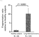
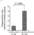
- Samples of urine from 124 cases of lung adenocarcinoma and 66 healthy subjects were subjected to an MRM method, thereby to determine the fragmentation ratio of a protein fragment in which the sequence of C-terminal amino acids derived from kininogen I was ESNEELTESCET.
- a patient's sample having a fragmentation ratio that was 1.25 times or more higher than the mean value of the healthy subjects group was considered to be positive for lung adenocarcinoma
- 8 cases out of the healthy subjects and 61 cases out of the lung adenocarcinoma cases were positive for lung adenocarcinoma.
- the fragmentation ratio was set to be 1.5 times the mean value of the healthy subjects group, 8 cases out of the healthy subjects and 53 cases out of the lung adenocarcinoma cases were positive for lung adenocarcinoma, and the sensitivity at the given reference value was 42.74%, the specificity was 87.88%, and the ROC-AUC value was 0.766.
- samples of urine from 124 cases of lung adenocarcinoma and 66 healthy subjects were subjected to an MRM method, thereby to determine the fragmentation ratio of a protein fragment in which the sequence of C-terminal amino acids derived from kininogen I was IYPTVNCQPLG.
- an MRM method determines the fragmentation ratio of a protein fragment in which the sequence of C-terminal amino acids derived from kininogen I was IYPTVNCQPLG.
- the fragmentation ratio was set to be 1.5 times the mean value of the healthy subjects group, 5 cases out of the healthy subjects cases and 68 cases out of the lung adenocarcinoma cases were positive for lung adenocarcinoma, and the sensitivity at the given reference value was the sensitivity at the given reference value was 54.84%, the specificity was 92.42%, and the ROC-AUC value was 0.814.
- Samples of urine from 61 cases of early lung adenocarcinoma at stages IA and IB and 66 healthy subjects were subjected to an MRM method, thereby to determine the fragmentation ratio of a protein fragment in which the sequence of C-terminal amino acids derived from kininogen I was ESNEELTESCET.
- a patient's sample having a fragmentation ratio that was 1.25 times or more higher than the mean value of the healthy subjects group was considered to be positive for lung adenocarcinoma
- 8 cases out of the healthy subjects and 21 cases out of the early lung adenocarcinoma cases were positive for lung adenocarcinoma.
- the fragmentation ratio was set to be 1.5 times the mean value of the healthy subjects group, 8 cases out of the healthy subjects and 18 cases out of the early lung adenocarcinoma cases were positive for lung adenocarcinoma, and the sensitivity at the given reference value was 29.51%, the specificity was 87.88%, and the ROC-AUC value was 0.676.
- samples of urine from 61 cases of early lung adenocarcinoma at stages IA and IB and 66 healthy subjects were subjected to an MRM method, thereby to determine the fragmentation ratio of a protein fragment in which the sequence of C-terminal amino acids derived from kininogen I was IYPTVNCQPLG.
- a patient's sample having a fragmentation ratio that was 1.25 times or more higher than the mean value of the healthy subjects group was considered to be positive for early lung adenocarcinoma, 7 cases out of the healthy subjects and 32 cases out of the early lung adenocarcinoma cases were positive for early lung adenocarcinoma.
- the fragmentation ratio was set to be 1.5 times the mean value of the healthy subjects group, 5 cases out of the healthy subjects and 26 cases out of the early lung adenocarcinoma cases were positive for lung adenocarcinoma, and the sensitivity at the given reference value was the sensitivity at the given reference value was 42.62%, the specificity was 92.42%, and the ROC-AUC value was 0.766.
- Samples of urine ( ⁇ 50 mL) were collected and markers derived from kininogen 1 were searched in a sequence of sample pretreatment, and obtaining and statistical analysis of analytical date.
- Sample pretreatment was performed to concentrate the urine samples 200 to 250 times using an Amicon Ultra-15 (having a molecular weight cut-off of 10 kDa) and an Amicon Ultra-4 (having a molecular weight cut-off of 10 kDa) (Merck Millipore), and remove low molecular weight molecules by washing three times with 3 mL of an aqueous solution of triethylammonium hydrogen carbonate containing 100 mM NaCl, prior to obtaining concentrated samples.
- the concentrated samples were subjected to protein quantification, and all the samples were adjusted to have a protein concentration, or a total protein amount, of 10 mg/mL using a buffer solution and used in a subsequent analysis step. Subsequently, the sample was subjected to reductive alkylation, followed by tryptic digestion in a buffer solution prepared using a particular concentration of H 2 18 O (a method for stable isotope labeling at the peptide C-terminus). After desalting and purification, trypsin-digested peptides were labeled by iTRAQ (8-plex, AB Sciex). Eight samples were mixed, and the mixture was desalted and purified.
- iTRAQ 8-plex, AB Sciex
- the fraction of interest was desalted and purified, and then only the C-terminal trypsin-digested peptide was selected and extracted with Nano-LC (on an LC column having an ID of 75 ⁇ m and a length of 100 mm and as a packing material, Inertsil C18 (a particle size of 3 ⁇ m), at a flow rate of 250 nL/min, using as a solvent, a concentration gradient of (from 3 to 80% of) acetonitrile in 0.1% trifluoroacetic acid/water for separating) /MALDI-MS/MS(AB Sciex) (using an in-house developed program "iSpec"; see, Fernandez-de-Cossio J., Takao T.
- Respective relative ratios to the peak intensity of a control sample were calculated for the peak intensity of each C-terminal trypsin-digested peptide in MS/MS spectra (Example 6) obtained from samples of urine from 58 cases of lung adenocarcinoma (5 cases at stage IA, 1 case at stage IB, 3 cases at stage IIIA, 8 cases at stage IIIb, and 41 cases at stage IV), 30 cases of benign respiratory diseases (bronchial asthma, empyema, lung fluke disease, nontuberculous mycobacteriosis, interstitial pneumonia, chronic obstructive pulmonary disease, sarcoidosis, inflammatory pseudotumor, bacterial pneumonia, allergic bronchopulmonary aspergillosis), and 25 cases of healthy subjects.
- benign respiratory diseases bronchial asthma, empyema, lung fluke disease, nontuberculous mycobacteriosis, interstitial pneumonia, chronic obstructive pulmonary disease, sarcoidosis, inflammatory pseudotumor,
- a protein fragment in which the sequence of C-terminal amino acids derived from kininogen 1 was TEHLASSSEDSTTPSA was detected in 26 cases out of the 58 lung adenocarcinoma cases and in 2 cases out of the 55 cases in the non-lung adenocarcinoma group, and met the above criterion (p value ⁇ 0.0001).
- the peak intensity detected in the healthy subjects in the non-lung adenocarcinoma group was used as a reference, the sensitivity, the specificity, and the ROC-AUC value at the reference value were 37.93%, 96.36%, and 0.708.
- Respective relative ratios to the peak intensity of a control sample were calculated for the peak intensity of each C-terminal trypsin-digested peptide in MS/MS spectra (Example 6) obtained from samples of urine from 25 cases of early lung adenocarcinoma (18 cases at stage IA, 5 cases at stage IB, and 1 case at stage IIA) and 25 cases of healthy subjects.
- MS/MS spectra obtained from samples of urine from 25 cases of early lung adenocarcinoma (18 cases at stage IA, 5 cases at stage IB, and 1 case at stage IIA) and 25 cases of healthy subjects.
- a comparison was made between the groups of the early lung adenocarcinoma cases and of the healthy subjects.
- samples of urine from 37 cases of lung adenocarcinoma (15 cases at stage IA, 3 cases at stage IB, 4 cases at stage IIA, 2 cases at stage IIIA, 3 cases at stage IIIB, and 10 cases at stage IV) and 55 healthy subjects were subjected to an MRM method, thereby to determine the fragmentation ratio of a protein fragment in which the sequence of C-terminal amino acids derived from kininogen I was IYPTVNCQPL.
- samples of urine from 22 cases of early lung adenocarcinoma (15 cases at stage IA, 3 cases at stage IB, and 4 cases at stage IIA) and 55 healthy subjects were subjected to an MRM method, thereby to determine the fragmentation ratio of a protein fragment in which the sequence of C-terminal amino acids derived from kininogen I was IYPTVNCQPL.
- a patient's sample having a fragmentation ratio that was 1.25 times or more higher than the mean value of the healthy subjects group (0.005) was considered to be positive for lung adenocarcinoma
- 12 cases out of the healthy subjects and 16 cases out of the lung adenocarcinoma cases were positive for lung adenocarcinoma.
- the sensitivity at the given reference value was 72.7%, the specificity was 78.2%, and the ROC-AUC value was 0.842.
- samples of urine from 37 cases of lung adenocarcinoma (15 cases at stage IA, 3 cases at stage IB, 4 cases at stage IIA, 2 cases at stage IIIA, 3 cases at stage IIIB, and 10 cases at stage IV) and 55 healthy subjects were subjected to an MRM method, thereby to determine the fragmentation ratio of a protein fragment in which the sequence of C-terminal amino acids derived from kininogen I was IYPTVNCQP.
- samples of urine from 22 cases of early lung adenocarcinoma (15 cases at stage IA, 3 cases at stage IB, and 4 cases at stage IIA) and 55 healthy subjects were subjected to an MRM method, thereby to determine the fragmentation ratio of a protein fragment in which the sequence of C-terminal amino acids derived from kininogen I was IYPTVNCQP.
- a patient's sample having a fragmentation ratio that was 1.25 times or more higher than the mean value of the healthy subjects group (0.044) was considered to be positive for lung adenocarcinoma
- 11 cases out of the healthy subjects and 16 cases out of the lung adenocarcinoma cases were positive for lung adenocarcinoma.
- the sensitivity at the given reference value was 72.3%, the specificity was 80%, and the ROC-AUC value was 0.842.
- the fragmentation ratio for each of the lung cancer cases was 0.125 for the pleomorphic cancer case, 0.162 for the carcinosarcoma case, and 0.135 for the large cell carcinoma case, and was higher than a reference value (0.055) that was 1.25 times the mean value of the healthy subjects group (0.044) .
- the fragmentation ratios of a protein fragment in which the sequence of C-terminal amino acids from kininogen I was IYPTVNCQP, in the cases of squamous cell carcinoma were 0.098 and 0.090 for the stage IB cases, 0.19 for the stage IIIA case, and 0.44 for the stage IIIB case, and all the 4 cases of squamous cell carcinoma gave a higher fragmentation ratio than a reference value (0.055) that was 1.25 times the mean value of the healthy subjects group (0.044).
- Samples of urine from 4 cases of small cell lung cancer, who were histopathologically diagnosed via transbronchial biopsy or surgical lung biopsy, and 55 healthy subjects were each subjected to MRM, thereby to determine the fragmentation ratio of a protein fragment in which the sequence of C-terminal amino acids derived from kininogen I was IYPTVNCQP.
- the respective fragmentation ratios of a protein fragment in which the sequence of C-terminal amino acids from kininogen I was IYPTVNCQP, in the cases of squamous cell carcinoma were 0.069, 0.131, 0.747, and 0.754, and all the 4 cases of small cell lung cancer gave a higher fragmentation ratio than a reference value (0.055) that was 1.25 times the mean value of the healthy subjects group (0.044).
- Samples of serum (10 ⁇ L) from 25 cases of early lung adenocarcinoma at stages IA and IB and 35 healthy subjects were subjected to an ELISA method, thereby to determine the concentration of a protein fragment in the serum, in which the sequence of C-terminal amino acids from kininogen I was IYPTVNCQPLG.
- an antibody was used that specifically recognized a fragment derived from kininogen I in which the sequence of C-terminal amino acids was CQPLG, which was recovered from the serum of a chicken which had been immunized by administration of an prepared immunogen solution.
- Samples of urine were collected over time from one case in which a 1.4-cm ground-glass nodular shadow suspected of lung adenocarcinoma was recognized in the periphery of the right lower lobe by a chest CT examination.
- the fragmentation ratio of a protein fragment in which the sequence of C-terminal amino acids from kininogen I was IYPTVNCQP, and the size of the nodular shadow were compared with those at the time when the lung adenocarcinoma was diagnosed by surgical lung biopsy.
- the post-operative stage in this case was T1bN0M0, pStageIA.
- results from the comparison of chest CT examinations between at 24 months prior to surgery and at the time thereof showed that the size of the nodular shadow was increased to 1.9 cm at the time of the surgery.
- the nodule was observed to have highly accumulated FDG in a PET-CT scan ( Fig. 8 ).
- the fragmentation ratio of the protein fragment in which the sequence of C-terminal amino acids from kininogen I was IYPTVNCQP was 0.468, 0.565, 0.58, 0.956, and 1.08, respectively, indicating that the fragmentation ratio was increased with growing adenocarcinoma.
- the fragmentation ratio of the protein fragment in which the sequence of C-terminal amino acids from kininogen I was IYPTVNCQP was decreased from 0.378 to 0.205, by 45.77% ( Fig. 9 ) .
- Samples of urine collected in 1 case of lung adenocarcinoma at stage IV who was positive for epidermal growth factor receptor (EGFR) gene mutation were subjected to an MRM method, whereby the fragmentation ratio of a protein fragment in which the sequence of C-terminal amino acids from kininogen I was IYPTVNCQP was compared between before and at 2 months after a treatment with an EGFR tyrosine kinase inhibitor.
- the therapeutic effect by the EGFR tyrosine kinase inhibitor was observed to lead to a tumor reduction rate of 37.1% ( Fig. 10 ), which met the criteria for Partial Response in the Response Evaluation Criteria In Solid Tumors (RECIST).
- Samples of urine from 27 cases of lung adenocarcinoma 13 cases at stage IA, 2 cases at stage IB, 4 cases at stage IIA, 2 cases at stage IIIA, and 6 cases at stage IV) and 49 healthy subjects were subjected to an MRM method, thereby to determine the fragmentation index (Fx) of a protein fragment in which the sequence of C-terminal amino acids from kininogen I was IYPTVNCQP.
- Samples of urine from 19 cases of early lung adenocarcinoma (13 cases at stage IA, 2 cases at stage IB, and 4 cases at stage IIA) and 49 healthy subjects were subjected to an MRM method, thereby to determine the fragmentation index (Fx) of a protein fragment in which the sequence of C-terminal amino acids from kininogen I was IYPTVNCQP.
- Samples of urine from 27 cases of lung adenocarcinoma 13 cases at stage IA, 2 cases at stage IB, 4 cases at stage IIA, 2 cases at stage IIIA, and 6 cases at stage IV) and 49 healthy subjects were subjected to an MRM method, thereby to determine the fragmentation index (Fx) of a protein fragment in which the sequence of C-terminal amino acids from kininogen I was IYPTVNCQPL.
- Samples of urine from 19 cases of early lung adenocarcinoma (13 cases at stage IA, 2 cases at stage IB, and 4 cases at stage IIA) and 49 healthy subjects were subjected to an MRM method, thereby to determine the fragmentation index (Fx) of a protein fragment in which the sequence of C-terminal amino acids from kininogen I was IYPTVNCQPL.
- Samples of urine from 15 cases of advanced lung adenocarcinoma (2 cases at stage IIIA, 3 cases at stage IIIB, and 10 cases at stage IV) and 55 healthy subjects were subjected to an MRM method, thereby to determine the amounts of presence of a protein fragment having the amino acid sequence IYPTVNCQPLGMISLM as a C-terminal portion from kininogen I and of a trypsin-digested peptide having the amino acid sequence YFIDFVAR as a C-terminal portion, to compare the ratio of presence of IYPTVNCQPLGMISLM, a C-terminal portion which is a normal protein fragment derived from kininogen I.
- the samples from the lung cancer patients showed a smaller mean peak-intensity ratio of 0.150 relative to the mean value of the healthy subjects group (0.353).
- the ratio of peak intensity of the trypsin-digested peptide LGQSLDCNAEVYVVPWEK to that of the trypsin-digested peptide ESNEELTESCETK that was 40% or more lower than the mean of the healthy subjects was considered to be positive for lung adenocarcinoma
- 6 cases out of the healthy subjects and 24 cases out of the lung adenocarcinoma cases were positive for lung adenocarcinoma.
- the sensitivity at the given reference value was 70.6%
- the specificity was 88.68%
- the ROC-AUC value was 0.9.
- the samples from the lung cancer patients showed a smaller mean peak-intensity ratio of 2.0 relative to the mean value of the healthy subjects group (5.423).
- the ratio of peak intensity of the trypsin-digested peptide ENFLFLTPDCK to that of the trypsin-digested peptide ESNEELTESCETK that was 40% or more lower than the mean of the healthy subjects was considered to be positive for lung adenocarcinoma
- 3 cases out of the healthy subjects and 31 cases out of the lung adenocarcinoma cases were positive for lung adenocarcinoma.
- the sensitivity at the given reference value was 91.2%, the specificity was 94.34%, and the ROC-AUC value was 0.972.
Landscapes
- Health & Medical Sciences (AREA)
- Life Sciences & Earth Sciences (AREA)
- Engineering & Computer Science (AREA)
- Immunology (AREA)
- Chemical & Material Sciences (AREA)
- Molecular Biology (AREA)
- Hematology (AREA)
- Biomedical Technology (AREA)
- Urology & Nephrology (AREA)
- Physics & Mathematics (AREA)
- Analytical Chemistry (AREA)
- Biotechnology (AREA)
- Biochemistry (AREA)
- General Health & Medical Sciences (AREA)
- Microbiology (AREA)
- Cell Biology (AREA)
- Pathology (AREA)
- Food Science & Technology (AREA)
- Medicinal Chemistry (AREA)
- General Physics & Mathematics (AREA)
- Bioinformatics & Cheminformatics (AREA)
- Proteomics, Peptides & Aminoacids (AREA)
- Organic Chemistry (AREA)
- Bioinformatics & Computational Biology (AREA)
- Biophysics (AREA)
- Zoology (AREA)
- Wood Science & Technology (AREA)
- Spectroscopy & Molecular Physics (AREA)
- Neurosurgery (AREA)
- General Engineering & Computer Science (AREA)
- Genetics & Genomics (AREA)
- Investigating Or Analysing Biological Materials (AREA)
- Peptides Or Proteins (AREA)
- Other Investigation Or Analysis Of Materials By Electrical Means (AREA)
Applications Claiming Priority (2)
| Application Number | Priority Date | Filing Date | Title |
|---|---|---|---|
| JP2017113289 | 2017-06-08 | ||
| PCT/JP2018/021901 WO2018225830A1 (fr) | 2017-06-08 | 2018-06-07 | Procédé de détection du cancer du poumon |
Publications (3)
| Publication Number | Publication Date |
|---|---|
| EP3637106A1 true EP3637106A1 (fr) | 2020-04-15 |
| EP3637106A4 EP3637106A4 (fr) | 2021-01-27 |
| EP3637106B1 EP3637106B1 (fr) | 2023-03-22 |
Family
ID=64565957
Family Applications (1)
| Application Number | Title | Priority Date | Filing Date |
|---|---|---|---|
| EP18813561.0A Active EP3637106B1 (fr) | 2017-06-08 | 2018-06-07 | Procédé de détection du cancer du poumon |
Country Status (4)
| Country | Link |
|---|---|
| US (1) | US20200124605A1 (fr) |
| EP (1) | EP3637106B1 (fr) |
| JP (1) | JP7144014B2 (fr) |
| WO (1) | WO2018225830A1 (fr) |
Cited By (1)
| Publication number | Priority date | Publication date | Assignee | Title |
|---|---|---|---|---|
| EP4169941A4 (fr) * | 2020-06-23 | 2024-06-12 | Mitsui Chemicals, Inc. | Procédé de détection d'adénocarcinome et kit d'examen |
Families Citing this family (2)
| Publication number | Priority date | Publication date | Assignee | Title |
|---|---|---|---|---|
| CN112240932A (zh) * | 2019-07-17 | 2021-01-19 | 张曼 | 肺癌患者尿液表达的kng1蛋白的应用 |
| CN112345762A (zh) * | 2019-08-08 | 2021-02-09 | 首都医科大学附属北京世纪坛医院 | 肺癌患者肺泡灌洗液表达的kng1蛋白的应用 |
Family Cites Families (10)
| Publication number | Priority date | Publication date | Assignee | Title |
|---|---|---|---|---|
| DE69627191T2 (de) * | 1995-06-09 | 2004-01-08 | The Regents Of The University Of Michigan, Ann Arbor | Bradikininanaloge als selektive thrombininhibitoren |
| EP2520935A3 (fr) * | 2006-08-09 | 2013-02-13 | Homestead Clinical Corporation | Protéines spécifiques d'organes et leurs procédés d'utilisation |
| EP1892303A1 (fr) * | 2006-08-22 | 2008-02-27 | Fraunhofer-Gesellschaft zur Förderung der angewandten Forschung e.V. | Méthodes d'identification de cibles thérapeutiques de tumeurs et méthodes de la détermination et du traitement de l'angiogenèse et de l'hémostase associés d'adénocarcinome pulmonaire |
| JP2014115186A (ja) | 2012-12-10 | 2014-06-26 | Toray Ind Inc | 胃癌、肺癌及び/又は食道癌の検出方法 |
| JP6317763B2 (ja) * | 2013-01-08 | 2018-04-25 | シュピーンゴテック ゲゼルシャフト ミット ベシュレンクテル ハフツング | 対象における癌となる危険性を予測し又は癌と診断する方法 |
| US20150147765A1 (en) * | 2013-11-28 | 2015-05-28 | Korea Basic Science Institute | Serological markers for cancer diagnosis using blood sample |
| KR101559101B1 (ko) * | 2013-11-28 | 2015-10-12 | 한국기초과학지원연구원 | 혈액유래 암 진단용 펩티드 마커 및 이를 이용한 암 진단방법 |
| EP3390439B1 (fr) * | 2015-12-15 | 2025-03-12 | Takeda Pharmaceutical Company Limited | Dosage de quantification de peptides permettant de distinguer entre un kininogène de masse moléculaire élevée et de pleine longueur (hmwk) et un hmwk clivé |
| CN112240932A (zh) | 2019-07-17 | 2021-01-19 | 张曼 | 肺癌患者尿液表达的kng1蛋白的应用 |
| CN112345762A (zh) | 2019-08-08 | 2021-02-09 | 首都医科大学附属北京世纪坛医院 | 肺癌患者肺泡灌洗液表达的kng1蛋白的应用 |
-
2018
- 2018-06-07 WO PCT/JP2018/021901 patent/WO2018225830A1/fr not_active Ceased
- 2018-06-07 JP JP2019523975A patent/JP7144014B2/ja active Active
- 2018-06-07 US US16/620,477 patent/US20200124605A1/en not_active Abandoned
- 2018-06-07 EP EP18813561.0A patent/EP3637106B1/fr active Active
Cited By (1)
| Publication number | Priority date | Publication date | Assignee | Title |
|---|---|---|---|---|
| EP4169941A4 (fr) * | 2020-06-23 | 2024-06-12 | Mitsui Chemicals, Inc. | Procédé de détection d'adénocarcinome et kit d'examen |
Also Published As
| Publication number | Publication date |
|---|---|
| JPWO2018225830A1 (ja) | 2020-04-09 |
| EP3637106A4 (fr) | 2021-01-27 |
| US20200124605A1 (en) | 2020-04-23 |
| EP3637106B1 (fr) | 2023-03-22 |
| WO2018225830A1 (fr) | 2018-12-13 |
| JP7144014B2 (ja) | 2022-09-29 |
Similar Documents
| Publication | Publication Date | Title |
|---|---|---|
| JP6672411B2 (ja) | 癌療法を示すためのsrmアッセイ | |
| CA2821908C (fr) | Nouveau biomarqueur et ses utilisations dans le diagnostic, le traitement de l'autisme | |
| EP3185014A1 (fr) | Protéines biomarqueurs du cancer de la vessie | |
| AU2018293090A1 (en) | Biomarker for detecting colorectal cancer | |
| AU2017201499B2 (en) | Her3 protein SRM/MRM assay | |
| EP3637106B1 (fr) | Procédé de détection du cancer du poumon | |
| EP3535587B1 (fr) | Procédés utilisant la spectrométrie de masse pour la détection de la circulation d'histones h3 et h2b dans le plasma de patients atteint de choc septique ou de sepsie | |
| EP3092495B1 (fr) | Dosage srm pour pd-l1 | |
| CA2824752C (fr) | Dosage par srm/mrm de la proteine 11 de type bcl-2 | |
| US11435354B2 (en) | Adenocarcinoma detection method | |
| JP2022527436A (ja) | 加齢黄斑変性の診断用複合マーカーおよびその用途 | |
| JP2020091243A (ja) | 膵臓がん、食道がん、乳がん、胃がん、大腸がん、胆道がん、肝臓がん又は胚細胞腫瘍の検出方法 | |
| AU2012318567B2 (en) | SRM/MRM assay for the receptor tyrosine-protein kinase erbB-4 protein (HER4) | |
| KR101535717B1 (ko) | 보체인자 i 단백질에 특이적으로 결합하는 폴리펩타이드 또는 항체를 포함하는 췌장암 진단용 조성물 | |
| JP2025131404A (ja) | 膵癌の検出方法 | |
| JP2013528284A (ja) | ウロキナーゼ型プラスミノーゲン活性化因子タンパク質/プラスミノーゲン活性化因子阻害物質1型タンパク質選択反応モニタリングアッセイ |
Legal Events
| Date | Code | Title | Description |
|---|---|---|---|
| STAA | Information on the status of an ep patent application or granted ep patent |
Free format text: STATUS: THE INTERNATIONAL PUBLICATION HAS BEEN MADE |
|
| PUAI | Public reference made under article 153(3) epc to a published international application that has entered the european phase |
Free format text: ORIGINAL CODE: 0009012 |
|
| STAA | Information on the status of an ep patent application or granted ep patent |
Free format text: STATUS: REQUEST FOR EXAMINATION WAS MADE |
|
| 17P | Request for examination filed |
Effective date: 20200107 |
|
| AK | Designated contracting states |
Kind code of ref document: A1 Designated state(s): AL AT BE BG CH CY CZ DE DK EE ES FI FR GB GR HR HU IE IS IT LI LT LU LV MC MK MT NL NO PL PT RO RS SE SI SK SM TR |
|
| AX | Request for extension of the european patent |
Extension state: BA ME |
|
| DAV | Request for validation of the european patent (deleted) | ||
| DAX | Request for extension of the european patent (deleted) | ||
| A4 | Supplementary search report drawn up and despatched |
Effective date: 20210112 |
|
| RIC1 | Information provided on ipc code assigned before grant |
Ipc: G01N 33/53 20060101ALI20201221BHEP Ipc: G01N 33/574 20060101AFI20201221BHEP |
|
| STAA | Information on the status of an ep patent application or granted ep patent |
Free format text: STATUS: EXAMINATION IS IN PROGRESS |
|
| 17Q | First examination report despatched |
Effective date: 20220216 |
|
| INTG | Intention to grant announced |
Effective date: 20221206 |
|
| GRAS | Grant fee paid |
Free format text: ORIGINAL CODE: EPIDOSNIGR3 |
|
| STAA | Information on the status of an ep patent application or granted ep patent |
Free format text: STATUS: GRANT OF PATENT IS INTENDED |
|
| GRAA | (expected) grant |
Free format text: ORIGINAL CODE: 0009210 |
|
| STAA | Information on the status of an ep patent application or granted ep patent |
Free format text: STATUS: THE PATENT HAS BEEN GRANTED |
|
| AK | Designated contracting states |
Kind code of ref document: B1 Designated state(s): AL AT BE BG CH CY CZ DE DK EE ES FI FR GB GR HR HU IE IS IT LI LT LU LV MC MK MT NL NO PL PT RO RS SE SI SK SM TR |
|
| REG | Reference to a national code |
Ref country code: GB Ref legal event code: FG4D |
|
| REG | Reference to a national code |
Ref country code: CH Ref legal event code: EP |
|
| REG | Reference to a national code |
Ref country code: DE Ref legal event code: R096 Ref document number: 602018047547 Country of ref document: DE |
|
| REG | Reference to a national code |
Ref country code: IE Ref legal event code: FG4D |
|
| REG | Reference to a national code |
Ref country code: AT Ref legal event code: REF Ref document number: 1555615 Country of ref document: AT Kind code of ref document: T Effective date: 20230415 |
|
| P01 | Opt-out of the competence of the unified patent court (upc) registered |
Effective date: 20230517 |
|
| REG | Reference to a national code |
Ref country code: LT Ref legal event code: MG9D |
|
| REG | Reference to a national code |
Ref country code: NL Ref legal event code: MP Effective date: 20230322 |
|
| PG25 | Lapsed in a contracting state [announced via postgrant information from national office to epo] |
Ref country code: RS Free format text: LAPSE BECAUSE OF FAILURE TO SUBMIT A TRANSLATION OF THE DESCRIPTION OR TO PAY THE FEE WITHIN THE PRESCRIBED TIME-LIMIT Effective date: 20230322 Ref country code: NO Free format text: LAPSE BECAUSE OF FAILURE TO SUBMIT A TRANSLATION OF THE DESCRIPTION OR TO PAY THE FEE WITHIN THE PRESCRIBED TIME-LIMIT Effective date: 20230622 Ref country code: LV Free format text: LAPSE BECAUSE OF FAILURE TO SUBMIT A TRANSLATION OF THE DESCRIPTION OR TO PAY THE FEE WITHIN THE PRESCRIBED TIME-LIMIT Effective date: 20230322 Ref country code: LT Free format text: LAPSE BECAUSE OF FAILURE TO SUBMIT A TRANSLATION OF THE DESCRIPTION OR TO PAY THE FEE WITHIN THE PRESCRIBED TIME-LIMIT Effective date: 20230322 Ref country code: HR Free format text: LAPSE BECAUSE OF FAILURE TO SUBMIT A TRANSLATION OF THE DESCRIPTION OR TO PAY THE FEE WITHIN THE PRESCRIBED TIME-LIMIT Effective date: 20230322 |
|
| REG | Reference to a national code |
Ref country code: AT Ref legal event code: MK05 Ref document number: 1555615 Country of ref document: AT Kind code of ref document: T Effective date: 20230322 |
|
| PG25 | Lapsed in a contracting state [announced via postgrant information from national office to epo] |
Ref country code: SE Free format text: LAPSE BECAUSE OF FAILURE TO SUBMIT A TRANSLATION OF THE DESCRIPTION OR TO PAY THE FEE WITHIN THE PRESCRIBED TIME-LIMIT Effective date: 20230322 Ref country code: NL Free format text: LAPSE BECAUSE OF FAILURE TO SUBMIT A TRANSLATION OF THE DESCRIPTION OR TO PAY THE FEE WITHIN THE PRESCRIBED TIME-LIMIT Effective date: 20230322 Ref country code: GR Free format text: LAPSE BECAUSE OF FAILURE TO SUBMIT A TRANSLATION OF THE DESCRIPTION OR TO PAY THE FEE WITHIN THE PRESCRIBED TIME-LIMIT Effective date: 20230623 Ref country code: FI Free format text: LAPSE BECAUSE OF FAILURE TO SUBMIT A TRANSLATION OF THE DESCRIPTION OR TO PAY THE FEE WITHIN THE PRESCRIBED TIME-LIMIT Effective date: 20230322 |
|
| PG25 | Lapsed in a contracting state [announced via postgrant information from national office to epo] |
Ref country code: SM Free format text: LAPSE BECAUSE OF FAILURE TO SUBMIT A TRANSLATION OF THE DESCRIPTION OR TO PAY THE FEE WITHIN THE PRESCRIBED TIME-LIMIT Effective date: 20230322 Ref country code: RO Free format text: LAPSE BECAUSE OF FAILURE TO SUBMIT A TRANSLATION OF THE DESCRIPTION OR TO PAY THE FEE WITHIN THE PRESCRIBED TIME-LIMIT Effective date: 20230322 Ref country code: PT Free format text: LAPSE BECAUSE OF FAILURE TO SUBMIT A TRANSLATION OF THE DESCRIPTION OR TO PAY THE FEE WITHIN THE PRESCRIBED TIME-LIMIT Effective date: 20230724 Ref country code: ES Free format text: LAPSE BECAUSE OF FAILURE TO SUBMIT A TRANSLATION OF THE DESCRIPTION OR TO PAY THE FEE WITHIN THE PRESCRIBED TIME-LIMIT Effective date: 20230322 Ref country code: EE Free format text: LAPSE BECAUSE OF FAILURE TO SUBMIT A TRANSLATION OF THE DESCRIPTION OR TO PAY THE FEE WITHIN THE PRESCRIBED TIME-LIMIT Effective date: 20230322 Ref country code: AT Free format text: LAPSE BECAUSE OF FAILURE TO SUBMIT A TRANSLATION OF THE DESCRIPTION OR TO PAY THE FEE WITHIN THE PRESCRIBED TIME-LIMIT Effective date: 20230322 |
|
| PG25 | Lapsed in a contracting state [announced via postgrant information from national office to epo] |
Ref country code: SK Free format text: LAPSE BECAUSE OF FAILURE TO SUBMIT A TRANSLATION OF THE DESCRIPTION OR TO PAY THE FEE WITHIN THE PRESCRIBED TIME-LIMIT Effective date: 20230322 Ref country code: PL Free format text: LAPSE BECAUSE OF FAILURE TO SUBMIT A TRANSLATION OF THE DESCRIPTION OR TO PAY THE FEE WITHIN THE PRESCRIBED TIME-LIMIT Effective date: 20230322 Ref country code: IS Free format text: LAPSE BECAUSE OF FAILURE TO SUBMIT A TRANSLATION OF THE DESCRIPTION OR TO PAY THE FEE WITHIN THE PRESCRIBED TIME-LIMIT Effective date: 20230722 |
|
| REG | Reference to a national code |
Ref country code: DE Ref legal event code: R097 Ref document number: 602018047547 Country of ref document: DE |
|
| PG25 | Lapsed in a contracting state [announced via postgrant information from national office to epo] |
Ref country code: MC Free format text: LAPSE BECAUSE OF FAILURE TO SUBMIT A TRANSLATION OF THE DESCRIPTION OR TO PAY THE FEE WITHIN THE PRESCRIBED TIME-LIMIT Effective date: 20230322 |
|
| PLBE | No opposition filed within time limit |
Free format text: ORIGINAL CODE: 0009261 |
|
| STAA | Information on the status of an ep patent application or granted ep patent |
Free format text: STATUS: NO OPPOSITION FILED WITHIN TIME LIMIT |
|
| PG25 | Lapsed in a contracting state [announced via postgrant information from national office to epo] |
Ref country code: SI Free format text: LAPSE BECAUSE OF FAILURE TO SUBMIT A TRANSLATION OF THE DESCRIPTION OR TO PAY THE FEE WITHIN THE PRESCRIBED TIME-LIMIT Effective date: 20230322 Ref country code: MC Free format text: LAPSE BECAUSE OF FAILURE TO SUBMIT A TRANSLATION OF THE DESCRIPTION OR TO PAY THE FEE WITHIN THE PRESCRIBED TIME-LIMIT Effective date: 20230322 Ref country code: DK Free format text: LAPSE BECAUSE OF FAILURE TO SUBMIT A TRANSLATION OF THE DESCRIPTION OR TO PAY THE FEE WITHIN THE PRESCRIBED TIME-LIMIT Effective date: 20230322 Ref country code: CZ Free format text: LAPSE BECAUSE OF FAILURE TO SUBMIT A TRANSLATION OF THE DESCRIPTION OR TO PAY THE FEE WITHIN THE PRESCRIBED TIME-LIMIT Effective date: 20230322 |
|
| REG | Reference to a national code |
Ref country code: CH Ref legal event code: PL |
|
| REG | Reference to a national code |
Ref country code: BE Ref legal event code: MM Effective date: 20230630 |
|
| 26N | No opposition filed |
Effective date: 20240102 |
|
| GBPC | Gb: european patent ceased through non-payment of renewal fee |
Effective date: 20230622 |
|
| PG25 | Lapsed in a contracting state [announced via postgrant information from national office to epo] |
Ref country code: LU Free format text: LAPSE BECAUSE OF NON-PAYMENT OF DUE FEES Effective date: 20230607 |
|
| REG | Reference to a national code |
Ref country code: IE Ref legal event code: MM4A |
|
| PG25 | Lapsed in a contracting state [announced via postgrant information from national office to epo] |
Ref country code: LU Free format text: LAPSE BECAUSE OF NON-PAYMENT OF DUE FEES Effective date: 20230607 |
|
| PG25 | Lapsed in a contracting state [announced via postgrant information from national office to epo] |
Ref country code: IE Free format text: LAPSE BECAUSE OF NON-PAYMENT OF DUE FEES Effective date: 20230607 |
|
| PG25 | Lapsed in a contracting state [announced via postgrant information from national office to epo] |
Ref country code: IE Free format text: LAPSE BECAUSE OF NON-PAYMENT OF DUE FEES Effective date: 20230607 Ref country code: CH Free format text: LAPSE BECAUSE OF NON-PAYMENT OF DUE FEES Effective date: 20230630 Ref country code: GB Free format text: LAPSE BECAUSE OF NON-PAYMENT OF DUE FEES Effective date: 20230622 |
|
| PG25 | Lapsed in a contracting state [announced via postgrant information from national office to epo] |
Ref country code: IT Free format text: LAPSE BECAUSE OF FAILURE TO SUBMIT A TRANSLATION OF THE DESCRIPTION OR TO PAY THE FEE WITHIN THE PRESCRIBED TIME-LIMIT Effective date: 20230322 Ref country code: BE Free format text: LAPSE BECAUSE OF NON-PAYMENT OF DUE FEES Effective date: 20230630 |
|
| PGFP | Annual fee paid to national office [announced via postgrant information from national office to epo] |
Ref country code: DE Payment date: 20240613 Year of fee payment: 7 |
|
| PGFP | Annual fee paid to national office [announced via postgrant information from national office to epo] |
Ref country code: FR Payment date: 20240510 Year of fee payment: 7 |
|
| PG25 | Lapsed in a contracting state [announced via postgrant information from national office to epo] |
Ref country code: BG Free format text: LAPSE BECAUSE OF FAILURE TO SUBMIT A TRANSLATION OF THE DESCRIPTION OR TO PAY THE FEE WITHIN THE PRESCRIBED TIME-LIMIT Effective date: 20230322 |
|
| PG25 | Lapsed in a contracting state [announced via postgrant information from national office to epo] |
Ref country code: BG Free format text: LAPSE BECAUSE OF FAILURE TO SUBMIT A TRANSLATION OF THE DESCRIPTION OR TO PAY THE FEE WITHIN THE PRESCRIBED TIME-LIMIT Effective date: 20230322 |
|
| PG25 | Lapsed in a contracting state [announced via postgrant information from national office to epo] |
Ref country code: CY Free format text: LAPSE BECAUSE OF FAILURE TO SUBMIT A TRANSLATION OF THE DESCRIPTION OR TO PAY THE FEE WITHIN THE PRESCRIBED TIME-LIMIT; INVALID AB INITIO Effective date: 20180607 |
|
| PG25 | Lapsed in a contracting state [announced via postgrant information from national office to epo] |
Ref country code: HU Free format text: LAPSE BECAUSE OF FAILURE TO SUBMIT A TRANSLATION OF THE DESCRIPTION OR TO PAY THE FEE WITHIN THE PRESCRIBED TIME-LIMIT; INVALID AB INITIO Effective date: 20180607 |
|
| REG | Reference to a national code |
Ref country code: DE Ref legal event code: R079 Ref document number: 602018047547 Country of ref document: DE Free format text: PREVIOUS MAIN CLASS: G01N0033574000 Ipc: G01N0033575000 |
|
| PG25 | Lapsed in a contracting state [announced via postgrant information from national office to epo] |
Ref country code: TR Free format text: LAPSE BECAUSE OF FAILURE TO SUBMIT A TRANSLATION OF THE DESCRIPTION OR TO PAY THE FEE WITHIN THE PRESCRIBED TIME-LIMIT Effective date: 20230322 |
|
| REG | Reference to a national code |
Ref country code: DE Ref legal event code: R119 Ref document number: 602018047547 Country of ref document: DE |
|
| PG25 | Lapsed in a contracting state [announced via postgrant information from national office to epo] |
Ref country code: DE Free format text: LAPSE BECAUSE OF NON-PAYMENT OF DUE FEES Effective date: 20260101 |
|
| PG25 | Lapsed in a contracting state [announced via postgrant information from national office to epo] |
Ref country code: FR Free format text: LAPSE BECAUSE OF NON-PAYMENT OF DUE FEES Effective date: 20250630 |