EP3647786A1 - Matériau de base permettant la fabrication d'un capteur d'analyse d'une cible de détection, capteur d'analyse d'une cible de détection, procédé d'analyse de cible de détection - Google Patents
Matériau de base permettant la fabrication d'un capteur d'analyse d'une cible de détection, capteur d'analyse d'une cible de détection, procédé d'analyse de cible de détection Download PDFInfo
- Publication number
- EP3647786A1 EP3647786A1 EP18809084.9A EP18809084A EP3647786A1 EP 3647786 A1 EP3647786 A1 EP 3647786A1 EP 18809084 A EP18809084 A EP 18809084A EP 3647786 A1 EP3647786 A1 EP 3647786A1
- Authority
- EP
- European Patent Office
- Prior art keywords
- group
- detection target
- binding
- sensor
- analysis
- Prior art date
- Legal status (The legal status is an assumption and is not a legal conclusion. Google has not performed a legal analysis and makes no representation as to the accuracy of the status listed.)
- Granted
Links
- SNURDPBGDLYVRE-UHFFFAOYSA-N CC(C(OCCSSc1ncccc1)=O)=C Chemical compound CC(C(OCCSSc1ncccc1)=O)=C SNURDPBGDLYVRE-UHFFFAOYSA-N 0.000 description 1
- 0 CCCCCOC(C)(C)CCOC(C)(C)C(CCC(O*(C(CC1)=C)C1=*)=O)=O Chemical compound CCCCCOC(C)(C)CCOC(C)(C)C(CCC(O*(C(CC1)=C)C1=*)=O)=O 0.000 description 1
- HAXFWIACAGNFHA-UHFFFAOYSA-N c1ccnc(SSc2ccccn2)c1 Chemical compound c1ccnc(SSc2ccccn2)c1 HAXFWIACAGNFHA-UHFFFAOYSA-N 0.000 description 1
Images





Classifications
-
- G—PHYSICS
- G01—MEASURING; TESTING
- G01N—INVESTIGATING OR ANALYSING MATERIALS BY DETERMINING THEIR CHEMICAL OR PHYSICAL PROPERTIES
- G01N33/00—Investigating or analysing materials by specific methods not covered by groups G01N1/00 - G01N31/00
- G01N33/48—Biological material, e.g. blood, urine; Haemocytometers
- G01N33/50—Chemical analysis of biological material, e.g. blood, urine; Testing involving biospecific ligand binding methods; Immunological testing
- G01N33/58—Chemical analysis of biological material, e.g. blood, urine; Testing involving biospecific ligand binding methods; Immunological testing involving labelled substances
- G01N33/582—Chemical analysis of biological material, e.g. blood, urine; Testing involving biospecific ligand binding methods; Immunological testing involving labelled substances with fluorescent label
-
- G—PHYSICS
- G01—MEASURING; TESTING
- G01N—INVESTIGATING OR ANALYSING MATERIALS BY DETERMINING THEIR CHEMICAL OR PHYSICAL PROPERTIES
- G01N33/00—Investigating or analysing materials by specific methods not covered by groups G01N1/00 - G01N31/00
- G01N33/48—Biological material, e.g. blood, urine; Haemocytometers
- G01N33/50—Chemical analysis of biological material, e.g. blood, urine; Testing involving biospecific ligand binding methods; Immunological testing
- G01N33/53—Immunoassay; Biospecific binding assay; Materials therefor
- G01N33/543—Immunoassay; Biospecific binding assay; Materials therefor with an insoluble carrier for immobilising immunochemicals
- G01N33/54366—Apparatus specially adapted for solid-phase testing
-
- G—PHYSICS
- G01—MEASURING; TESTING
- G01N—INVESTIGATING OR ANALYSING MATERIALS BY DETERMINING THEIR CHEMICAL OR PHYSICAL PROPERTIES
- G01N33/00—Investigating or analysing materials by specific methods not covered by groups G01N1/00 - G01N31/00
- G01N33/48—Biological material, e.g. blood, urine; Haemocytometers
- G01N33/50—Chemical analysis of biological material, e.g. blood, urine; Testing involving biospecific ligand binding methods; Immunological testing
- G01N33/53—Immunoassay; Biospecific binding assay; Materials therefor
-
- G—PHYSICS
- G01—MEASURING; TESTING
- G01N—INVESTIGATING OR ANALYSING MATERIALS BY DETERMINING THEIR CHEMICAL OR PHYSICAL PROPERTIES
- G01N33/00—Investigating or analysing materials by specific methods not covered by groups G01N1/00 - G01N31/00
- G01N33/48—Biological material, e.g. blood, urine; Haemocytometers
- G01N33/50—Chemical analysis of biological material, e.g. blood, urine; Testing involving biospecific ligand binding methods; Immunological testing
- G01N33/53—Immunoassay; Biospecific binding assay; Materials therefor
- G01N33/531—Production of immunochemical test materials
-
- G—PHYSICS
- G01—MEASURING; TESTING
- G01N—INVESTIGATING OR ANALYSING MATERIALS BY DETERMINING THEIR CHEMICAL OR PHYSICAL PROPERTIES
- G01N33/00—Investigating or analysing materials by specific methods not covered by groups G01N1/00 - G01N31/00
- G01N33/48—Biological material, e.g. blood, urine; Haemocytometers
- G01N33/50—Chemical analysis of biological material, e.g. blood, urine; Testing involving biospecific ligand binding methods; Immunological testing
- G01N33/53—Immunoassay; Biospecific binding assay; Materials therefor
- G01N33/543—Immunoassay; Biospecific binding assay; Materials therefor with an insoluble carrier for immobilising immunochemicals
Definitions
- the present invention relates to a technique for quickly detecting a detection target on a base material. More specifically, the present invention relates to a base material for producing a sensor for analysis of a detection target and a manufacturing method thereof, a sensor for analysis of a detection target and a manufacturing method thereof, and a method for analyzing a detection target.
- An exosome is one of vesicles released from a cell, and is a lipid bilayer membrane vesicle having a diameter of 20 to 150 nm.
- the exosome contains a protein and nucleic acids such as miRNA and mRNA in its inside and also has a protein on its surface. Because the exosome is characterized by such substances, it is thought that by analyzing the characteristics of the exosome, it is possible to presume what the cell that secreted the exosome is. The exosome has been confirmed to be present in various body fluids and can be collected relatively easily.
- An exosome secreted from a cancer cell contains a tumor-derived substance.
- diagnosis of cancer can be performed by analyzing substances contained in the exosome in a body fluid.
- an exosome is actively secreted by a cell, it is expected that the exosome exhibits some characteristics even at an early stage of cancer.
- Patent Document 1 discloses a method of capturing an exosome with a base plate surface-modified with an anchoring substance called BAM (Biocompatible anchor for membrane) and detecting the exosome using an antibody to an antigen molecule on the exosome.
- Patent Document 2 discloses a method of, in a fluid device including an exosome purification part, a biomolecule purification part, and a biomolecule detection part that are connected each other through a flow path, capturing and fracturing of an exosome at the exosome purification part modified with BAM, and purifying and detecting biomolecules inside the exosome at the biomolecule purification part and the biomolecule detection part, respectively.
- BAM Biocompatible anchor for membrane
- Patent Document 3 discloses a method of capturing an exosome with a 96-well plate having an immobilized capturing antibody to a surface antigen of the exosome, then adding a detection antibody to another surface antigen, and further adding an enzyme-labeled secondary antibody to the detection antibody to detect the exosome.
- Patent Document 4 discloses a method of, in a device having a concave in which an antibody to a surface antigen of an exosome is immobilized, capturing the exosome in the concave, then adding a bead on which an antibody to another surface antigen of the exosome is bound, and then counting the bead to detect the exosome.
- Patent Document 5 discloses a method of, using two types of antibodies of an antibody to a surface antigen of an exosome that is capable of binding to an excitation label and an antibody to another surface antigen of the exosome to which a signal generating label is bound, generating excitation light by energy transfer to detect the exosome when they bind to the exosome and are close each other in the presence of the excitation label.
- Patent Document 6 discloses a method of detecting an exosome by surface plasmon resonance, using a nanoplasmon sensor including a transparent planar base plate provided with a metal film having nano openings of a predetermined pattern on the surface to generate a detection region that generates surface plasmon resonance during illumination, and an exosome marker-specific capture agent provided on the metal film.
- Non-Patent Document 1 discloses a method of binding avidin to which an antibody to CD63, a surface antigen of an exosome, is bound to a biotinylated self-assembled monolayer provided on a gold base plate, and measuring the exosome bound via CD63 by surface plasmon resonance.
- Non-Patent Document 1 Anal. Chem. (2014) 86, 5929-5936
- exosome has been reported to play an important role as an intercellular communication tool that is deeply involved in immune regulation, development of neurodegenerative diseases, organ-specific metastasis of cancer and the like.
- achieving to quickly detect the exosome secreted from a cell and to grasp specifically what the detected object is by a simple method is important in detecting intercellular communication tools.
- the exosome detection method disclosed in Patent Document 1 after capturing the exosome, an additional step of binding the antibody is necessary, resulting in an inevitable time lag, and thus quick detection is impossible.
- the exosome detection method disclosed in Patent Document 2 a complicated system is necessary, in which the internal molecule generated by fracturing the exosome is purified and then the exosome is detected using a substance specific to the internal molecule, making the method complicated.
- the exosome detection method disclosed in Patent Document 3 is a method of detecting an exosome by sandwich ELISA, thus after capturing the exosome, an additional step of binding the antibody is necessary, resulting in an inevitable time lag, and thus quick detection is impossible.
- an object of the present invention is to provide a simple measurement system that allows quickly and highly specifically grasping a target to be detected.
- the present inventors have found that the above-mentioned object of the present invention can be achieved by configuring a base material so that a polymer film including a concave that receives a detection target will be formed on the surface, an antibody that ensures the binding specificity to the detection target will be placed inside the concave, and a signal substance will be placed only inside the concave.
- the present invention has been completed through further studies based on this finding.
- the present invention includes a base material for producing a sensor for analysis of a detection target and a manufacturing method thereof, a sensor for analysis of a detection target and a manufacturing method thereof, and a method for analyzing a detection target. That is, the present invention provides the invention having the aspects described below.
- a simple measurement system that allows to quickly and highly specifically grasp a target to be detected is provided.
- the base material for producing a sensor for analysis of a detection target of the present invention is a base material that is a material for producing the sensor for analysis of the invention below.
- This base material for producing a sensor for analysis is configured so that a user can easily customize the base material into a sensor capable of quickly detecting a detection target such as an exosome.
- the base material for producing a sensor for analysis of a detection target of the present invention includes: a base material; and a polymer film provided on a surface of the base material, wherein the polymer film includes a concave that receives a detection target, and inside the concave, a group for binding antibody substance and a group for binding signal substance.
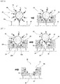
- a base material for producing a sensor for analysis of a detection target of the present invention is schematically shown in Fig. 1 .
- a base material for producing a sensor for analysis 10 includes a base material 20 and a polymer film 30.
- the polymer film 30 is provided on the surface of the base material 20 and includes a concave 31.
- the concave 31 is a hole formed in a size capable of receiving a detection target (a detection target 60 below).
- the base material for producing a sensor for analysis 10 includes a group for binding antibody substance 22 and a group for binding signal substance 32 inside the concave 31.
- the group for binding signal substance 32 is provided in the vicinity of the group for binding antibody substance 22.
- the material of the base material 20 can be a material selected from the group consisting of, for example, metal, glass, and resin.
- the metal include gold, silver, copper, aluminum, tungsten, and molybdenum.
- the resin include poly (meth)acrylate, polystyrene, ABS (acrylonitrile-butadiene-styrene copolymer), polycarbonate, polyester, polyethylene, polypropylene, nylon, polyurethane, silicone resin, fluororesin, methylpentene resin, phenol resin, melamine resin, epoxy resin, and vinyl chloride resin.
- the base material 20 can be formed by combining multiple materials selected from the materials above.
- the base material 20 can be a base material provided with a metal film on the surface of glass or resin.
- the shape of the base material 20 can be a plate shape or a particle shape. Preferred examples include a gold base plate, a glass base plate, a gold nanoparticle, and a glass bead.
- the polymer film 30 is layered on the base material 20 and includes multiple concaves 31.
- the concave 31 is a sensing place in the sensor for analysis of a detection target of the present invention.
- the concave 31 is not limited as long as it is formed so that a detection target can be received.
- the concave 31 can be a molecularly imprinted polymer (MIP) formed using molecularly imprinting polymerization as described below.
- MIP molecularly imprinted polymer
- the concave 31 is formed by a template (a template 40 below) used in molecular imprinting polymerization, and has a shape corresponding to a part of the surface shape of the template.
- the concave 31 only needs to be formed in a size capable of receiving a detection target, and thus the template of the concave 31 can be the same as the detection target or an object larger in size than the detection target.
- the concave 31 is formed in a size capable of receiving a detection target means that the size of the concave 31 on the surface of the base material 20 is sufficient to allow at least a part of a detection target to enter inside the concave 31 and approach to an antibody substance to bind it when the antibody substance (an antibody substance 52 below) and a signal substance (a signal substance 53 below) are bound to form a sensor for analysis (a sensor for analysis 50 below).
- the opening diameter of the concave 31 can vary depending on the detection target, thus it is not particularly limited, and it can be, for example, 1 nm to 10 ⁇ m.
- the thickness of the polymer film 30 can also vary depending on the detection target, thus it is not particularly limited, and it can be, for example, 1 nm to 1 ⁇ m.
- the polymer that constitutes the polymer film 30 can be, for example, a biocompatible polymer containing a component derived from a biocompatible monomer.
- Biocompatibility refers to a property of not inducing the adhesion of biological substances.
- the biocompatible monomer is preferably a hydrophilic monomer, more preferably a zwitterionic monomer.
- a zwitterionic monomer contains both an anionic group derived from an acidic functional group (for example, a phosphoric acid group, a sulfuric acid group, and a carboxyl group) and a cationic group derived from a basic functional group (for example, a primary amino group, a secondary amino group, a tertiary amino group, and a quaternary ammonium group) in one molecule.
- an acidic functional group for example, a phosphoric acid group, a sulfuric acid group, and a carboxyl group
- a cationic group derived from a basic functional group for example, a primary amino group, a secondary amino group, a tertiary amino group, and a quaternary ammonium group
- examples of the phosphobetaine include a molecule having a phosphorylcholine group in the side chain, and preferably include 2-methacryloyloxyethyl phosphorylcholine (MPC).
- MPC 2-methacryloyloxyethyl phosphorylcholine
- sulfobetaine examples include N,N-dimethyl-N-(3-sulfopropyl)-3'-methacryloylaminopropaneaminium inner salt (SPB) and N,N-dimethyl-N-(4-sulfobutyl)-3'-methacryloylaminopropaneaminium inner salt (SBB).
- carboxybetaine examples include N,N-dimethyl-N-(1-carboxymethyl)-2'-methacryloyloxyethaneaminium inner salt (CMB) and N,N-dimethyl-N-(2-carboxyethyl)-2'-methacryloyloxyethaneaminium inner salt (CEB).
- the percentage of the component derived from a biocompatible monomer in the polymer film 30 can be, for example, 5 mol% or more and 100 mol% or less.
- the content of the component derived from a biocompatible monomer of the above-mentioned lower limit or more is preferred in terms of inhibiting non-specific adsorption on the surface of the polymer film 30.
- the percentage of the component derived from a biocompatible monomer in the polymer film 30 can be preferably 10 mol% or more and 80 mol% or less, further preferably 20 mol% or more and 60 mol% or less.
- the group for binding antibody substance 22 is a group that allows an antibody substance to be introduced into the base material for producing a sensor for analysis 10 by directly or indirectly binding the antibody substance (an antibody substance 52 below).
- the user can freely target a detection target, freely select the antibody substance specific to the detection target to be targeted, and introduce it to the group for binding antibody substance 22.
- customization can be made so that the analysis of the detection target using multiple types of antibody substances will be achieved in one base material 20.
- both an exosome and a protein can be analyzed in one base material 20 by introducing an antibody substance that binds to an exosome into one concave 31 and introducing an antibody substance that binds to a protein into another concave 31.
- At least one group for binding antibody substance 22 needs to be provided in one concave 31, and multiple groups for binding antibody substance 22 can be provided in one concave 31.
- the group for binding antibody substance 22 is not provided on the part other than the concave 31 on the surface of the base material 20.
- the group for binding antibody substance 22 can be an irreversible binding group, a reversible binding group, a covalent binding group, or a non-covalent binding group.
- the group for binding antibody substance 22 is preferably a reversible binding group, more preferably a non-covalent binding group. Examples of such a group include a chelate-forming binding group.
- the chelate-forming binding group is capable of indirectly binding an antibody substance via a substance that binds to the Fc region of the antibody substance such as Protein G.
- the chelate-forming binding group is not particularly limited as long as it is a group that has a ligand having multiple coordination positions (a multidentate ligand) and binds (coordinates) to a metal ion.
- Examples thereof include an amino polycarboxylic acid chelating agent-derived group, a hydroxycarboxylic acid chelating agent-derived group, a deferoxamine-derived group, a deferacyclos-derived group, a deferiprone-derived group, and a histidine tag, and preferably include an amino polycarboxylic acid chelating agent-derived group.
- amino polycarboxylic acid examples include ethylenediaminetetraacetic acid (EDTA), ethylenediaminediacetic acid, hydroxyethylethylenediaminetriacetic acid (HEDTA), dihydroxyethylethylenediaminetetraacetic acid (DHEDDA), nitrilotriacetic acid (NTA), hydroxyethyliminodiacetic acid (HIDA), N-(2-hydroxyethyl) iminodiacetic acid, ⁇ -alanine diacetic acid, cyclohexanediaminetetraacetic acid, iminodiacetic acid, N-(2-hydroxyethyl) iminodiacetic acid, diethylenetriaminepentaacetic acid (DTPA), N-(2-hydroxyethyl)ethylenediaminetriacetic acid, glycol ether diamine tetraacetic acid, glutamic acid diacetic acid, aspartic acid diacetic acid, methylglycine diacetic acid, iminodisuccin
- the reversible binding group is a group capable of constituting a reversible linked group by binding to other reversible binding group (covalently or non-covalently), and reversible means that both conversion of a reversible binding group into a reversible linked group (bond) and conversion of a reversible linked group into a reversible binding group (cleavage) are bidirectionally possible (the same applies hereinafter).
- the group for binding signal substance 32 is a group that allows a signal substance to be introduced into the base material for producing a sensor for analysis 10 by binding the signal substance (a signal substance 53 below). The user can freely select the signal substance and introduce it into the group for binding signal substance 32.
- a signal substance 53 below The user can freely select the signal substance and introduce it into the group for binding signal substance 32.
- customization can be made so that different signal substances are provided depending on the type of antibody substances.
- multiple groups for binding signal substance 32 are usually provided for one concave 31.
- the group for binding signal substance 32 only needs to be provided in sufficient amount to detect the signal intensity change when sensing is performed in the concave 31 of the analysis sensor of the present invention (when the detection target is received in the concave 31).
- the amount of the group for binding signal substance 32 to be provided in one concave 31 is not particularly limited, and it can be, for example, about 100 to about 1000 per concave 31 depending on the size of the concave 31 and the size of the target substance to be detected.
- the group for binding signal substance 32 is not provided on the part other than the concave 31 on the surface of the base material 20.
- the group for binding signal substance 32 can be an irreversible binding group, a reversible binding group, a covalent binding group, or a non-covalent binding group.
- the group for binding signal substance 32 is preferably a reversible binding group, more preferably a covalent binding group.
- Examples of such a group include a thiol group (the corresponding reversible linked group is a disulfide group), an aminooxy group or a carbonyl group (the corresponding reversible linked group is an oxime group), a boronic acid group and a diol group (the corresponding reversible linked group is a cyclic diester group), an amino group and a carbonyl group (the corresponding reversible linked group is a Schiff base), and an aldehyde group or ketone group and alcohol (the corresponding reversible linked group is an acetal group).
- a thiol group the corresponding reversible linked group is a disulfide group
- an aminooxy group or a carbonyl group the corresponding reversible linked group is an oxime group
- a boronic acid group and a diol group the corresponding reversible linked group is a cyclic diester group
- an amino group and a carbonyl group the
- the base material for producing a sensor for analysis of a detection target of the present invention only needs to be configured so that at least one of the antibody substance and the signal substance can be customized by the user.
- an antibody substance specific to the detection target has already been bound to the group for binding antibody substance. In this case, the user can freely select and introduce the signal substance.
- the signal substance has already been bound to the group for binding signal substance.
- the user can freely target a detection target, and freely select and introduce the antibody substance specific to the detection target to be targeted.
- the sensor for analysis of a detection target of the present invention includes: the base material for producing a sensor for analysis of a detection target; an antibody substance specific to the detection target that is bound to the group for binding antibody substance; and a signal substance that is bound to the group for binding signal substance.
- a sensor for analysis of a detection target of the present invention is schematically shown in Fig. 2 .
- an antibody substance 52 is bound to the group for binding antibody substance 22, and a signal substance 53 is bound to the group for binding signal substance 32 in the base material for producing a sensor for analysis 10 above.
- the detection target (the detection target 60 below) of the sensor for analysis of the present invention is not particularly limited in principle as long as it has specific bindability to the antibody substance 52.
- Specific examples thereof include a low molecular weight substance, a protein, and a microparticle having a membrane structure.
- the low molecular weight substance include any substances such as a hormone, an agent, a herbicide, an agricultural chemical, sugar, cholesterol, a lipid, uric acid, an environmental hormone, and a peptide.
- the protein include any proteins such as HSA, IgG, fibrinogen, transferrin, AST, ALT, LDH, ALP, LAP, ⁇ -GTP, CRP, AFP, and PSA.
- Examples of the microparticle having a membrane structure include an extracellular microparticle, an intracellular vesicle, an organelles, and a cell.
- Examples of the membrane structure include a lipid bilayer membrane structure.
- Examples of the extracellular microparticle include an exosome, a microvesicle, and an apoptotic body.
- Examples of the intracellular vesicle include a lysosome and an endosome.
- Examples of the organelle include a mitochondrion.
- Examples of the cell include a cancer cell such as a circulating tumor cell (CTC) and other disease-related cells.
- CTC circulating tumor cell
- the detection target 60 is bound to the other of the fluorescent dye pair in advance.
- the antibody substance 52 only needs to have a specific bindability to the detection target.
- the antibody substance 52 is intended to include an antibody and an antibody-like substance.
- the antibody refers to a protein having a complete basic structure of an immunoglobulin, and the antibody-like substance refers to a fragment of an immunoglobulin (an antibody fragment).
- Examples of the antibody include an immunoglobulin (Ig) and a chimeric antibody, and more specifically include IgG, IgA, IgM, IgE, and IgD.
- Examples of the chimeric antibody include a humanized antibody.
- the antibody can be derived from mammals such as a mouse, a rabbit, a cow, a pig, a horse, a sheep, and a goat, birds such as a chicken, and animal species such as a human, and is not particularly limited.
- the antibody can be prepared, for example, from a serum derived from the above-mentioned animal species by a conventionally known method, or a commercially available antibody can be used.
- the antibody can be, for example, a polyclonal antibody or a monoclonal antibody, and is preferably a monoclonal antibody. Examples of the antibody-like substance include Fab, Fab' , F(ab') 2 , and ScFv.
- the antibody substance 52 can be appropriately selected by those skilled in the art according to the detection target.
- the detection target has a specific antigen on its surface
- one having specific bindability to the specific antigen can be used as the antibody substance 52.
- the detection target is an exosome
- the exosome has, for example, CD63, CD9, CD81, CD37, CD53, CD82, CD13, CD11, CD86, ICAM-1, Rab5, Annexin V, LAMP1 or the like as a membrane protein (an exosome-specific antigen)
- an antibody to the exosome-specific antigen can be used as the antibody substance 52.
- the detection target is a cancer cell
- the cancer cell has, for example, Caveolin-1, EpCAM, FasL, TRAIL, Galectine3, CD151, Tetraspanin 8, EGFR, HER2, RPN2, CD44, TGF- ⁇ or the like as a cancer cell-specific antigen
- an antibody to the cancer cell-specific antigen can be used as the antibody substance 52.
- an antibody to the disease cell-specific antigen can be used as the antibody substance 52.
- the modifying group in this case refers to a modifying group provided for a purpose other than affinity, such as the signal substance. Meanwhile, the modifying group does not include a group that contributes to the binding to the group for binding antibody substance 22 (for example, a histidine tag).
- the signal substance 53 functions as a reader of the information for binding between an antibody substance specific to the detection target 52 and the group for binding antibody substance 22.
- the signal substance 53 is not particularly limited as long as the detected signal intensity is changed or spectrum is changed (for example, the peak is shifted) by the binding of the detection target to the concave 31.
- Examples thereof include a fluorescent substance, a radioactive element-containing substance, and a magnetic substance. From the viewpoint of ease of detection and the like, the signal substance is preferably a fluorescent substance.
- the fluorescent substance examples include a fluorescein dye, a cyanine dye such as an indocyanine dye, a fluorescent dye such as rhodamine dyes, a fluorescent protein such as GFP, and a nanoparticle such as colloidal gold and a quantum dot.
- the radioactive element-containing substance examples include sugar, an amino acid, and a nucleic acid labeled with a radioactive isotope such as 18 F.
- the magnetic substance examples include those having a magnetic body such as ferrichrome, and those found in a ferrite nanoparticle and a nanomagnetic particle.
- the signal substance 53 can be configured as one of fluorescent dye pair that causes fluorescence resonance energy transfer (FRET).
- FRET fluorescence resonance energy transfer
- the fluorescent dye pair that causes FRET is not particularly limited, and a donor dye or an acceptor dye can be selected as the signal substance 53 without limitation.
- a donor dye can be selected as the signal substance 53.
- donor/acceptor dyes that constitute the fluorescent dye pair that causes FRET include fluorescein isothiocyanate (FITC)/tetramethylrhodamine isothiocyanate (TRITC), Alexa Fluor647 / Cy5.5, HiLyte Fluor647/Cy5.5, and R-phycoerythrin (R-PE)/allophycocyanin (APC).
- the manufacturing method of a base material for producing a sensor for analysis of a detection target of the present invention includes the following steps.
- An exosome can be used as a template.
- the manufacturing method of the base material for producing a sensor for analysis of a detection target of the present invention when an exosome is used as a template is schematically shown in Figs. 3 to 7 .
- the manufacturing method of the base material for producing a sensor for analysis of a detection target in this case includes the following steps.
- a molecular film-forming step of forming on a base material 20 a molecular film 21 having a binding functional group 22a and a polymerization initiating group 23a on a surface of the molecular film ( Fig. 3 ); a template introduction step of introducing a template 40 that specifically binds to an antibody substance 24 via a first reversible linked group 22b and the antibody substance 24 to the binding functional group 22a ( Fig. 4 ); a surface modification step of modifying a surface of the template 40 with a polymerizable functional group 33 via a second reversible linked group 32b ( Fig.
- a molecular film 21 having a binding functional group 22a and a polymerization initiating group 23a on the surface is formed on a base material 20.
- the binding functional group 22a is a group different from the polymerization initiating group 23a, and a group according to the reagent used for extending the molecular chain to the template 40 in the template introduction step below is appropriately determined by those skilled in the art. In the illustrated aspect, the case where the binding functional group 22a is an amino group is illustrated.
- the polymerization initiating group 23a is not particularly limited as long as it has a structure capable of functioning as a polymerization initiator, and it can be appropriately determined by those skilled in the art according to the polymerization reaction used in the polymerization step below.
- Examples of the polymerization initiating group 23a include a group having a structure that generates a radical during polymerization reaction, specifically, a carbon-halogen binding group derived from an organic halogen (-CX group; X represents a halogen atom).
- -CX group organic halogen
- the molecular film 21 can be formed by a conventionally known method as a monolayer formed by mixed self-assembly using a molecule having a binding functional group 22a at the end, and a molecule having a polymerization initiating group 23a at the end.
- the molecular film 21 can be formed as a mixed self-assembled monolayer (mixed SAMs).
- a template 40 that specifically binds to an antibody substance 24 is introduced via a first reversible linked group 22b and an antibody substance 24 to the binding functional group 22a.
- the template introduction step can include the following steps.
- the first reversible linked group 22b is not particularly limited as long as it is a group capable of bidirectional conversion from the first reversible linked group 22b into a reversible binding group (that is, the above-mentioned group for binding antibody substance 22) by cleavage and from a reversible binding group (that is, the above-mentioned group for binding antibody substance 22) into the first reversible linked group 22b by binding.
- the first reversible linked group 22b can be a chelate-bound group in which chelate-forming binding groups are bound each other via metal coordination.
- the first reversible linked group 22b a structure is illustrated in which an NTA group (a type of the group for binding antibody substance 22), which is an amino polycarboxylic acid chelating agent-derived group, and a histidine tag, which is a group 25 capable of forming a first reversible linked group 22b by binding to the group for binding antibody substance 22 (because the histidine tag in the figure is schematically shown, the exact molecular structure is not shown, and the imidazolyl group, a part of the histidine tag, is highlighted), are chelate bound via a nickel ion.
- NTA group a type of the group for binding antibody substance 22
- a histidine tag which is a group 25 capable of forming a first reversible linked group 22b by binding to the group for binding antibody substance 22
- the antibody substance-binding group 22c is not particularly limited as long as it is a group capable of binding to the antibody substance 24, it is preferably a group having specific bindability to the Fc region of the antibody substance 24 from the viewpoint of oriented binding of the antibody substance 24 (that is, binding in a direction in which the binding part of the antibody substance 24 to the template 40 faces outward).
- Protein G is illustrated as such a group.
- the antibody substance 24 can be determined according to the template 40. Specifically, an antibody substance having specific bindability to the template 40 is selected as the antibody substance 24.
- the template 40 is determined in consideration of the detection target. Specifically, an object that is the same as the detection target (the detection target 60 below) or an object larger in size than the detection target is selected as a template.
- the object larger in size than the detection target is not particularly limited as long as it has a shape and size that allow the concave 31 formed in the synthesis of the polymer film 30 in the polymerization step below to be a size that is large enough to receive the detection target on the surface of the base material 20, and examples thereof include a biomolecule such as a protein and a microparticle having a membrane structure.
- a template having on the surface an antigen to which the antibody substance 24 specifically binds is preferably used.
- an antibody to a specific antigen on the exosome surface a surface protein
- an exosome can be used as the template 40.
- an aspect is illustrated in which a molecule having an NTA group, which is an amino polycarboxylic acid chelating agent-derived group, is bound to the amino group as the binding functional group 22a, Protein G as the antibody substance-binding group 22c is introduced via the first reversible linked group 22b (a chelate group) by reacting recombinant Protein G with a histidine tag in the presence of nickel ions, then further, as an antibody substance 24, an antibody specific to the exosome surface protein is bound, and finally, an exosome as a template 40 is bound.
- NTA group which is an amino polycarboxylic acid chelating agent-derived group
- the surface of the template 40 is modified with a polymerizable functional group 33 via a second reversible linked group 32b.
- the surface modification step can include the following steps.
- the anchoring substance 41 is a substance including the anchoring group 43 and the reversible binding group 42.
- the anchoring group 43 is a group capable of being anchored to the template 40, and in particular, when the template 40 is a membrane body having a lipid bilayer membrane, the anchoring group 43 is preferably a hydrophobic chain group capable of being anchored to the lipid bilayer membrane.
- the hydrophobic chain can be of a single chain or multiple chains, can be a saturated hydrocarbon or an unsaturated hydrocarbon, and can be a hydrophobic chain with a substituent or a hydrophobic chain without a substituent.
- the hydrophobic chain is preferably a linear or branched alkyl group or alkenyl group having 6 to 24 carbon atoms, and more specific examples thereof include a hexyl group, a heptyl group, an octyl group, a nonyl group, a decyl group, an undecyl group, a dodecyl group, a tridecyl group, a tetradecyl group, a pentadecyl group, a hexadecyl group, a heptadecyl group, a stearyl group (an octadecyl group), a nonadecyl group, an icosyl group, a heicosyl group, a docosyl group, a tricosyl group, a tetracosyl group, a myristol group, a palmitoleyl group, an oleyl group, a lino
- the anchoring substance 41 can include a hydrophilic chain group in addition to the anchoring group 43 and the reversible binding group 42.
- the hydrophilic chain group can be a linking group located between the anchoring group 43 and the reversible binding group 42. Specific examples thereof include a protein, an oligopeptide, a polypeptide, polyacrylamide, polyethylene glycol (PEG), and a dextran, and more preferably include PEG.
- the reversible binding group 42 can be a group that is converted into a second reversible linked group 32b by binding to other reversible binding group (specifically the above-mentioned group for binding signal substance 32).
- examples thereof include a thiol group (the corresponding second reversible linked group 32b is a disulfide group), an aminooxy group or a carbonyl group (the corresponding second reversible linked group 32b is an oxime group), a boronic acid group and a diol group (the corresponding second reversible linked group 32b is a cyclic diester group), an amino group and a carbonyl group (the corresponding second reversible linked group 32b is a Schiff base), and an aldehyde group or ketone group and alcohol (the corresponding second reversible linked group 32b is an acetal group).
- n represents, for example, an integer of 0 to 182.
- the polymerizable functional group 33 only needs to have a polymerizable unsaturated bond, and representative examples thereof include a (meth)acryl group.
- an aspect is illustrated in which by anchoring of the anchoring substance 41 represented by the formula above to the lipid bilayer membrane of the exosome as the template 40, and subsequent disulfide exchange from a molecule including a (meth)acrylic group as a polymerizable functional group 33 and a disulfide bond 34 to the thiol group, a second reversible binding group 42 of the anchoring substance 41, the thiol group is converted into a disulfide group, the second reversible linked group 32b, and the surface of the template 40 is modified with the polymerizable functional group 33.
- the second reversible linked group 32b that is capable of generating the group for binding signal substance 32 by surface modification of the template 40, the second reversible linked group 32b can be delivered only to the surface of the template 40.
- the polymerizable monomer 35 is added, and a molecularly imprinted polymer corresponding to a part of the surface of the template 40 is synthesized using the polymerizable functional group 33 and the polymerizable monomer 35 as a substrate and the polymerization initiating group 23a as a polymerization initiator.
- the polymer film 30 including the concaves 31 is formed on the surface of the base material 20.
- a polymer synthesized by imprinting polymerization with a template is described as a molecular imprint polymer, and the molecularly imprinted polymer includes a polymer synthesized by imprinting polymerization with a template other than a molecule (for example, a cell).
- the biocompatible monomer is preferably a hydrophilic monomer, more preferably a zwitterionic monomer.
- a zwitterionic monomer contains both an anionic group derived from an acidic functional group (for example, a phosphoric acid group, a sulfuric acid group, and a carboxyl group) and a cationic group derived from a basic functional group (for example, a primary amino group, a secondary amino group, a tertiary amino group, and a quaternary ammonium group) in one molecule.
- an acidic functional group for example, a phosphoric acid group, a sulfuric acid group, and a carboxyl group
- a cationic group derived from a basic functional group for example, a primary amino group, a secondary amino group, a tertiary amino group, and a quaternary ammonium group
- examples of the phosphobetaine include a molecule having a phosphorylcholine group in the side chain, and preferably include 2-methacryloyloxyethyl phosphorylcholine (MPC).
- MPC 2-methacryloyloxyethyl phosphorylcholine
- the sulfobetaine include N,N-dimethyl-N-(3-sulfopropyl)-3'-methacryloylaminopropaneaminium inner salt (SPB) and N,N-dimethyl-N-(4-sulfobutyl)-3'-methacryloylaminopropaneaminium inner salt (SBB).
- carboxybetaine examples include N,N-dimethyl-N-(1-carboxymethyl)-2'-methacryloyloxyethanaminium inner salt (CMB) and N,N-dimethyl-N-(2-carboxyethyl)-2'-methacryloyloxyethaneaminium inner salt (CEB).
- the polymerization reaction system preferably further includes a transition metal complex formed by a transition metal or a transition metal compound and a ligand as a polymerization catalyst, and more preferably a reducing agent is used.
- transition metal or transition metal compound examples include metal copper or a copper compound, and examples of the copper compound include a chloride, a bromide, an iodide, a cyanide, an oxide, a hydroxide, an acetate, a sulfate, and a nitrate, and preferably include a bromide.
- the ligand is preferably a polydentate amine, and specific examples thereof include bidentate to hexadentate ligands.
- examples thereof preferably include a bidentate ligand, more preferably include 2,2-bipyridyl, 4,4'-di-(5-nonyl)-2,2'-bipyridyl, N-(n-propyl)pyridylmethanimine, and N-(n-octyl)pyridylmethanimine, and more preferably include 2,2-bipyridyl.
- the reducing agent include an alcohol, an aldehyde, a phenol, and an organic acid compound, and preferably include an organic acid compound.
- organic compound examples include citric acid, oxalic acid, ascorbic acid, a salt of ascorbic acid, and an ascorbic acid ester, preferably include ascorbic acid, a salt of ascorbic acid, and an ascorbic acid ester, and more preferably include ascorbic acid.
- a polymer chain extends from the polymerization initiating group 23a, a radical generation source, using the polymerizable monomer 35 as a substrate, the thickness of the polymer film increases, and the extending polymer chain also incorporates the polymerizable functional group 33 that is modifying the surface of the template 40 as a substrate when the extending polymer chain reaches the surface of the template 40, thereby a polymer is synthesized so that the concave 31 having a shape that conforms to the surface shape of the template 40 is formed.
- the polymer film can be grown to a thickness corresponding to about 1/2, preferably about 1/3 of the length from the top to the bottom of the template 40 (when the top of the drawing is the top) introduced into the base material 20.
- the reaction solvent in the polymerization reaction system an aqueous solvent such as a buffer is preferably used from the viewpoint of, for example, inhibiting denaturation of the template 40.
- the first reversible linked group 22b and the second reversible linked group 32b are cleaved and converted into the group for binding antibody substance 22 and the group for binding signal substance 32, respectively, and the antibody substance 24 and the template 40 are removed.
- the second reversible linked group 32b is delivered only to the surface of the template 40 in the surface modification step above, in the concave 31 of the polymer film 30, the trace of the removed template 40, the group for binding antibody substance 22 remains inside and the group for binding signal substance 32 produced from the second reversible linked group 32b is placed only inside the concave 31.
- the base material for producing a sensor for analysis 10 is obtained.
- an artificial particle can be used as a template.
- the base material for producing a sensor for analysis of a detection target can be manufactured according when an exosome is used as a template above.
- Using an artificial particle is suitable for industrial production because it does not need an antibody to immobilize the template onto a base plate, and the base material can be manufactured at low cost. Because the artificial particle used for the template itself is an industrial product and the particle size is controlled, size control and homogenization of the concave formed in the base material for producing a sensor for analysis are easy, and thus a sensor for analysis excellent in analytical property can be produced from the obtained base material for producing a sensor for analysis.
- the manufacturing method of a base material for producing a sensor for analysis of a detection target when an artificial particle is used as a template can include a molecular film-forming step of forming on a base material a molecular film having a binding functional group and a polymerization initiating group on a surface of the molecular film; a template introduction step of introducing an artificial particle as a template via a first reversible linked group to the binding functional group; a surface modification step of modifying a surface of the template with a polymerizable functional group via a second reversible linked group; a polymerization step of forming a polymer film on a surface of the base material by adding a polymerizable monomer and synthesizing a molecularly imprinted polymer corresponding to a part of the surface of the template using the polymerizable monomer as a substrate and the polymerization initiating group as a polymerization initiator; and a removal step of cleaving the first reversible linked group and the
- Fig. 8 shows the template introduction step
- Fig. 9 shows the surface modification step
- Fig. 10 shows the polymerization step
- Fig. 11 shows the removal step.
- the molecular film-forming step is the same as that when an exosome is used as a template, and specifically, it is as described in "3-1-1. Molecular film-forming step" and Fig. 3 .
- an artificial particle is introduced as the template 40 via a first reversible linked group 22b to the binding functional group 22a.
- the binding functional group 22 and the first reversible linked group 22b are as described in "3-1-1. Molecular film-forming step” and "3-1-2. Template introduction step" above, respectively.
- the binding functional group 22a is an amino group
- the first reversible linked group 22b is a group formed by binding of a nitrilotriacetic acid (NTA) group (a type of group for binding antibody substance 22) to a histidine tag (a type of the group 25 capable of forming a first reversible linked group 22b by binding to the group for binding antibody substance 22).
- NTA nitrilotriacetic acid
- the artificial particle used as the template 40 is not particularly limited as long as it can be used as a template, and examples thereof include an artificially manufactured inorganic particle and organic particle.
- the inorganic particle include a metal, a metal oxide, a nitride, a fluoride, a sulfide, a boride, a complex compound thereof, and hydroxyapatite, and preferably include silicon dioxide (silica).
- the organic particle include a latex cured product, dextran, chitosan, polylactic acid, poly (meth)acrylic acid, polystyrene, and polyethyleneimine. Because the size of the concave 31 (see Fig.
- the size of the artificial particle depends on the size of the template 40, the size of the artificial particle can be appropriately determined according to the size of the detection target.
- an artificial particle having a size same as or larger than the detection target can be used.
- the size of the artificial particle can be selected from 1 nm to 10 ⁇ m.
- the template 40 as an artificial particle includes on its surface a group 25 capable of forming the above-mentioned first reversible linked group 22b by binding to the above-mentioned group for binding antibody substance 22 and a reversible bond group 42 capable of forming the above-mentioned second reversible linked group 32b by binding to the above-mentioned group for binding signal substance 32.
- the group for binding antibody substance 22, the first reversible linked group 22b, the group 25 capable of forming a first reversible linked group 22b by binding to the group for binding antibody substance 22, the second reversible linked group 32b, and the reversible linked group 42 are as described above, and in the illustrated aspect, the group for binding antibody substance 22 is a nitrilotriacetic acid (NTA) group, the group 25 capable of forming a first reversible linked group 22b by binding to the group for binding antibody substance 22 is a histidine tag, and the reversible linked group 42 is a thiol group.
- NTA nitrilotriacetic acid
- the reversible linked group 42 shown as a thiol group binds to the group for binding signal substance 32 to form a second reversible linked group 32b.
- the method of modifying the surface of the artificial particle with a specific group in this way is widely known, and thus those skilled in the art can appropriately introduce the group 25 and the reversible bond group 42 to be introduced based on a known surface modification method, considering the type of those groups and components of the artificial particle.
- the template 40 of the artificial particle is introduced into the amino group, the binding functional group 22a on the base material 20, via the first reversible linked group 22b.
- the surface of the template 40 is modified with a polymerizable functional group 33 via a second reversible linked group 32b. More specifically, the reversible binding group 42 on the surface of the template 40 is converted into the second reversible linked group 32b and the polymerizable functional group 33 is introduced.
- the polymerizable functional group 33 is as described in "3-1-3. Surface modification step" above.
- an aspect is illustrated in which by disulfide exchange from a molecule including a (meth)acrylic group as a polymerizable functional group 33 and a disulfide bond 34 to the thiol group, a second reversible binding group 42 on the surface of the template 40, the thiol group is converted into a disulfide group, the second reversible linked group 32b, and the surface of the template 40 is modified with the polymerizable functional group 33.
- an artificial particle is used as the template 40, by using a template 40 having a thiol group, the second reversible binding group 42, on the surface in advance, the second reversible linked group 32b can be formed only on the surface of the template 40.
- the polymerizable monomer 35 is added, and a molecularly imprinted polymer corresponding to a part of the template 40 is synthesized using the polymerizable functional group 33 and the polymerizable monomer 35 as a substrate and the polymerization initiating group 23a as a polymerization initiator.
- the polymer film 30 including the concaves 31 is formed on the surface of the base material 20.
- the polymerizable monomer 35 and the polymer film 30 are as described in "3-1-4. Polymerization step" above.
- the first reversible linked group 22b and the second reversible linked group 32b are cleaved and converted into the group for binding antibody substance 22 and the group for binding signal substance 32, respectively, and the template 40 is removed. Because the second reversible linked group 32b is formed only on the surface of the template 40 in the surface modification step above, in the concave 31 of the polymer film 30, the trace of the removed template 40, the group for binding antibody substance 22 remains inside and the group for binding signal substance 32 produced from the second reversible linked group 32b is placed only inside the concave 31. Thus, the base material for producing a sensor for analysis 10 is obtained.
- the sensor for analysis of a detection target 50 is produced by only binding an antibody substance specific to the detection target 52 to the group for binding antibody substance 22, and binding a signal substance 53 to a group for binding signal substance 32 in the base material for producing a sensor for analysis 10.
- the antibody-like substance 52 is not particularly limited as long as it is specific to the detection target, and the antibody-like substance 52 can be the same antibody-like substance as the antibody-like substance 24, or a different antibody-like substance from the antibody-like substance 24 used in manufacturing of the base material for producing a sensor for analysis 10.
- the base material for producing a sensor for analysis 10 has a group for binding signal substance 32 only in the concave 31, which is a sensing place in the surface of the base plate 20, the signal substance 53 can be placed only in the concave 31.
- one type of the antibody-like substance 52 and one type of the signal substance 53 can be introduced into all concaves 31, or one type of the antibody-like substance can be introduced into one concave 31 and another type of the antibody-like substance can be introduced into another concave 31, and different types of signal substances 53 can be introduced according to the types of antibody-like substances.
- Fig. 12 shows a schematic diagram describing one example of a method for analyzing a detection target of the present invention.
- the analysis sample liquid containing the detection target 60 is contacted with the surface of the base material 20 of the sensor for analysis 50.
- the detection target 60 is not particularly limited in principle as long as it is a substance that specifically binds to the antibody substance 52, and examples thereof include a low molecular weight substance, a protein, and a microparticle having a membrane structure as described above.
- the low molecular weight substance include any substances such as a hormone, an agent, a herbicide, an agricultural chemical, sugar, cholesterol, a lipid, uric acid, an environmental hormone, and a peptide.
- the protein include any proteins such as HSA, IgG, fibrinogen, transferrin, AST, ALT, LDH, ALP, LAP, ⁇ -GTP, CRP, AFP, and PSA.
- Examples of the microparticle having a membrane structure include an extracellular microparticle, an intracellular vesicle, an organelle, and a cell.
- Examples of the membrane structure include a lipid bilayer membrane structure.
- Examples of the extracellular microparticle include an exosome, a microvesicle, and an apoptotic body.
- Examples of the intracellular vesicle include a lysosome and an endosome.
- Examples of the organelle include a mitochondrion.
- Examples of the cell include a cancer cell such as a circulating tumor cell (CTC) and other disease-related cells.
- CTC circulating tumor cell
- the aspect of the analysis sample liquid containing the detection target 60 is not particularly limited, the analysis sample liquid is preferably one that has not undergone the treatment that separates the detection target 60 from the viewpoint of swiftness.
- the treatment that separates the detection target 60 include ultracentrifugation, ultrafiltration, continuous flow electrophoresis, filtration with a size filter, and gel filtration chromatography.
- the analysis sample liquid containing the detection target 60 can be a sample obtained in an environment in which the detection target 60 is present (when the detection target 60 is a cell or an extracellular microparticle), or a sample obtained in an environment in which the detection target 60 can be produced (when the detection target 60 is an extracellular particle and is a product from a cell).
- the analysis sample liquid can be a biological sample containing a cell.
- the detection target 60 is an extracellular microparticle such as an exosome
- examples of the cell that produces the detection target 60 include a cancer cell, a mast cell, a dendritic cell, a reticulocyte, an epithelial cell, a B cells, and a nerve cell.
- examples of the analysis sample liquid containing the detection target 60 include body fluids such as blood, milk, urine, saliva, lymph, cerebrospinal fluid, amniotic fluid, tear fluid, sweat, and rhinorrhea, and further include a treated liquid obtained by subjecting these body fluids to a pretreatment such as removal of unnecessary components and a culture solution obtained by culturing cells contained in these body fluids.
- body fluids such as urine, saliva, tear fluid, sweat, and rhinorrhea are particularly preferred in terms of non-invasiveness and easy collection.
- the detection target 60 is specifically captured by the antibody substance 52 in the concave 31.
- the detection target 60 is an exosome
- the exosome is captured by specific binding to the antibody substance 52 via a membrane protein (an exosome-specific antigen), for example, CD63, CD9, CD81, CD37, CD53, CD82, CD13, CD11, CD86, ICAM-1, Rab5, Annexin V, and LAMP1.
- the cancer cell is captured by specific binding to the antibody substance 52 via a cancer cell-specific antigen, for example, Caveolin-1, EpCAM, FasL, TRAIL, Galectine 3, CD151, Tetraspanin 8, EGFR, HER2, RPN2, CD44, and TGF- ⁇ .
- a cancer cell-specific antigen for example, Caveolin-1, EpCAM, FasL, TRAIL, Galectine 3, CD151, Tetraspanin 8, EGFR, HER2, RPN2, CD44, and TGF- ⁇ .
- the detection target 60 is specifically captured by the antibody substance 52 in the concave 31, the signal substance 53 is shielded by the detection target 60, and thus the signal intensity detected from the signal substance 53 is reduced.
- the detection target 60 is detected by the signal intensity change. Because the capture of the detection target 60 and the signal intensity change occur almost simultaneously, there is no need to add a reagent for the detection of the detection target 60, and the detection can be performed quickly.
- the sensor for analysis 50 is configured so that the signal substance 53 will be one of the fluorescent dye pair that cause fluorescence resonance energy transfer (FRET), and the other of the fluorescent dye pair will be bound to the detection target 60 in advance, in the moment when the detection target 60 is specifically captured by the antibody substance 52 in the concave 31, the fluorescent dye in the signal substance 53 and the fluorescent dye in the detection target 60 are close to each other, and thus fluorescence is emitted by FRET.
- the detection target 60 is detected by the fluorescence emission by the FRET. Because the capture of the detection target 60 and the fluorescence emission by the FRET occur almost simultaneously, there is no need to add a reagent for the detection of the detection target 60, and the detection can be performed quickly.
- FRET fluorescence resonance energy transfer
- the fluorescent dye pair that causes FRET is not particularly limited, and a donor dye or an acceptor dye can be selected as the signal substance 53 without limitation.
- a donor dye can be selected as the signal substance 53.
- donor/acceptor dyes that constitute the fluorescent dye pair that causes FRET include fluorescein isothiocyanate (FITC)/tetramethylrhodamine isothiocyanate (TRITC), Alexa Fluor647/Cy5.5, HiLyte Fluor647/Cy5.5, and R-phycoerythrin (R-PE)/allophycocyanin (APC).
- the senor for analysis 50 of the present invention has no signal substance 53 in the part other than the concave 31 on the surface of the base material 20, the sensor is not affected by an undesired background even if there is non-specific adsorption in the part other than the concave 31 on the surface of the base material 20.
- the detection target 60 can be detected with high sensitivity.
- Example 1 Base material for producing sensor for analysis of detection target when exosome is used as template
- a mixed self-assembled monolayer having an amino group and a bromo group at the end on a gold thin film deposited glass base plate was produced (molecular film-forming step), an NTA group was introduced into the amino group at the end to form a NTA-Ni complex, then an antibody binding protein Protein G was bound by chelate bond, an Anti-CD9 antibody was immobilized, and further, an exosome was immobilized via the Anti-CD9 antibody (template introduction step). Then, the exosome membrane was modified with a methacrylic group using BAM (surface modification step), and a polymer thin film was synthesized by surface-initiated living radical polymerization (polymerization step). Finally, the exosome was removed (removal step) to obtain a base material for producing a sensor for analysis of a detection target.
- mixed SAMs mixed self-assembled monolayer having an amino group and a bromo group at the end on a gold thin film deposited glass base plate was produced (molecular film-
- Concentrated sulfuric acid and 30 w/v% hydrogen peroxide water were mixed at 3 : 1 (volume basis) to prepare a piranha solution.
- a gold thin film deposited glass base plate (SIA Kit Au from GE Healthcare was used when SPR measurement was performed, and a gold deposited mirror from JASCO Corporation was used when fluorescence measurement was performed) was immersed in a piranha solution at room temperature for 15 minutes to remove organic residues.
- the base plate was washed with pure water, then immersed in an EtOH solution of 0.5 mM Amino-EG6-undecanthiol hydrochloride (Dojindo Molecular Technologies, Inc.), 0.5 mM Bis [2-(2-bromoisobutyryloxy)undecyl]disulfide (Sigma-Aldrich Co. LLC), and allowed to stand at 25°C for 24 hours.
- EtOH solution 0.5 mM Amino-EG6-undecanthiol hydrochloride (Dojindo Molecular Technologies, Inc.), 0.5 mM Bis [2-(2-bromoisobutyryloxy)undecyl]disulfide (Sigma-Aldrich Co. LLC), and allowed to stand at 25°C for 24 hours.
- the base plate was washed with pure water, then 100 ⁇ L of 100 ⁇ M Recombinant His-tagged Protein G (BioVision Inc.) PBS (10 mM posphate, 140 mM NaCl, pH 7.4) solution was dropped onto the base plate, and the base plate was allowed to stand at 25°C for 30 minutes.
- the base plate was washed with PBS, then 100 ⁇ L of 0.3 ⁇ M Anti-CD9 (Human) mAb (MEDICAL & BIOLOGICAL LABORATORIES CO., LTD.) PBS (10 mM posphate, 140 mM NaCl, pH 7.4) solution was dropped onto the base plate, and the base plate was allowed to stand at 25°C for 30 minutes.
- the Protein G and Anti-CD9 antibody were immobilized. Immobilization of protein G and Anti-CD9 antibody was confirmed by the increase in SPR signal (response unit RU) ( ⁇ Ru was 4500 for Protein G and 5919 for Anti-CD9) using surface plasmon resonance analysis (measurement conditions: sample 1: 100 ⁇ M Protein G, sample 2: 0.3 ⁇ M Anti-CD9, flow rate: 30 ⁇ L/min, injection volume: 30 ⁇ L, running buffer: PBS (10 mM posphate, 140 mM NaCl, pH 7.4), temperature: 25°C).
- a surface plasmon resonance intermolecular interaction analyzer Biacore Q (GE Healthecare) was used (hereinafter, the same applies when SPR analysis was performed).
- the base plate was washed with PBS, then 100 ⁇ L of 20 ⁇ g/mL exosome (Exosomes from PC3 cell, HansaBioMed Life Sciences) PBS (10 mM posphate, 140 mM NaCl, pH 7.4) solution was dropped onto the base plate, and the base plate was allowed to stand at 25°C for 30 minutes. Thus, the exosome was immobilized.
- the immobilization of the exosome was confirmed as follows: a base plate same as above except that the Anti-CD9 antibody was not immobilized was separately produced, the binding behaviors of exosomes of both base plates were compared using SPR (measurement conditions: sample: exosome, concentration: 25, 50, 100, 250, 500, 1000, 2500, 5000, 10000 ng/mL, flow rate: 10 ⁇ L/min, injection volume: 30 ⁇ L, running buffer: PBS (10 mM posphate, 140 mM NaCl, pH 7.4), temperature: 25°C), and the base plate on which the Anti-CD9 antibody was immobilized was found to have a larger amount of exosome adsorption.
- SPR measurement conditions: sample: exosome, concentration: 25, 50, 100, 250, 500, 1000, 2500, 5000, 10000 ng/mL, flow rate: 10 ⁇ L/min, injection volume: 30 ⁇ L, running buffer: PBS (10 mM posphate, 140 m
- PB 10 mM posphate, pH 7.0
- 2-Aminoethanethiol hydrochloride Tokyo Chemical Industry Co., Ltd.
- the buffer of pH 7.0 was used to decrease the hydrolysis rate of the NHS group of BAM and increase the reactivity with the amino group.
- BAM-SH was diluted with PB to obtain a PB (10 mM posphate, pH 7.0) solution of 100 ⁇ M BAM-SH.
- the exosome-immobilized base plate was immersed in the solution, allowed to stand at 25°C for 1 hour, and washed with PBS (10 mM posphate, 140 mM NaCl, pH 7.4).
- 2-(2-pyridyl)dithioethylmethacrylate was dissolved in 50 ⁇ L of DMSO, then the solution was diluted to 100 ⁇ M with PBS (10 mM posphate, 140 mM NaCl, pH 7.4) (DMSO occupied about 0.9 wt% of the total) to prepare a PBS (pH 7.4) solution of 100 ⁇ M 2-(2-pyridyl)dithioethylmethacrylate.
- PBS pH 7.4
- SI-ATRP surface-initiated atom transfer radical polymerization
- the base plate was immersed in 1 M EDTA-4Na aqueous solution for 15 minutes to remove Cu 2+ .
- the base plate was immersed in 50 mM TCEP aqueous solution at 25°C for 3 hours to reduce disulfide bonds.
- the base plate was washed with pure water, then immersed in 50 mM acetate buffer (pH 4.0) containing 0.5 wt% SDS, and shaken at low speed for 1 hour to cut out the Protein G, the Anti-CD9, and the exosome from the polymer thin film.
- Example 2 Using the sensor for analysis obtained in Example 2, the binding behavior of the exosome was observed. Specifically, 10 ⁇ L each of exosome (estimated average molecular weight: 1.92 ⁇ 10 8 Da) solutions with different concentrations were dropped onto the produced base plate, and the base plate was covered with an 18 ⁇ 18 mm cover glass and allowed to stand at room temperature for 10 minutes. The base plate was allowed to stand with being shaded. The fluorescence intensity of the base plate after the reaction was measured.
- exosome estimated average molecular weight: 1.92 ⁇ 10 8 Da
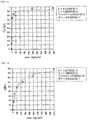
- a fluorescence microscope (inverted research microscope IX 73 (Olympus Corporation) was used for the measurement of fluorescence intensity, the measurement conditions were as follows: concentration: 2.5, 5, 10, 50, 100, 250, 500, 1000, 2500, 5000 ng/mL, filter: BW, objective lens: ⁇ 10, exposure time: 0.50 sec, and amount of light: 100%, and the average value from 5 points was taken as the measurement value. Andor SOLIS was used as the spectroscopic software. The fluorescence intensity change ((Io-I) / Io) with respect to the exosome concentration was measured to create a graph, which was subjected to curve fitting (regression analysis) to calculate the binding constant. The results are shown in Fig. 13 .
- Graph creation software DeltaGraph was used for graph creation and curve fitting, and the binding constant was calculated by the following equation. As a result, the binding constant Ka was calculated to be 1.50 ⁇ 10 14 [M -1 ].
- Y 1 + K ⁇ G + K ⁇ H ⁇ 1 + K ⁇ G + K ⁇ H 2 ⁇ 4 ⁇ K ⁇ K ⁇ H ⁇ G 1 ⁇ 2 ⁇ D 2 ⁇ KG Y: Fluorescence intensity change, H: Exosome concentration, G: Initial concentration of host molecule
- the binding constant was determined by surface plasmon resonance (SPR) analysis.
- SPR measurement conditions were as follows: sample: exosome, concentration: 25, 50, 100, 250, 500, 1000, 2500, 5000 ng/mL, flow rate: 10 ⁇ L/min, injection volume: 30 ⁇ L, running buffer: PBS (10 mM posphate, 140 mM NaCl, pH 7.4), and temperature: 25°C.
- a graph showing the SPR signal (response unit RU) with respect to the exosome concentration was subjected to curve fitting in the same manner as in Example 3 to calculate the binding constant. The results are shown in Fig. 14 .
- the binding constant Ka was calculated to be 2.03 ⁇ 10 12 [M -1 ].
- the reason why the calculated binding constant was larger than that in Example 3 is as follows: in Example 3, fluorescence change only in the concave, which is a sensing place, was specifically detected and even if there was non-specific adsorption in the part other than the sensing place, the non-specific adsorption did not affect the detection results, while in this Reference Example, in addition to the adsorption in the sensing place, the non-specific adsorption in the part other than the sensing place was also detected due to the principle of SPR analysis. Thus, according to the present invention, only the specifically recognized target can be detected.
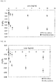
- Three sensors for analysis of a detection target were produced by the same method as in Example 2. Onto the three sensor for analysis of a detection target produced, exosome solutions with different concentrations were dropped and reacted by the same method as in Example 3, to measure the fluorescence intensity after the reaction. The fluorescence intensity was measured at 3 points for each sensor for analysis, 9 points in total. The fluorescence intensity change at 9 measurement points is shown in Fig. 15 .
- the horizontal axis indicates the exosome concentration (ng/ml), and the vertical axis indicates the fluorescence intensity change ((I - Io)/Io).
- the measurement error between the sensors for analysis was small, indicating that the sensors were produced with good reproducibility.
- reaction liquid was ultrafiltered (Amicon Ultra 0.5; 100 kDa) three times, 450 ⁇ L of 1 mM thiol-reactive rhodamine maleimide solution (10 mM PBS pH 7.4) was added to 27 ⁇ L of the collected solution, and the mixture was reacted in the dark at 4°C for 3 hours.
- the rhodamine-labeled exosome was purified by dialysis at 4°C for 24 hours using a dialysis membrane having a pore size of 10 kDa.
- a sensor for analysis of a detection target was produced in the same manner as in Example 2 except that 100 ⁇ M thiol-reactive fluorescein maleimide solution was dropped and the base plate was allowed to stand at room temperature for 3 hours instead of dropping 100 ⁇ L of 500 ⁇ M POLARIC-MLI (GORYO Chemical, Inc.) DMSO solution and allowing the base plate to stand at 25°C for 1 hour.
- 100 ⁇ M thiol-reactive fluorescein maleimide solution was dropped and the base plate was allowed to stand at room temperature for 3 hours instead of dropping 100 ⁇ L of 500 ⁇ M POLARIC-MLI (GORYO Chemical, Inc.) DMSO solution and allowing the base plate to stand at 25°C for 1 hour.
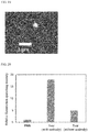
- Fig. 16 The measurement results are shown in Fig. 16 .
- the horizontal axis indicates the exosome concentration, and the vertical axis indicates the amount of change in fluorescence intensity ( ⁇ I).
- ⁇ I fluorescence intensity
- rhodamine fluorescence was observed at the excitation wavelength of fluorescein (that is, FRET was confirmed). FRET does not occur when fluorescein and rhodamine are not placed in close proximity to each other, and thus it was shown that the exosome actually bound to the fluorescein-labeled concave in the sensor for analysis.
- a sensor for analysis of a detection target was produced in the same manner as in Example 2 except that Alexa FluorTM 647 was used instead of POLARIC-MLI.
- 0.01, 0.05, 0.1, 0.5, 1, 5, and 10 ng / ml exosome PBS (10 mM phosphate, 140 mM NaCl, pH 7.4) solutions were prepared as samples in which free antibodies do not coexist, and 0.01, 0.05, 0.1, 0.5, 1, 5, and 10 ng / ml exosome PBS (10 mM phosphate, 140 mM NaCl, pH 7.4) solutions in which free antibodies coexist were prepared as samples in which free antibodies coexist by adding 100 ⁇ l of 0.3 ⁇ M Anti-CD9 antibody PBS (10 mM phosphate, 140 mM NaCl, pH 7.4) solution to 100 ⁇ l of 20 ng/ml exosome PBS (10 mM phosphate, 140 mM NaCl, pH 7.4) solution, incubating the mixture at 4°
- the fluorescence intensity change was measured in the same manner as in Example 3 for each of the samples in which free antibodies do not coexist and the samples in which free antibodies coexist. The results are shown in Fig. 17 . As shown in Fig. 17 , it was found that exosome binding is inhibited when free antibodies coexist. This strongly suggests that exosome binding occurs via antibodies in the binding space. Thus, it was found that the antibody and the fluorescent dye are provided in the concave in the sensor for analysis, and the exosome binding information can be read out.
- Example 8 Base material for producing sensor for analysis of a detection target when silica nanoparticle is used as template
- FITC-labeled silica nanoparticles (having 5 nmol of -COOH on the surface per 200 ⁇ l, and a particle size of 200 nm) (200 ⁇ l) were dispersed in dichloromethane (DCM) (silica nanoparticle dispersion).
- DCM dichloromethane
- NHS N-hydroxysuccinimide
- DIEA N,N-diisopropylethylamine
- the mixture was reacted overnight to modify the surface of the silica nanoparticles with NHS.
- a His-tag with 6 histidines linked by peptide bond (having a lysine residue at the end and having a free ⁇ -amino group: 0.10 ⁇ mol, 40 eq) and 2-aminoethanethiol hydrochloride (0.1 ⁇ mol, 40 eq) were added to the surface-modified silica nanoparticles, and reacted at room temperature.
- silica nanoparticles into which a thiol group and His-tag were introduced were purified by centrifugation and filtration.
- the base plate was washed with pure water, then SH/His-tagged silica nanoparticles dispersed in 10 mM posphate, 140 mM NaCl, pH 7.4, were dropped to obtain a base plate on which SH/His-tagged silica nanoparticles are immobilized.
- a base plate onto which a dispersion of silica nanoparticles that were not subjected to His-tag modification was also dropped in the same manner was also obtained.
- the fluorescence intensity derived from the fluorescent molecule fluorescein (excitation: 480 nm/fluorescence: 510 nm) introduced into the silica nanoparticles before and 1 hour after dropping of the silica nanoparticles was measured to confirm the immobilization of silica nanoparticles.
- the base plate was treated with an acidic buffer (0.5 wt% SDS 50 mM acetate buffer (pH 4.0)), and then the fluorescence intensity on the surface of the base plate was measured again.
- the fluorescence intensity increased after dropping the silica nanoparticles with His-tag modification onto the base plate, which confirms the immobilization of silica nanoparticles.
- an acidic buffer 0.5 wt% SDS 50 mM acetate buffer (pH 4.0)
- the fluorescence intensity decreased to the same level as that before dropping, and thus it was found that the silica nanoparticles bound via Ni-NTA were detached in the presence of acid and a surfactant.
- a mixed self-assembled monolayer having an amino group and a bromo group at the end on a gold thin film deposited glass base plate was produced (molecular film-forming step), an NTA group was introduced into the amino group at the end to form a NTA-Ni complex, then silica nanoparticles were immobilized by chelate bond (template introduction step). Then, the silica nanoparticles were modified with a methacrylic group (surface modification step), and a polymer thin film was synthesized by surface-initiated living radical polymerization (polymerization step). Finally, the silica nanoparticles were removed (removal step) to obtain a base material for producing a sensor for analysis of a detection target.
- mixed SAMs mixed self-assembled monolayer having an amino group and a bromo group at the end on a gold thin film deposited glass base plate was produced (molecular film-forming step)
- an NTA group was introduced into the amino group at the end to form a N
- the base plate was washed with DMSO and pure water, then 100 ⁇ L of 4 mM NiCl 2 aqueous solution was dropped onto the base plate, and the base plate was allowed to stand at room temperature for 15 minutes to form a Ni-NTA complex.
- a polymer thin film was synthesized on the base plate in the same manner as in Item 4 in Example 1 except that the polymerization time was 3 hours.
- a polymer thin film in which the methacryloyl group in the silica nanoparticles was also copolymerized together with the monomer in Table 1 was obtained on the base plate.
- the base plate was immersed in an aqueous solution of 1 M ethylenediaminetetraacetic acid-4Na for 15 minutes to remove Cu 2+ used for ATRP.
- the base plate was immersed in an aqueous solution of 50 mM tris(2-carboxyethyl)phosphine/HCl (TCEP) at 25°C for 3 hours to reduce and cleave the disulfide bond that binds the polymer to silica nanoparticles.
- TCEP tris(2-carboxyethyl)phosphine/HCl
- a free SH group remains on the polymer side, this SH group is derived from SH/His-tagged silica nanoparticles, thus it is not present in the part other than the concave corresponding to the template in the polymer thin film, and it is present only in the concave corresponding to the template.
- Ni-NTA again on the base material for producing a sensor for analysis (MIP base plate) obtained in Example 8, the MIP base plate was treated with an aqueous solution of 4 mM NiCh. Then, 100 ⁇ M His-tag Protein G dissolved in PBS was added to the base plate to immobilize Protein G capable of binding the antibody via His-tag. Finally, 0.3 ⁇ M Anti-CD9 antibody dissolved in PBS was added to the base plate to immobilize the Anti-CD9 antibody via Protein G. Because Protein G binds to the Fc region of the antibody, the orientation of the immobilized antibody is uniform.
- a fluorescent molecule was selectively introduced into the concave in the sensor for analysis using thiol-reactive Alexa Fluor (R) 647 C 2 Maleimide as the fluorescent molecule.
- the fluorescence intensity before introduction was 113 ⁇ 0.6 (n-3), and the fluorescence intensity after introduction was 151 ⁇ 2.1 (n-3), confirming the introduction of fluorescence.
- the sensor for analysis of a detection target was obtained.
- Fig. 18 The results are shown in Fig. 18 .
- the horizontal axis indicates the exosome concentration, and the vertical axis indicates the fluorescence intensity change.
- the estimated M w of the exosome is 2.33 ⁇ 10 8 Da, and thus the exosome was successfully detected with excellent sensitivity comparable to that when an exosome was used as a template.
- the obtained sensor for analysis was observed using a stimulated emission depletion (STED) microscope (SP8-STED3X manufactured by Leica Camera AG).
- STED stimulated emission depletion
- SP8-STED3X manufactured by Leica Camera AG.
- the obtained STED super-resolution image is shown in Fig. 19 .
- dot-like fluorescence of about 200 nm size was observed, and the size of the fluorescence was equivalent to the silica nanoparticles used for the template, and confirming that it is a concave formed by the template effect. Background fluorescence was hardly observed in the part other than the concave, and thus it was also shown that the fluorescent reporter molecule was selectively introduced into the concave.
- Tear fluid was collected using Schirmer test paper, then the test paper was immersed in 10 mM phosphate buffer (PBS) containing 500 ⁇ l of 140 mM NaCl at 4°C for 6 hours to collect exosomes contained in the tear fluid as a tear fluid sample.
- the tear fluid sample was subjected to fluorescence measurement using the sensor for analysis in Example 9 to measure exosomes in the tear fluid.
- the measurement of PBS with the sensor for analysis of a detection target in Example 9 and to confirm the binding of the tear fluid exosome to the antibody of the sensor for analysis, the measurement of the tear fluid sample with a base plate of the sensor for analysis in Example 9 on which the antibody is not immobilized were also performed.
- Fig. 20 The results are shown in Fig. 20 .
- PBS indicates the result of measurement in which PBS was subjected to the sensor for analysis in Example 9
- Tears (with an antibody) indicates the result of measurement in which the tear fluid sample was subjected to the sensor for analysis in Example 9
- Tear (without an antibody) indicates a result of measurement in which the tear fluid sample was subjected to the base plate of the sensor for analysis in Example 9 on which the antibody is not immobilized.
- the degree of fluorescence quenching was largest when the tear fluid sample was subjected to the sensor for analysis in Example 9 on which the antibody was immobilized, and thus it was shown that the exosome in tear fluid was measured.
Landscapes
- Health & Medical Sciences (AREA)
- Life Sciences & Earth Sciences (AREA)
- Immunology (AREA)
- Engineering & Computer Science (AREA)
- Molecular Biology (AREA)
- Biomedical Technology (AREA)
- Chemical & Material Sciences (AREA)
- Hematology (AREA)
- Urology & Nephrology (AREA)
- Biotechnology (AREA)
- Biochemistry (AREA)
- Cell Biology (AREA)
- Food Science & Technology (AREA)
- Medicinal Chemistry (AREA)
- Physics & Mathematics (AREA)
- Analytical Chemistry (AREA)
- Microbiology (AREA)
- General Health & Medical Sciences (AREA)
- General Physics & Mathematics (AREA)
- Pathology (AREA)
- Peptides Or Proteins (AREA)
- Investigating Or Analysing Materials By The Use Of Chemical Reactions (AREA)
- Investigating, Analyzing Materials By Fluorescence Or Luminescence (AREA)
Applications Claiming Priority (2)
| Application Number | Priority Date | Filing Date | Title |
|---|---|---|---|
| JP2017105588 | 2017-05-29 | ||
| PCT/JP2018/019292 WO2018221271A1 (fr) | 2017-05-29 | 2018-05-18 | Matériau de base permettant la fabrication d'un capteur d'analyse d'une cible de détection, capteur d'analyse d'une cible de détection, procédé d'analyse de cible de détection |
Publications (3)
| Publication Number | Publication Date |
|---|---|
| EP3647786A1 true EP3647786A1 (fr) | 2020-05-06 |
| EP3647786A4 EP3647786A4 (fr) | 2021-03-10 |
| EP3647786B1 EP3647786B1 (fr) | 2023-03-08 |
Family
ID=64456282
Family Applications (1)
| Application Number | Title | Priority Date | Filing Date |
|---|---|---|---|
| EP18809084.9A Active EP3647786B1 (fr) | 2017-05-29 | 2018-05-18 | Matériau de base permettant la fabrication d'un capteur d'analyse d'une cible de détection, capteur d'analyse d'une cible de détection, procédé d'analyse de cible de détection |
Country Status (4)
| Country | Link |
|---|---|
| US (2) | US12105085B2 (fr) |
| EP (1) | EP3647786B1 (fr) |
| JP (1) | JP6573351B2 (fr) |
| WO (1) | WO2018221271A1 (fr) |
Cited By (1)
| Publication number | Priority date | Publication date | Assignee | Title |
|---|---|---|---|---|
| EP4394382A4 (fr) * | 2021-08-27 | 2025-11-19 | Univ Kobe Nat Univ Corp | Technologie d'arrangement de particules sur un substrat sans agglutination |
Families Citing this family (14)
| Publication number | Priority date | Publication date | Assignee | Title |
|---|---|---|---|---|
| US12037430B2 (en) | 2018-10-17 | 2024-07-16 | National University Corporation Kobe University | Target substance polymer replica substrate, method for producing same, and method for producing substrate sensor |
| CN110487946A (zh) * | 2019-09-03 | 2019-11-22 | 中国人民解放军军事科学院军事医学研究院 | 一种尿液中外泌体的提取及其蛋白质组学和磷酸化蛋白质组学分析方法 |
| CN110702898B (zh) * | 2019-11-05 | 2020-07-14 | 山东农业大学 | 一种纳米酶标记仿生免疫分析检测磺胺嘧啶的方法 |
| JP7445913B2 (ja) * | 2019-11-15 | 2024-03-08 | 国立大学法人神戸大学 | 単一粒体分析用イムノセンサ及び単一粒体分析法 |
| CN112898624B (zh) * | 2019-12-04 | 2022-05-31 | 中国科学院大连化学物理研究所 | 用于特异性识别外泌体的替代模板印迹聚合物及其应用 |
| JP7227895B2 (ja) * | 2019-12-23 | 2023-02-22 | 株式会社堀場製作所 | 相互作用検出方法、相互作用検出装置及びバイオチップ再生キット |
| WO2021172591A1 (fr) * | 2020-02-27 | 2021-09-02 | 国立大学法人神戸大学 | Nanoparticules pour utilisation de détection et leur procédé de fabrication |
| US20230194510A1 (en) * | 2020-04-27 | 2023-06-22 | National University Corporation, Tokyo Medical And Dental University | Biological vesicle surface biomarker detection device |
| JP7454225B2 (ja) * | 2020-04-28 | 2024-03-22 | 国立大学法人神戸大学 | 検出対象の分析用センサ作製用基材、検出対象の分析用センサ、及び検出対象の分析法 |
| CN116324409A (zh) * | 2020-09-23 | 2023-06-23 | 国立大学法人神户大学 | 用于制作检测对象分析用传感器的基材、检测对象分析用传感器和检测对象的分析方法 |
| CN113189064B (zh) * | 2021-04-22 | 2022-12-09 | 西安交通大学 | 基于糖肽的荧光分子印迹聚合物及制备方法和在糖蛋白筛选和检测中的应用 |
| TW202337974A (zh) | 2021-08-27 | 2023-10-01 | 國立大學法人神戶大學 | 用於於基材上產率良好地形成空孔之技術之核殼模板分子、粒子 |
| JPWO2024143430A1 (fr) * | 2022-12-27 | 2024-07-04 | ||
| WO2025182733A1 (fr) * | 2024-02-27 | 2025-09-04 | 国立大学法人 奈良先端科学技術大学院大学 | Procédé et dispositif de détection |
Family Cites Families (13)
| Publication number | Priority date | Publication date | Assignee | Title |
|---|---|---|---|---|
| CN101415487A (zh) | 2003-12-08 | 2009-04-22 | 纽约州立大学研究基金会 | 传感器应用中的位点选择性标记和模板分子印记聚合物 |
| US8617806B2 (en) | 2008-01-25 | 2013-12-31 | Hansabiomed Ou | Method to measure and characterize microvesicles in the human body fluids |
| US20130196355A1 (en) | 2008-01-25 | 2013-08-01 | Hansabiomed Ou | Method and a kit to quantify and qualify exosomes for diagnosis of prostate cancer and prostate hyperplasia |
| US8097407B2 (en) | 2008-01-25 | 2012-01-17 | Hansabiomed Ou | Metastatic human tumor associated molecule, methods to detect both activated gene and protein and to interfere with gene expression |
| JP6029112B2 (ja) | 2011-12-22 | 2016-11-24 | テオリアサイエンス株式会社 | エクソソームの分析方法、エクソソーム分析用試薬およびエクソソーム分析装置 |
| JP6210004B2 (ja) | 2013-04-09 | 2017-10-11 | 株式会社Jvcケンウッド | 試料分析用デバイス及びエクソソームの捕捉方法 |
| JPWO2015029979A1 (ja) | 2013-08-30 | 2017-03-02 | 国立大学法人 東京大学 | エキソソームの分析方法、エキソソーム分析チップ、及びエキソソーム分析装置 |
| WO2015045666A1 (fr) | 2013-09-25 | 2015-04-02 | 国立大学法人東京大学 | Dispositif fluidique, procédé d'analyse d'exosomes, procédé d'analyse de biomolécules et procédé de détection de biomolécules |
| US10557847B2 (en) | 2013-12-02 | 2020-02-11 | The General Hospital Corporation | Nano-plasmonic sensor for exosome detection |
| JP6505490B2 (ja) | 2015-04-03 | 2019-04-24 | 国立大学法人神戸大学 | 分子インプリンティング膜、その製造方法、鋳型化合物、およびステロイドホルモン化合物の検出方法 |
| JP6653884B2 (ja) | 2015-07-10 | 2020-02-26 | 国立大学法人神戸大学 | 分子インプリントポリマーの製造方法、分子インプリントポリマー、および標的タンパク質の検出方法 |
| US12037430B2 (en) * | 2018-10-17 | 2024-07-16 | National University Corporation Kobe University | Target substance polymer replica substrate, method for producing same, and method for producing substrate sensor |
| JP7454225B2 (ja) * | 2020-04-28 | 2024-03-22 | 国立大学法人神戸大学 | 検出対象の分析用センサ作製用基材、検出対象の分析用センサ、及び検出対象の分析法 |
-
2018
- 2018-05-18 EP EP18809084.9A patent/EP3647786B1/fr active Active
- 2018-05-18 US US16/617,987 patent/US12105085B2/en active Active
- 2018-05-18 JP JP2019514336A patent/JP6573351B2/ja active Active
- 2018-05-18 WO PCT/JP2018/019292 patent/WO2018221271A1/fr not_active Ceased
-
2024
- 2024-09-06 US US18/827,348 patent/US20250003961A1/en active Pending
Cited By (1)
| Publication number | Priority date | Publication date | Assignee | Title |
|---|---|---|---|---|
| EP4394382A4 (fr) * | 2021-08-27 | 2025-11-19 | Univ Kobe Nat Univ Corp | Technologie d'arrangement de particules sur un substrat sans agglutination |
Also Published As
| Publication number | Publication date |
|---|---|
| WO2018221271A1 (fr) | 2018-12-06 |
| EP3647786B1 (fr) | 2023-03-08 |
| US12105085B2 (en) | 2024-10-01 |
| US20250003961A1 (en) | 2025-01-02 |
| US20200110084A1 (en) | 2020-04-09 |
| JP6573351B2 (ja) | 2019-09-11 |
| EP3647786A4 (fr) | 2021-03-10 |
| JPWO2018221271A1 (ja) | 2019-06-27 |
Similar Documents
| Publication | Publication Date | Title |
|---|---|---|
| US20250003961A1 (en) | Base material for manufacturing sensor for analyzing detection target, sensor for analyzing detection target, method for analyzing detection target | |
| Chen et al. | Surface plasmon resonance biosensor using hydrogel-AuNP supramolecular spheres for determination of prostate cancer-derived exosomes | |
| Szlag et al. | Molecular affinity agents for intrinsic surface-enhanced Raman scattering (SERS) sensors | |
| Yang et al. | Direct and highly selective drug optosensing in real, undiluted biological samples with quantum-dot-labeled hydrophilic molecularly imprinted polymer microparticles | |
| Zhuang et al. | Immunoglobulin G-encapsulated gold nanoclusters as fluorescent tags for dot-blot immunoassays | |
| CN104316678A (zh) | 固定有抗原的免疫荧光载片的制备方法和由该方法制备的免疫荧光载片 | |
| Tasso et al. | Oriented bioconjugation of unmodified antibodies to quantum dots capped with copolymeric ligands as versatile cellular imaging tools | |
| WO2016073833A1 (fr) | Immunopolymères à affinité élevée | |
| JP2017019992A (ja) | 分子インプリントポリマーの製造方法、分子インプリントポリマー、および標的タンパク質の検出方法 | |
| Morishige et al. | Post‐Imprinting‐Modified Molecularly Imprinted Nanocavities with Two Synergetic, Orthogonal, Glycoprotein‐Binding Sites to Transduce Binding Events into Fluorescence Changes | |
| Oliverio et al. | Versatile and high-throughput strategy for the quantification of proteins bound to nanoparticles | |
| Han et al. | Microfluidically partitioned dual channels for accurate background subtraction in cellular binding studies by surface plasmon resonance microscopy | |
| CN113552345B (zh) | 一种基于免疫荧光增强的外泌体定量检测方法 | |
| Alexandre et al. | Illuminating extracellular vesicles biology with super-resolution microscopy: insights into morphology and composition | |
| US12037430B2 (en) | Target substance polymer replica substrate, method for producing same, and method for producing substrate sensor | |
| JP2023037003A (ja) | 検出対象の分析用センサ作製用基材、検出対象の分析用センサ、及び検出対象の分析法 | |
| JP2021173702A (ja) | 検出対象の分析用センサ作製用基材、検出対象の分析用センサ、及び検出対象の分析法 | |
| JP7445913B2 (ja) | 単一粒体分析用イムノセンサ及び単一粒体分析法 | |
| US12385910B2 (en) | Solid phase carrier and kit | |
| EP3608665A1 (fr) | Procédé de fabrication d'un produit purifié de particule intégrée au phosphore modifié par une protéine, procédé de fabrication de liquide de coloration fluorescent, produit purifié de particule intégrée au phosphore modifié par une protéine, et filtre de purification d'un liquide de coloration fluorescent et d'une particule intégrée au phosphore modifiée par une protéine | |
| EP2281194B1 (fr) | Ensemble moléculaire comprenant de l'or et un liant pour la détection d'entités biochimiques | |
| Dallari et al. | Gold Nanostars Bioconjugation for Selective Targeting and SERS Detection of Biofluids. Nanomaterials 2021, 11, 665 | |
| JP2010002393A (ja) | 標的物質の検出方法 | |
| HK40092517A (en) | Base material for manufacturing sensor for analyzing detection object, sensor for analyzing detection object, and method for analyzing detection object | |
| Küçük | Thermoresponsive Polymer Integrated Metasurface Sensor for Capture and Release of Extracellular Vesicles in Real-Time |
Legal Events
| Date | Code | Title | Description |
|---|---|---|---|
| STAA | Information on the status of an ep patent application or granted ep patent |
Free format text: STATUS: THE INTERNATIONAL PUBLICATION HAS BEEN MADE |
|
| PUAI | Public reference made under article 153(3) epc to a published international application that has entered the european phase |
Free format text: ORIGINAL CODE: 0009012 |
|
| STAA | Information on the status of an ep patent application or granted ep patent |
Free format text: STATUS: REQUEST FOR EXAMINATION WAS MADE |
|
| 17P | Request for examination filed |
Effective date: 20191128 |
|
| AK | Designated contracting states |
Kind code of ref document: A1 Designated state(s): AL AT BE BG CH CY CZ DE DK EE ES FI FR GB GR HR HU IE IS IT LI LT LU LV MC MK MT NL NO PL PT RO RS SE SI SK SM TR |
|
| AX | Request for extension of the european patent |
Extension state: BA ME |
|
| DAV | Request for validation of the european patent (deleted) | ||
| DAX | Request for extension of the european patent (deleted) | ||
| A4 | Supplementary search report drawn up and despatched |
Effective date: 20210205 |
|
| RIC1 | Information provided on ipc code assigned before grant |
Ipc: G01N 33/543 20060101AFI20210201BHEP Ipc: G01N 33/531 20060101ALI20210201BHEP Ipc: G01N 33/53 20060101ALI20210201BHEP Ipc: G01N 33/58 20060101ALI20210201BHEP |
|
| STAA | Information on the status of an ep patent application or granted ep patent |
Free format text: STATUS: EXAMINATION IS IN PROGRESS |
|
| 17Q | First examination report despatched |
Effective date: 20220124 |
|
| GRAP | Despatch of communication of intention to grant a patent |
Free format text: ORIGINAL CODE: EPIDOSNIGR1 |
|
| STAA | Information on the status of an ep patent application or granted ep patent |
Free format text: STATUS: GRANT OF PATENT IS INTENDED |
|
| INTG | Intention to grant announced |
Effective date: 20220929 |
|
| GRAS | Grant fee paid |
Free format text: ORIGINAL CODE: EPIDOSNIGR3 |
|
| GRAA | (expected) grant |
Free format text: ORIGINAL CODE: 0009210 |
|
| STAA | Information on the status of an ep patent application or granted ep patent |
Free format text: STATUS: THE PATENT HAS BEEN GRANTED |
|
| AK | Designated contracting states |
Kind code of ref document: B1 Designated state(s): AL AT BE BG CH CY CZ DE DK EE ES FI FR GB GR HR HU IE IS IT LI LT LU LV MC MK MT NL NO PL PT RO RS SE SI SK SM TR |
|
| REG | Reference to a national code |
Ref country code: CH Ref legal event code: EP Ref country code: AT Ref legal event code: REF Ref document number: 1552918 Country of ref document: AT Kind code of ref document: T Effective date: 20230315 |
|
| REG | Reference to a national code |
Ref country code: IE Ref legal event code: FG4D |
|
| REG | Reference to a national code |
Ref country code: DE Ref legal event code: R096 Ref document number: 602018047044 Country of ref document: DE |
|
| REG | Reference to a national code |
Ref country code: LT Ref legal event code: MG9D |
|
| P01 | Opt-out of the competence of the unified patent court (upc) registered |
Effective date: 20230519 |
|
| REG | Reference to a national code |
Ref country code: NL Ref legal event code: MP Effective date: 20230308 |
|
| PG25 | Lapsed in a contracting state [announced via postgrant information from national office to epo] |
Ref country code: RS Free format text: LAPSE BECAUSE OF FAILURE TO SUBMIT A TRANSLATION OF THE DESCRIPTION OR TO PAY THE FEE WITHIN THE PRESCRIBED TIME-LIMIT Effective date: 20230308 Ref country code: NO Free format text: LAPSE BECAUSE OF FAILURE TO SUBMIT A TRANSLATION OF THE DESCRIPTION OR TO PAY THE FEE WITHIN THE PRESCRIBED TIME-LIMIT Effective date: 20230608 Ref country code: LV Free format text: LAPSE BECAUSE OF FAILURE TO SUBMIT A TRANSLATION OF THE DESCRIPTION OR TO PAY THE FEE WITHIN THE PRESCRIBED TIME-LIMIT Effective date: 20230308 Ref country code: LT Free format text: LAPSE BECAUSE OF FAILURE TO SUBMIT A TRANSLATION OF THE DESCRIPTION OR TO PAY THE FEE WITHIN THE PRESCRIBED TIME-LIMIT Effective date: 20230308 Ref country code: HR Free format text: LAPSE BECAUSE OF FAILURE TO SUBMIT A TRANSLATION OF THE DESCRIPTION OR TO PAY THE FEE WITHIN THE PRESCRIBED TIME-LIMIT Effective date: 20230308 Ref country code: ES Free format text: LAPSE BECAUSE OF FAILURE TO SUBMIT A TRANSLATION OF THE DESCRIPTION OR TO PAY THE FEE WITHIN THE PRESCRIBED TIME-LIMIT Effective date: 20230308 |
|
| REG | Reference to a national code |
Ref country code: AT Ref legal event code: MK05 Ref document number: 1552918 Country of ref document: AT Kind code of ref document: T Effective date: 20230308 |
|
| PG25 | Lapsed in a contracting state [announced via postgrant information from national office to epo] |
Ref country code: SE Free format text: LAPSE BECAUSE OF FAILURE TO SUBMIT A TRANSLATION OF THE DESCRIPTION OR TO PAY THE FEE WITHIN THE PRESCRIBED TIME-LIMIT Effective date: 20230308 Ref country code: NL Free format text: LAPSE BECAUSE OF FAILURE TO SUBMIT A TRANSLATION OF THE DESCRIPTION OR TO PAY THE FEE WITHIN THE PRESCRIBED TIME-LIMIT Effective date: 20230308 Ref country code: GR Free format text: LAPSE BECAUSE OF FAILURE TO SUBMIT A TRANSLATION OF THE DESCRIPTION OR TO PAY THE FEE WITHIN THE PRESCRIBED TIME-LIMIT Effective date: 20230609 Ref country code: FI Free format text: LAPSE BECAUSE OF FAILURE TO SUBMIT A TRANSLATION OF THE DESCRIPTION OR TO PAY THE FEE WITHIN THE PRESCRIBED TIME-LIMIT Effective date: 20230308 |
|
| PG25 | Lapsed in a contracting state [announced via postgrant information from national office to epo] |
Ref country code: SM Free format text: LAPSE BECAUSE OF FAILURE TO SUBMIT A TRANSLATION OF THE DESCRIPTION OR TO PAY THE FEE WITHIN THE PRESCRIBED TIME-LIMIT Effective date: 20230308 Ref country code: RO Free format text: LAPSE BECAUSE OF FAILURE TO SUBMIT A TRANSLATION OF THE DESCRIPTION OR TO PAY THE FEE WITHIN THE PRESCRIBED TIME-LIMIT Effective date: 20230308 Ref country code: PT Free format text: LAPSE BECAUSE OF FAILURE TO SUBMIT A TRANSLATION OF THE DESCRIPTION OR TO PAY THE FEE WITHIN THE PRESCRIBED TIME-LIMIT Effective date: 20230710 Ref country code: EE Free format text: LAPSE BECAUSE OF FAILURE TO SUBMIT A TRANSLATION OF THE DESCRIPTION OR TO PAY THE FEE WITHIN THE PRESCRIBED TIME-LIMIT Effective date: 20230308 Ref country code: CZ Free format text: LAPSE BECAUSE OF FAILURE TO SUBMIT A TRANSLATION OF THE DESCRIPTION OR TO PAY THE FEE WITHIN THE PRESCRIBED TIME-LIMIT Effective date: 20230308 Ref country code: AT Free format text: LAPSE BECAUSE OF FAILURE TO SUBMIT A TRANSLATION OF THE DESCRIPTION OR TO PAY THE FEE WITHIN THE PRESCRIBED TIME-LIMIT Effective date: 20230308 |
|
| PG25 | Lapsed in a contracting state [announced via postgrant information from national office to epo] |
Ref country code: SK Free format text: LAPSE BECAUSE OF FAILURE TO SUBMIT A TRANSLATION OF THE DESCRIPTION OR TO PAY THE FEE WITHIN THE PRESCRIBED TIME-LIMIT Effective date: 20230308 Ref country code: PL Free format text: LAPSE BECAUSE OF FAILURE TO SUBMIT A TRANSLATION OF THE DESCRIPTION OR TO PAY THE FEE WITHIN THE PRESCRIBED TIME-LIMIT Effective date: 20230308 Ref country code: IS Free format text: LAPSE BECAUSE OF FAILURE TO SUBMIT A TRANSLATION OF THE DESCRIPTION OR TO PAY THE FEE WITHIN THE PRESCRIBED TIME-LIMIT Effective date: 20230708 |
|
| REG | Reference to a national code |
Ref country code: DE Ref legal event code: R097 Ref document number: 602018047044 Country of ref document: DE |
|
| REG | Reference to a national code |
Ref country code: CH Ref legal event code: PL |
|
| PLBE | No opposition filed within time limit |
Free format text: ORIGINAL CODE: 0009261 |
|
| STAA | Information on the status of an ep patent application or granted ep patent |
Free format text: STATUS: NO OPPOSITION FILED WITHIN TIME LIMIT |
|
| PG25 | Lapsed in a contracting state [announced via postgrant information from national office to epo] |
Ref country code: MC Free format text: LAPSE BECAUSE OF FAILURE TO SUBMIT A TRANSLATION OF THE DESCRIPTION OR TO PAY THE FEE WITHIN THE PRESCRIBED TIME-LIMIT Effective date: 20230308 |
|
| REG | Reference to a national code |
Ref country code: BE Ref legal event code: MM Effective date: 20230531 |
|
| PG25 | Lapsed in a contracting state [announced via postgrant information from national office to epo] |
Ref country code: SI Free format text: LAPSE BECAUSE OF FAILURE TO SUBMIT A TRANSLATION OF THE DESCRIPTION OR TO PAY THE FEE WITHIN THE PRESCRIBED TIME-LIMIT Effective date: 20230308 Ref country code: MC Free format text: LAPSE BECAUSE OF FAILURE TO SUBMIT A TRANSLATION OF THE DESCRIPTION OR TO PAY THE FEE WITHIN THE PRESCRIBED TIME-LIMIT Effective date: 20230308 Ref country code: LU Free format text: LAPSE BECAUSE OF NON-PAYMENT OF DUE FEES Effective date: 20230518 Ref country code: LI Free format text: LAPSE BECAUSE OF NON-PAYMENT OF DUE FEES Effective date: 20230531 Ref country code: DK Free format text: LAPSE BECAUSE OF FAILURE TO SUBMIT A TRANSLATION OF THE DESCRIPTION OR TO PAY THE FEE WITHIN THE PRESCRIBED TIME-LIMIT Effective date: 20230308 Ref country code: CH Free format text: LAPSE BECAUSE OF NON-PAYMENT OF DUE FEES Effective date: 20230531 |
|
| 26N | No opposition filed |
Effective date: 20231211 |
|
| REG | Reference to a national code |
Ref country code: IE Ref legal event code: MM4A |
|
| PG25 | Lapsed in a contracting state [announced via postgrant information from national office to epo] |
Ref country code: IE Free format text: LAPSE BECAUSE OF NON-PAYMENT OF DUE FEES Effective date: 20230518 |
|
| PG25 | Lapsed in a contracting state [announced via postgrant information from national office to epo] |
Ref country code: IE Free format text: LAPSE BECAUSE OF NON-PAYMENT OF DUE FEES Effective date: 20230518 |
|
| PG25 | Lapsed in a contracting state [announced via postgrant information from national office to epo] |
Ref country code: IT Free format text: LAPSE BECAUSE OF FAILURE TO SUBMIT A TRANSLATION OF THE DESCRIPTION OR TO PAY THE FEE WITHIN THE PRESCRIBED TIME-LIMIT Effective date: 20230308 Ref country code: BE Free format text: LAPSE BECAUSE OF NON-PAYMENT OF DUE FEES Effective date: 20230531 |
|
| PG25 | Lapsed in a contracting state [announced via postgrant information from national office to epo] |
Ref country code: BG Free format text: LAPSE BECAUSE OF FAILURE TO SUBMIT A TRANSLATION OF THE DESCRIPTION OR TO PAY THE FEE WITHIN THE PRESCRIBED TIME-LIMIT Effective date: 20230308 |
|
| PG25 | Lapsed in a contracting state [announced via postgrant information from national office to epo] |
Ref country code: BG Free format text: LAPSE BECAUSE OF FAILURE TO SUBMIT A TRANSLATION OF THE DESCRIPTION OR TO PAY THE FEE WITHIN THE PRESCRIBED TIME-LIMIT Effective date: 20230308 |
|
| PGFP | Annual fee paid to national office [announced via postgrant information from national office to epo] |
Ref country code: DE Payment date: 20250527 Year of fee payment: 8 |
|
| PGFP | Annual fee paid to national office [announced via postgrant information from national office to epo] |
Ref country code: FR Payment date: 20250527 Year of fee payment: 8 |
|
| PG25 | Lapsed in a contracting state [announced via postgrant information from national office to epo] |
Ref country code: CY Free format text: LAPSE BECAUSE OF FAILURE TO SUBMIT A TRANSLATION OF THE DESCRIPTION OR TO PAY THE FEE WITHIN THE PRESCRIBED TIME-LIMIT; INVALID AB INITIO Effective date: 20180518 |
|
| PG25 | Lapsed in a contracting state [announced via postgrant information from national office to epo] |
Ref country code: HU Free format text: LAPSE BECAUSE OF FAILURE TO SUBMIT A TRANSLATION OF THE DESCRIPTION OR TO PAY THE FEE WITHIN THE PRESCRIBED TIME-LIMIT; INVALID AB INITIO Effective date: 20180518 |
|
| PG25 | Lapsed in a contracting state [announced via postgrant information from national office to epo] |
Ref country code: TR Free format text: LAPSE BECAUSE OF FAILURE TO SUBMIT A TRANSLATION OF THE DESCRIPTION OR TO PAY THE FEE WITHIN THE PRESCRIBED TIME-LIMIT Effective date: 20230308 |
|
| PGFP | Annual fee paid to national office [announced via postgrant information from national office to epo] |
Ref country code: GB Payment date: 20260326 Year of fee payment: 9 |