EP3659552A1 - Transkatheter-mitralklappenblattverlängerung - Google Patents
Transkatheter-mitralklappenblattverlängerung Download PDFInfo
- Publication number
- EP3659552A1 EP3659552A1 EP19211409.8A EP19211409A EP3659552A1 EP 3659552 A1 EP3659552 A1 EP 3659552A1 EP 19211409 A EP19211409 A EP 19211409A EP 3659552 A1 EP3659552 A1 EP 3659552A1
- Authority
- EP
- European Patent Office
- Prior art keywords
- disk
- leaflet
- occlusion device
- native
- native leaflet
- Prior art date
- Legal status (The legal status is an assumption and is not a legal conclusion. Google has not performed a legal analysis and makes no representation as to the accuracy of the status listed.)
- Withdrawn
Links
- 210000004115 mitral valve Anatomy 0.000 title description 31
- 238000000034 method Methods 0.000 claims abstract description 20
- 230000003190 augmentative effect Effects 0.000 claims abstract description 12
- 230000008878 coupling Effects 0.000 claims abstract description 7
- 238000010168 coupling process Methods 0.000 claims abstract description 7
- 238000005859 coupling reaction Methods 0.000 claims abstract description 7
- 230000017531 blood circulation Effects 0.000 claims description 11
- 239000000463 material Substances 0.000 claims description 8
- 229910001000 nickel titanium Inorganic materials 0.000 claims description 6
- HLXZNVUGXRDIFK-UHFFFAOYSA-N nickel titanium Chemical compound [Ti].[Ti].[Ti].[Ti].[Ti].[Ti].[Ti].[Ti].[Ti].[Ti].[Ti].[Ni].[Ni].[Ni].[Ni].[Ni].[Ni].[Ni].[Ni].[Ni].[Ni].[Ni].[Ni].[Ni].[Ni] HLXZNVUGXRDIFK-UHFFFAOYSA-N 0.000 claims description 6
- 229920000747 poly(lactic acid) Polymers 0.000 claims description 6
- 238000005259 measurement Methods 0.000 claims description 4
- 239000004744 fabric Substances 0.000 claims description 3
- 229910001285 shape-memory alloy Inorganic materials 0.000 claims description 3
- 210000003698 chordae tendineae Anatomy 0.000 description 12
- 210000005240 left ventricle Anatomy 0.000 description 12
- 210000005246 left atrium Anatomy 0.000 description 11
- 230000003416 augmentation Effects 0.000 description 8
- 210000001519 tissue Anatomy 0.000 description 7
- 206010027727 Mitral valve incompetence Diseases 0.000 description 5
- 239000008280 blood Substances 0.000 description 5
- 210000004369 blood Anatomy 0.000 description 5
- 230000008439 repair process Effects 0.000 description 5
- 230000006870 function Effects 0.000 description 4
- 210000003709 heart valve Anatomy 0.000 description 4
- 210000003540 papillary muscle Anatomy 0.000 description 3
- 230000002861 ventricular Effects 0.000 description 3
- 210000001015 abdomen Anatomy 0.000 description 2
- 238000013459 approach Methods 0.000 description 2
- 230000036772 blood pressure Effects 0.000 description 2
- 210000002837 heart atrium Anatomy 0.000 description 2
- 238000002513 implantation Methods 0.000 description 2
- 230000000302 ischemic effect Effects 0.000 description 2
- 210000004072 lung Anatomy 0.000 description 2
- 208000010125 myocardial infarction Diseases 0.000 description 2
- 230000007704 transition Effects 0.000 description 2
- 210000000591 tricuspid valve Anatomy 0.000 description 2
- 241000283690 Bos taurus Species 0.000 description 1
- 206010007559 Cardiac failure congestive Diseases 0.000 description 1
- 206010014666 Endocarditis bacterial Diseases 0.000 description 1
- JOYRKODLDBILNP-UHFFFAOYSA-N Ethyl urethane Chemical compound CCOC(N)=O JOYRKODLDBILNP-UHFFFAOYSA-N 0.000 description 1
- 206010019280 Heart failures Diseases 0.000 description 1
- 206010020772 Hypertension Diseases 0.000 description 1
- MWCLLHOVUTZFKS-UHFFFAOYSA-N Methyl cyanoacrylate Chemical compound COC(=O)C(=C)C#N MWCLLHOVUTZFKS-UHFFFAOYSA-N 0.000 description 1
- 208000001435 Thromboembolism Diseases 0.000 description 1
- 210000000709 aorta Anatomy 0.000 description 1
- 210000002376 aorta thoracic Anatomy 0.000 description 1
- 230000001746 atrial effect Effects 0.000 description 1
- 208000009361 bacterial endocarditis Diseases 0.000 description 1
- 230000000975 bioactive effect Effects 0.000 description 1
- 239000000560 biocompatible material Substances 0.000 description 1
- 239000012620 biological material Substances 0.000 description 1
- 230000015572 biosynthetic process Effects 0.000 description 1
- 238000002788 crimping Methods 0.000 description 1
- 201000010099 disease Diseases 0.000 description 1
- 208000037265 diseases, disorders, signs and symptoms Diseases 0.000 description 1
- 210000002889 endothelial cell Anatomy 0.000 description 1
- 201000007119 infective endocarditis Diseases 0.000 description 1
- 238000012986 modification Methods 0.000 description 1
- 230000004048 modification Effects 0.000 description 1
- 230000003387 muscular Effects 0.000 description 1
- 230000001991 pathophysiological effect Effects 0.000 description 1
- 210000003516 pericardium Anatomy 0.000 description 1
- 230000002093 peripheral effect Effects 0.000 description 1
- 229920000642 polymer Polymers 0.000 description 1
- 239000004810 polytetrafluoroethylene Substances 0.000 description 1
- 229920001343 polytetrafluoroethylene Polymers 0.000 description 1
- 208000002815 pulmonary hypertension Diseases 0.000 description 1
- 238000011084 recovery Methods 0.000 description 1
- 238000007634 remodeling Methods 0.000 description 1
- 210000005245 right atrium Anatomy 0.000 description 1
- 210000005241 right ventricle Anatomy 0.000 description 1
- 239000011343 solid material Substances 0.000 description 1
- 238000001356 surgical procedure Methods 0.000 description 1
Images


Classifications
-
- A—HUMAN NECESSITIES
- A61—MEDICAL OR VETERINARY SCIENCE; HYGIENE
- A61F—FILTERS IMPLANTABLE INTO BLOOD VESSELS; PROSTHESES; DEVICES PROVIDING PATENCY TO, OR PREVENTING COLLAPSING OF, TUBULAR STRUCTURES OF THE BODY, e.g. STENTS; ORTHOPAEDIC, NURSING OR CONTRACEPTIVE DEVICES; FOMENTATION; TREATMENT OR PROTECTION OF EYES OR EARS; BANDAGES, DRESSINGS OR ABSORBENT PADS; FIRST-AID KITS
- A61F2/00—Filters implantable into blood vessels; Prostheses, i.e. artificial substitutes or replacements for parts of the body; Appliances for connecting them with the body; Devices providing patency to, or preventing collapsing of, tubular structures of the body, e.g. stents
- A61F2/02—Prostheses implantable into the body
- A61F2/24—Heart valves ; Vascular valves, e.g. venous valves; Heart implants, e.g. passive devices for improving the function of the native valve or the heart muscle; Transmyocardial revascularisation [TMR] devices; Valves implantable in the body
- A61F2/2412—Heart valves ; Vascular valves, e.g. venous valves; Heart implants, e.g. passive devices for improving the function of the native valve or the heart muscle; Transmyocardial revascularisation [TMR] devices; Valves implantable in the body with soft flexible valve members, e.g. tissue valves shaped like natural valves
-
- A—HUMAN NECESSITIES
- A61—MEDICAL OR VETERINARY SCIENCE; HYGIENE
- A61F—FILTERS IMPLANTABLE INTO BLOOD VESSELS; PROSTHESES; DEVICES PROVIDING PATENCY TO, OR PREVENTING COLLAPSING OF, TUBULAR STRUCTURES OF THE BODY, e.g. STENTS; ORTHOPAEDIC, NURSING OR CONTRACEPTIVE DEVICES; FOMENTATION; TREATMENT OR PROTECTION OF EYES OR EARS; BANDAGES, DRESSINGS OR ABSORBENT PADS; FIRST-AID KITS
- A61F2/00—Filters implantable into blood vessels; Prostheses, i.e. artificial substitutes or replacements for parts of the body; Appliances for connecting them with the body; Devices providing patency to, or preventing collapsing of, tubular structures of the body, e.g. stents
- A61F2/02—Prostheses implantable into the body
- A61F2/24—Heart valves ; Vascular valves, e.g. venous valves; Heart implants, e.g. passive devices for improving the function of the native valve or the heart muscle; Transmyocardial revascularisation [TMR] devices; Valves implantable in the body
- A61F2/2442—Annuloplasty rings or inserts for correcting the valve shape; Implants for improving the function of a native heart valve
- A61F2/2454—Means for preventing inversion of the valve leaflets, e.g. chordae tendineae prostheses
-
- A—HUMAN NECESSITIES
- A61—MEDICAL OR VETERINARY SCIENCE; HYGIENE
- A61F—FILTERS IMPLANTABLE INTO BLOOD VESSELS; PROSTHESES; DEVICES PROVIDING PATENCY TO, OR PREVENTING COLLAPSING OF, TUBULAR STRUCTURES OF THE BODY, e.g. STENTS; ORTHOPAEDIC, NURSING OR CONTRACEPTIVE DEVICES; FOMENTATION; TREATMENT OR PROTECTION OF EYES OR EARS; BANDAGES, DRESSINGS OR ABSORBENT PADS; FIRST-AID KITS
- A61F2/00—Filters implantable into blood vessels; Prostheses, i.e. artificial substitutes or replacements for parts of the body; Appliances for connecting them with the body; Devices providing patency to, or preventing collapsing of, tubular structures of the body, e.g. stents
- A61F2/02—Prostheses implantable into the body
- A61F2/24—Heart valves ; Vascular valves, e.g. venous valves; Heart implants, e.g. passive devices for improving the function of the native valve or the heart muscle; Transmyocardial revascularisation [TMR] devices; Valves implantable in the body
- A61F2/2442—Annuloplasty rings or inserts for correcting the valve shape; Implants for improving the function of a native heart valve
-
- A—HUMAN NECESSITIES
- A61—MEDICAL OR VETERINARY SCIENCE; HYGIENE
- A61F—FILTERS IMPLANTABLE INTO BLOOD VESSELS; PROSTHESES; DEVICES PROVIDING PATENCY TO, OR PREVENTING COLLAPSING OF, TUBULAR STRUCTURES OF THE BODY, e.g. STENTS; ORTHOPAEDIC, NURSING OR CONTRACEPTIVE DEVICES; FOMENTATION; TREATMENT OR PROTECTION OF EYES OR EARS; BANDAGES, DRESSINGS OR ABSORBENT PADS; FIRST-AID KITS
- A61F2/00—Filters implantable into blood vessels; Prostheses, i.e. artificial substitutes or replacements for parts of the body; Appliances for connecting them with the body; Devices providing patency to, or preventing collapsing of, tubular structures of the body, e.g. stents
- A61F2/02—Prostheses implantable into the body
- A61F2/24—Heart valves ; Vascular valves, e.g. venous valves; Heart implants, e.g. passive devices for improving the function of the native valve or the heart muscle; Transmyocardial revascularisation [TMR] devices; Valves implantable in the body
- A61F2/2442—Annuloplasty rings or inserts for correcting the valve shape; Implants for improving the function of a native heart valve
- A61F2/2445—Annuloplasty rings in direct contact with the valve annulus
-
- A—HUMAN NECESSITIES
- A61—MEDICAL OR VETERINARY SCIENCE; HYGIENE
- A61F—FILTERS IMPLANTABLE INTO BLOOD VESSELS; PROSTHESES; DEVICES PROVIDING PATENCY TO, OR PREVENTING COLLAPSING OF, TUBULAR STRUCTURES OF THE BODY, e.g. STENTS; ORTHOPAEDIC, NURSING OR CONTRACEPTIVE DEVICES; FOMENTATION; TREATMENT OR PROTECTION OF EYES OR EARS; BANDAGES, DRESSINGS OR ABSORBENT PADS; FIRST-AID KITS
- A61F2/00—Filters implantable into blood vessels; Prostheses, i.e. artificial substitutes or replacements for parts of the body; Appliances for connecting them with the body; Devices providing patency to, or preventing collapsing of, tubular structures of the body, e.g. stents
- A61F2/02—Prostheses implantable into the body
- A61F2/24—Heart valves ; Vascular valves, e.g. venous valves; Heart implants, e.g. passive devices for improving the function of the native valve or the heart muscle; Transmyocardial revascularisation [TMR] devices; Valves implantable in the body
- A61F2/2442—Annuloplasty rings or inserts for correcting the valve shape; Implants for improving the function of a native heart valve
- A61F2/246—Devices for obstructing a leak through a native valve in a closed condition
-
- A—HUMAN NECESSITIES
- A61—MEDICAL OR VETERINARY SCIENCE; HYGIENE
- A61F—FILTERS IMPLANTABLE INTO BLOOD VESSELS; PROSTHESES; DEVICES PROVIDING PATENCY TO, OR PREVENTING COLLAPSING OF, TUBULAR STRUCTURES OF THE BODY, e.g. STENTS; ORTHOPAEDIC, NURSING OR CONTRACEPTIVE DEVICES; FOMENTATION; TREATMENT OR PROTECTION OF EYES OR EARS; BANDAGES, DRESSINGS OR ABSORBENT PADS; FIRST-AID KITS
- A61F2/00—Filters implantable into blood vessels; Prostheses, i.e. artificial substitutes or replacements for parts of the body; Appliances for connecting them with the body; Devices providing patency to, or preventing collapsing of, tubular structures of the body, e.g. stents
- A61F2/02—Prostheses implantable into the body
- A61F2/24—Heart valves ; Vascular valves, e.g. venous valves; Heart implants, e.g. passive devices for improving the function of the native valve or the heart muscle; Transmyocardial revascularisation [TMR] devices; Valves implantable in the body
- A61F2/2427—Devices for manipulating or deploying heart valves during implantation
-
- A—HUMAN NECESSITIES
- A61—MEDICAL OR VETERINARY SCIENCE; HYGIENE
- A61F—FILTERS IMPLANTABLE INTO BLOOD VESSELS; PROSTHESES; DEVICES PROVIDING PATENCY TO, OR PREVENTING COLLAPSING OF, TUBULAR STRUCTURES OF THE BODY, e.g. STENTS; ORTHOPAEDIC, NURSING OR CONTRACEPTIVE DEVICES; FOMENTATION; TREATMENT OR PROTECTION OF EYES OR EARS; BANDAGES, DRESSINGS OR ABSORBENT PADS; FIRST-AID KITS
- A61F2/00—Filters implantable into blood vessels; Prostheses, i.e. artificial substitutes or replacements for parts of the body; Appliances for connecting them with the body; Devices providing patency to, or preventing collapsing of, tubular structures of the body, e.g. stents
- A61F2/02—Prostheses implantable into the body
- A61F2/24—Heart valves ; Vascular valves, e.g. venous valves; Heart implants, e.g. passive devices for improving the function of the native valve or the heart muscle; Transmyocardial revascularisation [TMR] devices; Valves implantable in the body
- A61F2/2442—Annuloplasty rings or inserts for correcting the valve shape; Implants for improving the function of a native heart valve
- A61F2/2463—Implants forming part of the valve leaflets
-
- A—HUMAN NECESSITIES
- A61—MEDICAL OR VETERINARY SCIENCE; HYGIENE
- A61F—FILTERS IMPLANTABLE INTO BLOOD VESSELS; PROSTHESES; DEVICES PROVIDING PATENCY TO, OR PREVENTING COLLAPSING OF, TUBULAR STRUCTURES OF THE BODY, e.g. STENTS; ORTHOPAEDIC, NURSING OR CONTRACEPTIVE DEVICES; FOMENTATION; TREATMENT OR PROTECTION OF EYES OR EARS; BANDAGES, DRESSINGS OR ABSORBENT PADS; FIRST-AID KITS
- A61F2/00—Filters implantable into blood vessels; Prostheses, i.e. artificial substitutes or replacements for parts of the body; Appliances for connecting them with the body; Devices providing patency to, or preventing collapsing of, tubular structures of the body, e.g. stents
- A61F2/02—Prostheses implantable into the body
- A61F2/24—Heart valves ; Vascular valves, e.g. venous valves; Heart implants, e.g. passive devices for improving the function of the native valve or the heart muscle; Transmyocardial revascularisation [TMR] devices; Valves implantable in the body
- A61F2/2442—Annuloplasty rings or inserts for correcting the valve shape; Implants for improving the function of a native heart valve
- A61F2/2466—Delivery devices therefor
-
- A—HUMAN NECESSITIES
- A61—MEDICAL OR VETERINARY SCIENCE; HYGIENE
- A61F—FILTERS IMPLANTABLE INTO BLOOD VESSELS; PROSTHESES; DEVICES PROVIDING PATENCY TO, OR PREVENTING COLLAPSING OF, TUBULAR STRUCTURES OF THE BODY, e.g. STENTS; ORTHOPAEDIC, NURSING OR CONTRACEPTIVE DEVICES; FOMENTATION; TREATMENT OR PROTECTION OF EYES OR EARS; BANDAGES, DRESSINGS OR ABSORBENT PADS; FIRST-AID KITS
- A61F2210/00—Particular material properties of prostheses classified in groups A61F2/00 - A61F2/26 or A61F2/82 or A61F9/00 or A61F11/00 or subgroups thereof
- A61F2210/0014—Particular material properties of prostheses classified in groups A61F2/00 - A61F2/26 or A61F2/82 or A61F9/00 or A61F11/00 or subgroups thereof using shape memory or superelastic materials, e.g. nitinol
-
- A—HUMAN NECESSITIES
- A61—MEDICAL OR VETERINARY SCIENCE; HYGIENE
- A61F—FILTERS IMPLANTABLE INTO BLOOD VESSELS; PROSTHESES; DEVICES PROVIDING PATENCY TO, OR PREVENTING COLLAPSING OF, TUBULAR STRUCTURES OF THE BODY, e.g. STENTS; ORTHOPAEDIC, NURSING OR CONTRACEPTIVE DEVICES; FOMENTATION; TREATMENT OR PROTECTION OF EYES OR EARS; BANDAGES, DRESSINGS OR ABSORBENT PADS; FIRST-AID KITS
- A61F2230/00—Geometry of prostheses classified in groups A61F2/00 - A61F2/26 or A61F2/82 or A61F9/00 or A61F11/00 or subgroups thereof
- A61F2230/0063—Three-dimensional shapes
- A61F2230/0093—Umbrella-shaped, e.g. mushroom-shaped
-
- A—HUMAN NECESSITIES
- A61—MEDICAL OR VETERINARY SCIENCE; HYGIENE
- A61F—FILTERS IMPLANTABLE INTO BLOOD VESSELS; PROSTHESES; DEVICES PROVIDING PATENCY TO, OR PREVENTING COLLAPSING OF, TUBULAR STRUCTURES OF THE BODY, e.g. STENTS; ORTHOPAEDIC, NURSING OR CONTRACEPTIVE DEVICES; FOMENTATION; TREATMENT OR PROTECTION OF EYES OR EARS; BANDAGES, DRESSINGS OR ABSORBENT PADS; FIRST-AID KITS
- A61F2250/00—Special features of prostheses classified in groups A61F2/00 - A61F2/26 or A61F2/82 or A61F9/00 or A61F11/00 or subgroups thereof
- A61F2250/0014—Special features of prostheses classified in groups A61F2/00 - A61F2/26 or A61F2/82 or A61F9/00 or A61F11/00 or subgroups thereof having different values of a given property or geometrical feature, e.g. mechanical property or material property, at different locations within the same prosthesis
- A61F2250/0015—Special features of prostheses classified in groups A61F2/00 - A61F2/26 or A61F2/82 or A61F9/00 or A61F11/00 or subgroups thereof having different values of a given property or geometrical feature, e.g. mechanical property or material property, at different locations within the same prosthesis differing in density or specific weight
-
- A—HUMAN NECESSITIES
- A61—MEDICAL OR VETERINARY SCIENCE; HYGIENE
- A61F—FILTERS IMPLANTABLE INTO BLOOD VESSELS; PROSTHESES; DEVICES PROVIDING PATENCY TO, OR PREVENTING COLLAPSING OF, TUBULAR STRUCTURES OF THE BODY, e.g. STENTS; ORTHOPAEDIC, NURSING OR CONTRACEPTIVE DEVICES; FOMENTATION; TREATMENT OR PROTECTION OF EYES OR EARS; BANDAGES, DRESSINGS OR ABSORBENT PADS; FIRST-AID KITS
- A61F2250/00—Special features of prostheses classified in groups A61F2/00 - A61F2/26 or A61F2/82 or A61F9/00 or A61F11/00 or subgroups thereof
- A61F2250/0014—Special features of prostheses classified in groups A61F2/00 - A61F2/26 or A61F2/82 or A61F9/00 or A61F11/00 or subgroups thereof having different values of a given property or geometrical feature, e.g. mechanical property or material property, at different locations within the same prosthesis
- A61F2250/0018—Special features of prostheses classified in groups A61F2/00 - A61F2/26 or A61F2/82 or A61F9/00 or A61F11/00 or subgroups thereof having different values of a given property or geometrical feature, e.g. mechanical property or material property, at different locations within the same prosthesis differing in elasticity, stiffness or compressibility
Definitions
- the present invention generally relates to heart valve repair and, more particularly, to apparatus and methods for augmenting heart valve leaflets.
- Properly functioning heart valves can maintain unidirectional blood flow in the circulatory system by opening and closing, depending on the difference in pressure on opposite sides of the valve.
- the two atrioventricular valves (mitral and tricuspid valves) are multicuspid valves that prevent backflow from the ventricles into the atria during systole. They are anchored to the wall of the ventricle by chordae tendineae, which prevent the valves from inverting.
- the mitral valve is located at the gate between the left atrium and the left ventricle and is made up of two leaflets and a diaphanous incomplete ring around the valve, known as the mitral valve annulus.
- the mitral valve annulus When the valve opens, blood flows into the left ventricle. After the left ventricle fills with blood and contracts, the two leaflets of the mitral valve are pushed upwards and close, preventing blood from flowing back into the left atrium and the lungs.
- Mitral valve tenting is a type of valve disease in which the mitral valve leaflets tent (i.e., a portion of the affected leaflet is bulged or raised), preventing the leaflets from properly coapting. Accordingly, as the ventricle contracts, blood is allowed to return to the left atrium and the lungs. This phenomenon is known as mitral regurgitation.
- mitral valve tenting is ventricular enlargement due to pathophysiological remodeling, which may occur, for example, following a heart attack.
- Another cause of mitral valve tenting is shortened and/or inelastic chordae tendineae.
- the shortened and/or inflexible chordae tendineae prevent the leaflets to which they are attached from properly coapting and result in ischemic mitral regurgitation.
- Still yet another cause of mitral valve tenting is shortened and/or inelastic mitral valve leaflets that are incapable of properly coapting due to their shortened length. It has been discovered that as many as half of all myocardial infarction patients develop ischemic mitral regurgitation.
- mitral regurgitation may lead to congestive heart failure and pulmonary hypertension. For this reason, mitral regurgitation is often treated using annuloplasty rings, relocating papillary muscles, cutting chordae tendineae, or replacing the entire mitral valve.
- a method for augmenting the length of a native leaflet within a diseased atrioventricular valve is provided.
- the method allows the diseased valve to be repaired using minimally invasive techniques.
- One embodiment of the method includes delivering a cutting tool to a surgical site adjacent the native leaflet; cutting a slit through the native leaflet; inserting a collapsible and expandable occlusion device into the slit, the occlusion device having a first disk, a second disk, and an intermediate portion coupling the first disk to the second disk; expanding the occlusion device; engaging a first portion of the first disk with a first surface of the native leaflet; and engaging a first portion of the second disk with a second surface of the native leaflet opposite the first surface.
- the apparatus includes a delivery device having a lumen; and a collapsible and expandable plug having a first disk, a second disk, and an intermediate portion coupling the first to the second disk.
- the first disk has a concave surface for engaging the native leaflet and the second disk includes a convex surface for engaging the native leaflet.
- FIG. 1 is a schematic representation of a human heart H.
- the human heart includes two atria and two ventricles: a right atrium RA and a left atrium LA, and a right ventricle RV and a left ventricle LV. Blood flows through the heart H in the direction shown by arrows "B". As illustrated in FIG. 1 , the heart H further includes an aorta A and an aortic arch AA. Disposed between the left atrium LA and the left ventricle LV is the mitral valve MV.
- the mitral valve MV also known as the bicuspid valve or left atrioventricular valve, is a bi-leaflet valve that opens as a result of increased pressure in the left atrium, relative to the left ventricle, as the left atrium fills with blood.
- a typical mitral valve MV includes an annulus 12, a posterior leaflet 14, an anterior leaflet 16, and sub-valvular structure 18.
- Annulus 12 is a dense ring of fibrous tissue which lies at the juncture between the left atrium and the left ventricle.
- Posterior leaflet 14 and anterior leaflet 16 are attached to annulus 12 and extend toward the valve orifice.
- the portions of posterior and anterior leaflets 14, 16 that extend toward the valve orifice are known as free edges 20, 22, respectively.
- Each of posterior and anterior leaflets 14, 16 has an upper portion 24 that, when the leaflets are closed, extends from annulus 12 to coaptation line CL in a direction that is generally perpendicular to the direction of blood flow through the valve, and a lower portion 26 that, when the leaflets are closed, extends downward from the coaptation line to the free edge of the leaflet in a direction that is generally parallel to the direction of blood flow through the valve.
- Posterior leaflet 14 and anterior leaflet 16 both have three scalloped portions.
- Sub-valvular structure 18 includes two muscular projections that protrude from an inner wall of the left ventricle LV, known as papillary muscles 28a, 28b, and numerous chordae tendineae 30, thin fibrous bundles that emanate from the papillary muscles and attach to a bottom surface of the valve leaflets near the leaflet root.
- Atrial systole blood flows down the pressure gradient from the left atrium LA to the left ventricle LV.
- the increased blood pressure in the left ventricle LV causes the mitral valve leaflets to coapt, preventing the backflow of blood into the left atrium LA.
- posterior and anterior leaflets 14, 16 attempt to evert to the low pressure regions.
- Chordae tendineae 30 prevent the eversion by becoming tense, thus pulling posterior leaflet 14 and anterior leaflet 16 and holding them in a coapting or closed position.
- FIG. 2B is a schematic representation of a diseased mitral valve 10 during ventricular systole.
- a single shortened or inflexible chordae tendineae 30 is depicted.
- Diseased chordae tendineae 30 is fully extended such that the free edge 20 of posterior leaflet 14 is prevented from extending to coaptation line CL and coapting with anterior leaflet 16.
- anterior leaflet 16 is depicted in a properly closed position (i.e., adjacent coaptation line CL), although it is recognized that any of the chordae tendineae 30 could shorten or lose flexibility, preventing either or both of posterior leaflet 14 and anterior leaflet 16 from extending to coaptation line CL.
- the devices and methods described herein are adapted to repair diseased posterior or anterior leaflets 14, 16, to compensate for diseased chordae tendineae 30, and to restore proper function to the native valve. Instead of completely replacing the native valve, the device augments the diseased leaflets and restores proper coaptation between the leaflets. While the devices and methods are described herein in connection with the repair of the mitral valve, it will be appreciated that these concepts may be equally applicable to the repair of the tricuspid valve.
- An exemplary augmentation apparatus 100 shown schematically in FIG. 3 , includes a delivery device 102 and an occlusion device 104.
- Delivery device 102 may be a flexible, elongated catheter that extends in a longitudinal direction and that has a lumen 106 for housing occlusion device 104.
- Delivery device 102 may be delivered to a surgical site located adjacent a diseased leaflet using a trans femoral, transapical, transseptal or other approach known in the art.
- Augmentation apparatus 100 may optionally include a cutting device 108 at least partially disposed within the lumen 106 of delivery device 102 for cutting a slit in the diseased mitral valve leaflet.
- Cutting device 108 may be a mechanical device, for example, a blade, a saw, or scissors.
- Cutting device 108 may alternatively be a laser or any other device capable of cutting a slit through a native leaflet.
- occlusion device 104 is a collapsible and expandable plug having a first disk 110, a second disk 112, and an intermediate portion 114 coupling the first disk to the second disks.
- First disk 110 may sometimes be referred to herein as the proximal disk since, as described below, it is closest to the user during delivery and implantation into a patient.
- disk 112 may sometimes be referred to herein as the distal disk since it is farthest from the user during delivery and implantation.
- the occlusion device or plug 104 may be formed from a network of braided wires or mesh.
- the braided wire or mesh comprises a biocompatible material that is capable of self-expansion, for example, a shape memory alloy such as nitinol or a self-expanding and bioresorbable material that promotes ingrowth such as polylactide.
- the nitinol or polylactide mesh or braid may be configured such that plug 104 is substantially dumbbell shaped.
- plug 104 may include a plurality of braided layers, each of which may have a dumbbell shape.
- an outer braided layer may surround an intermediate braided layer which may surround an innermost braided layer.
- the innermost braided layer may be formed of a higher density braid than the intermediate braided layer which, in turn, may be formed of a higher density braid than the outer layer.
- braids that house a higher density are those that have a greater amount of solid material and a lesser amount of voids per unit of area.
- plug 104 may have a relatively dense core that is capable of substantially preventing blood flow therethrough without unduly prohibiting the outermost layer from crimping to the collapsed configuration.
- the outer structure of plug 104 may be covered with one or more layers of an impervious or relatively impervious material.
- an impervious or relatively impervious material may be a fabric, a suitable biological material, such as bovine or porcine pericardium, or a polymer, such as PTFE, urethane and the like or a combination of these materials.
- plug 104 includes multiple braided layers as described above, one or more layers of these materials may be provided between one or more of the braided layers.
- Plug 104 may additionally include bioactive molecules to promote tissue ingrowth of the native leaflet such that a new layer of endothelial cells may form over occlusion device 104 and reduce the possibility of bacterial endocarditis or the formation of thromboembolisms.
- occlusion device 104 may be radially crimped (e.g., toward a longitudinal axis L of the plug) to a diameter that allows it to be inserted into the lumen 106 of delivery device 102 for delivery to the surgical site, and then through a slit formed in the diseased mitral valve leaflet.
- proximal disk 110, distal disk 112 and intermediate portion 114 may have any peripheral shape, for example, circular, elliptical, oval, rectangular, hexagonal or octagonal.
- the expanded cross-settings of disks 110 and 112 in at least one direction orthogonal to longitudinal axis L is greater than the expanded cross-section of the intermediate portion 114 in that direction.
- Such configuration prevents plug 104 from being dislodged from the slit after it has been inserted therein and expanded.
- Proximal disk 110 has an inner surface 122 for engaging one surface of the native leaflet when assembled thereto and an outer surface 124 opposite the inner surface.
- the inner surface 122 of proximal disk 110 may be convex relative to a plane extending between disks 110 and 112 and orthogonal to the longitudinal axis L of plug 104 to substantially correspond to the morphology of the native leaflet.
- distal disk 112 has an inner surface 126 for engaging the opposite surface of the leaflet when assembled thereto and an outer surface 128 opposite the inner surface.
- the inner surface 126 of distal disk 112 may be concave relative to a plane extending between disks 110 and 112 and orthogonal to the longitudinal axis L of plug 104 to substantially correspond to the morphology of the native leaflet. In this manner, a substantial portion of the inner surfaces 122, 126 of plug 104 may engage opposing surfaces of the native leaflet and create a superior seal for preventing blood flow through the leaflet slit. This configuration also preserves the natural flapping motion of the native leaflets.
- Clamp 130 may be substantially cylindrical in shape and define a recess (not shown) in which the ends of braided wires or mesh are grouped together and secured. Clamp 130 may also include external or internal threads 132 adapted to engage a deployment device 134 (shown in FIG. 6C ) having corresponding threads.
- diseased mitral valve leaflets may shorten, lose flexibility, or be restrained by inflexible chordae tendineae, thereby preventing the free edge 20 of posterior leaflet 14 from coapting with the free edge 22 of anterior leaflet 16 along coaptation line CL.
- the distance between the leaflet's base 136 e.g., the portion of the leaflet that extends from annulus 12
- the free edge 20 of leaflet 14 before the augmentation procedure has been performed is herein referred to as the native length L1.
- Augmentation device 100 may be used to augment or lengthen one or more of the leaflets to a length that restores proper mitral valve function. This distance is referred to herein as the augmented length L2 (shown in FIG. 6D ) and is defined as the distance between the base 136 of leaflet 14 and the free edge 20 of the leaflet when proper mitral valve function has been restored.
- a user performing the augmentation procedure may use cutting device 108 to cut a slit 138 through the diseased leaflet as shown in FIG. 5A .
- the mitral valve leaflet may be cut using a separately introduced cutting device.
- Slit 138 is preferably cut in a direction substantially parallel to the free edge 20 of leaflet 14 and through tissue located in a belly 140 (e.g., between base 136 and free edge 20) of the leaflet. Slit 138 may be cut to a length that correlates to the desired length of the leaflet extension.
- the user may create a short slit if a small extension is desired, or a longer slit if a longer extension is desired.
- unsupported tissue located between the slit and free edge 20 may sag and cause the free edge to extend further from the base 136 of the leaflet, as shown in FIG. 5B .
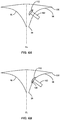
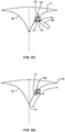
- the user may insert a leading end of the delivery device through slit 138. As shown in FIG. 6A , the user may then utilize deployment device 134 to urge distal disk 112 from the lumen 106 of delivery device 102. Upon exiting lumen 106, distal disk 112 will transition to its expanded configuration.
- distal disk 112 After distal disk 112 has been deployed, the user may retract delivery device 102 until the inner surface 126 of distal disk 112 engages with the distal surface of posterior leaflet 14, as shown in FIG. 6B . With the inner surface 126 of distal disk 112 engaged with leaflet 14, the user may urge plug 104 completely from the lumen 106 of delivery device 102, causing proximal disk 110 to expand and the inner surface 122 of proximal disk 110 to engage the proximal surface of the leaflet, as shown in FIG. 6C . Expansion of proximal and distal disks 110, 112 creates a clamping force on opposing sides of leaflet 14 and secures plug 104 to the leaflet.
- Plug 104 also causes intermediate portion 114 to expand between the portions of tissue on opposite sides of slit 138, pushing these tissue portions away from one another. This expands the slit opening in the longitudinal direction of leaflet 14 and causes the free edge 20 of the leaflet to move further from the base 136 thereof.
- Leaflet 14 is thus lengthened to its augmented length L2, which is sufficient to restore proper coaptation of the mitral valve leaflets.
- the user may then assess whether plug 104 has been properly positioned within leaflet 14. If the user determines that plug 104 has not been properly positioned, the user may retract deployment device 134, causing the plug to transition back to its collapsed configuration as it is withdrawn back into delivery device 102. The user may then redeploy plug 104 as described above until satisfied with its positioning. Once the user is satisfied with the positioning of plug 104 and leaflet 14 has been lengthened to its augmented length L2 such that proper coaptation has been restored, deployment device 134 may be rotated about its axis to unscrew the deployment device from clamp 130. Delivery device 102 may then be removed from the patient.
- the length of anterior leaflet 16 may be desirable to augment the length of anterior leaflet 16 rather than posterior leaflet 14, or it may be desirable to augment the length of both the posterior leaflet and the anterior leaflet. In these situations, the length of either or both of the leaflets may be augmented as described above.
- one aspect of the present disclosure is directed to a method of augmenting the length of a native leaflet.
- the method includes delivering a cutting tool to a surgical site adjacent the native leaflet; cutting a slit through the native leaflet; inserting a collapsible and expandable occlusion device into the slit, the occlusion device having a first disk, a second disk, and an intermediate portion coupling the first disk to the second disk; expanding the occlusion device; engaging a first surface of the first disk with a first surface of the native leaflet; and engaging a first surface of the second disk with a second surface of the native leaflet opposite the first surface; and/or the native leaflet may be one of an anterior leaflet or a posterior leaflet of a native mitral valve; and/or the slit may be cut through a belly of the native leaflet; and/or the slit may be cut through the native leaflet in a direction substantially parallel to the free edge of the native leaflet; and/or the step
- the apparatus includes a delivery device having a lumen; and a collapsible and expandable plug having a first disk, a second disk, an intermediate portion coupling the first disk to the second disk, and a longitudinal axis extending through the first disk, the intermediate portion and the second disk, the first disk having a concave surface relative to a plane passing between the first disk and the second disk in a direction orthogonal to the longitudinal axis, the concave surface being adapted to engage one surface of the native leaflet, the second disk having a convex surface relative to the plane, the convex surface being adapted to engage a surface of the native leaflet opposite the one surface; and/or the apparatus may further include a cutting device for cutting a slit through the native leaflet; and/or the occlusion device may be formed from braided nitinol or a polylactide mesh; and/or the braided nitin
Landscapes
- Health & Medical Sciences (AREA)
- Cardiology (AREA)
- Engineering & Computer Science (AREA)
- Biomedical Technology (AREA)
- Heart & Thoracic Surgery (AREA)
- Transplantation (AREA)
- Oral & Maxillofacial Surgery (AREA)
- Vascular Medicine (AREA)
- Life Sciences & Earth Sciences (AREA)
- Animal Behavior & Ethology (AREA)
- General Health & Medical Sciences (AREA)
- Public Health (AREA)
- Veterinary Medicine (AREA)
- Prostheses (AREA)
Applications Claiming Priority (1)
| Application Number | Priority Date | Filing Date | Title |
|---|---|---|---|
| US201862772188P | 2018-11-28 | 2018-11-28 |
Publications (1)
| Publication Number | Publication Date |
|---|---|
| EP3659552A1 true EP3659552A1 (de) | 2020-06-03 |
Family
ID=68696284
Family Applications (1)
| Application Number | Title | Priority Date | Filing Date |
|---|---|---|---|
| EP19211409.8A Withdrawn EP3659552A1 (de) | 2018-11-28 | 2019-11-26 | Transkatheter-mitralklappenblattverlängerung |
Country Status (2)
| Country | Link |
|---|---|
| US (1) | US20200163758A1 (de) |
| EP (1) | EP3659552A1 (de) |
Families Citing this family (1)
| Publication number | Priority date | Publication date | Assignee | Title |
|---|---|---|---|---|
| CN119033505B (zh) * | 2023-05-29 | 2025-09-23 | 科凯(南通)生命科学有限公司 | 心脏瓣膜修复用球囊体及心脏瓣膜修复组件 |
Citations (4)
| Publication number | Priority date | Publication date | Assignee | Title |
|---|---|---|---|---|
| US20090062841A1 (en) * | 2004-03-19 | 2009-03-05 | Aga Medical Corporation | Device for occluding vascular defects |
| US20100204662A1 (en) * | 2007-06-04 | 2010-08-12 | Mor Research Applications Ltd. | Cardiac valve leaflet augmentation |
| EP2410948A2 (de) * | 2005-02-07 | 2012-02-01 | Abbott Vascular | Verfahren, systeme und vorrichtungen zur reparatur von herzklappen |
| WO2017079153A1 (en) * | 2015-11-02 | 2017-05-11 | University Of Maryland, Baltimore | Distal anchor apparatus and methods for mitral valve repair |
-
2019
- 2019-08-27 US US16/552,195 patent/US20200163758A1/en not_active Abandoned
- 2019-11-26 EP EP19211409.8A patent/EP3659552A1/de not_active Withdrawn
Patent Citations (4)
| Publication number | Priority date | Publication date | Assignee | Title |
|---|---|---|---|---|
| US20090062841A1 (en) * | 2004-03-19 | 2009-03-05 | Aga Medical Corporation | Device for occluding vascular defects |
| EP2410948A2 (de) * | 2005-02-07 | 2012-02-01 | Abbott Vascular | Verfahren, systeme und vorrichtungen zur reparatur von herzklappen |
| US20100204662A1 (en) * | 2007-06-04 | 2010-08-12 | Mor Research Applications Ltd. | Cardiac valve leaflet augmentation |
| WO2017079153A1 (en) * | 2015-11-02 | 2017-05-11 | University Of Maryland, Baltimore | Distal anchor apparatus and methods for mitral valve repair |
Also Published As
| Publication number | Publication date |
|---|---|
| US20200163758A1 (en) | 2020-05-28 |
Similar Documents
| Publication | Publication Date | Title |
|---|---|---|
| US12138159B2 (en) | Methods and apparatus for engaging a valve prosthesis with tissue | |
| JP7337139B2 (ja) | 人工心臓弁 | |
| US11793640B2 (en) | Sequentially deployed transcatheter mitral valve prosthesis | |
| US11311375B2 (en) | Stents with anchoring sections | |
| US20220338977A1 (en) | Sequentially deployed transcatheter mitral valve prosthesis | |
| US10856974B2 (en) | Heart valve repair and replacement | |
| US10888424B2 (en) | Prosthetic mitral valve coaptation enhancement device | |
| US10639149B2 (en) | Sutureless prosthetic heart valve | |
| US10028821B2 (en) | Device for implantation in the heart of a mammal | |
| US8357195B2 (en) | Catheter based annuloplasty system and method | |
| CN111970996A (zh) | 心脏瓣膜密封装置及其递送装置 | |
| US20170079786A1 (en) | Self-expanding heart valves for coronary perfusion and sealing | |
| US20140371844A1 (en) | Transcatheter mitral valve and delivery system | |
| EP3659552A1 (de) | Transkatheter-mitralklappenblattverlängerung | |
| CN219354275U (zh) | 一种瓣膜修复装置及系统 | |
| WO2025049266A1 (en) | Prosthetic mitral valve for treating patients with small annulus dimensions |
Legal Events
| Date | Code | Title | Description |
|---|---|---|---|
| PUAI | Public reference made under article 153(3) epc to a published international application that has entered the european phase |
Free format text: ORIGINAL CODE: 0009012 |
|
| STAA | Information on the status of an ep patent application or granted ep patent |
Free format text: STATUS: THE APPLICATION HAS BEEN PUBLISHED |
|
| AK | Designated contracting states |
Kind code of ref document: A1 Designated state(s): AL AT BE BG CH CY CZ DE DK EE ES FI FR GB GR HR HU IE IS IT LI LT LU LV MC MK MT NL NO PL PT RO RS SE SI SK SM TR |
|
| AX | Request for extension of the european patent |
Extension state: BA ME |
|
| STAA | Information on the status of an ep patent application or granted ep patent |
Free format text: STATUS: THE APPLICATION IS DEEMED TO BE WITHDRAWN |
|
| 18D | Application deemed to be withdrawn |
Effective date: 20201204 |