EP3669772B1 - Verfahren zur visualisierung der darstellung von elektrophysiologischen wellen - Google Patents
Verfahren zur visualisierung der darstellung von elektrophysiologischen wellen Download PDFInfo
- Publication number
- EP3669772B1 EP3669772B1 EP19218127.9A EP19218127A EP3669772B1 EP 3669772 B1 EP3669772 B1 EP 3669772B1 EP 19218127 A EP19218127 A EP 19218127A EP 3669772 B1 EP3669772 B1 EP 3669772B1
- Authority
- EP
- European Patent Office
- Prior art keywords
- ecg
- anatomical map
- signal amplitudes
- processor
- map
- Prior art date
- Legal status (The legal status is an assumption and is not a legal conclusion. Google has not performed a legal analysis and makes no representation as to the accuracy of the status listed.)
- Active
Links
Images

Classifications
-
- A—HUMAN NECESSITIES
- A61—MEDICAL OR VETERINARY SCIENCE; HYGIENE
- A61B—DIAGNOSIS; SURGERY; IDENTIFICATION
- A61B5/00—Measuring for diagnostic purposes; Identification of persons
- A61B5/0002—Remote monitoring of patients using telemetry, e.g. transmission of vital signals via a communication network
- A61B5/0004—Remote monitoring of patients using telemetry, e.g. transmission of vital signals via a communication network characterised by the type of physiological signal transmitted
-
- A—HUMAN NECESSITIES
- A61—MEDICAL OR VETERINARY SCIENCE; HYGIENE
- A61B—DIAGNOSIS; SURGERY; IDENTIFICATION
- A61B5/00—Measuring for diagnostic purposes; Identification of persons
- A61B5/24—Detecting, measuring or recording bioelectric or biomagnetic signals of the body or parts thereof
- A61B5/316—Modalities, i.e. specific diagnostic methods
- A61B5/318—Heart-related electrical modalities, e.g. electrocardiography [ECG]
- A61B5/339—Displays specially adapted therefor
- A61B5/341—Vectorcardiography [VCG]
-
- A—HUMAN NECESSITIES
- A61—MEDICAL OR VETERINARY SCIENCE; HYGIENE
- A61B—DIAGNOSIS; SURGERY; IDENTIFICATION
- A61B5/00—Measuring for diagnostic purposes; Identification of persons
- A61B5/06—Devices, other than using radiation, for detecting or locating foreign bodies ; Determining position of diagnostic devices within or on the body of the patient
- A61B5/065—Determining position of the probe employing exclusively positioning means located on or in the probe, e.g. using position sensors arranged on the probe
- A61B5/068—Determining position of the probe employing exclusively positioning means located on or in the probe, e.g. using position sensors arranged on the probe using impedance sensors
-
- A—HUMAN NECESSITIES
- A61—MEDICAL OR VETERINARY SCIENCE; HYGIENE
- A61B—DIAGNOSIS; SURGERY; IDENTIFICATION
- A61B5/00—Measuring for diagnostic purposes; Identification of persons
- A61B5/24—Detecting, measuring or recording bioelectric or biomagnetic signals of the body or parts thereof
- A61B5/25—Bioelectric electrodes therefor
- A61B5/279—Bioelectric electrodes therefor specially adapted for particular uses
- A61B5/28—Bioelectric electrodes therefor specially adapted for particular uses for electrocardiography [ECG]
- A61B5/283—Invasive
-
- A—HUMAN NECESSITIES
- A61—MEDICAL OR VETERINARY SCIENCE; HYGIENE
- A61B—DIAGNOSIS; SURGERY; IDENTIFICATION
- A61B5/00—Measuring for diagnostic purposes; Identification of persons
- A61B5/24—Detecting, measuring or recording bioelectric or biomagnetic signals of the body or parts thereof
- A61B5/25—Bioelectric electrodes therefor
- A61B5/279—Bioelectric electrodes therefor specially adapted for particular uses
- A61B5/28—Bioelectric electrodes therefor specially adapted for particular uses for electrocardiography [ECG]
- A61B5/283—Invasive
- A61B5/287—Holders for multiple electrodes, e.g. electrode catheters for electrophysiological study [EPS]
-
- A—HUMAN NECESSITIES
- A61—MEDICAL OR VETERINARY SCIENCE; HYGIENE
- A61B—DIAGNOSIS; SURGERY; IDENTIFICATION
- A61B5/00—Measuring for diagnostic purposes; Identification of persons
- A61B5/24—Detecting, measuring or recording bioelectric or biomagnetic signals of the body or parts thereof
- A61B5/316—Modalities, i.e. specific diagnostic methods
- A61B5/318—Heart-related electrical modalities, e.g. electrocardiography [ECG]
- A61B5/33—Heart-related electrical modalities, e.g. electrocardiography [ECG] specially adapted for cooperation with other devices
-
- A—HUMAN NECESSITIES
- A61—MEDICAL OR VETERINARY SCIENCE; HYGIENE
- A61B—DIAGNOSIS; SURGERY; IDENTIFICATION
- A61B5/00—Measuring for diagnostic purposes; Identification of persons
- A61B5/24—Detecting, measuring or recording bioelectric or biomagnetic signals of the body or parts thereof
- A61B5/316—Modalities, i.e. specific diagnostic methods
- A61B5/318—Heart-related electrical modalities, e.g. electrocardiography [ECG]
- A61B5/339—Displays specially adapted therefor
-
- A—HUMAN NECESSITIES
- A61—MEDICAL OR VETERINARY SCIENCE; HYGIENE
- A61B—DIAGNOSIS; SURGERY; IDENTIFICATION
- A61B5/00—Measuring for diagnostic purposes; Identification of persons
- A61B5/68—Arrangements of detecting, measuring or recording means, e.g. sensors, in relation to patient
- A61B5/6846—Arrangements of detecting, measuring or recording means, e.g. sensors, in relation to patient specially adapted to be brought in contact with an internal body part, i.e. invasive
- A61B5/6847—Arrangements of detecting, measuring or recording means, e.g. sensors, in relation to patient specially adapted to be brought in contact with an internal body part, i.e. invasive mounted on an invasive device
- A61B5/6852—Catheters
-
- A—HUMAN NECESSITIES
- A61—MEDICAL OR VETERINARY SCIENCE; HYGIENE
- A61B—DIAGNOSIS; SURGERY; IDENTIFICATION
- A61B5/00—Measuring for diagnostic purposes; Identification of persons
- A61B5/74—Details of notification to user or communication with user or patient; User input means
- A61B5/742—Details of notification to user or communication with user or patient; User input means using visual displays
- A61B5/7425—Displaying combinations of multiple images regardless of image source, e.g. displaying a reference anatomical image with a live image
Definitions
- the present invention relates generally to electrophysiological mapping, and particularly to visualization of cardiac electrophysiological maps.
- Electrophysiological (EP) cardiac mapping may use visualizations methods previously proposed in the patent literature, to ease an interpretation of an EP map.
- 2017/0049348 describes a method for determining EP properties of cardiac tissue in order classify an arrhythmia.
- An eccentricity parameter reflecting the uniformity of a local conduction velocity, and divergence and curl-like sums or closed path integral parameters associated with the local velocity vectors are provided, and a rhythm classification responsive to catheter movement is displayed, thereby facilitating identification of types and causes of arrhythmia disorders.
- conduction velocity vector maps are coupled with local activation time (LAT) maps.
- LAT local activation time
- the display is updated immediately following each local depolarization and persisting or gradually fading out until the next local depolarization.
- some or all isochrones may be displayed as curved lines on the cardiac surface, for instance at specific intervals since the start of depolarization. This reduces visual clutter and allows a more interpretable superposition of conduction velocity arrows.
- U.S. Patent Application Publication 2010/0268059 describes a method that includes accessing cardiac information acquired via a catheter located at various positions in a venous network of a heart of a patient.
- the cardiac information comprises position information, electrical information and mechanical information.
- the method maps local electrical activation times to anatomic positions to generate an electrical activation time map.
- the method maps local mechanical activation times to anatomic positions to generate a mechanical activation time map.
- the method further generates an electromechanical delay map by subtracting local electrical activation times from corresponding local mechanical activation times, and renders at least the electromechanical delay map to a display.
- Cardiac ripple mapping A novel three-dimensional visualization method for use with electroanatomic mapping of cardiac arrhythmias
- Linton et al. Heart Rhythm, Elsevier, US, vol. 6, no 12
- electrogram visualization conveying both timing and morphology as well as location of each point within the chamber being studied.
- Data were used from six patients who had undergone electrophysiological study with the Carto electroanatomic mapping system.
- Software was written to construct a three-dimensional surface from the imported electrogram locations. Electrograms were time gated and displayed as dynamic bars that extend out from this surface, changing in length and color according to the local electrogram voltage-time relationship to create a ripple map of cardiac activation.
- An embodiment of the present invention provides a method including receiving an anatomical map of at least a portion of a heart. Positions and respective electrocardiogram (ECG) signal amplitudes measured at the positions are received for at least a region of the anatomical map. The ECG signal amplitudes are interpolated to derive a surface representation of the ECG signal amplitudes over the region. The surface representation of the ECG signal amplitudes is presented overlaid on the anatomical map. The anatomical map presents Local Activation Times (LAT).
- LAT Local Activation Times
- presenting the surface representation includes visualizing respective values of the surface representation as topographical heights above the anatomical map.
- presenting the surface representation includes presenting a semi-transparent surface that retains the anatomical map visible.
- interpolating the ECG signal amplitudes includes forming a shape including the interpolated and measured ECG amplitude values.
- a system including a memory and a processor.
- the memory is configured to store an anatomical map of at least a portion of a heart, and to store, for at least a region of the anatomical map, positions and respective electrocardiogram (ECG) signal amplitudes measured at the positions.
- the processor is configured to interpolate the ECG signal amplitudes to derive a surface representation of the ECG signal amplitudes over the region, and present the surface representation of the ECG signal amplitudes overlaid on the anatomical map.
- the anatomical map presents Local Activation Times (LAT).
- LAT Local Activation Times
- a catheter-based EP mapping system may be used for generating an EP map of least part of the heart of the patient, such as an EP map of a cardiac chamber.
- a distal end of a catheter which comprises sensing-electrodes, is inserted into the heart to sense EP signals.
- the EP mapping system acquires EP signals at various cardiac locations, as well as the respective positions of the distal end. Based on these acquired signals, a processor of the mapping system generates the required EP map.
- the processor of the EP mapping system presents the measured EP map overlaid on a heart anatomy visualized by, for example, a volume (3D) rendering of at least a portion of the heart.
- a volume (3D) rendering may be very useful in diagnosing cardiac irregularities.
- ECG "spikes" overlaid on an anatomical map may be used, where the height of the spikes gives a measure of the ECG signal amplitude at the spike position.
- the spike representation may indicate an anomalous conduction path causing an arrhythmia.
- this kind of visualization often tends to be too coarse and/or hides other features of diagnostic value.
- spikes only give a value of the signal at the given position, not in any intermediate positions, causing discontinuities in visualization, and furthermore the spikes may hide map details beneath or behind the spikes.
- Embodiments of the present invention that are described hereinafter use a processor to interpolate between measured values of the ECG signal at points near a selected location, and represent these interpolated values as topographical heights above the 3D rendering.
- the topographical heights are connected graphically to give a 3D surface referred to as a "sail,” having a continuous ripple, instead of a collection of spikes.
- the "sails" are made semi-transparent in order not to hide details.
- the processor is programmed in software containing a particular algorithm that enables the processor to conduct each of the processor related steps and functions outlined above.
- the disclosed visualization technique to overlay interpolated ECG ripples, that may be semi-transparent, on 3D cardiac anatomy, may improve the diagnostic value of catheter-based EP mapping procedures.
- Fig. 1 is a schematic, pictorial illustration of a cardiac three-dimensional (3D) navigation and electrophysiological (EP) signal analysis system 20, in accordance with an embodiment of the present invention.
- System 20 may be configured to analyze substantially any physiological parameter or combinations of such parameters.
- the signals analyzed are assumed to be intra-cardiac and/or extra-cardiac (body surface) electrocardiogram (ECG) potential-time relationships.
- ECG electrocardiogram
- a processor 40 uses the ECG signals to produce an EP map, such as a local activation time (LAT) map.
- LAT local activation time
- anatomical map refers to a map that models the 3D shape of at least a portion of the heart, and may have one or more parameters overlaid thereon.
- An EP map is one special case of an anatomical map, in which one or more electrophysiological parameters are overlaid.
- a LAT map is an example of an EP map, and thus also regarded as a type of anatomical map.
- Fig. 1 shows an investigative procedure wherein system 20 measures actual electrical activity of a heart 34, using a probe 24.
- probe 24 comprises a catheter which is inserted into the body of patient 26 during a mapping procedure performed by a physician 28 using system 20.
- a distal end 32 of probe 24 is assumed to have electrodes 22.
- patient 26 is assumed to be attached to a grounding electrode 23.
- electrodes 29 are assumed to be attached to the skin of patient 26, in the region of heart 34.
- probe 24 acquires local intra-cardiac ECG as the probe moves over a portion of the heart chamber. At these instances, probe 24 location is recorded as well.
- the measured signals are used, as noted above and among other usages, to create an LAT map of at least part of the wall tissue of heart 34 of a patient 26.
- System 20 is controlled by a system processor 40, comprising a processing unit 42 communicating with a memory 44.
- a memory 44 which is included in system processor 40, stores an LAT and/or ECG map 62 of at least part of wall tissue of heart 34 of patient 26.
- Processor 40 is typically mounted in a console 46, which comprises operating controls 38, typically including a pointing device 39 such as a mouse or trackball, that physician 28 uses to interact with the processor.
- Processor 40 (specifically processing unit 42) runs software, comprising a probe tracker module 30, an ECG module 36, and an ECG amplitude visualization module 35, used for visualizing ECG amplitudes over a 3D rendering of a portion of heart 26 anatomy (i.e., in the form of "sails"), as described above and described in further detail below.
- ECG module 36 is coupled to receive actual electrical signals from electrodes 22 and electrodes 29. The module is configured to analyze the actual signals and may present the results of the analysis in a standard ECG format, typically a graphical representation moving with time, on display 48.
- Probe tracker module 30 typically tracks the location of distal end 32 of probe 24 within the heart of patient 26.
- the tracker module may use any method for location tracking probes known in the art.
- module 30 may operate a magnetic-field-based location tracking sub-system. (For simplicity, components of such a sub-system are not shown in Fig. 1 .)
- processor 40 is able to measure locations of distal end 32.
- the processor is able to measure locations of the distal end, as well as LATs of the actual electrical signals detected at these particular locations.
- tracker module 30 may track probe 24 by measuring impedances between electrode 23, electrodes 29, and electrodes 22, as well as the impedances to other electrodes which may be located on the probe. (In this case electrodes 22 and/or electrodes 29 may provide both ECG and location tracking signals.)
- the Carto3 ® system produced by Biosense-Webster (Irvine, California), uses both magnetic field location tracking and impedance measurements for location tracking.
- Results of the operations and visualizations performed by processor 40 are presented to physician 28 on a display 48, which typically presents a graphic user interface to the physician, a visual representation of the ECG signals sensed by electrodes 22, and/or an image or map of heart 34 while it is being investigated.
- EP activation analysis module 35 presents to the physician a LAT map overlaid with the interpolated ECG amplitude "sails.”
- processor 40 may be downloaded to processor 40 in electronic form, over a network, for example, or it may, alternatively or additionally, be provided and/or stored on non-transitory tangible media, such as magnetic, optical, or electronic memory.
- processor 40 runs a dedicated algorithm that enables processor 40 to perform the disclosed steps, as described below.
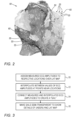
- Fig. 2 is a volume rendering showing ripple-mapping visualization of ECG amplitudes overlaid on a cardiac chamber 50 anatomy, in accordance with an embodiment of the present invention.
- ECG measured values i.e., spikes 55
- the disclosed visualization method not only gives the value of the ECG spikes at the given position, but also at any intermediate positions, which forms sail 60 shapes of the local ECG amplitudes.
- processor 40 interpolates values of ECG signals 55 at points near a selected location, and connects the topographical height values above the 3D rendering so as to produce the disclosed continuous ripple visualization.
- processor 40 generates semi-transparent sails 60 in order not to hide details behind them (i.e., to show details of underlying map) .
- ripple-mapping visualization shown in Fig. 2 is chosen purely for the sake of conceptual clarity.
- Various additional visualization tools may apply, such a presenting numbers, a magnifying glass effect to view sails 60 in detail, and others.
- Fig. 3 is flow chart that schematically illustrates a method for ripple-mapping visualization of ECG amplitudes shown in Fig. 2 , in accordance with an embodiment of the present invention.
- the algorithm carries out a process that begins with processor 40 assigning measured ECG amplitudes 55 to respective locations over an anatomical map of a cardiac chamber (i.e., overlaid on the electro-anatomically - such as LAT - mapped surface of chamber 50), at an ECG amplitude assigning step 70.
- processor 40 interpolates over values of ECG at points near each location.
- processor 40 connects measured and interpolated ECG amplitudes to create sails 60, at an interconnecting ECG amplitudes step 74.
- processor 40 further makes sails 60 semi-transparent, at a sail visualization step 76.
- Processor 40 presents the resulting visualization (LAT map with overlaid interpolated ECG amplitudes) to physician 28 on display 48.
- the example flow chart shown in Fig. 3 is chosen purely for the sake of conceptual clarity.
- the present embodiment also comprises additional steps of the algorithm. Examples include additional visualizations, such as conduction arrows between and under sails 60. Such additional steps have been omitted from the disclosure herein purposely on order to provide a more simplified flow chart.
Landscapes
- Health & Medical Sciences (AREA)
- Life Sciences & Earth Sciences (AREA)
- Engineering & Computer Science (AREA)
- Molecular Biology (AREA)
- Animal Behavior & Ethology (AREA)
- Pathology (AREA)
- Physics & Mathematics (AREA)
- Biomedical Technology (AREA)
- Heart & Thoracic Surgery (AREA)
- Medical Informatics (AREA)
- Veterinary Medicine (AREA)
- Surgery (AREA)
- Biophysics (AREA)
- General Health & Medical Sciences (AREA)
- Public Health (AREA)
- Cardiology (AREA)
- Nuclear Medicine, Radiotherapy & Molecular Imaging (AREA)
- Radiology & Medical Imaging (AREA)
- Physiology (AREA)
- Computer Networks & Wireless Communication (AREA)
- Human Computer Interaction (AREA)
- Measurement And Recording Of Electrical Phenomena And Electrical Characteristics Of The Living Body (AREA)
Claims (8)
- Verfahren, umfassend:Empfangen einer anatomischen Karte von mindestens einem Abschnitt eines Herzens;Empfangen, für mindestens eine Region der anatomischen Karte, von Positionen und jeweiligen an den Positionen gemessenen Elektrokardiogramm-(EKG-)Signalamplituden;Interpolieren der EKG-Signalamplituden, um eine Oberflächendarstellung der EKG-Signalamplituden über der Region abzuleiten; undDarstellen der Oberflächendarstellung der EKG-Signalamplituden, die auf der anatomischen Karte überlagert sind, wobei die anatomische Karte lokale Aktivierungszeiten (LATs) darstellt.
- Verfahren nach Anspruch 1, wobei das Darstellen der Oberflächendarstellung ein Visualisieren jeweiliger Werte der Oberflächendarstellung als topografische Höhen über der anatomischen Karte umfasst.
- Verfahren nach Anspruch 1, wobei das Darstellen der Oberflächendarstellung das Darstellen einer halbtransparenten Oberfläche umfasst, die die anatomische Karte sichtbar hält.
- Verfahren nach Anspruch 1, wobei das Interpolieren der EKG-Signalamplituden das Ausbilden einer Form umfasst, umfassend die interpolierten und gemessenen EKG-Amplitudenwerte.
- System, umfassend:
einen Speicher, der konfiguriert ist zum:Speichern einer anatomischen Karte von mindestens einem Abschnitt eines Herzens; undSpeichern, für mindestens eine Region der anatomischen Karte, von Positionen und jeweiligen an den Positionen gemessenen Elektrokardiogramm-(EKG-)Signalamplituden; undeinen Prozessor, der konfiguriert ist zum:Interpolieren der EKG-Signalamplituden, um eine Oberflächendarstellung der EKG-Signalamplituden über der Region abzuleiten; undDarstellen der Oberflächendarstellung der EKG-Signalamplituden, die auf der anatomischen Karte überlagert sind, wobei die anatomische Karte die lokalen Aktivierungszeiten (LATs) darstellt. - System nach Anspruch 5, wobei der Prozessor konfiguriert ist, um die Oberflächendarstellung durch Visualisieren jeweiliger Werte der Oberflächendarstellung als topografische Höhen über der anatomischen Karte darzustellen.
- System nach Anspruch 5, wobei der Prozessor konfiguriert ist, um die Oberflächendarstellung durch Darstellung einer halbtransparenten Oberfläche darzustellen, die die anatomische Karte sichtbar hält.
- System nach Anspruch 5, wobei der Prozessor konfiguriert ist, um die EKG-Signalamplituden zu interpolieren, durch Ausbilden einer Form, umfassend die interpolierten und gemessenen EKG-Amplitudenwerte.
Applications Claiming Priority (1)
| Application Number | Priority Date | Filing Date | Title |
|---|---|---|---|
| US16/228,426 US11123004B2 (en) | 2018-12-20 | 2018-12-20 | Electrophysiological ripple mapping visualization method |
Publications (3)
| Publication Number | Publication Date |
|---|---|
| EP3669772A1 EP3669772A1 (de) | 2020-06-24 |
| EP3669772C0 EP3669772C0 (de) | 2024-10-16 |
| EP3669772B1 true EP3669772B1 (de) | 2024-10-16 |
Family
ID=69410930
Family Applications (1)
| Application Number | Title | Priority Date | Filing Date |
|---|---|---|---|
| EP19218127.9A Active EP3669772B1 (de) | 2018-12-20 | 2019-12-19 | Verfahren zur visualisierung der darstellung von elektrophysiologischen wellen |
Country Status (5)
| Country | Link |
|---|---|
| US (1) | US11123004B2 (de) |
| EP (1) | EP3669772B1 (de) |
| JP (1) | JP2020099689A (de) |
| CN (1) | CN111345810B (de) |
| IL (1) | IL270433B (de) |
Families Citing this family (1)
| Publication number | Priority date | Publication date | Assignee | Title |
|---|---|---|---|---|
| US11160485B2 (en) * | 2020-04-01 | 2021-11-02 | Biosense Webster (Israel) Ltd. | Propagation map of a heart chamber with areas demonstrating fractionated electrograms |
Family Cites Families (22)
| Publication number | Priority date | Publication date | Assignee | Title |
|---|---|---|---|---|
| CN1124824C (zh) * | 1999-09-27 | 2003-10-22 | 复旦大学 | 动态心电标测方法及其装置 |
| US20070232949A1 (en) | 2006-03-31 | 2007-10-04 | Ep Medsystems, Inc. | Method For Simultaneous Bi-Atrial Mapping Of Atrial Fibrillation |
| US7774051B2 (en) * | 2006-05-17 | 2010-08-10 | St. Jude Medical, Atrial Fibrillation Division, Inc. | System and method for mapping electrophysiology information onto complex geometry |
| CN101460088B (zh) * | 2006-06-02 | 2011-06-15 | 皇家飞利浦电子股份有限公司 | 生物反馈系统和显示设备 |
| US7729752B2 (en) | 2006-06-13 | 2010-06-01 | Rhythmia Medical, Inc. | Non-contact cardiac mapping, including resolution map |
| GB0708781D0 (en) * | 2007-05-04 | 2007-06-13 | Imp Innovations Ltd | A Method of and apparatus for generating a model of a cardiac surface having a plurality of images representing electrogram voltages |
| JP5275340B2 (ja) * | 2007-05-08 | 2013-08-28 | シー・アール・バード・インコーポレーテッド | 多電極位置データを用いた迅速な3dマッピング |
| WO2009027812A2 (en) * | 2007-08-31 | 2009-03-05 | Medicalgorithmics Sp. Zo.O | Reconstruction of geometry of a body component and analysis of spatial distribution of electrophysiological values |
| JP5337367B2 (ja) * | 2007-10-31 | 2013-11-06 | 株式会社東芝 | 医用画像表示装置 |
| US8326419B2 (en) | 2009-04-07 | 2012-12-04 | Pacesetter, Inc. | Therapy optimization via multi-dimensional mapping |
| US9220425B2 (en) | 2012-09-17 | 2015-12-29 | Magnetecs Corp. | Method and apparatus for measuring biopotential and mapping ephaptic coupling employing a catheter with MOSFET sensor array |
| US9050011B2 (en) | 2012-12-26 | 2015-06-09 | Biosense Webster (Israel) Ltd. | Removal of artifacts from map data |
| WO2015130829A1 (en) | 2014-02-25 | 2015-09-03 | St. Jude Medical, Cardiology Division, Inc. | Systems and methods for using electrophysiology properties for classifying arrhythmia sources |
| JP6320634B2 (ja) * | 2014-10-15 | 2018-05-09 | セント・ジュード・メディカル,カーディオロジー・ディヴィジョン,インコーポレイテッド | 局所伝導速度をマッピングするための方法及びシステム |
| CN107072573B (zh) * | 2014-10-15 | 2018-11-16 | 圣犹达医疗用品心脏病学部门有限公司 | 用于生成针对心律失常的集成的基质标测图的方法和系统 |
| US9955889B2 (en) * | 2014-11-03 | 2018-05-01 | Biosense Webster (Israel) Ltd. | Registration maps using intra-cardiac signals |
| EP3206575A1 (de) * | 2015-01-07 | 2017-08-23 | St. Jude Medical, Cardiology Division, Inc. | System, verfahren und vorrichtung zur visualisierung von kardialen zeitinformationen unter verwendung von animationen |
| CN108601546B (zh) * | 2016-02-04 | 2021-03-26 | 日本电信电话株式会社 | 生物信号处理方法和生物信号处理设备 |
| US10360700B2 (en) * | 2016-02-11 | 2019-07-23 | Biosense Webster (Israel) Ltd. | Interpolation of dynamic three-dimensional maps |
| US10136828B2 (en) * | 2016-03-31 | 2018-11-27 | Biosense Webster (Israel) Ltd. | Mapping of atrial fibrillation |
| US10403053B2 (en) * | 2016-11-15 | 2019-09-03 | Biosense Webster (Israel) Ltd. | Marking sparse areas on maps |
| US10537261B2 (en) * | 2017-05-10 | 2020-01-21 | Boston Scientific Scimed Inc. | Region-of-interest representations for electroanatomical mapping |
-
2018
- 2018-12-20 US US16/228,426 patent/US11123004B2/en active Active
-
2019
- 2019-11-05 IL IL270433A patent/IL270433B/en unknown
- 2019-12-19 JP JP2019229067A patent/JP2020099689A/ja active Pending
- 2019-12-19 EP EP19218127.9A patent/EP3669772B1/de active Active
- 2019-12-20 CN CN201911324373.3A patent/CN111345810B/zh active Active
Also Published As
| Publication number | Publication date |
|---|---|
| CN111345810B (zh) | 2024-04-16 |
| EP3669772C0 (de) | 2024-10-16 |
| US20200196890A1 (en) | 2020-06-25 |
| CN111345810A (zh) | 2020-06-30 |
| IL270433B (en) | 2022-08-01 |
| EP3669772A1 (de) | 2020-06-24 |
| US11123004B2 (en) | 2021-09-21 |
| JP2020099689A (ja) | 2020-07-02 |
| IL270433A (en) | 2020-06-30 |
Similar Documents
| Publication | Publication Date | Title |
|---|---|---|
| US7774051B2 (en) | System and method for mapping electrophysiology information onto complex geometry | |
| EP2982293B1 (de) | Wellenfrontanalyse basierend auf ablationsparametern | |
| EP3750478B1 (de) | Visuell differenzierte primäre und sekundäre aktivierungen auf elektrophysiologischen karten | |
| EP2826418B1 (de) | Herztätigkeitsvisualisierung mit Frequenzdiskrimination | |
| EP3888553B1 (de) | Ausbreitungskarte einer herzkammer mit bereichen mit fraktionierten elektrogrammen | |
| EP3537444A1 (de) | Simulation einer schrittmachertherapie zur modellierung von arrhythmie | |
| US11123004B2 (en) | Electrophysiological ripple mapping visualization method | |
| EP4144295B1 (de) | Gewichtung der projizierten elektrophysiologischen wellengeschwindigkeit mit der sigmoidkurve | |
| IL294333A (en) | Local Latency Time (LAT) mapping with multiple session layers |
Legal Events
| Date | Code | Title | Description |
|---|---|---|---|
| PUAI | Public reference made under article 153(3) epc to a published international application that has entered the european phase |
Free format text: ORIGINAL CODE: 0009012 |
|
| STAA | Information on the status of an ep patent application or granted ep patent |
Free format text: STATUS: THE APPLICATION HAS BEEN PUBLISHED |
|
| AK | Designated contracting states |
Kind code of ref document: A1 Designated state(s): AL AT BE BG CH CY CZ DE DK EE ES FI FR GB GR HR HU IE IS IT LI LT LU LV MC MK MT NL NO PL PT RO RS SE SI SK SM TR |
|
| AX | Request for extension of the european patent |
Extension state: BA ME |
|
| STAA | Information on the status of an ep patent application or granted ep patent |
Free format text: STATUS: REQUEST FOR EXAMINATION WAS MADE |
|
| 17P | Request for examination filed |
Effective date: 20201217 |
|
| RBV | Designated contracting states (corrected) |
Designated state(s): AL AT BE BG CH CY CZ DE DK EE ES FI FR GB GR HR HU IE IS IT LI LT LU LV MC MK MT NL NO PL PT RO RS SE SI SK SM TR |
|
| STAA | Information on the status of an ep patent application or granted ep patent |
Free format text: STATUS: EXAMINATION IS IN PROGRESS |
|
| 17Q | First examination report despatched |
Effective date: 20231221 |
|
| REG | Reference to a national code |
Ref country code: DE Ref legal event code: R079 Free format text: PREVIOUS MAIN CLASS: A61B0005042000 Ref country code: DE Ref legal event code: R079 Ref document number: 602019060418 Country of ref document: DE Free format text: PREVIOUS MAIN CLASS: A61B0005042000 Ipc: A61B0005000000 |
|
| RIC1 | Information provided on ipc code assigned before grant |
Ipc: A61B 5/00 20060101AFI20240419BHEP |
|
| GRAP | Despatch of communication of intention to grant a patent |
Free format text: ORIGINAL CODE: EPIDOSNIGR1 |
|
| STAA | Information on the status of an ep patent application or granted ep patent |
Free format text: STATUS: GRANT OF PATENT IS INTENDED |
|
| INTG | Intention to grant announced |
Effective date: 20240603 |
|
| GRAS | Grant fee paid |
Free format text: ORIGINAL CODE: EPIDOSNIGR3 |
|
| GRAA | (expected) grant |
Free format text: ORIGINAL CODE: 0009210 |
|
| STAA | Information on the status of an ep patent application or granted ep patent |
Free format text: STATUS: THE PATENT HAS BEEN GRANTED |
|
| AK | Designated contracting states |
Kind code of ref document: B1 Designated state(s): AL AT BE BG CH CY CZ DE DK EE ES FI FR GB GR HR HU IE IS IT LI LT LU LV MC MK MT NL NO PL PT RO RS SE SI SK SM TR |
|
| REG | Reference to a national code |
Ref country code: GB Ref legal event code: FG4D |
|
| REG | Reference to a national code |
Ref country code: CH Ref legal event code: EP |
|
| REG | Reference to a national code |
Ref country code: IE Ref legal event code: FG4D |
|
| REG | Reference to a national code |
Ref country code: DE Ref legal event code: R096 Ref document number: 602019060418 Country of ref document: DE |
|
| U01 | Request for unitary effect filed |
Effective date: 20241031 |
|
| U07 | Unitary effect registered |
Designated state(s): AT BE BG DE DK EE FI FR IT LT LU LV MT NL PT RO SE SI Effective date: 20241113 |
|
| U20 | Renewal fee for the european patent with unitary effect paid |
Year of fee payment: 6 Effective date: 20241122 |
|
| PG25 | Lapsed in a contracting state [announced via postgrant information from national office to epo] |
Ref country code: HR Free format text: LAPSE BECAUSE OF FAILURE TO SUBMIT A TRANSLATION OF THE DESCRIPTION OR TO PAY THE FEE WITHIN THE PRESCRIBED TIME-LIMIT Effective date: 20241016 Ref country code: IS Free format text: LAPSE BECAUSE OF FAILURE TO SUBMIT A TRANSLATION OF THE DESCRIPTION OR TO PAY THE FEE WITHIN THE PRESCRIBED TIME-LIMIT Effective date: 20250216 |
|
| PG25 | Lapsed in a contracting state [announced via postgrant information from national office to epo] |
Ref country code: ES Free format text: LAPSE BECAUSE OF FAILURE TO SUBMIT A TRANSLATION OF THE DESCRIPTION OR TO PAY THE FEE WITHIN THE PRESCRIBED TIME-LIMIT Effective date: 20241016 |
|
| PG25 | Lapsed in a contracting state [announced via postgrant information from national office to epo] |
Ref country code: NO Free format text: LAPSE BECAUSE OF FAILURE TO SUBMIT A TRANSLATION OF THE DESCRIPTION OR TO PAY THE FEE WITHIN THE PRESCRIBED TIME-LIMIT Effective date: 20250116 |
|
| PG25 | Lapsed in a contracting state [announced via postgrant information from national office to epo] |
Ref country code: GR Free format text: LAPSE BECAUSE OF FAILURE TO SUBMIT A TRANSLATION OF THE DESCRIPTION OR TO PAY THE FEE WITHIN THE PRESCRIBED TIME-LIMIT Effective date: 20250117 |
|
| PG25 | Lapsed in a contracting state [announced via postgrant information from national office to epo] |
Ref country code: PL Free format text: LAPSE BECAUSE OF FAILURE TO SUBMIT A TRANSLATION OF THE DESCRIPTION OR TO PAY THE FEE WITHIN THE PRESCRIBED TIME-LIMIT Effective date: 20241016 |
|
| PG25 | Lapsed in a contracting state [announced via postgrant information from national office to epo] |
Ref country code: RS Free format text: LAPSE BECAUSE OF FAILURE TO SUBMIT A TRANSLATION OF THE DESCRIPTION OR TO PAY THE FEE WITHIN THE PRESCRIBED TIME-LIMIT Effective date: 20250116 |
|
| PG25 | Lapsed in a contracting state [announced via postgrant information from national office to epo] |
Ref country code: SM Free format text: LAPSE BECAUSE OF FAILURE TO SUBMIT A TRANSLATION OF THE DESCRIPTION OR TO PAY THE FEE WITHIN THE PRESCRIBED TIME-LIMIT Effective date: 20241016 |
|
| PG25 | Lapsed in a contracting state [announced via postgrant information from national office to epo] |
Ref country code: MC Free format text: LAPSE BECAUSE OF FAILURE TO SUBMIT A TRANSLATION OF THE DESCRIPTION OR TO PAY THE FEE WITHIN THE PRESCRIBED TIME-LIMIT Effective date: 20241016 |
|
| PG25 | Lapsed in a contracting state [announced via postgrant information from national office to epo] |
Ref country code: SK Free format text: LAPSE BECAUSE OF FAILURE TO SUBMIT A TRANSLATION OF THE DESCRIPTION OR TO PAY THE FEE WITHIN THE PRESCRIBED TIME-LIMIT Effective date: 20241016 |
|
| PG25 | Lapsed in a contracting state [announced via postgrant information from national office to epo] |
Ref country code: CZ Free format text: LAPSE BECAUSE OF FAILURE TO SUBMIT A TRANSLATION OF THE DESCRIPTION OR TO PAY THE FEE WITHIN THE PRESCRIBED TIME-LIMIT Effective date: 20241016 |
|
| REG | Reference to a national code |
Ref country code: CH Ref legal event code: PL |
|
| PLBE | No opposition filed within time limit |
Free format text: ORIGINAL CODE: 0009261 |
|
| STAA | Information on the status of an ep patent application or granted ep patent |
Free format text: STATUS: NO OPPOSITION FILED WITHIN TIME LIMIT |
|
| 26N | No opposition filed |
Effective date: 20250717 |
|
| PG25 | Lapsed in a contracting state [announced via postgrant information from national office to epo] |
Ref country code: CH Free format text: LAPSE BECAUSE OF NON-PAYMENT OF DUE FEES Effective date: 20241231 |
|
| PG25 | Lapsed in a contracting state [announced via postgrant information from national office to epo] |
Ref country code: IE Free format text: LAPSE BECAUSE OF NON-PAYMENT OF DUE FEES Effective date: 20241219 |
|
| U20 | Renewal fee for the european patent with unitary effect paid |
Year of fee payment: 7 Effective date: 20251110 |
|
| PGFP | Annual fee paid to national office [announced via postgrant information from national office to epo] |
Ref country code: GB Payment date: 20251030 Year of fee payment: 7 |