EP3672476B1 - Systeme und verfahren zur messung der ziliarmuskelaktivität - Google Patents
Systeme und verfahren zur messung der ziliarmuskelaktivität Download PDFInfo
- Publication number
- EP3672476B1 EP3672476B1 EP18797138.7A EP18797138A EP3672476B1 EP 3672476 B1 EP3672476 B1 EP 3672476B1 EP 18797138 A EP18797138 A EP 18797138A EP 3672476 B1 EP3672476 B1 EP 3672476B1
- Authority
- EP
- European Patent Office
- Prior art keywords
- signals
- ciliary muscle
- segments
- patient
- subset
- Prior art date
- Legal status (The legal status is an assumption and is not a legal conclusion. Google has not performed a legal analysis and makes no representation as to the accuracy of the status listed.)
- Active
Links
Images

Classifications
-
- A—HUMAN NECESSITIES
- A61—MEDICAL OR VETERINARY SCIENCE; HYGIENE
- A61F—FILTERS IMPLANTABLE INTO BLOOD VESSELS; PROSTHESES; DEVICES PROVIDING PATENCY TO, OR PREVENTING COLLAPSING OF, TUBULAR STRUCTURES OF THE BODY, e.g. STENTS; ORTHOPAEDIC, NURSING OR CONTRACEPTIVE DEVICES; FOMENTATION; TREATMENT OR PROTECTION OF EYES OR EARS; BANDAGES, DRESSINGS OR ABSORBENT PADS; FIRST-AID KITS
- A61F2/00—Filters implantable into blood vessels; Prostheses, i.e. artificial substitutes or replacements for parts of the body; Appliances for connecting them with the body; Devices providing patency to, or preventing collapsing of, tubular structures of the body, e.g. stents
- A61F2/02—Prostheses implantable into the body
- A61F2/14—Eye parts, e.g. lenses or corneal implants; Artificial eyes
- A61F2/16—Intraocular lenses
- A61F2/1613—Intraocular lenses having special lens configurations, e.g. multipart lenses; having particular optical properties, e.g. pseudo-accommodative lenses, lenses having aberration corrections, diffractive lenses, lenses for variably absorbing electromagnetic radiation, lenses having variable focus
- A61F2/1624—Intraocular lenses having special lens configurations, e.g. multipart lenses; having particular optical properties, e.g. pseudo-accommodative lenses, lenses having aberration corrections, diffractive lenses, lenses for variably absorbing electromagnetic radiation, lenses having variable focus having adjustable focus; power activated variable focus means, e.g. mechanically or electrically by the ciliary muscle or from the outside
-
- A—HUMAN NECESSITIES
- A61—MEDICAL OR VETERINARY SCIENCE; HYGIENE
- A61B—DIAGNOSIS; SURGERY; IDENTIFICATION
- A61B5/00—Measuring for diagnostic purposes; Identification of persons
- A61B5/24—Detecting, measuring or recording bioelectric or biomagnetic signals of the body or parts thereof
- A61B5/25—Bioelectric electrodes therefor
- A61B5/279—Bioelectric electrodes therefor specially adapted for particular uses
- A61B5/296—Bioelectric electrodes therefor specially adapted for particular uses for electromyography [EMG]
-
- A—HUMAN NECESSITIES
- A61—MEDICAL OR VETERINARY SCIENCE; HYGIENE
- A61B—DIAGNOSIS; SURGERY; IDENTIFICATION
- A61B5/00—Measuring for diagnostic purposes; Identification of persons
- A61B5/24—Detecting, measuring or recording bioelectric or biomagnetic signals of the body or parts thereof
- A61B5/25—Bioelectric electrodes therefor
- A61B5/279—Bioelectric electrodes therefor specially adapted for particular uses
- A61B5/297—Bioelectric electrodes therefor specially adapted for particular uses for electrooculography [EOG]: for electroretinography [ERG]
-
- A—HUMAN NECESSITIES
- A61—MEDICAL OR VETERINARY SCIENCE; HYGIENE
- A61B—DIAGNOSIS; SURGERY; IDENTIFICATION
- A61B5/00—Measuring for diagnostic purposes; Identification of persons
- A61B5/24—Detecting, measuring or recording bioelectric or biomagnetic signals of the body or parts thereof
- A61B5/316—Modalities, i.e. specific diagnostic methods
- A61B5/389—Electromyography [EMG]
-
- A—HUMAN NECESSITIES
- A61—MEDICAL OR VETERINARY SCIENCE; HYGIENE
- A61B—DIAGNOSIS; SURGERY; IDENTIFICATION
- A61B5/00—Measuring for diagnostic purposes; Identification of persons
- A61B5/24—Detecting, measuring or recording bioelectric or biomagnetic signals of the body or parts thereof
- A61B5/316—Modalities, i.e. specific diagnostic methods
- A61B5/398—Electrooculography [EOG], e.g. detecting nystagmus; Electroretinography [ERG]
-
- A—HUMAN NECESSITIES
- A61—MEDICAL OR VETERINARY SCIENCE; HYGIENE
- A61B—DIAGNOSIS; SURGERY; IDENTIFICATION
- A61B5/00—Measuring for diagnostic purposes; Identification of persons
- A61B5/68—Arrangements of detecting, measuring or recording means, e.g. sensors, in relation to patient
- A61B5/6801—Arrangements of detecting, measuring or recording means, e.g. sensors, in relation to patient specially adapted to be attached to or worn on the body surface
- A61B5/6813—Specially adapted to be attached to a specific body part
- A61B5/6814—Head
- A61B5/6821—Eye
-
- A—HUMAN NECESSITIES
- A61—MEDICAL OR VETERINARY SCIENCE; HYGIENE
- A61B—DIAGNOSIS; SURGERY; IDENTIFICATION
- A61B5/00—Measuring for diagnostic purposes; Identification of persons
- A61B5/68—Arrangements of detecting, measuring or recording means, e.g. sensors, in relation to patient
- A61B5/6801—Arrangements of detecting, measuring or recording means, e.g. sensors, in relation to patient specially adapted to be attached to or worn on the body surface
- A61B5/6844—Monitoring or controlling distance between sensor and tissue
-
- A—HUMAN NECESSITIES
- A61—MEDICAL OR VETERINARY SCIENCE; HYGIENE
- A61B—DIAGNOSIS; SURGERY; IDENTIFICATION
- A61B5/00—Measuring for diagnostic purposes; Identification of persons
- A61B5/68—Arrangements of detecting, measuring or recording means, e.g. sensors, in relation to patient
- A61B5/6846—Arrangements of detecting, measuring or recording means, e.g. sensors, in relation to patient specially adapted to be brought in contact with an internal body part, i.e. invasive
- A61B5/6847—Arrangements of detecting, measuring or recording means, e.g. sensors, in relation to patient specially adapted to be brought in contact with an internal body part, i.e. invasive mounted on an invasive device
-
- A—HUMAN NECESSITIES
- A61—MEDICAL OR VETERINARY SCIENCE; HYGIENE
- A61B—DIAGNOSIS; SURGERY; IDENTIFICATION
- A61B5/00—Measuring for diagnostic purposes; Identification of persons
- A61B5/72—Signal processing specially adapted for physiological signals or for diagnostic purposes
-
- A—HUMAN NECESSITIES
- A61—MEDICAL OR VETERINARY SCIENCE; HYGIENE
- A61F—FILTERS IMPLANTABLE INTO BLOOD VESSELS; PROSTHESES; DEVICES PROVIDING PATENCY TO, OR PREVENTING COLLAPSING OF, TUBULAR STRUCTURES OF THE BODY, e.g. STENTS; ORTHOPAEDIC, NURSING OR CONTRACEPTIVE DEVICES; FOMENTATION; TREATMENT OR PROTECTION OF EYES OR EARS; BANDAGES, DRESSINGS OR ABSORBENT PADS; FIRST-AID KITS
- A61F2/00—Filters implantable into blood vessels; Prostheses, i.e. artificial substitutes or replacements for parts of the body; Appliances for connecting them with the body; Devices providing patency to, or preventing collapsing of, tubular structures of the body, e.g. stents
- A61F2/02—Prostheses implantable into the body
- A61F2/14—Eye parts, e.g. lenses or corneal implants; Artificial eyes
- A61F2/16—Intraocular lenses
-
- A—HUMAN NECESSITIES
- A61—MEDICAL OR VETERINARY SCIENCE; HYGIENE
- A61F—FILTERS IMPLANTABLE INTO BLOOD VESSELS; PROSTHESES; DEVICES PROVIDING PATENCY TO, OR PREVENTING COLLAPSING OF, TUBULAR STRUCTURES OF THE BODY, e.g. STENTS; ORTHOPAEDIC, NURSING OR CONTRACEPTIVE DEVICES; FOMENTATION; TREATMENT OR PROTECTION OF EYES OR EARS; BANDAGES, DRESSINGS OR ABSORBENT PADS; FIRST-AID KITS
- A61F2/00—Filters implantable into blood vessels; Prostheses, i.e. artificial substitutes or replacements for parts of the body; Appliances for connecting them with the body; Devices providing patency to, or preventing collapsing of, tubular structures of the body, e.g. stents
- A61F2/02—Prostheses implantable into the body
- A61F2/14—Eye parts, e.g. lenses or corneal implants; Artificial eyes
- A61F2/16—Intraocular lenses
- A61F2/1662—Instruments for inserting intraocular lenses into the eye
-
- A—HUMAN NECESSITIES
- A61—MEDICAL OR VETERINARY SCIENCE; HYGIENE
- A61B—DIAGNOSIS; SURGERY; IDENTIFICATION
- A61B2562/00—Details of sensors; Constructional details of sensor housings or probes; Accessories for sensors
- A61B2562/02—Details of sensors specially adapted for in-vivo measurements
- A61B2562/0209—Special features of electrodes classified in A61B5/24, A61B5/25, A61B5/283, A61B5/291, A61B5/296, A61B5/053
-
- A—HUMAN NECESSITIES
- A61—MEDICAL OR VETERINARY SCIENCE; HYGIENE
- A61B—DIAGNOSIS; SURGERY; IDENTIFICATION
- A61B3/00—Apparatus for testing the eyes; Instruments for examining the eyes
- A61B3/10—Objective types, i.e. instruments for examining the eyes independent of the patients' perceptions or reactions
-
- A—HUMAN NECESSITIES
- A61—MEDICAL OR VETERINARY SCIENCE; HYGIENE
- A61F—FILTERS IMPLANTABLE INTO BLOOD VESSELS; PROSTHESES; DEVICES PROVIDING PATENCY TO, OR PREVENTING COLLAPSING OF, TUBULAR STRUCTURES OF THE BODY, e.g. STENTS; ORTHOPAEDIC, NURSING OR CONTRACEPTIVE DEVICES; FOMENTATION; TREATMENT OR PROTECTION OF EYES OR EARS; BANDAGES, DRESSINGS OR ABSORBENT PADS; FIRST-AID KITS
- A61F2/00—Filters implantable into blood vessels; Prostheses, i.e. artificial substitutes or replacements for parts of the body; Appliances for connecting them with the body; Devices providing patency to, or preventing collapsing of, tubular structures of the body, e.g. stents
- A61F2/02—Prostheses implantable into the body
- A61F2/14—Eye parts, e.g. lenses or corneal implants; Artificial eyes
- A61F2/16—Intraocular lenses
- A61F2/1613—Intraocular lenses having special lens configurations, e.g. multipart lenses; having particular optical properties, e.g. pseudo-accommodative lenses, lenses having aberration corrections, diffractive lenses, lenses for variably absorbing electromagnetic radiation, lenses having variable focus
- A61F2/1637—Correcting aberrations caused by inhomogeneities; correcting intrinsic aberrations, e.g. of the cornea, of the surface of the natural lens, aspheric, cylindrical, toric lenses
-
- A—HUMAN NECESSITIES
- A61—MEDICAL OR VETERINARY SCIENCE; HYGIENE
- A61F—FILTERS IMPLANTABLE INTO BLOOD VESSELS; PROSTHESES; DEVICES PROVIDING PATENCY TO, OR PREVENTING COLLAPSING OF, TUBULAR STRUCTURES OF THE BODY, e.g. STENTS; ORTHOPAEDIC, NURSING OR CONTRACEPTIVE DEVICES; FOMENTATION; TREATMENT OR PROTECTION OF EYES OR EARS; BANDAGES, DRESSINGS OR ABSORBENT PADS; FIRST-AID KITS
- A61F2250/00—Special features of prostheses classified in groups A61F2/00 - A61F2/26 or A61F2/82 or A61F9/00 or A61F11/00 or subgroups thereof
- A61F2250/0001—Means for transferring electromagnetic energy to implants
- A61F2250/0002—Means for transferring electromagnetic energy to implants for data transfer
-
- G—PHYSICS
- G02—OPTICS
- G02C—SPECTACLES; SUNGLASSES OR GOGGLES INSOFAR AS THEY HAVE THE SAME FEATURES AS SPECTACLES; CONTACT LENSES
- G02C7/00—Optical parts
- G02C7/02—Lenses; Lens systems ; Methods of designing lenses
- G02C7/04—Contact lenses for the eyes
- G02C7/041—Contact lenses for the eyes bifocal; multifocal
-
- G—PHYSICS
- G02—OPTICS
- G02C—SPECTACLES; SUNGLASSES OR GOGGLES INSOFAR AS THEY HAVE THE SAME FEATURES AS SPECTACLES; CONTACT LENSES
- G02C7/00—Optical parts
- G02C7/02—Lenses; Lens systems ; Methods of designing lenses
- G02C7/04—Contact lenses for the eyes
- G02C7/049—Contact lenses having special fitting or structural features achieved by special materials or material structures
-
- G—PHYSICS
- G02—OPTICS
- G02C—SPECTACLES; SUNGLASSES OR GOGGLES INSOFAR AS THEY HAVE THE SAME FEATURES AS SPECTACLES; CONTACT LENSES
- G02C7/00—Optical parts
- G02C7/02—Lenses; Lens systems ; Methods of designing lenses
- G02C7/08—Auxiliary lenses; Arrangements for varying focal length
- G02C7/081—Ophthalmic lenses with variable focal length
- G02C7/083—Electrooptic lenses
Definitions
- This disclosure relates generally to the field of ophthalmology.
- the human eye provides vision by transmitting light through a clear outer portion called the cornea, and focusing the image by way of a crystalline lens onto a retina.
- the quality of the focused image depends on many factors including the size and shape of the eye, and the transparency of the cornea and the lens.
- IOL intraocular lens
- phacoemulsification In the United States, the majority of cataractous lenses are removed by a surgical technique called phacoemulsification. During this procedure, an opening is made in the anterior capsule and a thin phacoemulsification cutting tip is inserted into the diseased lens and ultrasonically vibrated. The vibrating cutting tip liquefies or emulsifies the lens so that the lens may be aspirated out of the eye. The diseased lens, once removed, is replaced by an IOL.
- the natural lens In the natural lens, distance and near vision is provided by a mechanism known as accommodation.
- the natural lens is contained within the capsular bag and is soft early in life.
- the bag is suspended from the ciliary muscle by the zonules. Relaxation of the ciliary muscle tightens the zonules, and stretches the capsular bag. As a result, the natural lens tends to flatten. Tightening of the ciliary muscle relaxes the tension on the zonules, allowing the capsular bag and the natural lens to assume a more rounded shape. In this way, the natural lens can focus alternatively on near and far objects.
- presbyopia affects nearly all adults upon reaching the age of 45 to 50. Additionally, patients may also suffer from other conditions, such as age-related macular degeneration (AMD), which may require an even greater degree of magnification to be able to perform visual functions such as reading.
- AMD age-related macular degeneration
- an electro-active optical element in an ophthalmic lens, such as an intraocular lens (IOL) or contact lens.
- IOL intraocular lens
- Such an electro-active element may be designed to change optical power (and hence the patient's focal distance) in response to action by the ciliary muscle or detection of associated electrical activity.
- An exemplary approach is disclosed in U.S. Pat. No. 9,226,818 titled SENSORS FOR TRIGGERING ELECTRO-ACTIVE OPHTHALMIC LENSES.
- a computer implemented method for signal processing and analysis includes receiving a plurality of signals generated by a plurality of bipolar electrodes during a ciliary muscle assessment procedure, each of the plurality of signals indicating an electrical field associated with a patient's ciliary muscle and analyzing the signals to evaluate the patient's ciliary muscle accommodative potential.
- the ciliary muscle assessment procedure may comprise focusing on one or more targets at different distances from the patient.
- the method may further include providing a contact lens to be applied to a patient's eye, the contact lens comprising the plurality of bipolar electrodes.
- the at least one of the bipolar electrodes may be aligned with a perimeter of the patient's ciliary muscle when applied to the patient's eye.
- analyzing the signals to evaluate the patient's ciliary muscle accommodative potential comprises identifying a subset of the signals corresponding to the at least one of the bipolar electrodes aligned with the perimeter of the patient's ciliary muscle, calculating a value based on the identified subset of signals, and evaluating the patient's ciliary muscle accommodative potential based on the calculated value.
- Calculating a value based on the identified subset of signals may include calculating a sum of the identified subset of signals.
- Evaluating the patient's ciliary muscle accommodative potential based on the calculated value may include comparing the calculated sum of the identified subset of signals to a predetermined value.
- an ophthalmic system includes a contact lens configured to contact a surface of a patient's eye.
- the contact lens may include a plurality of bipolar electrodes, each configured to generate a signal indicating an electrical field associated with a patient's ciliary muscle.
- the system may further include a diagnostic system that includes a processor and memory configured to receive a plurality of signals generated by the plurality of bipolar electrodes during a ciliary muscle assessment procedure, each of the plurality of signals indicating an electrical field associated with a patient's ciliary muscle.
- the processor and memory of the diagnostic system may be further configured to analyze the received signals to identify a subset of signals which correspond to a subset of bipolar electrodes aligned with the patient's ciliary body, and calculate a value associated with the identified subset of electrodes.
- the system may further include a display communicatively coupled to the processor and configured to display the calculated value associated with the identified set of electrodes.
- the processor and memory of the diatnostic system are configured to analyze the signals to evaluate the patient's ciliary muscle accommodative potential by identifying a subset of the signals corresponding to the at least one of the bipolar electrodes aligned with a perimeter of the patient's ciliary muscle and calculating a value based on the identified subset of signals.
- Calculating a value based on the identified subset of signals may include calculating a sum of the identified subset of signals, and the processor and memory of the diagnostic system may be further configured to compare the calculated sum of the identified subset of signals to a predetermined value stored in the memory.
- Electromyography is a technique in which the electric field pattern surrounding the muscle is measured over time (such as by electric potential measurements) to determine the degree of muscle contraction.
- electromyography focuses on the electrical activity of the muscle itself, and as such, has proved to be a more reliable gauge of muscle activity. Furthermore, it can provide a continuous indication of the degree of muscle activity, and particularly the degree of force exerted by the muscle, rather than binary detection of a neurological signal.
- Movement of a muscle fiber is triggered by depolarization of within the muscle fiber, accompanied by movement of ions, which produces a change in electric field.
- a biphasic electric field signal is produced that switches signs from positive to negative as the depolarization wave moves along the fiber.
- Electromyography sensors detect this change in electric field, which allows the muscle activity to be measured. Measurements in skeletal muscle have demonstrated that the intensity varies monotonically and generally linearly with the force exerted by the muscle, so that the electric field can be used as an indicator of the amount of force exerted by the muscle.
- the correlation between accommodative demand, the degree to which muscle activity is demanded in response to visual stimuli, and the amount of electrical field in the muscles can be observed for purposes of calibrating the lens.
- the ciliary muscle continues to contract even in presbyopic eyes. This can provide an indication of accommodative demand that allows more granular detection than previous sensing techniques, such as detection of neural activity or gross detection of electrical activity as a trigger for accommodation.
- such a system could allow a continuous range of adjustment correlated to the electrical activity of the ciliary muscle tissue, which can in turn be calibrated based on the observed accommodation demand.
- Such calibration could be based on an average response in the population; alternatively, the calibration could be patient-specific.
- U.S. Patent No. 9,226,818 discloses exemplary electro-active ophthalmic lenses that include electromyography sensors configured to detect an electric field of a ciliary muscle, generate a signal indicative of the electric field, and adjust optical power for an electro-active optical element based on the signal.
- the sensor provides for automatic control of an electro-active lens.
- the sensor provides a user-controlled interface for operating the electro-active lens.
- accommodative IOLs are designed to change optical power (and hence the patient's focal distance) in response to action by the ciliary muscle.
- performance of such ophthalmic products can be limited due to physiological constraints within the eye, including residual ciliary muscle force.
- age-related degradation of the ciliary muscle may reduce or possibly eliminate its ability to contract.
- accommodating IOLs or other ophthalmic devices which work by detecting ciliary muscle electrical activity as an indicator to change optical power may not work effectively if the electrical signal obtained from a patient's ciliary muscle activity is substantially attenuated or absent.
- embodiments of the present disclosure provide a non-invasive screening technique and tool to preoperatively assess a patient's ciliary electrical activity and evaluate his or her potential to accommodate. This may be accomplished, for example, via sensing of ciliary muscle electrical activity prior to implanting an accommodative IOL or application of custom ciliary muscle-driven accommodating contact lenses, as described herein. Some embodiments may identify eyes in which a ciliary muscle-driven accommodative ophthalmic device would not function as intended, prior to surgery or purchase, to guide the decision on whether such devices are appropriate for a particular individual. While the following description focuses primarily on IOLs, the described techniques could also be used in contact lenses or spectacles driven by ciliary muscle activity.
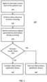
- FIG. 1 is a high-level overview demonstrating a method 100 of screening a patient for a ciliary-driven ophthalmic device, according to certain embodiments.
- a measuring device is applied to a patient's eye.
- the measuring device comprises an electrode contact lens as described below.
- the lens may be positioned on the eye such that one or more electrodes in or on the contact lens is adjacent to, on, around, and/or within the ciliary muscle (or a perimeter/circumference thereof) to acquire electrical signal data based on ciliary muscle movement.
- ciliary function screening is performed.
- a care provider may perform a preoperative exam to determine and characterize a patient's ciliary muscle activity. For example, once the electrode contact lens is placed on the eye, the patient may follow instructions to look at objects at varying distances, such as near (e.g., within 40cm) and far (e.g., beyond 3m), under an established procedure. As the patient changes focus (or attempts to change focus, as the case may be) to different target distances, the ciliary muscle attempts to change the focusing power of the natural lens accordingly. This causes a change in the electrical field of the patient's ciliary muscle(s) which can be detected and signaled b the electrode(s) on the contact lens.
- an electrical signal generated by the electrode(s) in response to the electrical field of the ciliary muscle at each distance may be set to and received, processed, and/or recorded by a computer 606 for subsequent presentation and evaluation.
- the electrical signals are transmitted via wired or wireless communication from the electrodes of the lens to the computer 606.
- a plurality of signals may be received from various electrodes on the lens during the screening procedure, and the received signals may be evaluated by the computer 606 (e.g., using summing, averaging, comparing, and/or statistical processing algorithms, etc.) in order to select those which provide the most accurate and/or reliable indication of ciliary muscle activity.
- the particular electrode signals selected may depend on the position of the contact lens on the patient's eye, as well as the characteristics of the patient's eye itself.
- the computer 606 may also process the signals and convert them to numerical values or other measurements which characterize ciliary muscle activity, responsiveness, strength, and/or accommodative capacity.
- Raw or processed signal data may be output by the computer and shown on a display 608.
- step 104 results of step 103 are evaluated.
- Signal values or measurements generated and/or displayed at step 103 may be reviewed and evaluated by computer 606 and/or a care provider to make a surgical recommendation or choice for the patient (e.g., whether or not to recommend or provide a ciliary-driven accommodative device).
- Step 104 may be performed manually by a care provider, or automatically by computer 606.
- the care provider may view and evaluate measurements of ciliary muscle activity generated at step 103 to determine whether they are above or below predetermined threshold(s), or within a predetermined range or target result deemed suitable for utilization of ciliary-driven ophthalmic devices.
- a computer 606 may execute instructions stored in memory to automatically analyze measurements of ciliary muscle activity generated at step 103 and automatically perform such an evaluation. Applicable threshold values, ranges, or targets for comparison may be stored in memory of the computer 606 and may be configured by a user.
- ciliary muscle activity generated at step 102 is satisfactory and passes the evaluation at step 104 (e.g., the muscle activity is above a threshold, at a target, or within a predetermined range)
- the care provider may proceed to step 106.
- ciliary muscle activity may be sufficient to support proper functioning of ciliary-driven ophthalmic devices, such as electro-active accommodative IOLs.
- the care provider may consider and evaluate ciliary-driven ophthalmic devices when choosing or formulating recommendations for the patient.
- ciliary muscle activity generated at step 102 is unsatisfactory and does not pass the evaluation at step 104 (e.g., the muscle activity is below a threshold, far from a target, or outside a predetermined range)
- the care provider may proceed to step 108.
- ciliary muscle activity may be insufficient to support proper functioning of ciliary-driven ophthalmic devices, such as electro-active accommodative IOLs, and the care provider may consider and evaluate alternatives to ciliary-driven ophthalmic devices when choosing or formulating recommendations for the patient.
- certain embodiments of the disclosure provide a technique for non-invasive screening for a ciliary-driven ophthalmic device, including implantable IOLs.
- electrodes for measuring ciliary muscle electrical signals may comprise metal or wire adhered or embedded in a contact lens. Such lenses may be placed on the eye to measure ciliary electrical signals.
- the location and spacing of the electrodes relative to the position of the ciliary muscle can have an impact on the ability to detect the electrical signal.
- the electrode position is decentered (e.g., misaligned superiorly, as shown in FIGS. 2B and 5B ) or moves on the eye (potentially due to gravity, eye movement, or blinking)
- the signal may be degraded or potentially inverted as the electrode moves off the muscle position.
- One approach to minimizing the effect of lens movement is to suction the lens onto the corneal surface. However, this can be uncomfortable and may not be entirely effective. Further, such an approach may not address potential signal changes related to ciliary muscle movement.
- Another factor which can impact the ability to measure ciliary electrical signal changes is a potential mismatch between the diameter of the ciliary muscle and the diameter(s) of the electrode(s). Indeed, even inter-patient variations in ciliary muscle diameter can be present. Moreover, this can be a static problem or could vary as a function of accommodation if the ciliary muscle contracts appreciably.
- bipolar electrodes may be used.
- Such electrodes can be segmented or multi-faceted, and may include, for example, one side of an electrode ring located outside a perimeter or circumference of the ciliary muscle and the opposite side electrode inside the perimeter or circumference of the ciliary muscle. This configuration may be used to cancel signals and/or reduce the overall signal magnitude.
- certain embodiments may divide the electrode into segments or individual components in order to generate a more comprehensive map of the ciliary electrical activity.
- different areas can be selected or disregarded (either manually by a care provider or automatically by a computer 606) as appropriate (e.g., based on alignment and position with respect to the perimeter or circumference of the ciliary muscle) to better characterize the true electrical signal (i.e., to obtain an accurate measurement of the electrical activity resulting from ciliary muscle movement).
- each individual electrode segment of an electrode contact lens may be connected to an ophthalmic diagnostic system (e.g., computer 606) that includes a processor and memory configured to receive, process, and display (e.g., via display 608) a measure of detected ciliary muscle activity.
- a care provider may inspect an alignment of various electrode segments with the patient's ciliary muscle to identify and select the appropriate segments (e.g., those which are best aligned with the ciliary muscle) on which to base an evaluation, as described by step 104.
- the diagnostic system automatically select the appropriate segments on which to base the evaluation.
- each segment may be continuously sampled or intermittently samples using a time-based multiplexing technique.
- One or more of these features may be used to optimize signal characterization and account for variables such as external lens movement, ciliary muscle movement with accommodation, and differences in ocular anatomy where otherwise the misaligned signal from one side could degrade the opposite side as the electrode position changes.
- FIGS. 2A and 2B illustrate an example of a contact lens with two embedded circular electrodes for measuring ciliary muscle activity and illustrating principles of the present disclosure.
- Eye 200 comprises a ciliary muscle 202 indicated by a circle around the iris (not labeled).
- a transparent contact lens placed on the cornea of eye 200 comprises a reference electrode 204 and a measurement electrode 206.
- Measurement electrode 206 sized to overlay the perimeter of ciliary muscle 202, while reference electrode 204 is larger and lies further away from ciliary muscle 202, outside the circumference.
- reference electrode 204 could be smaller than measurement electrode 206, lying further away from ciliary body 202 but closer to the pupil of eye 200.
- the difference in an electrical signal received from reference electrode 204 and measurement electrode 206 can be used to measure the magnitude of electrical signal in ciliary body 202.
- the difference in signal strength between reference electrode 204 and measurement electrode 206 will be the same or similar at any angle.
- the signal difference left of the pupil of eye 200 would be the same as the signal difference to the right of the pupil, or above or below it as well.
- the measured signal (over 360 degrees) may be determined by summing these differences across all angles.
- FIG. 2B illustrates the same features of FIG. 2A , but with reference electrode 204 and measurement electrode 206 misaligned superiorly with respect to ciliary muscle 202.
- reference electrode 206 is positioned over ciliary muscle 202, inferiorly and the difference signal (between reference electrode 204 and measurement electrode 206) in this position could dramatically depart from the analogous difference signal in the position showing in FIG. 2A .
- the inferior signal measured in FIG. 2B may be the inverse of that in FIG. 2A .
- FIG. 3 illustrates an electrode arrangement for a ciliary-activity detecting contact lens which can help overcome difficulties caused by displacement, according to certain embodiments.
- lens 300 comprises four concentric electrode rings 301-304, each divided into four segments shown as groups 310, 312, 314, and 316.
- Outer ring electrode 304 and each smaller ring 302, 303, and 301 can each measure electrical activity in each segment 310, 312, 314, and 316, providing a reading at sixteen separate segments or channels.
- a total of sixteen segments or channels is shown in FIG. 3
- the number of rings and their division may be varied as appropriate to optimize performance while managing complexity, as an increasing number of channels requires increasing complexity in electrical design and processing requirements.
- certain embodiments may include between two and six concentric electrode rings divided into between two and six groups, thereby providing anywhere between 4 and 36 segments or channels. Other variations are contemplated within the scope of the present disclosure.
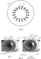
- FIG. 4 illustrates an embodiment of a lens 400 which includes an outer ring of electrodes 410 and an inner ring of electrodes 420, each including sixteen electrode segments. Accordingly, the embodiment of FIG. 4 may support up to 32 channels. Again, although a total of 32 channels are shown in FIG. 4 , the number of electrodes and rings may vary as appropriate to optimize performance while managing complexity, as an increasing number of channels requires increasing complexity in electrical design and processing requirements. For example, certain embodiments may include between two and six concentric electrode rings, each including between three and 20 electrodes, thereby providing anywhere between 6 and 120 channels. Other variations are contemplated within the scope of the present disclosure.
- FIG. 5 illustrates possible scenarios resulting from application of a contact lens comprising electrode rings (identical to lens 300 shown in FIG. 3 ) to a patient.
- lens 300 is centered on eye 200 and all four segments of the third ring from the center (corresponding to ring 303 of FIG. 3 ) are well-aligned with the perimeter or circumference of ciliary muscle 202. Summing the signals generated by each of the four segments of ring 303 yields a signal comparable to that obtained from measurement electrode 206 shown in FIG. 2A - a result that may be considered accurate and reliable.
- lens 300 of FIG. 5B is decentered (misaligned superiorly, analogous to FIG. 3B ) on eye 200.
- the segments of ring 303 are not well-aligned to ciliary body 202 in FIG. 5B , and summing the signals generated by each of the four segments of ring 303 would yield a signal comparable to that obtained from measurement electrode 206 shown in FIG. 2B - likely an unreliable and inaccurate result.
- the lower electrodes of ring 304 (toward the bottom of FIG. 5B , corresponding to segments 314 and 316 shown in FIG. 3 ) are well-aligned to ciliary body 202.
- the upper electrodes of ring 302 (toward the top of FIG. 5B , corresponding to segments 310 and 312 shown in FIG. 3 ) are suitably aligned to ciliary body 202.
- a lens according to the invention comprising a plurality of electrode segments configured to independently generate and transmit signals may be used to accurately evaluate ciliary muscle activity in accordance with the method of FIG. 1 , even if the lens is misaligned with respect to the ciliary body.
- This can be achieved by selecting signals from electrodes which are well-aligned with the ciliary muscle. Such a selection may be peformed manually by a care provider, or automatically by a computer 606, as discussed below.
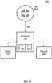
- FIG. 6 illustrates an ophthalmic system 600 which may be used to perform method 100, according to certain embodiments.
- the system includes one or more electrode contact lenses 602 are designed for placement on the surface of the cornea of a patient's eye for a ciliary muscle evaluation procedure.
- Lens 602 may comprise multiple electrodes and segments as shown in FIGS. 3 and 5 , but is not limited to the arrangement shown in those embodiments.
- a care provider may perform the steps illustrated and described above with respect to step 102 of FIG. 1 .
- the ciliary muscle attempts to change the focusing power of the natural lens accordingly. This causes a change in the electrical field of the patient's ciliary muscle(s).
- electrical signals 604 generated by each electrode or channel of lens 602 may be transmitted to and received by a computer 606 which includes a processor and memory configured to execute instructions for processing the signals 604.
- Signals 604 may be transmitted to computer 606 via wired or wireless communication.
- particular segments or channels of the multi-electrode lens 602 aligned with the patient's ciliary body may be identified and selected by computer 606, as discussed above with respect to the example of FIG. 5B .
- Computer 606 comprises one or more processors 612 and memory 614.
- Memory 614 may include persistent and volatile media, fixed and removable media, and magnetic and semiconductor media.
- Memory 614 is operable to store programs, codes, scripts, instructions, data, and the like.
- Memory 614 as shown includes sets or sequences of instructions, namely, an operating system, and an ophthalmic diagnostic program.
- the operating system may be a UNIX or UNIX-like operating system, a Windows ® family operating system, an Apple ® family operating system (e.g., macOS, iOS), or another suitable operating system. Instructions and data stored in memory 614 are accessible to processor 612 and are executable by the processor 612 to perform the steps discussed herein.
- the processor 612 may be or include a general purpose microprocessor, as well as a specialized co-processor or another type of data processing apparatus. In some cases, the processor 612 performs high level operation of the ciliary function diagnostic evaluation discussed herein.
- the processor 612 may be configured to execute or interpret software, scripts, programs, functions, executables, or other instructions stored in the memory 614 to receive, interpret, process, and evaluate signals generated by electrodes during ciliary muscle screening (e.g., as described in the process 100 of FIG. 1 ). Accordingly, computer 606 is specially adapted to perform ophthalmic-specific processes related to ciliary muscle function, as described herein.
- a processor of computer 606 may execute instructions to compare and evaluate signals 604 received from each electrode or channel of lens 602 to determine and select the best signals for further processing and/or evaluation.
- the processor of computer 604 may execute summing, averaging, comparing, and/or statistical processing algorithms using the received signals to identify and select a subset of signals which which provide the most accurate and/or reliable indication of ciliary muscle activity. In some examples, this may include identifying and selecting signals which, based on the algoritm performed by the processor, are best situated within and aligned to the perimeter or circumference of the ciliary muscle.
- Algorithms executed by the processor may include also comparing each received signal with upper and lower thresholds, mean or median values (which may be calculated based on received signals), or other indicia useful for evaluating the quality and/or reliability of the received signal.
- the subset of electrode signals selected may depend on the position of the contact lens on the patient's eye, as well as the characteristics of the patient's eye itself.
- the instructions can be encoded as preprogrammed or re-programmable logic circuits, logic gates, or other types of hardware or firmware components.
- a user may use an input device 610 (e.g., keyboard, mouse, touch screen, voice recognition, etc.) to assist with the identification and selection of particular segments or channels of the multi-electrode lens 602 for the evaluation step.
- computer 606 may identify and select particular segments or channels of the multi-electrode lens 602 for the evaluation step automatically, based on algorithms noted above.
- the processor of computer 606 may execute instructions to process raw signal data and convert them to numerical values or other measurements which characterize ciliary muscle activity, responsiveness, strength, and/or accommodative capacity.
- Raw or processed signal data may be output by the computer and shown on a display 608 (e.g., a monitor, screen, heads-up display, tablet device, etc.).
- the care provider may then proceed to step 104 and evaluate the results and data obtained from multi-electrode lens 602.
- the processor of computer 606 may compare the sleected measurement values and data to predetermined thresholds, targets, and ranges to provide a notification, recommendation, or alert to the care provider via display 608.
- the processor of computer 606 may include one or more CPUs, microprocessors, field-programmable gate arrays (FPGAs), application-specific integrated circuits (ASICs), digital-signal processors (DSPs), system-on-chip (SoC) processors, or analogous components.
- the memory of computer 606 may include volatile or non-volatile memory including, magnetic media, optical media, random access memory (RAM), read-only memory (ROM), removable media, or analogous components.
- the memory of computer 606 may store instructions for software programs and algorithms that, when executed by the processor, analyze signals received from lens 602 as discussed above.
- processor As used in the claims, the terms "processor,” “memory,” “instructions,” and the like refer to classes of structures that are well-known to those skilled in the art. As such, these terms are to be understood as denoting structural rather than functional elements of the disclosed system.
- embodiments of the present disclosure provide novel and useful systems and methods for preoperatively assessing a patient's ciliary electrical activity.
- a care provider may identify eyes in which a ciliary muscle-driven accommodative ophthalmic device would not function as intended, prior to surgery or purchase, to guide the decision on whether such devices are appropriate for a particular individual.
- a care provider may identify eyes in which a ciliary muscle-driven accommodative ophthalmic device would likely function well, prior to surgery or purchase, to guide the decision on whether such devices are appropriate for a particular individual.
Landscapes
- Health & Medical Sciences (AREA)
- Life Sciences & Earth Sciences (AREA)
- Engineering & Computer Science (AREA)
- General Health & Medical Sciences (AREA)
- Animal Behavior & Ethology (AREA)
- Veterinary Medicine (AREA)
- Biomedical Technology (AREA)
- Heart & Thoracic Surgery (AREA)
- Public Health (AREA)
- Molecular Biology (AREA)
- Surgery (AREA)
- Physics & Mathematics (AREA)
- Medical Informatics (AREA)
- Biophysics (AREA)
- Pathology (AREA)
- Ophthalmology & Optometry (AREA)
- Transplantation (AREA)
- Oral & Maxillofacial Surgery (AREA)
- Cardiology (AREA)
- Vascular Medicine (AREA)
- Artificial Intelligence (AREA)
- Computer Vision & Pattern Recognition (AREA)
- Physiology (AREA)
- Psychiatry (AREA)
- Signal Processing (AREA)
- Eye Examination Apparatus (AREA)
- Measurement And Recording Of Electrical Phenomena And Electrical Characteristics Of The Living Body (AREA)
Claims (5)
- Ophthalmisches System (600), umfassend:eine Kontaktlinse (602), die dafür ausgelegt ist, eine Oberfläche des Auges eines Patienten zu berühren, wobei die Kontaktlinse eine Vielzahl von bipolaren Elektroden umfasst, die eine Vielzahl von konzentrischen Ringen (301-304) umfassen, wobei jeder konzentrische Ring eine Vielzahl von Segmenten (310, 312, 314, 316) umfasst, wobei jedes der Vielzahl von Segmenten (310, 312, 314, 316) dafür ausgelegt ist, ein Signal zu erzeugen, das ein elektrisches Feld anzeigt, das dem Ziliarmuskel des Patienten zugeordnet ist; undein Diagnosesystem (606), umfassend einen oder mehrere Prozessoren (612) und einen Speicher (614), der Anweisungen zum Bewerten des Akkommodationspotenzials des Ziliarmuskels des Patienten umfasst;wobei das Diagnosesystem (606) für folgende Vorgänge ausgelegt ist:Empfangen einer Vielzahl von Signalen (604), die von der Vielzahl von Segmenten (310, 312, 314, 316) der konzentrischen Ringe (301-304) während eines Ziliarmuskelbewertungsverfahrens erzeugt werden, wobei jedes der Vielzahl von Signalen ein elektrisches Feld anzeigt, das dem Ziliarmuskel des Patienten zugeordnet ist;Analysieren der empfangenen Signale, beinhaltend das Vergleichen der empfangenen Signale zum Bewerten von Unterschieden in der Qualität und der Genauigkeit der Vielzahl von Signalen, die von der Vielzahl von Segmenten empfangen wurden;Identifizieren einer Teilmenge von Signalen, die eine genaue Messung der elektrischen Aktivität des Ziliarmuskels bereitstellen, zur Verwendung beim Bewerten des Ziliarmuskels des Patienten, wobei die Teilmenge von Signalen, die durch die Analyse identifiziert werden, von einer Teilmenge der Vielzahl von Segmenten der konzentrischen Ringe empfangen werden, die mit dem Ziliarkörper des Patienten ausgerichtet sind, wobei das Bewerten der Vielzahl von Signalen, die von der Vielzahl von Segmenten (310, 312, 314, 316) empfangen werden, ferner das Vergleichen der Signale mit einem Schwellenwert, einem Mittelwert oder einem Medianwert umfasst; undBerechnen eines Wertes, der der identifizierten Teilmenge der Vielzahl von Segmenten (310, 312, 314, 316) zugeordnet ist, wobei das Berechnen des Wertes das Summieren der Signale der Teilmenge von Signalen umfasst, die von den Segmenten (310, 312, 314, 316) erzeugt werden, die mit dem Ziliarkörper des Patienten ausgerichtet sind; undeine Anzeige (608), die kommunikativ mit dem Prozessor gekoppelt und dafür ausgelegt ist, den berechneten Wert anzuzeigen, der der identifizierten Teilmenge der Vielzahl von Segmenten zugeordnet ist.
- System nach Anspruch 1, wobei das Bewerten der Vielzahl von Signalen, die von der Vielzahl von Segmenten empfangen werden, ferner das Verarbeiten der Signale umfasst, einschließlich einem oder mehreren von Summierung, Mittelwertbildung, Vergleich oder statistische Verarbeitung zum Identifizieren und Auswählen einer Teilmenge von Signalen, die die genaueste und/oder die zuverlässigste Angabe von Ziliarmuskelaktivität bereitstellt.
- System nach Anspruch 1, wobei das Identifizieren der Teilmenge von Signalen durch einen vom Diagnosesystem ausgeführten Verarbeitungsalgorithmus zur Verwendung beim Bewerten von Ziliarmuskelaktivität das Identifizieren der Signale beinhaltet, die von der Teilmenge von Segmenten empfangen werden, die sich innerhalb des Umfangs oder der Peripherie des Ziliarmuskels befinden und damit ausgerichtet sind.
- System nach Anspruch 1, wobei die Anweisungen bei Ausführung durch den einen oder die mehreren Prozessoren dazu geeignet sind, die berechnete Summe der ausgewählten Teilmenge von Signalen mit einem vorbestimmten Wert zu vergleichen.
- System nach einem der vorhergehenden Ansprüche, das dafür ausgelegt ist, Änderungen in der Vielzahl von empfangenen Signalen zu erfassen, wenn der Ziliarmuskel die Fokussierungskraft beim Fokussieren auf ein oder mehrere Ziele in unterschiedlichen Entfernungen ändert.
Applications Claiming Priority (2)
| Application Number | Priority Date | Filing Date | Title |
|---|---|---|---|
| US201762573186P | 2017-10-17 | 2017-10-17 | |
| PCT/IB2018/058008 WO2019077488A1 (en) | 2017-10-17 | 2018-10-16 | SYSTEMS AND METHODS FOR MEASURING CILIARY MUSCLE ACTIVITY |
Publications (2)
| Publication Number | Publication Date |
|---|---|
| EP3672476A1 EP3672476A1 (de) | 2020-07-01 |
| EP3672476B1 true EP3672476B1 (de) | 2024-09-25 |
Family
ID=64109966
Family Applications (1)
| Application Number | Title | Priority Date | Filing Date |
|---|---|---|---|
| EP18797138.7A Active EP3672476B1 (de) | 2017-10-17 | 2018-10-16 | Systeme und verfahren zur messung der ziliarmuskelaktivität |
Country Status (7)
| Country | Link |
|---|---|
| US (2) | US11259916B2 (de) |
| EP (1) | EP3672476B1 (de) |
| JP (2) | JP7155257B2 (de) |
| CN (2) | CN116172565B (de) |
| AU (1) | AU2018353527B2 (de) |
| CA (1) | CA3076463A1 (de) |
| WO (1) | WO2019077488A1 (de) |
Families Citing this family (5)
| Publication number | Priority date | Publication date | Assignee | Title |
|---|---|---|---|---|
| EP3672476B1 (de) * | 2017-10-17 | 2024-09-25 | Alcon Inc. | Systeme und verfahren zur messung der ziliarmuskelaktivität |
| JP2023170284A (ja) * | 2022-05-18 | 2023-12-01 | ポーラ化成工業株式会社 | 目の評価方法及び眼輪筋の筋力測定方法 |
| US12279881B1 (en) * | 2024-08-09 | 2025-04-22 | Neuralytix, Llc | System and method for assessing nerve health during a spinal decompression procedure using a calculated nerve function index |
| US12279879B1 (en) | 2024-08-09 | 2025-04-22 | Neuralytix, Llc | System and method for empirical assessment of nerve health during a spinal decompression procedure |
| US12279880B1 (en) | 2024-08-09 | 2025-04-22 | Neuralytix, Llc | System and method for assessing nerve health during a spinal decompression procedure |
Family Cites Families (18)
| Publication number | Priority date | Publication date | Assignee | Title |
|---|---|---|---|---|
| RU2063199C1 (ru) * | 1989-11-02 | 1996-07-10 | Александр Робертович Гуськов | Способ формирования воздействия для электростимуляции цилиарной мышцы, электростимулятор цилиарной мышцы для лечения нарушения аккомодации и электродное устройство |
| CN1064611A (zh) * | 1991-03-11 | 1992-09-23 | 罗伯特·G·威利 | 具有散光矫正能力的可变光焦度的人工晶状体 |
| US20070203478A1 (en) * | 2006-02-21 | 2007-08-30 | Herekar Satish V | Method and system for elasto-modulation of ocular tissue |
| US20110022117A1 (en) * | 2009-07-22 | 2011-01-27 | Casey Lind | Cytokine Production Via Electrical Stimulation to Treat Glaucoma |
| CN101766473B (zh) * | 2010-02-09 | 2011-09-07 | 北京大学人民医院 | 眼压监测系统 |
| EP2593040B1 (de) * | 2010-07-12 | 2016-03-30 | Katholieke Universiteit Leuven | Bionische kontaktlinse |
| US9931203B2 (en) * | 2011-08-02 | 2018-04-03 | Valdemar Portney | Presbyopia correcting wireless optical system |
| US9241669B2 (en) * | 2012-07-18 | 2016-01-26 | Johnson & Johnson Vision Care, Inc. | Neuromuscular sensing for variable-optic electronic ophthalmic lens |
| US8965478B2 (en) * | 2012-10-12 | 2015-02-24 | Google Inc. | Microelectrodes in an ophthalmic electrochemical sensor |
| WO2014084958A1 (en) * | 2012-11-30 | 2014-06-05 | Novartis Ag | Sensors for triggering electro-active ophthalmic lenses |
| DE102013000429A1 (de) * | 2013-01-10 | 2014-07-24 | Karlsruher Institut für Technologie | Sensorsystem für die Erfassung der Ansteuersignale eines Ziliarmuskels |
| WO2014200864A1 (en) * | 2013-06-14 | 2014-12-18 | University Of Houston System | Accommodation stimulation and recording device |
| US20150029463A1 (en) * | 2013-07-23 | 2015-01-29 | Retmap, Inc. | Device for electrophysiological recording from the eye |
| PT3046459T (pt) | 2013-12-20 | 2019-10-15 | Novartis Ag | Sistema e método para avaliação de acomodação residual em olhos presbiópicos |
| US9933634B2 (en) * | 2014-06-13 | 2018-04-03 | Verily Life Sciences Llc | Apparatus, system and method for gaze tracking based on photodetection by an eye-mountable device |
| US10702375B2 (en) * | 2015-09-18 | 2020-07-07 | Vista Ocular, Llc | Electromyographic sensing and vision modification |
| CN106963607B (zh) * | 2017-05-16 | 2018-01-16 | 刘永宏 | 智能控制睫状肌训练仪 |
| EP3672476B1 (de) * | 2017-10-17 | 2024-09-25 | Alcon Inc. | Systeme und verfahren zur messung der ziliarmuskelaktivität |
-
2018
- 2018-10-16 EP EP18797138.7A patent/EP3672476B1/de active Active
- 2018-10-16 CN CN202310203138.0A patent/CN116172565B/zh active Active
- 2018-10-16 CA CA3076463A patent/CA3076463A1/en active Pending
- 2018-10-16 WO PCT/IB2018/058008 patent/WO2019077488A1/en not_active Ceased
- 2018-10-16 US US16/161,130 patent/US11259916B2/en active Active
- 2018-10-16 JP JP2020520493A patent/JP7155257B2/ja active Active
- 2018-10-16 CN CN201880067727.8A patent/CN111225602B/zh active Active
- 2018-10-16 AU AU2018353527A patent/AU2018353527B2/en active Active
-
2022
- 2022-01-24 US US17/648,695 patent/US12127933B2/en active Active
- 2022-10-05 JP JP2022160799A patent/JP2022180630A/ja active Pending
Also Published As
| Publication number | Publication date |
|---|---|
| AU2018353527A1 (en) | 2020-04-09 |
| JP2020537564A (ja) | 2020-12-24 |
| CA3076463A1 (en) | 2019-04-25 |
| AU2018353527B2 (en) | 2024-09-05 |
| CN116172565A (zh) | 2023-05-30 |
| US11259916B2 (en) | 2022-03-01 |
| CN111225602A (zh) | 2020-06-02 |
| US20220192821A1 (en) | 2022-06-23 |
| JP7155257B2 (ja) | 2022-10-18 |
| EP3672476A1 (de) | 2020-07-01 |
| US12127933B2 (en) | 2024-10-29 |
| US20190110888A1 (en) | 2019-04-18 |
| WO2019077488A1 (en) | 2019-04-25 |
| CN111225602B (zh) | 2023-03-14 |
| JP2022180630A (ja) | 2022-12-06 |
| CN116172565B (zh) | 2025-12-09 |
Similar Documents
| Publication | Publication Date | Title |
|---|---|---|
| US12127933B2 (en) | Systems and methods for measuring ciliary muscle activity | |
| US9119561B2 (en) | Optimizing vision correction procedures | |
| AU2017202009B2 (en) | Neuromuscular sensing for variable-optic electronic ophthalmic lens | |
| RU2604942C2 (ru) | Измерение/отображение/запись/воспроизведение данных волнового фронта для использования в процедурах коррекции зрения | |
| US20140156000A1 (en) | Sensors for triggering electro-active ophthalmic lenses | |
| JP5255277B2 (ja) | 網膜疾患の検診装置 | |
| EP3349648A1 (de) | Elektromyografische messung und visionsänderung | |
| TW201835563A (zh) | 用於感測睫狀肌阻抗的電極結構 | |
| WO2026005721A1 (en) | A focus-tunable lens system with enhanced accommodation | |
| Symons et al. | Functional screening devices for diabetic retinopathy. |
Legal Events
| Date | Code | Title | Description |
|---|---|---|---|
| STAA | Information on the status of an ep patent application or granted ep patent |
Free format text: STATUS: UNKNOWN |
|
| STAA | Information on the status of an ep patent application or granted ep patent |
Free format text: STATUS: THE INTERNATIONAL PUBLICATION HAS BEEN MADE |
|
| PUAI | Public reference made under article 153(3) epc to a published international application that has entered the european phase |
Free format text: ORIGINAL CODE: 0009012 |
|
| STAA | Information on the status of an ep patent application or granted ep patent |
Free format text: STATUS: REQUEST FOR EXAMINATION WAS MADE |
|
| 17P | Request for examination filed |
Effective date: 20200325 |
|
| AK | Designated contracting states |
Kind code of ref document: A1 Designated state(s): AL AT BE BG CH CY CZ DE DK EE ES FI FR GB GR HR HU IE IS IT LI LT LU LV MC MK MT NL NO PL PT RO RS SE SI SK SM TR |
|
| AX | Request for extension of the european patent |
Extension state: BA ME |
|
| DAV | Request for validation of the european patent (deleted) | ||
| DAX | Request for extension of the european patent (deleted) | ||
| STAA | Information on the status of an ep patent application or granted ep patent |
Free format text: STATUS: EXAMINATION IS IN PROGRESS |
|
| 17Q | First examination report despatched |
Effective date: 20220823 |
|
| P01 | Opt-out of the competence of the unified patent court (upc) registered |
Effective date: 20230507 |
|
| REG | Reference to a national code |
Ref country code: DE Ref legal event code: R079 Free format text: PREVIOUS MAIN CLASS: A61B0005000000 Ipc: A61B0003100000 Ref document number: 602018074752 Country of ref document: DE |
|
| GRAP | Despatch of communication of intention to grant a patent |
Free format text: ORIGINAL CODE: EPIDOSNIGR1 |
|
| STAA | Information on the status of an ep patent application or granted ep patent |
Free format text: STATUS: GRANT OF PATENT IS INTENDED |
|
| RIC1 | Information provided on ipc code assigned before grant |
Ipc: A61B 5/296 20210101ALI20240319BHEP Ipc: A61B 5/00 20060101ALI20240319BHEP Ipc: A61F 2/16 20060101ALI20240319BHEP Ipc: A61B 3/10 20060101AFI20240319BHEP |
|
| INTG | Intention to grant announced |
Effective date: 20240416 |
|
| GRAS | Grant fee paid |
Free format text: ORIGINAL CODE: EPIDOSNIGR3 |
|
| GRAA | (expected) grant |
Free format text: ORIGINAL CODE: 0009210 |
|
| STAA | Information on the status of an ep patent application or granted ep patent |
Free format text: STATUS: THE PATENT HAS BEEN GRANTED |
|
| AK | Designated contracting states |
Kind code of ref document: B1 Designated state(s): AL AT BE BG CH CY CZ DE DK EE ES FI FR GB GR HR HU IE IS IT LI LT LU LV MC MK MT NL NO PL PT RO RS SE SI SK SM TR |
|
| REG | Reference to a national code |
Ref country code: GB Ref legal event code: FG4D |
|
| REG | Reference to a national code |
Ref country code: CH Ref legal event code: EP |
|
| REG | Reference to a national code |
Ref country code: DE Ref legal event code: R096 Ref document number: 602018074752 Country of ref document: DE |
|
| REG | Reference to a national code |
Ref country code: IE Ref legal event code: FG4D |
|
| REG | Reference to a national code |
Ref country code: LT Ref legal event code: MG9D |
|
| PG25 | Lapsed in a contracting state [announced via postgrant information from national office to epo] |
Ref country code: NO Free format text: LAPSE BECAUSE OF FAILURE TO SUBMIT A TRANSLATION OF THE DESCRIPTION OR TO PAY THE FEE WITHIN THE PRESCRIBED TIME-LIMIT Effective date: 20241225 |
|
| PG25 | Lapsed in a contracting state [announced via postgrant information from national office to epo] |
Ref country code: GR Free format text: LAPSE BECAUSE OF FAILURE TO SUBMIT A TRANSLATION OF THE DESCRIPTION OR TO PAY THE FEE WITHIN THE PRESCRIBED TIME-LIMIT Effective date: 20241226 Ref country code: FI Free format text: LAPSE BECAUSE OF FAILURE TO SUBMIT A TRANSLATION OF THE DESCRIPTION OR TO PAY THE FEE WITHIN THE PRESCRIBED TIME-LIMIT Effective date: 20240925 |
|
| PG25 | Lapsed in a contracting state [announced via postgrant information from national office to epo] |
Ref country code: BG Free format text: LAPSE BECAUSE OF FAILURE TO SUBMIT A TRANSLATION OF THE DESCRIPTION OR TO PAY THE FEE WITHIN THE PRESCRIBED TIME-LIMIT Effective date: 20240925 |
|
| PG25 | Lapsed in a contracting state [announced via postgrant information from national office to epo] |
Ref country code: LV Free format text: LAPSE BECAUSE OF FAILURE TO SUBMIT A TRANSLATION OF THE DESCRIPTION OR TO PAY THE FEE WITHIN THE PRESCRIBED TIME-LIMIT Effective date: 20240925 |
|
| PG25 | Lapsed in a contracting state [announced via postgrant information from national office to epo] |
Ref country code: RS Free format text: LAPSE BECAUSE OF FAILURE TO SUBMIT A TRANSLATION OF THE DESCRIPTION OR TO PAY THE FEE WITHIN THE PRESCRIBED TIME-LIMIT Effective date: 20241225 |
|
| REG | Reference to a national code |
Ref country code: NL Ref legal event code: MP Effective date: 20240925 |
|
| PG25 | Lapsed in a contracting state [announced via postgrant information from national office to epo] |
Ref country code: RS Free format text: LAPSE BECAUSE OF FAILURE TO SUBMIT A TRANSLATION OF THE DESCRIPTION OR TO PAY THE FEE WITHIN THE PRESCRIBED TIME-LIMIT Effective date: 20241225 Ref country code: NO Free format text: LAPSE BECAUSE OF FAILURE TO SUBMIT A TRANSLATION OF THE DESCRIPTION OR TO PAY THE FEE WITHIN THE PRESCRIBED TIME-LIMIT Effective date: 20241225 Ref country code: LV Free format text: LAPSE BECAUSE OF FAILURE TO SUBMIT A TRANSLATION OF THE DESCRIPTION OR TO PAY THE FEE WITHIN THE PRESCRIBED TIME-LIMIT Effective date: 20240925 Ref country code: GR Free format text: LAPSE BECAUSE OF FAILURE TO SUBMIT A TRANSLATION OF THE DESCRIPTION OR TO PAY THE FEE WITHIN THE PRESCRIBED TIME-LIMIT Effective date: 20241226 Ref country code: FI Free format text: LAPSE BECAUSE OF FAILURE TO SUBMIT A TRANSLATION OF THE DESCRIPTION OR TO PAY THE FEE WITHIN THE PRESCRIBED TIME-LIMIT Effective date: 20240925 Ref country code: BG Free format text: LAPSE BECAUSE OF FAILURE TO SUBMIT A TRANSLATION OF THE DESCRIPTION OR TO PAY THE FEE WITHIN THE PRESCRIBED TIME-LIMIT Effective date: 20240925 |
|
| REG | Reference to a national code |
Ref country code: AT Ref legal event code: MK05 Ref document number: 1725933 Country of ref document: AT Kind code of ref document: T Effective date: 20240925 |
|
| PG25 | Lapsed in a contracting state [announced via postgrant information from national office to epo] |
Ref country code: NL Free format text: LAPSE BECAUSE OF FAILURE TO SUBMIT A TRANSLATION OF THE DESCRIPTION OR TO PAY THE FEE WITHIN THE PRESCRIBED TIME-LIMIT Effective date: 20240925 |
|
| PG25 | Lapsed in a contracting state [announced via postgrant information from national office to epo] |
Ref country code: PT Free format text: LAPSE BECAUSE OF FAILURE TO SUBMIT A TRANSLATION OF THE DESCRIPTION OR TO PAY THE FEE WITHIN THE PRESCRIBED TIME-LIMIT Effective date: 20250127 Ref country code: IS Free format text: LAPSE BECAUSE OF FAILURE TO SUBMIT A TRANSLATION OF THE DESCRIPTION OR TO PAY THE FEE WITHIN THE PRESCRIBED TIME-LIMIT Effective date: 20250125 |
|
| PG25 | Lapsed in a contracting state [announced via postgrant information from national office to epo] |
Ref country code: RO Free format text: LAPSE BECAUSE OF FAILURE TO SUBMIT A TRANSLATION OF THE DESCRIPTION OR TO PAY THE FEE WITHIN THE PRESCRIBED TIME-LIMIT Effective date: 20240925 Ref country code: SM Free format text: LAPSE BECAUSE OF FAILURE TO SUBMIT A TRANSLATION OF THE DESCRIPTION OR TO PAY THE FEE WITHIN THE PRESCRIBED TIME-LIMIT Effective date: 20240925 |
|
| PG25 | Lapsed in a contracting state [announced via postgrant information from national office to epo] |
Ref country code: ES Free format text: LAPSE BECAUSE OF FAILURE TO SUBMIT A TRANSLATION OF THE DESCRIPTION OR TO PAY THE FEE WITHIN THE PRESCRIBED TIME-LIMIT Effective date: 20240925 |
|
| PG25 | Lapsed in a contracting state [announced via postgrant information from national office to epo] |
Ref country code: EE Free format text: LAPSE BECAUSE OF FAILURE TO SUBMIT A TRANSLATION OF THE DESCRIPTION OR TO PAY THE FEE WITHIN THE PRESCRIBED TIME-LIMIT Effective date: 20240925 Ref country code: AT Free format text: LAPSE BECAUSE OF FAILURE TO SUBMIT A TRANSLATION OF THE DESCRIPTION OR TO PAY THE FEE WITHIN THE PRESCRIBED TIME-LIMIT Effective date: 20240925 |
|
| PG25 | Lapsed in a contracting state [announced via postgrant information from national office to epo] |
Ref country code: PL Free format text: LAPSE BECAUSE OF FAILURE TO SUBMIT A TRANSLATION OF THE DESCRIPTION OR TO PAY THE FEE WITHIN THE PRESCRIBED TIME-LIMIT Effective date: 20240925 Ref country code: CZ Free format text: LAPSE BECAUSE OF FAILURE TO SUBMIT A TRANSLATION OF THE DESCRIPTION OR TO PAY THE FEE WITHIN THE PRESCRIBED TIME-LIMIT Effective date: 20240925 |
|
| PG25 | Lapsed in a contracting state [announced via postgrant information from national office to epo] |
Ref country code: IT Free format text: LAPSE BECAUSE OF FAILURE TO SUBMIT A TRANSLATION OF THE DESCRIPTION OR TO PAY THE FEE WITHIN THE PRESCRIBED TIME-LIMIT Effective date: 20240925 Ref country code: SK Free format text: LAPSE BECAUSE OF FAILURE TO SUBMIT A TRANSLATION OF THE DESCRIPTION OR TO PAY THE FEE WITHIN THE PRESCRIBED TIME-LIMIT Effective date: 20240925 |
|
| REG | Reference to a national code |
Ref country code: CH Ref legal event code: PL |
|
| REG | Reference to a national code |
Ref country code: DE Ref legal event code: R097 Ref document number: 602018074752 Country of ref document: DE |
|
| PG25 | Lapsed in a contracting state [announced via postgrant information from national office to epo] |
Ref country code: MC Free format text: LAPSE BECAUSE OF FAILURE TO SUBMIT A TRANSLATION OF THE DESCRIPTION OR TO PAY THE FEE WITHIN THE PRESCRIBED TIME-LIMIT Effective date: 20240925 |
|
| PG25 | Lapsed in a contracting state [announced via postgrant information from national office to epo] |
Ref country code: DK Free format text: LAPSE BECAUSE OF FAILURE TO SUBMIT A TRANSLATION OF THE DESCRIPTION OR TO PAY THE FEE WITHIN THE PRESCRIBED TIME-LIMIT Effective date: 20240925 |
|
| PG25 | Lapsed in a contracting state [announced via postgrant information from national office to epo] |
Ref country code: BE Free format text: LAPSE BECAUSE OF NON-PAYMENT OF DUE FEES Effective date: 20241031 Ref country code: LU Free format text: LAPSE BECAUSE OF NON-PAYMENT OF DUE FEES Effective date: 20241016 |
|
| PG25 | Lapsed in a contracting state [announced via postgrant information from national office to epo] |
Ref country code: CH Free format text: LAPSE BECAUSE OF NON-PAYMENT OF DUE FEES Effective date: 20241031 |
|
| PLBE | No opposition filed within time limit |
Free format text: ORIGINAL CODE: 0009261 |
|
| STAA | Information on the status of an ep patent application or granted ep patent |
Free format text: STATUS: NO OPPOSITION FILED WITHIN TIME LIMIT |
|
| REG | Reference to a national code |
Ref country code: BE Ref legal event code: MM Effective date: 20241031 |
|
| 26N | No opposition filed |
Effective date: 20250626 |
|
| PG25 | Lapsed in a contracting state [announced via postgrant information from national office to epo] |
Ref country code: SE Free format text: LAPSE BECAUSE OF FAILURE TO SUBMIT A TRANSLATION OF THE DESCRIPTION OR TO PAY THE FEE WITHIN THE PRESCRIBED TIME-LIMIT Effective date: 20240925 |
|
| PGFP | Annual fee paid to national office [announced via postgrant information from national office to epo] |
Ref country code: GB Payment date: 20250918 Year of fee payment: 8 |
|
| PGFP | Annual fee paid to national office [announced via postgrant information from national office to epo] |
Ref country code: FR Payment date: 20250922 Year of fee payment: 8 |
|
| PG25 | Lapsed in a contracting state [announced via postgrant information from national office to epo] |
Ref country code: IE Free format text: LAPSE BECAUSE OF NON-PAYMENT OF DUE FEES Effective date: 20241016 |
|
| PGFP | Annual fee paid to national office [announced via postgrant information from national office to epo] |
Ref country code: DE Payment date: 20250916 Year of fee payment: 8 |
|
| PG25 | Lapsed in a contracting state [announced via postgrant information from national office to epo] |
Ref country code: HR Free format text: LAPSE BECAUSE OF FAILURE TO SUBMIT A TRANSLATION OF THE DESCRIPTION OR TO PAY THE FEE WITHIN THE PRESCRIBED TIME-LIMIT Effective date: 20240925 |
|
| PG25 | Lapsed in a contracting state [announced via postgrant information from national office to epo] |
Ref country code: CY Free format text: LAPSE BECAUSE OF FAILURE TO SUBMIT A TRANSLATION OF THE DESCRIPTION OR TO PAY THE FEE WITHIN THE PRESCRIBED TIME-LIMIT; INVALID AB INITIO Effective date: 20181016 |
|
| PG25 | Lapsed in a contracting state [announced via postgrant information from national office to epo] |
Ref country code: HU Free format text: LAPSE BECAUSE OF FAILURE TO SUBMIT A TRANSLATION OF THE DESCRIPTION OR TO PAY THE FEE WITHIN THE PRESCRIBED TIME-LIMIT; INVALID AB INITIO Effective date: 20181016 |
|
| PG25 | Lapsed in a contracting state [announced via postgrant information from national office to epo] |
Ref country code: TR Free format text: LAPSE BECAUSE OF FAILURE TO SUBMIT A TRANSLATION OF THE DESCRIPTION OR TO PAY THE FEE WITHIN THE PRESCRIBED TIME-LIMIT Effective date: 20240925 |
|
| PG25 | Lapsed in a contracting state [announced via postgrant information from national office to epo] |
Ref country code: SI Free format text: LAPSE BECAUSE OF FAILURE TO SUBMIT A TRANSLATION OF THE DESCRIPTION OR TO PAY THE FEE WITHIN THE PRESCRIBED TIME-LIMIT Effective date: 20240925 |
|
| PG25 | Lapsed in a contracting state [announced via postgrant information from national office to epo] |
Ref country code: LT Free format text: LAPSE BECAUSE OF FAILURE TO SUBMIT A TRANSLATION OF THE DESCRIPTION OR TO PAY THE FEE WITHIN THE PRESCRIBED TIME-LIMIT Effective date: 20240925 |