EP3672688B1 - Procédé permettant de fournir des données de résultat appropriées pour être utilisées dans une planification d'une irradiation d'un patient - Google Patents
Procédé permettant de fournir des données de résultat appropriées pour être utilisées dans une planification d'une irradiation d'un patient Download PDFInfo
- Publication number
- EP3672688B1 EP3672688B1 EP18755715.2A EP18755715A EP3672688B1 EP 3672688 B1 EP3672688 B1 EP 3672688B1 EP 18755715 A EP18755715 A EP 18755715A EP 3672688 B1 EP3672688 B1 EP 3672688B1
- Authority
- EP
- European Patent Office
- Prior art keywords
- measurement data
- acquisition
- patient
- ray detector
- irradiation
- Prior art date
- Legal status (The legal status is an assumption and is not a legal conclusion. Google has not performed a legal analysis and makes no representation as to the accuracy of the status listed.)
- Active
Links
Images


Classifications
-
- A—HUMAN NECESSITIES
- A61—MEDICAL OR VETERINARY SCIENCE; HYGIENE
- A61N—ELECTROTHERAPY; MAGNETOTHERAPY; RADIATION THERAPY; ULTRASOUND THERAPY
- A61N5/00—Radiation therapy
- A61N5/10—X-ray therapy; Gamma-ray therapy; Particle-irradiation therapy
- A61N5/103—Treatment planning systems
- A61N5/1031—Treatment planning systems using a specific method of dose optimization
-
- A—HUMAN NECESSITIES
- A61—MEDICAL OR VETERINARY SCIENCE; HYGIENE
- A61N—ELECTROTHERAPY; MAGNETOTHERAPY; RADIATION THERAPY; ULTRASOUND THERAPY
- A61N5/00—Radiation therapy
- A61N5/10—X-ray therapy; Gamma-ray therapy; Particle-irradiation therapy
- A61N5/103—Treatment planning systems
- A61N5/1039—Treatment planning systems using functional images, e.g. PET or MRI
-
- A—HUMAN NECESSITIES
- A61—MEDICAL OR VETERINARY SCIENCE; HYGIENE
- A61N—ELECTROTHERAPY; MAGNETOTHERAPY; RADIATION THERAPY; ULTRASOUND THERAPY
- A61N5/00—Radiation therapy
- A61N5/10—X-ray therapy; Gamma-ray therapy; Particle-irradiation therapy
- A61N5/1048—Monitoring, verifying, controlling systems and methods
- A61N5/1071—Monitoring, verifying, controlling systems and methods for verifying the dose delivered by the treatment plan
-
- A—HUMAN NECESSITIES
- A61—MEDICAL OR VETERINARY SCIENCE; HYGIENE
- A61N—ELECTROTHERAPY; MAGNETOTHERAPY; RADIATION THERAPY; ULTRASOUND THERAPY
- A61N5/00—Radiation therapy
- A61N5/10—X-ray therapy; Gamma-ray therapy; Particle-irradiation therapy
- A61N5/1048—Monitoring, verifying, controlling systems and methods
- A61N5/1049—Monitoring, verifying, controlling systems and methods for verifying the position of the patient with respect to the radiation beam
- A61N2005/1061—Monitoring, verifying, controlling systems and methods for verifying the position of the patient with respect to the radiation beam using an x-ray imaging system having a separate imaging source
Definitions
- the invention relates to a method for providing result data which are suitable for use in planning an irradiation of a patient, a computing unit, a CT device and a computer program product.
- a target volume for example a tumor
- ionizing radiation External radiation therapy, which includes irradiation of a patient's body from outside the body, is known here.
- External radiation therapy which includes irradiation of a patient's body from outside the body
- Internal radiation therapy also known as brachytherapy
- radiation sources which include radioactive substances, are introduced into a patient's body in order to damage or destroy the tumor tissue in the target volume locally in the patient's body.
- an irradiation plan is usually created using medical measurement data of the patient, which were created using a three-dimensional imaging method.
- Computed tomography measurement data acquired by means of a computed tomography device (CT device) are usually used for this purpose (CT measurement data) used.
- CT measurement data computed tomography measurement data
- the target volume for the irradiation can be determined on the one hand and the surrounding risk volume can be localized on the other.
- the intensity values of the CT measurement data represent an electron density at the corresponding location in the patient's body to a good approximation, since the intensity values are based on absorption of the X-ray radiation at the corresponding locations.
- the CT measurement data can be converted into an electron density map in a particularly simple manner for planning the irradiation. Since the intensity of the interaction of the radiation correlates with the electron density in the body during irradiation, the attenuation of the radiation as it passes through the patient's body can be calculated relatively easily from the CT measurement data.
- CT measurement data have only a small geometric distortion and thus enable a suitable definition of a reference geometry for the planning of the irradiation and the implementation of the irradiation. Due to this property, CT measurement data has so far been used preferentially when planning radiation therapy.
- a quantum-counting X-ray detector also called a direct-converting X-ray detector or photon-counting X-ray detector, enables a direct conversion of a high-energy photon into electron-hole pairs when the high-energy photon hits a semiconductor material of the quantum-counting X-ray detector.
- the electrons generated in the semiconductor material can then be converted into an electrical signal pulse in an integrated circuit.
- a pulse height of the signal pulse can correlate with an energy of the high-energy photon.
- the use of quantum-counting X-ray detectors enables various advantages.
- the CT measurement data acquired by means of the CT device can thus have an intrinsic spectral sensitivity, since the energy of the detected photons can be detected directly, as explained in the previous section.
- the CT measurement data acquired by means of the CT device have an intrinsically high resolution, since the detector elements of the quantum-counting x-ray detector are typically smaller than the detector elements of conventional x-ray detectors.
- quantum-counting X-ray detectors usually have a good performance at low signal intensity, since electronic noise is typically almost completely suppressed, since the electronic noise is below the first set energy threshold.
- the counting of individual photons enables a reduction in an inherent energy weighting of the quantum-counting X-ray detector.
- the DE 10 2005 059210 A1 describes a radiotherapeutic device with a radiotherapeutic irradiation unit with a radiation source for generating radiotherapeutic radiation and a beam guidance and/or beam shaping device in order to direct the radiotherapeutic radiation in a defined manner to a specific irradiation area.
- the radiotherapeutic device has an imaging unit which includes a radionuclide emission tomography recording unit.
- the Post-Released EP 3 238 780 A2 describes a method to support the planning of an irradiation of a patient, in which magnetic resonance image data are assigned to computed tomography image data of a patient, wherein the magnetic resonance image data have been acquired from an examination subject different from the patient, and in which the computed tomography image data are combined be provided with the magnetic resonance image data assigned to the computed tomography image data to support the planning of the irradiation of the patient.
- the DE 10 2006 026945 A1 describes a computed tomographic imaging method in which projection images of the X-rays transmitted through the examination object are recorded along several projection vectors while an examination object is irradiated with polychromatic X-rays and a location-dependent attenuation coefficient of the examination object is calculated using a reconstruction algorithm on the basis of the projection images.
- the invention is based on the object of specifying a method for providing result data which are particularly suitable for use in planning an irradiation of a patient.
- the object is solved by the features of the independent claims.
- Advantageous configurations are described in the dependent claims.
- the use of a quantum-counting X-ray detector can enable the acquisition of CT measurement data, which are particularly suitable for use in planning the irradiation of the patient. Furthermore, by means of suitable further processing of the acquired CT measurement data, which is particularly advantageously tailored to the specific information content of the CT measurement data, result data can be generated which are particularly suitable for use in planning the irradiation of the patient.
- CT measurement data and/or result data further processed from the CT measurement data can be particularly suitable for use in planning the irradiation of the patient: It can be advantageous for the CT measurement data to have a high resolution and/or a high soft-tissue contrast (with or without contrast medium), so that a target volume and/or volume at risk for the radiation can be identified and identified as precisely as possible during further processing of the CT measurement data can be delimited. This prerequisite can also continue to be decisive if there is a movement of the patient's organs, for example in the thoracic, abdominal or pelvis region.
- the CT measurement data should in particular be suitable for the identification of organ boundaries of the organs, which, for example, by means of a manual, semi-automatic or automatic contouring and / or segmentation takes place.
- a demarcation between muscle tissue and fat tissue or between gray and white brain matter may be necessary during further processing of the CT measurement data.
- functional CT measurement data such as perfusion measurement data or ventilation measurement data, can advantageously be used in the contouring of the target volume and/or volume at risk.
- CT measurement data in particular cone-beam CT measurement data, which are acquired by means of a quantum-counting flat-panel detector, can be used particularly advantageously for tracking the target volume and/or the volume at risk.
- the CT measurement data acquired by means of the quantum-counting X-ray detector can then be used particularly advantageously to position the To verify the patient, especially the target volume, for the irradiation. It is also conceivable to carry out online dosimetry.
- CT measurement data can be used particularly advantageously for observing (monitoring) a course of treatment, for example in follow-up examinations.
- a reaction of the tissue to the radiation for example in the form of an inflammation, tumor regression or tumor progression, can be tracked using the CT measurement data.
- irradiation parameters for the irradiation of the patient can then be suitably adapted in terms of adaptive irradiation.
- a patient's response to the radiation can also be examined by evaluating tumor parameters, such as tumor size and/or tumor volume and/or angiogenesis, and by comparing these tumor parameters to previous measurements.
- the CT measurement data acquired by means of the quantum-counting X-ray detector can be used in a particularly suitable manner in combination with a detection of certain image features, for example texture parameters, in the target volume and/or volume at risk in order to support or enable treatment decisions for the patient.
- CT measurement data can be acquired by using the quantum-counting X-ray detector, which CT measurement data can at least partially meet the stated requirements in a particularly suitable manner.
- a particularly suitable further processing of the CT measurement data for use in planning the irradiation of the patient should also be discussed.
- the CT device can be a typical CT device which is used to acquire planning image data for planning the irradiation of the patient.
- the CT measurement data are usually acquired before the start of the first irradiation of the patient.
- the irradiation of the patient then usually takes place at least one day after the acquisition of the CT measurement data.
- the CT device is typically positioned in an examination room which is spatially separated from a treatment room in which the patient is irradiated by means of the radiation therapy device.
- the CT device can be installed together with the radiation therapy device in the treatment room, in particular a radiation bunker, in which the patient is irradiated by means of the radiation therapy device.
- the CT device can then represent part of the radiation therapy device or be installed separately from the radiation therapy device.
- the patient support device can move back and forth between the radiation therapy device and the CT device.
- the acquired CT measurement data can be used particularly advantageously for an adaptive planning of the irradiation of the patient, in particular for an adjustment of an already existing irradiation plan, and/or for a verification of a positioning of the patient for the irradiation.
- the CT device in special cases, it is also conceivable for the CT device to have a C-arm, with the X-ray source and the at least one quantum-counting X-ray detector being attached to opposite ends of the C-arm.
- CT measurement data are in particular projection data acquired by means of the CT device or image data reconstructed from the projection data.
- the acquisition of the CT measurement data can include loading the CT measurement data acquired by means of the quantum-counting X-ray detector from a database.
- the acquisition of the CT measurement data can also include the acquisition of the CT measurement data using the quantum-counting x-ray detector.
- the CBCT measurement data can be used for one or any combination of the mentioned applications.
- the quantum-counting X-ray detector can advantageously detect small differences in the amount of radiation dose, which different organs of the patients, for example the lungs or bones, recognize.
- the further processing of the CT measurement data takes place in particular by means of a further processing algorithm, with the CT measurement data entering the further processing algorithm as input parameters.
- the output parameters of the further processing algorithm, in particular the result data, are generated.
- the further processing of the CT measurement data includes in particular at least one further processing step such that result data which are suitable for use in planning the irradiation of the patient are generated from the CT measurement data.
- the further processing of the CT measurement data can include a conversion of the CT measurement data, for example into a spatially resolved distribution of a material coefficient.
- Further processing of the CT measurement data can also include contouring and/or segmentation of an organ structure, in particular of the target volume and/or risk volume in the CT measurement data.
- Other options for further processing of the CT measurement data are of course conceivable.
- Various options for further processing of the CT measurement data are described in the description of the specific embodiments.
- the specific information content of the CT measurement data results from the use of the quantum-counting X-ray detector during the acquisition of the CT measurement data.
- the specific information content of the CT measurement data can be a spectral resolution of the CT measurement data, which results from the inherent energy sensitivity of the quantum-counting X-ray detector. It is also conceivable that the specific information content is a particularly high spatial resolution of the CT measurement data, which results from the use of the quanta-counting X-ray detector when acquiring the CT measurement data.
- the use of the quantum-counting X-ray detector can also simplify the further processing of the CT measurement data, so that suitable result data can be generated from the CT measurement data in a particularly simple manner.
- the specific information content of the CT measurement data lies in the particular suitability of the CT measurement data for further processing to form the result data, which are suitable for use in planning the irradiation of the patient.
- the specific information content of the CT measurement data can also result from a particularly skillful acquisition of the CT measurement data, which is only possible through the use of the quantum-counting X-ray detector.
- the specific information content of the CT measurement data is taken into account in the further processing of the CT measurement data in such a way that particularly simple and/or efficient further processing of the CT measurement data for generating the result data is made possible.
- the specific information content of the CT measurement data can also make suitable further processing of the CT measurement data possible at all.
- result data can be generated which are particularly suitable for use in planning the irradiation of the patient.
- the result data can have different forms, for example in the form of a spatially resolved distribution of a material coefficient or a contour of an organ structure.
- the result data represent suitable input parameters for an irradiation planning algorithm, by means of which an irradiation plan for the irradiation of the patient can be created.
- other data can also be included as input parameters in the irradiation planning algorithm.
- the CT measurement data acquired by means of the quantum-counting x-ray detector can, for example, be combined with measurement data from other imaging modalities, for example MR measurement data or PET measurement data, when planning the irradiation of the patient.
- the irradiation planning algorithm can in particular access the interface, in particular the result data provided at the interface, in order to carry out the actual planning to carry out the irradiation of the patient.
- the planning of the irradiation of the patient is a particularly advantageous step, in particular a step that follows the method according to the invention.
- the method includes the following additional method step: planning the irradiation of the patient, the result data being retrieved from the interface and being used when planning the irradiation of the patient.
- the irradiation planning algorithm accesses in particular the results data stored in the interface in order to call them up and use them when planning the irradiation of the patient.
- the result data can represent a spatially resolved distribution of a material coefficient, which can be used as an input parameter for a dose calculation and an irradiation planning algorithm.
- the result data include a contouring of a target volume and/or volume at risk, which can be used as a basis for planning the irradiation.
- the result data can, of course, also be included in the planning of the irradiation in other ways that appear sensible to a person skilled in the art.
- the acquisition of the CT measurement data comprises an acquisition of spectrally resolved CT measurement data resulting from the acquisition using the quantum-counting X-ray detector
- the further processing of the CT measurement data includes the calculation of a spatially resolved distribution of a quantitative material coefficient includes the spectrally resolved CT measurement data and providing the result data includes providing the spatially resolved distribution of the quantitative material coefficient to the interface.
- the quantitative material coefficient characterizes a physical property of the tissue.
- the quantitative material coefficient usually has a physical unit.
- the quantitative material coefficient is calculated for each voxel of the CT measurement data.
- the quantitative material coefficient can be chosen from the following list: an electron density, a mass density, an effective atomic number, a linear attenuation coefficient for a certain energy or for a spectrum of several energies, a stopping power (stopping power), a water equivalent path length (water equivalent path length, WEPL), a quantitative elemental composition of the tissue.
- stopping power stopping power
- WEPL water equivalent path length
- the spatially resolved distribution of the quantitative material coefficient can be calculated particularly suitably from the CT measurement data acquired by means of the quantum-counting X-ray detector.
- the main reason for this is in particular that the CT measurement data can be spectrally resolved due to the acquisition using the quantum-counting X-ray detector.
- the quantum-counting x-ray detector and the x-ray tube of the CT device are suitably configured such that CT measurement data are acquired for at least two different energy spectra. Due to the inherent energy resolution, the quantitative material coefficient can also be calculated retrospectively from spectrally resolved CT measurement data originally acquired for another purpose using the quantum counting X-ray detector. This procedure is described in more detail in one of the following embodiments.
- spectrally resolved CT measurement data in the calculation of the quantitative material coefficient is opposed the use of conventional CT measurement data, which are only available for a single energy or a single energy spectrum. This is because the quantitative material coefficient can in particular be calculated directly from the spectrally resolved CT measurement data. In particular, only the spectrally resolved CT measurement data can then be included in the calculation of the spatially resolved distribution of the quantitative material coefficient.
- the quantitative material coefficient can typically only be calculated indirectly from the conventional CT measurement data. This is because the Hounsfield values (HU values) of the conventional CT measurement data typically cannot be used directly for the calculation of the quantitative material coefficient since they do not contain spectrally resolved energy information, but the attenuation in a given material is usually a function of the energy . So that the quantitative material coefficient can be calculated from conventional CT measurement data, a calibration measurement and storage of a conversion rule in a look-up table is therefore typically necessary. It follows from this that limitations usually have to be implemented for conventional CT acquisition, for example the tube voltage must not be changed.
- a quality or an accuracy of the quantitative material coefficient reconstructed from the spectrally resolved CT measurement data is also advantageously higher than from conventional CT measurement data.
- Reasons for this are, for example, that the dependency on the calibration measurement and thus on the acquisition parameters selected during the acquisition can be omitted.
- a dependency on an elementary composition of the tissue, which may influence the conversion specification determined in the calibration, can also be omitted.
- the spectrally resolved CT measurement data can be acquired by means of the quantum-counting x-ray detector in an optimized manner for additional tasks, for example particularly optimized for a subsequent contouring of the target volume and/or volume at risk.
- a particularly suitable contrast-to-noise ratio (CNR) can be ensured in the spectrally resolved CT measurement data, so that the spectrally resolved CT measurement data is also particularly suitable for the contouring in addition to the calculation of the quantitative material coefficient.
- CNR contrast-to-noise ratio
- the spatially resolved distribution of the quantitative material coefficient is provided in particular for a dose calculation algorithm.
- other applications of the quantitative material coefficient in relation to the planning of the irradiation of the patient are also conceivable.
- a standardized acquisition protocol is advantageously defined for the acquisition of the spectrally resolved CT measurement data.
- the quantitative material coefficient can thus advantageously be reconstructed in a standardized manner directly from the spectrally resolved CT measurement data.
- the standardized acquisition protocol includes standardized energy thresholds, for example, which are particularly suitable for calculating the quantitative material coefficient, for example the electron density.
- the standardized acquisition of the spectrally resolved CT measurement data can facilitate further processing of the spatially resolved distribution of a quantitative material coefficient obtained from the spectrally resolved CT measurement data.
- a standardized calculation of spatially resolved distributions of two different quantitative material coefficients from the spectrally resolved CT measurement data is particularly advantageous.
- the quantitative material coefficient calculated using the CT measurement data can also be used in a suitable manner for radiomics studies or follow-up studies.
- a quantitative material coefficient which is characteristic of a specific body region of the patient, can be acquired, in particular with an adjustment of the detector parameter, which is described in more detail in one of the following paragraphs.
- This characteristic quantitative material coefficient can be used to identify a feature pattern in a database, for example by means of a statistical method or a machine learning method.
- the characteristic quantitative material coefficient can also be used to classify a tissue, for example to quantify a success of the irradiation of the tissue or to quantify a toxicity of the irradiation with respect to the tissue.
- the use of the quantum-counting X-ray detector enables an inherent spectral sensitivity of the acquired CT measurement data.
- two quantitative material coefficients can be calculated from any two-parameter model.
- a quantification with respect to two linearly independent base materials is also possible, as described in more detail in one of the following embodiments.
- the spatially resolved distribution of the quantitative material coefficient in particular the electron density, scatter characteristics of the patient can be defined.
- the spatially resolved distribution of the material coefficient can also be used as an input parameter for a scatter correction algorithm for the CT measurement data.
- the further processing of the CT measurement data includes calculating a first spatially resolved distribution of a first quantitative material coefficient and a second spatially resolved distribution of a second quantitative material coefficient from the spectrally resolved CT measurement data.
- both the first spatially resolved distribution and the second spatially resolved distribution can contain the result data form.
- the first quantitative material coefficient and the second quantitative material coefficient are in particular designed differently and describe in particular different physical tissue properties.
- both the first spatially resolved distribution and the second spatially resolved distribution can be calculated on the basis of a single set of spectrally resolved measurement data.
- provision of the result data includes providing the first spatially resolved distribution of the first quantitative material coefficient for a first dose calculation algorithm and providing the second spatially resolved distribution of the second quantitative material coefficient for a second dose calculation algorithm.
- the first dose calculation algorithm uses in particular the first spatially resolved distribution of the first quantitative material coefficient as an input parameter for the dose calculation.
- a first dose distribution is calculated using the first dose calculation algorithm, taking into account the first spatially resolved distribution of the first quantitative material coefficient.
- the second dose calculation algorithm uses in particular the second spatially resolved distribution of the second quantitative material coefficient as an input parameter for the dose calculation.
- a second dose distribution is calculated in particular by means of the second dose calculation algorithm, taking into account the second spatially resolved distribution of the second quantitative material coefficient.
- the first dose calculation algorithm and the second dose calculation algorithm are designed differently. Especially considering the different spatially resolved distributions of the quantitative material coefficient In this way, the first dose calculation algorithm and the second dose calculation algorithm can have different dose distributions as the calculation result.
- the first quantitative material coefficient and the second quantitative material coefficient are suitably selected.
- the first material quantitative coefficient is an electron density and the second material quantitative coefficient is an effective atomic number.
- the first quantitative material coefficient and the second quantitative material coefficient are based on two basic materials that are linearly independent of one another.
- An example of two basic materials that are linearly independent of one another are water and calcium. Another example is water and iodine.
- the further processing of the CT measurement data includes separate further processing of the first spatially resolved distribution of the first quantitative material coefficient and the second spatially resolved distribution of the second quantitative material coefficient and generation of the result data by combining the partial result data generated during separate further processing .
- the first quantitative material coefficient is determined in particular independently of the second quantitative material coefficient.
- the algorithms can be used, for example, to contour the target volume and/or volume at risk.
- the algorithms can also be used to analyze image properties in manually or semi-automatically generated contoured image volumes.
- One embodiment provides that the acquisition of the CT measurement data includes loading previously acquired spectrally resolved CT measurement data from a database to calculate the spatially resolved distribution of the quantitative material coefficient.
- the quantitative material coefficient can also be calculated retrospectively from spectrally resolved CT measurement data originally acquired for another purpose using the quantum counting X-ray detector. Nevertheless, the spatially resolved distribution of the quantitative material coefficient can be subsequently calculated from the spectrally resolved CT measurement data.
- the CT measurement data is acquired, for example, at least one day before loading from the database for calculating the spatially resolved distribution of the quantitative material coefficient from the CT measurement data.
- the further processing of the CT measurement data includes, in addition to calculating the spatially resolved distribution of the quantitative material coefficient from the spectrally resolved CT measurement data, an identification of a target volume and/or volume at risk in the spectrally resolved CT measurement data.
- the spectrally resolved CT measurement data can perform an advantageous dual function, namely serving both as a basis for calculating the spatially resolved distribution of the quantitative material coefficient and for identifying the target volume and/or volume at risk.
- the acquisition of the CT measurement data can be matched particularly suitably to the identification of the target volume and/or volume at risk. This is possible in particular because the acquisition of the CT measurement data does not have to have any special features due to the inherent spectral sensitivity of the quantum-counting X-ray detector, so that the calculation of the spatially resolved distribution of the quantitative Material coefficients from the spectrally resolved CT measurement data is possible.
- Dual-energy applications can be used as standard in further processing of the spectrally resolved CT measurement data without the need for a special dual-energy acquisition is. In this way, it can only be determined after the acquisition of the spectrally resolved CT measurement data that a dual-energy application is to be used in the further processing of the spectrally resolved CT measurement data.
- Dual-energy applications enable different further processing of the spectrally resolved CT measurement data and can, for example, lead to improved differentiation between bone tissue and other tissues.
- Other possible dual-energy applications enable a particularly advantageous evaluation in certain organ regions, such as in the bone marrow, in blood vessels, in the brain, etc. Dual-energy applications tailored to specific diseases, such as gout, are also conceivable.
- One embodiment provides that the spectrally resolved CT measurement data have been acquired using such acquisition parameters of the quantum-counting X-ray detector that the spectrally resolved CT measurement data have a particularly suitable contrast between different tissue types for identifying the target volume and/or volume at risk.
- the spectrally resolved CT measurement data are accordingly acquired in such a way that they are particularly suitable for identifying the target volume and/or volume at risk.
- the focus is therefore in particular on the following identification of the target volume and/or risk volume and not on the calculation of the spatially resolved distribution of the quantitative material coefficients, which is typically possible in any case due to the use of inherent spectral sensitivity of the quantum-counting X-ray detector in the acquisition of the CT measurement data.
- the procedure is advantageous in that the spatially resolved distribution of the linear attenuation coefficient for a specific photon energy is reconstructed from the spectrally resolved CT measurement data.
- the determined photon energy is advantageously selected in such a way that the spectrally resolved CT measurement data have a particularly suitable contrast between the different tissue types for identifying the target volume and/or volume at risk.
- the linear attenuation coefficient is reconstructed for different energies in order to be able to distinguish between different tissue types in a particularly suitable manner.
- a reconstruction is also conceivable here in which the energy for which the linear attenuation coefficient is determined varies as a function of location. In this way, the best possible contrast-to-noise ratio can be obtained locally for identifying the target volume and/or volume at risk.
- the spectrally resolved CT measurement data cover a measurement field which, in an axial measurement slice, includes both the entire body of the patient and positioning aids used for positioning the patient.
- the spatially resolved distribution of the material coefficient calculated from the spectrally resolved CT measurement data covers the measurement field mentioned.
- FOV field of view
- the spectrally resolved CT measurement data can be calculated particularly easily for the entire measurement field.
- the entire field of view of the CT device can serve as a measuring field.
- the use of the quantum-counting X-ray detector in the data acquisition can ensure that the spectrally resolved CT measurement data can be acquired with consistent accuracy for the entire measurement field. A particularly accurate calculation of the quantitative material coefficient for the entire measurement field can be achieved in this way.
- the further processing of the CT measurement data includes a beam hardening correction of the spectrally resolved CT measurement data using spectral information contained in the spectrally resolved CT measurement data, with the calculation of the spatially resolved distribution of the quantitative material coefficients from the Beam hardening correction corrected spectrally resolved CT measurement data.
- the beam hardening in the CT measurement data can be corrected particularly advantageously by means of the spectral information contained in the spectrally resolved CT measurement data.
- the beam hardening correction of the spectrally resolved CT measurement data can include a decomposition of the spectrally resolved CT measurement data in water tissue and bone tissue using the spectral information contained in the spectrally resolved CT measurement data. This decomposition can advantageously replace or complement conventional thresholding techniques used in beam hardening correction. Using the beam hardening correction systematic errors in the calculation of the quantitative material coefficient can be avoided in a particularly advantageous manner. In this way, the accuracy of a dose calculation can advantageously be increased by means of the spatially resolved distribution of the material coefficient calculated in this way.
- One embodiment provides that the further processing of the CT measurement data includes an identification of a target volume and/or volume at risk in the CT measurement data, with the provision of the result data including providing the identified target volume and/or volume at risk to the interface.
- the identification of the target volume and/or risk volume can be understood to mean automatic, semi-automatic or manual segmentation and/or contouring of the target volume and/or risk volume.
- the result data generated by means of this further processing has information about which spatial area within the CT measurement data has the identified target volume and/or risk volume.
- the result data can also contain information about the course of an outer boundary of a contour of the identified target volume and/or risk volume within the CT measurement data.
- the result data can accordingly contain information about the spatial position of the target volume and/or risk volume within the CT measurement data that is necessary for planning the irradiation.
- multiple target volumes and/or multiple risk volumes can also be identified in the spectrally resolved CT measurement data.
- the identification of the target volume and/or volume at risk includes a delimitation of a tissue of the target volume and/or volume at risk from the surrounding tissue, the delimitation of the tissue of the target volume and/or volume at risk from the surrounding tissue using the specific information content of the CT -measurement data, which results from the use of the at least one quantum-counting X-ray detector in the acquisition of the CT measurement data.
- the use of the quanta-counting X-ray detector in the acquisition of the CT measurement data means that each X-ray photon contributes equally to the signal in the CT measurement data.
- the effective x-ray spectrum detected in the CT measurement data acquired by means of the quanta-counting x-ray detector is typically shifted to lower energies compared to CT measurement data acquired by means of integrating detectors. At these lower energies, contributions from the photoelectric effect predominate over contributions from the Compton effect to the absorption of the X-radiation.
- the increased contributions of the photoelectric effect mean that higher differences between materials with different elemental compositions can be measured in the CT measurement data.
- the contrast between tissue types with a similar electron density but different elemental composition in particular with regard to elements with higher atomic numbers, can be increased by using the quantum-counting X-ray detector when acquiring the CT measurement data.
- the tissue of the target volume and/or risk volume can be delimited particularly clearly from the surrounding tissue by means of the CT measurement data acquired in this way.
- the identification of the target volume and/or volume at risk includes a delimitation of fat tissue and muscle tissue, the delimitation of the fat tissue and muscle tissue using the specific information content of the CT measurement data, which results from the use of the at least one quantum-counting X-ray detector results from the acquisition of the CT measurement data.
- This procedure is based in particular on the consideration that the use of the quantum-counting X-ray detector in the acquisition of the CT measurement data leads to an increased contrast between fat tissue and muscle tissue. In this way, a particularly suitable differentiation between these two types of tissue in the CT measurement data is possible. In contrast, with a conventional acquisition using an integrating X-ray detector, both tissue types would appear similar in the CT measurement data, since they have a similar electron density.
- the identification of the target volume and / or volume at risk includes a demarcation of gray brain matter and white brain matter, wherein the demarcation of the gray brain matter and white brain matter using the specific information content of the CT measurement data, which results from the use of at least a quantum-counting X-ray detector during the acquisition of the CT measurement data.
- This procedure is based in particular on the consideration that the use of the quantum-counting X-ray detector in the acquisition of the CT measurement data leads to an increased contrast between gray brain matter and white brain matter. In this way, a particularly suitable differentiation between these two types of tissue in the CT measurement data is possible. In contrast, with a conventional acquisition using an integrating X-ray detector, both tissue types would appear similar in the CT measurement data, since they have a similar electron density.
- the acquisition of the CT measurement data comprises an acquisition of spectrally resolved CT measurement data resulting from the acquisition using the quantum-counting X-ray detector
- the further processing of the CT measurement data includes a beam hardening correction of the spectrally resolved includes CT measurement data using spectral information contained in the spectrally resolved CT measurement data
- the identification of the target volume and/or volume at risk includes a delineation of bone structures in the spectrally resolved CT measurement data corrected by the beam hardening correction.
- the beam hardening correction can be carried out particularly advantageously in the spectrally resolved CT measurement data.
- the bone structures can be distinguished particularly well from one another in the spectrally resolved CT measurement data corrected by the beam hardening correction. If the target volume and/or volume at risk includes a bone structure or a body region located directly adjacent to a bone structure, an improved identification of the target volume and/or volume at risk is possible in this way.
- One embodiment provides that the bone structures in a skull base region of the patient are delimited from one another.
- the small bone structures in the skull base region in particular can be distinguished particularly well from one another in the spectrally resolved CT measurement data corrected by the beam hardening correction.
- the CT measurement data comprises a first CT measurement data set and a second CT measurement data set, the first CT measurement data set being acquired using a higher energy threshold of the quantum-counting X-ray detector than the second CT measurement data set, the further processing of the CT measurement data comprises creating a weighted combination of the first CT measurement data set and the second CT measurement data set, combined CT measurement data being generated and the target volume and/or risk volume being identified in the combined CT measurement data.
- the procedure described in this embodiment can also be referred to as photon weighting.
- the first CT measurement data set and the second CT measurement data set are acquired in particular by means of different configurations of the quantum-counting X-ray detector with regard to the energy thresholds.
- the fact that the first CT measurement data set was acquired using a higher energy threshold of the quantum-counting X-ray detector than the second CT measurement data set can mean in particular that the first CT measurement data set was acquired on average at higher energy thresholds than the second CT measurement data set.
- the first CT measurement data set therefore advantageously has information about higher-energy photons than the second CT measurement data set.
- the first CT measurement data set and the second CT measurement data set cover the same measurement field.
- the combined CT measurement data generated by means of the photon weighting are particularly well suited for identifying the target volume and/or volume at risk.
- the information about different photon energies from the first CT measurement data set and the second CT measurement data set can be combined in a particularly advantageous manner by a suitable choice of the weighting factors, so that an improved identification of the target volume and/or volume at risk in the combined CT measurement data is possible.
- Weighting factors for forming the weighted combination can be generated in a post-processing based on a content of the first CT measurement data set and/or second CT measurement data set. As described in more detail below, the weighting factors can be determined using a rough pre-segmentation of the CT measurement data. Alternatively or additionally, it is also conceivable that weighting factors, by means of which combined CT measurement data that is particularly suitable for planning the irradiation can be generated, are obtained by means of a machine learning method, for example an artificial neural network.
- the further processing of the CT measurement data includes extracting information for a beam hardening correction from the first CT measurement data set and a beam hardening correction of the combined CT measurement data based on the extracted information, with the identification of the target volume and/or volume at risk in the combined CT measurement data corrected by the beam hardening correction.
- the information from the first CT measurement data record which in particular characterizes higher-energy photons than the second CT measurement data record, can be used particularly suitably for the beam hardening correction.
- One embodiment provides that the weighted combination of the first CT measurement dataset and the second CT measurement dataset is created using spatially varying weighting factors.
- a different weighting factor is used in particular for creating the weighted combination at a first point in space of the CT measurement data than for a second point in space of the CT measurement data.
- the weighting factors can vary over different body regions of the patient.
- One embodiment provides that the further processing of the CT measurement data initially includes a rough identification of the target volume and/or volume at risk, with the spatially varying weighting factors being defined on the basis of the roughly segmented target volume and/or volume at risk, and then a fine identification of the target volume and/or risk volume in the combined CT measurement data.
- a different weighting factor can be set for the target volume and/or risk volume than surrounding body areas for the target volume and/or volume at risk.
- the rough identification of the target volume and/or volume at risk advantageously serves to determine the weighting factors, by means of which a particularly advantageous weighting of the first CT measurement data set and second CT measurement data set can then be carried out, so that the fine identification of the target volume and/or volume at risk is particularly suitably weighted combined CT measurement data.
- the acquisition of the CT measurement data comprises an acquisition of high-resolution CT measurement data by means of the quantum-counting X-ray detector, with a high-resolution mode of the quantum-counting X-ray detector being used to acquire the high-resolution CT measurement data, with in the high-resolution mode each pixel of the quantum counting X-ray detector is counted separately.
- the use of the quantum-counting X-ray detector intrinsically leads to an increase in resolution compared to conventional CT acquisitions, since individual detector elements, also called detector pixels, of the quantum-counting X-ray detector have to be smaller than detector elements of conventional CT detectors, since there are no separating layers between individual pixels of the quantum-counting X-ray detector , which reduce quantum efficiency at higher pixel resolution, are necessary.
- the problem of cross-talk between adjacent detector pixels which typically makes it difficult to reduce the size of the detector pixels of conventional CT detectors, usually does not exist in the case of the quantum-counting X-ray detector.
- a high resolution of the CT measurement data can be achieved in all spatial directions (x, y, z) and over the entire measurement field, even when using a large bore radius (large bore system).
- the resolution of the acquired CT measurement data typically depends on the Number of pixels of the quantum-counting X-ray detector for which the counting pulses are counted together.
- a number of pixels of the quantum-counting x-ray detector are typically counted together, for example a pixel matrix made up of 2 ⁇ 2 pixels or a different type of interconnection of pixels.
- the proposed use of the ultra-high resolution mode (UHR mode), in which each pixel of the quantum-counting X-ray detector is counted separately, can lead to a particularly high resolution of the CT measurement data.
- This particularly high-resolution CT measurement data can then be used particularly advantageously when planning the irradiation of the patient.
- the further processing of the CT measurement data includes calculating a spatially resolved distribution of a quantitative material coefficient from the high-resolution CT measurement data and the provision of the result data includes providing the spatially resolved distribution of the quantitative material coefficient to the interface.
- the high-resolution CT measurement data can be used particularly advantageously in the calculation of the quantitative material coefficient for bone tissue.
- the high resolution of the CT measurement data can, for example, enable improved differentiation between different bone densities of the bone tissue.
- One embodiment provides that the high-resolution CT measurement data go into the calculation of the spatially resolved distribution of the quantitative material coefficient for bone tissue of the patient in an unchanged form and the high-resolution CT measurement data go into the calculation of the spatially resolved distribution of the quantitative material coefficient for a Soft tissue of the patient with a reduced resolution.
- the reduced resolution of the high-resolution CT measurement data is generated by filtering the high-resolution CT measurement data using a filter kernel.
- the calculation of the spatially resolved distribution of the quantitative material coefficient includes calculating a spatially resolved distribution of a braking capacity from the high-resolution CT measurement data and providing the result data includes providing the spatially resolved distribution of the braking capacity for planning particle irradiation of the patients included.
- the particle irradiation takes place in particular by means of protons, helium ions or carbon ions. Since a high level of accuracy is typically required for the dose calculation in the case of particle irradiation, the spatially resolved distribution of the braking capacity for planning the particle irradiation can be calculated particularly advantageously using the high-resolution CT measurement data.
- the spatially resolved distribution of the quantitative material coefficient is calculated from the high-resolution CT measurement data for at least one of the following body regions of the patient: an eye region of the patient, a body region described by a course of the trigeminal nerve of the patient, a skull base of the patient, a lung region of the patient.
- the high-resolution CT measurement data can be used particularly advantageously for planning the irradiation of body regions for which a particularly high level of accuracy is required.
- a particularly precise irradiation of the mentioned body regions for example a particle irradiation or a brachytherapy irradiation, is advantageously possible here.
- the influence of small bone structures for example, can advantageously be taken into account in the particularly precise irradiation.
- the acquisition of the CT measurement data comprises an acquisition of spectrally resolved and time-resolved CT measurement data, which result from the acquisition using the quantum-counting X-ray detector
- the further processing of the CT measurement data includes the calculation of a spatially resolved and includes time-resolved distribution of a quantitative material coefficient from the spectrally-resolved and time-resolved CT measurement data and providing the result data includes providing the spatially-resolved and time-resolved distribution of the quantitative material coefficient to the interface.
- Time-resolved CT measurement data are often important for planning the irradiation of the patient in order to be able to take into account a movement of the patient, in particular a cyclic respiratory movement, during the planning. This can be particularly important when the target volume is close to organs, such as the liver, which move particularly strongly with the breathing movement.
- spectrally resolved CT measurement data can also be acquired dynamically over the course of the patient's breathing movement over time.
- This spectrally resolved CT measurement data can have complete temporal and spatial coherence over the course of the patient's breathing movement over time.
- the dual-energy applications mentioned can advantageously be used in the further processing of the spectrally-resolved and time-resolved CT measurement data.
- certain tissues of the patient can be suitably highlighted in the CT measurement data over the course of the patient's breathing movement over time, or time-resolved dosimetry, in particular for a target volume located in the lungs for particle therapy, is conceivable. Contrast removal or beam hardening correction can also be performed in the time-resolved CT measurement data.
- the spectrally resolved and temporally resolved CT measurement data are acquired from at least one of the following body regions of the patient: a liver region of the patient, a pancreas region of the patient, a thoracic region of the patient.
- the spectrally-resolved and temporally-resolved CT measurement data can offer a particularly advantageous basis for planning the irradiation, particularly in the case of imaging of these body regions, in particular with contrast agent-enhanced imaging.
- the acquisition of the CT measurement data of the patient includes the acquisition of the CT measurement data by means of the CT device, which has the quantum-counting X-ray detector, with a detector parameter of the quantum-counting X-ray detector being changed during the acquisition of the CT measurement data.
- the detector parameter can be changed for different sections of the acquisition of the CT measurement data.
- a dynamic, continuous change in the detector parameters during the acquisition of the CT measurement data is also conceivable.
- Said detector parameter can also be adjusted after the acquisition of the CT measurement data during the reconstruction of the CT measurement data.
- a spatial variation of the resolution and/or of energy thresholds in the reconstruction of the CT measurement data is conceivable. It is also conceivable that strips of detector elements of the quantum-counting x-ray detector are set with different detector parameters. This may allow specific reconstructions for helical acquisition. It is also conceivable that, based on the acquisition protocol and/or a positioning of the target volume or volume at risk, exclusively fixed detector parameters are used for certain areas of the quantum-counting x-ray detector. This fixation of the detector parameters can vary with respect to the rotation of the detector-emitter system and/or a change in the acquisition along the z-position.
- One embodiment provides that an energy threshold of the quantum-counting X-ray detector is changed during the acquisition of the CT measurement data.
- an energy threshold of the quantum-counting x-ray detector or also a plurality of energy thresholds of the quantum-counting x-ray detector can be changed during the acquisition of the CT measurement data. If several energy thresholds of the quantum-counting X-ray detector are changed during the acquisition of the CT measurement data, the quantum-counting X-ray detector is operated with a different set of energy thresholds than at a second point in time of the acquisition of the CT measurement data, in particular at a first point in time of the acquisition of the CT measurement data. Different photon energies in keV can be set as energy thresholds. The different energy thresholds can be set, for example, as photon energy, as voltage or as current.
- the quantum-counting X-ray detector can be set in such a way that the CT measurement data are acquired in a particularly suitable manner for planning the irradiation, for example locally have a particularly suitable contrast between different tissue types for identifying the target volume and/or volume at risk.
- One embodiment provides that during the acquisition of the CT measurement data, the quantum-counting X-ray detector is resolved is changed with respect to a combination of pixels of the quantum counting X-ray detector.
- the quanta-counting x-ray detector can be operated with a higher resolution at a first point in time of the acquisition of the CT measurement data than at a second point in time of the acquisition of the CT measurement data. Accordingly, at the first point in time, in particular fewer pixels of the quantum-counting x-ray detector are counted together than at the second point in time.
- the application is useful that high-resolution CT measurement data is acquired from bone tissue of the patient, while low-resolution CT measurement data is acquired from soft tissue of the patient. Furthermore, a higher resolution of the CT measurement data in the area of the target volume and/or volume at risk than in the rest of the patient's body is also useful.
- the quantum-counting x-ray detector can be adapted to the changed photon flow, in particular by adapting a bit depth of the transmission.
- a photon flow to be expected, in particular according to a set modulation of the x-ray tube, can thus be used as an input parameter for adjusting the bit depth. For example, if a maximum count rate to be expected at the quantum-counting x-ray detector is known, the bit depth of the transmission can be throttled accordingly. If, for example, a photon flux maximum is expected, the number of threshold values and/or detector rows and/or information about individual detector pixels to be transmitted can be increased.
- the photon flow can only be changed in at least one of the three spatial directions (x, y, z).
- the advantageous application is conceivable that in the vicinity of the target volume of the irradiation, the resolution of the quantum-counting X-ray detector is increased and at the same time a particularly high dose rate for the acquisition from this area is set. In this way there can be sufficient dose for the size of the pixels of the quantum counting X-ray detector.
- CT measurement data with a possibly suitable setting of the detector parameters are acquired for different body regions.
- the detector parameter of the quantum-counting X-ray detector is changed during the acquisition of the CT measurement data in such a way that a first setting of the detector parameter is set for an acquisition of the CT measurement data at a first z-position along a longitudinal direction of the patient and for an acquisition of the CT measurement data at a second z-position along the longitudinal direction of the patient, a second setting of the detector parameter is set.
- the detector parameter of the quantum-counting X-ray detector is changed during the acquisition of the CT measurement data in such a way that for an acquisition of the CT measurement data at a first rotational position of a detector-radiator system of the CT device, a first setting of the detector parameter is set and a second setting of the detector parameter is set for an acquisition of the CT measurement data at a second rotational position of the detector-radiator system of the CT device.
- the detector parameter of the quantum-counting X-ray detector is changed during the acquisition of the CT measurement data in such a way that a first setting of the detector parameter is set for an acquisition of the CT measurement data of a first body region of the patient and one for an acquisition of the CT measurement data second body region of the patient a second setting of the detector parameter is set.
- the possibility is described in more detail that the first setting of the detector parameter is set for the acquisition of the CT measurement data of the first body region of the patient and the second setting of the detector parameter is set for the acquisition of the CT measurement data of the second body region of the patient .
- the explanations can largely also be applied to the other options.
- One embodiment provides that the first body region and the second body region are defined prior to the acquisition of the CT measurement data using medical image data already available from the patient and/or using an atlas database.
- a contour of an organ structure can already exist for the medical image data already available from the patient.
- the contour of the organ structure can be used in defining the first body region and the second body region. It is also conceivable that the contour of the organ structure is determined based on information contained in the Atlas database. As a result of this procedure, a different detector parameter can be set for the acquisition of the CT measurement data from the organ structure than for the acquisition of the remaining CT measurement data.
- the first body region comprises a target volume of the irradiation and the second body region comprises a body region lying outside the target volume of the irradiation.
- the first body region is designed in such a way that it is positioned in a beam area when the patient is irradiated
- the second body region is designed in such a way that it is positioned outside of a beam area when the patient is irradiated.
- the beam area is known in particular from the beam settings of the radiation therapy device, which are defined by an already existing radiation plan.
- One embodiment provides that the first body region and the second body region are designed in such a way that when the patient is irradiated, a higher radiation dose is provided for the first body region than for the second body region.
- a known three-dimensional dose distribution from an already existing irradiation plan can be used as input information for setting the detector parameters.
- the detector parameters can then be defined in such a way that suitable CT measurement data can be acquired, particularly for an optimization or adaptation of the already existing radiation treatment plan.
- This procedure is based in particular on the consideration that more precise imaging is required for the first body region, for which a higher radiation dose is provided, than for the second body region, for which a lower radiation dose is provided. It is also conceivable that the CT measurement data must have greater precision for that body region in which there is a steep dose gradient, for example near the Bragg peak in particle therapy or near a position of brachytherapy applicators or near the target volume or .
- One embodiment provides that during the acquisition of the CT measurement data, the detector parameters of the quantum-counting X-ray detector are changed in such a way that a higher resolution of the quantum-counting X-ray detector is set for the acquisition of the CT measurement data from the first body region of the patient than for the acquisition of the CT -Measurement data from the second body region of the patient.
- This advantageous procedure is based on the consideration that higher-resolution CT measurement data are useful for the first body region than for the second body region.
- external camera data for example 3D camera data
- the body region for example the neck and shoulder region, in which the detector parameters for the acquisition using the quantum-counting X-ray detector are to be adjusted.
- the interface acquires further measurement data, which is acquired by means of an imaging device of a radiation therapy device used for irradiating the patient, with the result data being related to the further measurement data for planning the irradiation of the patient.
- the result data can be more easily related to the other measurement data. For example, a simpler correction for scattered radiation is possible.
- the imaging device of the radiation therapy device used for the irradiation of the patient also has a quantum-counting X-ray detector, the result data using a specific information content of the CT measurement data and the further CT measurement data, which results from the use of the quantum-counting X-ray detector the acquisition of the CT measurement data and the further measurement data can be set in relation to the further measurement data.
- the CT measurement data can be related to the further measurement data in a particularly simple manner.
- the CT measurement data comprises at least two CT measurement data sets, which have been recorded at different times, the at least two CT measurement data sets using the specific information content of the CT measurement data, which results from the use of the at least one Quantum-counting X-ray detector results in the acquisition of the CT measurement data, are registered, the registered at least two CT measurement data sets being provided to the interface.
- registration of the at least two CT measurement datasets can be improved.
- This can be useful for special CT applications, for example perfusion imaging or functional imaging, in particular of the patient's lungs.
- the first embodiment provides that a contrast agent has been used during the acquisition of the CT measurement data , wherein the further processing of the CT measurement data is a material decomposition of the CT measurement data in tissue, which has contrast agent, and other tissue using the specific information content of the CT measurement data, which results from the use of the at least one quantum-counting X-ray detector in the acquisition of the CT Measurement data results includes.
- the second embodiment provides that a contrast agent has been used during the acquisition of the CT measurement data, with the further processing of the CT measurement data creating a virtual non-contrasted CT image using the specific information content of the CT measurement data, which consists of the use of the at least one quantum-counting X-ray detector in the acquisition of the CT measurement data.
- k-edge imaging can be used to create the virtual non-contrasted image.
- the further processing of the CT measurement data includes a detection and/or quantification of metal nanoparticles that are in the patient to increase an effect of the irradiation of the patient, using the specific information content of the CT measurement data, which resulting from the use of the at least one quantum-counting X-ray detector in the acquisition of the CT measurement data.
- the arithmetic unit according to the invention comprises at least one arithmetic module, the arithmetic unit being designed to carry out a method according to the invention.
- the arithmetic unit is designed to execute a method for providing result data which are suitable for use in planning an irradiation of a patient.
- the computing module is used to record CT measurement data of the patient, which has been acquired by means of a CT device which has a quantum-counting X-ray detector, for further processing of the CT measurement data, with a specific information content of the CT Measurement data, which results from the use of the quantum-counting X-ray detector in the acquisition of the CT measurement data, is taken into account, with the further processing of the CT measurement data generating result data which are suitable for use in planning the irradiation of the patient, and Providing the result data to an interface so that the result data can be used for planning the irradiation of the patient.
- the components of the processing unit can for the most part be embodied in the form of software components. In principle, however, these components can also be partially implemented in the form of software-supported hardware components, for example FPGAs or the like, particularly when particularly fast calculations are involved.
- the required interfaces can be designed as software interfaces, for example when it is only a matter of taking over data from other software components. However, they can also be in the form of hardware interfaces that are controlled by suitable software.
- suitable software it is also conceivable that several of the components mentioned are combined in the form of a single software component or software-supported hardware component.
- the CT device according to the invention includes the computing unit according to the invention.
- the computing unit can be designed to send control signals to the CT device and/or to receive and/or process control signals in order to carry out a method according to the invention.
- the computing unit can be integrated into the CT device.
- the computing unit can also be installed separately from the CT device.
- the processing unit can be connected to the CT device.
- the computer program product according to the invention can be loaded directly into a memory of a programmable computing unit and has program code means in order to carry out a method according to the invention when the computer program product is executed in the computing unit.
- the computer program product can be a computer program or can comprise a computer program.
- the method according to the invention can be carried out quickly, in an identically repeatable and robust manner.
- the computer program product is configured in such a way that it can carry out the method steps according to the invention using the computing unit.
- the arithmetic unit must in each case have the prerequisites such as, for example, a corresponding main memory, a corresponding graphics card or a corresponding logic unit, so that the respective method steps can be carried out efficiently.
- the computer program product is stored, for example, on a computer-readable medium or on a network or Server stored, from where it can be loaded into the processor of a local processing unit.
- control information of the computer program product can be stored on an electronically readable data carrier.
- the control information of the electronically readable data carrier can be designed in such a way that it executes a method according to the invention when the data carrier is used in a computing unit.
- the computer program product can also represent the electronically readable data carrier. Examples of electronically readable data carriers are a DVD, a magnetic tape, a hard disk or a USB stick on which electronically readable control information, in particular software (see above), is stored.
- control information (software) is read from the data carrier and stored in a controller and/or arithmetic unit, all of the inventive embodiments of the methods described above can be carried out.
- the invention can also proceed from said computer-readable medium and/or said electronically readable data carrier.
- FIG. 1 shows a CT device 1 according to the invention with a computing unit 35 according to the invention.
- the CT device 1 has a gantry 20 , a tunnel-shaped opening 9 , a patient positioning device 10 and a control device 30 .
- the gantry 20 has a stationary support frame 21 and a rotor 24 .
- the rotor 24 is arranged on the stationary support frame 21 so as to be rotatable about a rotation axis relative to the stationary support frame 21 by means of a rotary bearing device.
- a patient 13 can be introduced into the tunnel-shaped opening 9 .
- An acquisition area 4 is located in the tunnel-shaped opening 9.
- an area of the patient 13 to be imaged in particular the measuring field, can be positioned in such a way that electromagnetic radiation 27 from a radiation source 26 to the area to be imaged and after an interaction with the area to be imaged Area can reach a quantum-counting X-ray detector 28.
- the patient positioning device 10 has a positioning table 11 and a transfer plate 12 for positioning the patient 13 .
- the transfer plate 12 is arranged on the support table 11 such that it can be moved relative to the support table 11 such that the transfer plate 12 can be inserted into the acquisition region 4 in a longitudinal direction of the transfer plate 12 .
- the CT device 1 is designed to acquire CT measurement data based on the electromagnetic radiation 27 .
- the CT device 1 comprises a CT measurement data acquisition unit with the radiation source 26, in particular an X-ray source, and the quanta-counting X-ray detector 28.
- the radiation source 26 is arranged on the rotor 24 and for emitting radiation 27, in particular X-ray radiation, with radiation quanta 27 educated.
- the quanta-counting x-ray detector 28 is arranged on the rotor 24 and is designed to detect the radiation quanta 27 .
- the radiation quanta 27 can travel from the radiation source 26 to the area of the patient 13 to be imaged and, after interacting with the area to be imaged, impinge on the quanta-counting x-ray detector 28 . In this way, the acquisition unit can be used to acquire CT measurement data of the region to be imaged.
- a control device 30 is designed to receive and further process the CT measurement data recorded by the recording unit.
- the control device 30 is designed to control the CT device 1 .
- the CT device 1 has an input unit 38 and a display unit 39 which are each connected to the control device 30 .
- the input unit 38 is for entering control information, e.g. B. image reconstruction parameters and / or examination parameters.
- the display unit 39 is designed in particular to display the acquired CT measurement data, in particular CT image data reconstructed from the CT measurement data.
- the illustrated CT device 1 can, of course, include other components that CT devices 1 usually have.
- a general mode of operation of a CT device 1 is also known to a person skilled in the art, so that a detailed description of the other components is not provided.
- the arithmetic unit 35 shown comprises at least one arithmetic module 36.
- the CT device 1 is thus designed together with the arithmetic unit 35 to carry out a method according to the invention.
- the computing unit can acquire the CT measurement data from the control device 30 of the CT device 1 .
- the computing unit 35 is advantageously connected to the control device 30 of the CT device 11 with regard to data exchange.
- the processing unit 35 can also be designed solely for carrying out a method according to the invention.
- the computing unit 35 will typically load the CT measurement data from a database and/or retrieve it from the CT device 1 .
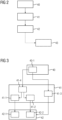
- FIG. 2 shows a flow chart of a first embodiment of a method according to the invention for providing result data which are suitable for use in planning an irradiation of a patient.
- a method step 40 CT measurement data of the patient are recorded, which have been acquired by means of a CT device which has a quantum-counting X-ray detector.
- the CT measurement data is processed further, with a specific information content of the CT measurement data resulting from the use of the quantum-counting X-ray detector in the acquisition of the CT measurement data being taken into account during further processing of the CT measurement data, with the further processing of the CT measurement data generates result data which are suitable for use in planning the irradiation of the patient.
- the results data are made available to an interface so that the results data can be used for planning the irradiation of the patient.
- the method according to the invention can be expanded in such a way that in a method step 43 the planning of the irradiation of the patient is carried out, the result data being retrieved from the interface and being used in the planning of the irradiation of the patient.
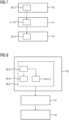
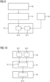
- FIG. 3 shows a flowchart of a second embodiment of a method according to the invention.
- the acquisition of the CT measurement data in method step 40 comprises, in a method step 40-1, an acquisition of spectrally resolved CT measurement data which result from the acquisition using the quantum-counting x-ray detector.
- the acquisition of the CT measurement data can include loading previously acquired, spectrally resolved CT measurement data from a database to calculate the spatially resolved distribution of the quantitative material coefficient.
- the spectrally resolved CT measurement data advantageously cover a measurement field which, in an axial measurement slice, includes both the patient's entire body and positioning aids used for positioning the patient.
- the further processing of the CT measurement data in method step 41 includes calculating a spatially resolved distribution of a quantitative material coefficient from the spectrally resolved CT measurement data in a method step 41-1.
- Providing the result data includes providing the spatially resolved distribution of the quantitative material coefficient to the interface in a method step 42-1.
- the further processing of the CT measurement data includes a calculation of two spatially resolved distributions of two different quantitative material coefficients.
- a first spatially resolved distribution of a first quantitative material coefficient is calculated in method step 41-1 and a second spatially resolved distribution of a second quantitative material coefficient is calculated from the spectrally resolved CT measurement data in method step 41-2.
- the provision of the result data in method step 42 according to 3 providing the first spatially resolved distribution of the first quantitative material coefficient for a first dose calculation algorithm in a method step 42-1 and providing the second spatially resolved distribution of the second quantitative material coefficient for a second dose calculation algorithm in a process step 42-2.
- the first quantitative material coefficient is an electron density and the second quantitative material coefficient is an effective atomic number. It is also conceivable that the first quantitative material coefficient and the second quantitative material coefficient are based on two base materials that are linearly independent of one another.
- the further processing of the CT measurement data requires separate further processing of the first spatially resolved distribution of the first quantitative material coefficient and the second spatially resolved distribution of the second quantitative material coefficient and generation of the result data by means of a combination of the partial result data generated during separate further processing includes.
- the procedure is also shown that the further processing of the CT measurement data in a method step 41-3 in addition to calculating the spatially resolved distribution of the quantitative material coefficient from the spectrally resolved CT measurement data an identification of a target volume and / or volume at risk in the spectrally resolved CT -Measurement data included.
- the advantageous procedure is conceivable that the spectrally resolved CT measurement data have been acquired using such recording parameters of the quantum-counting X-ray detector that the spectrally resolved CT measurement data have a particularly suitable contrast between different tissue types for the identification of the target volume and / or volume at risk .
- the further processing of the CT measurement data in a method step 41-4 includes a beam hardening correction of the spectrally resolved CT measurement data using spectral information contained in the spectrally resolved CT measurement data, with the calculation of the spatially resolved distribution of the quantitative material coefficients from the spectrally resolved CT measurement data corrected by the beam hardening correction.
- the quantitative material coefficient can also be calculated directly from the uncorrected CT measurement data.
- FIG. 4 shows a flowchart of a third embodiment of a method according to the invention.
- the further processing of the CT measurement data in a method step 41-5 includes an identification of a target volume and/or volume at risk in the CT measurement data.
- providing the result data includes providing the identified target volume and/or risk volume to the interface in a method step 42-3.
- the identification of the target volume and/or volume at risk in a method step 41-5-1 includes a delimitation of a tissue of the target volume and/or volume at risk from the surrounding tissue, wherein the delimitation of the tissue of the target volume and/or volume at risk from the surrounding tissue using the specific information content of the CT measurement data, which results from the use of the at least one quantum-counting X-ray detector in the acquisition of the CT measurement data.
- the identification of the target volume and/or volume at risk includes a differentiation between fat tissue and muscle tissue, the differentiation of fat tissue and muscle tissue using the specific information content of the CT measurement data, which results from the use of the at least one Quantum-counting X-ray detector results in the acquisition of the CT measurement data takes place.
- the procedure is also conceivable that the identification of the target volume and/or risk volume delimits gray brain matter and white brain matter includes, wherein the demarcation of the gray brain matter and white brain matter using the specific information content of the CT measurement data, which results from the use of the at least one quantum-counting X-ray detector in the acquisition of the CT measurement data, takes place.
- the procedure is shown that in a method step 40-1 the acquisition of the CT measurement data comprises an acquisition of spectrally resolved CT measurement data which result from the acquisition using the quantum-counting X-ray detector, in a method step 41-4 the further processing of the CT -Measurement data includes a beam hardening correction of the spectrally resolved CT measurement data using spectral information contained in the spectrally resolved CT measurement data and in a method step 41-5-2 the identification of the target volume and / or volume at risk a delimitation of bone structures in the by the beam hardening correction includes corrected spectrally resolved CT measurement data.
- the bone structures in a skull base region of the patient are advantageously delimited from one another.
- the identification of the target volume and/or volume at risk in method step 41-5-1 can also include the identification of the bone structure and/or the demarcation between gray brain matter and white brain matter and/or the demarcation of fat tissue and muscle tissue.
- figure 5 shows a flowchart of a fourth embodiment of a method according to the invention.
- FIG 5 shows the procedure in which the acquisition of the CT measurement data comprises an acquisition of a first CT measurement dataset in a method step 40-2 and an acquisition of a second CT measurement dataset in a method step 40-3.
- the first CT measurement data record is by means of a higher Energy threshold of the quantum-counting X-ray detector has been acquired as the second CT measurement data set.
- the further processing of the CT measurement data includes creating a weighted combination of the first CT measurement data set and the second CT measurement data set, with combined CT measurement data being generated. In this way, the target volume and/or risk volume is identified in the combined CT measurement data in a method step 41-5.
- FIG 5 the advantageous procedure shown that the further processing of the CT measurement data in a method step 41-7 an extraction of information for a beam hardening correction from the first CT measurement data set and in a method step 41-8 a beam hardening correction of the combined CT measurement data based on the extracted information includes.
- the target volume and/or risk volume is identified in the combined CT measurement data corrected by the beam hardening correction.
- method step 41-6-1 the weighted combination of the first CT measurement dataset and the second CT measurement dataset is created using spatially varying weighting factors.
- the further processing of the CT measurement data in a method step 41-6-2 initially involves a rough identification of the target volume and/or volume at risk, with the spatially varying weighting factors being defined on the basis of the roughly segmented target volume and/or volume at risk, and then a fine identification of the target volume and/or risk volume in the combined CT measurement data.
- FIG. 6 shows a flow chart of a fifth embodiment of a method according to the invention.
- the acquisition of the CT measurement data according to 6 includes in a method step 40-4 an acquisition of high-resolution CT measurement data by means of the quantum-counting X-ray detector, wherein a high-resolution mode of the quantum-counting X-ray detector is used to acquire the high-resolution CT measurement data, wherein in the high-resolution mode each Pixel of the quantum counting X-ray detector is counted separately.
- the further processing of the CT measurement data thus includes calculating a spatially resolved distribution of a quantitative material coefficient from the high-resolution CT measurement data. Consequently, providing the result data includes providing the spatially resolved distribution of the quantitative material coefficient to the interface in a method step 42-4.
- FIG. 6 shows the advantageous procedure that in a method step 41-9-1 the high-resolution CT measurement data are included in the calculation of the spatially resolved distribution of the quantitative material coefficient for bone tissue of the patient in unchanged form and in a method step 41-9-3 the high-resolution CT measurement data go into the calculation of the spatially resolved distribution of the quantitative material coefficient for a soft tissue of the patient in a reduced resolution.
- the reduced resolution of the high-resolution CT measurement data is generated in a method step 41-9-2 by filtering the high-resolution CT measurement data using a filter kernel.
- FIG. 7 shows a flowchart of a sixth embodiment of a method according to the invention.
- FIG. 8 shows a flow chart of a seventh embodiment of a method according to the invention.
- According to 8 includes the acquisition of the CT measurement data of the patient in a method step 40-6 the acquisition of the CT measurement data by means of the CT device, which has the quantum-counting X-ray detector, wherein a detector parameter of the quantum-counting X-ray detector is changed during the acquisition of the CT measurement data .
- the detector parameter of the quantum-counting X-ray detector is changed during the acquisition of the CT measurement data in such a way that in a method step 40-6-1 for an acquisition of the CT measurement data of a first body region of the patient, a first setting of the detector parameter is set and in a Method step 40-6-2 a second setting of the detector parameter is set for an acquisition of the CT measurement data of a second body region of the patient.
- a method step 40-6-3 the first body region and the second body region are defined before the acquisition of the CT measurement data using medical image data already available from the patient and/or using an atlas database.
- the detector parameters of the quantum-counting X-ray detector are advantageously changed in such a way that a higher resolution of the quantum-counting X-ray detector is set for the acquisition of the CT measurement data from the first body region of the patient than for the acquisition of the CT measurement data from the second body region of the patient.
- the procedure not shown is also conceivable that the detector parameters of the quantum-counting X-ray detector are changed during the acquisition of the CT measurement data in such a way that a first setting is required for an acquisition of the CT measurement data at a first z-position along a longitudinal direction of the patient of the detector parameter is set and a second setting of the detector parameter is set for an acquisition of the CT measurement data at a second z-position along the longitudinal direction of the patient.
- the procedure not shown is also conceivable that the detector parameters of the quantum-counting X-ray detector are changed during the acquisition of the CT measurement data in such a way that for an acquisition of the CT measurement data at a first rotational position of a detector-radiator system of the CT device a first setting of the detector parameter is set and a second setting of the detector parameter is set for an acquisition of the CT measurement data at a second rotational position of the detector-radiator system of the CT device.
- FIG. 9 shows a flowchart of an eighth embodiment of a method according to the invention.
- the interface captures further measurement data, which is captured by means of an imaging device of a radiation therapy device used for the irradiation of the patient, with the result data being related to the further measurement data in a method step 42-8 for planning the irradiation of the patient become.
- the imaging device of the radiation therapy device used for the irradiation of the patient also has a quantum-counting X-ray detector, the result data using a specific information content of the CT measurement data and the further CT measurement data, which results from the use of the quantum-counting X-ray detector in the acquisition of the CT -Measurement data and the further measurement data are set in relation to the further measurement data.
- FIG. 10 shows a flow chart of a ninth embodiment of a method according to the invention.
- a first CT measurement data record is recorded in a method step 40-7 and a second CT measurement data record is recorded in a method step 40-8. Both CT measurement data sets were recorded at different points in time.
- the at least two CT measurement data sets are registered using the specific information content of the CT measurement data, which results from the use of the at least one quantum-counting X-ray detector when acquiring the CT measurement data.
- the registered at least two sets of CT measurement data are made available to the interface.
- FIG. 11 shows a flow chart of a tenth embodiment of a method according to the invention.
- a contrast agent has been used in a method step 40-9 during the acquisition of the CT measurement data.
- the further processing of the CT measurement data in a method step 41-12 comprises a material decomposition of the CT measurement data in tissue that has contrast agent and other tissue using the specific information content of the CT measurement data, which results from the use of the at least a quantum-counting X-ray detector when acquiring the CT measurement data.
- the material decomposition is provided to the interface in a method step 42-10.
- the second possible application can be used, that in a method step 41-13 the further processing of the CT measurement data involves the creation of a virtual non-contrasted CT image using the specific information content of the CT measurement data, which results from the use of the at least a quantum-counting x-ray detector in the acquisition of the CT measurement data, wherein the virtual non-contrasted CT image is provided to the interface in a method step 42-11.
- the computing unit includes the software required for this and/or computer programs stored in a memory unit of the processing unit.
- the software and/or computer programs include programming means that are designed to execute the method according to the invention when the computer program and/or the software is executed in the processing unit by means of a processor unit of the processing unit.
Landscapes
- Health & Medical Sciences (AREA)
- Engineering & Computer Science (AREA)
- Biomedical Technology (AREA)
- Life Sciences & Earth Sciences (AREA)
- Nuclear Medicine, Radiotherapy & Molecular Imaging (AREA)
- Radiology & Medical Imaging (AREA)
- Pathology (AREA)
- Animal Behavior & Ethology (AREA)
- General Health & Medical Sciences (AREA)
- Public Health (AREA)
- Veterinary Medicine (AREA)
- Apparatus For Radiation Diagnosis (AREA)
- Radiation-Therapy Devices (AREA)
Claims (50)
- Procédé pour disposer de données de résultat, qui sont propres à une utilisation dans une planification d'une exposition d'un patient à du rayonnement, comprenant les stades de procédé suivants :- on saisit des données de mesure CT du patient, qui ont été acquises au moyen d'un appareil CT, qui a un détecteur de rayons X comptant les quantas,- on traite ultérieurement les données de mesure CT, dans lequel, lors du traitement ultérieur du traitement de données de mesure CT, on prend en compte un contenu informationnel spécifique des données de mesure CT, qui provient de l'utilisation du détecteur de rayons X comptant les quantas, lors de l'acquisition des données de mesure CT, dans lequel, lors du traitement ultérieur des données de mesure CT, on produit des données de résultat, qui sont propres à être utilisées dans la planification de l'exposition du patient à du rayonnement,- on met les données de résultat à disposition d'une interface, de manière à pouvoir utiliser les données de résultat pour la planification de l'exposition du patient à du rayonnement.
- Procédé suivant la revendication 1, dans lequel le procédé comprend le stade de procédé supplémentaire suivant :- on effectue la planification de l'exposition du patient à du rayonnement, dans lequel on appelle les données de résultat de l'interface et on les utilise lors de la planification de l'exposition du patient à du rayonnement.
- Procédé suivant l'une des revendications précédentes, dans lequel- la saisie des données de mesure CT comprend une saisie de données de mesure CT à résolution spectrale, qui proviennent de l'acquisition au moyen du détecteur de rayons X comptant les quantas,- le traitement ultérieur des données de mesure CT comprend un calcul d'une distribution à résolution spatiale d'un coefficient quantitatif de matériau, à partir des données de mesure CT à résolution spectrale, et- la fourniture des données de résultat comprend une mise de la distribution à résolution spatiale du coefficient quantitatif de matériau à disposition de l'interface.
- Procédé suivant la revendication 3, dans lequel le traitement ultérieur des données de mesure CT comprend un calcul d'une première distribution à résolution spatiale d'un premier coefficient quantitatif de matériau et une deuxième distribution à résolution spatiale d'un deuxième coefficient quantitatif de matériau, à partir des données de mesure CT à résolution spectrale.
- Procédé suivant la revendication 4, dans lequel la fourniture des données de résultat comprend une mise à disposition de la première distribution à résolution spatiale du premier coefficient quantitatif de matériau pour un premier algorithme de calcul de dose et une mise à disposition de la deuxième distribution à résolution spatiale du deuxième coefficient quantitatif de matériau pour un deuxième algorithme de calcul de dose.
- Procédé suivant l'une des revendications 4 à 5, dans lequel le premier coefficient quantitatif de matériau est une densité électronique et le deuxième coefficient quantitatif de matériau est un numéro atomique effectif.
- Procédé suivant l'une des revendications 4 à 5, dans lequel le premier coefficient quantitatif de matériau et le deuxième coefficient quantitatif de matériau reposent sur des matériaux de base indépendants linéairement l'un de l'autre.
- Procédé suivant l'une des revendications 4 à 7, dans lequel le traitement ultérieur des données de mesure CT comprend un traitement ultérieur distinct de la première distribution à résolution spatiale du premier coefficient quantitatif de matériau et de la deuxième distribution à résolution spatiale du deuxième coefficient quantitatif de matériau et une production des données de résultat au moyen d'une réunion des données de résultat partiel produits dans le traitement ultérieur distinct.
- Procédé suivant l'une des revendications 3 à 8, dans lequel la saisie des données de mesure CT comprend une charge de données de mesure CT à résolution spatiale acquises auparavant à partir d'une base de données pour le calcul de la distribution à résolution spatiale du coefficient quantitatif de matériau.
- Procédé suivant l'une des revendications 3 à 9, dans lequel le traitement ultérieur des données de mesure CT comprend, en outre, pour le calcul de la distribution à résolution spatiale du coefficient quantitatif de matériau à partir des données de mesure CT à résolution spectrale, une identification d'un volume cible et/ou d'un volume à risque dans les données de mesure CT à résolution spectrale.
- Procédé suivant la revendication 10, dans lequel on a acquis les données de mesure CT à résolution spectrale au moyen de paramètres d'enregistrement du détecteur à rayons X comptant les quantas, en ce que les données de mesure CT à résolution spectrale ont, pour l'identification du volume cible et/ou du volume à risque, un contraste particulièrement approprié entre divers types de tissus.
- Procédé suivant l'une des revendications 3 à 11, dans lequel les données de mesure CT à résolution spectrale recouvrent un champ de mesure, qui comprend, dans une couche de mesure axiale, tant tout le corps du patient qu'également les auxiliaires de mise en position pour coucher le patient.
- Procédé suivant l'une des revendications 3 à 12, dans lequel le traitement ultérieur des données de mesure CT comprend une correction de dureté du rayonnement des données de mesure CT à résolution spectrale, en utilisant des informations spectrales contenues dans les données de mesure CT à résolution spectrale, dans lequel de la distribution à résolution spatiale du coefficient quantitatif de matériau s'effectue à partir des données de mesure CT à résolution spectrale corrigées par la correction de dureté du rayonnement.
- Procédé suivant l'une des revendications précédentes, dans lequel le traitement ultérieur des données de mesure CT comprend une identification de volume cible et/ou de volume à risque dans les données de mesure CT, dans lequel la mise des données de résultat à disposition de l'interface comprend une mise à disposition du volume cible et/ou du volume à risque identifié.
- Procédé suivant la revendication 14, dans lequel l'identification du volume cible et/ou du volume à risque comprend une délimitation d'un tissu du volume cible et/ou du volume à risque par rapport à des tissus environnants, la délimitation du tissu du volume cible et/ou du volume à risque par rapport aux tissus environnants a lieu en utilisant le contenu informationnel spécifique des données de mesure CT, qui provient de l'utilisation du au moins un détecteur à rayons X comptant les quantas, lors de l'acquisition des données de mesure CT.
- Procédé suivant l'une des revendications 14 à 15, dans lequel l'identification du volume cible et/ou du volume à risque comprend une délimitation de tissu adipeux et de tissu musculaire, dans lequel la délimitation du tissu adipeux et du tissu musculaire a lieu en utilisant le contenu informationnel spécifique des données de mesure CT, qui provient de l'utilisation du au moins un détecteur de rayons X comptant les quantas lors de l'acquisition des données de mesure CT.
- Procédé suivant l'une des revendications 14 à 16, dans lequel l'identification du volume cible et/ou du volume à risque comprend une délimitation de substance grise du cerveau et de substance blanche du cerveau, dans lequel la délimitation de la substance grise du cerveau et de la substance blanche du cerveau a lieu en utilisant le contenu informationnel spécifique des données de mesure CT, qui provient de l'utilisation du au moins un détecteur à rayons X comptant les quantas lors de l'acquisition des données de mesure CT.
- Procédé suivant l'une des revendications 14 à 17, dans lequel- la saisie des données de mesure CT comprend une saisie de données de mesure CT à résolution spectrale, qui provient de l'acquisition au moyen du détecteur à rayons X comptant les quantas,- le traitement ultérieur des données de mesure CT comprend une correction de dureté de rayonnement des données de mesure CT à résolution spectrale en utilisant des informations spectrales contenues dans les données de mesure CT à résolution spectrale, et- l'identification du volume cible et/ou du volume à risque comprend une délimitation de structures osseuses dans les données de mesure CT à résolution spectrale corrigées par la correction de dureté du rayonnement.
- Procédé suivant la revendication 18, dans lequel on délimite entre elles les structures osseuses dans une région de base du crâne du patient.
- Procédé suivant l'une des revendications 14 à 19, dans lequel- les données de mesure CT comprennent un premier ensemble de données de mesure CT et un deuxième ensemble de données de mesure CT, dans lequel le premier ensemble de données de mesure CT a été acquis au moyen d'un seuil d'énergie plus haut du détecteur à rayons X comptant les quantas que le deuxième ensemble de données de mesure CT,- le traitement ultérieur des données de mesure CT comprend un établissement d'une combinaison pondérée du premier ensemble de données de mesure CT et du deuxième ensemble de données de mesure CT, des données de mesure CT combinées étant produites, et- l'identification du volume cible et/ou du volume à risque a lieu dans les données de mesure CT combinées.
- Procédé suivant la revendication 20, dans lequel- le traitement ultérieur des données de mesure CT comprend une extraction d'informations pour une correction de dureté de rayonnement à partir du premier jeu de données de mesure CT, et une correction de dureté de rayonnement des données de mesure CT combinées sur la base des informations extraites, dans lequel- l'identification du volume cible et/ou du volume à risque a lieu dans les données de mesure CT combinées corrigées par la correction de dureté du rayonnement.
- Procédé suivant l'une des revendications 20 à 21, dans lequel l'établissement de la combinaison pondérée du premier ensemble de données de mesure CT et du deuxième ensemble de données de mesure CT a lieu en utilisant des facteurs de pondération variant dans l'espace.
- Procédé suivant la revendication 22, dans lequel- le traitement ultérieur des données de mesure CT comprend d'abord une identification grossière du volume cible et/ou du volume à risque, dans lequel on définit les facteurs de pondération variant dans l'espace, à l'aide du volume cible et/ou du volume à risque segmenté grossièrement et a lieu ensuite une identification fine du volume cible et/ou du volume à risque dans les données de mesure CT combinées.
- Procédé suivant l'une des revendications précédentes, dans lequel la saisie des données de mesure CT comprend une acquisition de données de mesure CT à haute résolution au moyen du détecteur à rayons X comptant les quantas, dans lequel on utilise un mode à haute résolution du détecteur à rayons X comptant les quantas pour l'acquisition des données de mesure CT à haute résolution, dans lequel on compte séparément dans le mode à haute résolution chaque pixel du détecteur à rayons X comptant les quantas.
- Procédé suivant la revendication 24, dans lequel- le traitement ultérieur des données de mesure CT comprend un calcul d'une distribution à résolution spatiale d'un coefficient quantitatif de matériau à partir des données de mesure CT à haute résolution, et- la mise à disposition des données de résultat comprend une mise de la distribution à résolution spatiale du coefficient quantitatif de matériau à disposition de l'interface.
- Procédé suivant la revendication 25, dans lequel- les données de mesure CT à haute résolution entrent, sous une forme inchangée, dans le calcul de la distribution à résolution spatiale du coefficient quantitatif de matériau pour un tissu osseux du patient, et- les données de mesure CT à haute résolution entrent, suivant une résolution réduite, dans le calcul de la distribution à résolution spatiale du coefficient quantitatif de matériau pour un tissu de partie molle du patient.
- Procédé suivant la revendication 26, dans lequel on produit la résolution réduite des données de mesure CT à haute résolution au moyen d'un filtrage des données de mesure CT à haute résolution en utilisant un noyau de filtre.
- Procédé suivant l'une des revendications 25 à 27, dans lequel- le calcul de la distribution à résolution spatiale du coefficient quantitatif de matériau comprend un calcul d'une distribution à résolution spatiale d'un pouvoir de ralentissement à partir des données de mesure CT à haute résolution, et- la mise à disposition des données de résultat comprend une mise à disposition de la distribution à résolution spatiale du pouvoir de ralentissement pour une planification de l'exposition du patient à des particules.
- Procédé suivant l'une des revendications 25 à 28, dans lequel le calcul de la distribution à résolution spatiale du coefficient quantitatif de matériau à partir des données de mesure CT à haute résolution a lieu pour au moins l'une des régions du corps suivante du patient :- une région de l'œil du patient,- une région du corps décrite par un tracé du nerf du trijumeau du patient,- une base du crâne du patient,- une région des poumons du patient.
- Procédé suivant l'une des revendications précédentes, dans lequel- la saisie des données de mesure CT comprend une saisie de données de mesure CT à résolution spectrale et à résolution temporelle, qui proviennent de l'acquisition au moyen du détecteur à rayons X comptant les quantas,- le traitement ultérieur des données de mesure CT comprend un calcul d'une distribution à résolution spatiale et à résolution temporelle d'un coefficient quantitatif de matériau à partir des données de mesure CT à résolution spectrale et à résolution temporelle, et- la mise à disposition des données de résultat comprend une mise de la distribution à résolution spatiale et à résolution temporelle du coefficient quantitatif de matériau à disposition de l'interface.
- Procédé suivant la revendication 30, dans lequel on saisit les données de mesure CT à résolution spectrale et à résolution temporelle à partir d'au moins l'une des régions du corps suivante du patient :- une région du foie du patient,- une région du pancréas du patient,- une région du thorax du patient.
- Procédé suivant l'une des revendications précédentes, dans lequel la saisie des données de mesure CT du patient comprend l'acquisition des données de mesure CT au moyen de l'appareil CT, qui a le détecteur de rayons X comptant les quantas, dans lequel, pendant l'acquisition des données de mesure CT, on modifie un paramètre du détecteur de rayons X comptant les quantas.
- Procédé suivant la revendication 32, dans lequel on modifie, pendant l'acquisition des données de mesure CT, un seuil d'énergie du détecteur à rayons X comptant les quantas.
- Procédé suivant l'une des revendications 32 à 33, dans lequel, pendant l'acquisition des données de mesure CT, on modifie une résolution du détecteur de rayons X comptant les quantas, en ce qui concerne une combinaison de pixels du détecteur à rayons X comptant les quantas.
- Procédé suivant l'une des revendications 32 à 34, dans lequel on modifie le paramètre du détecteur de rayons X comptant les quantas pendant l'acquisition des données de mesure CT, de manière à mettre, pendant une acquisition des données de mesure CT à une première position z le long d'une direction longitudinale du patient, un premier réglage du paramètre du détecteur, et pendant une acquisition des données de mesure CT à une deuxième position z le long de la direction longitudinale du patient, un deuxième réglage du paramètre du détecteur.
- Procédé suivant l'une des revendications 32 à 35, dans lequel on modifie le paramètre du détecteur de rayons X comptant les quantas pendant l'acquisition des données de mesure CT, de manière à mettre, pendant une acquisition des données de mesure CT pour une première position en rotation d'un système de détecteur - émetteur de l'appareil CT, un premier réglage du paramètre du détecteur, et pendant une acquisition des données de mesure CT à une deuxième position en rotation du système détecteur - émetteur de l'appareil CT, un deuxième réglage du paramètre du détecteur.
- Procédé suivant l'une des revendications 32 à 36 précédentes, dans lequel on modifie le paramètre du détecteur à rayons X comptant les quantas, pendant l'acquisition des données de mesure CT, de manière à mettre, pendant une acquisition des données de mesure CT d'une première région du corps du patient, un premier réglage du paramètre du détecteur, et pendant une acquisition des données de mesure CT d'une deuxième région du corps du patient, un deuxième réglage du paramètre du détecteur.
- Procédé suivant la revendication 37, dans lequel on fixe la première région du corps et la deuxième région du corps avant l'acquisition des données de mesure CT, à l'aide de données d'images médicales déjà présentent du patient et/ou en utilisant une base de données formant atlas.
- Procédé suivant l'une des revendications 37 à 38, dans lequel la première région du corps comprend un volume cible à exposer au rayonnement et la deuxième région du corps comprend une région du corps se trouvant à l'extérieur du volume cible à exposer au rayonnement.
- Procédé suivant l'une des revendications 37 à 39, dans lequel la première région du corps est constituée de manière à être placée, lors de l'exposition du patient à du rayonnement, dans une zone de rayonnement, et la deuxième région du corps est constituée de manière à être placée, lors de l'exposition du patient à du rayonnement, en dehors d'une zone de rayonnement.
- Procédé suivant l'une des revendications 37 à 40, dans lequel la première région du corps et la deuxième région du corps sont constitués de manière à ce que, lors de l'exposition du patient à du rayonnement, il soit prévu une dose de rayonnement plus grande pour la première région du corps que pour la deuxième région du corps.
- Procédé suivant l'une des revendications 37 à 41, dans lequel, pendant l'acquisition des données de mesure CT, on modifie le paramètre du détecteur de rayons X comptant les quantas, de manière à mettre, pendant l'acquisition des données de mesure CT à partir de la première région du corps du patient, une résolution plus haute du détecteur de rayons X comptant les quantas, que pour l'acquisition des données de mesure CT à partir de la deuxième région du corps du patient.
- Procédé suivant l'une des revendications précédentes, dans lequel l'interface saisit d'autres données de mesure que l'on saisit au moyen d'une installation d'imagerie d'un appareil de radiothérapie utilisée pour l'exposition du patient à du rayonnement, dans lequel, pour la planification de l'exposition du patient à du rayonnement, on règle les données du résultat par rapport aux autres données de mesure.
- Procédé suivant la revendication 43, dans lequel l'installation d'imagerie de l'appareil de radiothérapie utilisée pour l'exposition du patient à du rayonnement a également un détecteur de rayons X comptant les quantas, dans lequel on règle, par rapport aux autres données de mesure, les données de résultat en utilisant un contenu informationnel spécifique des données de mesure CT et des autres données de mesure CT, qui proviennent de l'utilisation du détecteur à rayons X comptant les quantas lors de l'acquisition des données de mesure CT et des autres données de mesure CT.
- Procédé suivant l'une des revendications précédentes, dans lequel les données de mesure CT comprennent au moins deux ensembles de données de mesure CT, qui ont été saisies à des instants différents, dans lequel on enregistre les au moins deux ensembles de données de mesure CT, en utilisant le contenu informationnel spécifique des données de mesure CT, qui provient de l'utilisation du au moins un détecteur à rayons X comptant les quantas lors de l'acquisition des données de mesure CT, dans lequel on met à disposition de l'interface les au moins deux ensembles de données de mesure CT enregistrés.
- Procédé suivant l'une des revendications précédentes, dans lequel on a utilisé un agent de contraste pendant l'acquisition des données de mesure CT, dans lequel le traitement ultérieur des données de mesure CT comprend une décomposition de matière des données de mesure CT dans du tissu, qui a l'agent de contraste, et dans le reste du tissu en utilisant le contenu informationnel spécifique des données de mesure CT, qui provient de l'utilisation, lors de l'acquisition des données de mesure CT, du au moins un détecteur de rayons X comptant les quantas.
- Procédé suivant l'une des revendications précédentes, dans lequel on a utilisé, pendant l'acquisition des données de mesure CT, un agent de contraste, dans lequel le traitement ultérieur des données de mesure CT comprend une production d'une image CT virtuelle non contrastée en utilisant le contenu informationnel spécifique des données de mesure CT, qui provient de l'utilisation, lors de l'acquisition des données de mesure CT, du au moins un détecteur de rayons X comptant les quantas.
- Unité informatique comprenant au moins un module de calcul, dans lequel l'unité informatique est constituée pour effectuer un procédé suivant l'une des revendications précédentes.
- Appareil CT comprenant une unité informatique suivant la revendication 48.
- Produit de programme d'ordinateur, qui peut être chargé directement dans une mémoire d'une unité informatique programmable, comprenant des moyens de code de programme pour effectuer un procédé suivant l'une des revendications 1 à 47, lorsque le produit de programme d'ordinateur est réalisé dans l'unité informatique.
Applications Claiming Priority (2)
| Application Number | Priority Date | Filing Date | Title |
|---|---|---|---|
| US201762549175P | 2017-08-23 | 2017-08-23 | |
| PCT/EP2018/069777 WO2019037977A1 (fr) | 2017-08-23 | 2018-07-20 | Procédé permettant de fournir des données de résultat appropriées pour être utilisées dans une planification d'une irradiation d'un patient |
Publications (2)
| Publication Number | Publication Date |
|---|---|
| EP3672688A1 EP3672688A1 (fr) | 2020-07-01 |
| EP3672688B1 true EP3672688B1 (fr) | 2023-03-01 |
Family
ID=63244545
Family Applications (1)
| Application Number | Title | Priority Date | Filing Date |
|---|---|---|---|
| EP18755715.2A Active EP3672688B1 (fr) | 2017-08-23 | 2018-07-20 | Procédé permettant de fournir des données de résultat appropriées pour être utilisées dans une planification d'une irradiation d'un patient |
Country Status (5)
| Country | Link |
|---|---|
| US (3) | US10898727B2 (fr) |
| EP (1) | EP3672688B1 (fr) |
| JP (1) | JP7321997B2 (fr) |
| CN (2) | CN114917492A (fr) |
| WO (1) | WO2019037977A1 (fr) |
Families Citing this family (5)
| Publication number | Priority date | Publication date | Assignee | Title |
|---|---|---|---|---|
| CN111569279B (zh) * | 2020-05-26 | 2021-08-17 | 杭州珞珈质子科技有限公司 | 质子治疗的参数监测装置及系统 |
| CN116999720A (zh) * | 2020-11-25 | 2023-11-07 | 中硼(厦门)医疗器械有限公司 | 中子捕获治疗设备 |
| CN114782375B (zh) * | 2022-04-27 | 2023-06-23 | 影图(成都)医疗科技有限公司 | 骨质密度测量方法、装置和设备 |
| CN118657801B (zh) * | 2024-08-20 | 2024-10-29 | 川北医学院附属医院 | 一种三叉神经磁共振成像分割分类系统 |
| EP4710999A1 (fr) * | 2024-09-12 | 2026-03-18 | Siemens Healthineers AG | Procédé de détermination d'un surdosage en radiothérapie |
Family Cites Families (27)
| Publication number | Priority date | Publication date | Assignee | Title |
|---|---|---|---|---|
| US20060104414A1 (en) | 2002-01-30 | 2006-05-18 | Mayo William E | Combinatorial contraband detection using energy dispersive x-ray diffraction |
| DE10212638A1 (de) | 2002-03-21 | 2003-10-16 | Siemens Ag | Computertomograph und Verfahren zum Nachweis von Röntgenstrahlung mit einer aus einer Vielzahl von Detektoren bestehenden Detektoreinheit |
| DE102004039681B4 (de) * | 2004-08-16 | 2006-06-01 | Siemens Ag | Tomographiegerät und Verfahren für ein Tomographiegerät |
| US7968853B2 (en) | 2005-04-26 | 2011-06-28 | Koninklijke Philips Electronics N.V. | Double decker detector for spectral CT |
| DE102005059210B4 (de) * | 2005-12-12 | 2008-03-20 | Siemens Ag | Radiotherapeutische Vorrichtung |
| CA2572713C (fr) * | 2006-01-18 | 2014-09-30 | Sunnybrook And Women's College And Health Sciences Centre | Detecteur radioscopique cerenkov pour imagerie par centrage |
| DE102006026945B4 (de) | 2006-06-09 | 2008-04-10 | Siemens Ag | Computertomographisches Bildaufnahmeverfahren, Verfahren zur Bestimmung der ortsabhängigen Konzentration einer Anzahl vorgegebener Stoffe in einem Untersuchungsobjekt und zugehöriger Computertomograph |
| DE102007034982B4 (de) | 2007-07-26 | 2016-07-07 | Siemens Healthcare Gmbh | Verfahren zum Betreiben eines getakteten, zählenden Röntgenstrahlendetektors |
| JP5171215B2 (ja) * | 2007-11-08 | 2013-03-27 | ジーイー・メディカル・システムズ・グローバル・テクノロジー・カンパニー・エルエルシー | X線ct装置 |
| EP2303125B1 (fr) | 2008-07-18 | 2019-10-30 | Koninklijke Philips N.V. | Imagerie spectrale |
| JP2010246883A (ja) * | 2009-03-27 | 2010-11-04 | Mitsubishi Electric Corp | 患者位置決めシステム |
| JP5279637B2 (ja) * | 2009-07-02 | 2013-09-04 | 株式会社日立製作所 | ベッド位置決めシステム及びベッド位置決め方法 |
| DE102009032252B3 (de) | 2009-07-08 | 2010-08-05 | Siemens Aktiengesellschaft | Verfahren zur Erzeugung von Röntgenbildern mit einem Mehrenergie-Röntgendetektionssystem sowie Vorrichtung zur Durchführung des Verfahrens |
| JP5535733B2 (ja) * | 2010-04-05 | 2014-07-02 | 住友重機械工業株式会社 | 実効原子番号及び電子密度を求める方法、その方法を実行させるためのプログラム、そのプログラムを記録したコンピュータ読み取り可能な記録媒体及びct装置 |
| DE102011077859B4 (de) * | 2011-06-21 | 2014-01-23 | Siemens Aktiengesellschaft | Quantenzählender Strahlungsdetektor |
| US9415240B2 (en) * | 2011-10-21 | 2016-08-16 | Accuray Incorporated | Apparatus for generating multi-energy x-ray images and methods of using the same |
| CN104135930B (zh) * | 2012-02-21 | 2018-03-30 | 皇家飞利浦有限公司 | 利用感兴趣组织谱成像和跟踪进行的自适应放射治疗 |
| CN103458967B (zh) * | 2012-04-11 | 2016-08-10 | 东芝医疗系统株式会社 | 放射线治疗系统以及治疗计划装置 |
| US9364191B2 (en) * | 2013-02-11 | 2016-06-14 | University Of Rochester | Method and apparatus of spectral differential phase-contrast cone-beam CT and hybrid cone-beam CT |
| CN104036452B (zh) | 2013-03-06 | 2017-12-05 | 东芝医疗系统株式会社 | 图像处理装置和方法以及医学图像设备 |
| CN103393408B (zh) * | 2013-08-20 | 2015-12-02 | 中国科学院自动化研究所 | 一种光声和荧光循环互提升成像方法 |
| JP6351164B2 (ja) * | 2014-06-12 | 2018-07-04 | 国立研究開発法人量子科学技術研究開発機構 | ビーム照射対象確認装置、ビーム照射対象確認プログラム、および阻止能比算出プログラム |
| DE102015217421B4 (de) * | 2015-09-11 | 2023-05-17 | Siemens Healthcare Gmbh | Spektrale Filterung von Röntgenstrahlung für energieselektive Röntgenbildgebung |
| US10219775B2 (en) * | 2015-11-02 | 2019-03-05 | Toshiba Medical Systems Corporation | Photon-counting X-ray CT apparatus and image processing apparatus |
| JP6985004B2 (ja) * | 2015-11-02 | 2021-12-22 | キヤノンメディカルシステムズ株式会社 | 光子計数型x線ct装置及び画像処理装置 |
| DE102016215109B4 (de) * | 2016-08-12 | 2025-05-28 | Siemens Healthineers Ag | Verfahren und Datenverarbeitungseinheit zum Optimieren eines Bildrekonstruktionsalgorithmus |
| DE102016219496A1 (de) | 2016-10-07 | 2018-04-26 | Siemens Healthcare Gmbh | Verfahren zur Unterstützung einer Planung einer Bestrahlung eines Patienten |
-
2018
- 2018-07-20 US US16/640,960 patent/US10898727B2/en active Active
- 2018-07-20 CN CN202210373223.7A patent/CN114917492A/zh active Pending
- 2018-07-20 WO PCT/EP2018/069777 patent/WO2019037977A1/fr not_active Ceased
- 2018-07-20 EP EP18755715.2A patent/EP3672688B1/fr active Active
- 2018-07-20 JP JP2020510538A patent/JP7321997B2/ja active Active
- 2018-07-20 CN CN201880067153.4A patent/CN111212680B/zh active Active
-
2020
- 2020-12-16 US US17/123,594 patent/US11844961B2/en active Active
-
2023
- 2023-11-06 US US18/502,308 patent/US12214220B2/en active Active
Also Published As
| Publication number | Publication date |
|---|---|
| CN111212680A (zh) | 2020-05-29 |
| US12214220B2 (en) | 2025-02-04 |
| US20240066320A1 (en) | 2024-02-29 |
| JP7321997B2 (ja) | 2023-08-07 |
| US20210101022A1 (en) | 2021-04-08 |
| CN114917492A (zh) | 2022-08-19 |
| CN111212680B (zh) | 2022-04-29 |
| US10898727B2 (en) | 2021-01-26 |
| WO2019037977A1 (fr) | 2019-02-28 |
| EP3672688A1 (fr) | 2020-07-01 |
| JP2020531132A (ja) | 2020-11-05 |
| US20200171324A1 (en) | 2020-06-04 |
| US11844961B2 (en) | 2023-12-19 |
Similar Documents
| Publication | Publication Date | Title |
|---|---|---|
| EP3672688B1 (fr) | Procédé permettant de fournir des données de résultat appropriées pour être utilisées dans une planification d'une irradiation d'un patient | |
| EP3143592B1 (fr) | Procédé et dispositif de réduction d'artéfacts dans des images obtenues par tomodensitométrie | |
| EP2150179B1 (fr) | Procédé de sélection de deux agents de contraste destinés à être utilisés lors d'un examen par ct à double énergie, combinaison d'agents de contraste et production d'images de ct à l'aide d'une combinaison d'agents de contraste et de spectres d'énergie différents | |
| DE102013200337B4 (de) | Verfahren, Computertomopraph und Computerprogrammprodukt zum Bestimmen von Intensitätswerten einer Röntgenstrahlung zur Dosismodulation | |
| EP2041719B1 (fr) | Procédé de détermination d'une carte de propriété pour un objet, en particulier pour un être humain, basé sur au moins une première image, en particulier une image par résonance magnétique nucléaire | |
| DE102008058488B4 (de) | Verfahren und Vorrichtung zur Aufbereitung von kombinierten MR-Emissionstomographieaufnahmen | |
| EP3238780B1 (fr) | Procédé pour la planification d'irradiation d'un patient | |
| EP1325471B1 (fr) | Procede et dispositif pour la representation d'un objet au moyen d'une irradiation ainsi que pour la reconstruction | |
| DE102018216370B4 (de) | Bereitstellen eines annotierten medizinischen Bilddatensatzes für eine Radiotherapieplanung eines Patienten | |
| DE102008036478A1 (de) | Vorrichtung und Verfahren zur Auswertung einer Aktivitätsverteilung sowie Bestrahlungsanlage | |
| DE102016223831B3 (de) | Verfahren zum Bereitstellen von mittels eines Dual-Source CT-Geräts aus einem Messfeld erfassten quantitativen CT-Bilddaten, sowie Recheneinheit, Dual-Source CT-Gerät und Computerprogrammprodukt | |
| WO2012097801A1 (fr) | Procédé de production d'une représentation radiographique assistée par agent de contraste et système radiographique approprié | |
| DE102016218359B3 (de) | Verfahren und Vorrichtung zur Korrektur einer synthetischen Elektronendichtekarte | |
| DE102007017629A1 (de) | Verfahren zur Differenzierung zwischen vier Materialien in tomographischen Aufnahmen eines 2-Energie-CT-Systems | |
| DE102015213730B4 (de) | Qualitätskontrolle einer Bestrahlungsplanung durch Vergleich erster und zweiter MR-basierter Dosisverteilungen im Planungsvolumen | |
| DE102012222714A1 (de) | Ermittlung eines Mehrfachenergie-Bildes | |
| JP2020531132A5 (fr) | ||
| DE102011005161B4 (de) | Verfahren, Bilddatensatzrekonstruktionseinrichtung, Röntgen-System und Computerprogramm zur Artefaktkorrektur | |
| EP3400877A1 (fr) | Procédé d'imagerie d'une zone d'examen au moyen d'un premier agent de contraste et d'un second agent de contraste | |
| DE102014218924A1 (de) | Ermittlung einer Position eines Objekts mittels MRT-Aufnahmen | |
| EP3511053B1 (fr) | Procédé et dispositif de détermination de la puissance de freinage pour une thérapie protonique | |
| DE102021213557B4 (de) | Verfahren und Vorrichtung zur Bereitstellung eines Perfusionsbilddatensatzes eines Patienten | |
| DE102021213439B3 (de) | Verfahren und Vorrichtung zur Erzeugung eines Ergebnisbilddatensatzes eines Patienten | |
| DE102023204265B4 (de) | Verfahren zum Generieren eines Volumenmodells eines Untersuchungsobjekts, Steuereinrichtung, Röntgenvorrichtung, Computerprogramm und elektronisch lesbarer Datenträger | |
| EP4113431A1 (fr) | Procédé et dispositif de détermination d'un ensemble de données d'imagerie radiographiques corrigées |
Legal Events
| Date | Code | Title | Description |
|---|---|---|---|
| STAA | Information on the status of an ep patent application or granted ep patent |
Free format text: STATUS: UNKNOWN |
|
| STAA | Information on the status of an ep patent application or granted ep patent |
Free format text: STATUS: THE INTERNATIONAL PUBLICATION HAS BEEN MADE |
|
| PUAI | Public reference made under article 153(3) epc to a published international application that has entered the european phase |
Free format text: ORIGINAL CODE: 0009012 |
|
| STAA | Information on the status of an ep patent application or granted ep patent |
Free format text: STATUS: REQUEST FOR EXAMINATION WAS MADE |
|
| 17P | Request for examination filed |
Effective date: 20200131 |
|
| AK | Designated contracting states |
Kind code of ref document: A1 Designated state(s): AL AT BE BG CH CY CZ DE DK EE ES FI FR GB GR HR HU IE IS IT LI LT LU LV MC MK MT NL NO PL PT RO RS SE SI SK SM TR |
|
| AX | Request for extension of the european patent |
Extension state: BA ME |
|
| DAV | Request for validation of the european patent (deleted) | ||
| DAX | Request for extension of the european patent (deleted) | ||
| GRAP | Despatch of communication of intention to grant a patent |
Free format text: ORIGINAL CODE: EPIDOSNIGR1 |
|
| STAA | Information on the status of an ep patent application or granted ep patent |
Free format text: STATUS: GRANT OF PATENT IS INTENDED |
|
| INTG | Intention to grant announced |
Effective date: 20221021 |
|
| GRAS | Grant fee paid |
Free format text: ORIGINAL CODE: EPIDOSNIGR3 |
|
| GRAA | (expected) grant |
Free format text: ORIGINAL CODE: 0009210 |
|
| STAA | Information on the status of an ep patent application or granted ep patent |
Free format text: STATUS: THE PATENT HAS BEEN GRANTED |
|
| AK | Designated contracting states |
Kind code of ref document: B1 Designated state(s): AL AT BE BG CH CY CZ DE DK EE ES FI FR GB GR HR HU IE IS IT LI LT LU LV MC MK MT NL NO PL PT RO RS SE SI SK SM TR |
|
| REG | Reference to a national code |
Ref country code: GB Ref legal event code: FG4D Free format text: NOT ENGLISH |
|
| REG | Reference to a national code |
Ref country code: CH Ref legal event code: EP Ref country code: AT Ref legal event code: REF Ref document number: 1550580 Country of ref document: AT Kind code of ref document: T Effective date: 20230315 |
|
| REG | Reference to a national code |
Ref country code: DE Ref legal event code: R096 Ref document number: 502018011697 Country of ref document: DE |
|
| REG | Reference to a national code |
Ref country code: IE Ref legal event code: FG4D Free format text: LANGUAGE OF EP DOCUMENT: GERMAN |
|
| REG | Reference to a national code |
Ref country code: NL Ref legal event code: FP |
|
| REG | Reference to a national code |
Ref country code: LT Ref legal event code: MG9D |
|
| PG25 | Lapsed in a contracting state [announced via postgrant information from national office to epo] |
Ref country code: RS Free format text: LAPSE BECAUSE OF FAILURE TO SUBMIT A TRANSLATION OF THE DESCRIPTION OR TO PAY THE FEE WITHIN THE PRESCRIBED TIME-LIMIT Effective date: 20230301 Ref country code: NO Free format text: LAPSE BECAUSE OF FAILURE TO SUBMIT A TRANSLATION OF THE DESCRIPTION OR TO PAY THE FEE WITHIN THE PRESCRIBED TIME-LIMIT Effective date: 20230601 Ref country code: LV Free format text: LAPSE BECAUSE OF FAILURE TO SUBMIT A TRANSLATION OF THE DESCRIPTION OR TO PAY THE FEE WITHIN THE PRESCRIBED TIME-LIMIT Effective date: 20230301 Ref country code: LT Free format text: LAPSE BECAUSE OF FAILURE TO SUBMIT A TRANSLATION OF THE DESCRIPTION OR TO PAY THE FEE WITHIN THE PRESCRIBED TIME-LIMIT Effective date: 20230301 Ref country code: HR Free format text: LAPSE BECAUSE OF FAILURE TO SUBMIT A TRANSLATION OF THE DESCRIPTION OR TO PAY THE FEE WITHIN THE PRESCRIBED TIME-LIMIT Effective date: 20230301 Ref country code: ES Free format text: LAPSE BECAUSE OF FAILURE TO SUBMIT A TRANSLATION OF THE DESCRIPTION OR TO PAY THE FEE WITHIN THE PRESCRIBED TIME-LIMIT Effective date: 20230301 |
|
| PG25 | Lapsed in a contracting state [announced via postgrant information from national office to epo] |
Ref country code: SE Free format text: LAPSE BECAUSE OF FAILURE TO SUBMIT A TRANSLATION OF THE DESCRIPTION OR TO PAY THE FEE WITHIN THE PRESCRIBED TIME-LIMIT Effective date: 20230301 Ref country code: PL Free format text: LAPSE BECAUSE OF FAILURE TO SUBMIT A TRANSLATION OF THE DESCRIPTION OR TO PAY THE FEE WITHIN THE PRESCRIBED TIME-LIMIT Effective date: 20230301 Ref country code: GR Free format text: LAPSE BECAUSE OF FAILURE TO SUBMIT A TRANSLATION OF THE DESCRIPTION OR TO PAY THE FEE WITHIN THE PRESCRIBED TIME-LIMIT Effective date: 20230602 Ref country code: FI Free format text: LAPSE BECAUSE OF FAILURE TO SUBMIT A TRANSLATION OF THE DESCRIPTION OR TO PAY THE FEE WITHIN THE PRESCRIBED TIME-LIMIT Effective date: 20230301 |
|
| PG25 | Lapsed in a contracting state [announced via postgrant information from national office to epo] |
Ref country code: SM Free format text: LAPSE BECAUSE OF FAILURE TO SUBMIT A TRANSLATION OF THE DESCRIPTION OR TO PAY THE FEE WITHIN THE PRESCRIBED TIME-LIMIT Effective date: 20230301 Ref country code: RO Free format text: LAPSE BECAUSE OF FAILURE TO SUBMIT A TRANSLATION OF THE DESCRIPTION OR TO PAY THE FEE WITHIN THE PRESCRIBED TIME-LIMIT Effective date: 20230301 Ref country code: PT Free format text: LAPSE BECAUSE OF FAILURE TO SUBMIT A TRANSLATION OF THE DESCRIPTION OR TO PAY THE FEE WITHIN THE PRESCRIBED TIME-LIMIT Effective date: 20230703 Ref country code: EE Free format text: LAPSE BECAUSE OF FAILURE TO SUBMIT A TRANSLATION OF THE DESCRIPTION OR TO PAY THE FEE WITHIN THE PRESCRIBED TIME-LIMIT Effective date: 20230301 Ref country code: CZ Free format text: LAPSE BECAUSE OF FAILURE TO SUBMIT A TRANSLATION OF THE DESCRIPTION OR TO PAY THE FEE WITHIN THE PRESCRIBED TIME-LIMIT Effective date: 20230301 |
|
| PG25 | Lapsed in a contracting state [announced via postgrant information from national office to epo] |
Ref country code: SK Free format text: LAPSE BECAUSE OF FAILURE TO SUBMIT A TRANSLATION OF THE DESCRIPTION OR TO PAY THE FEE WITHIN THE PRESCRIBED TIME-LIMIT Effective date: 20230301 Ref country code: IS Free format text: LAPSE BECAUSE OF FAILURE TO SUBMIT A TRANSLATION OF THE DESCRIPTION OR TO PAY THE FEE WITHIN THE PRESCRIBED TIME-LIMIT Effective date: 20230701 |
|
| REG | Reference to a national code |
Ref country code: DE Ref legal event code: R097 Ref document number: 502018011697 Country of ref document: DE |
|
| PLBE | No opposition filed within time limit |
Free format text: ORIGINAL CODE: 0009261 |
|
| STAA | Information on the status of an ep patent application or granted ep patent |
Free format text: STATUS: NO OPPOSITION FILED WITHIN TIME LIMIT |
|
| PG25 | Lapsed in a contracting state [announced via postgrant information from national office to epo] |
Ref country code: SI Free format text: LAPSE BECAUSE OF FAILURE TO SUBMIT A TRANSLATION OF THE DESCRIPTION OR TO PAY THE FEE WITHIN THE PRESCRIBED TIME-LIMIT Effective date: 20230301 Ref country code: DK Free format text: LAPSE BECAUSE OF FAILURE TO SUBMIT A TRANSLATION OF THE DESCRIPTION OR TO PAY THE FEE WITHIN THE PRESCRIBED TIME-LIMIT Effective date: 20230301 |
|
| 26N | No opposition filed |
Effective date: 20231204 |
|
| PG25 | Lapsed in a contracting state [announced via postgrant information from national office to epo] |
Ref country code: MC Free format text: LAPSE BECAUSE OF FAILURE TO SUBMIT A TRANSLATION OF THE DESCRIPTION OR TO PAY THE FEE WITHIN THE PRESCRIBED TIME-LIMIT Effective date: 20230301 |
|
| REG | Reference to a national code |
Ref country code: DE Ref legal event code: R081 Ref document number: 502018011697 Country of ref document: DE Owner name: SIEMENS HEALTHINEERS AG, DE Free format text: FORMER OWNER: SIEMENS HEALTHCARE GMBH, MUENCHEN, DE |
|
| PG25 | Lapsed in a contracting state [announced via postgrant information from national office to epo] |
Ref country code: MC Free format text: LAPSE BECAUSE OF FAILURE TO SUBMIT A TRANSLATION OF THE DESCRIPTION OR TO PAY THE FEE WITHIN THE PRESCRIBED TIME-LIMIT Effective date: 20230301 |
|
| REG | Reference to a national code |
Ref country code: CH Ref legal event code: PL |
|
| REG | Reference to a national code |
Ref country code: BE Ref legal event code: MM Effective date: 20230731 |
|
| PG25 | Lapsed in a contracting state [announced via postgrant information from national office to epo] |
Ref country code: LU Free format text: LAPSE BECAUSE OF NON-PAYMENT OF DUE FEES Effective date: 20230720 |
|
| PG25 | Lapsed in a contracting state [announced via postgrant information from national office to epo] |
Ref country code: LU Free format text: LAPSE BECAUSE OF NON-PAYMENT OF DUE FEES Effective date: 20230720 |
|
| REG | Reference to a national code |
Ref country code: IE Ref legal event code: MM4A |
|
| PG25 | Lapsed in a contracting state [announced via postgrant information from national office to epo] |
Ref country code: CH Free format text: LAPSE BECAUSE OF NON-PAYMENT OF DUE FEES Effective date: 20230731 |
|
| PG25 | Lapsed in a contracting state [announced via postgrant information from national office to epo] |
Ref country code: BE Free format text: LAPSE BECAUSE OF NON-PAYMENT OF DUE FEES Effective date: 20230731 |
|
| PG25 | Lapsed in a contracting state [announced via postgrant information from national office to epo] |
Ref country code: IE Free format text: LAPSE BECAUSE OF NON-PAYMENT OF DUE FEES Effective date: 20230720 |
|
| PG25 | Lapsed in a contracting state [announced via postgrant information from national office to epo] |
Ref country code: IE Free format text: LAPSE BECAUSE OF NON-PAYMENT OF DUE FEES Effective date: 20230720 |
|
| REG | Reference to a national code |
Ref country code: AT Ref legal event code: MM01 Ref document number: 1550580 Country of ref document: AT Kind code of ref document: T Effective date: 20230720 |
|
| PG25 | Lapsed in a contracting state [announced via postgrant information from national office to epo] |
Ref country code: AT Free format text: LAPSE BECAUSE OF NON-PAYMENT OF DUE FEES Effective date: 20230720 |
|
| PG25 | Lapsed in a contracting state [announced via postgrant information from national office to epo] |
Ref country code: AT Free format text: LAPSE BECAUSE OF NON-PAYMENT OF DUE FEES Effective date: 20230720 |
|
| PG25 | Lapsed in a contracting state [announced via postgrant information from national office to epo] |
Ref country code: BG Free format text: LAPSE BECAUSE OF FAILURE TO SUBMIT A TRANSLATION OF THE DESCRIPTION OR TO PAY THE FEE WITHIN THE PRESCRIBED TIME-LIMIT Effective date: 20230301 |
|
| PG25 | Lapsed in a contracting state [announced via postgrant information from national office to epo] |
Ref country code: BG Free format text: LAPSE BECAUSE OF FAILURE TO SUBMIT A TRANSLATION OF THE DESCRIPTION OR TO PAY THE FEE WITHIN THE PRESCRIBED TIME-LIMIT Effective date: 20230301 |
|
| PG25 | Lapsed in a contracting state [announced via postgrant information from national office to epo] |
Ref country code: CY Free format text: LAPSE BECAUSE OF FAILURE TO SUBMIT A TRANSLATION OF THE DESCRIPTION OR TO PAY THE FEE WITHIN THE PRESCRIBED TIME-LIMIT; INVALID AB INITIO Effective date: 20180720 |
|
| PG25 | Lapsed in a contracting state [announced via postgrant information from national office to epo] |
Ref country code: HU Free format text: LAPSE BECAUSE OF FAILURE TO SUBMIT A TRANSLATION OF THE DESCRIPTION OR TO PAY THE FEE WITHIN THE PRESCRIBED TIME-LIMIT; INVALID AB INITIO Effective date: 20180720 |
|
| PGFP | Annual fee paid to national office [announced via postgrant information from national office to epo] |
Ref country code: NL Payment date: 20250709 Year of fee payment: 8 |
|
| PGFP | Annual fee paid to national office [announced via postgrant information from national office to epo] |
Ref country code: DE Payment date: 20250919 Year of fee payment: 8 |
|
| PGFP | Annual fee paid to national office [announced via postgrant information from national office to epo] |
Ref country code: IT Payment date: 20250718 Year of fee payment: 8 |
|
| PGFP | Annual fee paid to national office [announced via postgrant information from national office to epo] |
Ref country code: GB Payment date: 20250811 Year of fee payment: 8 |
|
| PGFP | Annual fee paid to national office [announced via postgrant information from national office to epo] |
Ref country code: FR Payment date: 20250716 Year of fee payment: 8 |
|
| PG25 | Lapsed in a contracting state [announced via postgrant information from national office to epo] |
Ref country code: TR Free format text: LAPSE BECAUSE OF FAILURE TO SUBMIT A TRANSLATION OF THE DESCRIPTION OR TO PAY THE FEE WITHIN THE PRESCRIBED TIME-LIMIT Effective date: 20230301 |