EP3673864A1 - Segmentation de dents à l'aide d'un enregistrement de dents - Google Patents
Segmentation de dents à l'aide d'un enregistrement de dents Download PDFInfo
- Publication number
- EP3673864A1 EP3673864A1 EP18306870.9A EP18306870A EP3673864A1 EP 3673864 A1 EP3673864 A1 EP 3673864A1 EP 18306870 A EP18306870 A EP 18306870A EP 3673864 A1 EP3673864 A1 EP 3673864A1
- Authority
- EP
- European Patent Office
- Prior art keywords
- model image
- tooth
- segmentation
- obtaining
- model
- Prior art date
- Legal status (The legal status is an assumption and is not a legal conclusion. Google has not performed a legal analysis and makes no representation as to the accuracy of the status listed.)
- Granted
Links
Images




Classifications
-
- G—PHYSICS
- G06—COMPUTING OR CALCULATING; COUNTING
- G06T—IMAGE DATA PROCESSING OR GENERATION, IN GENERAL
- G06T7/00—Image analysis
- G06T7/10—Segmentation; Edge detection
- G06T7/12—Edge-based segmentation
-
- A—HUMAN NECESSITIES
- A61—MEDICAL OR VETERINARY SCIENCE; HYGIENE
- A61B—DIAGNOSIS; SURGERY; IDENTIFICATION
- A61B6/00—Apparatus or devices for radiation diagnosis; Apparatus or devices for radiation diagnosis combined with radiation therapy equipment
- A61B6/02—Arrangements for diagnosis sequentially in different planes; Stereoscopic radiation diagnosis
- A61B6/03—Computed tomography [CT]
- A61B6/032—Transmission computed tomography [CT]
-
- A—HUMAN NECESSITIES
- A61—MEDICAL OR VETERINARY SCIENCE; HYGIENE
- A61B—DIAGNOSIS; SURGERY; IDENTIFICATION
- A61B6/00—Apparatus or devices for radiation diagnosis; Apparatus or devices for radiation diagnosis combined with radiation therapy equipment
- A61B6/40—Arrangements for generating radiation specially adapted for radiation diagnosis
- A61B6/4064—Arrangements for generating radiation specially adapted for radiation diagnosis specially adapted for producing a particular type of beam
- A61B6/4085—Cone-beams
-
- A—HUMAN NECESSITIES
- A61—MEDICAL OR VETERINARY SCIENCE; HYGIENE
- A61B—DIAGNOSIS; SURGERY; IDENTIFICATION
- A61B6/00—Apparatus or devices for radiation diagnosis; Apparatus or devices for radiation diagnosis combined with radiation therapy equipment
- A61B6/50—Apparatus or devices for radiation diagnosis; Apparatus or devices for radiation diagnosis combined with radiation therapy equipment specially adapted for specific body parts; specially adapted for specific clinical applications
- A61B6/51—Apparatus or devices for radiation diagnosis; Apparatus or devices for radiation diagnosis combined with radiation therapy equipment specially adapted for specific body parts; specially adapted for specific clinical applications for dentistry
-
- A—HUMAN NECESSITIES
- A61—MEDICAL OR VETERINARY SCIENCE; HYGIENE
- A61B—DIAGNOSIS; SURGERY; IDENTIFICATION
- A61B6/00—Apparatus or devices for radiation diagnosis; Apparatus or devices for radiation diagnosis combined with radiation therapy equipment
- A61B6/50—Apparatus or devices for radiation diagnosis; Apparatus or devices for radiation diagnosis combined with radiation therapy equipment specially adapted for specific body parts; specially adapted for specific clinical applications
- A61B6/51—Apparatus or devices for radiation diagnosis; Apparatus or devices for radiation diagnosis combined with radiation therapy equipment specially adapted for specific body parts; specially adapted for specific clinical applications for dentistry
- A61B6/512—Intraoral means
-
- A—HUMAN NECESSITIES
- A61—MEDICAL OR VETERINARY SCIENCE; HYGIENE
- A61C—DENTISTRY; APPARATUS OR METHODS FOR ORAL OR DENTAL HYGIENE
- A61C7/00—Orthodontics, i.e. obtaining or maintaining the desired position of teeth, e.g. by straightening, evening, regulating, separating, or by correcting malocclusions
- A61C7/002—Orthodontic computer assisted systems
-
- A—HUMAN NECESSITIES
- A61—MEDICAL OR VETERINARY SCIENCE; HYGIENE
- A61C—DENTISTRY; APPARATUS OR METHODS FOR ORAL OR DENTAL HYGIENE
- A61C9/00—Impression cups, i.e. impression trays; Impression methods
- A61C9/004—Means or methods for taking digitized impressions
- A61C9/0046—Data acquisition means or methods
- A61C9/0053—Optical means or methods, e.g. scanning the teeth by a laser or light beam
- A61C9/006—Optical means or methods, e.g. scanning the teeth by a laser or light beam projecting one or more stripes or patterns on the teeth
-
- G—PHYSICS
- G06—COMPUTING OR CALCULATING; COUNTING
- G06T—IMAGE DATA PROCESSING OR GENERATION, IN GENERAL
- G06T7/00—Image analysis
- G06T7/0002—Inspection of images, e.g. flaw detection
- G06T7/0012—Biomedical image inspection
-
- G—PHYSICS
- G06—COMPUTING OR CALCULATING; COUNTING
- G06T—IMAGE DATA PROCESSING OR GENERATION, IN GENERAL
- G06T7/00—Image analysis
- G06T7/30—Determination of transform parameters for the alignment of images, i.e. image registration
- G06T7/33—Determination of transform parameters for the alignment of images, i.e. image registration using feature-based methods
-
- G—PHYSICS
- G06—COMPUTING OR CALCULATING; COUNTING
- G06T—IMAGE DATA PROCESSING OR GENERATION, IN GENERAL
- G06T7/00—Image analysis
- G06T7/30—Determination of transform parameters for the alignment of images, i.e. image registration
- G06T7/33—Determination of transform parameters for the alignment of images, i.e. image registration using feature-based methods
- G06T7/344—Determination of transform parameters for the alignment of images, i.e. image registration using feature-based methods involving models
-
- A—HUMAN NECESSITIES
- A61—MEDICAL OR VETERINARY SCIENCE; HYGIENE
- A61C—DENTISTRY; APPARATUS OR METHODS FOR ORAL OR DENTAL HYGIENE
- A61C7/00—Orthodontics, i.e. obtaining or maintaining the desired position of teeth, e.g. by straightening, evening, regulating, separating, or by correcting malocclusions
- A61C7/002—Orthodontic computer assisted systems
- A61C2007/004—Automatic construction of a set of axes for a tooth or a plurality of teeth
-
- G—PHYSICS
- G06—COMPUTING OR CALCULATING; COUNTING
- G06T—IMAGE DATA PROCESSING OR GENERATION, IN GENERAL
- G06T2207/00—Indexing scheme for image analysis or image enhancement
- G06T2207/10—Image acquisition modality
- G06T2207/10028—Range image; Depth image; 3D point clouds
-
- G—PHYSICS
- G06—COMPUTING OR CALCULATING; COUNTING
- G06T—IMAGE DATA PROCESSING OR GENERATION, IN GENERAL
- G06T2207/00—Indexing scheme for image analysis or image enhancement
- G06T2207/10—Image acquisition modality
- G06T2207/10072—Tomographic images
- G06T2207/10081—Computed x-ray tomography [CT]
-
- G—PHYSICS
- G06—COMPUTING OR CALCULATING; COUNTING
- G06T—IMAGE DATA PROCESSING OR GENERATION, IN GENERAL
- G06T2207/00—Indexing scheme for image analysis or image enhancement
- G06T2207/30—Subject of image; Context of image processing
- G06T2207/30004—Biomedical image processing
- G06T2207/30036—Dental; Teeth
Definitions
- the disclosure relates generally to dental imaging and more particularly relates to methods and apparatus for segmentation of intraoral features.
- Optical intraoral scans produce contours of dentition objects and have been helpful in improving visualization of teeth, gums, and other intra-oral structures.
- Surface contour information can be particularly useful for assessment of tooth condition and has recognized value for various types of dental procedures, such as for restorative dentistry. This can provide a valuable tool to assist the dental practitioner in identifying various problems and in validating other measurements and observations related to the patient's teeth and supporting structures.
- Surface contour information can also be used to generate 3D models of dentition components such as individual teeth; the position and orientation information related to individual teeth can then be used in assessing orthodontic treatment progress.
- a model of patient dentition is generated, initially and at various stages of the process, using surface contour information that can be acquired from an intraoral scanner.
- the model can be formed as a point cloud or mesh formed from scanned image content showing the surface contour.
- a number of standardized metrics can then be applied to the models for comparison and to track overall progress of the treatment regimen.
- segmentation enables the individual tooth to be identified from within the model and its features and allows its orientation to be correctly analyzed as part of the treatment evaluation.
- segmentation defines the tooth at each position in the model, such as along the point cloud or mesh, and identifies the tooth label. Segmentation may also identify features such as the cervical limit and the tooth cusp and fossae. Segmentation can also provide needed information for determining the tooth axis (mesio-distal and main axis). For example, segmentation allows the practitioner to identify and isolate the crown and to distinguish other visible portions of the tooth from gums and related supporting structure.
- An object of the present disclosure is to address the need for improved tooth segmentation and workflow in restorative and orthodontic imaging.
- Embodiments of the present disclosure support segmentation processing with the additional leverage available from the overall treatment plan and from previous segmentation results.
- a method for segmenting a 3D model image of a patient's dentition comprising:
- viewer In the context of the present disclosure, the terms “viewer”, “operator”, and “user” are considered to be equivalent and refer to the viewing practitioner, technician, or other person who views and manipulates an image, such as a dental image, on a display monitor.
- An "operator instruction” or “viewer instruction” is obtained from explicit commands entered by the viewer, such as by clicking a button on a scanner or by using a computer mouse or by touch screen or keyboard entry.
- the term "subject” is generally used to denote the patient who is imaged as the "object” of an optical system.
- the terms “subject” and “object” can thus be used interchangeably when referring to the imaged patient.
- the subject or object could alternately be a dental impression or a cast or other model obtained from the dental impression.
- the phrase "in signal communication” indicates that two or more devices and/or components are capable of communicating with each other via signals that travel over some type of signal path.
- Signal communication may be wired or wireless.
- the signals may be communication, power, data, or energy signals.
- the signal paths may include physical, electrical, magnetic, electromagnetic, optical, wired, and/or wireless connections between the first device and/or component and second device and/or component.
- the signal paths may also include additional devices and/or components between the first device and/or component and second device and/or component.
- Mesh representation can be provided using either 3D volume radiographic methods such as CBCT or structured light and other reflectance imaging methods using an intraoral scanner, or by using some combination of radiographic and reflection imaging.
- 3D model may be used synonymously in the context of the present disclosure for image structures that visually represent the 3D surface contour of imaged teeth.
- a dense point cloud is formed using techniques familiar to those skilled in the volume imaging arts for surface contour representation and relates generally to methods that identify points in space corresponding to surface features.
- a dense point cloud can be generated, for example, using the reconstructed contour data from one or more reflectance images.
- a mesh can be generated using the same acquired surface contour to identify vertices that serve as the basis for a polygon model for tooth and gum surfaces.
- the mesh and point cloud representations for a 3D surface can have the same visual appearance depending on magnification; computed coordinates for vertices of the mesh and particular points in the point cloud, however, need not be identical.
- the CBCT apparatus can be used for acquiring 3D volume content usable for generating a 3D model image of patient dentition.
- the CBCT apparatus rotates an x-ray source and a detector about the subject and acquires a set having a series of radiographic 2D projection images at different angles about the subject. Reconstruction processes are then used to form a reconstructed 3D volume image of the subject or the object using the set of 2D projection images.
- the CBCT apparatus is typically employed to scan a mold or imprint of patient dentition.
- a computer or other type of dedicated logic processor can act as control logic processor for obtaining, processing, and storing image data as part of the CBCT system, along with one or more displays for viewing image results.
- the acquired 3D volume from the CBCT system can be used for generating a model of patient dentition, which may be in the form of a mesh or point cloud, as described subsequently in more detail.
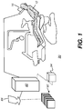
- FIG. 1 shows a surface contour imaging apparatus 90 that can be used for obtaining 3D content for model generation from a succession of reflectance images according to an embodiment of the present disclosure.
- An imaging device 16 typically a hand-held digital camera, a color depth camera, handheld 3D scanner, or intra-oral 3D scanner, is scanned through the mouth of patient 12 for acquiring a set having multiple reflectance images and associated depth information.
- a control logic processor 80 configurable to execute programmed instructions, is in signal communication with imaging device 16 and a display 84. Processor 80 obtains image data from imaging device 16 and processes this image data along with depth information in order to generate individual 3D views 92. Control logic processor 80 then combines the scanned 3D views in order to generate, store, and optionally render, on display 84, a composite 3D model surface 94.
- FIG. 2 is a schematic diagram showing an imaging apparatus 70 that can operate as a video camera 24 for polychromatic reflectance image data capture as well as a scanner 28 for executing related projecting and imaging functions used to characterize surface contour with structured light patterns 46.
- a handheld imaging apparatus 70 uses a video camera 24 for image acquisition for both contour scanning and image capture functions according to an embodiment of the present disclosure.
- Control logic processor 80 or other type of computer that may be part of camera 24, controls the operation of an illumination array 10 that generates the structured light and directs the light toward a surface position and controls operation of an imaging sensor array 30.
- Image data from surface 20, such as from a tooth 22, is obtained from imaging sensor array 30 and stored as video image data in a memory 72.
- Imaging sensor array 30 is part of a sensing apparatus 40 that includes an objective lens 34 and associated elements for acquiring video image content.
- Control logic processor 80 in signal communication with camera 24 components that acquire the image, processes the received image data and stores the mapping in memory 72.
- the resulting image from memory 72 is then optionally rendered and displayed on a display 74, which may be part of another computer 75 used for some portion of the processing described herein.
- Memory 72 may also include a display buffer.
- One or more sensors 42 such as a motion sensor, can also be provided as part of scanner 28 circuitry.
- a pattern of lines or other shapes is projected from illumination array 10 toward the surface of an object from a given angle.
- the projected pattern from the illuminated surface position is then viewed from another angle as a contour image, taking advantage of triangulation in order to analyze surface information based on the appearance of contour lines.
- Phase shifting in which the projected pattern is incrementally shifted spatially for obtaining additional measurements at the new locations, is typically applied as part of structured light imaging, used in order to complete the contour mapping of the surface and to increase overall resolution in the contour image.
- use of structured light patterns for surface contour characterization is described in commonly assigned U.S. Patent Application Publications No. US2013/0120532 and No. US2013/0120533 , both entitled “3D INTRAORAL MEASUREMENTS USING OPTICAL MULTILINE METHOD” and incorporated herein in their entirety.
- a computer and software can use triangulation methods to compute the coordinates of numerous illuminated surface points relative to a plane. As the plane is moved to intersect eventually with some or all of the surface of the object, the coordinates of an increasing number of points are accumulated. As a result of this image acquisition, a point cloud of vertex points or vertices can be identified and used to represent the extent of a 3D surface within a volume. The points in the point cloud then represent actual, measured points on the three-dimensional surface of an object.
- a mesh can alternately be constructed, such as by connecting points on the point cloud as vertices that define individual congruent polygonal faces (typically triangular faces) that characterize the surface shape.
- the full 3D surface image model can then be formed by combining the surface contour information provided by the mesh with monochromatic or polychromatic image content obtained from a camera, such as camera 24 of FIG. 2 .
- Polychromatic image content can be provided in a number of ways, including the use of a single monochrome imaging sensor with a succession of images obtained using illumination of different primary colors, one color at a time, for example. Alternately, a color imaging sensor could be used.
- Image processing at control logic processor 80 can generate a 3D contour surface model using line scan data from structured light imaging, or using point cloud or mesh data or CBCT volume image data.
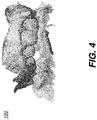
- FIGs. 3 and 4 show a point cloud or mesh 150 generated from a succession of structured light images. Further processing of the point cloud content can be used to generate a mesh as an alternative contour surface model.
- Reflectance imaging can be used for obtaining intraoral surface contour data used to generate the 3D surface model.
- Surface contour information can be obtained using time-of-flight imaging or range imaging methods, such as structure-from-motion processing, for example.
- An embodiment of the present disclosure provides an improved workflow for tooth segmentation that takes advantage of previous tooth segmentation data in order to advance and streamline the segmentation process.
- the simplified workflow logic diagram of FIG. 5 gives an overview of the overall workflow sequence for obtaining segmentation of a 3D surface model using tooth registration.
- a first 3D surface model image of patient dentition is acquired.
- the first model can be a 3D mesh virtual model initially formed prior to the beginning of patient treatment.
- the surface contour characterization that is obtained can be from an intraoral scanner or can be calculated from a CBCT scan of the imprint or mold of the patient dentition. Alternately, intraoral scanner and CBCT information can be combined to obtain the surface contour for the 3D model image.
- Initial segmentation step S820 is performed on the first 3D model image and the resulting segmentation data are stored for subsequent use.
- Initial segmentation step S820 provides segmentation of the individual teeth from the first 3D model image, along with tooth labeling and axis information as determined from the tooth crown.
- axis Mesio-distal, vestibule-lingual, main axis
- cusps cusps
- fossae and largest contour, cervical limit for example.
- a subsequent acquisition step S830 executes, acquiring a second 3D model image, a 3D virtual model showing the surface as a mesh or point cloud with an updated surface contour characterization of the patient.
- a registration process S840 then takes, as input, the initial tooth segmentation of step S820 and the second 3D model data from step S830 for streamlined segmentation processing.
- An intra-model registration step S850 executes, coarsely registering key features of the first 3D model image to the second 3D model image, then registering each tooth of the first 3D model image to the surface of the second 3D model image.
- Leveraged segmentation step S860 then takes the registration information from step S850 and performs segmentation, providing labeling and axis information for each tooth of the second model 3D image corresponding to each segmented tooth of the first 3D model image.
- a display step S870 then displays the segmented content.
- the segmented content may further or alternatively be transmitted.
- an initial features registration step S842 registers particular features of the second model with the initial segmentation results.
- segmented molars from the first 3D model image can be positionally registered with the molars from the second model.
- Registration of the molars from the first segmented model to the volume acquired in the second scan can be generally straightforward since molar position is typically unchanged for an orthodontic treatment. Registration can use any of a number of image matching methods, such as ICP (iterative closest point) techniques.
- a tooth position step S844 performs additional tooth positioning to find the best position of a tooth onto the second 3D model image.
- a Feature Matching Algorithm can be used for this function.
- feature matching begins with molars, proceeding tooth by tooth to match individual features using appropriate feature-matching algorithms, familiar to those skilled in the imaging arts.
- Normal information for each point of the surface contour can be estimated using well-known techniques in computer graphics, such as those described, for example, at the pointclouds.org website under documentation/tutorials/normal_estimation.php.
- a local neighborhood of points is defined.
- the covariance matrix is computed using the local neighborhood of points.
- Superscript "T” denotes the matrix transpose.
- This square, symmetric 3x3 covariance matrix can provide eigenvectors and associated eigenvalues for feature-matching computation. If a least-squares fit of the local neighborhood of points is performed using an ellipsoid, the ellipsoid axes are the eigenvectors and the axis length is related to the corresponding eigenvalues. The smallest eigenvalue of the covariance matrix represents the shortest ellipsoid axis and the associated eigenvector gives the direction of the local normal. The direction of the local normal can then be flipped if necessary to match the observation direction from the scanner. This local normal can then be assigned to the query point, which allows the computation of a surface contour with normals and colors.
- the stitching procedure of the surface contour with normals and colors from the image data onto the growing surface contour has the following generic steps:
- a validation step S845 tests for likelihood of positional validity, using measurements of shape and relationship to matched structures from registration step S842. Any needed adjustments can be made as necessary. If validation step S845 fails to meet pre-determined conditions for accuracy, the search area is reduced in an area reduction step S846 and tooth position step S844 repeats to attempt a repositioning over the reduced area. For example, the search area can be limited to the left or right portions of the jaw structure for subsequent reprocessing in step S844. If validation step S845 succeeds, a save registration step S848 executes, storing the registration results for the teeth. Registration results can contain a registration matrix which transforms the tooth from the first 3D model image to the second 3D model image. At this stage of processing, each tooth from the second 3D acquisition is registered to a corresponding tooth from the first model. Subsequent processing then refines tooth position and shape information in order to provide segmentation of the second model using segmentation established for the first model.
- structures of the un-segmented second 3D model image are associated with, or correlated to, corresponding segmented structures of the first model. Further refinement of the tooth shape and axis orientation in subsequent processing then helps to complete the segmentation of the second model.
- a positioning definition step S852 further defines tooth shape for the second model using tooth positioning results.
- a cervical limit refinement step S854 more accurately defines the tooth shape for each of the teeth in the second 3D model image. For each registered tooth of the second model, step S852 and S854 procedures project the cervical limit for the tooth of the first 3D model image onto the second 3D model image and optionally refine the cervical limit definition using any of a number of curvature detection techniques familiar to those skilled in the image processing art. The corresponding tooth surface contour of the second 3D model image can then be extracted using the projected cervical limit or the refined cervical limit.
- a labeling and axis definition step S862 assigns a number or other label to the tooth from the second model and applies a registration matrix to the axis and other features (such as cusps) of the tooth of the first model, to identify the axis and other features of the tooth of the second model.
- This step obtains and stores the data used for segmentation of each tooth in the second model, including position, axis, and cervical limit.
- a complete segmentation step S866 performs final segmentation processing for the second model, generating a segmented model.
- a display step S870 then provides display rendering, storage, or transmission of the second model segmentation.
- FIG. 7 shows an example of tooth segmentation from a first 3D model image of the lower jaw.
- FIG. 8 shows tooth segmentation for a second 3D model image of the same dentition, taken subsequently.
- each tooth of the first model can be registered onto the second model.
- FIG. 9 shows results of feature matching for registration of the first model onto the second model.
- FIG. 10 illustrates processing steps using an example of a single tooth (tooth 47).
- a figure 1101 shows tooth 47 segmented in the first 3D model image.
- a figure 1102 shows tooth 47 of the first model registered onto the second model.
- a figure 1103 shows cervical limits C1 and C2 detected for the first and second 3D model images, respectively, for tooth 47 (ISO numbering). Cervical limit C2 curvature is computed for the second 3D model image.
- a figure 1104 shows the refined cervical limit C2' for the second model. allowing tooth 47 to be segmented.
Landscapes
- Health & Medical Sciences (AREA)
- Engineering & Computer Science (AREA)
- Life Sciences & Earth Sciences (AREA)
- Physics & Mathematics (AREA)
- Medical Informatics (AREA)
- General Health & Medical Sciences (AREA)
- Animal Behavior & Ethology (AREA)
- Veterinary Medicine (AREA)
- Public Health (AREA)
- Theoretical Computer Science (AREA)
- General Physics & Mathematics (AREA)
- Nuclear Medicine, Radiotherapy & Molecular Imaging (AREA)
- Computer Vision & Pattern Recognition (AREA)
- Radiology & Medical Imaging (AREA)
- Optics & Photonics (AREA)
- Oral & Maxillofacial Surgery (AREA)
- Dentistry (AREA)
- Heart & Thoracic Surgery (AREA)
- Surgery (AREA)
- Molecular Biology (AREA)
- Biomedical Technology (AREA)
- Pathology (AREA)
- High Energy & Nuclear Physics (AREA)
- Biophysics (AREA)
- Epidemiology (AREA)
- General Engineering & Computer Science (AREA)
- Quality & Reliability (AREA)
- Pulmonology (AREA)
- Dental Tools And Instruments Or Auxiliary Dental Instruments (AREA)
- Apparatus For Radiation Diagnosis (AREA)
Priority Applications (5)
| Application Number | Priority Date | Filing Date | Title |
|---|---|---|---|
| ES18306870T ES3052991T3 (en) | 2018-12-28 | 2018-12-28 | Apparatus for generating a 3d model image of a patient's dentition |
| EP18306870.9A EP3673864B1 (fr) | 2018-12-28 | 2018-12-28 | Appareil pour générer une image modélisée en 3d de la dentition d'un patient |
| US17/418,634 US11961238B2 (en) | 2018-12-28 | 2019-12-27 | Tooth segmentation using tooth registration |
| JP2021537957A JP7667738B2 (ja) | 2018-12-28 | 2019-12-27 | 歯の位置合わせを用いた歯のセグメンテーション |
| PCT/EP2019/087083 WO2020136243A1 (fr) | 2018-12-28 | 2019-12-27 | Segmentation de dent à l'aide d'un alignement de dent |
Applications Claiming Priority (1)
| Application Number | Priority Date | Filing Date | Title |
|---|---|---|---|
| EP18306870.9A EP3673864B1 (fr) | 2018-12-28 | 2018-12-28 | Appareil pour générer une image modélisée en 3d de la dentition d'un patient |
Publications (3)
| Publication Number | Publication Date |
|---|---|
| EP3673864A1 true EP3673864A1 (fr) | 2020-07-01 |
| EP3673864B1 EP3673864B1 (fr) | 2025-10-22 |
| EP3673864C0 EP3673864C0 (fr) | 2025-10-22 |
Family
ID=65200553
Family Applications (1)
| Application Number | Title | Priority Date | Filing Date |
|---|---|---|---|
| EP18306870.9A Active EP3673864B1 (fr) | 2018-12-28 | 2018-12-28 | Appareil pour générer une image modélisée en 3d de la dentition d'un patient |
Country Status (5)
| Country | Link |
|---|---|
| US (1) | US11961238B2 (fr) |
| EP (1) | EP3673864B1 (fr) |
| JP (1) | JP7667738B2 (fr) |
| ES (1) | ES3052991T3 (fr) |
| WO (1) | WO2020136243A1 (fr) |
Cited By (7)
| Publication number | Priority date | Publication date | Assignee | Title |
|---|---|---|---|---|
| CN114073540A (zh) * | 2020-08-20 | 2022-02-22 | 易美构有限公司 | 用于配准3d牙科数据的自动化方法以及具有用于执行该方法的程序的计算机可读介质 |
| US11278377B1 (en) | 2021-06-02 | 2022-03-22 | Oxilio Ltd | System and method for performing digital separation of teeth |
| WO2022174747A1 (fr) * | 2021-02-18 | 2022-08-25 | 无锡时代天使医疗器械科技有限公司 | Procédé de segmentation d'image de dents par tomographie assistée par ordinateur |
| CN117670956A (zh) * | 2023-12-07 | 2024-03-08 | 北京瑞医博科技有限公司 | 牙齿边界的自动提取方法、图像矫正方法及牙齿治疗系统 |
| AU2020464339B2 (en) * | 2020-08-20 | 2025-04-10 | Imagoworks Inc. | Method for automating dental three-dimensional data position alignment, and computer-readable recording medium having program for executing same on computer recorded thereon |
| WO2025263672A1 (fr) * | 2024-06-20 | 2025-12-26 | 이마고웍스 주식회사 | Procédé de segmentation de dents de données de balayage buccal tridimensionnel à l'aide d'un modèle de bibliothèque de dents, et support d'enregistrement lisible par ordinateur sur lequel est enregistré un programme pour l'exécuter dans un ordinateur |
| WO2026025841A1 (fr) * | 2024-08-02 | 2026-02-05 | 正雅齿科科技(上海)有限公司 | Procédé de segmentation de modèle de dent et conception de schéma orthodontique, appareil orthodontique et procédé de fabrication |
Families Citing this family (12)
| Publication number | Priority date | Publication date | Assignee | Title |
|---|---|---|---|---|
| CN113873947A (zh) * | 2019-05-22 | 2021-12-31 | 株式会社迪耀 | 神经检测方法及装置 |
| US12370016B2 (en) * | 2020-09-11 | 2025-07-29 | Align Technology, Inc. | Automatic segmentation quality assessment for secondary treatment plans |
| KR20240166528A (ko) * | 2022-04-20 | 2024-11-26 | 주식회사 메디트 | 3차원 스캐너의 3차원 이미지 모델의 생성과 정렬을 위한 전자 장치, 방법 및 기록 매체 |
| EP4280155A1 (fr) * | 2022-05-16 | 2023-11-22 | Institut Straumann AG | Fabrication améliorée d'implants dentaires basée sur l'alignement des données de balayage numérique |
| CN115393315A (zh) * | 2022-08-23 | 2022-11-25 | 广西医科大学附属武鸣医院(广西医科大学武鸣临床医学院、南宁市武鸣区人民医院) | 前列腺经腹超声影像的自动标注方法、系统、电子设备及存储介质 |
| CN115457093B (zh) * | 2022-08-24 | 2024-03-22 | 北京百度网讯科技有限公司 | 一种牙齿图像的处理方法、装置、电子设备及存储介质 |
| CN116958169B (zh) * | 2023-06-30 | 2026-01-06 | 电子科技大学 | 一种三维牙颌模型牙齿分割方法 |
| US20250049546A1 (en) * | 2023-08-09 | 2025-02-13 | Rtrs Investment, Llc | Implants, functionalized implant surfaces and related systems, devices, computer program products, and methods |
| CN116934821B (zh) * | 2023-09-15 | 2023-12-22 | 深圳市家鸿口腔医疗股份有限公司 | 一种个性化假牙三维图像模型配准方法及系统 |
| CN117830317B (zh) * | 2024-03-05 | 2024-06-18 | 天津医科大学口腔医院 | 基于图像处理的正畸自动检测方法及系统 |
| CN118512278B (zh) * | 2024-07-22 | 2024-10-29 | 水护健康科技(温州)有限公司 | 一种用于牙齿3d打印前的ai建模方法及装置 |
| DE102024121325B3 (de) * | 2024-07-26 | 2025-12-31 | Institut Straumann Ag | Verfahren und System zum Generieren eines Modells zur Verwendung in der intraoralen Navigation in einem Patienten |
Citations (11)
| Publication number | Priority date | Publication date | Assignee | Title |
|---|---|---|---|---|
| US5270926A (en) | 1990-12-21 | 1993-12-14 | General Electric Company | Method and apparatus for reconstructing a three-dimensional computerized tomography (CT) image of an object from incomplete cone beam projection data |
| US5999587A (en) | 1997-07-03 | 1999-12-07 | University Of Rochester | Method of and system for cone-beam tomography reconstruction |
| US20030039389A1 (en) | 1997-06-20 | 2003-02-27 | Align Technology, Inc. | Manipulating a digital dentition model to form models of individual dentition components |
| US8416984B2 (en) * | 2011-01-20 | 2013-04-09 | Carestream Health, Inc. | Automatic tooth charting using digital images |
| US20130120532A1 (en) | 2011-11-10 | 2013-05-16 | James R. Milch | 3d intraoral measurements using optical multiline method |
| US20130120533A1 (en) | 2011-11-10 | 2013-05-16 | Carestream Health, Inc. | 3d intraoral measurements using optical multiline method |
| US8670521B2 (en) | 2011-06-02 | 2014-03-11 | Carestream Health, Inc. | Method for generating an intraoral volume image |
| US20150305830A1 (en) * | 2001-04-13 | 2015-10-29 | Orametrix, Inc. | Tooth positioning appliance and uses thereof |
| US20160004811A1 (en) * | 2014-07-01 | 2016-01-07 | 3M Innovative Properties Company | Detecting tooth wear using intra-oral 3d scans |
| US20180005371A1 (en) * | 2016-06-30 | 2018-01-04 | Align Technology, Inc. | Historical scan reference for intraoral scans |
| CN108665533A (zh) * | 2018-05-09 | 2018-10-16 | 西安增材制造国家研究院有限公司 | 一种通过牙齿ct图像和三维扫描数据重建牙列的方法 |
Family Cites Families (7)
| Publication number | Priority date | Publication date | Assignee | Title |
|---|---|---|---|---|
| US20140067334A1 (en) * | 2012-09-06 | 2014-03-06 | Align Technology Inc. | Method and a system usable in creating a subsequent dental appliance |
| US9135498B2 (en) * | 2012-12-14 | 2015-09-15 | Ormco Corporation | Integration of intra-oral imagery and volumetric imagery |
| US20140227655A1 (en) * | 2013-02-12 | 2014-08-14 | Ormco Corporation | Integration of model data, surface data, and volumetric data |
| US11147652B2 (en) * | 2014-11-13 | 2021-10-19 | Align Technology, Inc. | Method for tracking, predicting, and proactively correcting malocclusion and related issues |
| US9770217B2 (en) * | 2015-01-30 | 2017-09-26 | Dental Imaging Technologies Corporation | Dental variation tracking and prediction |
| CN105761252B (zh) * | 2016-02-02 | 2017-03-29 | 北京正齐口腔医疗技术有限公司 | 图像分割的方法及装置 |
| CN106228549B (zh) * | 2016-07-14 | 2019-04-19 | 嘉兴学院 | 一种基于路径规划的三角网格牙齿分割方法 |
-
2018
- 2018-12-28 ES ES18306870T patent/ES3052991T3/es active Active
- 2018-12-28 EP EP18306870.9A patent/EP3673864B1/fr active Active
-
2019
- 2019-12-27 US US17/418,634 patent/US11961238B2/en active Active
- 2019-12-27 JP JP2021537957A patent/JP7667738B2/ja active Active
- 2019-12-27 WO PCT/EP2019/087083 patent/WO2020136243A1/fr not_active Ceased
Patent Citations (11)
| Publication number | Priority date | Publication date | Assignee | Title |
|---|---|---|---|---|
| US5270926A (en) | 1990-12-21 | 1993-12-14 | General Electric Company | Method and apparatus for reconstructing a three-dimensional computerized tomography (CT) image of an object from incomplete cone beam projection data |
| US20030039389A1 (en) | 1997-06-20 | 2003-02-27 | Align Technology, Inc. | Manipulating a digital dentition model to form models of individual dentition components |
| US5999587A (en) | 1997-07-03 | 1999-12-07 | University Of Rochester | Method of and system for cone-beam tomography reconstruction |
| US20150305830A1 (en) * | 2001-04-13 | 2015-10-29 | Orametrix, Inc. | Tooth positioning appliance and uses thereof |
| US8416984B2 (en) * | 2011-01-20 | 2013-04-09 | Carestream Health, Inc. | Automatic tooth charting using digital images |
| US8670521B2 (en) | 2011-06-02 | 2014-03-11 | Carestream Health, Inc. | Method for generating an intraoral volume image |
| US20130120532A1 (en) | 2011-11-10 | 2013-05-16 | James R. Milch | 3d intraoral measurements using optical multiline method |
| US20130120533A1 (en) | 2011-11-10 | 2013-05-16 | Carestream Health, Inc. | 3d intraoral measurements using optical multiline method |
| US20160004811A1 (en) * | 2014-07-01 | 2016-01-07 | 3M Innovative Properties Company | Detecting tooth wear using intra-oral 3d scans |
| US20180005371A1 (en) * | 2016-06-30 | 2018-01-04 | Align Technology, Inc. | Historical scan reference for intraoral scans |
| CN108665533A (zh) * | 2018-05-09 | 2018-10-16 | 西安增材制造国家研究院有限公司 | 一种通过牙齿ct图像和三维扫描数据重建牙列的方法 |
Non-Patent Citations (5)
| Title |
|---|
| AKHOONDALI ET AL.: "Rapid Automatic Segmentation and Visualization of Teeth in CT-Scan Data", JOURNAL OF APPLIED SCIENCES, 2009, pages 2031 - 2044, XP055176486, DOI: doi:10.3923/jas.2009.2031.2044 |
| GAO ET AL.: "Tooth Region Separation for Dental CT Images", PROCEEDINGS OF THE 2008 THIRD INTERNATIONAL CONFERENCE ON CONVERGENCE AND HYBRID INFORMATION TECHNOLOGY, 2008, pages 897 - 901, XP055176488, DOI: doi:10.1109/ICCIT.2008.342 |
| PAGE, D.L. ET AL.: "Perception-based 3D Triangle Mesh Segmentation Using Fast Marching Watershed", PROC. CVPI, vol. II, 2003 |
| RUSU, R. B., SEMANTIC 3D OBJECT MAPS FOR EVERYDAY MANIPULATION IN HUMAN LIVING ENVIRONMENTS, 17 August 2009 (2009-08-17) |
| THOMAS KRONFELD ET AL.: "Snake-Based Segmentation of Teeth from Virtual Dental Casts", COMPUTER-AIDED DESIGN & APPLICATIONS, vol. 7, no. a, 2010 |
Cited By (11)
| Publication number | Priority date | Publication date | Assignee | Title |
|---|---|---|---|---|
| CN114073540A (zh) * | 2020-08-20 | 2022-02-22 | 易美构有限公司 | 用于配准3d牙科数据的自动化方法以及具有用于执行该方法的程序的计算机可读介质 |
| EP3958213A1 (fr) * | 2020-08-20 | 2022-02-23 | Imagoworks Inc. | Procédé automatisé d'alignement de données dentaires en 3d et support lisible par ordinateur comportant un programme permettant de mettre en uvre ce procédé |
| US12023221B2 (en) | 2020-08-20 | 2024-07-02 | Imagoworks Inc. | Automated method for aligning 3D dental data and computer readable medium having program for performing the method |
| AU2020464339B2 (en) * | 2020-08-20 | 2025-04-10 | Imagoworks Inc. | Method for automating dental three-dimensional data position alignment, and computer-readable recording medium having program for executing same on computer recorded thereon |
| CN114073540B (zh) * | 2020-08-20 | 2025-07-11 | 易美构有限公司 | 用于配准3d牙科数据的自动化方法以及具有用于执行该方法的程序的计算机可读介质 |
| WO2022174747A1 (fr) * | 2021-02-18 | 2022-08-25 | 无锡时代天使医疗器械科技有限公司 | Procédé de segmentation d'image de dents par tomographie assistée par ordinateur |
| US11278377B1 (en) | 2021-06-02 | 2022-03-22 | Oxilio Ltd | System and method for performing digital separation of teeth |
| US11751976B2 (en) | 2021-06-02 | 2023-09-12 | Oxilio Ltd | System and method for performing digital separation of teeth |
| CN117670956A (zh) * | 2023-12-07 | 2024-03-08 | 北京瑞医博科技有限公司 | 牙齿边界的自动提取方法、图像矫正方法及牙齿治疗系统 |
| WO2025263672A1 (fr) * | 2024-06-20 | 2025-12-26 | 이마고웍스 주식회사 | Procédé de segmentation de dents de données de balayage buccal tridimensionnel à l'aide d'un modèle de bibliothèque de dents, et support d'enregistrement lisible par ordinateur sur lequel est enregistré un programme pour l'exécuter dans un ordinateur |
| WO2026025841A1 (fr) * | 2024-08-02 | 2026-02-05 | 正雅齿科科技(上海)有限公司 | Procédé de segmentation de modèle de dent et conception de schéma orthodontique, appareil orthodontique et procédé de fabrication |
Also Published As
| Publication number | Publication date |
|---|---|
| ES3052991T3 (en) | 2026-01-16 |
| EP3673864B1 (fr) | 2025-10-22 |
| JP7667738B2 (ja) | 2025-04-23 |
| WO2020136243A1 (fr) | 2020-07-02 |
| EP3673864C0 (fr) | 2025-10-22 |
| JP2022516488A (ja) | 2022-02-28 |
| US20220122264A1 (en) | 2022-04-21 |
| US11961238B2 (en) | 2024-04-16 |
Similar Documents
| Publication | Publication Date | Title |
|---|---|---|
| US11961238B2 (en) | Tooth segmentation using tooth registration | |
| JP7289026B2 (ja) | ハイブリッドメッシュセグメンテーションのための方法及び装置 | |
| US11896455B2 (en) | Method and system for braces removal from dentition mesh | |
| US11058514B2 (en) | Method and system for dentition mesh braces removal | |
| US10339649B2 (en) | Method and system for hybrid mesh segmentation | |
| US10810738B1 (en) | Marker-less alignment of digital 3D face and jaw models | |
| CN109414306A (zh) | 用于口内扫描的历史扫描参考 | |
| US12056836B2 (en) | Dental model superimposition using clinical indications | |
| JP7245335B2 (ja) | 口腔内スキャナを使用して3dデータを取得する混合方法 | |
| US20220068039A1 (en) | 3d segmentation for mandible and maxilla | |
| WO2020136245A1 (fr) | Optimisation d'impression 3d faisant intervenir des indications cliniques | |
| EP4404139A1 (fr) | Procédé et système d'alignement de représentations 3d | |
| EP4631008A1 (fr) | Procédé et système d'alignement de représentations 3d | |
| Matsopoulos et al. | Image registration based on lifting process: an application to digital subtraction radiography | |
| CN120388129A (zh) | 多模态融合的口腔内窥图像三维重建方法及系统 |
Legal Events
| Date | Code | Title | Description |
|---|---|---|---|
| PUAI | Public reference made under article 153(3) epc to a published international application that has entered the european phase |
Free format text: ORIGINAL CODE: 0009012 |
|
| STAA | Information on the status of an ep patent application or granted ep patent |
Free format text: STATUS: THE APPLICATION HAS BEEN PUBLISHED |
|
| AK | Designated contracting states |
Kind code of ref document: A1 Designated state(s): AL AT BE BG CH CY CZ DE DK EE ES FI FR GB GR HR HU IE IS IT LI LT LU LV MC MK MT NL NO PL PT RO RS SE SI SK SM TR |
|
| AX | Request for extension of the european patent |
Extension state: BA ME |
|
| STAA | Information on the status of an ep patent application or granted ep patent |
Free format text: STATUS: REQUEST FOR EXAMINATION WAS MADE |
|
| 17P | Request for examination filed |
Effective date: 20201216 |
|
| RBV | Designated contracting states (corrected) |
Designated state(s): AL AT BE BG CH CY CZ DE DK EE ES FI FR GB GR HR HU IE IS IT LI LT LU LV MC MK MT NL NO PL PT RO RS SE SI SK SM TR |
|
| STAA | Information on the status of an ep patent application or granted ep patent |
Free format text: STATUS: EXAMINATION IS IN PROGRESS |
|
| 17Q | First examination report despatched |
Effective date: 20210623 |
|
| RAP1 | Party data changed (applicant data changed or rights of an application transferred) |
Owner name: NOBEL BIOCARE SERVICES AG |
|
| GRAP | Despatch of communication of intention to grant a patent |
Free format text: ORIGINAL CODE: EPIDOSNIGR1 |
|
| STAA | Information on the status of an ep patent application or granted ep patent |
Free format text: STATUS: GRANT OF PATENT IS INTENDED |
|
| RIC1 | Information provided on ipc code assigned before grant |
Ipc: G06T 7/38 20170101ALI20241122BHEP Ipc: G06T 7/33 20170101ALI20241122BHEP Ipc: G06T 7/10 20170101ALI20241122BHEP Ipc: A61C 9/00 20060101ALI20241122BHEP Ipc: A61C 7/00 20060101AFI20241122BHEP |
|
| INTG | Intention to grant announced |
Effective date: 20241216 |
|
| GRAJ | Information related to disapproval of communication of intention to grant by the applicant or resumption of examination proceedings by the epo deleted |
Free format text: ORIGINAL CODE: EPIDOSDIGR1 |
|
| STAA | Information on the status of an ep patent application or granted ep patent |
Free format text: STATUS: EXAMINATION IS IN PROGRESS |
|
| GRAP | Despatch of communication of intention to grant a patent |
Free format text: ORIGINAL CODE: EPIDOSNIGR1 |
|
| STAA | Information on the status of an ep patent application or granted ep patent |
Free format text: STATUS: GRANT OF PATENT IS INTENDED |
|
| INTC | Intention to grant announced (deleted) | ||
| INTG | Intention to grant announced |
Effective date: 20250515 |
|
| GRAS | Grant fee paid |
Free format text: ORIGINAL CODE: EPIDOSNIGR3 |
|
| GRAA | (expected) grant |
Free format text: ORIGINAL CODE: 0009210 |
|
| STAA | Information on the status of an ep patent application or granted ep patent |
Free format text: STATUS: THE PATENT HAS BEEN GRANTED |
|
| AK | Designated contracting states |
Kind code of ref document: B1 Designated state(s): AL AT BE BG CH CY CZ DE DK EE ES FI FR GB GR HR HU IE IS IT LI LT LU LV MC MK MT NL NO PL PT RO RS SE SI SK SM TR |
|
| REG | Reference to a national code |
Ref country code: CH Ref legal event code: F10 Free format text: ST27 STATUS EVENT CODE: U-0-0-F10-F00 (AS PROVIDED BY THE NATIONAL OFFICE) Effective date: 20251022 Ref country code: GB Ref legal event code: FG4D |
|
| REG | Reference to a national code |
Ref country code: DE Ref legal event code: R096 Ref document number: 602018086524 Country of ref document: DE |
|
| REG | Reference to a national code |
Ref country code: IE Ref legal event code: FG4D |
|
| U01 | Request for unitary effect filed |
Effective date: 20251113 |
|
| U07 | Unitary effect registered |
Designated state(s): AT BE BG DE DK EE FI FR IT LT LU LV MT NL PT RO SE SI Effective date: 20251120 |
|
| REG | Reference to a national code |
Ref country code: CH Ref legal event code: U11 Free format text: ST27 STATUS EVENT CODE: U-0-0-U10-U11 (AS PROVIDED BY THE NATIONAL OFFICE) Effective date: 20260101 |
|
| REG | Reference to a national code |
Ref country code: ES Ref legal event code: FG2A Ref document number: 3052991 Country of ref document: ES Kind code of ref document: T3 Effective date: 20260116 |
|
| U20 | Renewal fee for the european patent with unitary effect paid |
Year of fee payment: 8 Effective date: 20260220 |
|
| PGFP | Annual fee paid to national office [announced via postgrant information from national office to epo] |
Ref country code: ES Payment date: 20260114 Year of fee payment: 8 |
|
| PG25 | Lapsed in a contracting state [announced via postgrant information from national office to epo] |
Ref country code: NO Free format text: LAPSE BECAUSE OF FAILURE TO SUBMIT A TRANSLATION OF THE DESCRIPTION OR TO PAY THE FEE WITHIN THE PRESCRIBED TIME-LIMIT Effective date: 20260122 |
|
| PG25 | Lapsed in a contracting state [announced via postgrant information from national office to epo] |
Ref country code: HR Free format text: LAPSE BECAUSE OF FAILURE TO SUBMIT A TRANSLATION OF THE DESCRIPTION OR TO PAY THE FEE WITHIN THE PRESCRIBED TIME-LIMIT Effective date: 20251022 |
|
| PG25 | Lapsed in a contracting state [announced via postgrant information from national office to epo] |
Ref country code: RS Free format text: LAPSE BECAUSE OF FAILURE TO SUBMIT A TRANSLATION OF THE DESCRIPTION OR TO PAY THE FEE WITHIN THE PRESCRIBED TIME-LIMIT Effective date: 20260122 |
|
| PG25 | Lapsed in a contracting state [announced via postgrant information from national office to epo] |
Ref country code: IS Free format text: LAPSE BECAUSE OF FAILURE TO SUBMIT A TRANSLATION OF THE DESCRIPTION OR TO PAY THE FEE WITHIN THE PRESCRIBED TIME-LIMIT Effective date: 20260222 |
|
| PGFP | Annual fee paid to national office [announced via postgrant information from national office to epo] |
Ref country code: CH Payment date: 20260101 Year of fee payment: 8 |
|
| PG25 | Lapsed in a contracting state [announced via postgrant information from national office to epo] |
Ref country code: PL Free format text: LAPSE BECAUSE OF FAILURE TO SUBMIT A TRANSLATION OF THE DESCRIPTION OR TO PAY THE FEE WITHIN THE PRESCRIBED TIME-LIMIT Effective date: 20251022 |