EP3689609B1 - Graft scaffold for cartilage repair and process for making same - Google Patents
Graft scaffold for cartilage repair and process for making same Download PDFInfo
- Publication number
- EP3689609B1 EP3689609B1 EP20160362.8A EP20160362A EP3689609B1 EP 3689609 B1 EP3689609 B1 EP 3689609B1 EP 20160362 A EP20160362 A EP 20160362A EP 3689609 B1 EP3689609 B1 EP 3689609B1
- Authority
- EP
- European Patent Office
- Prior art keywords
- printing
- cartilage
- cells
- particles
- bioink
- Prior art date
- Legal status (The legal status is an assumption and is not a legal conclusion. Google has not performed a legal analysis and makes no representation as to the accuracy of the status listed.)
- Active
Links
Images





Classifications
-
- A—HUMAN NECESSITIES
- A61—MEDICAL OR VETERINARY SCIENCE; HYGIENE
- A61K—PREPARATIONS FOR MEDICAL, DENTAL OR TOILETRY PURPOSES
- A61K9/00—Medicinal preparations characterised by special physical form
-
- A—HUMAN NECESSITIES
- A61—MEDICAL OR VETERINARY SCIENCE; HYGIENE
- A61L—METHODS OR APPARATUS FOR STERILISING MATERIALS OR OBJECTS IN GENERAL; DISINFECTION, STERILISATION OR DEODORISATION OF AIR; CHEMICAL ASPECTS OF BANDAGES, DRESSINGS, ABSORBENT PADS OR SURGICAL ARTICLES; MATERIALS FOR BANDAGES, DRESSINGS, ABSORBENT PADS OR SURGICAL ARTICLES
- A61L27/00—Materials for grafts or prostheses or for coating grafts or prostheses
- A61L27/14—Macromolecular materials
- A61L27/20—Polysaccharides
-
- A—HUMAN NECESSITIES
- A61—MEDICAL OR VETERINARY SCIENCE; HYGIENE
- A61L—METHODS OR APPARATUS FOR STERILISING MATERIALS OR OBJECTS IN GENERAL; DISINFECTION, STERILISATION OR DEODORISATION OF AIR; CHEMICAL ASPECTS OF BANDAGES, DRESSINGS, ABSORBENT PADS OR SURGICAL ARTICLES; MATERIALS FOR BANDAGES, DRESSINGS, ABSORBENT PADS OR SURGICAL ARTICLES
- A61L27/00—Materials for grafts or prostheses or for coating grafts or prostheses
- A61L27/14—Macromolecular materials
- A61L27/26—Mixtures of macromolecular compounds
-
- A—HUMAN NECESSITIES
- A61—MEDICAL OR VETERINARY SCIENCE; HYGIENE
- A61L—METHODS OR APPARATUS FOR STERILISING MATERIALS OR OBJECTS IN GENERAL; DISINFECTION, STERILISATION OR DEODORISATION OF AIR; CHEMICAL ASPECTS OF BANDAGES, DRESSINGS, ABSORBENT PADS OR SURGICAL ARTICLES; MATERIALS FOR BANDAGES, DRESSINGS, ABSORBENT PADS OR SURGICAL ARTICLES
- A61L27/00—Materials for grafts or prostheses or for coating grafts or prostheses
- A61L27/36—Materials for grafts or prostheses or for coating grafts or prostheses containing ingredients of undetermined constitution or reaction products thereof, e.g. transplant tissue, natural bone, extracellular matrix
- A61L27/3604—Materials for grafts or prostheses or for coating grafts or prostheses containing ingredients of undetermined constitution or reaction products thereof, e.g. transplant tissue, natural bone, extracellular matrix characterised by the human or animal origin of the biological material, e.g. hair, fascia, fish scales, silk, shellac, pericardium, pleura, renal tissue, amniotic membrane, parenchymal tissue, fetal tissue, muscle tissue, fat tissue, enamel
- A61L27/3612—Cartilage, synovial fluid
-
- A—HUMAN NECESSITIES
- A61—MEDICAL OR VETERINARY SCIENCE; HYGIENE
- A61L—METHODS OR APPARATUS FOR STERILISING MATERIALS OR OBJECTS IN GENERAL; DISINFECTION, STERILISATION OR DEODORISATION OF AIR; CHEMICAL ASPECTS OF BANDAGES, DRESSINGS, ABSORBENT PADS OR SURGICAL ARTICLES; MATERIALS FOR BANDAGES, DRESSINGS, ABSORBENT PADS OR SURGICAL ARTICLES
- A61L27/00—Materials for grafts or prostheses or for coating grafts or prostheses
- A61L27/36—Materials for grafts or prostheses or for coating grafts or prostheses containing ingredients of undetermined constitution or reaction products thereof, e.g. transplant tissue, natural bone, extracellular matrix
- A61L27/38—Materials for grafts or prostheses or for coating grafts or prostheses containing ingredients of undetermined constitution or reaction products thereof, e.g. transplant tissue, natural bone, extracellular matrix containing added animal cells
- A61L27/3804—Materials for grafts or prostheses or for coating grafts or prostheses containing ingredients of undetermined constitution or reaction products thereof, e.g. transplant tissue, natural bone, extracellular matrix containing added animal cells characterised by specific cells or progenitors thereof, e.g. fibroblasts, connective tissue cells, kidney cells
- A61L27/3817—Cartilage-forming cells, e.g. pre-chondrocytes
-
- A—HUMAN NECESSITIES
- A61—MEDICAL OR VETERINARY SCIENCE; HYGIENE
- A61L—METHODS OR APPARATUS FOR STERILISING MATERIALS OR OBJECTS IN GENERAL; DISINFECTION, STERILISATION OR DEODORISATION OF AIR; CHEMICAL ASPECTS OF BANDAGES, DRESSINGS, ABSORBENT PADS OR SURGICAL ARTICLES; MATERIALS FOR BANDAGES, DRESSINGS, ABSORBENT PADS OR SURGICAL ARTICLES
- A61L27/00—Materials for grafts or prostheses or for coating grafts or prostheses
- A61L27/36—Materials for grafts or prostheses or for coating grafts or prostheses containing ingredients of undetermined constitution or reaction products thereof, e.g. transplant tissue, natural bone, extracellular matrix
- A61L27/38—Materials for grafts or prostheses or for coating grafts or prostheses containing ingredients of undetermined constitution or reaction products thereof, e.g. transplant tissue, natural bone, extracellular matrix containing added animal cells
- A61L27/3839—Materials for grafts or prostheses or for coating grafts or prostheses containing ingredients of undetermined constitution or reaction products thereof, e.g. transplant tissue, natural bone, extracellular matrix containing added animal cells characterised by the site of application in the body
- A61L27/3843—Connective tissue
- A61L27/3852—Cartilage, e.g. meniscus
-
- A—HUMAN NECESSITIES
- A61—MEDICAL OR VETERINARY SCIENCE; HYGIENE
- A61L—METHODS OR APPARATUS FOR STERILISING MATERIALS OR OBJECTS IN GENERAL; DISINFECTION, STERILISATION OR DEODORISATION OF AIR; CHEMICAL ASPECTS OF BANDAGES, DRESSINGS, ABSORBENT PADS OR SURGICAL ARTICLES; MATERIALS FOR BANDAGES, DRESSINGS, ABSORBENT PADS OR SURGICAL ARTICLES
- A61L27/00—Materials for grafts or prostheses or for coating grafts or prostheses
- A61L27/36—Materials for grafts or prostheses or for coating grafts or prostheses containing ingredients of undetermined constitution or reaction products thereof, e.g. transplant tissue, natural bone, extracellular matrix
- A61L27/38—Materials for grafts or prostheses or for coating grafts or prostheses containing ingredients of undetermined constitution or reaction products thereof, e.g. transplant tissue, natural bone, extracellular matrix containing added animal cells
- A61L27/3839—Materials for grafts or prostheses or for coating grafts or prostheses containing ingredients of undetermined constitution or reaction products thereof, e.g. transplant tissue, natural bone, extracellular matrix containing added animal cells characterised by the site of application in the body
- A61L27/3843—Connective tissue
- A61L27/3852—Cartilage, e.g. meniscus
- A61L27/3856—Intervertebral discs
-
- A—HUMAN NECESSITIES
- A61—MEDICAL OR VETERINARY SCIENCE; HYGIENE
- A61L—METHODS OR APPARATUS FOR STERILISING MATERIALS OR OBJECTS IN GENERAL; DISINFECTION, STERILISATION OR DEODORISATION OF AIR; CHEMICAL ASPECTS OF BANDAGES, DRESSINGS, ABSORBENT PADS OR SURGICAL ARTICLES; MATERIALS FOR BANDAGES, DRESSINGS, ABSORBENT PADS OR SURGICAL ARTICLES
- A61L27/00—Materials for grafts or prostheses or for coating grafts or prostheses
- A61L27/40—Composite materials, i.e. containing one material dispersed in a matrix of the same or different material
- A61L27/44—Composite materials, i.e. containing one material dispersed in a matrix of the same or different material having a macromolecular matrix
- A61L27/46—Composite materials, i.e. containing one material dispersed in a matrix of the same or different material having a macromolecular matrix with phosphorus-containing inorganic fillers
-
- A—HUMAN NECESSITIES
- A61—MEDICAL OR VETERINARY SCIENCE; HYGIENE
- A61L—METHODS OR APPARATUS FOR STERILISING MATERIALS OR OBJECTS IN GENERAL; DISINFECTION, STERILISATION OR DEODORISATION OF AIR; CHEMICAL ASPECTS OF BANDAGES, DRESSINGS, ABSORBENT PADS OR SURGICAL ARTICLES; MATERIALS FOR BANDAGES, DRESSINGS, ABSORBENT PADS OR SURGICAL ARTICLES
- A61L27/00—Materials for grafts or prostheses or for coating grafts or prostheses
- A61L27/40—Composite materials, i.e. containing one material dispersed in a matrix of the same or different material
- A61L27/44—Composite materials, i.e. containing one material dispersed in a matrix of the same or different material having a macromolecular matrix
- A61L27/48—Composite materials, i.e. containing one material dispersed in a matrix of the same or different material having a macromolecular matrix with macromolecular fillers
-
- A—HUMAN NECESSITIES
- A61—MEDICAL OR VETERINARY SCIENCE; HYGIENE
- A61L—METHODS OR APPARATUS FOR STERILISING MATERIALS OR OBJECTS IN GENERAL; DISINFECTION, STERILISATION OR DEODORISATION OF AIR; CHEMICAL ASPECTS OF BANDAGES, DRESSINGS, ABSORBENT PADS OR SURGICAL ARTICLES; MATERIALS FOR BANDAGES, DRESSINGS, ABSORBENT PADS OR SURGICAL ARTICLES
- A61L27/00—Materials for grafts or prostheses or for coating grafts or prostheses
- A61L27/50—Materials characterised by their function or physical properties, e.g. injectable or lubricating compositions, shape-memory materials, surface modified materials
-
- A—HUMAN NECESSITIES
- A61—MEDICAL OR VETERINARY SCIENCE; HYGIENE
- A61L—METHODS OR APPARATUS FOR STERILISING MATERIALS OR OBJECTS IN GENERAL; DISINFECTION, STERILISATION OR DEODORISATION OF AIR; CHEMICAL ASPECTS OF BANDAGES, DRESSINGS, ABSORBENT PADS OR SURGICAL ARTICLES; MATERIALS FOR BANDAGES, DRESSINGS, ABSORBENT PADS OR SURGICAL ARTICLES
- A61L27/00—Materials for grafts or prostheses or for coating grafts or prostheses
- A61L27/50—Materials characterised by their function or physical properties, e.g. injectable or lubricating compositions, shape-memory materials, surface modified materials
- A61L27/52—Hydrogels or hydrocolloids
-
- A—HUMAN NECESSITIES
- A61—MEDICAL OR VETERINARY SCIENCE; HYGIENE
- A61L—METHODS OR APPARATUS FOR STERILISING MATERIALS OR OBJECTS IN GENERAL; DISINFECTION, STERILISATION OR DEODORISATION OF AIR; CHEMICAL ASPECTS OF BANDAGES, DRESSINGS, ABSORBENT PADS OR SURGICAL ARTICLES; MATERIALS FOR BANDAGES, DRESSINGS, ABSORBENT PADS OR SURGICAL ARTICLES
- A61L27/00—Materials for grafts or prostheses or for coating grafts or prostheses
- A61L27/50—Materials characterised by their function or physical properties, e.g. injectable or lubricating compositions, shape-memory materials, surface modified materials
- A61L27/54—Biologically active materials, e.g. therapeutic substances
-
- B—PERFORMING OPERATIONS; TRANSPORTING
- B33—ADDITIVE MANUFACTURING TECHNOLOGY
- B33Y—ADDITIVE MANUFACTURING, i.e. MANUFACTURING OF THREE-DIMENSIONAL [3D] OBJECTS BY ADDITIVE DEPOSITION, ADDITIVE AGGLOMERATION OR ADDITIVE LAYERING, e.g. BY 3D PRINTING, STEREOLITHOGRAPHY OR SELECTIVE LASER SINTERING
- B33Y10/00—Processes of additive manufacturing
-
- B—PERFORMING OPERATIONS; TRANSPORTING
- B33—ADDITIVE MANUFACTURING TECHNOLOGY
- B33Y—ADDITIVE MANUFACTURING, i.e. MANUFACTURING OF THREE-DIMENSIONAL [3D] OBJECTS BY ADDITIVE DEPOSITION, ADDITIVE AGGLOMERATION OR ADDITIVE LAYERING, e.g. BY 3D PRINTING, STEREOLITHOGRAPHY OR SELECTIVE LASER SINTERING
- B33Y30/00—Apparatus for additive manufacturing; Details thereof or accessories therefor
-
- B—PERFORMING OPERATIONS; TRANSPORTING
- B33—ADDITIVE MANUFACTURING TECHNOLOGY
- B33Y—ADDITIVE MANUFACTURING, i.e. MANUFACTURING OF THREE-DIMENSIONAL [3D] OBJECTS BY ADDITIVE DEPOSITION, ADDITIVE AGGLOMERATION OR ADDITIVE LAYERING, e.g. BY 3D PRINTING, STEREOLITHOGRAPHY OR SELECTIVE LASER SINTERING
- B33Y50/00—Data acquisition or data processing for additive manufacturing
- B33Y50/02—Data acquisition or data processing for additive manufacturing for controlling or regulating additive manufacturing processes
-
- B—PERFORMING OPERATIONS; TRANSPORTING
- B33—ADDITIVE MANUFACTURING TECHNOLOGY
- B33Y—ADDITIVE MANUFACTURING, i.e. MANUFACTURING OF THREE-DIMENSIONAL [3D] OBJECTS BY ADDITIVE DEPOSITION, ADDITIVE AGGLOMERATION OR ADDITIVE LAYERING, e.g. BY 3D PRINTING, STEREOLITHOGRAPHY OR SELECTIVE LASER SINTERING
- B33Y70/00—Materials specially adapted for additive manufacturing
- B33Y70/10—Composites of different types of material, e.g. mixtures of ceramics and polymers or mixtures of metals and biomaterials
-
- B—PERFORMING OPERATIONS; TRANSPORTING
- B33—ADDITIVE MANUFACTURING TECHNOLOGY
- B33Y—ADDITIVE MANUFACTURING, i.e. MANUFACTURING OF THREE-DIMENSIONAL [3D] OBJECTS BY ADDITIVE DEPOSITION, ADDITIVE AGGLOMERATION OR ADDITIVE LAYERING, e.g. BY 3D PRINTING, STEREOLITHOGRAPHY OR SELECTIVE LASER SINTERING
- B33Y80/00—Products made by additive manufacturing
-
- G—PHYSICS
- G05—CONTROLLING; REGULATING
- G05B—CONTROL OR REGULATING SYSTEMS IN GENERAL; FUNCTIONAL ELEMENTS OF SUCH SYSTEMS; MONITORING OR TESTING ARRANGEMENTS FOR SUCH SYSTEMS OR ELEMENTS
- G05B19/00—Program-control systems
- G05B19/02—Program-control systems electric
- G05B19/18—Numerical control [NC], i.e. automatically operating machines, in particular machine tools, e.g. in a manufacturing environment, so as to execute positioning, movement or co-ordinated operations by means of program data in numerical form
- G05B19/4097—Numerical control [NC], i.e. automatically operating machines, in particular machine tools, e.g. in a manufacturing environment, so as to execute positioning, movement or co-ordinated operations by means of program data in numerical form characterised by using design data to control NC machines, e.g. CAD/CAM
- G05B19/4099—Surface or curve machining, making three-dimensional [3D] objects, e.g. desktop manufacturing
-
- A—HUMAN NECESSITIES
- A61—MEDICAL OR VETERINARY SCIENCE; HYGIENE
- A61L—METHODS OR APPARATUS FOR STERILISING MATERIALS OR OBJECTS IN GENERAL; DISINFECTION, STERILISATION OR DEODORISATION OF AIR; CHEMICAL ASPECTS OF BANDAGES, DRESSINGS, ABSORBENT PADS OR SURGICAL ARTICLES; MATERIALS FOR BANDAGES, DRESSINGS, ABSORBENT PADS OR SURGICAL ARTICLES
- A61L2300/00—Biologically active materials used in bandages, wound dressings, absorbent pads or medical devices
- A61L2300/40—Biologically active materials used in bandages, wound dressings, absorbent pads or medical devices characterised by a specific therapeutic activity or mode of action
- A61L2300/412—Tissue-regenerating or healing or proliferative agents
- A61L2300/414—Growth factors
-
- A—HUMAN NECESSITIES
- A61—MEDICAL OR VETERINARY SCIENCE; HYGIENE
- A61L—METHODS OR APPARATUS FOR STERILISING MATERIALS OR OBJECTS IN GENERAL; DISINFECTION, STERILISATION OR DEODORISATION OF AIR; CHEMICAL ASPECTS OF BANDAGES, DRESSINGS, ABSORBENT PADS OR SURGICAL ARTICLES; MATERIALS FOR BANDAGES, DRESSINGS, ABSORBENT PADS OR SURGICAL ARTICLES
- A61L2430/00—Materials or treatment for tissue regeneration
- A61L2430/06—Materials or treatment for tissue regeneration for cartilage reconstruction, e.g. meniscus
-
- G—PHYSICS
- G05—CONTROLLING; REGULATING
- G05B—CONTROL OR REGULATING SYSTEMS IN GENERAL; FUNCTIONAL ELEMENTS OF SUCH SYSTEMS; MONITORING OR TESTING ARRANGEMENTS FOR SUCH SYSTEMS OR ELEMENTS
- G05B2219/00—Program-control systems
- G05B2219/30—Nc systems
- G05B2219/35—Nc in input of data, input till input file format
- G05B2219/35134—3-D cad-cam
-
- G—PHYSICS
- G05—CONTROLLING; REGULATING
- G05B—CONTROL OR REGULATING SYSTEMS IN GENERAL; FUNCTIONAL ELEMENTS OF SUCH SYSTEMS; MONITORING OR TESTING ARRANGEMENTS FOR SUCH SYSTEMS OR ELEMENTS
- G05B2219/00—Program-control systems
- G05B2219/30—Nc systems
- G05B2219/49—Nc machine tool, till multiple
- G05B2219/49007—Making, forming 3-D object, model, surface
Definitions
- the present invention relates to a three-dimensional graft, particularly for repair of craniofacial features and injured joints, and to a process of producing patient specific grafts using computer aided modelling and three-dimensional bio-printing with biocompatible inks.
- Auricular reconstruction is applicable to congenital deformities, microtia, melanoma related tissue sacrifice and injuries including accidents and severe burns. Ears are involved in approximately 90 % of burns involving the head and neck.
- the mostly frequently used standard treatment for total auricular reconstruction in the United States and the European Union is based on a two- to three-stage surgical technique using autologous costal cartilage harvested from the sixth, seventh and eighth rib which is sculpted into an ear-like shape to the extent possible by the limited amount of harvested tissue.
- osteochondral lesions which occur as a result of sport injury, trauma and degenerative diseases such as osteoarthritis.
- Current methodologies to treat this involve transplantation of osteochondral grafts, which are either autologous or derived from bone banks. This treatment has several disadvantages including donor site morbidity, scarcity of donor tissue, surgical difficulty and the fact the graft consists of multiple pieces, each which can come loose or be mis-positioned in the height.
- WO 20117119059 describes a photo-crosslinkable gellan gum-based hydrogel.
- AU 2013 204 780 describes a bio-ink for inkjet printing comprising a gelling polymer (e.g. gellan gum, alginate, xanthan gum), a salt and living cells.
- a gelling polymer e.g. gellan gum, alginate, xanthan gum
- US 2012/089238 describes a 3D construct comprising viable cells, which is formed in a two-step process.
- WO 2014 032748 describes a scaffold for cartilage repair comprising a polymer and cells or minced tissue.
- the objective of the present invention is to provide methods and means for providing patient specific grafts that improve on the above mentioned deficiencies of the state of the art. This objective is attained by the subject matter of the claims of the present specification.
- a method of providing a graft scaffold comprises the steps of:
- a graft as obtained or obtainable by any of the preceding aspects of the invention, or any of their specific embodiments, is provided, particularly for use in a method for craniofacial or joint repair.
- a first aspect of the invention concerns a method of providing a graft scaffold, particularly in a human patient, comprising the steps of:
- the printing mix comprises an aqueous solution of a gelling polysaccharide and particles. In certain embodiments, the printing mix comprises an aqueous solution of a gelling polysaccharide and fibres.
- Fibres and / or particles particularly when derived from cartilaginous tissue, may comprise factors aiding in supporting the growth of cells within the graft.
- the printing mix comprises an aqueous solution of a gelling polysaccharide and both particles and fibres.
- the printing mix comprises an aqueous solution of a gelling polysaccharide and cells. In certain embodiments, the printing mix comprises an aqueous solution of a gelling polysaccharide and cells and one or several growth factors. The inventors have surprisingly found that even in the absence of cartilaginous particles or fibres, the provision of gelling material and cells may be sufficient, particularly in presence of growth factors, to sustain cell viability and proliferation.
- the printing mix comprises an aqueous solution of a gelling polysaccharide and particles and cells. In certain embodiments, the printing mix comprises an aqueous solution of a gelling polysaccharide and particles and fibres and cells.
- said particles consist of, or comprise, tissue particles. In certain embodiments, said particles consist of, or comprise, cartilage particles. In certain embodiments, said particles consist of, or comprise, particles consisting of lyophilized cartilage tissue. In certain embodiments, said particles consist of, or comprise, human cartilage tissue. In certain preferred embodiments, said particles consist of, or comprise, autologous cartilage tissue. In certain preferred embodiments the particles can be clinical products of micronized matrix including BioCartilage, Amniofix, Alloderm-Cymetra, Cook Biotech Small Intestial Muscosa (SIS) particles. In certain preferred embodiments the particles can be hydroxyapatite or calcium phosphate.
- the particles and/or fibres are made of a synthetic polymer, particularly a polymer selected from the group consisting of polymers, or polymers derived from, polyethylene glycol, polypropylene glycol, gel forming poloxamers F108, F127, F68, F88, polyoxazolines, polyethylenimine, polyvinyl alcohol, polyvinyl acetate, polymethylvinylether-co-maleic anhydride, polylactide, poly-N-isopropylacrylamide, polyglycolic acid, polymethylmethacrylate, polyacrylamide, polyacrylic acid, and polyallylamine or co-polymers of these or block-copolymers of these.
- a synthetic polymer particularly a polymer selected from the group consisting of polymers, or polymers derived from, polyethylene glycol, polypropylene glycol, gel forming poloxamers F108, F127, F68, F88, polyoxazolines, polyethylenimine, polyvinyl alcohol, poly
- the particles and/ or fibres comprise or are predominantly or exclusively composed of minced tissue.
- the minced tissue is derived from tissue selected from the group consisting of auricular cartilage, nasal cartilage, nucleus pulposus, meniscus, trachea, nasal cartilage, rib cartilage, articular cartilage, synovial fluid, vitreous humor, brain, spinal cord, muscle, connective tissues, small intestinal submucosa and liver.
- the minced tissue is in the range of from 5 ⁇ m - 50 ⁇ m, 50 - 200 ⁇ m and 200 - 1000 ⁇ m or a combination of these.
- said gelling polysaccharide is selected from guar gum, cassia gum, konjac gum, Arabic gum, ghatti gum, locust bean gum, xanthan gum, xanthan gum sulfate, carrageen, carrageen sulfate, or a mixture of any of the above gelling polysaccharides.
- said solution of a gelling polysaccharide comprises a cytocompatible polymer as an additive in addition to the gelling polysaccharide, wherein the cytocompatible polymer is alginate.
- this additive is comprised in combination with gellan gum, acylated and/or sulfated gellan gum.
- Gellan gum is a water-soluble polysaccharide produced by the bacterium Pseudomonas elodea.
- the repeating unit of the polymer is a tetrasaccharide, which consists of two residues of D glucose and one of each residues of L-rhamnose and D-glucuronic acid.
- the repeat has the following structure: [D-Glc( ⁇ 1 ⁇ 47D-GlcA( ⁇ 1 ⁇ 4)Djhbn-Glc( ⁇ 877 ⁇ u8ir)L-Rha( ⁇ 1 ⁇ 3)] n
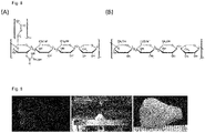
- Acylated gellan gum is a term known in the art and refers to gellan that comprises acetyl in some or all oxygen 5' positions and glycerylic acid in some of all oxygen 2' positions of the glucose unit. See Fig. 8 : Acylated gellan (A) is a raw product gellan after bacterial fermentation and when it is purified acyl and glyseryl side chains can be cleaved (B). This enhances the gelation and different stiffness can be achieved. Certain embodiments of the present invention combine acylated and purified gellan together to achieve better flexibility for the structures.
- the solution of a gelling polysaccharide comprises gellan gum or acetylated gellan gum, or a sulfation product of acylated gellan gum, as the gelling polysaccharide, and alginate as a cytocompatible polymer additive.
- an aqueous solution of a salt comprising monovalent, divalent and/or trivalent cations is added to said gelling polysaccharide to effect gelation.
- said aqueous solution comprises between 10 and 150 mmol/I of divalent ions.
- said aqueous solution comprises strontium ions (Sr 2+ ).
- said aqueous solution comprises barium ions (Ba 2+ ).
- said aqueous solution comprises calcium ions (Ca 2+ ).
- said aqueous solution comprises a total of between 10 and 150 mmol/I of divalent ions. In certain embodiments, said aqueous solution comprises between 10 and 150 mmol/I of strontium ions (Sr 2+ ), particularly between 15 and 50 mmol/I Sr 2+ . In certain embodiments, said aqueous solution comprises between 10 and 150 mmol/l of barium ions (Ba 2+ ), particularly between 15 and 50 mmol/l Ba 2+ . In certain embodiments, said aqueous solution comprises between 10 and 150 mmol/l of calcium ions (Ca 2+ ), particularly between 15 and 100 mmol/l Ca 2+ .
- said aqueous solution comprises a total of between 10 and 150 mmol/l of Sr 2+ and Ba 2+ , particularly between 15 and 50 mmol/l of Sr 2+ and Ba 2+ . In certain embodiments, said aqueous solution comprises a total of between 10 and 150 mmol/l of Ca 2+ and Ba 2+ , particularly between 15 and 50 mmol/l of Ca 2+ and Ba 2+ . In certain embodiments, said aqueous solution comprises a total of between 10 and 150 mmol/l of Sr 2+ and Ca 2+ , particularly between 15 and 50 mmol/l of Sr 2+ and Ca 2+ .
- said solution of a gelling polysaccharide comprises a monosaccharide sugar or disaccharide sugar, particularly glucose, mannose or arabinose, at physiologic osmolarity. This addition can be important to safeguard viability of the cells embedded in the printing mix.
- said particles and/or fibres consist of, or comprise,
- particles are formed from a biopolymer mentioned above in oil emulsion or by precipitation.
- such biopolymer is alginate.
- said tissue particles are derived from tissue selected from the group consisting of auricular cartilage, nasal cartilage, nucleus pulposus, meniscus, trachea, nasal cartilage, rib cartilage, articular cartilage, synovial fluid, vitreous humor, brain, spinal cord, muscle, connective tissues, small intestinal submucosa and liver.
- the cytocompatible polymer is a natural polymer.
- the cytocompatible polymer is gellan gum of varying acylation degree, particularly acylation ranging between 100% to 10% acylation, with 100% being high, and optionally comprises an additive selected from the group consisting of alginate, alginate sulfate, heparin, fibrin, heparin sulfate, elastin, tropoelastin, chondroitin sulfate, dermatan sulfate, hyaluronic acid, hyaluronan sulfate, cellulose, dextran, dextran sulfate, poly-l-lysine, chitosan, silk and collagen of varying type and sulfated versions of these.
- an additive selected from the group consisting of alginate, alginate sulfate, heparin, fibrin, heparin sulfate, elastin, tropoelastin, chondroitin sulfate, dermatan sulfate, hy
- ⁇ 90%, ⁇ 95% or ⁇ 98% of said particles are in the range of from 5 ⁇ m - 1000 ⁇ m, particularly from 5 ⁇ m to 50 ⁇ m, 5 ⁇ m to 200 ⁇ m, 50 ⁇ m - 200 ⁇ m or 200 ⁇ m to 1000 ⁇ m.
- said fibers are sized in the range of 5 ⁇ m - 50 ⁇ m and 50 - 500 ⁇ m in length having an aspect ratio ranging between 2-1000, particularly an aspect range of 10 - 500, more particularly 100 - 500, 100 - 1000, 200 - 1000 or from 500 to 1000.
- silk fibres are used having a diameter of 1 ⁇ m or less, and a length of 500 to 1000 ⁇ m or more.
- the aspect ratio for the purpose of the term's use in the context of the present specification is defined as the ratio of fiber length to diameter.
- said mammalian cells are cartilage cells, cartilage precursor cells or stem cells capable of differentiating into cartilage precursor cells or cartilage cells.
- the mammalian cells are selected from the group consisting of primary autologous chondrocytes, primary allogenic chondrocytes, chondroprogenitor cells, chondroblasts, mesenchymal stem cells, induced pluripotent stem cells and adipose-derived stem cells.
- the printing mix is deposited together with a sacrificial polymer. This allows the generation of overhanging structures, such as are especially important in shaping certain features of the nose and ear.
- the printing mix is deposited onto a sacrificial polymer scaffold.
- the sacrificial polymer scaffold is co-deposited with the printing mix.
- the sacrificial polymer mix and/or scaffold comprises divalent cations or other agents of gelation/polymerization.
- these cations or other agents of gelation/polymerization allow for a rapid formation of the three dimensional structure of the scaffold.
- the three dimensional form and/or said sacrificial polymer scaffold is derived by 3-D-printing methods, particularly on the basis of a three dimensional computer model of a contralateral organ of said patient.
- the three dimensional model is obtained by computed tomography, magnetic resonance imaging, laser scanning or utilizing three dimensional cameras.
- the computer model is created to support load bearing in gradients and to create internal structures for better cell survival and porosity.
- the polymer scaffold is derived by additive manufacturing methods.
- the additive manufacturing method is ink jet printing, bioprinting, extrusion printing or layer-by-layer method.
- the polymer scaffold is characterized by internal polymer gradients, porosity and support regions.
- additional polymers are added to increase the matrix liquid viscosity so that the ink can be extruded consistently and is not blocked due to filter pressing phenomena.
- the sacrificial polymer is removed prior or subsequent to said cell culture step.
- the tissue particles and/or bioink comprise a growth factor or a combination of growth factors, particularly selected from BMP-2, BMP-7, TGF- ⁇ 1, TGF- ⁇ 2, TGF- ⁇ 3, and/or FGF-2, and/or mitogenic factors, particularly IGF-1, to promote healing and regeneration.
- growth factors particularly selected from BMP-2, BMP-7, TGF- ⁇ 1, TGF- ⁇ 2, TGF- ⁇ 3, and/or FGF-2, and/or mitogenic factors, particularly IGF-1, to promote healing and regeneration.
- growth factors can be directly loaded into the bioink mixture.
- concentration of said growth factor(s) is in the range of from 0.1-5 mg/ml, 5-50 ng/ml or 50-500 ng/ml of one growth factor or a combination of several growth factors.
- the growth factors are selected from BMP-2, BMP-7, TGF- ⁇ 1, 2, 3, IGF-1 and/or FGF-2.
- the printing mix comprises 10 ng/ml TGF beta 3.
- the printing mix particularly the particles, comprise additional components, particularly components selected from growth factors, antioxidants, cytokines, drugs and biologics.
- the sacrificial polymer concludes an agent for initiating the crosslinking, said agent being a monovalent, divalent and trivalent cation, enzyme, hydrogen peroxide, horseradish peroxidase, radiation polymerizable monomers such as lithium phenyl-2,4,6-trimethylbenzoylphosphinate.
- crosslinking initiating groups are present in the printing mix, particularly selected from groups that participate in light exposure, cation-mediated crosslinking and enzyme-mediated crosslinking.
- depositing the printing mix in a three-dimensional form is performed by deposition of lines of the printing mix, wherein each line has a width of 700 to 1100 ⁇ m, and the lines overlap by 20% to 60%.
- Another aspect of the invention concerns a graft scaffold obtainable by, or obtained by, the method according to any one of the preceding methods of the invention, or any specific embodiment or combination of features provided by the specific embodiments.
- the present invention provides patient-specific craniofacial reconstructive grafts produced by additive manufacturing methods.
- the cartilage tissue graft abolishes the need for cartilage harvesting thus decreasing patient discomfort, reducing the surgical time and allowing better replication of shape, size and mechanical flexibility. Furthermore faster tissue regeneration and increased cell proliferation can be achieved to enhance the surgical recovery.
- this technique can be combined with currently used skin augmentation treatment (i.e. expanders) or other natural or synthetic skin grafts.
- patient specific auricular and nasal grafts are produced based on the three-dimensional scanned models from the patient by utilizing additive manufacturing methods such as but not limited to extrusion printing, inkjet printing and other layer-by-layer deposition methods.
- Clinical computed tomography (CT), magnetic resonance imaging (MRI) or other three dimensional imaging tools such as laser scanners, 3D cameras or combinations of these are used to produce the computerized model of the patient specific implant.
- CT computed tomography
- MRI magnetic resonance imaging
- 3D cameras three dimensional imaging tools
- the image can be mirrored to produce a computational model precisely mimicking the contralateral ear for tissue graft production.
- a library of graft models can be used to provide choice of grafts for the patient especially in a case where a normal contralateral scan cannot be performed.
- Avascularized cartilage can be designed to host vascular structures for over laying skin and other tissues in its proximity to prevent necrosis.
- the printed cartilage framework can be used as a bioactive template for the construction of overlying tissues, releasing growth factors and other secretory molecules to enhance the viability of neighboring cells. This release can be specially designed by having sulfated polymers in the mixture to bind growth factors to the proximity of the cells and slowly releasing the molecules.
- the 3D form can be created as a computer model to support load bearing in gradients and to created internal structures for better cell survival and porosity.
- the bioink material comprises at least one cytocompatible polymer and at least one of particles and cells, the crosslinking being provided by spontaneous or externally triggered reaction of reactive groups and molecules, at least one of these types being present on at least one of the polymer, minced tissue and cells.
- the cytocompatible polymerfor use in the method according to the invention is alginate.
- At least one of the groups being present on at least one of the polymer, particles and cells is meant that the added reactive groups may be present on all or any of these entities.
- Particles incorporated to polymer solution can consist of but are not limited to extracellular matrix tissue particles, loaded or unloaded beads and fibers in size range between 5 - 500 microns.
- hydrogel based on the material combination, particles and cells can be initiated by many factors or agents, including but not limited to mono-, di-, trivalent cations, enzymes and radical initiators. Additionally, physical and physical-chemical methods may be employed, for example, treatment in low or high pH solution and different temperature regions during the manufacturing process.
- either one of said printing mix and said polymer scaffold comprises reactive groups covalently attached thereto, particularly reactive groups facilitating linking of said printing mix, or its constituent components, to said particles, by crosslinking by spontaneous or externally triggered reaction, wherein reactive groups are present on at least one of the polymer, minced tissue and cells to reconstruct functional and native cartilage like tissue grafts.
- the size of the minced tissue to be used may be any suitable size, but in a particular embodiment, it is from 5 microns - 500 microns, so that it can be extruded without clogging the dispensing unit such as needle or valve.
- the minced tissue for use in the method may be any suitable tissue, but it is advantageously tissue of a similar or identical nature to that of the cartilage.
- tissue examples include articular cartilage, nucleus pulposus, meniscus, trachea, nasal cartilage, rib cartilage, ear cartilage, synovial fluid, tracheal cartilage, vitreous humor, brain, liver, spinal cord, muscle, connective tissues and subcutaneous fat, intrapatellar fat pad, small intestinal submucosa.
- tissue with high content of elastin and glycosaminoglycan particular examples being any type of cartilage, nucleus pulposus and meniscus.
- the tissue may be minced by any suitable method, exemplary and non-limiting methods including homogenizing, cryomilling, dry milling, cutting, chopping, crushing and slicing.
- the tissue may be subject to decellularization to remove epitopes which can cause acute inflammatory responses and pathogens including HIV.
- decellularized tissues that is, tissue in which the cells have been killed and their remnants removed, have attracted interest as scaffold material alternatives to simpler approaches where the scaffold is composed of a single material ( Hoshiba et al. "Decellularized matrices for tissue engineering". Expert Opinion on Biological Therapy. 2010;10:1717-28 ).
- Tissue decellularization results in a scaffold of extracellular matrix ideally suited for regenerating injured or diseased tissue since it retains the high resolution architecture and biological cues necessary for recapitulation of function.
- Decellularization may be done, for example, by using detergents, hydrogen peroxide, sodium hydroxide and enzymes, RNase and DNase.
- Particles can be manufactured by methods such as but not limited to colloid formation by hydrophilic/hydrophobic interactions, two phase emulsions and in oil interfaces.
- Fibers can be manufactured by methods such as but not limited to electrospinning, fiber extrusion and fiber pulling.
- Particles and fibers of any kind may be minced by any suitable method, exemplary and non-limiting methods including homogenizing, cryomilling, dry milling, cutting, chopping, crushing and slicing.
- These additive tissue pieces, particles and fibers may be further modified with functional groups binding to carrier polymer or combination of these materials or treated to expose reactive groups for crosslinking.
- growth factors, antioxidants and drug molecules may be loaded in or on the added polymers, tissue pieces, particles and fibers.
- cells encompasses not only individual cells, particularly mammalian cells, more particularly human cells, most particularly autologous human cells, but also encompasses agglomerations of the described cells which form spheroids, pellets, and microtissues , which are well known to and commonly used by the art.
- the cells for use in the method are advantageously cells of a similar type as those present on the cartilage tissue.
- suitable cell types include primary autologous chondrocytes, primary allogenic chondrocytes, chondroprogenitor cells, chondroblasts, mesenchymal stem cells, induced pluripotent stem cells and adipose-derived stem cells, neural crest derived stem cells.
- printing mix in the context of the present specification refers to an extruded mass comprising the key constituent components:
- the composition of the printing mix material may be varied across a wide range, depending on the nature of the materials and the end-use.
- the polymers are typically present in a weight proportion of from 0.5- 20%.
- the printing mix comprises 3.5% (w/v) of said gelling polysaccharide, and the solution of a gelling polysaccharide further comprises 2.5% or 3% (w/v) of alginate.
- minced tissue, particles or fibers are present, they are typically present at a weight proportion of from 10-40% of dry polymers or equally 1-20% in total weight.
- cells are present, they are typically used at concentrations of 3 ⁇ 10 6 cells/ml - 50 ⁇ 10 6 cells/ml.
- the crosslinkable material may include other materials, present to confer particular properties on the material.
- One particular example is elastin, which is abundant in auricular and nasal ECM to provide the elasticity of the tissue and other examples include growth factors, cytokines, drugs, biologics, siRNA, DNA, antioxidants such as polyphenols into the polymeric solutions, which could augment regeneration of the tissues.
- Added growth factors could be bound to sulfated polymers or unmodified polymer for enhanced delivery and effectivity in the proximity of the cells residing in the printing mix.
- the printing mix material in its ready-to-use form is a readily thermally gelled state that can easily be applied to take desired shape in the manufacturing process.
- Powders of the molecules and lyophilized minced tissue, particles and fibers can be stored and sterilized separately. All the components can be combined before packaging or rehydrated just prior to use thus preserving the growth factors and proteins for long periods of time.
- Patient specific tissue grafts are tailored for each patient or certain model catalogs can be created for situations where patient imaging is not desired or not possible.
- the three-dimensional model obtained from external ear and nose scans can be modified to contain internal support structures, gradient of polymers for versatile mechanical properties and porosity for enhanced cell survival in large constructs.
- region could be tuned in terms of stiffness, growth factor cocktail and concentration, for example, to induce regional variations in cell proliferation.
- the periphery of the cartilage graft could be more porous or softer allowing more nutrient flow into the deep structure.
- the regional specificity and tissue types are found in these constructs, for example, in the lobe of the ear fat is the main tissue and is responsible for the mechanical properties.
- the regional properties and specified structures can be easily built in a layer-by-layer manner.
- the crosslinking mechanism would take place not only within individual layers, but also between adjacent layers, thus forming a completely integrated continuous structure. This can be achieved by initiating crosslinking in the periphery of the construct in contact with support structure containing the reactive molecule reservoir.
- Support structure can be co-deposited with the printing mix material to support overhanging structures, to initiate crosslinking or to prevent drying of the material during deposition.
- Support material can contain crosslinking factors including but not limited to mono-, di-, trivalent cations, enzymes and radical initiators. Additionally, physical and physical-chemical methods may be employed by support material interactions to modify the pH and molecule concentration. After the construct manufacturing the support structure can be eluted. Elution can be due to but not limited to temperature change, pH change or degrading molecules.
- the result is a cartilage repair that is quick, effective and long-lasting.
- the longevity is an important factor in the graft to preserve the mechanical properties until sufficient ECM production of the cells has been achieved to produce a native cartilage-like structure.
- Example 1a Bioprinting of patient specified tissue grafts
- Fig. 1 Clinical computed tomography imaging was performed and the resulting computational three-dimensional object ( FIG. 1 ) was obtained.
- the patient specific external ear model was then mirrored for the contralateral side and a new 3D model was generated. Together with the new model the external support structure model was generated to support the ear structure especially in the overhanging regions during the printing.
- Support structure was designed to be in contact with the ink in the strategically important places to initiate the crosslinking and to support the overhanging features ( Fig. 5 ).
- the co-extrusion of the support material was shown to preserve horizontal bioink lines without sagging and the printed shape accurately after elution of the support.
- the internal support structure of more dense polymers was prepared to allow better force distribution in internal structure ( Fig. 2 , 3 ). All models were converted into machine code in STL-converter (RegenHU) and transferred into the bioprinter (BioFactory, RegenHU) for the printing process.
- Two-component intervertebral disc grafts could be printed with two bioink compositions mimicking the nucleus pulposus and the annulus fibrosus.
- Example 1b Production of cartilage particles for three-dimensional printing purposes
- Cartilage was harvested from the fresh bovine articular or auricular cartilage by removing thin layers of cartilage into a petri dish containing PBS and penicillin-streptomycin 1%. The harvested cartilage was transferred into cryomill (Retsch) and milled for three cycles in 30 Hz intensity. Milled cartilage was collected and lyophilized to obtain dry powder that could be sieved into the desired particle size range. These particles can be further loaded with growth factors or other molecules to enhance the proliferation and other cell responses. After loading the particles were lyophilyzed and cryopreserved to maximize the biomolecule availability for prolonged shelf life.
- Example 1c printing mix material preparation and the printing process
- Printing mix material (“Bio-Ink”) was produced by combining gellan gum in 3.5% concentration with the alginate 3%. Gellan gum was dialyzed against ultrapure water to minimize the cation residues in the material. Dialysis was performed over three days in 70-80°C ultrapure water changing the water one to two times a day. Gellan was further lyophilized to obtain a dry powder. Purified gellan gum was dissolved into glucose containing deionized water making it more cell compatible and alginate solution was added to obtain final concentration of polymers. The polymer blend was mixed with ECM particles and 6 ⁇ 10 6 cells/ml to obtain the final printing mix material. This printing mix material stimulated the cell proliferation significantly compared to positive control ( Fig. 4 ).
- Cartilage extracellular matrix production was evaluated with histology and immunostaining after 8 weeks in culture for Bioink alone and Bioink+ECM with and without growth factor TGF- ⁇ 3.
- the Bioink+ECM without growth factors stimulated cell proliferation above Bioink alone which was clearly visible in H&E staining.
- the Bioink+Cartilage particles showed a slight increase in Alcian blue staining and a slight collagen II staining was observed suggesting the need for additional growth factor stimulation. Cells were often seen proliferating around the particles without growth factor whereas the Bioink+Cartilage particles with TGF- ⁇ 3 had no site-specific proliferation which suggests that the particles are a source of mitogenic growth factors.
- the gross appearance of the scaffolds suggested that the growth factor stimulation had a clear effect on cartilage matrix production as seen in the size and opaque appearance of TGF- ⁇ 3 supplemented samples.
- Both supplemented bioink compositions showed a significant increase in cartilage ECM components and had areas which began to resemble the cell density and GAG content of native cartilage.
- collagen II deposition was strong throughout the graft in the growth factor supplemented conditions while only pericellular staining was seen in the samples cultured without TGF- ⁇ 3. Collagen type I and alizarin red staining were performed to determine the fibrocartilage production and calcification.
- Collagen I was found in Bioink+Cartilage particles and in both TGF- ⁇ 3 supplemented conditions suggesting some fibrocartilage production, perhaps due to the passaging of the cells. In all the conditions calcification was absent suggesting the cartilage phenotype of the chondrocytes was stable.
- the printing mix material was printed onto a substrate and the support polymer Pluronic F127 was co-extruded to fill subsequent layer.
- Pluronic contained 20mM of SrCl 2 to initiate the bioink crosslinking upon contact with the ink. Cations diffused into the printing mix material due to osmotic balance and electrostatic forces which initiated the crosslinking. Structures were generated with 410 ⁇ m needles and 800mm/minute feedrate. Pressure applied to extrusion syringe varied between 1.2 - 1.4 bar. After layer-by-layer deposition of the material into desired form, the sacrificial support Pluronic was eluted in a 20mM SrCl 2 bath for few minutes before the construct was transferred to 37°C cell culture medium.
- Fig. 1 illustrates the ear cartilage internal support structure and Fig. 6 shows the nose grafts generated by this technique.
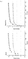
- Fig. 2 illustrates the initial storage modulus being 100 kPa after crosslinking which is comparable to high stiffness hydrogels
- Example 2 Bioink composition optimized for its mechanical properties and growth factor retention
- Bioink preparation Gellan was added to D-glucose (300mM) containing ultra-pure water at 90°C to achieve a 3.5% solution, of which 85% was low-acyl gellan gum and 25% was high-acyl gellan gum. Alginate was added to the mixture to achieve 2.5% solution. The boiling flask was kept at 90°C with agitation until the solution was homogeneous, typically for one hour. The homogeneous solution was cooled down to 30°C prior the cell mixing. Briefly, the bovine chondrocytes (4 ⁇ 10 6 cells/ml) were mixed in the DMEM solution and added to the bioink in culture medium in 1:10 volume ratio to pre-crosslink the bioink. Mixing was performed until the solution reached room temperature and the printing syringes were loaded.
- Gellan gum high acyl (GG-HA) and low acyl (GG-LA) compositions contribute to the stiffness and the elasticity of the final bioink.
- GG-HA Gellan gum high acyl
- GG-LA low acyl
- Fig. 8 Gellan gum high acyl
- the materials can crosslink tighter yielding stiffer matrix whereas by disrupting the tight packing of the polymer chains in crosslinking more elastic matrix can be produced.
- An exemplary bioink is a blend of gellan and alginate mixed with human micronized Cartilage particles or HA particles ( ⁇ 40 ⁇ m size).
- gelation occurs as the helices aggregate into junction zones which are linked into a three dimensional network via the coiled part of the molecule.
- the printing process is divided into three stages namely bioink pre-printing, printing process and post-printing crosslinking. Initially the bioink was loaded into a syringe and the support polymer into a second syringe. At this stage, a small amount of cations were present in the bioink to increase viscosity and enhance printing properties.
- the support can be eluted in 4°C cation-supplemented medium.
- the cation related viscosity enhancement and crosslinking properties can be investigated with rheology and mechanical testing.
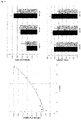
- Rheological properties of the Bioink, Bioink+HA, and Bioink+Cartilage Particles were measured with an Anton Paar MCR 301 (Anton Paar, Zofingen, Switzerland) rheometer to determine the shear behavior and shear recovery. All of the bioink compositions showed shear thinning behavior which is critical for extrusion ( Fig. 11 a) . Furthermore, all the compositions had a yield point (weak gel formation) prior to extrusion which is important in preventing particle and cell sedimentation in the syringe (Table 1). Table 1. Summary of the rheological measurements. The yield points were calculated using the Herschel/Bulkley equation.
- Bioink Bioink+HA Bioink+Cartilage Particles Yield point 15.6 Pa ⁇ 0.7 Pa 17.7 Pa ⁇ 6.5 Pa 122 Pa ⁇ 22 Pa Cessation in 10s* 21% 90% 98% Maximum G' 152 kPa ⁇ 3.0 kPa 110 kPa ⁇ 2.0 kPa 96 kPa ⁇ 1.0 kPa
- the shear recovery curves ( Fig. 11b ) illustrate the recovery of the bioink structure afte the printing process. Shear recovery after the second shear sequence was 98% in Bioink+Cartilage Particles and 90% in Bioink+HA after ten seconds. At the same time the Bioink alone recovered to only 21% of the original modulus.
- Fig. 11c illustrates the storage modulus G' after cation-induced crosslinking of Bioink alone where the cation concentration and source had a clear influence.
- Fig. 11d illustrates the final storage modulus for the three bioink compositions.
- the young adult sized nose graft had decreased viability in the center of the scaffold (60% viable cells at day 7) compared to 96% viability in the periphery ( Figure 13b ).
- interconnected porosity into 1.5 cm high cubes the viability in the center of the structure was as high as in the periphery.
- Such nutrition channels [ or engineered porosity can be incorporated into the bioprinted structures by extruding the support polymer within the grafts, which could later be cleared in subsequent washing/crosslinking steps. With this technique a complex 3D interconnected porous network can be created that is used to perfuse the grafts with nutrient-rich medium.
- grafts can also be pre-conditioned in dynamic bioreactors.
- Example 4e Extracellular matrix production and cartilage formation
- Cartilage extracellular matrix production was evaluated in bioink alone and bioink+Cartilage particles with histology and immunostaining after 3 and 8 weeks in culture. Histological evaluation after 3 weeks revealed a clear increase in cell number, GAG synthesis and collagen II production in both bioink compositions supplemented with TGF- ⁇ 3 (10ng/ml). Furthermore, Bioink+Cartilage particles without growth factors stimulated cell proliferation above Bioink alone which was clearly visible with 3 and 8 week H&E staining. At both time points the Bioink+Cartilage particles showed a slight increase in Alcian blue staining and at the 8 week time point a slight collagen II staining was observed suggesting the need for additional growth factor stimulation.
- collagen II deposition was strong throughout the graft in the growth factor supplemented conditions while only pericellular staining was seen in the samples cultured without TGF- ⁇ 3.
- Collagen I was found in Bioink+Cartilage particles and in both TGF- ⁇ 3 supplemented conditions suggesting some fibrocartilage production, perhaps due to the passaging of the cells. In all the conditions calcification was absent suggesting the cartilage phenotype of the chondrocytes was stable.
- the optimal line spacing is affected by the bioink in question however by increasing the overlapping the probability of internal printing process related defects decreases.
- the line thickness can be freely chosen by changing the process parameters such as pressure, printing speed and needle diameter.
Landscapes
- Health & Medical Sciences (AREA)
- Chemical & Material Sciences (AREA)
- Engineering & Computer Science (AREA)
- Life Sciences & Earth Sciences (AREA)
- Biomedical Technology (AREA)
- Medicinal Chemistry (AREA)
- Veterinary Medicine (AREA)
- Public Health (AREA)
- General Health & Medical Sciences (AREA)
- Animal Behavior & Ethology (AREA)
- Epidemiology (AREA)
- Oral & Maxillofacial Surgery (AREA)
- Transplantation (AREA)
- Dermatology (AREA)
- Materials Engineering (AREA)
- Manufacturing & Machinery (AREA)
- Chemical Kinetics & Catalysis (AREA)
- Cell Biology (AREA)
- Botany (AREA)
- Zoology (AREA)
- Composite Materials (AREA)
- Urology & Nephrology (AREA)
- Molecular Biology (AREA)
- Vascular Medicine (AREA)
- Rheumatology (AREA)
- Inorganic Chemistry (AREA)
- Ceramic Engineering (AREA)
- Structural Engineering (AREA)
- Civil Engineering (AREA)
- Dispersion Chemistry (AREA)
- General Physics & Mathematics (AREA)
- Automation & Control Theory (AREA)
- Physics & Mathematics (AREA)
- Human Computer Interaction (AREA)
- Pharmacology & Pharmacy (AREA)
- Materials For Medical Uses (AREA)
- Prostheses (AREA)
- Polymers & Plastics (AREA)
- Organic Chemistry (AREA)
- Coating Apparatus (AREA)
Applications Claiming Priority (4)
| Application Number | Priority Date | Filing Date | Title |
|---|---|---|---|
| EP14197449 | 2014-12-11 | ||
| EP15158224 | 2015-03-09 | ||
| PCT/EP2015/079502 WO2016092106A1 (en) | 2014-12-11 | 2015-12-11 | Graft scaffold for cartilage repair and process for making same |
| EP15817126.4A EP3230044B1 (en) | 2014-12-11 | 2015-12-11 | Graft scaffold for cartilage repair and process for making same |
Related Parent Applications (2)
| Application Number | Title | Priority Date | Filing Date |
|---|---|---|---|
| EP15817126.4A Division EP3230044B1 (en) | 2014-12-11 | 2015-12-11 | Graft scaffold for cartilage repair and process for making same |
| EP15817126.4A Division-Into EP3230044B1 (en) | 2014-12-11 | 2015-12-11 | Graft scaffold for cartilage repair and process for making same |
Publications (2)
| Publication Number | Publication Date |
|---|---|
| EP3689609A1 EP3689609A1 (en) | 2020-08-05 |
| EP3689609B1 true EP3689609B1 (en) | 2022-04-20 |
Family
ID=55027702
Family Applications (2)
| Application Number | Title | Priority Date | Filing Date |
|---|---|---|---|
| EP15817126.4A Active EP3230044B1 (en) | 2014-12-11 | 2015-12-11 | Graft scaffold for cartilage repair and process for making same |
| EP20160362.8A Active EP3689609B1 (en) | 2014-12-11 | 2015-12-11 | Graft scaffold for cartilage repair and process for making same |
Family Applications Before (1)
| Application Number | Title | Priority Date | Filing Date |
|---|---|---|---|
| EP15817126.4A Active EP3230044B1 (en) | 2014-12-11 | 2015-12-11 | Graft scaffold for cartilage repair and process for making same |
Country Status (19)
| Country | Link |
|---|---|
| US (4) | US10532126B2 (it) |
| EP (2) | EP3230044B1 (it) |
| JP (1) | JP6762936B2 (it) |
| CN (1) | CN106999635B (it) |
| AU (1) | AU2015359286B2 (it) |
| CA (1) | CA2967162C (it) |
| CY (1) | CY1123170T1 (it) |
| DK (2) | DK3689609T3 (it) |
| ES (1) | ES2809457T3 (it) |
| HR (1) | HRP20201252T1 (it) |
| HU (1) | HUE050420T2 (it) |
| IL (1) | IL252771B (it) |
| LT (1) | LT3230044T (it) |
| PL (1) | PL3230044T3 (it) |
| PT (1) | PT3230044T (it) |
| RS (1) | RS60700B1 (it) |
| SI (1) | SI3230044T1 (it) |
| SM (1) | SMT202000466T1 (it) |
| WO (1) | WO2016092106A1 (it) |
Families Citing this family (80)
| Publication number | Priority date | Publication date | Assignee | Title |
|---|---|---|---|---|
| US10675379B2 (en) | 2014-12-18 | 2020-06-09 | Cellink Ab | Cellulose nanofibrillar bioink for 3D bioprinting for cell culturing, tissue engineering and regenerative medicine applications |
| US11052175B2 (en) | 2015-08-19 | 2021-07-06 | Musculoskeletal Transplant Foundation | Cartilage-derived implants and methods of making and using same |
| SG11201811226PA (en) | 2016-06-16 | 2019-01-30 | Aspect Biosystems Ltd | Bioprinted meniscus implant and methods of using same |
| US11850324B2 (en) * | 2016-10-12 | 2023-12-26 | Advanced Biomatrix, Inc. | Three-dimensional (3-D) printing inks made from natural extracellular matrix molecules |
| CN106620873A (zh) * | 2016-11-17 | 2017-05-10 | 太原理工大学 | 一种复合水凝胶软骨修复材料及其制备方法 |
| WO2018118862A1 (en) * | 2016-12-23 | 2018-06-28 | Lifecell Corporation | Injectable hydrogels and applications thereof |
| CN106727706A (zh) * | 2016-12-26 | 2017-05-31 | 湖南新起源医疗技术有限公司 | 一种用于皮肤损伤修复的细胞制剂的制备方法 |
| CN106943632A (zh) * | 2017-03-23 | 2017-07-14 | 江南大学 | 一种胶原/硫酸软骨素复合人工眼角膜及其制备方法 |
| CN108686272B (zh) * | 2017-03-29 | 2021-04-09 | 复旦大学附属华山医院 | 静电混纺肝素-瑞舒伐他汀钙壳芯结构纳米纤维动脉瘤覆膜支架及制备方法 |
| SG11201909419XA (en) * | 2017-04-10 | 2019-11-28 | TheWell Bioscience | Hydrogel for cell culture and biomedical applications |
| EP3635045A1 (en) | 2017-05-26 | 2020-04-15 | Infinite Material Solutions, LLC | Water soluble polymer compositions |
| EP3427949B1 (en) * | 2017-07-12 | 2024-10-09 | MAPTECH Holdings UG | Mechanically tunable bioinks for bioprinting |
| CN107501612B (zh) * | 2017-08-07 | 2020-04-28 | 华南理工大学 | 3d打印氧化石墨烯/纤维素复合材料及其制备方法与应用 |
| JP7057076B2 (ja) | 2017-08-07 | 2022-04-19 | 株式会社クボタ | 植物体の生産方法 |
| KR102573462B1 (ko) * | 2017-08-28 | 2023-09-04 | 디에스엠 아이피 어셋츠 비.브이. | 폴리우레탄과 폴리옥사졸린을 포함하는 합성 멤브레인 조성물 |
| EP4309686A3 (en) | 2017-10-19 | 2024-02-07 | LifeCell Corporation | Flowable acellular tissue matrix products and methods of production |
| US11246994B2 (en) | 2017-10-19 | 2022-02-15 | Lifecell Corporation | Methods for introduction of flowable acellular tissue matrix products into a hand |
| US11485072B2 (en) * | 2017-10-27 | 2022-11-01 | DePuy Synthes Products, Inc. | Selective laser sintering of asymmetric particles |
| CN107823711B (zh) * | 2017-11-09 | 2020-10-30 | 华中科技大学同济医学院附属协和医院 | 核壳结构复合材料的制备及利用其构建组织工程微组织的方法 |
| EP3485918A1 (en) * | 2017-11-21 | 2019-05-22 | ETH Zürich | Gel composition for 3d printing of cell suspensions |
| CN111655451A (zh) * | 2017-11-29 | 2020-09-11 | 奥里根生物治疗公司 | 无菌增材制造系统 |
| JP7420721B2 (ja) * | 2017-12-20 | 2024-01-23 | アスペクト バイオシステムズ リミティド | バイオ印刷された半月板インプラント及びその使用方法 |
| WO2019122351A1 (en) * | 2017-12-22 | 2019-06-27 | Cellink Ab | Tissue-specific human bioinks for the physiological 3d-bioprinting of human tissues for in vitro culture and transplantation |
| CN108159500A (zh) * | 2017-12-27 | 2018-06-15 | 天津宝坻紫荆科技有限公司 | 一种3d打印人工骨修复材料及其制备方法 |
| WO2019145795A2 (en) | 2018-01-26 | 2019-08-01 | Cellink Ab | Systems and methods for optical assessments of bioink printability |
| CN108355175A (zh) * | 2018-02-12 | 2018-08-03 | 华中科技大学鄂州工业技术研究院 | 一种复合纳米纤维支架的制备方法 |
| CN108187137A (zh) * | 2018-02-27 | 2018-06-22 | 崔友军 | 一种可生物降解神经修复支架的制备方法 |
| CN110240799A (zh) * | 2018-03-09 | 2019-09-17 | 中国石油化工股份有限公司 | 3d打印组合物及其制备方法和应用 |
| CN108578617B (zh) * | 2018-04-09 | 2021-03-30 | 深圳市莱利赛生物科技有限公司 | 促进膝关节软骨再生用脐血间质干细胞药物的制备方法 |
| CN108379659A (zh) * | 2018-05-06 | 2018-08-10 | 西北工业大学 | 一种细胞密度多梯度人工软骨制备方法 |
| CN108355168A (zh) * | 2018-05-14 | 2018-08-03 | 上海交通大学医学院附属第九人民医院 | 一种结合三维打印技术制备髓核回植体的方法 |
| CN108690205A (zh) * | 2018-05-28 | 2018-10-23 | 深圳市第二人民医院 | 一种ⅱ型胶原和聚丙烯酰胺复合水凝胶及其制备和应用 |
| CN108853582A (zh) * | 2018-06-15 | 2018-11-23 | 南京冬尚生物科技有限公司 | 3D打印PCL-Cu骨组织工程支架及其制备方法 |
| US20200040296A1 (en) * | 2018-08-06 | 2020-02-06 | University Of South Carolina | Decellularized Tissue as a Microcarrier for Cell Culture and Expansion |
| US11426818B2 (en) | 2018-08-10 | 2022-08-30 | The Research Foundation for the State University | Additive manufacturing processes and additively manufactured products |
| US11186736B2 (en) | 2018-10-10 | 2021-11-30 | Cellink Ab | Double network bioinks |
| US20210001009A1 (en) * | 2018-10-25 | 2021-01-07 | Cellink Ab | Biogum and botanical gum hydrogel bioinks for the physiological 3d bioprinting of tissue constructs for in vitro culture and transplantation |
| CN109432501A (zh) * | 2018-10-30 | 2019-03-08 | 上海交通大学医学院附属第九人民医院 | 复合纤维支架在制备鼻整形的复合材料中的用途 |
| CN109513037B (zh) * | 2018-11-14 | 2021-09-17 | 华中科技大学同济医学院附属协和医院 | 一种负载介孔生物玻璃的小肠粘膜下层创面敷料 |
| CN109248009B (zh) * | 2018-11-23 | 2020-01-14 | 吉林大学 | 一种利用3d打印技术制作人工胸骨的方法 |
| KR102302770B1 (ko) * | 2018-11-30 | 2021-09-16 | 주식회사 엘앤씨바이오 | 인체 유래 성분을 함유하고 조직 특이적 세포 분화 효과를 갖는 3d 프린팅 바이오 잉크 조성물 및 그 제조방법 |
| KR102098134B1 (ko) * | 2019-02-13 | 2020-04-08 | 성균관대학교산학협력단 | 나노 섬유질 콜라겐 3차원 구조체 제조방법 |
| WO2020184974A1 (ko) * | 2019-03-11 | 2020-09-17 | 주식회사 로킷헬스케어 | 동결건조 유리연골 파우더를 이용한 연골 재생용 조성물의 제조방법, 이를 이용하여 제조된 연골 재생용 조성물, 연골 재생용 조성물을 이용한 환자 맞춤형 연골 재생용 스캐폴드의 제조방법 및 환자 맞춤형 연골 재생용 스캐폴드 |
| MX2021011881A (es) | 2019-03-29 | 2021-10-26 | Innovative Orthopedics Llc | Composiciones y metodos de reemplazo de cartilago. |
| KR20220060501A (ko) * | 2019-03-29 | 2022-05-11 | 티디비티 아이피 인코포레이티드 | 조직 및 장기 대체물 그리고 이들의 제조 방법 |
| CN110331124B (zh) * | 2019-06-14 | 2022-03-22 | 浙江大学 | 一种导电聚吡咯/细胞外基质复合薄膜及其制备方法 |
| CN110464879A (zh) * | 2019-07-15 | 2019-11-19 | 暨南大学 | 一种骨组织工程支架的3d打印方法 |
| CN110302428B (zh) * | 2019-07-30 | 2021-07-13 | 中国人民解放军陆军军医大学第一附属医院 | 基于活细胞3d打印的软骨-骨-骨髓复合组织结构及方法 |
| CN110538006B (zh) * | 2019-08-13 | 2022-06-17 | 上海亚朋生物技术有限公司 | 纤维增强的三维打印软骨脱细胞基质支架的制作方法 |
| CN110433331B (zh) * | 2019-08-26 | 2021-08-24 | 四川大学 | 一种生物活性支架及其制备方法 |
| CN110450401A (zh) * | 2019-09-05 | 2019-11-15 | 上海交通大学医学院附属第九人民医院 | 一种液体支撑介质内生物三维打印人工关节的方法 |
| US11826951B2 (en) | 2019-09-06 | 2023-11-28 | Cellink Ab | Temperature-controlled multi-material overprinting |
| CN110478529A (zh) * | 2019-09-24 | 2019-11-22 | 广西医科大学 | 一种3d打印魔芋胶水凝胶支架及制备方法和应用 |
| KR20220084043A (ko) * | 2019-10-18 | 2022-06-21 | 카네기 멜론 유니버시티 | 연질 재료의 개선된 3d 프린팅을 위한 유변학 및 기계 경로 지정의 수정 |
| CN110575568A (zh) * | 2019-10-22 | 2019-12-17 | 上海交通大学医学院附属第九人民医院 | 一种用于3d打印的水凝胶材料、制备方法及用途 |
| CN110680954A (zh) * | 2019-11-06 | 2020-01-14 | 广西医科大学 | 一种3d打印黄原胶水凝胶支架及其制备方法 |
| CN110755685A (zh) * | 2019-11-06 | 2020-02-07 | 广西医科大学 | 一种3d打印瓜尔胶水凝胶支架及其制备方法 |
| KR102228619B1 (ko) * | 2019-11-25 | 2021-03-17 | 경희대학교 산학협력단 | 히알루론산 및 폴리에틸렌글리콜을 포함하여 제조된 세포 시트 및 이의 제조방법 |
| BR112022011148A2 (pt) * | 2019-12-18 | 2022-08-23 | Grifols Worldwide Operations Ltd | Compósito de osso, composições de múltiplas partes para a impressão 3d do mesmo, seus usos, método para a preparação do referido compósito de osso, e kit |
| CN111320767B (zh) * | 2020-03-04 | 2023-01-10 | 西南交通大学 | 一种用于3d生物打印的可触变性水凝胶的制备方法 |
| CN114080244A (zh) * | 2020-03-06 | 2022-02-22 | 爱恩斯生物科技(昆山)有限公司 | 利用基于软骨成分的生物墨水的治疗小耳症目的结构体制造用组合物及其制造方法 |
| US12371581B2 (en) | 2020-03-25 | 2025-07-29 | Infinite Material Solutions, Llc | High performance water soluble polymer compositions |
| US20230105586A1 (en) * | 2020-03-26 | 2023-04-06 | Osaka University | Supporting bath for three-dimensional (3d) tissue culture |
| CN113456897A (zh) * | 2020-03-31 | 2021-10-01 | 威斯顿股份有限公司 | 生物材料及其用于促进组织再生的用途 |
| MX2022015741A (es) * | 2020-06-17 | 2023-01-19 | Auregen Biotherapeutics Sa | Composiciones impresas tridimensionales maduradas y usos de las mismas. |
| CN112354017B (zh) * | 2020-11-19 | 2022-07-26 | 中国医学科学院生物医学工程研究所 | 一种3d打印的力学仿生耳廓软骨组织工程支架及其制造方法 |
| EP4271325A4 (en) * | 2021-01-04 | 2024-11-27 | Technion Research & Development Foundation Limited | Auricular reconstruction using 3d printed autologous cartilage tissue |
| JP7696580B2 (ja) * | 2021-02-16 | 2025-06-23 | 株式会社ニッピ | バイオインク、成型体、製品、および成型体の製造方法 |
| CN113069596A (zh) * | 2021-04-07 | 2021-07-06 | 山东建筑大学 | 一种基于3d打印半月板的复合生物陶瓷修复材料 |
| CN113244455B (zh) * | 2021-05-17 | 2022-08-05 | 广西医科大学 | 一种3d打印亚麻籽水凝胶支架及制备方法和应用 |
| US20250073168A1 (en) | 2021-06-16 | 2025-03-06 | Auregen Biotherapeutics Sa | Gellan gum compositions and method for their preparation |
| US11944726B2 (en) | 2021-06-18 | 2024-04-02 | Dimension Inx Corp | Method for fabrication of additively manufactured, self-gelling structures and their use |
| WO2023247801A1 (en) * | 2022-06-24 | 2023-12-28 | Katholieke Universiteit Leuven | Printing of cell aggregates |
| WO2024004696A1 (ja) * | 2022-06-30 | 2024-01-04 | 国立大学法人大阪大学 | 三次元造形物の製造方法および三次元造形物作製用インクセット |
| CN119768265A (zh) * | 2022-08-26 | 2025-04-04 | 国立大学法人大阪大学 | 三维造型物的制造方法和三维造型物制作用墨组 |
| CN115519788B (zh) * | 2022-09-06 | 2024-03-15 | 深圳先进技术研究院 | 组织工程支架的打印方法、打印系统、终端及存储介质 |
| CN120344275A (zh) * | 2022-10-13 | 2025-07-18 | 北卡罗莱纳州立大学 | 可植入支架及其使用方法 |
| CN116139341B (zh) * | 2022-12-14 | 2024-06-25 | 四川大学 | 一种定向冷冻聚乙烯醇/交联脱细胞基质复合材料及其制备方法和应用 |
| CN116970304B (zh) * | 2023-06-13 | 2025-02-14 | 华南理工大学 | 一种MnO2-蛋白纤维复合颗粒凝胶墨水及其制备与应用 |
| WO2025070369A1 (ja) * | 2023-09-25 | 2025-04-03 | 国立大学法人大阪大学 | 三次元造形物の製造方法および三次元造形物作製用インクセット |
Family Cites Families (8)
| Publication number | Priority date | Publication date | Assignee | Title |
|---|---|---|---|---|
| US6126690A (en) * | 1996-07-03 | 2000-10-03 | The Trustees Of Columbia University In The City Of New York | Anatomically correct prosthesis and method and apparatus for manufacturing prosthesis |
| JP4975013B2 (ja) * | 2005-03-11 | 2012-07-11 | ウエイク・フオレスト・ユニバーシテイ・ヘルス・サイエンシズ | 組織が工作された心臓弁の製造 |
| US7531503B2 (en) * | 2005-03-11 | 2009-05-12 | Wake Forest University Health Sciences | Cell scaffold matrices with incorporated therapeutic agents |
| AU2008311957A1 (en) * | 2007-10-15 | 2009-04-23 | Wake Forest University | Methods and compositions for printing biologically compatible nanotube composites of autologous tissue |
| US9012415B2 (en) * | 2010-03-26 | 2015-04-21 | Stemmatters, Biotecnologia E Medicina Regenerativa S.A. | Photo-crosslinked gellan gum-based hydrogels: preparation methods and uses thereof |
| US20120089238A1 (en) * | 2010-10-06 | 2012-04-12 | Hyun-Wook Kang | Integrated organ and tissue printing methods, system and apparatus |
| WO2014032748A1 (en) * | 2012-08-31 | 2014-03-06 | Eth Zurich | Process of cartilage repair |
| AU2013204780A1 (en) * | 2013-04-12 | 2014-10-30 | University Of Wollongong | Formulation and Method |
-
2015
- 2015-12-11 ES ES15817126T patent/ES2809457T3/es active Active
- 2015-12-11 PT PT158171264T patent/PT3230044T/pt unknown
- 2015-12-11 WO PCT/EP2015/079502 patent/WO2016092106A1/en not_active Ceased
- 2015-12-11 CN CN201580064029.9A patent/CN106999635B/zh active Active
- 2015-12-11 DK DK20160362.8T patent/DK3689609T3/da active
- 2015-12-11 JP JP2017529659A patent/JP6762936B2/ja active Active
- 2015-12-11 CA CA2967162A patent/CA2967162C/en active Active
- 2015-12-11 HR HRP20201252TT patent/HRP20201252T1/hr unknown
- 2015-12-11 LT LTEP15817126.4T patent/LT3230044T/lt unknown
- 2015-12-11 EP EP15817126.4A patent/EP3230044B1/en active Active
- 2015-12-11 DK DK15817126.4T patent/DK3230044T3/da active
- 2015-12-11 AU AU2015359286A patent/AU2015359286B2/en active Active
- 2015-12-11 US US15/535,053 patent/US10532126B2/en active Active
- 2015-12-11 SI SI201531312T patent/SI3230044T1/sl unknown
- 2015-12-11 RS RS20200960A patent/RS60700B1/sr unknown
- 2015-12-11 PL PL15817126T patent/PL3230044T3/pl unknown
- 2015-12-11 EP EP20160362.8A patent/EP3689609B1/en active Active
- 2015-12-11 HU HUE15817126A patent/HUE050420T2/hu unknown
- 2015-12-11 SM SM20200466T patent/SMT202000466T1/it unknown
-
2017
- 2017-06-08 IL IL252771A patent/IL252771B/en unknown
-
2020
- 2020-01-07 US US16/735,751 patent/US11633518B2/en active Active
- 2020-08-04 CY CY20201100721T patent/CY1123170T1/el unknown
-
2023
- 2023-03-30 US US18/192,682 patent/US20230321320A1/en not_active Abandoned
-
2025
- 2025-03-25 US US19/089,058 patent/US20250222166A1/en active Pending
Also Published As
| Publication number | Publication date |
|---|---|
| CA2967162A1 (en) | 2016-06-16 |
| US20250222166A1 (en) | 2025-07-10 |
| ES2809457T3 (es) | 2021-03-04 |
| EP3689609A1 (en) | 2020-08-05 |
| US20230321320A1 (en) | 2023-10-12 |
| US10532126B2 (en) | 2020-01-14 |
| WO2016092106A1 (en) | 2016-06-16 |
| US20200139007A1 (en) | 2020-05-07 |
| EP3230044B1 (en) | 2020-07-01 |
| LT3230044T (lt) | 2020-08-25 |
| AU2015359286A1 (en) | 2017-07-27 |
| EP3230044A1 (en) | 2017-10-18 |
| HRP20201252T1 (hr) | 2020-11-27 |
| CY1123170T1 (el) | 2021-10-29 |
| US20170348458A1 (en) | 2017-12-07 |
| DK3689609T3 (da) | 2022-07-04 |
| CN106999635A (zh) | 2017-08-01 |
| AU2015359286B2 (en) | 2019-05-02 |
| DK3230044T3 (da) | 2020-08-17 |
| IL252771A0 (en) | 2017-08-31 |
| CN106999635B (zh) | 2021-06-11 |
| JP6762936B2 (ja) | 2020-09-30 |
| PT3230044T (pt) | 2020-08-03 |
| SMT202000466T1 (it) | 2020-11-10 |
| JP2018501845A (ja) | 2018-01-25 |
| US11633518B2 (en) | 2023-04-25 |
| SI3230044T1 (sl) | 2020-10-30 |
| HUE050420T2 (hu) | 2020-12-28 |
| RS60700B1 (sr) | 2020-09-30 |
| IL252771B (en) | 2022-06-01 |
| PL3230044T3 (pl) | 2021-02-08 |
| CA2967162C (en) | 2019-07-02 |
Similar Documents
| Publication | Publication Date | Title |
|---|---|---|
| US20250222166A1 (en) | Graft scaffold for cartilage repair and process for making same | |
| Jahangirian et al. | A review of using green chemistry methods for biomaterials in tissue engineering | |
| US6991652B2 (en) | Tissue engineering composite | |
| CA2672495C (en) | Novel injectable chitosan mixtures forming hydrogels | |
| US20130295081A1 (en) | Polyurethane Composite for Wound Healing and Methods Thereof | |
| US20100233267A1 (en) | Composite hydrogel | |
| CN111432853A (zh) | 交联的蛋白质泡沫及其使用多用途细胞支架的方法 | |
| EP2919763A1 (en) | Flexible tissue matrix | |
| CA2547668A1 (en) | A biomimetic composition reinforced by a polyelectrolytic complex of hyaluronic acid and chitosan | |
| Chen et al. | Hyaluronic acid-based biphasic scaffold with layer-specific induction capacity for osteochondral defect regeneration | |
| KR20200007747A (ko) | 골치료를 위한 fk506을 포함하는 키토산/템포 산화 셀룰로오스 나노 섬유 하이드로겔 및 이의 제조방법 | |
| EP4259223B1 (en) | Particulate materials for tissue mimics | |
| Chen et al. | A lubcan cross-linked polyethylene glycol dimethyl ether hydrogel for hyaluronic acid replacement as soft tissue engineering fillers | |
| Chhibber et al. | Hydrogels in tissue engineering | |
| JP7433692B2 (ja) | 異物反応が低減された三次元埋入型マトリックス | |
| Mueller et al. | Clinical translation of 3D bioprinting for cartilage repair: a biomaterial perspective | |
| CN114080244A (zh) | 利用基于软骨成分的生物墨水的治疗小耳症目的结构体制造用组合物及其制造方法 | |
| Hosseini et al. | Fabrication and morphology of biomaterials based on the used synthesis methods | |
| Namkaew et al. | Polyvinyl alcohol-carboxymethyl cellulose scaffolds for cartilage tissue formation | |
| Kesti | Bioprinting technologies for auricular cartilage tissue engineering | |
| Fredriksson et al. | Exploration of de novo tissue regeneration: With applications for skin and cartilage | |
| 김수진 | Development of hydrogel scaffolds for adipose tissue regeneration | |
| WO2022009125A1 (en) | Tridimensional bioactive porous body for bone tissue regeneration and process for its preparation | |
| Spiller | Semi-degradable, multi-functional hydrogels for the repair of articular cartilage defects | |
| Lu | The Use of Synthetic ECMs in Tissue Engineering |
Legal Events
| Date | Code | Title | Description |
|---|---|---|---|
| PUAI | Public reference made under article 153(3) epc to a published international application that has entered the european phase |
Free format text: ORIGINAL CODE: 0009012 |
|
| STAA | Information on the status of an ep patent application or granted ep patent |
Free format text: STATUS: THE APPLICATION HAS BEEN PUBLISHED |
|
| AC | Divisional application: reference to earlier application |
Ref document number: 3230044 Country of ref document: EP Kind code of ref document: P |
|
| AK | Designated contracting states |
Kind code of ref document: A1 Designated state(s): AL AT BE BG CH CY CZ DE DK EE ES FI FR GB GR HR HU IE IS IT LI LT LU LV MC MK MT NL NO PL PT RO RS SE SI SK SM TR |
|
| STAA | Information on the status of an ep patent application or granted ep patent |
Free format text: STATUS: REQUEST FOR EXAMINATION WAS MADE |
|
| 17P | Request for examination filed |
Effective date: 20210127 |
|
| RBV | Designated contracting states (corrected) |
Designated state(s): AL AT BE BG CH CY CZ DE DK EE ES FI FR GB GR HR HU IE IS IT LI LT LU LV MC MK MT NL NO PL PT RO RS SE SI SK SM TR |
|
| GRAP | Despatch of communication of intention to grant a patent |
Free format text: ORIGINAL CODE: EPIDOSNIGR1 |
|
| STAA | Information on the status of an ep patent application or granted ep patent |
Free format text: STATUS: GRANT OF PATENT IS INTENDED |
|
| INTG | Intention to grant announced |
Effective date: 20210601 |
|
| GRAJ | Information related to disapproval of communication of intention to grant by the applicant or resumption of examination proceedings by the epo deleted |
Free format text: ORIGINAL CODE: EPIDOSDIGR1 |
|
| STAA | Information on the status of an ep patent application or granted ep patent |
Free format text: STATUS: REQUEST FOR EXAMINATION WAS MADE |
|
| GRAP | Despatch of communication of intention to grant a patent |
Free format text: ORIGINAL CODE: EPIDOSNIGR1 |
|
| STAA | Information on the status of an ep patent application or granted ep patent |
Free format text: STATUS: GRANT OF PATENT IS INTENDED |
|
| INTC | Intention to grant announced (deleted) | ||
| INTG | Intention to grant announced |
Effective date: 20211108 |
|
| GRAS | Grant fee paid |
Free format text: ORIGINAL CODE: EPIDOSNIGR3 |
|
| GRAA | (expected) grant |
Free format text: ORIGINAL CODE: 0009210 |
|
| STAA | Information on the status of an ep patent application or granted ep patent |
Free format text: STATUS: THE PATENT HAS BEEN GRANTED |
|
| AC | Divisional application: reference to earlier application |
Ref document number: 3230044 Country of ref document: EP Kind code of ref document: P |
|
| AK | Designated contracting states |
Kind code of ref document: B1 Designated state(s): AL AT BE BG CH CY CZ DE DK EE ES FI FR GB GR HR HU IE IS IT LI LT LU LV MC MK MT NL NO PL PT RO RS SE SI SK SM TR |
|
| REG | Reference to a national code |
Ref country code: GB Ref legal event code: FG4D |
|
| REG | Reference to a national code |
Ref country code: CH Ref legal event code: EP |
|
| REG | Reference to a national code |
Ref country code: IE Ref legal event code: FG4D |
|
| REG | Reference to a national code |
Ref country code: DE Ref legal event code: R096 Ref document number: 602015078467 Country of ref document: DE |
|
| REG | Reference to a national code |
Ref country code: AT Ref legal event code: REF Ref document number: 1484874 Country of ref document: AT Kind code of ref document: T Effective date: 20220515 |
|
| REG | Reference to a national code |
Ref country code: DK Ref legal event code: T3 Effective date: 20220628 |
|
| REG | Reference to a national code |
Ref country code: FI Ref legal event code: FGE |
|
| REG | Reference to a national code |
Ref country code: SE Ref legal event code: TRGR |
|
| REG | Reference to a national code |
Ref country code: NL Ref legal event code: FP |
|
| REG | Reference to a national code |
Ref country code: NO Ref legal event code: T2 Effective date: 20220420 |
|
| REG | Reference to a national code |
Ref country code: LT Ref legal event code: MG9D |
|
| REG | Reference to a national code |
Ref country code: AT Ref legal event code: MK05 Ref document number: 1484874 Country of ref document: AT Kind code of ref document: T Effective date: 20220420 |
|
| PG25 | Lapsed in a contracting state [announced via postgrant information from national office to epo] |
Ref country code: PT Free format text: LAPSE BECAUSE OF FAILURE TO SUBMIT A TRANSLATION OF THE DESCRIPTION OR TO PAY THE FEE WITHIN THE PRESCRIBED TIME-LIMIT Effective date: 20220822 Ref country code: LT Free format text: LAPSE BECAUSE OF FAILURE TO SUBMIT A TRANSLATION OF THE DESCRIPTION OR TO PAY THE FEE WITHIN THE PRESCRIBED TIME-LIMIT Effective date: 20220420 Ref country code: HR Free format text: LAPSE BECAUSE OF FAILURE TO SUBMIT A TRANSLATION OF THE DESCRIPTION OR TO PAY THE FEE WITHIN THE PRESCRIBED TIME-LIMIT Effective date: 20220420 Ref country code: GR Free format text: LAPSE BECAUSE OF FAILURE TO SUBMIT A TRANSLATION OF THE DESCRIPTION OR TO PAY THE FEE WITHIN THE PRESCRIBED TIME-LIMIT Effective date: 20220721 Ref country code: ES Free format text: LAPSE BECAUSE OF FAILURE TO SUBMIT A TRANSLATION OF THE DESCRIPTION OR TO PAY THE FEE WITHIN THE PRESCRIBED TIME-LIMIT Effective date: 20220420 Ref country code: BG Free format text: LAPSE BECAUSE OF FAILURE TO SUBMIT A TRANSLATION OF THE DESCRIPTION OR TO PAY THE FEE WITHIN THE PRESCRIBED TIME-LIMIT Effective date: 20220720 Ref country code: AT Free format text: LAPSE BECAUSE OF FAILURE TO SUBMIT A TRANSLATION OF THE DESCRIPTION OR TO PAY THE FEE WITHIN THE PRESCRIBED TIME-LIMIT Effective date: 20220420 |
|
| PG25 | Lapsed in a contracting state [announced via postgrant information from national office to epo] |
Ref country code: RS Free format text: LAPSE BECAUSE OF FAILURE TO SUBMIT A TRANSLATION OF THE DESCRIPTION OR TO PAY THE FEE WITHIN THE PRESCRIBED TIME-LIMIT Effective date: 20220420 Ref country code: PL Free format text: LAPSE BECAUSE OF FAILURE TO SUBMIT A TRANSLATION OF THE DESCRIPTION OR TO PAY THE FEE WITHIN THE PRESCRIBED TIME-LIMIT Effective date: 20220420 Ref country code: LV Free format text: LAPSE BECAUSE OF FAILURE TO SUBMIT A TRANSLATION OF THE DESCRIPTION OR TO PAY THE FEE WITHIN THE PRESCRIBED TIME-LIMIT Effective date: 20220420 Ref country code: IS Free format text: LAPSE BECAUSE OF FAILURE TO SUBMIT A TRANSLATION OF THE DESCRIPTION OR TO PAY THE FEE WITHIN THE PRESCRIBED TIME-LIMIT Effective date: 20220820 |
|
| REG | Reference to a national code |
Ref country code: DE Ref legal event code: R097 Ref document number: 602015078467 Country of ref document: DE |
|
| PG25 | Lapsed in a contracting state [announced via postgrant information from national office to epo] |
Ref country code: SM Free format text: LAPSE BECAUSE OF FAILURE TO SUBMIT A TRANSLATION OF THE DESCRIPTION OR TO PAY THE FEE WITHIN THE PRESCRIBED TIME-LIMIT Effective date: 20220420 Ref country code: SK Free format text: LAPSE BECAUSE OF FAILURE TO SUBMIT A TRANSLATION OF THE DESCRIPTION OR TO PAY THE FEE WITHIN THE PRESCRIBED TIME-LIMIT Effective date: 20220420 Ref country code: RO Free format text: LAPSE BECAUSE OF FAILURE TO SUBMIT A TRANSLATION OF THE DESCRIPTION OR TO PAY THE FEE WITHIN THE PRESCRIBED TIME-LIMIT Effective date: 20220420 Ref country code: EE Free format text: LAPSE BECAUSE OF FAILURE TO SUBMIT A TRANSLATION OF THE DESCRIPTION OR TO PAY THE FEE WITHIN THE PRESCRIBED TIME-LIMIT Effective date: 20220420 Ref country code: CZ Free format text: LAPSE BECAUSE OF FAILURE TO SUBMIT A TRANSLATION OF THE DESCRIPTION OR TO PAY THE FEE WITHIN THE PRESCRIBED TIME-LIMIT Effective date: 20220420 |
|
| PLBE | No opposition filed within time limit |
Free format text: ORIGINAL CODE: 0009261 |
|
| STAA | Information on the status of an ep patent application or granted ep patent |
Free format text: STATUS: NO OPPOSITION FILED WITHIN TIME LIMIT |
|
| 26N | No opposition filed |
Effective date: 20230123 |
|
| PG25 | Lapsed in a contracting state [announced via postgrant information from national office to epo] |
Ref country code: AL Free format text: LAPSE BECAUSE OF FAILURE TO SUBMIT A TRANSLATION OF THE DESCRIPTION OR TO PAY THE FEE WITHIN THE PRESCRIBED TIME-LIMIT Effective date: 20220420 |
|
| PG25 | Lapsed in a contracting state [announced via postgrant information from national office to epo] |
Ref country code: SI Free format text: LAPSE BECAUSE OF FAILURE TO SUBMIT A TRANSLATION OF THE DESCRIPTION OR TO PAY THE FEE WITHIN THE PRESCRIBED TIME-LIMIT Effective date: 20220420 |
|
| P01 | Opt-out of the competence of the unified patent court (upc) registered |
Effective date: 20230526 |
|
| PG25 | Lapsed in a contracting state [announced via postgrant information from national office to epo] |
Ref country code: LU Free format text: LAPSE BECAUSE OF NON-PAYMENT OF DUE FEES Effective date: 20221211 |
|
| PG25 | Lapsed in a contracting state [announced via postgrant information from national office to epo] |
Ref country code: IT Free format text: LAPSE BECAUSE OF FAILURE TO SUBMIT A TRANSLATION OF THE DESCRIPTION OR TO PAY THE FEE WITHIN THE PRESCRIBED TIME-LIMIT Effective date: 20220420 |
|
| PG25 | Lapsed in a contracting state [announced via postgrant information from national office to epo] |
Ref country code: HU Free format text: LAPSE BECAUSE OF FAILURE TO SUBMIT A TRANSLATION OF THE DESCRIPTION OR TO PAY THE FEE WITHIN THE PRESCRIBED TIME-LIMIT; INVALID AB INITIO Effective date: 20151211 |
|
| PG25 | Lapsed in a contracting state [announced via postgrant information from national office to epo] |
Ref country code: CY Free format text: LAPSE BECAUSE OF FAILURE TO SUBMIT A TRANSLATION OF THE DESCRIPTION OR TO PAY THE FEE WITHIN THE PRESCRIBED TIME-LIMIT Effective date: 20220420 |
|
| PG25 | Lapsed in a contracting state [announced via postgrant information from national office to epo] |
Ref country code: MK Free format text: LAPSE BECAUSE OF FAILURE TO SUBMIT A TRANSLATION OF THE DESCRIPTION OR TO PAY THE FEE WITHIN THE PRESCRIBED TIME-LIMIT Effective date: 20220420 |
|
| PG25 | Lapsed in a contracting state [announced via postgrant information from national office to epo] |
Ref country code: MT Free format text: LAPSE BECAUSE OF FAILURE TO SUBMIT A TRANSLATION OF THE DESCRIPTION OR TO PAY THE FEE WITHIN THE PRESCRIBED TIME-LIMIT Effective date: 20220420 |
|
| PG25 | Lapsed in a contracting state [announced via postgrant information from national office to epo] |
Ref country code: BG Free format text: LAPSE BECAUSE OF FAILURE TO SUBMIT A TRANSLATION OF THE DESCRIPTION OR TO PAY THE FEE WITHIN THE PRESCRIBED TIME-LIMIT Effective date: 20220420 |
|
| PG25 | Lapsed in a contracting state [announced via postgrant information from national office to epo] |
Ref country code: BG Free format text: LAPSE BECAUSE OF FAILURE TO SUBMIT A TRANSLATION OF THE DESCRIPTION OR TO PAY THE FEE WITHIN THE PRESCRIBED TIME-LIMIT Effective date: 20220420 |
|
| PG25 | Lapsed in a contracting state [announced via postgrant information from national office to epo] |
Ref country code: TR Free format text: LAPSE BECAUSE OF FAILURE TO SUBMIT A TRANSLATION OF THE DESCRIPTION OR TO PAY THE FEE WITHIN THE PRESCRIBED TIME-LIMIT Effective date: 20220420 |
|
| REG | Reference to a national code |
Ref country code: CH Ref legal event code: U11 Free format text: ST27 STATUS EVENT CODE: U-0-0-U10-U11 (AS PROVIDED BY THE NATIONAL OFFICE) Effective date: 20260101 |
|
| PGFP | Annual fee paid to national office [announced via postgrant information from national office to epo] |
Ref country code: GB Payment date: 20251218 Year of fee payment: 11 |
|
| PGFP | Annual fee paid to national office [announced via postgrant information from national office to epo] |
Ref country code: NO Payment date: 20251216 Year of fee payment: 11 Ref country code: MC Payment date: 20251217 Year of fee payment: 11 |
|
| PGFP | Annual fee paid to national office [announced via postgrant information from national office to epo] |
Ref country code: FI Payment date: 20251216 Year of fee payment: 11 Ref country code: DK Payment date: 20251217 Year of fee payment: 11 |
|
| PGFP | Annual fee paid to national office [announced via postgrant information from national office to epo] |
Ref country code: NL Payment date: 20251217 Year of fee payment: 11 Ref country code: FR Payment date: 20251218 Year of fee payment: 11 |
|
| PGFP | Annual fee paid to national office [announced via postgrant information from national office to epo] |
Ref country code: BE Payment date: 20251223 Year of fee payment: 11 |
|
| PGFP | Annual fee paid to national office [announced via postgrant information from national office to epo] |
Ref country code: SE Payment date: 20251217 Year of fee payment: 11 |
|
| PGFP | Annual fee paid to national office [announced via postgrant information from national office to epo] |
Ref country code: IE Payment date: 20251218 Year of fee payment: 11 |
|
| PGFP | Annual fee paid to national office [announced via postgrant information from national office to epo] |
Ref country code: DE Payment date: 20251222 Year of fee payment: 11 |
|
| PGFP | Annual fee paid to national office [announced via postgrant information from national office to epo] |
Ref country code: CH Payment date: 20260101 Year of fee payment: 11 |