EP3698144B1 - Compositions, méthodes et trousses pour le diagnostic du cancer du poumon - Google Patents
Compositions, méthodes et trousses pour le diagnostic du cancer du poumon Download PDFInfo
- Publication number
- EP3698144B1 EP3698144B1 EP18868355.1A EP18868355A EP3698144B1 EP 3698144 B1 EP3698144 B1 EP 3698144B1 EP 18868355 A EP18868355 A EP 18868355A EP 3698144 B1 EP3698144 B1 EP 3698144B1
- Authority
- EP
- European Patent Office
- Prior art keywords
- proteins
- samples
- protein
- classifier
- cancer
- Prior art date
- Legal status (The legal status is an assumption and is not a legal conclusion. Google has not performed a legal analysis and makes no representation as to the accuracy of the status listed.)
- Active
Links
Images


Classifications
-
- G—PHYSICS
- G01—MEASURING; TESTING
- G01N—INVESTIGATING OR ANALYSING MATERIALS BY DETERMINING THEIR CHEMICAL OR PHYSICAL PROPERTIES
- G01N33/00—Investigating or analysing materials by specific methods not covered by groups G01N1/00 - G01N31/00
- G01N33/48—Biological material, e.g. blood, urine; Haemocytometers
- G01N33/50—Chemical analysis of biological material, e.g. blood, urine; Testing involving biospecific ligand binding methods; Immunological testing
- G01N33/53—Immunoassay; Biospecific binding assay; Materials therefor
- G01N33/575—Immunoassay; Biospecific binding assay; Materials therefor for cancer
- G01N33/5752—Immunoassay; Biospecific binding assay; Materials therefor for cancer of the lungs
Definitions
- PNs Pulmonary nodules
- CT computed tomography
- PNs Pulmonary nodules
- indeterminate nodules are located in the lung and are often discovered during screening of both high risk patients or incidentally.
- the number of PNs identified is expected to rise due to increased numbers of patients with access to health care, the rapid adoption of screening techniques and an aging population. It is estimated that over 3 million PNs are identified annually in the US. Although the majority of PNs are benign, some are malignant leading to additional interventions. For patients considered low risk for malignant nodules, current medical practice dictates scans every three to six months for at least two years to monitor for lung cancer.
- the time period between identification of a PN and diagnosis is a time of medical surveillance or "watchful waiting" and may induce stress on the patient and lead to significant risk and expense due to repeated imaging studies. If a biopsy is performed on a patient who is found to have a benign nodule, the costs and potential for harm to the patient increase unnecessarily. Major surgery is indicated in order to excise a specimen for tissue biopsy and diagnosis. All of these procedures are associated with risk to the patient including: illness, injury and death as well as high economic costs.
- the present disclosure provides a method of determining the likelihood that a lung condition in a subject is cancer by measuring an abundance of a panel of proteins in a sample obtained from the subject; calculating a probability of cancer score based on the protein measurements and ruling out cancer for the subject if the score is lower than a pre-determined score.
- Treatment protocols include for example pulmonary function test (PFT), pulmonary imaging, a biopsy, a surgery, a chemotherapy, a radiotherapy, or any combination thereof.
- the imaging is an x-ray, a chest computed tomography (CT) scan, or a positron emission tomography (PET) scan.
- the present disclosure further provides a method of ruling in the likelihood of cancer for a subject by measuring an abundance of panel of proteins in a sample obtained from the subject, calculating a probability of cancer score based on the protein measurements and ruling in the likelihood of cancer for the subject if the score is higher than a pre-determined score.
- the disclosure further provides a method of determining the likelihood of the presence of a lung condition in a subject by measuring an abundance of panel of proteins in a sample obtained from the subject, calculating a probability of cancer score based on the protein measurements and concluding the presence of said lung condition if the score is equal or greater than a pre-determined score.
- the lung condition is lung cancer such as for example, non-small cell lung cancer (NSCLC).
- NSCLC non-small cell lung cancer
- the subject is at risk of developing lung cancer.
- the disclosure provides a method of determining the likelihood that a pulmonary nodule in a subject is not lung cancer, comprising: (a) measuring the expression levels of a panel of proteins present in a blood sample obtained from the subject, wherein the panel of proteins comprises, consisting essentially of, or consisting of LG3BP and C163A; (b) calculating a probability of lung cancer score based on the expression levels of the panel of proteins of step (a); and (c) ruling out lung cancer for the subject if the score in step (b) is lower than a pre-determined score.
- the panel of proteins consists of LG3BP and C163A.
- LC-SRM-MS is one method that provides for both quantification and identification of circulating proteins in plasma. Changes in protein expression levels, such as but not limited to signaling factors, growth factors, cleaved surface proteins and secreted proteins, can be detected using such a sensitive technology to assay cancer.
- archival plasma samples from subjects presenting with PNs were analyzed for differential protein expression by mass spectrometry and the results were used to identify biomarker proteins and panels of biomarker proteins that are differentially expressed in conjunction with various lung conditions (cancer vs. non-cancer).
- one hundred and sixty three panels were discovered that allow for the classification of PN as being benign or malignant. These panels include those listed on Table 1.
- the panel according to the disclosure includes measuring 1, 2, 3, 4, 5 or more proteins selected from ISLR, ALDOA, KIT, GRP78, AIFM1, CD14, COIA1, IBP3, TSP1, BGH3 , TETN, FRI, LG3BP, GGH, PRDX1 or LRP1.
- the panel includes any panel or protein exemplified on Table 1.
- the panel includes ALDOA, GRP78, CD14, COIA1, IBP3, FRIL, LG3BP, and LRP1.
- the panel of proteins consists of LG3BP and C163A.
- the one hundred best random panels of proteins out of the million generated are shown in Table 2.
- Preferred panels for ruling in treatment for a subject include the panels listed on Table 3 and 4.
- the panels according to the include measuring at least 2, 3, 4, 5, 6, 7, or more of the proteins listed on Tables 2 and 3.
- the panel of proteins consists of LG3BP and C163A.
- a preferred normalizer panel is listed in Table 5.
- pulmonary nodules refers to lung lesions that can be visualized by radiographic techniques.
- a pulmonary nodule is any nodules less than or equal to three centimeters in diameter. In one example a pulmonary nodule has a diameter of about 0.8 cm to 2 cm.
- masses or “pulmonary masses” refers to lung nodules that are greater than three centimeters maximal diameter.
- blood biopsy refers to a diagnostic study of the blood to determine whether a patient presenting with a nodule has a condition that may be classified as either benign or malignant.
- acceptance criteria refers to the set of criteria to which an assay, test, diagnostic or product should conform to be considered acceptable for its intended use.
- acceptance criteria are a list of tests, references to analytical procedures, and appropriate measures, which are defined for an assay or product that will be used in a diagnostic.
- the acceptance criteria for the classifier refers to a set of predetermined ranges of coefficients.
- average maximal AUC refers to the methodology of calculating performance.
- a plot can be generated with performance (AUC or partial AUC score on the Y axis and proteins on the X axis) the point which maximizes performance indicates the number and set of proteins the gives the best result.
- partial AUC factor or pAUC factor is greater than expected by random prediction.
- the pAUC factor is the trapezoidal area under the ROC curve from 0.9 to 1.0 Specificity / (0.1*0.1 / 2).
- Incremental information refers to information that may be used with other diagnostic information to enhance diagnostic accuracy. Incremental information is independent of clinical factors such as including nodule size, age, or gender.
- score refers to the refers to calculating a probability likelihood for a sample.
- values closer to 1.0 are used to represent the likelihood that a sample is cancer
- values closer to 0.0 represent the likelihood that a sample is benign.
- the model coefficient and the coefficient of variation (CV) of each protein's model coefficient may increase or decrease, dependent upon the method (or model) of measurement of the protein classifier.
- CV coefficient of variation
- best team players refers to the proteins that rank the best in the random panel selection algorithm, i.e., perform well on panels. When combined into a classifier these proteins can segregate cancer from benign samples.
- Best team player proteins proteins is synonymous with “cooperative proteins”.
- cooperative proteins refers proteins that appear more frequently on high performing panels of proteins than expected by chance. This gives rise to a protein's cooperative score which measures how (in)frequently it appears on high performing panels. For example, a protein with a cooperative score of 1.5 appears on high performing panels 1.5x more than would be expected by chance alone.
- classifying refers to the act of compiling and analyzing expression data for using statistical techniques to provide a classification to aid in diagnosis of a lung condition, particularly lung cancer.
- classifier refers to an algorithm that discriminates between disease states with a predetermined level of statistical significance.
- a two-class classifier is an algorithm that uses data points from measurements from a sample and classifies the data into one of two groups.
- the data used in the classifier is the relative expression of proteins in a biological sample. Protein expression levels in a subject can be compared to levels in patients previously diagnosed as disease free or with a specified condition.
- the "classifier” maximizes the probability of distinguishing a randomly selected cancer sample from a randomly selected benign sample, i.e., the AUC of ROC curve.
- classifier In addition to the classifier's constituent proteins with differential expression, it may also include proteins with minimal or no biologic variation to enable assessment of variability, or the lack thereof, within or between clinical specimens; these proteins may be termed endogenous proteins and serve as internal controls for the other classifier proteins.
- normalization refers to the expression of a differential value in terms of a standard value to adjust for effects which arise from technical variation due to sample handling, sample preparation and mass spectrometry measurement rather than biological variation of protein concentration in a sample.
- the absolute value for the expression of the protein can be expressed in terms of an absolute value for the expression of a standard protein that is substantially constant in expression. This prevents the technical variation of sample preparation and mass spectrometry measurement from impeding the measurement of protein concentration levels in the sample.
- condition refers generally to a disease, event, or change in health status.
- treatment protocol including further diagnostic testing typically performed to determine whether a pulmonary nodule is benign or malignant.
- Treatment protocols include diagnostic tests typically used to diagnose pulmonary nodules or masses such as for example, CT scan, positron emission tomography (PET) scan, bronchoscopy or tissue biopsy.
- PET positron emission tomography
- Treatment protocol as used herein is also meant to include therapeutic treatments typically used to treat malignant pulmonary nodules and/or lung cancer such as for example, chemotherapy, radiation or surgery.
- diagnosis also encompass the terms “prognosis” and “prognostics”, respectively, as well as the applications of such procedures over two or more time points to monitor the diagnosis and/or prognosis over time, and statistical modeling based thereupon.
- diagnosis includes: a. prediction (determining if a patient will likely develop a hyperproliferative disease) b. prognosis (predicting whether a patient will likely have a better or worse outcome at a pre-selected time in the future) c. therapy selection d. therapeutic drug monitoring e. relapse monitoring.
- classification of a biological sample as being derived from a subject with a lung condition may refer to the results and related reports generated by a laboratory, while diagnosis may refer to the act of a medical professional in using the classification to identify or verify the lung condition.
- providing refers to directly or indirectly obtaining the biological sample from a subject.
- providing may refer to the act of directly obtaining the biological sample from a subject (e.g., by a blood draw, tissue biopsy, lavage and the like).
- providing may refer to the act of indirectly obtaining the biological sample.
- providing may refer to the act of a laboratory receiving the sample from the party that directly obtained the sample, or to the act of obtaining the sample from an archive.
- lung cancer preferably refers to cancers of the lung, but may include any disease or other disorder of the respiratory system of a human or other mammal.
- Respiratory neoplastic disorders include, for example small cell carcinoma or small cell lung cancer (SCLC), non-small cell carcinoma or non-small cell lung cancer (NSCLC), squamous cell carcinoma, adenocarcinoma, broncho-alveolar carcinoma, mixed pulmonary carcinoma, malignant pleural mesothelioma, undifferentiated large cell carcinoma, giant cell carcinoma, synchronous tumors, large cell neuroendocrine carcinoma, adenosquamous carcinoma, undifferentiated carcinoma; and small cell carcinoma, including oat cell cancer, mixed small cell/large cell carcinoma, and combined small cell carcinoma; as well as adenoid cystic carcinoma, hamartomas, mucoepidermoid tumors, typical carcinoid lung tumors, atypical carcinoid lung tumors, peripheral carcinoid lung tumors, central car
- Lung cancers may be of any stage or grade.
- the term may be used to refer collectively to any dysplasia, hyperplasia, neoplasia, or metastasis in which the protein biomarkers expressed above normal levels as may be determined, for example, by comparison to adjacent healthy tissue.
- non-cancerous lung condition examples include chronic obstructive pulmonary disease (COPD), benign tumors or masses of cells (e.g., hamartoma, fibroma, neurofibroma), granuloma, sarcoidosis, and infections caused by bacterial (e.g., tuberculosis) or fungal (e.g. histoplasmosis) pathogens.
- COPD chronic obstructive pulmonary disease
- benign tumors or masses of cells e.g., hamartoma, fibroma, neurofibroma
- granuloma e.g., sarcoidosis
- bacterial e.g., tuberculosis
- fungal e.g. histoplasmosis
- “Accuracy” refers to the degree of conformity of a measured or calculated quantity (a test reported value) to its actual (or true) value. Clinical accuracy relates to the proportion of true outcomes (true positives (TP) or true negatives (TN) versus misclassified outcomes (false positives (FP) or false negatives (FN)), and may be stated as a sensitivity, specificity, positive predictive values (PPV) or negative predictive values (NPV), or as a likelihood, odds ratio, among other measures.
- biological sample refers to any sample of biological origin potentially containing one or more biomarker proteins.
- biological samples include tissue, organs, or bodily fluids such as whole blood, plasma, serum, tissue, lavage or any other specimen used for detection of disease.
- the sample is a blood sample.
- subject refers to a mammal, preferably a human.
- biomarker protein refers to a polypeptide in a biological sample from a subject with a lung condition versus a biological sample from a control subject.
- a biomarker protein includes not only the polypeptide itself, but also minor variations thereof, including for example one or more amino acid substitutions or modifications such as glycosylation or phosphorylation.
- biomarker protein panel refers to a plurality of biomarker proteins.
- the expression levels of the proteins in the panels can be correlated with the existence of a lung condition in a subject.
- biomarker protein panels comprise 2, 3, 4, 5, 6, 7, 8, 9, 10, 11, 12, 13, 14, 15, 16, 17, 18, 19, 20, 21, 22, 23, 24, 25, 26, 27, 28, 29, 30, 31, 32, 33, 34, 35, 36, 37, 38, 39, 40, 41, 42, 43, 44, 45, 46, 47, 48, 49, 50, 60, 70, 80, 90 or 100 proteins.
- the biomarker proteins panels comprise from 100-125 proteins, 125-150 proteins, 150-200 proteins or more.
- the panel of proteins consists of LG3BP and C163A.
- Treating” or “treatment” as used herein with regard to a condition may refer to preventing the condition, slowing the onset or rate of development of the condition, reducing the risk of developing the condition, preventing or delaying the development of symptoms associated with the condition, reducing or ending symptoms associated with the condition, generating a complete or partial regression of the condition, or some combination thereof.
- rule out is meant that the subject is selected not to receive a treatment protocol.
- rule-in as used herein is meant that the subject is selected to receive a treatment protocol.
- Biomarker levels may change due to treatment of the disease.
- the changes in biomarker levels may be measured by the present invention. Changes in biomarker levels may be used to monitor the progression of disease or therapy.
- a change may be an increase or decrease by 1%, 5%, 10%, 20%,30%, 40%, 50%, 60%, 70%, 80%, 90%, 95%, 99%, 100%, or more, or any value in between 0% and 100%.
- the change may be 1-fold, 1.5- fold 2-fold, 3-fold, 4-fold, 5-fold or more, or any values in between 1-fold and five-fold.
- the change may be statistically significant with a p value of 0.1, 0.05, 0.001, or 0.0001.
- a clinical assessment of a patient is first performed. If there exists is a higher likelihood for cancer, the clinician may rule in the disease which will require the pursuit of diagnostic testing options yielding data which increase and/or substantiate the likelihood of the diagnosis. "Rule in" of a disease requires a test with a high specificity.
- FN is false negative, which for a disease state test means classifying a disease subject incorrectly as non-disease or normal.
- FP is false positive, which for a disease state test means classifying a normal subject incorrectly as having disease.
- rule in refers to a diagnostic test with high specificity that coupled with a clinical assessment indicates a higher likelihood for cancer. If the clinical assessment is a lower likelihood for cancer, the clinician may adopt a stance to rule out the disease, which will require diagnostic tests which yield data that decrease the likelihood of the diagnosis. "Rule out” requires a test with a high sensitivity.
- rule out refers to a diagnostic test with high sensitivity that coupled with a clinical assessment indicates a lower likelihood for cancer.
- NDV negative predictive value
- MS analysis levels are measured by MS.
- MS analyzes the mass spectrum produced by an ion after its production by the vaporization of its parent protein and its separation from other ions based on its mass-to-charge ratio.
- the most common modes of acquiring MS data are 1) full scan acquisition resulting in the typical total ion current plot (TIC), 2) selected ion monitoring (SIM), and 3) selected reaction monitoring (SRM).
- LC-SRM-MS is a highly selective method of tandem mass spectrometry which has the potential to effectively filter out all molecules and contaminants except the desired analyte(s). This is particularly beneficial if the analysis sample is a complex mixture which may comprise several isobaric species within a defined analytical window.
- LC-SRM-MS methods may utilize a triple quadrupole mass spectrometer which, as is known in the art, includes three quadrupole rod sets. A first stage of mass selection is performed in the first quadrupole rod set, and the selectively transmitted ions are fragmented in the second quadrupole rod set.
- the resultant transition (product) ions are conveyed to the third quadrupole rod set, which performs a second stage of mass selection.
- the product ions transmitted through the third quadrupole rod set are measured by a detector, which generates a signal representative of the numbers of selectively transmitted product ions.
- the RF and DC potentials applied to the first and third quadrupoles are tuned to select (respectively) precursor and product ions that have m/z values lying within narrow specified ranges.
- a peptide corresponding to a targeted protein may be measured with high degrees of sensitivity and selectivity.
- Signal-to-noise ratio is superior to conventional tandem mass spectrometry (MS/MS) experiments, which select one mass window in the first quadrupole and then measure all generated transitions in the ion detector.
- LC-SRM-MS liquid crystal resonance
- an LC-SRM-MS assay for use in diagnosing lung cancer may measure the intensity of five transitions that correspond to selected peptides associated with each biomarker protein.
- the achievable limit of quantification (LOQ) may be estimated for each peptide according to the observed signal intensities during this analysis. For examples, for sets of target proteins associated with lung cancer see Table 12.
- the expression level of a biomarker protein can be measured using any suitable method known in the art, including but not limited to mass spectrometry (MS), reverse transcriptase-polymerase chain reaction (RT-PCR), microarray, serial analysis of gene expression (SAGE), gene expression analysis by massively parallel signature sequencing (MPSS), immunoassays (e.g., ELISA), immunohistochemistry (IHC), transcriptomics, and proteomics.
- MS mass spectrometry
- RT-PCR reverse transcriptase-polymerase chain reaction
- MPSS massively parallel signature sequencing
- immunoassays e.g., ELISA
- IHC immunohistochemistry
- transcriptomics transcriptomics
- proteomics proteomics
- a ROC curve is generated for each significant transition.
- AUC represents the area under the ROC curve.
- the AUC is an overall indication of the diagnostic accuracy of 1) a biomarker or a panel of biomarkers and 2) a ROC curve.
- AUC is determined by the "trapezoidal rule.” For a given curve, the data points are connected by straight line segments, perpendiculars are erected from the abscissa to each data point, and the sum of the areas of the triangles and trapezoids so constructed is computed.
- a biomarker protein has an AUC in the range of about 0.75 to 1.0. In certain of these embodiments, the AUC is in the range of about 0.8 to 0.8, 0.9 to 0.95, or 0.95 to 1.0.
- the present disclosure provides a method of determining the likelihood that a lung condition in a subject is cancer by measuring an abundance of a panel of proteins in a sample obtained from the subject; calculating a probability of cancer score based on the protein measurements and ruling out cancer for the subject if the score) is lower than a pre-determined score, wherein when cancer is ruled out the subject does not receive a treatment protocol.
- Treatment protocols include for example pulmonary function test (PFT), pulmonary imaging, a biopsy, a surgery, a chemotherapy, a radiotherapy, or any combination thereof.
- the imaging is an x-ray, a chest computed tomography (CT) scan, or a positron emission tomography (PET) scan.
- the present disclosure further provides a method of ruling in the likelihood of cancer for a subject by measuring an abundance of panel of proteins in a sample obtained from the subject, calculating a probability of cancer score based on the protein measurements and ruling in the likelihood of cancer for the subject if the score in step is higher than a pre-determined score.
- the panel includes at least 4 proteins selected from ALDOA, FRIL, LG3BP, IBP3, LRP1, ISLR, TSP1, COIA1, GRP78, TETN, PRDX1 and CD14. In some instances, the panel further includes at least one protein selected from BGH3, COIA1, TETN, GRP78, PRDX, FIBA and GSLG1.
- the panel includes at least 3 proteins selected from ALDOA, FRIL, LG3BP, IBP3, LRP1, ISLR, TSP1, COIA1, GRP78, TETN, PRDX1 and CD14. In some instances, the panel comprises at least 1, 2, 3, or 4 proteins selected from LRP1, COIA1, ALDOA, and LG3BP. In some instances, the panel comprises at least 1, 2, 3, 4, 5, 6, 7, or 8 proteins selected from LRP1, COIA1, ALDOA, LG3BP, BGH3, PRDX1, TETN, and ISLR.
- the panel includes at least 1, 2, 3, 4, 5, 6, 7, 8, 9, 10, 11, 12, 13, 14, 15, 16, 17, 18, 19, 20, 21, 22, 23, 24, 25, 26, 27, 28, 29, 30, 31, 32, 33, 34, 35, or 36 proteins selected from TSP1, COIA1, ISLR, TETN, FRIL, GRP78, ALDOA, BGH3, LG3BP, LRP1, FIBA, PRDX1, GSLG1, KIT, CD14, EF1A1, TENX, AIFM1, GGH, IBP3, ENPL, ERO1A, 6PGD, ICAM1, PTPA, NCF4, SEM3G, 1433T, RAP2B, MMP9, FOLH1, GSTP1, EF2, RAN, SODM, and DSG2.
- the panel includes at least 1, 2, 3, 4, 5, 6, 7, 8, 9, 10, 11, 12, 13, 14, 15, 16, 17, 18, 19, 20, 21, 22, 23, 24, 25, 26, 27, 28, 29, 30, 31, 32, 33, 34, 35, 36, 37, 38, 39, 40, 41, 42, 43, 44 proteins selected from FRIL, TSP1, LRP1, PRDX1, TETN, TBB3, COIA1, GGH, A1AG1, AIFM1, AMPN, CRP, GSLG1, IBP3, KIT, NRP1, 6PGD, CH10, CLIC1, COF1, CSF1, CYTB, DMKN, DSG2, EREG, ERO1A, FOLH1, ILEU, K1C19, LYOX, MMP7, NCF4, PDIA3, PTGIS, PTPA, RAN, SCF, SEM3G, TBA1B, TCPA, TERA, TIMP1, TNF12, and UGPA.
- the subject has or is suspected of having a pulmonary nodule.
- the pulmonary nodule has a diameter of less than or equal to 3 cm. In one embodiment, the pulmonary nodule has a diameter of about 0.8cm to 2.0cm.
- the subject may have stage IA lung cancer (i.e., the tumor is smaller than 3 cm).
- the score is calculated from a logistic regression model applied to the protein measurements. For example, the score is determined as where ⁇ i,s is logarithmically transformed and normalized intensity of transition i in said sample ( s ), ⁇ i is the corresponding logistic regression coefficient, ⁇ was a panel-specific constant, and N was the total number of transitions in said panel.
- the diagnostic methods disclosed herein are used to rule out a treatment protocol for a subject, measuring the abundance of a panel of proteins in a sample obtained from the subject, calculating a probability of cancer score based on the protein measurements and ruling out the treatment protocol for the subject if the score determined in the sample is lower than a pre-determined score.
- the panel contains at least 3 proteins selected ALDOA, FRIL, LG3BP, IBP3, LRP1, ISLR, TSP1 ,COIA1, GRP78, TETN, PRDX1 and CD14.
- the panel further comprises one or more proteins selected from ERO1A, 6PGD, GSTP1, GGH, PRDX1, CD14, PTPA, ICAM1, FOLH1, SODM, FIBA, GSLG1, RAP2B, or C163A or one or more proteins selected from LRP1, COIA1, TSP1, ALDOA, GRP78, FRIL, LG3BP, BGH3, ISLR, PRDX1, FIBA, or GSLG.
- the panel contains at least TSP1, LG3BP, LRP1, ALDOA, and COIA1.
- the panel contains at least TSP1, LRP1, ALDOA and COIA1.
- the diagnostic methods disclosed herein are used to rule in a treatment protocol for a subject by measuring the abundance of a panel of proteins in a sample obtained from the subject, calculating a probability of cancer score based on the protein measurements and ruling in the treatment protocol for the subject if the score determined in the sample is greater than a pre-determined score.
- the panel contains at least 3 proteins selected ALDOA, FRIL, LG3BP, IBP3, LRP1, ISLR or TSP1 or ALDOA, FRIL, LG3BP, IBP3, LRP1, ISLR, TSP1, COIA1, GRP78, TETN, PRDX1 and CD14.
- the panel further comprises one or more proteins selected from ERO1A, 6PGD, GSTP1, COIA1, GGH, PRDX1, SEM3G, GRP78, TETN, AIFM1, MPRI, TNF12, MMP9 or OSTP or COIA1,TETN, GRP78, APOE or TBB3.
- proteins selected from ERO1A, 6PGD, GSTP1, COIA1, GGH, PRDX1, SEM3G, GRP78, TETN, AIFM1, MPRI, TNF12, MMP9 or OSTP or COIA1,TETN, GRP78, APOE or TBB3.
- a biological sample may be subjected to enrichment, separation, and/or depletion prior to assaying biomarker or putative biomarker protein expression levels.
- blood proteins may be initially processed by a glycocapture method, which enriches for glycosylated proteins, allowing quantification assays to detect proteins in the high pg/ml to low ng/ml concentration range.
- a glycocapture method which enriches for glycosylated proteins, allowing quantification assays to detect proteins in the high pg/ml to low ng/ml concentration range.
- Exemplary methods of glycocapture are well known in the art (see, e.g., U.S. Patent No. 7,183,188 ; U.S. Patent Appl. Publ. No. 2007/0099251; U.S. Patent Appl. Publ. No. 2007/0202539; U.S.
- blood proteins may be initially processed by a protein depletion method, which allows for detection of commonly obscured biomarkers in samples by removing abundant proteins.
- the protein depletion method is a Supermix (Sigma) depletion method.
- the study design and analytical plan prioritizes the control:experimental group pairings set forth in Table 9. Additional clinical and molecular insights may be gained by selective inclusion of phenotypes, e.g. effect of smoking, in the assignment of experimental and control groups. Demographic information available in the clinical database will enable further refinements in sample selection via the stratification or matching of samples in the case-control analyses with respect to clinical parameters, e.g., age and nodule size.
- LC-SRM-MS is performed to identify and quantify various plasma proteins in the plasma samples. Prior to LC-SRM-MS analysis, each sample is depleted using IgY14/Supermix (Sigma) and then trypsin-digested. Samples from each control or experimental group are batched randomly and processed together on a QTrap 5500 instrument (AB SCIEX, Foster City, CA) for unbiased comparisons. Each sample analysis takes approximately 30 minutes. Peak areas for two transitions (native and heavy label) are collected and reported for all peptides and proteins. The data output for each protein analyzed by LC-SRM-MS typically yields four measurements consisting of two transition measurements from each of two peptides from the same protein. These measurements enable an inference of the relative abundance of the target protein, which will be used as its expression level in the bioinformatics and statistical analyses.
- biomarker proteins having differential expression levels between the control and experimental groups yields one or more novel proteomic profiles. For example, biomarker proteins are identified with expression levels that differ in subjects with PNs who are diagnosed with NSCLC versus those without an NSCLC diagnosis, or in subjects with PNs who are diagnosed with NSCLC versus an inflammatory disorder. Panels of biomarker proteins are also identified which can collectively discriminate between dichotomous disease states.
- the sequence of steps for determining statistical significance for differential expression of an individual protein includes the following: 1) assessing and correlating the calibrated values of transitions of a single protein (a quality control measure); 2) comparing paired analysis of groups to control for other influences using the Mann-Whitney U-test (rank sum) to determine statistical significance; and 3) determining its significance based on a pre-defined significance threshold. Transitions within a protein that are not correlated across samples (e.g., Pearson correlation ⁇ 0.5) will be deemed unreliable and excluded from the analysis.
- Comparison of calibrated samples between two cohorts requires pairing or matching using a variety of clinical parameters such as nodule size, age and gender. Such pairing controls for the potential influence of these other parameters on the actual comparison goal, e.g. cancer and non-cancer.
- a non-parametric test such as the Mann-Whitney U-test (rank sum) will then be applied to measure the statistical difference between the groups. The resulting p value can be adjusted using multiple testing corrections such as the false discovery rate. Permutation tests can be used for further significance assessments.
- SRM Single Reaction Monitoring
- MS mass spectrometry
- the study consisted of 242 plasma samples from three sites (Laval, UPenn and NYU). The number of benign and malignant samples from each site are indicated in Table 10.
- the study consisted of 144 plasma samples from patients with PNs of size 2cm or less and of 98 samples from patients with PNs of size larger than 2cm. This resulted in an estimated power of 94% for discovering proteins with blood concentrations of 1.5 fold or more between benign and malignant cancer samples of size 2cm or less. Power is 74% for PNs of size larger than 2cm.
- the AUC for each of the nine batches in the study is calculated as follows. If the transition is detected in fewer than half of the cancer samples and in fewer than half of the benign samples then the batch AUC is 'ND'. Otherwise, the batch AUC is calculated comparing the benign and cancer samples in the batch.
- the batch AUC values are transformed into percentile AUC scores for each transition. That is, if a normalized transition is in the 82nd percentile of AUC scores for all transitions then it is assigned percentile AUC 0.82 for that batch.
- Reproducible transitions are those satisfying at least one of the following criteria:
- the classifier has the structure
- Example 6 Cooperative Proteins for Diagnosing Pulmonary Nodules.
- Plasma membranes were isolated from both endothelial and epithelial cells and analyzed by tandem mass spectrometry to identify cell surface proteins over expressed on tumor cells.
- Golgi apparatus were isolated to identify over-secreted proteins from tumor cells. Proteins with evidence of being present in blood or secreted were prioritized resulting in a set of 217 proteins. See Example 7: Materials and Methods for details.
- tissue (217) and literature (319) candidates overlapped by 148 proteins resulting in a final candidate list of 388 protein candidates. See Example 7: Materials and Methods.
- SRM assays for the 388 proteins were developed using standard synthetic peptide techniques (See Example 7: Materials and Methods). Of the 388 candidates, SRM assays were successfully developed for 371 candidates. The 371 SRM assays were applied to benign and lung cancer plasma samples to evaluate detection rate in blood. 190 (51% success rate) of the SRM assays were detected. This success rate compares favorably to similar attempts to develop large scale SRM assays for detection of cancer markers in plasma. Recently 182 SRM assays for general cancer markers were developed from 1172 candidates (16% success rate) [15]. Despite focusing only on lung cancer markers, the 3-fold increase in efficiency is likely due to sourcing candidates from cancer tissues with prior evidence of presence in blood.
- Each batch contained a set of randomly selected cancer-benign pairs and three plasma standards, included for calibration and quality control purposes.
- the 13 protein classifier was trained to a logistic regression model by Monte Carlo cross validation (MCCV) with a hold out rate of 20% and 20,000 iterations.
- MCCV Monte Carlo cross validation
- the thirteen proteins for the rule-out classifier are listed in Table 18 along with their highest intensity transition and model coefficient.
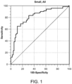
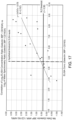
- the discovery and validation curves for NPV and ROR are similar with the discovery curves superior as expected. This demonstrates the reproducibility of performance on an independent set of samples.
- a Discovery classifier rule out threshold of 0.40 achieves NPV of 96% and 90%, whereas ROR is 33% and 23%, for the discovery samples and the validation samples, respectively.
- Final classifier rule threshold of 0.60 achieves NPV of 91% and 90%, whereas ROR is 45% and 43%, for all samples and all samples restricted to be 8mm-20mm, respectively.
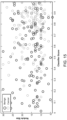
- Figure 9 presents the application of the final classifier to all 247 samples from the discovery and validation sets.
- the intent of Figure 9 is to contrast the clinical risk factors of smoking (measured in pack years) and nodule size (proportional to the size of each circle) to the classifier score assigned to each sample.
- classifier provides molecular information about the disease status of an IPN that is incremental upon risk factors such as nodule size and smoking status. Consequently, it is a clinical tool for physicians to make more informed decisions around the clinical management of an IPN.
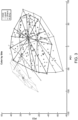
- the 13 classifier proteins are also highly specific to these three networks (lung cancer, response to oxidative stress and lung inflammation). This is summarized in Figure 10 where the classifier proteins (green), transcription factors (blue) and the three merged networks (orange) are depicted. Only ISLR is not connected through these three lung specific networks to the other proteins, although it is connected through cancer networks not specific to cancer. In summary, the modulation of the 13 classifier proteins can be tracked back to a few transcription factors specific to lung cancer, lung inflammation and oxidative stress networks.
- Plasma membranes of each pair of samples were then isolated from the epithelial cells of 30 patients (19 adenocarcinoma, 6 squamous, 5 large cell carcinoma) and endothelial cells of 38 patients (13 adenocarcinoma, 18 squamous, 7 large cell carcinoma) using immune-affinity protocols.
- Golgi apparatus were isolated from each pair of samples from 33 patients (18 adenocarcinoma, 14 squamous, 1 adenosquamous) using isopycnic centrifugation followed by ammonium carbonate extraction. Plasma membrane isolations and Golgi isolations were then analyzed by tandem mass spectrometry to identify proteins overexpressed in lung cancer tissue over normal tissue, for both plasma membranes and Golgi.
- Candidate lung cancer biomarkers were identified from two public and one commercial database: Entrez, NBK3836, UniProt and NextBio. Terminologies were predefined for the database queries which were automated using PERL scripts. The mining was carried out on May 6, 2010 (UniProt), May 17, 2010 (Entrez) and July 8, 2010 (NextBio), respectively. Biomarkers were then assembled and mapped to UniProt identifiers.
- tissue-sourced and literature-source biomarker candidates were required to have evidence of presence in blood. For evidence by mass spectrometry detection, three datasets were used. HUPO9504 contains 9504 human proteins identified by tandem mass spectrometry [13]. HUPO889, a higher confidence subset of HUPO9504, contains 889 human proteins [18]. The PeptideAtlas (November 2009 build) was also used. A biomarker candidate was marked as previously detected if it contained at least one HUPO889, or at least two HUPO9504 peptides, or at least two PeptideAtlas peptides.
- a panel of six normalization transitions were used to normalize raw SRM data for two purposes: (A) to reduce sample-to-sample intensity variations within same study and (B) to reduce intensity variations between different studies.
- a scaling factor was calculated for each sample so that the intensities of the six normalization transitions of the sample were aligned with the corresponding median intensities of all HGS samples.
- N i,s is the intensity of a normalization transition i in sample s
- N ⁇ i the corresponding median intensity of all HGS samples
- the scaling factor for sample s is given by ⁇ / S s , where is the median of the intensity ratios and ⁇ is the median of S s over all samples in the study.
- Endothelial plasma membrane proteins were isolated from normal and tumor lung tissue samples that were obtained from fresh lung resections. Briefly, tissues were washed in buffer and homogenates were prepared by disrupting the tissues with a Polytron. Homogenates were filtered through a 180- ⁇ m mesh and filtrates were centrifuged at 900 x g for 10 min, at 4°C. Supernatants were centrifuged on top of a 50%(w:v) sucrose cushion at 218,000 x g for 60 min at 4°C to pellet the membranes. Pellets were resuspended and treated with micrococcal nuclease.
- Membranes from endothelial cells were incubated with a combination of anti-thrombomodulin, anti-ACE, anti-CD34 and anti-CD144 antibodies, and then centrifuged on top of a 50%(w:v) sucrose cushion at 280,000 x g for 60 min at 4°C. After pellets were resuspended, endothelial cell plasma membranes were isolated using MACS microbeads, treated with potassium iodide to remove cytoplasmic peripheral proteins.
- Epithelial plasma membrane proteins from normal and tumor lung tissue samples were isolated from fresh lung resections. Tissues were washed and homogenates as described above for endothelial plasma membrane proteins preparation. Membranes from epithelial cells were labeled with a combination of anti-ESA, anti-CEA, anti-CD66c and anti-EMA antibodies, and then centrifuged on top of a 50%(w:v) sucrose cushion at 218,000 x g for 60 min at 4°C. Epithelial cell plasma membranes were isolated using MACS microbeads and the eluate was centrifuged at 337,000 x g for 30 minutes at 4°C over a 33%(w:v) sucrose cushion. After removing the supernatant and sucrose cushion, the pellet was resuspended in Laemmli/Urea/DTT.
- Secreted proteins were isolated from normal and tumor lung tissue samples that were isolated from fresh lung resections. Tissues were washed and homogenized using a Polytron homogenization. The density of the homogenates was adjusted to 1.4 M with concentrated sucrose prior to isolating the secretory vesicles by isopycnic centrifugation at 100,000 x g for 2hr at 4°C on a 0.8 and 1.2 M discontinuous sucrose gradient. Vesicles concentrating at the 0.8 / 1.2 M interface were collected and further incubated for 25 minutes with 0.5 M KCl (final concentration) to remove loosely bound peripheral proteins.
- Vesicles were recuperated by ultracentrifugation at 150,000 x g for one hour at 4°C and then opened with 100 mM ammonium carbonate pH 11.0 for 30 minutes at 4°C. Secreted proteins were recovered in the supernatant following a 1-hour ultracentrifugation at 150,000 x g at 4°C.
- a pathway analysis demonstrated that the classifier proteins are likely modulated by a small number of transcription regulators (AHR, NF2L2, MYC, FOS) highly associated with lung cancer, lung inflammation and oxidative stress response networks/processes. Chronic lung inflammation and oxidative stress response are both linked to NSCLC development.
- a strength of the classifier is that it monitors multiple proteins from these multiple lung cancer associated processes. This multiple protein, multiple process survey accounts for the high sensitivity of the classifier for detecting the circulating signature emitted by malignant nodules, and so, high NPV when the classifier calls a nodule benign.
- Candidate Plasma Proteins Two approaches were employed to identify candidate proteins for a lung cancer classifier, including analysis of the proteome of lung tissues with a histopathologic diagnosis of NSCLC and a search of literature databases for lung cancer-associated proteins. All candidate proteins were also assessed for evidence of blood circulation and satisfied one or more requirement(s) for the evidence.
- SRM assays for candidate proteins were developed based on synthetic peptides, as previously described. After identification and synthesis of up to five suitable peptides per protein, SRM triggered MS/MS spectra were collected on a 5500 QTrap ® mass spectrometer for both doubly and triply charged precursor ions. The obtained MS/MS spectra were assigned to individual peptides using MASCOT and with a minimum cutoff score of 15. Up to four transitions per precursor ion were then selected for optimization. The resulting corresponding optimal retention time, declustering potential and collision energy were assembled for all transitions.
- the intensity ratio defined as the ratio between the intensities of the two best transitions of a peptide in the synthetic peptide mixture, was used to assess the specificity of the transitions in a biological sample. Transitions demonstrating interference with other transitions were not selected. A method to ensure the observed transitions corresponded to the peptides and proteins they were intended to measure was developed. In particular, 93% of peptide transitions developed had an error rate below 5%.
- Sample eligibility for the proteomic analysis was based on the satisfaction of the study inclusion and exclusion criteria, including the subject's demographic information; the subject's corresponding lung nodule radiographic characterization by chest CT scan and a maximal linear dimension of 30 mm; and the histopathology of the lung nodule obtained at the time of diagnostic surgical resection, i.e. either NSCLC or a benign, i.e. non-malignant, process.
- Each cancer-benign sample pair was matched, as much as possible among eligible samples, by gender, nodule size ( ⁇ 10 mm), age ( ⁇ 10 years), smoking history pack-years ( ⁇ 20 pack-years), and by center.
- HIPAA Health Insurance Portability and Accountability Act
- the logistic regression classification method was used to combine a panel of transitions into a classifier and to calculate a classification probability score between 0 and 1 for each sample.
- the probability score ( P s ) of a sample was determined as where ⁇ i,s was the logarithmically transformed (base 2), normalized intensity of transition i in sample s, ⁇ i was the corresponding logistic regression coefficient, ⁇ was a classifier-specific constant, and N was the total number of transitions in the classifier.
- a sample was classified as benign if P s was less than a reference value or cancer otherwise.
- the reference value can be increased or decreased depending on the desired NPV.
- the panel of transitions i.e. proteins
- their coefficients the normalization transitions
- classifier coefficient ⁇ and the reference value must be learned (i.e. trained) from the discovery study and then confirmed using the validation study.
- Lung Nodule Classifier Development The goal of the discovery study was to derive a multivariate classifier with a target performance sufficient for clinical utility in the intended use population, i.e. a classifier having an NPV of 90% or higher. This goal was incorporated in the data analysis strategies.
- the classifier development included the following: normalization and filtering of raw SRM-MS data; identification of candidate proteins that occurred with a high frequency in top-performing panels; evaluation of candidate proteins based on SRM-MS signal quality; selection of candidate proteins for the final classifier based on their stability in performance; and training to a logistic regression model to derive the final classifier. Table 28 provides a summary overview of the primary steps.
- Normalization of raw SRM-MS data was performed to reduce sample-to-sample intensity variations using a panel of six endogenous proteins. After data normalization, SRM-MS data were filtered down to transitions having the highest intensities of the corresponding proteins and satisfying the criterion for detection in a minimum of 50% of the cancer or 50% of the benign samples. A total of 125 proteins satisfied these criteria of reproducible detection. Missing values were replaced by half the minimum detected values of the corresponding transitions in all samples.
- MCCV Monte Carlo cross validation
- the cooperative score is defined as its frequency on the 100 high performance panels divided by the expected frequency. Highly cooperative proteins had a score of 1.75 or higher (the corresponding one-sided p value ⁇ 0.05) while non-cooperative proteins had a score of 1 or less. Note that one million panels were sampled to ensure that the 100 top performing panels were exceptional (empirical p value ⁇ 10 -4 ). In addition, panels of size 10 were used in this procedure based on empirical evidence that larger panels did not change the resulting list of cooperative proteins. We also wanted to avoid overfitting the logistic regression model. In total, 36 cooperative proteins were identified, including 15 highly cooperative proteins.
- Remaining candidate proteins were then evaluated in an iterative, stepwise procedure to derive the final classifier.
- MCCV was performed using a holdout rate of 20% and 104 sample permutations to train the remaining candidate proteins to a logistic regression model and to assess the variability, i.e. stability, of the coefficient derived for each protein by the model.
- the protein having the least stable coefficient was identified and removed.
- Proteins for the final classifier were identified when the corresponding partial AUC was optimal. Seven of the 13 proteins in the final classifier were highly cooperative.
- Proteins in the final classifier were further trained to a logistic regression model by MCCV with a holdout rate of 20% and 2x10 4 sample permutations.
- Lung Nodule Classifier Validation The design of the validation study was identical to that of the discovery study, but involved K2-EDTA plasma samples associated with independent subjects and independent lung nodules not evaluated in the discovery study. Additional specimens were obtained from Vanderbilt University (Nashville, TN) with similar requirements for patient consent, IRB approval, and satisfaction of HIPAA requirements. Of the 104 total cancer and benign samples in the validation study, half were analyzed immediately after the discovery study, while the other half was analyzed later. The study was powered to observe the expected 95% confidence interval (CI) of NPV being 90 ⁇ 8%.
- CI 95% confidence interval
- the raw SRM-MS dataset in the validation study was normalized in the same way as the discovery dataset. Variability between the discovery and the validation studies was mitigated by utilizing human plasma standard (HPS) samples in both studies as external calibrator. Missing data in the validation study were then replaced by half the minimum detected values of the corresponding transitions in the discovery study. Transition intensities were applied to the logistic regression model of the final classifier learned previously in the training phase, from which classifier scores were assigned to individual samples. The performance of the lung nodule classifier on the validation samples was then assessed based on the classifier scores.
- HPS human plasma standard
- IPA Pathway Analysis Standard parameters were used. Specifically, in the search for nuclear transcription regulators, requirements were p-value ⁇ 0.01 with a minimum of 3 proteins modulated. Significance was determined using a right-tailed Fisher's exact test using the IPA Knowledge Database as background.
- Candidate Biomarkers Identified by Tissue Proteomics. Specimens of resected NSCLC (adenocarcinoma, squamous cell and large cell) lung tumors and non-adjacent normal tissue in the same lobe were obtained from patients who provided informed consent in studies approved by the Ethics Review Boards at the Centre Hospitalier de l'liable de Quebec and the McGill University Health Centre.
- Membrane proteins from endothelial cells or epithelial cells and secreted proteins were isolated from normal or tumor tissues from fresh lung resections after washing in buffer and disruption with a Polytron to prepare homogenates.
- the cell membrane protocol included filtration using 180 ⁇ m mesh and centrifugation at 900 x g for 10 min at 4°C, supernatants prior to layering on 50% (w:v) sucrose and centrifugation at 218,000 x g for 1 h at 4°C to pellet the membranes.
- Membrane pellets were resuspended and treated with micrococcal nuclease, and incubated with the following antibodies specified by plasma membrane type: endothelial membranes (anti-thrombomodulin, anti-ACE, anti-CD34 and anti-CD144 antibodies); epithelial membranes (anti-ESA, anti-CEA, anti-CD66c and anti-EMA antibodies), prior to centrifugation on top of a 50%(w:v) sucrose cushion at 280,000 x g (endothelial) or 218,000 x g (epithelial) for 1 h at 4°C. After pellet resuspension, plasma membranes were isolated using MACS microbeads.
- Xpresys Lung has been developed to differentiate benign from malignant lung nodules.
- Xpresys Lung is a blood test for proteins that combines expertise in proteomics and computer science using large data sets. Mass spectrometry has been employed as a technology for molecular diagnostics for decades and recent advances in instrumentation allows measurement of hundreds of proteins at a time. Cancers secrete and shed proteins that are different from normal cells and some of these proteins circulate in the blood. InDi started with 388 protein candidates and blood samples stored from both patients with benign and malignant lung nodules. The initial analyses discovered and validated a predictor for benign nodules using a combination of 11 proteins.
Landscapes
- Health & Medical Sciences (AREA)
- Life Sciences & Earth Sciences (AREA)
- Immunology (AREA)
- Engineering & Computer Science (AREA)
- Molecular Biology (AREA)
- Biomedical Technology (AREA)
- Chemical & Material Sciences (AREA)
- Hematology (AREA)
- Urology & Nephrology (AREA)
- Biotechnology (AREA)
- Microbiology (AREA)
- Cell Biology (AREA)
- Food Science & Technology (AREA)
- Medicinal Chemistry (AREA)
- Physics & Mathematics (AREA)
- Analytical Chemistry (AREA)
- Biochemistry (AREA)
- General Health & Medical Sciences (AREA)
- General Physics & Mathematics (AREA)
- Pathology (AREA)
- Investigating Or Analysing Biological Materials (AREA)
- Other Investigation Or Analysis Of Materials By Electrical Means (AREA)
Claims (9)
- Procédé de détermination de la probabilité qu'un nodule pulmonaire chez un sujet ne soit pas un cancer du poumon, comprenant :(a) la mesure des niveaux d'expression d'un panel de protéines présentes dans un échantillon de sang obtenu du sujet, dans lequel le panel de protéines est constitué de LG3BP et de C163A ;(b) le calcul d'une probabilité de score de cancer du poumon sur la base des niveaux d'expression du panel de protéines de l'étape (a) et de facteurs de risque cliniques comprenant l'âge, les antécédents de tabagisme, et la taille de nodule ; et(c) l'exclusion du cancer du poumon pour le sujet si le score dans l'étape (b) est inférieur à un score prédéterminé ;dans lequel les niveaux d'expression du panel de protéines sont mesurés par un dosage immunologique.
- Procédé selon la revendication 1, dans lequel les niveaux d'expression du panel de protéines sont mesurés par un dosage immuno-enzymatique (ELISA) comprenant la mise en contact de l'échantillon de sang avec un anticorps LG3BP et un anticorps C163A.
- Procédé selon la revendication 1 ou la revendication 2, dans lequel lorsque le cancer du poumon est exclu, le sujet ne reçoit pas de protocole de traitement.
- Procédé selon la revendication 3, dans lequel le protocole de traitement est un test de fonction pulmonaire (TFP), une imagerie pulmonaire, une biopsie, une chirurgie, une chimiothérapie, une radiothérapie ou toute combinaison de ceux-ci.
- Procédé selon la revendication 4, où l'imagerie pulmonaire est une radiographie, une tomodensitométrie thoracique (TDM), ou une tomodensitométrie par émission de positons (TEP).
- Procédé selon la revendication 1 ou la revendication 2, dans lequel le nodule pulmonaire a un diamètre inférieur ou égal à 3 cm.
- Procédé selon la revendication 1 ou la revendication 2, dans lequel le nodule pulmonaire a un diamètre d'environ 0,8 cm à 3,0 cm.
- Procédé selon la revendication 1 ou la revendication 2, dans lequel le sujet est à risque de développer un cancer du poumon.
- Procédé selon la revendication 1 ou la revendication 2, dans lequel le sujet est âgé de 40 ans ou plus.
Applications Claiming Priority (2)
| Application Number | Priority Date | Filing Date | Title |
|---|---|---|---|
| US15/786,924 US11913957B2 (en) | 2011-12-21 | 2017-10-18 | Compositions, methods and kits for diagnosis of lung cancer |
| PCT/US2018/056570 WO2019079635A1 (fr) | 2017-10-18 | 2018-10-18 | Compositions, méthodes et trousses pour le diagnostic du cancer du poumon |
Publications (3)
| Publication Number | Publication Date |
|---|---|
| EP3698144A1 EP3698144A1 (fr) | 2020-08-26 |
| EP3698144A4 EP3698144A4 (fr) | 2021-07-14 |
| EP3698144B1 true EP3698144B1 (fr) | 2025-07-09 |
Family
ID=66173523
Family Applications (1)
| Application Number | Title | Priority Date | Filing Date |
|---|---|---|---|
| EP18868355.1A Active EP3698144B1 (fr) | 2017-10-18 | 2018-10-18 | Compositions, méthodes et trousses pour le diagnostic du cancer du poumon |
Country Status (4)
| Country | Link |
|---|---|
| EP (1) | EP3698144B1 (fr) |
| CN (1) | CN111788486A (fr) |
| ES (1) | ES3043532T3 (fr) |
| WO (1) | WO2019079635A1 (fr) |
Families Citing this family (7)
| Publication number | Priority date | Publication date | Assignee | Title |
|---|---|---|---|---|
| US11913957B2 (en) | 2011-12-21 | 2024-02-27 | Biodesix, Inc. | Compositions, methods and kits for diagnosis of lung cancer |
| CN113388683A (zh) * | 2021-06-29 | 2021-09-14 | 北京泱深生物信息技术有限公司 | 与肺癌预后相关的生物标志物及其应用 |
| US11542340B1 (en) | 2021-10-29 | 2023-01-03 | Biodesix, Inc. | Antibodies targeting pulmonary nodule specific biomarkers and uses thereof |
| EP4494152A4 (fr) * | 2023-06-09 | 2025-10-08 | Seekin Inc Shenzhen China | Méthodes pour détecter le cancer |
| GB202310137D0 (en) * | 2023-07-03 | 2023-08-16 | Belgian Volition Srl | Method for the detection of cancer |
| CN117347643B (zh) * | 2023-12-05 | 2024-02-06 | 成都泰莱生物科技有限公司 | 用于判断肺部结节良恶性的代谢标志物组合及其筛选方法和应用 |
| CN118275695B (zh) * | 2024-05-07 | 2024-12-13 | 烟台至公生物医药科技有限公司 | 一种基于质谱检测的肿瘤组织判别方法及系统 |
Family Cites Families (6)
| Publication number | Priority date | Publication date | Assignee | Title |
|---|---|---|---|---|
| EP2295570A1 (fr) * | 2005-07-27 | 2011-03-16 | Oncotherapy Science, Inc. | Procédé de diagnostic du cancer pulmonaire à petites cellules |
| CA2860298A1 (fr) * | 2011-12-21 | 2013-06-27 | Biodesix, Inc. | Compositions, procedes et trousses pour le diagnostic du cancer du poumon |
| WO2014100717A2 (fr) * | 2012-12-21 | 2014-06-26 | Integrated Diagnostics, Inc. | Compositions, procédés et kits pour le diagnostic d'un cancer du poumon |
| US9297805B2 (en) * | 2013-07-26 | 2016-03-29 | Integrated Diagnostics, Inc. | Compositions, methods and kits for diagnosis of lung cancer |
| US20150087728A1 (en) * | 2013-09-20 | 2015-03-26 | Integrated Diagnostics, Inc. | Compositions, methods and kits for diagnosis of lung cancer |
| US20170269090A1 (en) * | 2016-03-18 | 2017-09-21 | Integrated Diagnostics, Inc. | Compositions, methods and kits for diagnosis of lung cancer |
-
2018
- 2018-10-18 EP EP18868355.1A patent/EP3698144B1/fr active Active
- 2018-10-18 ES ES18868355T patent/ES3043532T3/es active Active
- 2018-10-18 WO PCT/US2018/056570 patent/WO2019079635A1/fr not_active Ceased
- 2018-10-18 CN CN201880081583.1A patent/CN111788486A/zh active Pending
Non-Patent Citations (1)
| Title |
|---|
| HOOFNAGLE A N ET AL: "The fundamental flaws of immunoassays and potential solutions using tandem mass spectrometry", JOURNAL OF IMMUNOLOGICAL METHODS, ELSEVIER SCIENCE PUBLISHERS B.V.,AMSTERDAM, NL, vol. 347, no. 1-2, 15 August 2009 (2009-08-15), pages 3 - 11, XP026337366, ISSN: 0022-1759, [retrieved on 20090616], DOI: 10.1016/J.JIM.2009.06.003 * |
Also Published As
| Publication number | Publication date |
|---|---|
| CN111788486A (zh) | 2020-10-16 |
| ES3043532T3 (en) | 2025-11-25 |
| EP3698144A4 (fr) | 2021-07-14 |
| WO2019079635A1 (fr) | 2019-04-25 |
| EP3698144A1 (fr) | 2020-08-26 |
Similar Documents
| Publication | Publication Date | Title |
|---|---|---|
| EP3698144B1 (fr) | Compositions, méthodes et trousses pour le diagnostic du cancer du poumon | |
| JP6082026B2 (ja) | 肺癌診断用の組成物、方法及びキット | |
| US12247980B2 (en) | Compositions, methods and kits for diagnosis of lung cancer | |
| US9304137B2 (en) | Compositions, methods and kits for diagnosis of lung cancer | |
| US20240159753A1 (en) | Methods for the detection and treatment of lung cancer | |
| US11193935B2 (en) | Compositions, methods and kits for diagnosis of lung cancer | |
| WO2017192965A2 (fr) | Compositions, méthodes et trousses pour le diagnostic du cancer du poumon | |
| CN106461647A (zh) | 用于检测结肠直肠肿瘤的蛋白质生物标志物谱 | |
| WO2014100717A2 (fr) | Compositions, procédés et kits pour le diagnostic d'un cancer du poumon | |
| Widlak et al. | Serum mass profile signature as a biomarker of early lung cancer | |
| US20170168058A1 (en) | Compositions, methods and kits for diagnosis of lung cancer | |
| WO2024107923A1 (fr) | Méthodes pour la détection et le traitement du cancer du poumon | |
| US20170269090A1 (en) | Compositions, methods and kits for diagnosis of lung cancer | |
| Brown et al. | High-sensitivity multicancer detection of stage 1 cancer in dogs | |
| HK40039966A (en) | Compositions, methods and kits for diagnosis of lung cancer | |
| Hu et al. | Aspergillus-specific Immunoglobulin G Seropositivity and Lung Function Decline in Patients with Chronic Lung Diseases: A Prospective Cohort Study | |
| HK40012915A (en) | Compositions, methods and kits for diagnosis of lung cancer | |
| HK1203091B (en) | Methods for diagnosis of lung cancer |
Legal Events
| Date | Code | Title | Description |
|---|---|---|---|
| STAA | Information on the status of an ep patent application or granted ep patent |
Free format text: STATUS: THE INTERNATIONAL PUBLICATION HAS BEEN MADE |
|
| PUAI | Public reference made under article 153(3) epc to a published international application that has entered the european phase |
Free format text: ORIGINAL CODE: 0009012 |
|
| STAA | Information on the status of an ep patent application or granted ep patent |
Free format text: STATUS: REQUEST FOR EXAMINATION WAS MADE |
|
| 17P | Request for examination filed |
Effective date: 20200518 |
|
| AK | Designated contracting states |
Kind code of ref document: A1 Designated state(s): AL AT BE BG CH CY CZ DE DK EE ES FI FR GB GR HR HU IE IS IT LI LT LU LV MC MK MT NL NO PL PT RO RS SE SI SK SM TR |
|
| AX | Request for extension of the european patent |
Extension state: BA ME |
|
| DAV | Request for validation of the european patent (deleted) | ||
| DAX | Request for extension of the european patent (deleted) | ||
| A4 | Supplementary search report drawn up and despatched |
Effective date: 20210616 |
|
| RIC1 | Information provided on ipc code assigned before grant |
Ipc: G01N 33/68 20060101AFI20210610BHEP Ipc: G01N 33/574 20060101ALI20210610BHEP |
|
| STAA | Information on the status of an ep patent application or granted ep patent |
Free format text: STATUS: EXAMINATION IS IN PROGRESS |
|
| 17Q | First examination report despatched |
Effective date: 20220902 |
|
| P01 | Opt-out of the competence of the unified patent court (upc) registered |
Effective date: 20230907 |
|
| RAP3 | Party data changed (applicant data changed or rights of an application transferred) |
Owner name: BIODESIX, INC. |
|
| GRAP | Despatch of communication of intention to grant a patent |
Free format text: ORIGINAL CODE: EPIDOSNIGR1 |
|
| STAA | Information on the status of an ep patent application or granted ep patent |
Free format text: STATUS: GRANT OF PATENT IS INTENDED |
|
| INTG | Intention to grant announced |
Effective date: 20241216 |
|
| GRAJ | Information related to disapproval of communication of intention to grant by the applicant or resumption of examination proceedings by the epo deleted |
Free format text: ORIGINAL CODE: EPIDOSDIGR1 |
|
| STAA | Information on the status of an ep patent application or granted ep patent |
Free format text: STATUS: EXAMINATION IS IN PROGRESS |
|
| GRAP | Despatch of communication of intention to grant a patent |
Free format text: ORIGINAL CODE: EPIDOSNIGR1 |
|
| STAA | Information on the status of an ep patent application or granted ep patent |
Free format text: STATUS: GRANT OF PATENT IS INTENDED |
|
| INTG | Intention to grant announced |
Effective date: 20250307 |
|
| GRAS | Grant fee paid |
Free format text: ORIGINAL CODE: EPIDOSNIGR3 |
|
| GRAA | (expected) grant |
Free format text: ORIGINAL CODE: 0009210 |
|
| STAA | Information on the status of an ep patent application or granted ep patent |
Free format text: STATUS: THE PATENT HAS BEEN GRANTED |
|
| AK | Designated contracting states |
Kind code of ref document: B1 Designated state(s): AL AT BE BG CH CY CZ DE DK EE ES FI FR GB GR HR HU IE IS IT LI LT LU LV MC MK MT NL NO PL PT RO RS SE SI SK SM TR |
|
| REG | Reference to a national code |
Ref country code: GB Ref legal event code: FG4D |
|
| REG | Reference to a national code |
Ref country code: CH Ref legal event code: EP |
|
| REG | Reference to a national code |
Ref country code: IE Ref legal event code: FG4D |
|
| REG | Reference to a national code |
Ref country code: DE Ref legal event code: R096 Ref document number: 602018083490 Country of ref document: DE |
|
| PGFP | Annual fee paid to national office [announced via postgrant information from national office to epo] |
Ref country code: IT Payment date: 20250922 Year of fee payment: 8 |
|
| PGFP | Annual fee paid to national office [announced via postgrant information from national office to epo] |
Ref country code: GB Payment date: 20250828 Year of fee payment: 8 |
|
| PGFP | Annual fee paid to national office [announced via postgrant information from national office to epo] |
Ref country code: FR Payment date: 20250908 Year of fee payment: 8 |
|
| REG | Reference to a national code |
Ref country code: CH Ref legal event code: R17 Free format text: ST27 STATUS EVENT CODE: U-0-0-R10-R17 (AS PROVIDED BY THE NATIONAL OFFICE) Effective date: 20251024 |
|
| REG | Reference to a national code |
Ref country code: CH Ref legal event code: U11 Free format text: ST27 STATUS EVENT CODE: U-0-0-U10-U11 (AS PROVIDED BY THE NATIONAL OFFICE) Effective date: 20251101 |
|
| REG | Reference to a national code |
Ref country code: NL Ref legal event code: MP Effective date: 20250709 |
|
| REG | Reference to a national code |
Ref country code: ES Ref legal event code: FG2A Ref document number: 3043532 Country of ref document: ES Kind code of ref document: T3 Effective date: 20251125 |
|
| PG25 | Lapsed in a contracting state [announced via postgrant information from national office to epo] |
Ref country code: PT Free format text: LAPSE BECAUSE OF FAILURE TO SUBMIT A TRANSLATION OF THE DESCRIPTION OR TO PAY THE FEE WITHIN THE PRESCRIBED TIME-LIMIT Effective date: 20251110 |
|
| PG25 | Lapsed in a contracting state [announced via postgrant information from national office to epo] |
Ref country code: NL Free format text: LAPSE BECAUSE OF FAILURE TO SUBMIT A TRANSLATION OF THE DESCRIPTION OR TO PAY THE FEE WITHIN THE PRESCRIBED TIME-LIMIT Effective date: 20250709 |
|
| REG | Reference to a national code |
Ref country code: AT Ref legal event code: MK05 Ref document number: 1812250 Country of ref document: AT Kind code of ref document: T Effective date: 20250709 |
|
| PG25 | Lapsed in a contracting state [announced via postgrant information from national office to epo] |
Ref country code: IS Free format text: LAPSE BECAUSE OF FAILURE TO SUBMIT A TRANSLATION OF THE DESCRIPTION OR TO PAY THE FEE WITHIN THE PRESCRIBED TIME-LIMIT Effective date: 20251109 |
|
| PGFP | Annual fee paid to national office [announced via postgrant information from national office to epo] |
Ref country code: DE Payment date: 20250827 Year of fee payment: 8 |
|
| PG25 | Lapsed in a contracting state [announced via postgrant information from national office to epo] |
Ref country code: NO Free format text: LAPSE BECAUSE OF FAILURE TO SUBMIT A TRANSLATION OF THE DESCRIPTION OR TO PAY THE FEE WITHIN THE PRESCRIBED TIME-LIMIT Effective date: 20251009 |
|
| REG | Reference to a national code |
Ref country code: LT Ref legal event code: MG9D |
|
| PG25 | Lapsed in a contracting state [announced via postgrant information from national office to epo] |
Ref country code: AT Free format text: LAPSE BECAUSE OF FAILURE TO SUBMIT A TRANSLATION OF THE DESCRIPTION OR TO PAY THE FEE WITHIN THE PRESCRIBED TIME-LIMIT Effective date: 20250709 |
|
| PG25 | Lapsed in a contracting state [announced via postgrant information from national office to epo] |
Ref country code: FI Free format text: LAPSE BECAUSE OF FAILURE TO SUBMIT A TRANSLATION OF THE DESCRIPTION OR TO PAY THE FEE WITHIN THE PRESCRIBED TIME-LIMIT Effective date: 20250709 |
|
| PG25 | Lapsed in a contracting state [announced via postgrant information from national office to epo] |
Ref country code: HR Free format text: LAPSE BECAUSE OF FAILURE TO SUBMIT A TRANSLATION OF THE DESCRIPTION OR TO PAY THE FEE WITHIN THE PRESCRIBED TIME-LIMIT Effective date: 20250709 |
|
| PG25 | Lapsed in a contracting state [announced via postgrant information from national office to epo] |
Ref country code: GR Free format text: LAPSE BECAUSE OF FAILURE TO SUBMIT A TRANSLATION OF THE DESCRIPTION OR TO PAY THE FEE WITHIN THE PRESCRIBED TIME-LIMIT Effective date: 20251010 |
|
| PGFP | Annual fee paid to national office [announced via postgrant information from national office to epo] |
Ref country code: CH Payment date: 20251101 Year of fee payment: 8 |
|
| PG25 | Lapsed in a contracting state [announced via postgrant information from national office to epo] |
Ref country code: SE Free format text: LAPSE BECAUSE OF FAILURE TO SUBMIT A TRANSLATION OF THE DESCRIPTION OR TO PAY THE FEE WITHIN THE PRESCRIBED TIME-LIMIT Effective date: 20250709 |
|
| PG25 | Lapsed in a contracting state [announced via postgrant information from national office to epo] |
Ref country code: LV Free format text: LAPSE BECAUSE OF FAILURE TO SUBMIT A TRANSLATION OF THE DESCRIPTION OR TO PAY THE FEE WITHIN THE PRESCRIBED TIME-LIMIT Effective date: 20250709 |
|
| PG25 | Lapsed in a contracting state [announced via postgrant information from national office to epo] |
Ref country code: BG Free format text: LAPSE BECAUSE OF FAILURE TO SUBMIT A TRANSLATION OF THE DESCRIPTION OR TO PAY THE FEE WITHIN THE PRESCRIBED TIME-LIMIT Effective date: 20250709 Ref country code: PL Free format text: LAPSE BECAUSE OF FAILURE TO SUBMIT A TRANSLATION OF THE DESCRIPTION OR TO PAY THE FEE WITHIN THE PRESCRIBED TIME-LIMIT Effective date: 20250709 |
|
| PG25 | Lapsed in a contracting state [announced via postgrant information from national office to epo] |
Ref country code: RS Free format text: LAPSE BECAUSE OF FAILURE TO SUBMIT A TRANSLATION OF THE DESCRIPTION OR TO PAY THE FEE WITHIN THE PRESCRIBED TIME-LIMIT Effective date: 20251009 |
|
| PGFP | Annual fee paid to national office [announced via postgrant information from national office to epo] |
Ref country code: ES Payment date: 20251106 Year of fee payment: 8 |
|
| PG25 | Lapsed in a contracting state [announced via postgrant information from national office to epo] |
Ref country code: RO Free format text: LAPSE BECAUSE OF FAILURE TO SUBMIT A TRANSLATION OF THE DESCRIPTION OR TO PAY THE FEE WITHIN THE PRESCRIBED TIME-LIMIT Effective date: 20250709 |
|
| PG25 | Lapsed in a contracting state [announced via postgrant information from national office to epo] |
Ref country code: SM Free format text: LAPSE BECAUSE OF FAILURE TO SUBMIT A TRANSLATION OF THE DESCRIPTION OR TO PAY THE FEE WITHIN THE PRESCRIBED TIME-LIMIT Effective date: 20250709 |
|
| PG25 | Lapsed in a contracting state [announced via postgrant information from national office to epo] |
Ref country code: DK Free format text: LAPSE BECAUSE OF FAILURE TO SUBMIT A TRANSLATION OF THE DESCRIPTION OR TO PAY THE FEE WITHIN THE PRESCRIBED TIME-LIMIT Effective date: 20250709 |
|
| PG25 | Lapsed in a contracting state [announced via postgrant information from national office to epo] |
Ref country code: CZ Free format text: LAPSE BECAUSE OF FAILURE TO SUBMIT A TRANSLATION OF THE DESCRIPTION OR TO PAY THE FEE WITHIN THE PRESCRIBED TIME-LIMIT Effective date: 20250709 |
|
| PG25 | Lapsed in a contracting state [announced via postgrant information from national office to epo] |
Ref country code: EE Free format text: LAPSE BECAUSE OF FAILURE TO SUBMIT A TRANSLATION OF THE DESCRIPTION OR TO PAY THE FEE WITHIN THE PRESCRIBED TIME-LIMIT Effective date: 20250709 Ref country code: SK Free format text: LAPSE BECAUSE OF FAILURE TO SUBMIT A TRANSLATION OF THE DESCRIPTION OR TO PAY THE FEE WITHIN THE PRESCRIBED TIME-LIMIT Effective date: 20250709 |