EP3701873B1 - Système et procédé pour indiquer le risque de maladie coronarienne - Google Patents
Système et procédé pour indiquer le risque de maladie coronarienne Download PDFInfo
- Publication number
- EP3701873B1 EP3701873B1 EP20168782.9A EP20168782A EP3701873B1 EP 3701873 B1 EP3701873 B1 EP 3701873B1 EP 20168782 A EP20168782 A EP 20168782A EP 3701873 B1 EP3701873 B1 EP 3701873B1
- Authority
- EP
- European Patent Office
- Prior art keywords
- low frequency
- power
- cad
- frequency power
- periods
- Prior art date
- Legal status (The legal status is an assumption and is not a legal conclusion. Google has not performed a legal analysis and makes no representation as to the accuracy of the status listed.)
- Active
Links
Images




Classifications
-
- A—HUMAN NECESSITIES
- A61—MEDICAL OR VETERINARY SCIENCE; HYGIENE
- A61B—DIAGNOSIS; SURGERY; IDENTIFICATION
- A61B5/00—Measuring for diagnostic purposes; Identification of persons
- A61B5/72—Signal processing specially adapted for physiological signals or for diagnostic purposes
- A61B5/7271—Specific aspects of physiological measurement analysis
- A61B5/7275—Determining trends in physiological measurement data; Predicting development of a medical condition based on physiological measurements, e.g. determining a risk factor
-
- A—HUMAN NECESSITIES
- A61—MEDICAL OR VETERINARY SCIENCE; HYGIENE
- A61B—DIAGNOSIS; SURGERY; IDENTIFICATION
- A61B7/00—Instruments for auscultation
- A61B7/02—Stethoscopes
- A61B7/04—Electric stethoscopes
-
- A—HUMAN NECESSITIES
- A61—MEDICAL OR VETERINARY SCIENCE; HYGIENE
- A61B—DIAGNOSIS; SURGERY; IDENTIFICATION
- A61B5/00—Measuring for diagnostic purposes; Identification of persons
- A61B5/02—Detecting, measuring or recording for evaluating the cardiovascular system, e.g. pulse, heart rate, blood pressure or blood flow
- A61B5/02007—Evaluating blood vessel condition, e.g. elasticity, compliance
-
- A—HUMAN NECESSITIES
- A61—MEDICAL OR VETERINARY SCIENCE; HYGIENE
- A61B—DIAGNOSIS; SURGERY; IDENTIFICATION
- A61B5/00—Measuring for diagnostic purposes; Identification of persons
- A61B5/02—Detecting, measuring or recording for evaluating the cardiovascular system, e.g. pulse, heart rate, blood pressure or blood flow
- A61B5/024—Measuring pulse rate or heart rate
-
- A—HUMAN NECESSITIES
- A61—MEDICAL OR VETERINARY SCIENCE; HYGIENE
- A61B—DIAGNOSIS; SURGERY; IDENTIFICATION
- A61B5/00—Measuring for diagnostic purposes; Identification of persons
- A61B5/72—Signal processing specially adapted for physiological signals or for diagnostic purposes
- A61B5/7235—Details of waveform analysis
- A61B5/725—Details of waveform analysis using specific filters therefor, e.g. Kalman or adaptive filters
Definitions
- the present invention relates to a system and a method for indicating a risk of coronary artery disease (CAD), and in particular to an electronic stethoscope configured to indicate a risk of CAD from measured acoustic signals.
- CAD coronary artery disease
- Coronary artery disease is a condition in which plaque builds up inside the coronary arteries. These arteries supply the heart muscle with oxygen-rich blood. Plaque narrows the arteries and reduces blood flow to the heart, which can cause angina or a heart attack. Over time, coronary artery disease may weaken the heart muscle and lead to heart failure and arrhythmias. Coronary heart disease is the most common type of heart disease. Lifestyle changes, medicines and/or medical procedures can effectively prevent or treat the disease in most people.
- a stethoscope is often used to examine the sound of the heart. Although the role of the stethoscope in the modern clinic seems to be fading, new electronic stethoscopes with integrated diagnostic algorithms might alter the trend and again expand the clinical potential of the stethoscope.
- An example of such a type of algorithms is a detector of coronary artery disease.
- S 1 is usually the loudest heart sound and is the first heart sound during ventricular contraction.
- S 1 is often described as a "lubb" sound.
- S 1 occurs at the beginning of ventricular systole and relates to the closure of atrioventicular valves between the atria and the ventricles.
- S2 is often described as a "dubb" sound.
- S2 occurs at the beginning of the diastole and relates to the closing of the semilunar valves separating the aorta and pulmonary artery from the left and right ventricles, respectively.
- S1 and S2 can be easily heard with a stethoscope ("normal heart sounds").
- S3 and S4 can usually not be heard in the normal heart ("abnormal heart sounds") of a person over 40 years old.
- S3, also referred to as "ventricular gallop” occurs in the early diastolic period and is caused by the ventricular wall distending to the point it reaches its elastic limit.
- S4, also referred to as "atrial gallop” occurs near the end of atrial contraction and is also caused by the ventricular wall distending until it reaches its elastic limit.
- Heart sounds can be used to augment the diagnosis and to help assess the severity of important types of cardiac disease. For example, after age 40, S3 can indicate congestive heart failure, and S4 can indicate hypertension, acute myocardial infarction, or coronary artery disease. Unfortunately, studies have shown that even highly experienced physicians do not reliably detect important heart sounds. Therefore various systems have been developed to support physicians in detecting possible heart diseases.
- US-7,096,060 relates to a method and system for automatically detecting heart sounds.
- the sound system may use ECG data to identify various locations, e.g. an R peak, within a beat and use those locations to assist in detection of heart sounds.
- ECG data may be used to identify various locations, e.g. an R peak, within a beat and use those locations to assist in detection of heart sounds.
- US-5,159,932 relates to a myocardial ischemia detection system for non invasively monitoring the motion of the patient's heart, to detect and display ischemia induced variations in the heart's motion which indicate coronary artery disease.
- an accelerometer is used as a compression wave transducer which must have a wide bandwidth and should exhibit a flat frequency response from 0.025 Hz to 800 Hz.
- the purpose of the compression wave transducer is to translate with high accuracy the very low amplitude mechanical motion at the surface of the patient into an electrical signal for further processing.
- waveforms including identified morphological features are processed, displayed and used for diagnosis along with a reference ECG.
- US-2009/0177107 relates to an electronic stethoscope system that automatically detects coronary artery disease in patients.
- the system uses an electronic stethoscope to record acoustic data from the fourth left intercostal space of a patient.
- a processing technique is then applied in order to filter the data and produce Fast Fourier Transform (FFT) data of magnitude versus frequency. If a bell curve is identified in the data between a predefined frequency range (e.g. 50 and 80 Hz) with a peak magnitude of greater than a predefined threshold, e.g. 2.5 units, the system automatically provides an output indicating that the patient is likely to have 50 to 99 percent stenosis of the coronary artery.
- a predefined frequency range e.g. 50 and 80 Hz
- a predefined threshold e.g. 2.5 units
- diastolic sounds from CAD patients differ from non-CAD patients. This is described in US-5,036,857 . The difference is likely caused by weak murmurs originating from post-stenotic turbulence in the coronary arteries.
- One method for quantifying the difference in diastolic heart sounds is based on autoregressive (AR) models of the diastolic sounds based upon the fact that the pole magnitudes in AR-models of the diastolic heart sound in CAD patients differed from non-CAD patients.
- AR autoregressive
- US-6,048,319 relates to a non-invasive acoustic screening device for detecting coronary stenosis by identifying S 1 and S2 heart sounds, heart rate, and determines the diastolic interval of the subject and thereafter estimates the acoustic energy levels within a 2 octave band around approximately 20 Hz during diastole. Based upon such estimation, a diagnosis can be rendered as to the presence and degree of stenosis from the coronary artery.
- the object of the present invention is to provide an improved analysis system and method in order to improve diagnosis of CAD.
- the above-mentioned object is achieved by a system for detection of frequency power for diagnosing of coronary artery disease (CAD) according to independent claim 1.
- the system comprises an acoustic sensor adapted to be placed on the chest of a patient.
- the system further comprises: at least one memory adapted to store acoustic signals S A from the acoustic sensor; and a control unit adapted to receive said acoustic signals S A .
- the parts of the system can be placed separately, e.g. the sensor and memory may be part of an electronic stethoscope capable of sending recorded data to an external unit comprising said control unit, for further processing of the data.
- the object is achieved by a method for detecting frequency power according to independent claim 7.
- the method according to the invention can be used to improve other methods for diagnosing CAD.
- the detection of frequency power in relation to coronary artery disease comprises the following steps:
- the low frequent components of the heart sounds are most likely related to movements of the ventricle.
- the estimation of the power in the low frequency band in the diastolic or systolic periods can be estimated using one of the following methods:
- a median low frequency band power is preferably calculated for several heart-beats.
- the low frequency power measure may also be corrected against differences caused by body mass index (BMI), gender, age, disease or similar estimates of the tissue dampening factor.
- BMI body mass index
- a risk of CAD may then be determined by comparing the low frequency power measure with a reference value. An increased low frequency power measure compared to the reference value is according to studies an indication that the patient has CAD.
- Abdominal noise was the least common noise source, present in 10.8% of the recordings.
- the best pole, with respect to detection of CAD, extracted from the 250-1000 Hz frequency band was most sensitive to noise, since the AUC dropped from 0.70 in recordings without noise to 0.57 when noisy recordings were included, whereas the best pole from the 25-250 Hz frequency band was relatively robust against noise, since the AUC dropped from 0.73 to only 0.70 when noisy recordings were included.
- the study according to the examples in the description demonstrated that noise contamination is a frequent problem and that features from lower frequency bands are more robust against noise than features from higher frequency bands.
- Both low frequency and high frequency sounds are produced during the normal heart cycle. Increased high frequency heart sounds have been found associated with coronary artery disease and originating from the vibrating stenosed coronary arteries. Increased low frequency heart sounds have not been found to originate from the stenotic coronary arteries, and are as such independent in origin.
- the combination of a low-frequency power spectrum and a high-frequency power spectrum could provide an even more certain coronary artery disease diagnosis, than either the low frequency power spectrum or the high frequency power spectrum alone.
- the low frequency signals are less sensitive to disturbances than the high frequency signals, thus, information may be retracted from the low frequency signals that is not possible from the high frequency signals contributing to a more secure and accurate diagnose of CAD.
- Figure 1 shows a system for detection of low frequency power.
- the system can be used for diagnosing of coronary artery disease (CAD), and in particular for classification of CAD together with other indicators.
- CAD coronary artery disease
- the system comprises an acoustic sensor adapted to be placed on the chest of a patient, and to generate acoustic signals S A .
- the acoustic sensor comprises e.g. a microphone, an accelerometer or an optical beam device.
- the sensor may be adapted to sense acoustic sounds down to 5 Hz or may be adapted to sense acoustic sounds down to 20 Hz.
- the system further comprises at least one memory adapted to store acoustic signals S A from the acoustic sensor; and a control unit adapted to receive said acoustic signals S A .
- the acoustic sensor and the memory may be incorporated in a separate device e.g. an electronic stethoscope adapted to transmit the sensed acoustic signals by wire or wirelessly to an external unit comprising said control unit.
- a separate device i.e. the stethoscope
- the separate device i.e. the stethoscope, then comprises a transmitting unit adapted to transmit the signals (not shown in the figure).
- the signals may be processed in the external unit instead of in the stethoscope.
- the memory may be a detachable memory such as a USB-device, which can be plugged into the external unit.
- the control unit comprises an identification unit adapted to identify diastolic or systolic periods in a predetermined time period of the stored acoustic signals S A , and to generate a period signal S P indicating the identified periods.
- the predetermined time period may include a predetermined number of diastolic or systolic periods.
- S2-sound occurs in the beginning of the diastolic period and relates to the closing of the semilunar valves separating the aorta and pulmonary artery from the left and right ventricles, respectively.
- the S 1-sound occurs in the beginning of ventricular systole and relates to the closure of atrioventicular valves between the atria and the ventricles.
- the control unit further comprises a filtering unit adapted to apply at least one filter to the identified periods and to generate a low frequency band signal S LFB indicating low frequency bands of the identified periods.
- the filtering unit may also be applied when the filtering is performed by using the power spectrum density (PSD) method (as discussed in example 8) if the low frequency energy is estimated using PSD where at least one filter is applied to said identified periods and to empathize frequencies relevant for estimation of low frequency power.
- PSD power spectrum density
- the heart sounds in the lower frequency range of the periods have according to studies shown to be less sensitive to disturbances than the heart sounds in other frequency ranges.
- the low frequency band in between 5-70 Hz, preferably 10-60 Hz, more preferably 15-50 Hz and most preferably 20-40 Hz.
- the control unit comprises a calculation unit adapted to estimate the power in the low frequency band of the identified periods.
- the control unit is adapted to estimate the power by band pass filtering said identified periods using a 5-70 Hz, preferably 10-60 Hz, more preferably 15-50 Hz, and most preferably 20-40 Hz band pass filter frequency and calculating the variance during said periods.
- the filtering unit band pass filters the identified periods with a between 5-70 Hz, preferably 10-60 Hz, more preferably 15-50 Hz and most preferably 20-40 Hz band pass filter frequency, and the calculation unit calculates the variance during the band pass filtered period.
- the calculated variance is named low frequency power.
- the examples 1-7 relate to results from two different studies, one study based on recordings obtained using a standard electronic stethoscope (3M Littmann E4000) and another study using a custom made sensor adapted to sense low frequencies.
- Figure 3 shows recordings of heart sounds with an electronic stethoscope of two different patients, filtered with a 20 to 50 Hz band pass filter.
- the top chart shows recordings from a non-CAD patient, and the lower chart shows recordings from a CAD patient.
- the recordings are from two males with approximately the same body mass index (BMI).
- BMI body mass index
- the calculation unit is adapted to estimate the power by generating a power spectrum directly or indirectly with at least one auto-regressive (AR) - model of the low frequency band and calculate the power in the low frequency band.
- a power spectrum can be calculated directly by calculating using Fast Fourier Transform (FFT), and indirectly by calculating the power spectrum from a model of the signal.
- FFT Fast Fourier Transform
- An autoregressive model can be viewed as the output of an all-pole infinite response filter whose input is white noise.
- the pole magnitudes in AR-models of the diastolic heart sounds in CAD patients have been found to differ from non-CAD patients. For example, a sub-segmentation method can be used to extract the pole-magnitude of the AR-model(s).
- a sub-segmentation method was used.
- the polemagnitudes of the AR-models were extracted from the diastolic segments.
- the sub-segmentation method divided the diastolic periods into sub-segments of short duration and removed sub-segments with high variance, before the AR-pole magnitudes were calculated from the remaining sub-segments.
- the sub-segmentation method improved the robustness against noise of short duration, such as friction spikes.
- AR-models with order 2, 4, 6, 8 and 10 were constructed using the foreword backward method. The pole magnitude which obtained the best classification performance in recordings without insignificant noise was selected for the evaluation of the classification performance for all signals.
- the performances of the AR-poles were evaluated by calculating the Area Under the receiver operating Characteristic (AUC).
- AUC Area Under the receiver operating Characteristic
- the best performing feature was the pole magnitude of the first pole in the 6 order AR model.
- the AUC for the "clean recordings” was 0.725, and dropped to 0.7 when all noisy recordings were included. The results indicates that the noise does not affect the AUC considerably when the 25-250 Hz band was used. Only when the noise exceeds 4 seconds a slight drop in AUC is observed for respiration noise and ambient noise. Therefore, the recommend noise threshold is maximum 4 seconds for respiration noise and ambient noise. 458 of the recordings fulfill these criteria and the AUC was 0.725 if the criteria were applied.
- the robustness to noise and the AUC performances of the pole extracted from the 25-250 Hz frequency band indicate that features extracted from the lower frequencies of the heart sounds are most suitable for detection of CAD markers if the heart sounds are recorded with a handheld electronic stethoscope in a clinical environment.
- the calculation unit is further adapted to calculate a low frequency power measure based upon the estimated power and to generate a low frequency power measure signal S LFP indicating said low frequency power measure.
- the low frequency power measure may be determined by calculating the median low frequency power of several heart beats, i.e. of several identified diastolic or systolic periods. Thus a measure of the low frequency power is obtained, which value indicates if the patient is suffering from CAD.
- the histogram in figure 4 shows the diastolic power in the 20 to 40 Hz frequency band in 264 recordings from 70 non-CAD subjects and 246 recordings from 63 CAD subjects, from the study according to the examples with an electronic stethoscope.
- the power was calculated by filtering the recordings with a 20 to 40 Hz band pass filter and calculating the variance in the diastoles.
- the feature is named low frequency power.
- the classification performance of the low frequency energy is measured with the area under the receiver operating characteristic curve (AUC).
- AUC of the 20-40 Hz diastolic power was 69.44%.
- Figure 5 show the mean power calculated using a running variance in end-systole and start-diastole, obtained with the custom made sensor.
- the lower window shows the P-values used for determining the significance of the power difference between the two groups of CAD patients and non-CAD patients, respectively.
- the P-values are calculated using a T-test. As seen is the power difference significant (5%) in both diastole and systole, but not at the time were the S2 is present.
- the system comprises a display unit adapted to indicate the low frequency power measure.
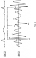
- FIG. 6 shows simultaneously recorded seismocardiogram (SCG) and electrocardiogram (ECG) with timing of seismocardiographic waves during cardiac cycle.
- SCG seismocardiogram
- ECG electrocardiogram
- systolic events were identified as mitral valve closure (MC), isovolumic movement (IM), aortic valve opening (AO), rapid systolic ejection (RE) and aortic valve closure (AC).
- Diastolic events include mitral valve opening (MO), early rapid filling (RF) and atrial systole (AS).
- the system comprises a risk determining unit adapted to determine a risk of CAD by comparing the low frequency power measure with at least one reference value for CAD, and generating a risk-of-CAD-signal S CAD indicating the result of said comparison.
- the result may be indicated on the display unit, thus, the display unit is adapted to indicate said risk of CAD.
- the inventors have found that the combination of a low-frequency power spectrum and a high-frequency power spectrum provides an even more certain coronary artery disease diagnosis, than either the low frequency power spectrum or the high frequency power spectrum alone. This is illustrated by the schematic block diagram in figure 2 .
- the filtering unit is adapted to apply a filter to said identified periods and to generate a high frequency band signal S HFB indicating high frequency bands of said identified periods.
- the high frequency band is between 250-1000 Hz.
- the calculation unit is adapted to calculate a high frequency feature measure based upon an estimated feature in high frequency bands of said periods and to generate a high frequency feature measure signal S HFF indicating said high frequency feature measure.
- the risk determining unit is then in addition adapted to compare said high frequency feature measure with a further feature reference value for CAD and to generate a combined risk-of-CAD-signal S' CAD indicating said risk in dependence of said comparison.
- Features from high frequency bands can be of different type such as power, power ratio or complexity. For different features, different feature reference values for CAD are used. Thus, an even more certain coronary artery disease diagnosis is then achieved.
- the amplitude of the recordings may be affected by tissue damping factors.
- the calculation unit is accorded to one embodiment adapted to correct the low frequency power measure against at least one tissue damping factor, such as body mass index (BMI), gender, age and disease.
- BMI body mass index
- the amplitude of the low frequency recordings is most likely to be related to BMI, since the distance from the ventricle to the recording spot will affect the amplitude.
- corrected low frequency power low frequency power + 0.43 ⁇ BMI + 11.6
- the classification performance AUC was 72.6% before correction and 73.1% after correction. Thus, the accuracy of the low frequency power was improved.
- the low frequency power score is likely to be related to gender since the women's heart is smaller and the breasts increases the distance from the ventricle to the recording spot.
- the mean female low frequency power was 13.9 dB in the healthy patients and 15.8 dB in the diseased female patients, were mean power was respectively 20.3 dB and 23.4 dB for the males. This clearly demonstrates that the power levels are lower in the female group compared to the male group.
- the influence of gender may be corrected for to remove the difference between the results between men and women. If the influence is not corrected for, different reference values for the low frequency power measure should be used for men and women respectively.
- the calculation unit is adapted to calculate low frequency power measures for both diastolic and systolic periods, to compare the measures and generate a correlation value based upon the comparison indicating the correlation between the low frequency power measures. Thus, if the values correlate, this is indicated in the correlation value and supports the assumption that a patient has CAD.
- Power generated by low frequency components, in this case in the frequency band 10-90 Hz, of the heart signal can be more precisely estimated by normalizing the generated power over other, higher frequency band.
- this larger frequency band is 90-300 Hz.
- PSD the power spectrum density
- the power generated in the low frequency band is normalized against the power of the higher frequency band, i.e. power ratio of the two power values is calculated. In this way, noise and/or variations between individuals may be removed.
- Figure 6 shows, as expected, that the averaged diastolic power value for CAD-patients in low frequency band is greater than corresponding value of the control group.
- PSD the dynamic range of PSD is high, approximately 70 dB, meaning that the low frequency part of the signal is 70 higher than the high frequency part of the signal. Therefore the HF part of the signal is covered by the LF part. This is due to a phenomena called spectral leakage.
- a so-called whitening filter By adjusting a so-called whitening filter to make the PSD of average healthy subject white, all frequencies will have the same power. Thereby the dynamic range of the frequency spectrum is reduced, which will reduce spectral leakage, and thereby it will be possible to estimate low frequency power and high frequency power from the same spectrum.
- the area under the curve is 78.21%. Moreover, if threshold is defined, a sensitivity of 73.42 % and specificity of 75.76 % is obtained.
- the example shows that by combining the estimated power ratio obtained in the previous example 8 (i.e. by normalizing power obtained in the frequency band 10-90 Hz over power obtained in the frequency band 90-300 Hz) with the analogously obtained, estimated power ratio in respectively 700-900 Hz and 900-1300 Hz frequency band, an improved classification model is obtained.
- AUC Area Under Curve
- the power in the low frequency band may be measured in different ways. It may, for instance, be measured in a predefined time interval of the diastole starting 200-300 ms after the S2 sound. One way to perform the measurement is then to, in each individual recording, identify a time interval named "diastasis", which is the last stage of diastole, i.e. time period between S3 and S4 sounds. Since diastasis is, as it may also be seen in Figure 9 , a relatively quiescent period, one way to do this is to identify the time window in the diastole with the lowest power. This time window might be 100 ms or longer and is found in the interval from approximately 200 ms after the S2 and 100 ms before S 1, but not later than 450 ms after S2.
- the electronic stethoscope is adapted to convert acoustic sound waves to electrical signals which can be further processed.
- the stethoscope comprises an acoustic sensor adapted to be placed on the chest of a patient, and to generate acoustic signals S A .
- the stethoscope further comprises at least one memory adapted to store acoustic signals S A from the acoustic sensor; and a control unit adapted to receive said acoustic signals S A . Because the sounds are transmitted electronically, the electronic stethoscope can be a wireless device, a recording device, and may provide noise reduction, signal enhancement, and both visual and audio output.
- the electronic stethoscope may comprise a transmitting unit adapted to transmit the signal(s) referred to herein to an external unit.
- the control unit comprises an identification unit adapted to identify diastolic or systolic periods in a predetermined time period of the stored acoustic signals S A , and to generate a period signal S P indicating said identified periods; a filtering unit adapted to apply at least one filter to said identified periods and to generate a low frequency band signal S LFB indicating low frequency bands of said identified periods; and a calculation unit adapted to estimate the power in said low frequency band of said identified periods, to calculate a low frequency power measure based upon said estimated power and to generate a low frequency power measure signal S LFP indicating said low frequency power measure.
- the stethoscope may comprise a risk determining unit adapted to determine a risk of CAD by comparing the low frequency power measure with at least one reference value for CAD, and generating a risk-of-CAD-signal S CAD indicating the result of the comparison.
- the stethoscope may comprise a display unit adapted to indicate said low frequency power measure and/or the result of the above-mentioned comparison, e.g. the patient's risk of CAD.
- the stethoscope also may be provided with the necessary means required to generate the combined risk-of-CAD-signal S' CAD , i.e. the means described e.g. in connection with the embodiment illustrated by figure 2 .
- the present disclosure also relates to a method for detecting frequency power for diagnosing of coronary artery disease (CAD) using a system according to the invention.
- CAD coronary artery disease
- the method comprises the first step A1 of recording acoustic data with a system for detection of frequency power comprising an acoustic sensor placed on the chest of a patient, and generating acoustic signals S A indicating said data.
- the sensor is preferably placed closely to the patient's heart, to be able to sense and record sound from heart beats.
- S 1, S2, S3 and S4 The most significant sounds of the heart are referred to as S 1, S2, S3 and S4 as has been explained in the preceding text.
- diastolic or systolic periods are identified in a predetermined time period of the stored acoustic data, and a period signal S P indicating said identified periods is generated.
- a period signal S P indicating said identified periods is generated.
- the duration of the diastolic or systolic periods is identified. Only the diastolic or systolic periods may be present in the period signal S P .
- a third step A3 at least one filter is applied to the period signal S P and a low frequency band signal S LFB is generated indicating low frequency bands of the identified periods. Preferably frequencies above 100 Hz are filtered away.
- the period signal S P is band pass filtered using a 5-70 Hz, preferably 10-60 Hz, more preferably 15-50 Hz, and most preferably 20-40 Hz band pass filter frequency.
- a fourth step A4 the power in the low frequency band of said identified periods is estimated.
- the estimation may be performed using one of the two following methods:
- a low frequency power measure is calculated based upon said estimated power, and a low frequency power measure signal S LFP is generated indicating said low frequency power measure.
- the method comprises the step (A6) of comparing said low frequency power measure with at least one reference value for CAD, and generating a risk-of-CAD-signal S CAD indicating the result of said comparison.
- the risk of CAD may be shown on a display, or audibly transmitted to the practitioner.
- the method comprises the steps of:
- the present invention relates to a method for detecting frequency power for diagnosing of coronary artery disease (CAD) using a system for detection of frequency power.
- CAD coronary artery disease
- the recorded values may be affected by tissue damping factors such as BMI, gender, age or disease.
- the method comprises according to one embodiment the step of correcting said low frequency power measure against at least one tissue damping factor. How this can be made has been previously explained.
- the method comprises the step of transmitting any of the aforementioned signals to an external unit for further processing or saving.
- the invention also relates to a computer program product, comprising computer program instructions for instructing a computer system in a system according to claims 1-6 or stethoscope to perform the steps according to the preceding method steps, when the data program instructions are executed on said computer system.
Landscapes
- Health & Medical Sciences (AREA)
- Life Sciences & Earth Sciences (AREA)
- Engineering & Computer Science (AREA)
- Physics & Mathematics (AREA)
- General Health & Medical Sciences (AREA)
- Surgery (AREA)
- Public Health (AREA)
- Animal Behavior & Ethology (AREA)
- Veterinary Medicine (AREA)
- Biomedical Technology (AREA)
- Heart & Thoracic Surgery (AREA)
- Medical Informatics (AREA)
- Molecular Biology (AREA)
- Biophysics (AREA)
- Pathology (AREA)
- Physiology (AREA)
- Acoustics & Sound (AREA)
- Cardiology (AREA)
- Artificial Intelligence (AREA)
- Computer Vision & Pattern Recognition (AREA)
- Psychiatry (AREA)
- Signal Processing (AREA)
- Vascular Medicine (AREA)
- Measuring Pulse, Heart Rate, Blood Pressure Or Blood Flow (AREA)
- Measurement And Recording Of Electrical Phenomena And Electrical Characteristics Of The Living Body (AREA)
Claims (11)
- Système de détection de puissance fréquentielle pour le diagnostic de maladie coronarienne (CAD), comprenant :- un capteur acoustique conçu pour être placé sur la poitrine d'un patient, et pour générer des signaux acoustiques SA ;- au moins une mémoire conçue pour stocker les signaux acoustiques SA provenant du capteur acoustique ;- une unité de commande conçue pour recevoir lesdits signaux acoustiques SA ;dans lequel ladite unité de commande comprend également :- une unité d'identification conçue pour identifier des périodes diastoliques ou systoliques dans une période prédéterminée des signaux acoustiques stockés SA, et pour générer un signal de période SP indiquant lesdites périodes identifiées ; caractérisé en ce que ladite unité de commande comprend également :- une unité de filtrage conçue pour appliquer au moins un filtre auxdites périodes identifiées et pour générer un signal de bande basse fréquence SLFB indiquant les bandes basses fréquences desdites périodes identifiées, dans lequel lesdites bandes basses fréquences sont comprises entre 5 et 70 Hz, et pour appliquer un autre filtre auxdites périodes identifiées et générer un signal de bande haute fréquence SHFB indiquant les bandes hautes fréquences desdites périodes identifiées ;- une unité de calcul conçue pour estimer la puissance dans ladite bande basse fréquence desdites périodes identifiées, pour calculer une mesure de puissance basse fréquence sur la base de ladite puissance estimée et pour générer un signal de mesure de puissance basse fréquence SLFP indiquant ladite mesure de puissance basse fréquence, et dans lequel ladite unité de calcul est en outre conçue pour calculer une mesure de puissance haute fréquence sur la base d'une puissance estimée dans des bandes haute fréquence desdites périodes et pour générer un signal de mesure de puissance haute fréquence SHFF indiquant ladite mesure de puissance haute fréquence,dans lequel ledit système comprend également une unité de détermination de risque conçue pour déterminer un risque de maladie coronarienne (CAD) par combinaison de ladite mesure de puissance basse fréquence et de ladite mesure de puissance haute fréquence et pour comparer une valeur de rapport de puissance combinée résultante avec une autre valeur de référence de caractéristique pour CAD et pour générer un signal de risque de CAD combiné S'CAD indiquant ledit risque en fonction de ladite comparaison.
- Système selon la revendication 1, dans lequel ladite bande basse fréquence est comprise entre 10 et 60 Hz, de préférence entre 15 et 50 Hz, de manière davantage préférée entre 20 et 40 Hz.
- Système selon l'une quelconque des revendications précédentes, dans lequel ladite unité de calcul est conçue pour corriger ladite mesure de puissance basse fréquence par rapport à au moins un facteur d'amortissement tissulaire, et en ce que ledit ou lesdits facteurs d'amortissement tissulaire sont un ou plusieurs parmi : IMC (Indice de Masse Corporelle), sexe, âge et maladie.
- Système selon l'une quelconque des revendications précédentes, dans lequel ladite unité de commande est conçue pour estimer la puissance basse fréquence par filtrage passe-bande desdites périodes identifiées à l'aide d'une fréquence de filtrage passe-bande de 5 à 70 Hz, de préférence de 10 à 60 Hz, de manière davantage préférée de 15 à 50 Hz, et de manière préférée entre toutes de 20 à 40 Hz et par calcul de la variance au cours desdites périodes.
- Système selon l'une quelconque des revendications précédentes, dans lequel ladite unité de calcul est conçue pour estimer la puissance basse fréquence en générant un spectre de puissance directement ou indirectement avec au moins un modèle AR de ladite bande basse fréquence et en calculant la puissance dans ladite bande basse fréquence.
- Système selon l'une quelconque des revendications précédentes, dans lequel au moins des parties dudit système sont réalisées par un stéthoscope électronique.
- Procédé de détection de puissance fréquentielle pour le diagnostic de maladie coronarienne (CAD) à l'aide d'un système de détection de puissance basse fréquence, le procédé comprenant :- l'enregistrement de données acoustiques par ledit système comprenant un capteur acoustique placé sur la poitrine d'un patient et la génération de signaux acoustiques SA indiquant lesdites données ;- l'identification de périodes diastoliques ou systoliques dans une période prédéterminée des données acoustiques stockées, et la génération d'un signal de période SP indiquant lesdites périodes identifiées ;- l'application d'au moins un filtre au signal de période SP et la génération d'un signal de bande basse fréquence SLFB indiquant des bandes basse fréquence desdites périodes identifiées et l'application d'un autre filtre au signal de période SP et la génération d'un signal de bande haute fréquence SHFB indiquant des bandes haute fréquence desdites périodes identifiées, dans lequel lesdites bandes basse fréquence sont comprises entre 5 et 70 Hz ;- l'estimation de la puissance dans lesdites bandes basse fréquence desdites périodes identifiées ;- le calcul d'une mesure de puissance basse fréquence sur la base de ladite puissance estimée, et la génération d'un signal de mesure de puissance basse fréquence SLFP indiquant ladite mesure de puissance basse fréquence ;- le calcul d'une mesure de puissance haute fréquence sur la base d'une puissance estimée dans des bandes haute fréquence desdites périodes, et la génération d'un signal de mesure de puissance haute fréquence SHFF indiquant ladite mesure de puissance haute fréquence, et- la combinaison de ladite mesure de puissance basse fréquence et de ladite mesure de puissance haute fréquence, et la comparaison d'une valeur de rapport de puissance combinée résultante avec une autre valeur de référence de caractéristique pour la CAD afin de générer un signal de risque de CAD combiné S'CAD indiquant ledit risque en fonction de ladite comparaison.'
- Procédé selon la revendication 7, comprenant l'estimation de la puissance dans lesdites bandes basse fréquence par filtrage passe-bande desdites périodes identifiées à l'aide d'une fréquence de filtrage passe-bande de 10 à 60 Hz, de préférence de 15 à 50 Hz, et de manière davantage préférée de 20 à 40 Hz et le calcul de la variance au cours desdites périodes.
- Procédé selon la revendication 7 ou 8, comprenant l'estimation de la puissance basse fréquence en générant un spectre de puissance directement ou indirectement avec au moins un modèle AR de ladite bande basse fréquence et le calcul de la puissance dans ladite bande basse fréquence.
- Procédé selon l'une quelconque des revendications 7 à 9, comprenant la correction de ladite mesure de puissance basse fréquence par rapport à au moins un facteur d'amortissement tissulaire et caractérisé en ce que ledit ou lesdits facteurs d'amortissement tissulaire sont un ou plusieurs parmi : IMC (Indice de Masse Corporelle), sexe, âge et maladie.
- Produit-programme informatique, comprenant des instructions de programme informatique pour ordonner à un système informatique dans un système selon l'une quelconque des revendications 1 à 6 de réaliser les étapes selon le procédé selon l'une quelconque des revendications 7 à 10, dans lequel les instructions de programme de données sont exécutées sur ledit système informatique, ou dans lequel les instructions de programme informatique sont stockées sur un support lisible par ordinateur.
Applications Claiming Priority (3)
| Application Number | Priority Date | Filing Date | Title |
|---|---|---|---|
| EP10194756A EP2462871A1 (fr) | 2010-12-13 | 2010-12-13 | Système, stéthoscope et procédé pour indiquer le risque de maladie coronarienne |
| EP11794179.9A EP2651304B8 (fr) | 2010-12-13 | 2011-12-13 | Système, stéthoscope et procédé pour indiquer le risque d'une coronaropathie |
| PCT/EP2011/072537 WO2012080209A1 (fr) | 2010-12-13 | 2011-12-13 | Système, stéthoscope et procédé pour indiquer le risque d'une coronaropathie |
Related Parent Applications (3)
| Application Number | Title | Priority Date | Filing Date |
|---|---|---|---|
| PCT/EP2011/072537 Previously-Filed-Application WO2012080209A1 (fr) | 2010-12-13 | 2011-12-13 | Système, stéthoscope et procédé pour indiquer le risque d'une coronaropathie |
| EP11794179.9A Division-Into EP2651304B8 (fr) | 2010-12-13 | 2011-12-13 | Système, stéthoscope et procédé pour indiquer le risque d'une coronaropathie |
| EP11794179.9A Division EP2651304B8 (fr) | 2010-12-13 | 2011-12-13 | Système, stéthoscope et procédé pour indiquer le risque d'une coronaropathie |
Publications (2)
| Publication Number | Publication Date |
|---|---|
| EP3701873A1 EP3701873A1 (fr) | 2020-09-02 |
| EP3701873B1 true EP3701873B1 (fr) | 2024-02-28 |
Family
ID=43618366
Family Applications (3)
| Application Number | Title | Priority Date | Filing Date |
|---|---|---|---|
| EP10194756A Withdrawn EP2462871A1 (fr) | 2010-12-13 | 2010-12-13 | Système, stéthoscope et procédé pour indiquer le risque de maladie coronarienne |
| EP20168782.9A Active EP3701873B1 (fr) | 2010-12-13 | 2011-12-13 | Système et procédé pour indiquer le risque de maladie coronarienne |
| EP11794179.9A Active EP2651304B8 (fr) | 2010-12-13 | 2011-12-13 | Système, stéthoscope et procédé pour indiquer le risque d'une coronaropathie |
Family Applications Before (1)
| Application Number | Title | Priority Date | Filing Date |
|---|---|---|---|
| EP10194756A Withdrawn EP2462871A1 (fr) | 2010-12-13 | 2010-12-13 | Système, stéthoscope et procédé pour indiquer le risque de maladie coronarienne |
Family Applications After (1)
| Application Number | Title | Priority Date | Filing Date |
|---|---|---|---|
| EP11794179.9A Active EP2651304B8 (fr) | 2010-12-13 | 2011-12-13 | Système, stéthoscope et procédé pour indiquer le risque d'une coronaropathie |
Country Status (9)
| Country | Link |
|---|---|
| US (2) | US9125564B2 (fr) |
| EP (3) | EP2462871A1 (fr) |
| JP (1) | JP6073799B2 (fr) |
| CN (1) | CN103313662B (fr) |
| AU (1) | AU2011344281B2 (fr) |
| BR (1) | BR112013014674A2 (fr) |
| CA (1) | CA2819293C (fr) |
| RU (1) | RU2571333C2 (fr) |
| WO (1) | WO2012080209A1 (fr) |
Families Citing this family (25)
| Publication number | Priority date | Publication date | Assignee | Title |
|---|---|---|---|---|
| US7520860B2 (en) | 2005-04-13 | 2009-04-21 | Marie G. Johnson | Detection of coronary artery disease using an electronic stethoscope |
| US9403254B2 (en) * | 2011-08-17 | 2016-08-02 | Taiwan Semiconductor Manufacturing Company, Ltd. | Methods for real-time error detection in CMP processing |
| JP6036174B2 (ja) * | 2012-10-31 | 2016-11-30 | 株式会社Jvcケンウッド | 心音情報処理装置、心音情報処理方法および心音情報処理プログラム |
| CA2907400A1 (fr) * | 2013-03-18 | 2014-09-25 | PhonoFlow Medical, LLC | Analyse spectrale de flux sanguin turbulent d'artere coronaire |
| US9504844B2 (en) * | 2013-06-12 | 2016-11-29 | Medical Energetics Ltd | Health applications for using bio-feedback to control an electromagnetic field |
| EP3078329A1 (fr) | 2015-04-09 | 2016-10-12 | Acarix A/S | Indication du risque de coronaropathie |
| JP6534566B2 (ja) * | 2015-06-30 | 2019-06-26 | 旭化成株式会社 | 心疾患診断装置、心疾患診断プログラム及び媒体 |
| DE102015226365A1 (de) * | 2015-12-21 | 2017-06-22 | Robert Bosch Gmbh | Verfahren zur Messung der Varianz in einem Messsignal, Verfahren zur Datenfusion, Computerprogramm, Maschinenlesbares Speichermedium und Vorrichtung |
| CN105595991B (zh) * | 2016-01-29 | 2019-09-27 | 北京心量科技有限公司 | 一种心脏相干指数的测量方法及装置 |
| CN109475340B (zh) * | 2016-06-07 | 2022-02-15 | 远程护理科技第一公司 | 用于测量孕妇的中心脉搏波速的方法和系统 |
| EP3409197A1 (fr) * | 2017-05-30 | 2018-12-05 | Koninklijke Philips N.V. | Systèmes et procédés permettant de supprimer des crêtes dans un sismocardiogramme |
| CA3075930A1 (fr) * | 2017-09-19 | 2019-03-28 | Ausculsciences, Inc. | Systeme et procede de detection du decouplage d'un capteur sonore d'auscultation d'un sujet de test |
| US11284827B2 (en) | 2017-10-21 | 2022-03-29 | Ausculsciences, Inc. | Medical decision support system |
| EP3476299A1 (fr) * | 2017-10-26 | 2019-05-01 | Acarix A/S | Détermination de risque de coronaropathie |
| US11504047B2 (en) | 2017-12-28 | 2022-11-22 | Heart Force Medical Inc. | Sensor apparatuses, methods of operating same, and systems including same, and methods and systems for sensing and analyzing electromechanical characteristics of a heart |
| CN111770722B (zh) * | 2018-02-27 | 2024-01-02 | 罗伯特·博世有限公司 | 具有心脏振动描记信号的自动参考的可穿戴健康设备系统 |
| EP3539464B1 (fr) * | 2018-03-16 | 2025-01-15 | Tata Consultancy Services Limited | Système de classification des maladies coronariennes à base de métadonnées et de signaux cardiovasculaires |
| CN109512456A (zh) * | 2018-12-25 | 2019-03-26 | 河北铭心堂生物科技有限公司 | 一种八通道冠状动脉疾病诊断仪 |
| US11240579B2 (en) | 2020-05-08 | 2022-02-01 | Level 42 Ai | Sensor systems and methods for characterizing health conditions |
| KR102554084B1 (ko) | 2020-07-24 | 2023-07-10 | 중앙대학교 산학협력단 | 청진기 음파 분석을 통한 혈관 협착 평가 방법 |
| US11751774B2 (en) | 2020-11-12 | 2023-09-12 | Unitedhealth Group Incorporated | Electronic auscultation and improved identification of auscultation audio samples |
| US11545256B2 (en) | 2020-11-12 | 2023-01-03 | Unitedhealth Group Incorporated | Remote monitoring using an array of audio sensors and improved jugular venous pressure (JVP) measurement |
| JP7295368B2 (ja) * | 2020-12-31 | 2023-06-21 | Yanchers株式会社 | 聴診音の解析システム |
| KR102483149B1 (ko) | 2021-01-29 | 2022-12-30 | 중앙대학교 산학협력단 | 머신러닝을 활용한 경동맥 협착증 판별을 위한 정보제공 방법 |
| TWI884070B (zh) * | 2024-08-23 | 2025-05-11 | 廣達電腦股份有限公司 | 主動降噪聽診器 |
Family Cites Families (22)
| Publication number | Priority date | Publication date | Assignee | Title |
|---|---|---|---|---|
| US5020540A (en) * | 1987-10-09 | 1991-06-04 | Biometrak Corporation | Cardiac biopotential analysis system and method |
| US5109863A (en) * | 1989-10-26 | 1992-05-05 | Rutgers, The State University Of New Jersey | Noninvasive diagnostic system for coronary artery disease |
| US5036857A (en) | 1989-10-26 | 1991-08-06 | Rutgers, The State University Of New Jersey | Noninvasive diagnostic system for coronary artery disease |
| CA2078136A1 (fr) | 1990-03-16 | 1991-09-17 | John M. Zanetti | Systeme de depistage de l'ischemie myocardique |
| RU2130755C1 (ru) * | 1992-12-18 | 1999-05-27 | Удмуртский государственный университет | Датчик электронного стетоскопа |
| US5638823A (en) * | 1995-08-28 | 1997-06-17 | Rutgers University | System and method for noninvasive detection of arterial stenosis |
| US6135966A (en) * | 1998-05-01 | 2000-10-24 | Ko; Gary Kam-Yuen | Method and apparatus for non-invasive diagnosis of cardiovascular and related disorders |
| JP3577417B2 (ja) * | 1998-08-18 | 2004-10-13 | 博志 松本 | 冠状動脈病変診断装置および診断方法 |
| US6048319A (en) * | 1998-10-01 | 2000-04-11 | Integrated Medical Systems, Inc. | Non-invasive acoustic screening device for coronary stenosis |
| WO2002096293A1 (fr) * | 2001-05-28 | 2002-12-05 | Health Devices Pte Ltd. | Systeme de diagnostic cardiaque |
| RU2229843C2 (ru) * | 2001-12-24 | 2004-06-10 | Антонов Андрей Васильевич | Многоканальный электронный стетоскоп |
| EP1485013A2 (fr) * | 2002-03-18 | 2004-12-15 | Sonomedica, Llc | Procede et systeme permettant de generer un coefficient de vraisemblance pour une maladie cardio-vasculaire, d'analyser des signaux sonores cardio-vasculaires |
| CA2526932A1 (fr) * | 2003-04-23 | 2005-02-17 | Hemchandra Shertukde | Appareil et methode de diagnostic non invasif de coronaropathie |
| US7096060B2 (en) | 2003-06-27 | 2006-08-22 | Innovise Medical, Inc. | Method and system for detection of heart sounds |
| US7300405B2 (en) * | 2003-10-22 | 2007-11-27 | 3M Innovative Properties Company | Analysis of auscultatory sounds using single value decomposition |
| RU2316256C2 (ru) * | 2004-01-22 | 2008-02-10 | Сергей Валерьевич Владимиров | Индивидуальный электронный стетоскоп |
| CN101137977A (zh) * | 2005-01-20 | 2008-03-05 | 麦德斯科恩索尼科公司 | 冠状动脉异常的声学诊断装置 |
| US7520860B2 (en) | 2005-04-13 | 2009-04-21 | Marie G. Johnson | Detection of coronary artery disease using an electronic stethoscope |
| US7922669B2 (en) * | 2005-06-08 | 2011-04-12 | Cardiac Pacemakers, Inc. | Ischemia detection using a heart sound sensor |
| WO2008000254A1 (fr) * | 2006-06-26 | 2008-01-03 | Coloplast A/S | Classification multiparamétrique de bruits cardio-vasculaires |
| JP5207174B2 (ja) * | 2008-03-31 | 2013-06-12 | 国立大学法人山口大学 | 心音周波数解析装置 |
| US20110137210A1 (en) * | 2009-12-08 | 2011-06-09 | Johnson Marie A | Systems and methods for detecting cardiovascular disease |
-
2010
- 2010-12-13 EP EP10194756A patent/EP2462871A1/fr not_active Withdrawn
-
2011
- 2011-12-13 EP EP20168782.9A patent/EP3701873B1/fr active Active
- 2011-12-13 CA CA2819293A patent/CA2819293C/fr active Active
- 2011-12-13 RU RU2013132214/14A patent/RU2571333C2/ru active
- 2011-12-13 AU AU2011344281A patent/AU2011344281B2/en active Active
- 2011-12-13 EP EP11794179.9A patent/EP2651304B8/fr active Active
- 2011-12-13 JP JP2013543698A patent/JP6073799B2/ja active Active
- 2011-12-13 WO PCT/EP2011/072537 patent/WO2012080209A1/fr not_active Ceased
- 2011-12-13 BR BR112013014674A patent/BR112013014674A2/pt not_active Application Discontinuation
- 2011-12-13 CN CN201180059941.7A patent/CN103313662B/zh active Active
- 2011-12-13 US US13/994,089 patent/US9125564B2/en active Active
-
2015
- 2015-05-01 US US14/702,660 patent/US9451921B2/en active Active
Also Published As
| Publication number | Publication date |
|---|---|
| AU2011344281B2 (en) | 2016-01-07 |
| AU2011344281A1 (en) | 2013-06-20 |
| BR112013014674A2 (pt) | 2016-09-27 |
| JP2014504192A (ja) | 2014-02-20 |
| JP6073799B2 (ja) | 2017-02-01 |
| EP2651304B8 (fr) | 2020-07-15 |
| EP2651304B1 (fr) | 2020-05-27 |
| RU2571333C2 (ru) | 2015-12-20 |
| CA2819293C (fr) | 2020-04-28 |
| US20150305687A1 (en) | 2015-10-29 |
| RU2013132214A (ru) | 2015-01-20 |
| EP3701873A1 (fr) | 2020-09-02 |
| US20130261484A1 (en) | 2013-10-03 |
| CN103313662B (zh) | 2016-01-27 |
| EP2462871A1 (fr) | 2012-06-13 |
| US9125564B2 (en) | 2015-09-08 |
| EP2651304A1 (fr) | 2013-10-23 |
| US9451921B2 (en) | 2016-09-27 |
| CA2819293A1 (fr) | 2012-06-21 |
| WO2012080209A1 (fr) | 2012-06-21 |
| CN103313662A (zh) | 2013-09-18 |
Similar Documents
| Publication | Publication Date | Title |
|---|---|---|
| EP3701873B1 (fr) | Système et procédé pour indiquer le risque de maladie coronarienne | |
| TWI669096B (zh) | 具有確定頸動脈血壓的多功能量測裝置 | |
| JP5576116B2 (ja) | 心血管音のマルチパラメータ分類 | |
| US20120071767A1 (en) | Pulmonary artery pressure estimator | |
| CN107106118B (zh) | 检测重搏切迹的方法 | |
| US10004473B2 (en) | Heart rate detection method and device using heart sound acquired from auscultation positions | |
| CN101594826A (zh) | 分析体音的方法和系统 | |
| JP6547054B1 (ja) | 医療機器及びプログラム | |
| US20160206287A1 (en) | Wearable Doppler Ultrasound Based Cardiac Monitoring | |
| JP5534674B2 (ja) | 非正弦波周期性の流動作用を測定又は検出する超音波法及び装置 | |
| JP2009525117A5 (fr) | ||
| Balogh et al. | Application of phonocardiography on preterm infants with patent ductus arteriosus | |
| Sang | Analysis of Seismocardiogram Signals From Healthy and Unhealthy Subjects Using an Accelerometer Contact Microphone | |
| HK1129821B (en) | Multi parametric classification of cardiovascular sound |
Legal Events
| Date | Code | Title | Description |
|---|---|---|---|
| PUAI | Public reference made under article 153(3) epc to a published international application that has entered the european phase |
Free format text: ORIGINAL CODE: 0009012 |
|
| STAA | Information on the status of an ep patent application or granted ep patent |
Free format text: STATUS: THE APPLICATION HAS BEEN PUBLISHED |
|
| AC | Divisional application: reference to earlier application |
Ref document number: 2651304 Country of ref document: EP Kind code of ref document: P |
|
| AK | Designated contracting states |
Kind code of ref document: A1 Designated state(s): AL AT BE BG CH CY CZ DE DK EE ES FI FR GB GR HR HU IE IS IT LI LT LU LV MC MK MT NL NO PL PT RO RS SE SI SK SM TR |
|
| STAA | Information on the status of an ep patent application or granted ep patent |
Free format text: STATUS: REQUEST FOR EXAMINATION WAS MADE |
|
| 17P | Request for examination filed |
Effective date: 20210219 |
|
| RBV | Designated contracting states (corrected) |
Designated state(s): AL AT BE BG CH CY CZ DE DK EE ES FI FR GB GR HR HU IE IS IT LI LT LU LV MC MK MT NL NO PL PT RO RS SE SI SK SM TR |
|
| GRAP | Despatch of communication of intention to grant a patent |
Free format text: ORIGINAL CODE: EPIDOSNIGR1 |
|
| STAA | Information on the status of an ep patent application or granted ep patent |
Free format text: STATUS: GRANT OF PATENT IS INTENDED |
|
| INTG | Intention to grant announced |
Effective date: 20230509 |
|
| GRAJ | Information related to disapproval of communication of intention to grant by the applicant or resumption of examination proceedings by the epo deleted |
Free format text: ORIGINAL CODE: EPIDOSDIGR1 |
|
| STAA | Information on the status of an ep patent application or granted ep patent |
Free format text: STATUS: REQUEST FOR EXAMINATION WAS MADE |
|
| RAP3 | Party data changed (applicant data changed or rights of an application transferred) |
Owner name: ACARIX A/S |
|
| GRAP | Despatch of communication of intention to grant a patent |
Free format text: ORIGINAL CODE: EPIDOSNIGR1 |
|
| INTC | Intention to grant announced (deleted) | ||
| STAA | Information on the status of an ep patent application or granted ep patent |
Free format text: STATUS: GRANT OF PATENT IS INTENDED |
|
| INTG | Intention to grant announced |
Effective date: 20230928 |
|
| GRAS | Grant fee paid |
Free format text: ORIGINAL CODE: EPIDOSNIGR3 |
|
| GRAA | (expected) grant |
Free format text: ORIGINAL CODE: 0009210 |
|
| STAA | Information on the status of an ep patent application or granted ep patent |
Free format text: STATUS: THE PATENT HAS BEEN GRANTED |
|
| AC | Divisional application: reference to earlier application |
Ref document number: 2651304 Country of ref document: EP Kind code of ref document: P |
|
| AK | Designated contracting states |
Kind code of ref document: B1 Designated state(s): AL AT BE BG CH CY CZ DE DK EE ES FI FR GB GR HR HU IE IS IT LI LT LU LV MC MK MT NL NO PL PT RO RS SE SI SK SM TR |
|
| REG | Reference to a national code |
Ref country code: GB Ref legal event code: FG4D |
|
| REG | Reference to a national code |
Ref country code: CH Ref legal event code: EP |
|
| REG | Reference to a national code |
Ref country code: DE Ref legal event code: R096 Ref document number: 602011074614 Country of ref document: DE |
|
| REG | Reference to a national code |
Ref country code: IE Ref legal event code: FG4D |
|
| P01 | Opt-out of the competence of the unified patent court (upc) registered |
Effective date: 20240402 |
|
| REG | Reference to a national code |
Ref country code: LT Ref legal event code: MG9D |
|
| PG25 | Lapsed in a contracting state [announced via postgrant information from national office to epo] |
Ref country code: IS Free format text: LAPSE BECAUSE OF FAILURE TO SUBMIT A TRANSLATION OF THE DESCRIPTION OR TO PAY THE FEE WITHIN THE PRESCRIBED TIME-LIMIT Effective date: 20240628 |
|
| REG | Reference to a national code |
Ref country code: NL Ref legal event code: MP Effective date: 20240228 |
|
| PG25 | Lapsed in a contracting state [announced via postgrant information from national office to epo] |
Ref country code: LT Free format text: LAPSE BECAUSE OF FAILURE TO SUBMIT A TRANSLATION OF THE DESCRIPTION OR TO PAY THE FEE WITHIN THE PRESCRIBED TIME-LIMIT Effective date: 20240228 |
|
| PG25 | Lapsed in a contracting state [announced via postgrant information from national office to epo] |
Ref country code: GR Free format text: LAPSE BECAUSE OF FAILURE TO SUBMIT A TRANSLATION OF THE DESCRIPTION OR TO PAY THE FEE WITHIN THE PRESCRIBED TIME-LIMIT Effective date: 20240529 |
|
| PG25 | Lapsed in a contracting state [announced via postgrant information from national office to epo] |
Ref country code: HR Free format text: LAPSE BECAUSE OF FAILURE TO SUBMIT A TRANSLATION OF THE DESCRIPTION OR TO PAY THE FEE WITHIN THE PRESCRIBED TIME-LIMIT Effective date: 20240228 Ref country code: RS Free format text: LAPSE BECAUSE OF FAILURE TO SUBMIT A TRANSLATION OF THE DESCRIPTION OR TO PAY THE FEE WITHIN THE PRESCRIBED TIME-LIMIT Effective date: 20240528 Ref country code: NL Free format text: LAPSE BECAUSE OF FAILURE TO SUBMIT A TRANSLATION OF THE DESCRIPTION OR TO PAY THE FEE WITHIN THE PRESCRIBED TIME-LIMIT Effective date: 20240228 |
|
| PG25 | Lapsed in a contracting state [announced via postgrant information from national office to epo] |
Ref country code: ES Free format text: LAPSE BECAUSE OF FAILURE TO SUBMIT A TRANSLATION OF THE DESCRIPTION OR TO PAY THE FEE WITHIN THE PRESCRIBED TIME-LIMIT Effective date: 20240228 |
|
| PG25 | Lapsed in a contracting state [announced via postgrant information from national office to epo] |
Ref country code: RS Free format text: LAPSE BECAUSE OF FAILURE TO SUBMIT A TRANSLATION OF THE DESCRIPTION OR TO PAY THE FEE WITHIN THE PRESCRIBED TIME-LIMIT Effective date: 20240528 Ref country code: NO Free format text: LAPSE BECAUSE OF FAILURE TO SUBMIT A TRANSLATION OF THE DESCRIPTION OR TO PAY THE FEE WITHIN THE PRESCRIBED TIME-LIMIT Effective date: 20240528 Ref country code: NL Free format text: LAPSE BECAUSE OF FAILURE TO SUBMIT A TRANSLATION OF THE DESCRIPTION OR TO PAY THE FEE WITHIN THE PRESCRIBED TIME-LIMIT Effective date: 20240228 Ref country code: LT Free format text: LAPSE BECAUSE OF FAILURE TO SUBMIT A TRANSLATION OF THE DESCRIPTION OR TO PAY THE FEE WITHIN THE PRESCRIBED TIME-LIMIT Effective date: 20240228 Ref country code: IS Free format text: LAPSE BECAUSE OF FAILURE TO SUBMIT A TRANSLATION OF THE DESCRIPTION OR TO PAY THE FEE WITHIN THE PRESCRIBED TIME-LIMIT Effective date: 20240628 Ref country code: HR Free format text: LAPSE BECAUSE OF FAILURE TO SUBMIT A TRANSLATION OF THE DESCRIPTION OR TO PAY THE FEE WITHIN THE PRESCRIBED TIME-LIMIT Effective date: 20240228 Ref country code: GR Free format text: LAPSE BECAUSE OF FAILURE TO SUBMIT A TRANSLATION OF THE DESCRIPTION OR TO PAY THE FEE WITHIN THE PRESCRIBED TIME-LIMIT Effective date: 20240529 Ref country code: FI Free format text: LAPSE BECAUSE OF FAILURE TO SUBMIT A TRANSLATION OF THE DESCRIPTION OR TO PAY THE FEE WITHIN THE PRESCRIBED TIME-LIMIT Effective date: 20240228 Ref country code: ES Free format text: LAPSE BECAUSE OF FAILURE TO SUBMIT A TRANSLATION OF THE DESCRIPTION OR TO PAY THE FEE WITHIN THE PRESCRIBED TIME-LIMIT Effective date: 20240228 Ref country code: BG Free format text: LAPSE BECAUSE OF FAILURE TO SUBMIT A TRANSLATION OF THE DESCRIPTION OR TO PAY THE FEE WITHIN THE PRESCRIBED TIME-LIMIT Effective date: 20240228 |
|
| PG25 | Lapsed in a contracting state [announced via postgrant information from national office to epo] |
Ref country code: PT Free format text: LAPSE BECAUSE OF FAILURE TO SUBMIT A TRANSLATION OF THE DESCRIPTION OR TO PAY THE FEE WITHIN THE PRESCRIBED TIME-LIMIT Effective date: 20240628 Ref country code: PL Free format text: LAPSE BECAUSE OF FAILURE TO SUBMIT A TRANSLATION OF THE DESCRIPTION OR TO PAY THE FEE WITHIN THE PRESCRIBED TIME-LIMIT Effective date: 20240228 |
|
| REG | Reference to a national code |
Ref country code: AT Ref legal event code: MK05 Ref document number: 1660462 Country of ref document: AT Kind code of ref document: T Effective date: 20240228 |
|
| PG25 | Lapsed in a contracting state [announced via postgrant information from national office to epo] |
Ref country code: SE Free format text: LAPSE BECAUSE OF FAILURE TO SUBMIT A TRANSLATION OF THE DESCRIPTION OR TO PAY THE FEE WITHIN THE PRESCRIBED TIME-LIMIT Effective date: 20240228 Ref country code: PT Free format text: LAPSE BECAUSE OF FAILURE TO SUBMIT A TRANSLATION OF THE DESCRIPTION OR TO PAY THE FEE WITHIN THE PRESCRIBED TIME-LIMIT Effective date: 20240628 Ref country code: PL Free format text: LAPSE BECAUSE OF FAILURE TO SUBMIT A TRANSLATION OF THE DESCRIPTION OR TO PAY THE FEE WITHIN THE PRESCRIBED TIME-LIMIT Effective date: 20240228 Ref country code: LV Free format text: LAPSE BECAUSE OF FAILURE TO SUBMIT A TRANSLATION OF THE DESCRIPTION OR TO PAY THE FEE WITHIN THE PRESCRIBED TIME-LIMIT Effective date: 20240228 |
|
| PG25 | Lapsed in a contracting state [announced via postgrant information from national office to epo] |
Ref country code: DK Free format text: LAPSE BECAUSE OF FAILURE TO SUBMIT A TRANSLATION OF THE DESCRIPTION OR TO PAY THE FEE WITHIN THE PRESCRIBED TIME-LIMIT Effective date: 20240228 |
|
| PG25 | Lapsed in a contracting state [announced via postgrant information from national office to epo] |
Ref country code: SM Free format text: LAPSE BECAUSE OF FAILURE TO SUBMIT A TRANSLATION OF THE DESCRIPTION OR TO PAY THE FEE WITHIN THE PRESCRIBED TIME-LIMIT Effective date: 20240228 |
|
| PG25 | Lapsed in a contracting state [announced via postgrant information from national office to epo] |
Ref country code: CZ Free format text: LAPSE BECAUSE OF FAILURE TO SUBMIT A TRANSLATION OF THE DESCRIPTION OR TO PAY THE FEE WITHIN THE PRESCRIBED TIME-LIMIT Effective date: 20240228 Ref country code: EE Free format text: LAPSE BECAUSE OF FAILURE TO SUBMIT A TRANSLATION OF THE DESCRIPTION OR TO PAY THE FEE WITHIN THE PRESCRIBED TIME-LIMIT Effective date: 20240228 |
|
| PG25 | Lapsed in a contracting state [announced via postgrant information from national office to epo] |
Ref country code: AT Free format text: LAPSE BECAUSE OF FAILURE TO SUBMIT A TRANSLATION OF THE DESCRIPTION OR TO PAY THE FEE WITHIN THE PRESCRIBED TIME-LIMIT Effective date: 20240228 |
|
| PG25 | Lapsed in a contracting state [announced via postgrant information from national office to epo] |
Ref country code: SK Free format text: LAPSE BECAUSE OF FAILURE TO SUBMIT A TRANSLATION OF THE DESCRIPTION OR TO PAY THE FEE WITHIN THE PRESCRIBED TIME-LIMIT Effective date: 20240228 |
|
| PG25 | Lapsed in a contracting state [announced via postgrant information from national office to epo] |
Ref country code: SM Free format text: LAPSE BECAUSE OF FAILURE TO SUBMIT A TRANSLATION OF THE DESCRIPTION OR TO PAY THE FEE WITHIN THE PRESCRIBED TIME-LIMIT Effective date: 20240228 Ref country code: SK Free format text: LAPSE BECAUSE OF FAILURE TO SUBMIT A TRANSLATION OF THE DESCRIPTION OR TO PAY THE FEE WITHIN THE PRESCRIBED TIME-LIMIT Effective date: 20240228 Ref country code: RO Free format text: LAPSE BECAUSE OF FAILURE TO SUBMIT A TRANSLATION OF THE DESCRIPTION OR TO PAY THE FEE WITHIN THE PRESCRIBED TIME-LIMIT Effective date: 20240228 Ref country code: EE Free format text: LAPSE BECAUSE OF FAILURE TO SUBMIT A TRANSLATION OF THE DESCRIPTION OR TO PAY THE FEE WITHIN THE PRESCRIBED TIME-LIMIT Effective date: 20240228 Ref country code: DK Free format text: LAPSE BECAUSE OF FAILURE TO SUBMIT A TRANSLATION OF THE DESCRIPTION OR TO PAY THE FEE WITHIN THE PRESCRIBED TIME-LIMIT Effective date: 20240228 Ref country code: CZ Free format text: LAPSE BECAUSE OF FAILURE TO SUBMIT A TRANSLATION OF THE DESCRIPTION OR TO PAY THE FEE WITHIN THE PRESCRIBED TIME-LIMIT Effective date: 20240228 Ref country code: AT Free format text: LAPSE BECAUSE OF FAILURE TO SUBMIT A TRANSLATION OF THE DESCRIPTION OR TO PAY THE FEE WITHIN THE PRESCRIBED TIME-LIMIT Effective date: 20240228 |
|
| REG | Reference to a national code |
Ref country code: DE Ref legal event code: R097 Ref document number: 602011074614 Country of ref document: DE |
|
| PG25 | Lapsed in a contracting state [announced via postgrant information from national office to epo] |
Ref country code: IT Free format text: LAPSE BECAUSE OF FAILURE TO SUBMIT A TRANSLATION OF THE DESCRIPTION OR TO PAY THE FEE WITHIN THE PRESCRIBED TIME-LIMIT Effective date: 20240228 |
|
| PG25 | Lapsed in a contracting state [announced via postgrant information from national office to epo] |
Ref country code: IT Free format text: LAPSE BECAUSE OF FAILURE TO SUBMIT A TRANSLATION OF THE DESCRIPTION OR TO PAY THE FEE WITHIN THE PRESCRIBED TIME-LIMIT Effective date: 20240228 |
|
| PLBE | No opposition filed within time limit |
Free format text: ORIGINAL CODE: 0009261 |
|
| STAA | Information on the status of an ep patent application or granted ep patent |
Free format text: STATUS: NO OPPOSITION FILED WITHIN TIME LIMIT |
|
| 26N | No opposition filed |
Effective date: 20241129 |
|
| PG25 | Lapsed in a contracting state [announced via postgrant information from national office to epo] |
Ref country code: SI Free format text: LAPSE BECAUSE OF FAILURE TO SUBMIT A TRANSLATION OF THE DESCRIPTION OR TO PAY THE FEE WITHIN THE PRESCRIBED TIME-LIMIT Effective date: 20240228 |
|
| PG25 | Lapsed in a contracting state [announced via postgrant information from national office to epo] |
Ref country code: MC Free format text: LAPSE BECAUSE OF FAILURE TO SUBMIT A TRANSLATION OF THE DESCRIPTION OR TO PAY THE FEE WITHIN THE PRESCRIBED TIME-LIMIT Effective date: 20240228 |
|
| REG | Reference to a national code |
Ref country code: CH Ref legal event code: PL |
|
| PG25 | Lapsed in a contracting state [announced via postgrant information from national office to epo] |
Ref country code: LU Free format text: LAPSE BECAUSE OF NON-PAYMENT OF DUE FEES Effective date: 20241213 |
|
| REG | Reference to a national code |
Ref country code: BE Ref legal event code: MM Effective date: 20241231 |
|
| PG25 | Lapsed in a contracting state [announced via postgrant information from national office to epo] |
Ref country code: BE Free format text: LAPSE BECAUSE OF NON-PAYMENT OF DUE FEES Effective date: 20241231 |
|
| PG25 | Lapsed in a contracting state [announced via postgrant information from national office to epo] |
Ref country code: CH Free format text: LAPSE BECAUSE OF NON-PAYMENT OF DUE FEES Effective date: 20241231 |
|
| PG25 | Lapsed in a contracting state [announced via postgrant information from national office to epo] |
Ref country code: IE Free format text: LAPSE BECAUSE OF NON-PAYMENT OF DUE FEES Effective date: 20241213 |
|
| PGFP | Annual fee paid to national office [announced via postgrant information from national office to epo] |
Ref country code: GB Payment date: 20251219 Year of fee payment: 15 |
|
| PGFP | Annual fee paid to national office [announced via postgrant information from national office to epo] |
Ref country code: FR Payment date: 20251211 Year of fee payment: 15 |
|
| PGFP | Annual fee paid to national office [announced via postgrant information from national office to epo] |
Ref country code: DE Payment date: 20251222 Year of fee payment: 15 |