EP3716207B1 - Verfahren und vorrichtung zur diagnostischen analyse der funktion und morphologie von mikrozirkulationsänderungen - Google Patents
Verfahren und vorrichtung zur diagnostischen analyse der funktion und morphologie von mikrozirkulationsänderungen Download PDFInfo
- Publication number
- EP3716207B1 EP3716207B1 EP20165861.4A EP20165861A EP3716207B1 EP 3716207 B1 EP3716207 B1 EP 3716207B1 EP 20165861 A EP20165861 A EP 20165861A EP 3716207 B1 EP3716207 B1 EP 3716207B1
- Authority
- EP
- European Patent Office
- Prior art keywords
- ivm
- disease
- identify
- analysis
- blood
- Prior art date
- Legal status (The legal status is an assumption and is not a legal conclusion. Google has not performed a legal analysis and makes no representation as to the accuracy of the status listed.)
- Active
Links
Images




Classifications
-
- G—PHYSICS
- G02—OPTICS
- G02B—OPTICAL ELEMENTS, SYSTEMS OR APPARATUS
- G02B21/00—Microscopes
- G02B21/36—Microscopes arranged for photographic purposes or projection purposes or digital imaging or video purposes including associated control and data processing arrangements
-
- G—PHYSICS
- G06—COMPUTING OR CALCULATING; COUNTING
- G06T—IMAGE DATA PROCESSING OR GENERATION, IN GENERAL
- G06T7/00—Image analysis
- G06T7/0002—Inspection of images, e.g. flaw detection
- G06T7/0012—Biomedical image inspection
-
- A—HUMAN NECESSITIES
- A61—MEDICAL OR VETERINARY SCIENCE; HYGIENE
- A61B—DIAGNOSIS; SURGERY; IDENTIFICATION
- A61B5/00—Measuring for diagnostic purposes; Identification of persons
- A61B5/20—Measuring for diagnostic purposes; Identification of persons for measuring urological functions restricted to the evaluation of the urinary system
- A61B5/201—Assessing renal or kidney functions
-
- G—PHYSICS
- G02—OPTICS
- G02B—OPTICAL ELEMENTS, SYSTEMS OR APPARATUS
- G02B21/00—Microscopes
- G02B21/0004—Microscopes specially adapted for specific applications
- G02B21/0012—Surgical microscopes
-
- G—PHYSICS
- G06—COMPUTING OR CALCULATING; COUNTING
- G06N—COMPUTING ARRANGEMENTS BASED ON SPECIFIC COMPUTATIONAL MODELS
- G06N3/00—Computing arrangements based on biological models
- G06N3/02—Neural networks
- G06N3/04—Architecture, e.g. interconnection topology
- G06N3/0464—Convolutional networks [CNN, ConvNet]
-
- G—PHYSICS
- G06—COMPUTING OR CALCULATING; COUNTING
- G06N—COMPUTING ARRANGEMENTS BASED ON SPECIFIC COMPUTATIONAL MODELS
- G06N3/00—Computing arrangements based on biological models
- G06N3/02—Neural networks
- G06N3/06—Physical realisation, i.e. hardware implementation of neural networks, neurons or parts of neurons
- G06N3/063—Physical realisation, i.e. hardware implementation of neural networks, neurons or parts of neurons using electronic means
-
- G—PHYSICS
- G06—COMPUTING OR CALCULATING; COUNTING
- G06N—COMPUTING ARRANGEMENTS BASED ON SPECIFIC COMPUTATIONAL MODELS
- G06N3/00—Computing arrangements based on biological models
- G06N3/02—Neural networks
- G06N3/08—Learning methods
-
- G—PHYSICS
- G06—COMPUTING OR CALCULATING; COUNTING
- G06N—COMPUTING ARRANGEMENTS BASED ON SPECIFIC COMPUTATIONAL MODELS
- G06N3/00—Computing arrangements based on biological models
- G06N3/02—Neural networks
- G06N3/08—Learning methods
- G06N3/09—Supervised learning
-
- G—PHYSICS
- G16—INFORMATION AND COMMUNICATION TECHNOLOGY [ICT] SPECIALLY ADAPTED FOR SPECIFIC APPLICATION FIELDS
- G16H—HEALTHCARE INFORMATICS, i.e. INFORMATION AND COMMUNICATION TECHNOLOGY [ICT] SPECIALLY ADAPTED FOR THE HANDLING OR PROCESSING OF MEDICAL OR HEALTHCARE DATA
- G16H50/00—ICT specially adapted for medical diagnosis, medical simulation or medical data mining; ICT specially adapted for detecting, monitoring or modelling epidemics or pandemics
- G16H50/20—ICT specially adapted for medical diagnosis, medical simulation or medical data mining; ICT specially adapted for detecting, monitoring or modelling epidemics or pandemics for computer-aided diagnosis, e.g. based on medical expert systems
-
- A—HUMAN NECESSITIES
- A61—MEDICAL OR VETERINARY SCIENCE; HYGIENE
- A61B—DIAGNOSIS; SURGERY; IDENTIFICATION
- A61B5/00—Measuring for diagnostic purposes; Identification of persons
- A61B5/02—Detecting, measuring or recording for evaluating the cardiovascular system, e.g. pulse, heart rate, blood pressure or blood flow
- A61B5/026—Measuring blood flow
- A61B5/0261—Measuring blood flow using optical means, e.g. infrared light
-
- A—HUMAN NECESSITIES
- A61—MEDICAL OR VETERINARY SCIENCE; HYGIENE
- A61B—DIAGNOSIS; SURGERY; IDENTIFICATION
- A61B5/00—Measuring for diagnostic purposes; Identification of persons
- A61B5/72—Signal processing specially adapted for physiological signals or for diagnostic purposes
- A61B5/7235—Details of waveform analysis
- A61B5/7264—Classification of physiological signals or data, e.g. using neural networks, statistical classifiers, expert systems or fuzzy systems
-
- G—PHYSICS
- G06—COMPUTING OR CALCULATING; COUNTING
- G06T—IMAGE DATA PROCESSING OR GENERATION, IN GENERAL
- G06T2207/00—Indexing scheme for image analysis or image enhancement
- G06T2207/10—Image acquisition modality
- G06T2207/10016—Video; Image sequence
-
- G—PHYSICS
- G06—COMPUTING OR CALCULATING; COUNTING
- G06T—IMAGE DATA PROCESSING OR GENERATION, IN GENERAL
- G06T2207/00—Indexing scheme for image analysis or image enhancement
- G06T2207/10—Image acquisition modality
- G06T2207/10056—Microscopic image
-
- G—PHYSICS
- G06—COMPUTING OR CALCULATING; COUNTING
- G06T—IMAGE DATA PROCESSING OR GENERATION, IN GENERAL
- G06T2207/00—Indexing scheme for image analysis or image enhancement
- G06T2207/20—Special algorithmic details
- G06T2207/20084—Artificial neural networks [ANN]
-
- G—PHYSICS
- G06—COMPUTING OR CALCULATING; COUNTING
- G06T—IMAGE DATA PROCESSING OR GENERATION, IN GENERAL
- G06T2207/00—Indexing scheme for image analysis or image enhancement
- G06T2207/30—Subject of image; Context of image processing
- G06T2207/30004—Biomedical image processing
-
- Y—GENERAL TAGGING OF NEW TECHNOLOGICAL DEVELOPMENTS; GENERAL TAGGING OF CROSS-SECTIONAL TECHNOLOGIES SPANNING OVER SEVERAL SECTIONS OF THE IPC; TECHNICAL SUBJECTS COVERED BY FORMER USPC CROSS-REFERENCE ART COLLECTIONS [XRACs] AND DIGESTS
- Y02—TECHNOLOGIES OR APPLICATIONS FOR MITIGATION OR ADAPTATION AGAINST CLIMATE CHANGE
- Y02A—TECHNOLOGIES FOR ADAPTATION TO CLIMATE CHANGE
- Y02A90/00—Technologies having an indirect contribution to adaptation to climate change
- Y02A90/10—Information and communication technologies [ICT] supporting adaptation to climate change, e.g. for weather forecasting or climate simulation
Definitions
- the field of this invention relates to a method and apparatus for processing at least one or sequence of vital microscopy image(s) and performing a diagnostic analysis of a function and morphology of microcirculation alterations of humans related to health and disease, for example for identifying a disease of a subject and an associated therapeutic resolution
- the inventors of the present invention have recognised and appreciated that this issue is key in adequately and correctly quantifying MC function, as the inability of these clinical techniques to image the capillaries makes it impossible to obtain quantitative parameters of the MC.
- HVM hand-held vital microscopes
- HVMs consist of a pen-like light guide with a magnification lens at its end and which, with suitable illumination and image acquisition, is able to image the microcirculation within it to capture the flowing blood cells.
- HVM has provided much detailed information about the microcirculatory and cellular basis for diseases and therapy in patients. Since HVM provides microscopic images of the microcirculation, including all the various vessels combined with flowing cells, it allows quantitative measures to be made of the function of the microcirculation, and thus makes it a unique technology in the range of technologies present whose aim is to measure the microcirculation.
- Measurements using HVM provide complex images of the (micro)vasculature with red and white blood cells moving therein. Measurements are performed on organ surfaces (e.g. during operations) but are most routinely used for measurement of the microcirculation sublingually. Functional parameters related to the function of the microcirculation have been shown to provide sensitive information regarding the cause and therapeutic resolution of a host of disease states in advance of conventional physiological parameters being measured.
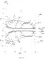
- FIG. 1 illustrates three known handheld microscope devices, each of which includes as its basic components a video or image capture sensor, a focus mechanism, a magnifying lens and an illumination source, such that surface reflections of the organ surfaces are avoided.
- the devices can be powered by either a battery or a mains supply. Images of the microcirculation (with therein moving blood cells) are saved either on computer memories or on video recorders, and are then analysed off-line using specialized software.
- a first known handheld microscope device is an orthogonal polarization spectroscopy (OPS) imaging device 100.
- Orthogonal polarized spectral imaging OPS imaging (5) works by eliminating directly reflected green (550 ⁇ 70 nm) polarized light from tissue surface via an orthogonally placed analyzer allowing structures below the surface to be visualized.
- the known OPS imaging device 100 includes a light source 105 that is coupled to a polarizer 110 such that it transmits a polarized incident light into a beam splitter 115, Reflected (polarized) light 120 is input to a scatterer 125 that scatters the image of the target tissue and transmits the scattered (de-polarized) light to a charge coupled device (CCD) 135.
- CCD charge coupled device
- the CCD is an integrated circuit etched onto a silicon surface forming light sensitive elements called pixels. Photons incident on this surface generate charge that can be read by an electronics device/element.
- the CCD 135 outputs a digital copy of the light patterns falling on the device to a video recorder/monitor 140 and off-line image analysis 145 may be subsequently performed.
- a second known handheld microscope device is a side-stream dark field (SDF) imaging device 150.
- SDF imaging works by emitting green (540 ⁇ 50 nm) light from light-emitting diodes (LEDs) arranged in a ring formation optically isolated from the central core of the light guide directly illuminating the tissue microcirculation.
- a SDF imaging device comprises a stroboscopic LED 155 ring-based imaging modality, which was introduced for clinical observation of the microcirculation 160.
- the microcirculation 160 can be viewed through a magnifying lens 165 and still or moving images of the MC can be taken by camera 170.
- the camera 170 is able to output a digital copy of the microcirculation 160 image adjacent the SDF imaging device 150 to a video recorder/monitor 140 and off-line image analysis 145 may be subsequently performed.
- a third known handheld microscope device is an incident dark field (IDF) imaging device 150.
- IDF imaging is a technique that, similar to OPS and SDF imaging, allows real-time visualisation of the microcirculation 160. Based upon the illumination of microvessels covered by a thin epithelial layer, it may be thought of as the successor to both orthogonal polarization spectroscopy (OPS), and more recently, sidestream dark field (SDF) imaging.
- An IDF imaging device comprises a light emitting diode (LED) 155 that also allows real-time clinical observation of the microcirculation 160.
- LED light emitting diode
- the microcirculation 160 can be viewed through a magnifying lens 165 and video images can be taken by image sensor 180.
- the image sensor 180 is able to output a digital copy of the microcirculation 160 image to a video recorder/monitor 140 and off-line image analysis 145 may be subsequently performed.
- Each of the three types of known HVM device 100, 150, 175 is optically configured using three different types of optical configuration in order to eliminate surface reflections of incident light to allow observation of the microcirculation below the surface of the organ.
- Green light is used because it is absorbed by haemoglobin present in the red blood cells allowing visualization of these cells as they flow in the microcirculation.
- Vessels not containing red blood cells cannot be observed in this way and remain invisible to this type of HVM (as under rest conditions about 30% of microvessels are not filled with red blood cells). They can, however, be filled red blood cells by opening the microvessels by topical application of a vasodilator such as nitro-glycerine.
- segmentation of (micro) vascular structures can be performed on images taken by, for example, the HVMs of FIG. 1 . Sublingually, these segmented vascular structures can be drawn manually to identify the various anatomic structures.
- Several types of hardware and software proposals have been put forward to identify the microcirculation and kinetics of the moving blood cells therein (see references [1]-[4]).
- a known hardware implementation in this general field includes WO0122741 (A2), titled: 'Medical applications of orthogonal polarization spectral imaging'.
- WO2001022741A2 describes a use of an OPS imaging device in order to capture images of the microcirculation of organ surfaces, in order to derive information of states of disease. The device is configured to only capture the microcirculation image and does so using OPS imaging.
- a further known hardware implementation in this general field includes US2014369588 (A1), titled: 'Darkfield imaging system and methods for automated screening of cells.
- EP1673007B / US2012089031 : titled 'System and method for imaging the reflectance of a substrate'.
- the microcirculation is identified as an asset to, in addition to the macrocirculation, and in order to gain such pertinent clinical information regarding the state of the cardiovascular system, EP1673007B proposes to monitor the microcirculation, using a side-stream dark field microscopy device (see reference [5]).
- the suggested device only yields image sequences, and the inventors of the present invention have recognised and appreciated that this has limited value in practical scenarios where identification of MC abnormalities coupled to a specific disease state, advice on the optimal type therapy and the prognosis of the state of disease is required.
- US2012269420 (A1), titled: ⁇ Image processing and machine learning for diagnostic analysis of microcirculation'; GB2510176 (A ), titled: 'Determining patient prognosis by microcirculation analysis'.
- US2012269420 describes a MC study approach that specifically mentions a calculation of only three parameters PPV, FCD and PVD. On the basis of this classification the authors of US2012269420 decide whether someone has a haemorrhage or is normal. The authors of US2012269420 focus their methodology on a decision as to whether (or not) to resuscitate a patient. The inventors of the present application have recognised and appreciated that calculation of these three parameters (FCD, PPV, PVD) provide very limited sensitivity and specificity to identify states of disease.
- GB2510176A proposes to use changes in FCD, pericapillary bleeding and capillary flow in order to determine a prognosis of patients (see reference [6]).
- GB2510176A specifies technical details on the image capture (camera) setup and mentions that flow analysis and counting of the capillaries may be carried out manually.
- WO 2014/114814 A1 describes a computer assisted video microscope (CAVM) device to capture microcirculatory images of neonates in the skin with a separate fibre-based spectroscope (DSR) that measures the spectrum.
- CAVM computer assisted video microscope
- US 2019/0139221 A1 describes an apparatus for detecting severe neutropenia based on images of nailfold capillaries.
- the images are taken in terms of microscopy videos of the microcirculation in nailfold capillaries of patients.
- the apparatus classifies a user to a user type of a set of user types, wherein at least one of the user types is associated with a diagnosis of neutropenia, based on a number of cellular events, such as the presence of red and white blood cells, detected in the set of capillaries.
- the inventors have recognised and appreciated that more relevant and more detailed information needs to be extracted from an MC in order to gain a deeper understanding of health, disease and therapy of these segmented microvascular structures. Furthermore, the inventors have recognised and appreciated that a speed of accessing, and accurate processing of, MC image data is needed in order to better advise clinicians of a health, disease and/or therapy of a patient.
- the invention seeks to mitigate, alleviate or eliminate one or more of the above mentioned disadvantages, either singly or in any combination.
- aspects of the invention provide an intelligent vital microscopy, IVM, device (which in some examples encompasses other microscopic configurations to image the microcirculation in humans for example including non-held microscopic devices, which can be placed or fixated on an organ surface for continuous monitoring and imaging of the microcirculation) and a method therefor, as described in the appended claims.
- an intelligent vital microscopy, IVM, device that includes: a receiver configured to receive at least one IVM image of a human microcirculation, MC, of an organ surface; and a learning processor coupled to the receiver and configured to: process the at least one IVM image that visualises cellular constituents of the MC, wherein the MC comprises arterioles, capillaries and venules, representing blood vessels, their flowing cells including the fluid in which they are in, and tissue cells surrounding the blood vessels of the at least one IVM image, and extract at least one MC variable therefrom, wherein the extracted at least one MC variable comprises at least one quantitative functional parameter of the MC that comprises at least one of: tissue red blood cell perfusion, tRBCp; total vessel density, TVD; blood volume; a proportion of perfused vessel density, PVD; a vessel diameter, VD; a RBC velocity; a proportion of perfused vessels, PPV; the oxygen saturation of haemoglobin in red
- the learning processor configured as above may facilitate obtaining clinical information from the diagnostic analysis, for example regarding an origin of the microcirculatory abnormalities. Besides, the learning processor facilitates obtaining clinical information regarding a presence of a pathogen or identification of the underlying disease. The learning processor may also facilitate obtaining clinical information regarding a type and amount of therapy that is deemed to be most effective in correcting, say, a pathogenic state.
- At least one functional parameter may be either: measured in steady state or for identification of a maximum present number of capillaries by application of a challenge, where the challenge is one of: metabolic, vasodilator, blood transfusion, exercise, focus quality, depth of focus, image movement and content of microcirculatory structures.
- such a MC variable may also include at least a portion or an entirety of the properties and structure of a trained neuronal network, incorporated in the IVM device trained for performing a specific task such as provide; output as feedback to a user as one or more of: (i) a quality of the at least one IVM image of a human sublingual microcirculation, (ii) a classification of identified MC abnormality, (iii) an origin of a cause of the identified MC abnormality, (iv) a recommended therapeutic strategy to normalize the MC abnormalities and resolve a disease state.
- the claimed IVM device is able to enhance MC images and recognise patterns. In doing so the claimed IVM device may be able to identify and quantify abnormal red and white blood cell flow kinetics, and/or provide distribution of blood cell velocities. In addition, the claimed IVM device may be able to calculate capillary hematocrit, discharge hematocrit and tissue red blood cell perfusion. The latter may be used in some examples as a resuscitation end point. Monitoring of the MC parameters from the extraction may also be performed, say on a routine basis by nurses who use the IVM device, so that they can detect abnormalities that can be used to trigger a further examination of the patient or clinical examination. Similarly, use can be made in a personal-care environment as part of home care for patients having cardiovascular disease.
- the identified extracted at least one MC variable includes at least one of: a quantification of a morphological parameter of the MC
- the identified at least one morphological parameter may comprise at least one of: functional microcirculatory structure, a total vessel density, TVD; a tortuosity; one or more fractal dimension; bifurcations of MC vessels.
- the functional microcirculatory structure may include at least one of: sputum glands, orifices, vessel loops, rectal crypts, cell to cell junctions, one or more cell dimensions, a functional state of mitochondria, properties of nuclei, microcirculatory units related to organ function, intestinal villi, renal tubular structures, liver lobule, alveoli, glycocalyx dimensions.
- an identified, extracted at least one MC variable enables a quantification of a morphological parameter of the MC, to analyse, say, morphological features of the vasculature (fractal dimensions, tortoisity) and to unexpected blood flow properties as well as the presence of abnormal microcirculatory structures.
- identified, extracted at least one MC variable may address a combination of various abnormalities, which may signify impending disease or an abnormal response to therapy. Analysis of such complex relationships would be very difficult in a supervised fashion, which is why unsupervised analysis using the claimed learning processor of such images as proposed herein provides a solution to alleviate this need, thereby providing superior diagnostic capacity of the IVM device.
- the identified disease may include a presence of sepsis and the learning processor may be configured to distinguish between inflammation and infection from early sepsis or septic shock based on the nature of a microcirculatory alteration.
- the learning processor may be configured to distinguish between inflammation and infection from early sepsis or septic shock based on the nature of a microcirculatory alteration.
- Analysis of the microcirculation according to the claimed invention may provide a sensitive and specific methodology for showing the presence of sepsis.
- the use of the claimed IVM device may provide a much more sensitive methodology to detect in a point-of-care setting the presence of sepsis and in extension to immediately start therapy and evaluate whether there is a suitable response.
- the learning processor may be configured to process the at least one IVM image and identify a type of shock that a patient is suffering from, and identify at least one of: a resuscitation strategy based on a type of fluid or blood, a futility of resuscitation, an area of the MC that requires resuscitating; a target for titration of intravenous fluids and blood.
- a resuscitation strategy based on a type of fluid or blood
- a futility of resuscitation an area of the MC that requires resuscitating
- a target for titration of intravenous fluids and blood Currently it is very difficult, and requires a high degree of skill, to be able to distinguish between the five categories of shock.
- the claimed IVM device may classify and distinguish between at least the following types of shock: cardiogenic, obstructive, distributive (septic shock), hypovolemic (haemhorragic) shock, anaemic shock (too few red blood cells caused by too much fluid administration or disease acquired such as in cancer).
- the IVM device may include in this analysis other clinical variables, such as blood pressure, cardiac output, blood gasses, lactate and biomarkers of organ injury, etc. Having identified the type of shock the claimed IVM device may identify an optimal resuscitation strategy to given (e.g. cardiopulmonary resuscitation, vasopressor agents, fluid administration, blood transfusion, extracorporeal assist device).
- the claimed IVM device may be able to identify that resuscitation from shock has been successful in correcting MC alterations induced by shock.
- the MC variable may also include a portion of an entirety of the properties and the structure of a trained neuronal network (e.g., supervised or unsupervised learning), incorporated in the IVM device and trained for performing a specific task.
- a trained neuronal network e.g., supervised or unsupervised learning
- the learning processor may evaluate a severity of a patient's wounds based on the MC analysis and, in response thereto: identify a wound healing of scar formation; and output a recommended fluid resuscitation or drug strategy that reduces edema formation, promotes wound healing and reduces scar formation.
- the claimed IVM device is able to analyse and identify a growth of the microcirculation, which is essential for normal wound healing since it transports oxygen to the growing tissue during wound repair.
- outputting this to, say, a clinician may provide a real-time (patho)physiological state of the wound, as well as the patient's response to therapy. Wound healing is dependent upon the development of scar formation, such as occurs the healing of surgical wounds including sternites.
- Wounds can be infected or be present in deeper tissue structure. Diabetic ulcer wounds are indicative of such.
- tissue regenerative therapies or wound dressings can enhance wound healing. Wounds as a result of burns can benefit from fluid resuscitation.
- such wounds and wound healing can be verified by the IVM device.
- the learning processor may, based on the MC analysis, identify heart failure of a patient, which carries with it a risk for myocardial infarction (Ml).
- Ml myocardial infarction
- Employing the IVM device herein described enables an observation sublingually on the MC image to identify if the heart is functioning well enough to achieve adequate microcirculatory perfusion. Abnormalities in blood flow kinetics might indicate myocardial instability.
- analysis of microcirculatory patterns may also provide sensitive information regarding the functioning of the failing heart being supported either by extracorporeal assist device or using medication.
- Identifying the success of such therapy is very difficult, which is greatly eased by using the IVM device described herein, as it can provide a tool for evaluating heart function at the level of the microcirculation and identify a success (or failure) of therapeutic support of the failing heart.
- the learning processor may, based on the MC analysis, identify a presence of infectious disease and a potential burden of infectious agents, which can otherwise only be shown by blood withdrawal and analysis.
- an inexpensive methodology, allowing easy measurement in large populations, is needed. This is especially needed in a setting of tropical disease, such as malaria and denge.
- the claimed IVM device has been shown to be sensitive in identifying microcirculatory alterations in malaria and denge.
- the inventors of the present invention have recognised and appreciated a need for a point-of-care methodology, for identifying infected people the infection itself and possibly then identify the infectious agent without having to withdraw blood.
- the IVM device herein described may identify abnormal microvascular structures on tumour surfaces as well ischemic boundaries associated with surgical reconstructions, which may lead to anastomotic leakage with great precision.
- the IVM device can be used to identify boundaries of pathology and healthy tissue or the nature of pathology of tissues, such as a tumour (or absence thereof) or ischemia (or absence thereof).
- the IVM device may allow direct diagnosis and identification of abnormal tissue and provide the surgeon important information in support of clinical decision making during surgery.
- HVM devices have proven successful in identifying morphological changes in vessel structures and subcellular components associated with the severity of diabetes. Differentiation and quantification of the severity of disease in diabetes may be achieved by the IVM device using, say, AI analysis of the different components of the microcirculation, such as the morphology, leucocytes kinetics, subcellular structures glycocalyx. In some examples, the IVM device may provide/output this information in a point-of-care manner, as well as potentially providing a self-care type of IVM device.
- hypovolemia is a condition where there is insufficient volume of blood in the circulation to allow the heart to sufficiently pump blood to perfuse the microcirculation with oxygen rich blood. It can occur as a result of blood loss or dehydration (which is the primary cause of death amongst children worldwide). Hypovolemia is difficult to diagnose.
- the IVM device allows precise identification and also enables a distinguishing between blood loss and dehydration.
- the IVM device described herein is able to image and interpret the properties of the vasculture that would allow an individual (and also across a population) to track as a vascular bioprint the development of the vasculuture. Since the microcirculation is that part of the cardiovascular system that is closest to the tissues, it can be regarded as the most relevant to follow in this respect. It can be visualized by the IVM device, but needs interpretation and quantification, which can also be provided by the described IVM device. Being able to measure the microcirculation and interpret its health and response to lifestyle would allow one to track ones vascular health over time. Since it is known that exercise benefits vascular health, the IVM device can allow an individual (or patient) to assess the benefit of exercise for the development of the (micro) vasculture needed for cardiovascular health.
- Extracorporeal organ support comprises a number of different technologies, whose aim is to mechanically support failing organ systems, such as can occur for the lung (mechanical ventilation, venous-venous extracorporeal membrane oxygenation (VVECMO), the heart (left ventricular assist devices, vaneous arterial extracorporeal membrane oxygenation (VA-ECMO), the liver (molecular adsorbent recirculating system MARS) and the kidney (hemodialysis and continuous renal replacement therapy (CRRT).
- EOS can also be supplemented by specific extra devices such as adsorbers meant to extract various toxin. However objective criteria for attachment of such additional devices and evaluation of their efficacy in terms of benefit remain unavailable, prior to the IVM device described herein.
- the mentioned EOS technologies also include artificial organs placed in patients, such as artificial heart, kidneys and livers.
- the efficacy of these known devices in supporting the cardiovascular system is uncertain, as no real quantitative clinical parameters exist that allows determination of whether the EOS is effective in supporting organ function and their microcirculation.
- the interpretation of MC images requires time consuming analysis off line and there is no current known technology that allows bedside interpretation of changes in response to, and/or providing advice on, how to proceed with EOS.
- the IVM device described herein advantageously provides such a technology platform to solve these problems.
- kidney disease is chronic or is in its acute form (acute kidney injury AKI) as it is difficult to predict and also difficult to therapeutically treat successfully.
- therapies for such diseases besides treatment with drugs also include a use of renal support or replacement devices, including hemodialysis and continuous renal replacement therapy (CRRT).
- CRRT continuous renal replacement therapy
- Known devices can also be supplemented by specific extra devices, such as adsorbers (specialized filters) that are meant to extract various toxins from the blood.
- adsorbers specialized filters
- the herein described IVM device is able to provide diagnostic bedside technology which is able to show a presence of disease, severity of disease and response to therapy, with the needed sensitivity and specificity, and identify the progress of disease and response to therapy.
- the IVM device described herein has been able to show a presence of MC alterations associated with kidney disease and its therapies, indicating that the IVM device provides a window of diagnostic capacity.
- the IVM device provides an interpretation of images and provides advice for therapeutic success, therapeutic titration and/or advice on applying alternative therapeutic strategies.
- the IVM device herein is able to analyse the MC and examine the functional properties of the vascular network, both in and on the brain, as well as in distant locations, and may provide information regarding the risk, presence or response to therapy regarding brain disease.
- the IVM device is able to be applied sublingually (small and large vessel disease) or adapted to the retina as well as applied during neurosurgery in order to provide a window of diagnostic possibility.
- a mechanism to identify a high blood cell concentration, where there are abnormally high amount of red and white blood cell count such as occurs in polycythemia and leukemia can be provided by the IVM device described herein, without a need for the current requirement to withdraw blood.
- a blood-less, non-invasive methodology by the IVM device enables a clinician to visualize red and white blood cells.
- the IVM device provides an alternative approach, by observing abnormalities of the MC by IVM endoscopy, for example combined with AI for identification and gradation of tissue pathology.
- This IVM device approach can also advantageously be used during gynaecological oncosurgery, for identification of a presence of tumours and micrometasis associated with for example ovarian cancer.
- AI analysis and identification may aid a surgeon to identify cancer, resection margins and or provide a decision for surgical resolution of such cancers and/or for initiating chemotherapy or radiation therapy for point-of-care of decision making.
- the IVM device described herein may also be applied to gynaecology and urogynaecological diseases, including identification for a success of therapy for treatment of prolapse surgery (e.g., laser or surgical), as well as other urogynaecological disorders such as vaginal atrophy and its treatment by oestrogen therapy.
- prolapse surgery e.g., laser or surgical
- other urogynaecological disorders such as vaginal atrophy and its treatment by oestrogen therapy.
- the described IVM device is able to identify the severity of atrophy and the efficacy and timing of oestrogen therapy.
- SCA sickle cell anaemia
- the method includes: receiving at least one IVM image of a human microcirculation, MC, of an organ surface; processing the at least one IVM image of a human MC that visualises cellular constituents of the MC, wherein the MC comprises arterioles, capillaries and venules, representing blood vessels, their flowing cells including the fluid in which they are in, and tissue cells surrounding the blood vessels of the at least one IVM image by a learning processor; extracting at least one MC variable from the processed at least one IVM image; wherein the extracted at least one MC variable is according to the first aspect of the invention; identifying from the extracted at least one MC variable of the at least one IVM image, based on MC analysis of training data images repeatedly and previously input to the learning processor for analyzing a function and morphology of MC, at least one of: an underlying cause for an observed abnormality, an intervention, a disease state, a disease diagnosis, a presence of a pathogen
- IVM intelligent vital microscopy
- HVM hand-held vital microscope
- non-held microscopic devices such as intra-vital microscopes, endoscopes, non-hand held miniature vital microscope fixated to an organ surface, fundus cameras and surgical microscopes equipped with suitable optics to image the microcirculation.
- IVM device used herein is intended to encompass all such vital microscopes, which are embedded or placed on tissue surfaces for on-line visualization of the MC. These include microscopy embedded in operating microscopes, intravital microscopes or endoscopes.
- Miniaturized non-held IVM devices can be fixed or implanted in the oral region or other locations in the body to enable continuous visualization of the microcirculation and assessment by the learning processor. Such miniaturized non-held IVM devices will benefit largely from the concepts described herein because continuous evaluation of an evolution of the MC will allow early therapy to be initiated in advance of clinical deterioration of the patient. Thus, examples of a IVM device described herein, as well as the term IVM device' used herein, is intended to additionally cover this range of devices.
- examples of the invention are described with reference to an intelligent vital microscopy, IVM, device to observe sublingual microcirculation of a processed image or sequence of images, it is envisaged that the concepts described herein are equally applicable to observe sublingual microcirculation of a processed image or sequence of images of any organ surface.
- examples of an IVM device described herein to observe sublingual microcirculation from a processed image or sequence of images is intended to cover an observation of microcirculation of any organ surface from a processed image or sequence of images.
- the invention described herein proposes a use of a learning processor embedded in the IVM device in order to provide real-time, on-line analysis of images for identification of an underlying disease.
- the real-time, on-line analysis of images for identification of an underlying disease may also include a suggestion for a use of therapy, for example both for clinical decision making at a bedside and needed for titrating therapy.
- These learning processor approaches may provide sensitive analysis of images in order to identify MC alterations and identify (for example in an unsupervised format) as yet uncovered information regarding the health and disease of patients and information obtained from one or more MC parameters extracted from a processed one or more IVM image.
- an artificial neural network which encompasses a network of artificial neurons and nodes designed for solving learning processor problems, such as those in artificial intelligence (Al).
- the connections of the artificial neurons may be weighted to model excitatory or inhibitory connections, which may then be summed.
- an activation process may be used to define an amplitude of the output.
- the artificial neural networks may be used for predictive modelling and adaptive control. In this manner, such neural networks may also be trained via a dataset.
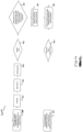
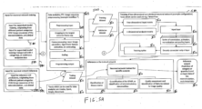
- self-learning or unsupervised learning resulting from analysis of data sets as illustrated in FIG. 5A and FIG.
- processed MC images may occur within the neural networks, which allows the neural network to derive conclusions from a seemingly unrelated information set.
- such methodology may be integrated into the functionality of an IVM device, for example at a bedside in a point-of-care fashion, in order to provide direct information regarding an origin of a functional state of the MC, its related disease state, advice for therapy and identifying an outcome, etc.
- the IVM device may be able to remotely obtain such information, if stored in a large database that the IVM device is connected to, for example in a wireless (or indeed wireline) configuration.
- artificial intelligence when used herein, it is intended to encompass any form of supervised processor learning or unsupervised processor learning, as well as machine learning (ML). Examples of such processes are shown in FIG. 5A and FIG. 5B .
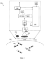
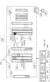
- FIG. 2 illustrates an example block diagram of an intelligent vital microscopy, IVM, device 200 embodied with a learning processor to enable training and inference of neuronal networks for microcirculation analysis, in accordance with example embodiments of the invention.
- the IVM device 200 includes a controller 214 that maintains overall operational control of the IVM device 200.
- the controller 214 is coupled to a signal processor 208, which is generally realized by a Digital Signal Processor (DSP).
- DSP Digital Signal Processor
- the signal processor 208 may include a health and clinical decision making circuit 221 or processor that is configured to analyse AI data related to at least one function and morphology of a microcirculation of humans, in order to evaluate a state of health of a subject.
- the clinical decision making circuit 221 may exist external to the IVM device 200.
- the controller 214 is also coupled to a buffer module 217 and a memory device 216 that selectively stores data relating to operating functions of the IVM device 200.
- a timer 218 is operably coupled to the controller 214 to control the timing of operations (e.g. transmission or reception of time-dependent signals) within the IVM device 200.
- the IVM device 200 includes a learning processor 222, which in some examples may be implemented as a neural network, as shown in FIG. 9 .
- the learning processor 222 may be operationally responsive to the controller 214.
- the signal processing module 208 and/or controller 214 may receive inputs from one or more input device or sensor modules 220, for example which may receive one or more clinical variables.
- the learning processor 222 may include (or be operably coupled to) an image acquisition and pre-processing circuit 221, which may be configured to organise the input data into a suitable format.
- the input image data received by the image acquisition and pre-processing circuit 221 may be provided by a camera 225 coupled to a receiver 228 comprising a video camera/image capture circuit 227.
- the video camera/image capture circuit 227 may include or be operably coupled to a focusing circuit/mechanism to focus the image provided by the camera 225.
- the IVM device 200 includes a disposable, see-through cap 285, to allow unfettered access to the subject's tissue 290.
- An HVM is usually housed in a tube consisting of a magnifying lens 275, focus mechanism, and image sensor/video camera and a data acquisition module (not shown).
- the IVM device 200 to analyze the function and morphology of the microcirculation of humans in order to evaluate a state of health of a subject may also consist of different coloured LEDs 280 allowing multiple wavelength analysis of the images to extract spectroscopic information regarding the oxygen saturation of the haemoglobin in the red blood cells 295 or spectroscopic information about the composition of the tissue 290 cells.
- the tissue surface 290 is shown with microcirculation microvessels embedded with flowing blood cells 295 visible.
- IVM device In alternative examples of the IVM device, other microscopic modalities may be incorporated into the IVM device in order to enhance the imaging capacity of the IVM device to identify sub-cellular structures (e.g., endothelial glycocalyx, cell to cell junctions, mitochondria, nuclei) and platelets, as well identification of the type of blood cells present in the microcirculation. Furthermore, in some examples, it is envisaged that the IVM device may be used to observe microorganisms, such as viruses, parasites and bacteria.
- sub-cellular structures e.g., endothelial glycocalyx, cell to cell junctions, mitochondria, nuclei
- platelets e.g., cell to cell junctions, mitochondria, nuclei
- the IVM device may be used to observe microorganisms, such as viruses, parasites and bacteria.
- the IVM device may be adapted such that spectroscopy may be used, for example to allow composition of cells to be determinants, such as an amount of collagen in cells obtained by polarization spectroscopy and the redox state of the mitochondria obtained by NADH fluorescence. Additionally, in some examples, it is envisaged that the IVM device may be configured to analyse more than just a single instance in time, in that it may be configured to also analyse changes occurring over time, in order to identify progress of health or disease or response to therapy.
- the IVM device 200 is a handheld-microscope that is able to image moving blood cells in the microcirculation, as well as imaging the structure of the microvessels (as shown in FIG. 3 ). In operation, the IVM device 200 is held on or above an organ surface or a subject's/patient's tissue, in order to provide an on-line, real-time visualization of the microcirculation and their cellular constituents embedded within flowing blood cells and observed through the microvessels. In some examples, the IVM device 200 is a miniaturized non-held IVM device, which can be fixed or implanted in the oral region or other locations in the body to enable continuous visualization of the microcirculation and assessment by the learning processor.
- Such a miniaturized non-held IVM device will benefit largely from the concepts described herein because continuous evaluation of an evolution of the MC will allow early therapy to be initiated in advance of clinical deterioration of the patient.
- the use of real-time AI techniques in this process may assist immediate processing and classification of microcirculation microvessels images and enable interpretation of the image data for an accurate diagnosis, choice of therapy and/or assist clinical decision making.
- the IVM device 200 also includes a power supply (not shown), a light guide with a magnifying lens 275 at its tip and suitable illumination modality for illuminating an organ surface. Acquired images are routed to an image acquisition circuit, which can be a video camera or an image acquisition sensor. In other examples, the IVM device 200 may also include a conventional intra-vital microscope, or a hands free vital microscope fixated on to a tissue surface, or an operation microscope or endoscope, equipped with suitable magnification to view the microcirculation in organ surfaces.
- the learning processor 222 which in some instances may be implemented as an artificial intelligence (Al) processor, may be integrated in the IVM device 200 in order to allow identification, quantification, optimization, classification and interpretation of the images as well as identifying a health care strategy based on AI insight.
- the learning processor 222 is configured to extract at least one MC variable from a processed MC image or sequence of MC images, and identify from the extracted at least one MC variable of the at least one IVM image at least one of: an underlying cause for an observed abnormality, an intervention, a disease state, a disease diagnosis, a medical state of the human; a presence of a pathogen.
- the AI circuit can be physically integrated into an IVM device (or system) or be placed off-line in a PC, mobile telephone or in the cloud, for example.
- the IVM device 200 includes an output 206 coupled to the learning processor 222, for example via the controller 214, and configured to output from the learning processor 222, an identification of the extracted at least one MC variable from the MC image or sequence of MC images.
- an intelligent vital microscopy, IVM, device 200 comprises: a receiver 228 configured to receive at least one IVM image of a human microcirculation, MC, of an organ surface; a learning processor 222 coupled to the receiver and configured to: process the at least one IVM image and extract at least one MC variable therefrom, and identify from the extracted at least one MC variable of the at least one IVM image at least one of: an underlying cause for an observed abnormality, an intervention, a disease state, a disease diagnosis, a medical state of the human; a presence of a pathogen; and an output 206 coupled to the learning processor 222, for example via the controller 214, and configured to output the identification.
- IVM device 200 can be realized in discrete or integrated component form, with an ultimate structure therefore being application-specific or design-based.
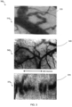
- FIG. 3 a series of pictures 300 of white and red blood cells in microcirculation analysis is illustrated, which can be analysed using AI techniques according to examples of the invention.
- white blood cells are shown as white cells 315 in the arched capillary in the middle of the picture.
- microcircirculation vessels 345 are shown with blood flow therein, as seen in moving cells.
- flowing single red blood cells are illustrated 375 in sublingual in magnifed microcirculation recorded by an HVM device. Red blood cells have a diameter of about 5 micrometers in this example. For clarity purposes only, note that the dimensions of the field of view in these illustrations are approximately 200 microns.
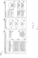
- each module is the input size of the next one, where the acronym RELU refers to a rectifier linear unit.
- Table 1 describes the composition of the convolutional neuronal network used for prediction of microcirculatory vasodilation.
- the input layer is connected via rectified linear units activation functions (RELU) to a module consisting of a two-dimensional convolution layer (2D conv) followed by batch normalization to reduce covariance shift, sample-based discretization via two-dimensional max pooling, and coadaptation prevention via a 25 % dropout layer (module A).
- RELU rectified linear units activation functions
- the output is then routed via RELU to a module consisting of a two-dimensional convolution layer followed by batch normalization (module B) followed by a module A using an increasing number of filters to reduce the matrix size.
- module B batch normalization
- module A using an increasing number of filters to reduce the matrix size.
- the output is routed via RELU to a dense layer, again batch normalized and routed to a 50 % dropout layer to a final dense layer that produces the two-category output via a softmax activation function.
- the main function of the microcirculation is to deliver oxygen to the tissues by oxygen carrying red blood cells (RBC) to meet their metabolic needs for optimal cellular function required to enable them to support organ function. It is also the main compartment involved in the immune system where leucocytes interact with vessels and tissue cells to exert their functional activity. In states of infection and inflammation leucocytes and endothelial cells become activated and leucocytes will stick and roll along the endothelial wall and eventually extravagate into the tissues. The activity of the leucocytes can be observed and quantified using IVM device observations and indicate the presence of inflammation (see reference [9]).
- the alterations seen in the microcirculation during sepsis and septic shock are very specific including heterogeneous perfusion of the microcirculation with plugged vessels next to ones where there is flow.
- Alterations in the MC represent the most sensitive and specific hemodynamic alteration seen in the cardiovascular system more so than systemic hemodynamic variables.
- Such sepsis associated alterations detected under the tongue can be associated with severe organ dysfunction and lack of responsiveness to therapy whereas systemic variables do not show such discrimination. It is envisaged that the ability to distinguish between infection and sepsis and its response to therapy, which currently is an important clinical problem, may be possible using IVM analysis of the MC.
- the functional state of the microcirculation is key in the interaction between the metabolic requirements of the tumour and its microcirculation.
- the inventors have recognised and appreciated that knowledge relating to the microcirculation, either on the tumour or at a distant location, would be beneficial in identifying the stage of the tumour and the optimal therapy and therapeutic target that needs to be applied.
- Other chronic disease states such as diabetes, kidney disease, heart failure and hypertension can also be identified by known HVM observations, although there is no known HVM methodology that presently distinguishes between these different disease states.
- the learning processor is configured to process and extract at least one MC variable from a processed MC image or, optionally, from a processed sequence of MC images.
- the extracted at least one MC variable may include a quantification of a morphological parameter of the MC.
- the extracted at least one MC variable includes at least one quantitative functional parameter of the MC.
- the extracted at least one MC variable may be the Functional Capillary Density (FCD) or leucocyte kinetics.
- the at least one quantitative functional parameter of the MC may include at least one of: MC hemodynamic values; capillary; a venule; arteriolar blood flow; an identification of at least one type of vessel; a proportion of functional capillary density, FCD, of flowing red blood cells, RBC, that carry oxygen; tissue RBC perfusion; a leucocyte velocity; a microvascular flow index of a flow heterogeneity, MFlhet; rolling and sticking leukocytes, fluorescence spectroscopy of a number of platelets and leucocytes.
- the at least one quantitative functional parameter of the MC includes at least one of: tissue red blood cell perfusion (tRBCp); total vessel density (TVD); blood volume; a proportion of perfused vessel density (PVD); a vessel diameter (VD); a RBC velocity; a proportion of perfused vessels (PPV); oxygen saturation of haemoglobin in red blood cells; a capillary tube and discharge haematocrit.
- the macro-vasculature blood vessels >100 ⁇ m including arteries and veins
- the microcirculation blood vessels ⁇ 100 ⁇ m consisting of capillaries, arterioles and venules.
- the former vessels can be detected by MRI, angiography, echo, optical coherence tomography, fluorescence angiography of such organs as heart, brain and retinal vascular structures.
- the microcirculation can be detected by the IVM device, intravital microscopy (including fluorescence and capillaroscopy).
- the IVM device can directly image the capillaries and the movement of single blood cells in them, thereby allowing uniquely the ability to provide quantitative information regarding the functionality of the MC.
- the term IVM is used to also include non-hand-held vital microscopes, which are embedded or placed on tissue surfaces for on-line visualization of the MC, such as being embedded in operating microscopes, intravital microscopes or endoscopes.
- Fluorescence spectroscopy can be incorporated into the IVM device in order to obtain spectroscopic information about plasma and tissue molecular and particle constituents.
- histological information concerning microcirculatory morphology can be obtained from biopsy analysis and optical coherence tomography. These can also be used as input variables for Al.
- Miniature devices implanted in the oral region or other locations in the body will enable continuous visualization of the microcirculation. These will benefit greatly from the examples described herein because continuous evaluation of the evolution of the MC will allow early therapy to be initiated in advance of clinical deterioration of the patient.
- AI will follow changes the functional state of the MC and identify the state of health and disease and response to therapy.
- HVM hand-held vital microscopes
- OPS imaging see reference [5]
- SDF imaging see reference [6]
- IDF imaging devices see reference [8]
- stand-alone i.e. non hand-held but placed at a location in the body for continuous monitoring
- These cameras have been used extensively to observe sublingual and oral microvascular structures and function in disease and health (see reference [11]).
- These techniques have also been used to observe microcirculatory structure in other human orifices as well as on human organ surfaces during surgery. Structures that are observed include flowing red and white blood cells, vessel structures and morphology, parenchymal cells as well as subcellular structures such as endothelial glycocalyx, platelets, cell and vascular and cell junctions as well as nuclei.
- Spectrophotometry can be used to evaluate the state of oxygenation of the microcirculation and can be used in combination with the IVM device as well as spectroscopy (e.g. Raman spectroscopy) to obtain molecular information about plasma and tissue molecule information providing additive integrative information about microcirculatory hemodynamic and oxygenation variables together.
- oxygen electrodes or BOLD can be used to measure the tissue oxygenation as well the assessment of mitochondrial oxygen tension using the decay of delayed fluorescence of mitochondrial protoporphyrin.
- An IVM can also be implemented as a stand-alone device where a microscope sensor placed on an organ surface independently monitoring continuously to obtain microscopic images of the MC. Such one-spot measurements would allow AI analysis of the behaviour of blood cells in single micro vessels.
- Such images could be analyzed locally in a point-of-care setting or be analyzed in a remote location (e.g. telemetrically to a mobile cell phone).
- Addition of spectrophotometry or other spectroscopic techniques can be used to obtain even more detailed MC and tissue information.
- Other techniques which can be used to observe the MC include fluorescence or con focal microscopy which can be incorporated into such IVM devices.
- Such microscopic information can also be obtained by microscopic endoscopy where a magnifying lens is attached to the end of an endoscopy and imaging of the MC is achieved either by optical fibers or by placement of a high definition camera placed at the tip of the endoscope.
- imaging capsules introduced intra-gastrically can also be used observe (micro)vascular structures. All of the above measurements are considered to provide information regarding the MC.
- any reference to AI encompasses a mathematical methodology to extract information from signals and images that may be similar to that used in cognitive functions in humans associated with learning or pattern recognition, in a supervised or unsupervised deep learning setting (see FIG. 5A and FIG. 5B ).
- flexible adaption is able to extract information previously not necessarily recognized or hypothesized or based on manually derived algorithms specific to the measurement of defined parameters.
- AI in the setting of unsupervised learning is able to exploit serendipity in much the same way as scientific discoveries are made by chance.
- any reference to ML may be used as a mathematical methodology that refers to mathematical and statistical methods, which can without supervision, analyze and identify a specific outcome related to in the case of this paper health and disease. It applies to algorithms and iterative methodologies to arrive at an optimal identification of a set task. It is used for data mining.
- Some examples of the invention described herein propose ML to be used to quantify morphological and functional aspects of the microcirculation being used as input variables with the aim of identifying health disease or outcome of therapy.
- any reference to AI examples of the invention described herein encompass supervised and unsupervised learning, as well as Machine Language.
- An artificial neural network is a network of artificial neurons and nodes meant for solving learning processor-type problems, such as artificial intelligence (Al) problems.
- the connections of the neuron are weighted to model excitatory or inhibitory connections and summed.
- An activation function defines the amplitude of the output.
- these neural networks may be used for predictive modelling and adaptive control.
- these neural networks may also be trained via a dataset, such as the examples described herein. Self-learning or unsupervised learning, resulting from analysis of datasets, can occur within the network, which allows the network to derive conclusions from a seemingly unrelated information set.
- the dataset which the AI algorithms will use as a learning platform will include, but not limited to, data from patients and healthy individuals including all ages from new born and onwards.
- Data will consist primarily of microcirculatory images (still and moving images) obtained mainly, but not limited to, sublingual and oral microcirculation as well images from all other internal and external body and organ surfaces. These microcirculatory images are obtained from various types of vital microscopes described above. Still histological images of the microcirculation as obtained from biopsies are also to be included in the data set. Movies or images of the subjects can either be obtained at a single time point or be followed over the course of time either short time (e.g.
- the data set will include detailed information concerning single individuals as well as databases of categories of healthy subjects as well patients suffering from particular disease states or undergoing specific therapeutic interventions.
- microcirculatory variables derived from the various MC techniques discussed can be derived using conventional analysis or derived using AI methodologies. These MC functional parameters will be then used as a learning set for AI to evaluate the various example embodiments described herein.
- MC functional parameters which will be used as input variables AI learning set will include but not limited to: Microcirculatory hemodynamic values: capillary, venule and arteriolar blood flow and capillary blood volume and haematocrit (see reference [11]), identification of types of vessels (e.g. capillaries, capillary loops, arterioles, venules), Proportion of perfused vessel density or functional capillary density (PVD [mm/mm2].
- FCD density of functional capillaries where flowing red blood cells (RBC) carrying oxygen are measured ⁇ ; Vessel diameters (VD [ ⁇ m]); RBC velocity (RBCv [ ⁇ m /sec] or arbitrary units such as obtained from Laser Speckle Imaging; Leucocyte velocity ( ⁇ m /sec) and number; proportion of perfused vessels (PPV [%]); Microvascular flow index; Flow heterogeneity (MFlhet (see reference [11]) or MC expressed as a function of a histogram of MC variables); Rolling and sticking leukocytes ( ⁇ m/sec), number of leucocytes, microcirculatory RBC Hb saturation, capillary tube and discharge haematocrit.
- the metric describing the number and/or density of functionally flowing red blood cell in a field of view is referred to as the parameter tissue red blood cell perfusion (tRBCp), and can be calculated using MicroTools or the IVM device and is used as target value for resuscitation following a state of shock.
- tRBCp tissue red blood cell perfusion
- a microcirculatory functional parameter can be measured in steady state, but also as a result of a challenge.
- These challenges can identify the reserve microcirculation which can be therapeutically targeted for recruitment with the aim of improving the capacity of the tissues to extract oxygen from the capillaries due to the shorter diffusion distances.
- Such challenges to the microcirculation include the administration either topically or systemically of a vasodilator such as nitro glycerine or other challenges such as a metabolic challenge (a meal), blood transfusion and exercise.
- Morphological parameters including but not limited to total vessel density (TVD [mm/mm2]); functional microcirculatory structures (sputum glands, orifices, vessel loops, rectal crypts, microcirculatory units related to organ function, intestinal villi, renal tubular structures, liver lobule, alveoli, glycocalyx dimensions (e.g. measured using the IVM device and/or RBC perfused boundary region as a measure of glycocalyx dimensions), tortuosity, fractal dimensions, bifurcations of MC vessels. AI identification of mean values but also of histograms of the distributions of said parameters within a population or within a single measurement of the microcirculation (field of view), or within a single capillary.
- spectroscopic measurements measuring tissue and plasma molecular and particular constituents as well morphometric information about the MC obtained from optical coherence tomography and confocal microscopy are included as input variables in the assessment of the MC.
- MC variables extracted from such techniques such as intravital microscopy (including fluorescence and capillaroscopy), spectrophotometry, laser speckle imaging, near infrared spectroscopy, fluorescence vital microscopy (see reference [11]). Fluorescence spectroscopy can be incorporated into the IVM device to obtain spectroscopic information about plasma and tissue molecular and particle constituents. In addition, histological information concerning microcirculatory morphology can be obtained from biopsy analysis and optical coherence tomography. All MC variables extracted from these techniques either by AI or conventional methodologies can be used as input variables for the data set required for AI learning.
- microcirculatory alterations including capillary refill time, peripheral temperature, arterial venous CO2 gap, contrast enhanced ultra sound derived microcirculatory flow measures (intra-renal microcirculatory transit time, laser speckle imaging, laser Doppler flow measurements, gastric tonometry, near infrared spectroscopy. Although these measures do not image the MC, in some examples of the invention they may be used as input variables to AI analysis, providing extra information related to states of health, disease, indications for therapy and outcome.
- contrast enhanced ultra sound derived microcirculatory flow measures intra-renal microcirculatory transit time, laser speckle imaging, laser Doppler flow measurements, gastric tonometry, near infrared spectroscopy.
- AI analysis will also analyze the quality of the IVM device's obtained measurements during a measurement session, prior to being accepted for analysis and for being included into the data set.
- Clinical and health related data set Included as part of the data set and paired with the above microcirculatory variables associated with each patient and subject are the demographics of the subjects and patients, information about the presence of co morbidities (e.g. hypertension, diabetes, heart failure, COPD, obesity, fragility, mental disease, cancer) and clinical information relating to these conditions (imaging, pathological information, clinical chemistry, functional tests, genomic analysis of genes related to states of disease) with the microcirculatory variables listed above.
- co morbidities e.g. hypertension, diabetes, heart failure, COPD, obesity, fragility, mental disease, cancer
- clinical information relating to these conditions imagingg, pathological information, clinical chemistry, functional tests, genomic analysis of genes related to states of disease
- the full range of conventional clinical parameters including, but not limited to, the measurements of hemodynamic variables, field potentials (e.g. ECG, EEG), ultrasound and other variables related to the measurement of organ function and identification of disease are included as input variables. Also, as input variables for the data set and output parameters to show Al related benefit are considered scoring systems related to health and disease.
- DALY Disability-Adjusted Life Year
- APACHE score the "Acute Physiology, Age, Chronic Health Evaluation score is a severity-of-disease classification system used in the ICU
- SOFA Simple Organ Failure Assessment
- organ function e.g. renal failure see reference [18], heart failure
- liver failure and brain function is included as well as biomarkers related to several disease states such as cancer, sepsis kidney disease heart failure and diabetes.
- This section is a brief outline of the type of input parameters which can be given to an IVM device in support clinical decision making procedures.
- Physical examination is a corner stone of input variables for clinical decision making and is well described in the medical literature (age, blood pressure, BMI, co-morbidities, sex, lacatate and acid base disorder and arterial venous CO2 gap, etc.). Measurements made in association with such examinations include blood chemistry analysis where among other variables indicators of organ function in terms of biomarkers are assessed (e.g., liver, kidney and heart function), humoral, metabolic immunological and electrolyte biochemical variables as well as haematological variables (blood gas, blood components, haemostasis, immunological variables) can be assessed as and used as input parameter.
- blood chemistry analysis where among other variables indicators of organ function in terms of biomarkers are assessed (e.g., liver, kidney and heart function), humoral, metabolic immunological and electrolyte biochemical variables as well as haematological variables (blood gas, blood components, haemostasis
- Similar measurements can be made in the analysis of urine and faeces. Besides such measurements invasive measurements can provide a range of hemodynamic variables related to the function of the cardiovascular system (blood gas, heart function, blood pressure, blood flow). Field potentials can give important information regarding organ function and include ECG, EEG and EMG. Imaging modalities used in clinical medicine include angiograms, MRI, CT and echo scans. Further information can be obtained by histological examination of biopsy material as well by endoscopic examination. Included here is also genetic information obtained by gene sequencing.
- FIFE Frailty Index For Elders
- PRISM Paediatric Risk of Mortality
- GCS Glasgow coma scale
- the microcirculation refers to that part of the cardiovascular system that embodies the smallest vessels of the body, which vascularizes the tissue cells of the various organ systems. Its main function is to transport oxygen carrying red blood cells and nutrients to the tissue cells to support their respiratory needs. In addition, it is the interface between circulating blood and tissues cells where immunological and humoral actions occurs.
- the microcirculation contains blood vessels with flowing blood cells in plasma with vascular diameters less than I00 ⁇ m.
- the arterioles, capillaries and venules, their flowing cells including the fluid in which they are in (plasma) and the tissue cells surrounding the blood vessels (endothelial cells, smooth muscle cells and neighbouring parenchymal cells) are collectively referred to as the microcirculation.
- Their physical diameters, density, morphology and function e.g.
- diabetes cancer, hypertension, sepsis, cardiovascular compromise, heart and kidney failure, arteriosclerosis, intestinal ischemia, venous insufficiency, pre-eclampsia, small and large vessel disease, eye disease, Alzheimer's ) affecting morphology and blood flow kinetics often as a result of a vascular or metabolic challenge as well acute disease states such as stroke, surgery, myocardial infarction, cardiac arrest, trauma and states of shock (see reference [11]).
- Some examples of the invention described herein propose relating abnormal variables related to the blood flow kinetics and morphology of the microcirculation to other physiological properties of individuals and analyzing these using AI and assessing and identifying health and disease advising on therapy and identifying their outcome.
- microcirculatory abnormalities or alterations refer to changes in the parameters with respect to their values in normal healthy individuals and signal a presence of disease. These alterations have been described in reference [10] and [11]. In these references a differentiation of microcirculatory abnormalities have been distinguished into four types of abnormalities:
- Examples of therapies based on these observations of MC abnormalities include, for Type 1 abnormality: antibiotics, anti-inflammatories, blood transfusions, fluid therapy; for Type 2 abnormality: a reduction in the type or amount of fluid being administered, the administration of blood or RBC enhancing therapy (iron or EPO); for Type 3 abnormality: a reduction in the vasopressor agent, administration of a vasodilatory agent, treatment of a tamponade by relieving an obstruction; for Type 4 abnormality: a resolution of the capillary leak removal of excessive fluid by diuretic therapy and or haemodialysis.
- microcirculatory abnormalities consist of a slowing of the velocity of blood cells. This defect is associated with either a failing heart (heart failure) or the presence of insufficient volume (hypovolemic shock).
- Associated therapies can be, respectively, the administration of volume (fluids or blood) or of cardiotonics in support of heart contractility (such as inotropics) or for the attachment of a left ventricular assist device.
- Second type of abnormality consists of a decrease in functional vascular density. Such a condition can be associated with a disease state where vasodilator therapy is indicated or blood transfusion.
- leucocytes The presence of an abnormal kinetics of leucocytes , such as leucocyte count be up, or leucocyte rolling and sticking are observed are associated with the presence of inflammation and or haematological disorder.
- Appropriate therapies which are indicated include chemo/radio therapy, blood transfusions and anti-inflammatory drugs.
- pathogens in the MC such as the presence of microorganism such as bacteria, viruses, fungi and parasites underlying disease states.
- the IVM device described herein may be trained by an appropriate learning set including a multitude of clinical and microcirculatory parameters mentioned in our patent, will provide a much more sensitive and specific methodology to identify specific states of disease and advice specific therapeutic strategies, which will improve outcome much more readily than available diagnostic strategies.
- HVM hand-held vital microscopes
- microcirculation examples of the invention described herein discuss the measurement of the microcirculation (MC) and its cellular constituents, for example at a patient's bedside and describe the methodologies that can be used.
- the microcirculation can be measured in several ways in humans.
- HVM vital microscopy
- OPS orthogonal spectral imaging
- SDF side stream dark field
- IDF incident dark field
- Visible light can be used but also fluorescent imaging as a consequence of in vivo labelling of cells using in vivo methodologies (e.g. Na-fluorescein, indocyanine green and other fluorophores which can adhere to cells, plasma or particles allowing detection by fluorescence spectroscopy).
- in vivo methodologies e.g. Na-fluorescein, indocyanine green and other fluorophores which can adhere to cells, plasma or particles allowing detection by fluorescence spectroscopy.
- Other techniques for monitoring the microcirculation include Laser Speckle Imaging and Laser Doppler Imaging which are also covered in this document when referring to the measurement of the MC.
- Different functional tissue variables related to the MC can be measured using different wave length of light. The most obvious one in this respect is the use of different wavelengths of light to measure the oxygen saturation of Hb in the flowing red blood cells. Different wavelengths of light can also be used to identify the presence of leucocytes.
- Information regarding the functional and anatomical properties of the MC structures are used as input variables to AI algorithms to relate them to health, disease and response to therapy.
- MC images can be analyzed and functional variables extricated from images of moving cells in the MC by analysis of moving cells in the images using advanced image processing methodology.
- Several software methodologies have been introduced to analyze microcirculatory images in this manner (see references [2], [3], [4], [12]) but as yet no one has used AI to analyze such MC images.
- a proof of concept study is demonstrated herein wherein the application of AI methodology for the analysis and classification (the presence of vasodilation or not) of microcirculatory images are comprehensively presented.
- AI methodology for the analysis and classification (the presence of vasodilation or not) of microcirculatory images are comprehensively presented.
- Such analysis is carried out by manual or semiquantitative analysis having no intelligence and requiring much time and having no aspect of self-learning or AI (see references [7], [13]).
- Examples herein described disclose in sufficient detail, the method and system for analysis of MC variables as input learning for development of an IVM device in combination with other markers of health and disease to identify the state of health and response to therapy.
- the AI based analysis method described here is used to identify disease states and indicators of health previously not known by analysis of (micro)vascular morphology and function in combination with other physiological medical or clinical parameters.
- functional parameters can be obtained by different techniques including invasive and non-invasive monitoring using echo, (f)mri and endoscopy, hemodynamic and biochemical biomarkers (blood chemistry, biomarkers related to the function of various organ systems) as well as analysis of gene sequence analysis related to the evaluation of health and disease.
- Hemodynamic parameters included can be such variables as systolic, mean and diastolic blood pressure (BP); stroke volume; cardiac output; lung water, total peripheral resistance (TPR); whole blood viscosity (BV); plasma viscosity; haematocrit; RBC aggregation and deformability; blood chemistry levels including but not limited to haemoglobin (Hb), electrolytes, lactate, glucose, insulin, albumin, biomarkers of organ dysfunction (e.g. creatinine, troponin, NGAL, etc.), leucocyte and platelet count. Similar analyses are made of other bodily fluids such as urine, sweat, tears, sputum, ascites and Broncho-alveolar lavage fluids.
- the learning sets for the establishment of AI network can be obtained by analysis of databases of populations of humans in health and disease where microcirculatory measurements (“Datasets") are included. Said Datasets can be generated from populations of patients being supported by various therapeutic modalities including but not limited to exercise and extracorporeal organ support (ECOS) devices (including ECMO (extracorporeal membrane oxygenation), LVAD (left ventricular assist devices), mechanical ventilation, haemodialysis and cardiopulmonary bypass devices).
- ECOS extracorporeal organ support
- ECMO extracorporeal membrane oxygenation
- LVAD left ventricular assist devices
- mechanical ventilation haemodialysis and cardiopulmonary bypass devices
- the learning sets for AI are obtained by the continuous evaluation of physiological multi parameters of a single patient and/or individual.
- example IVM device and method of analysis described herein of the microcirculation is performed in a point-of-care setting for personalized medicine continuously evolving in time and/or in response to therapy in the short term but also in the long term over years.
- the example IVM device and method described herein are also used to analyze the epidemiology of large data sets of information where microcirculation analysis forms a part of the data sets to evaluate the evolution of health and disease.
- Kinetic information relating to the hemodynamic of the microcirculation includes the velocity profile of the various blood cells, but also includes the sticking and rolling of leucocytes as they interact with the endothelial cells as a consequence of inflammation.
- the IVM device will identify states of inflammation and infection related to disease by analysis of red and white blood flow kinetics. Not all blood vessels of the MC are filled with red blood cells carrying oxygen at rest (about 30% unfilled capillaries). They remain invisible to the IVM device according to examples of the invention, because there are no Hb filled RBCs needed for IVM detection. These as yet unfilled vessels constitute the physiological reserve of the MC which can be recruited during stress or exercise.
- These recruitable vessels can be identified by use of fluorophore labelled platelets which pass into these capillaries (either flowing or stagnant plasma). Their presence can also be identified by giving a challenge by topical application of a vasodilatory compound (e.g. nitro-glycerine, lidocaine or acetylcholine) which then opens all the vessels allowing evaluation of the maximal available functional vessels, which can be functionally recruited for enhancing oxygen availability during states of stress or disease.
- a vasodilatory compound e.g. nitro-glycerine, lidocaine or acetylcholine
- These manoeuvres allow the quantification of the maximum recruit able vessels in the microcirculation. This quantity is important to determine since if resuscitation manoeuvres results in the maximum filling of vessels then further resuscitation is futile since no more vessels can be recruited and a maximum oxygen transport capacity has at that moment been established.
- IVM device and method therefor also provide an important diagnostic target for a point-of-care methodology in emergency and critical care medicine where currently resuscitation targets are wanting.
- Microcirculatory parameters involving metrics of convection (red blood cell flow, tube and discharge haematocrit) and diffusion (TVD, functional capillary density) are parameters related to the oxygen transport capacity function of the microcirculation. These kinetic parameters are determined either at steady state or as a result of a challenge or therapeutic intervention. Evaluation of morphometric parameters such as tortuosity, fractal dimension, number of bifurcations, presence of twisting structures allows identification various forms of chronic condition, such as cardiovascular disease e.g. hypertension, diabetes and cancer. Such input variables can be obtained by the techniques described above.
- Integrative evaluation of the above microcirculatory values in addition to other physiological parameters related to health, disease and therapy will increase the sensitivity of the AI analysis of MC parameters to identify severity of disease and response to therapy and be used as a health metric and which can be followed and evaluated over years in combination with other indicators of health and physiology. Their integrative evaluation will provide a medical finger print as to the state of health of an individual.
- Microcirculation as an indicator of health is an indicator of health.
- a mapping of the microvascular structures intra-orally e.g. sublingually, in the cheeks, inside of the lip, in skin as well as MC structures in other parts of the body to be analyzed by Al
- a finger print of (cardio)vascular health for either personal or medical or clinical use.
- the aim of this example is to provide a proof-of-concept for the application of a deep learning processor algorithm to identify interventions or disease states from intelligent vital microscopy, IVM, device image sequences recorded of the human sublingual microcirculation.
- This example provides an application of an AI algorithm as described in FIG. 4 and FIG. 5A and FIG. 5B to the analysis of the microcirculation in a two-dimensional convolutional neuronal network as a system and a method for detection of whether there is local microcirculatory vasodilation.
- FIG. 4 illustrates an example of a neuronal network flow diagram for an intelligent vital microscopy, IVM, device using Al, in accordance with example embodiments of the invention.
- the image sequences of the microcirculation and clinical data are acquired and input 410 from an output of the IVM device camera (such as camera 250 of IVM device 200 of FIG. 2 ), and are then pre-processed by cropping 401 and stabilization of movements 402 in the images and are then assessed to allocate a quality grade of the pre-processed images 403.
- a decision is made at 404 as to whether (or not) the quality of the graded, pre-processed images at 403 is acceptable.
- the user may be advised to make a new recording (such a decision can also be made on an AI platform) and the neuronal network flow diagram 400 loops back to the start with new image sequences of the microcirculation and clinical data being acquired and input 410.
- the evaluation at 405 may entail repeated analysis after clinical intervention, e.g. a local vasodilatory challenge, a volume challenge, a vasoactive drug inotrope, etc. These analysed image sequences are then collected ready for processing at 406 as part of an inference or training data set of an AI algorithm at 409. Parallel to the acquisition of sequence of images, clinical or other data related to the health or disease of the patient may be acquired and attached to each image sequence at 407, 408.
- FIG. 5A an example flow chart 500 of a neuronal network for training and inference for classification (identification of features, e.g. disease states) or quantification (of relevant physiological microcirculatory parameters (RPMP)) of intelligent vital microscopy, IVM, device image sequences of the microcirculation and clinical data in a supervised leaning model is illustrated.
- identification of features e.g. disease states
- quantification of relevant physiological microcirculatory parameters (RPMP) of intelligent vital microscopy
- IVM device image sequences of the microcirculation
- clinical data in a supervised leaning model is illustrated.
- the process starts with the input for neural network training 501, obtained for example from the output of FIG. 4 (for example step 409).
- these can include inputs for model training, originating from different patient categories: IVM image sequences of the microcirculation, and/or clinical data at 506.
- the endpoint input data for supervised model training may additionally or alternatively consist of categorical (with n categories), or continuous endpoint data 507.
- the input for neuronal network inference and identifications 501 may also originate from different patient categories 502, for example including, but not limited to, IVM image sequences of the microcirculation, and clinical data 508.
- This input 508 or that of 506 can form as input to a Data analytics circuit or process 503, where image sequence pre-processing (example workflow) can occur.
- the image sequence pre-processing may take the form of image acquisition and pre-processing circuit 221 of FIG. 2 , for example consisting of pre-processing 509, cropping (e.g. to a largest common frame size) 510, frame processing (e.g. to a mean image, as well as calculation, algorithmic feature extraction and/or contrasting) 511 and scaling 512 operations, resulting in a pre-processing output 513 that forms the training 514 or validation 515 data set.
- tools 523 that can be used for data analytics may include but not restricted to are Pandas TM , Spark TM , Graph TM and Microtools TM ' own developed data analytics software (see reference [7]).
- the output of data analytics process 503 may also be used as a testing data set 522, which can serve as an input to a neural network training for specific purposes 524.
- the input for the training of two-dimensional convolutional neuronal network (example configuration) where tools such as TensorFlow can be used 504 include the training dataset 514 and data validation dataset 515, set as well as categorical or continuous endpoint data 507.
- the training network process 504 may consist of different dimensional matrices 516, 517, which can serve as input layers 518 to a series of convolution, activation, normalization and/or pooling layers 519, resulting in output layer 520, which together are used in a series of repeated, iterative, training cycles 521.
- the result of this training network process 504 may then be used as an input to the inference circuit or process 505, which consists of a trained neuronal network designed for processing specific tasks 524.
- the trained neuronal network designed for processing specific tasks 524 may be used for inference and identifications, originating from different patient categories, including but not limited to IVM image sequences of the microcirculation, and clinical data 508 processed as a testing data set 522.
- the inference circuit or process 505 can include classification of disease states 525, quantification of relevant physiological microcirculatory parameters (RPMP) 526 or (but not limited to) quality assessment and identification of features related to image quality 527. It is envisaged that the output of the inference circuit or process 505 may also be used for identification of category or quantity based on input data of the microcirculation, and clinical data from a new data set 528.
- RPMP physiological microcirculatory parameters
- the fitted model is then able to identify vasodilation status of the human sublingual microcirculation.
- FIG. 5B an example flow chart 555 of a neuronal network block diagram for unsupervised training and inference for classification (identification of features, e.g. disease states) of intelligent vital microscopy, IVM, device image sequences of the microcirculation and clinical data is shown.
- the process starts with the input for neural network training 551, obtained for example from the output of FIG. 4 (for example step 409).
- these can include inputs for model training, originating from different patient categories: IVM image sequences of the microcirculation, and/or clinical data at 556.
- the input for neuronal network inference and identifications 551 may also originate from different patient categories 552, for example including, but not limited to, IVM image sequences of the microcirculation, and clinical data 558.
- This input 558 or that of 556 can form as input to a Data analytics circuit or process 553, where image sequence pre-processing (example workflow) can occur.
- the image sequence pre-processing may take the form of image acquisition and pre-processing circuit 221 of FIG. 2 , for example consisting of pre-processing 559, cropping (e.g. to a largest common frame size) 560, frame processing (e.g. to a mean image, as well as calculation, algorithmic feature extraction and/or contrasting) 561 and scaling 562 operations, resulting in a pre-processing output 563 that forms the training 564 or a training output of categories formed in unsupervised learning and prediction category 565 based on input data of the MC and clinical data.
- tools 573 that can be used for data analytics may include but not restricted to are Pandas TM , Spark TM , Graph TM and Microtools TM ' own developed data analytics software (see reference [7]).
- the output of data analytics process 553 may also be used as a testing data set 572, which can serve as an input to a neural network training for specific purposes 574.
- the input for the training of two-dimensional convolutional neuronal network (example configuration) where tools such as TensorFlow can be used 554 include the training dataset 564 and data validation dataset 565, set as well as categorical or continuous endpoint data 557.
- the training network process 554 may consist of different dimensional matrices 566, which can serve as input layers 568 to a series of convolution, activation, normalization and/or pooling layers 569, resulting in output layer 570, which together are used in a series of repeated, iterative, training cycles 571.
- the result of this training network process 554 may then be used as an input to the inference circuit or process 555, which consists of a trained neuronal network designed for processing specific tasks 574.
- the trained neuronal network designed for processing specific tasks 574 may be used for inference and identifications, originating from different patient categories, including but not limited to IVM image sequences of the microcirculation, and clinical data 558 processed as a testing data set 572.
- the inference circuit or process 555 can include classification of disease states 575, quantification of relevant physiological microcirculatory parameters (RPMP) 576 or (but not limited to) quality assessment and identification of features related to image quality 577. It is envisaged that the output of the inference circuit or process 555 may also be used for identification of category based on input data of the microcirculation, and clinical data from a new data set 578.
- RPMP physiological microcirculatory parameters
- the fitted model is then able to identify vasodilation status of the human sublingual microcirculation.
- FIG. 6 Illustrates the results 600 of an example of the training 620 and validation 630 of a two dimensional convolutional neuronal network for identifying whether (or not) in a MC data set microcirculatory vasodilation has occurred. Images were obtained from volunteers before and after a challenge with a vasodilator. Part of the data set was used for training 620 and part for inference.
- results and methods are discussed in more detail in the text below relating to a description of the worked example of how IVM (for example where the AI may be performed off-line) obtained microcirculatory image sequences can be AI analyzed and the ability of the AI methodology to accurately identify changes in microcirculatory variables associated with vasodilation validated with receiver operating characteristics of the identification in FIG. 7 .
- nitro-glycerine 5 ⁇ g of nitro-glycerine (2.2-10- 2 ⁇ mol of nitro-glycerine as 0.05 ml of 1% (4.4 ⁇ 10- 2 M) nitro-glycerine solution (Perlinganit isotonic infusion solution, UBC Pharma, Bulle, Switzerland) diluted 1:102 with 0.9 % sodium chloride) were applied to the sublingual area. This intervention has previously been demonstrated as intended to consistently vasodilate the local microcirculation but not the systemic circulation. The study was conducted with permission from the institutional Ethics Board of the University of Bern (KEK 226/12, ClinicalTrials.gov identifier NCT01953198) and after obtaining informed consent preoperatively from each volunteer.
- the IVM image sequences were then analyzed according to international consensus on examination of the sublingual microcirculation see reference [11], measuring capillary total vessel density (TVD), functional capillary density (FCD), proportion of perfused vessels (PPV) and red blood cell velocity (RBCv).
- TVD capillary total vessel density
- FCD functional capillary density
- PV perfused vessels
- RBCv red blood cell velocity
- the IVM image sequences were recorded and stabilized using the CCtools 1.7.12 software (Braedius Medical, Huizen, The Netherlands) associated with the IDF imaging IVM device 175 shown in FIG 1 (Cytocam, Braedius Medical). Thereafter, they were analyzed using an advanced computer vision algorithm called MicroTools version 63, which was specifically developed for analysis of the sublingual microcirculation.
- MicroTools version 63 One example of an algorithm and software are described elsewhere (see reference [7]).
- the neuronal network used in this study was developed and applied using the R environment for statistical computing, version 3.5.1 (see reference [14]) with the Keras package version 2.2.0 and the TensorFlow package version 1.9 (see reference [15]).
- a fully scripted and reproducible data management pathway was created for pre-processing of training 620, validation 630 and testing data and for training and application of the neuronal network.
- the neuronal network consists of sequential blocks of two-dimensional convolution using rectifier linear units (RELU) for activation, batch normalization and two-dimensional maximum pooling.
- the first two-dimensional convolution layer serves as input layer.
- the output layer is a dense layer utilizing a SoftMax activation function for categorical output. See Table 1 for a detailed description of the neuronal network.
- IVM image sequences stabilized by CCtools contain moving black borders whose size corresponds to the translation vector used for image stabilization.
- the maximum size of these stabilization artefacts was detected on each border of the image and the frames cropped by that amount.
- cropping of the right and lower border to the maximum common frame size across all image sequences was performed, resulting in equally-sized frames of maximized size with respect to the amount of necessary stabilization within the dataset.
- a mean image was created out of all frames within an IVM image sequence, containing the mean grey scale values of the corresponding pixels across all frames. The mean images were then scaled to a width of 100 pixels to generate the input matrix for the neural network.
- the entire dataset was randomly sampled for a subset consisting of 90% of the data points, with weighting applied for vasodilation status.
- the resulting subset was used as the training and validation dataset, whereas during fitting of the neuronal network model, a randomly sampled 20% subset of the training dataset was used for continual validation. The remaining 10% of the original dataset was used for testing of the neuronal network model after completion of fitting.
- FIG. 7 a graphical identification example 700 of microcirculatory vasodilation using standard analysis of intravital microscopy image sequences is illustrated, in accordance with some example embodiments of the invention
- Predictions made by the neuronal network of the vasodilation state of the sublingual microcirculation were assessed using their true-positive rate 750 in a testing dataset (versus a false positive rate 760) that is entirely separate from the training and validation datasets.
- the predictive value of the vasodilation state of the sublingual microcirculation according to parameters such as TVD 740, FCD 710, PPV 720 and RBCv 730 was examined using receiver operating characteristics analysis and calculation of the area under the curve across the entire dataset. For all statistical analyses a fully scripted and reproducible data management pathway was created within the R environment for statistical computing, version 3.5.1. Receiver operating characteristics analysis was performed using the R library plotROC version 2.2.1 [16]. Graphical output was generated using the R library ggplot2, version 2.2.1, see reference [17] as shown in FIG. 6 .
- the dataset contained 28.9 x 10 9 pixels of image data, which were pre-processed as described above, yielding a 93 x 100 x 80 x 1 matrix that was used to train and validate the neuronal network.
- a steady decrease in loss as illustrated at 615 of the loss graph 610 of FIG. 6
- an increase in accuracy as illustrated at 655 of the accuracy graph 650 of FIG. 6 .
- the graphs 600 of FIG. 6 illustrate the results of an example of the training 620 and validation 630 of a two dimensional convolutional neuronal network for identification of microcirculatory vasodilation. Images were obtained from volunteers before and after a challenge with a vasodilator. Part of the data set was used for training and part for inference. Results and methods are discussed in more detail in the text below relating to a description of the worked example of how IVM obtained microcirculatory image sequences can be Al analyzed.
- Receiver operating characteristics analysis for identification of vasodilation status by current standard parameters for microcirculatory analysis yielded an area under the curve of 0.8 for TVD, 0.8 for FCD, 0.5 for PPV and 0.7 for RBCv ( FIG. 4 400).
- FIG. 7 illustrates receiver operating characteristics Al analysis for identification of vasodilation status by current standard parameters for microcirculatory analysis. As can be seen from the area under the curve an excellent identification of physiological microcirculatory parameters of 0.8 for TVD (total vessel density), 0.8 for FCD (functional capillary density), 0.5 for PPV and 0.7 for RBCvel was obtained
- a model is fitted that allows for identification of maximal vasodilation of the microcirculation versus healthy volunteers in IVM image sequences of the sublingual microcirculation, or indeed, in some examples of the invention, the model may be compared with identified diseased volunteers/humans.
- examples of the present invention propose a generic learning processor algorithm that achieves a similar result as did a complex algorithm that was specifically designed to analyze the sublingual microcirculation.
- an example flowchart 800 or system diagram of on-site inference on a trained neuronal network for a specific purpose e.g., identification of disease patterns in an IVM device of the microcirculation and clinical data, AI conversational search, and data collection for continuing training of neuronal networks in remote locations is illustrated, in accordance with some example embodiments of the invention.
- the example flowchart 800 illustrates a flow process of an AI circuit (such as learning processor 222 of the IVM device 200 of FIG. 2 ) embedded as part of (but not limited to) an IVM device for imaging and analyzing AI aided MC features.
- an AI circuit such as learning processor 222 of the IVM device 200 of FIG. 2
- the flowchart 800 or system diagram of on-site inference is shown on a trained neuronal network for a specific purpose (e.g., identification of disease patterns in intelligent vital microscopy, IVM, device images of the microcirculation and clinical and/or health related data of a specific patient or volunteer).
- a specific purpose e.g., identification of disease patterns in intelligent vital microscopy, IVM, device images of the microcirculation and clinical and/or health related data of a specific patient or volunteer.
- the flow process of AI embedded in the IVM devices for on-site inference on trained models is shown in 801.
- on-site data acquisition of the microcirculation and clinical data in human subjects/ patients is accomplished at 803, and as shown in FIG. 4 .
- on site data pre-processing and merging can occur at 804, and as shown in FIG. 5 .
- on-site inference on a copy of a trained neuronal network (which can be periodically updated) for a specific purpose (e.g., identification of disease patterns) is performed at 805.
- This modality can then provide on-site real-time feedback on such features as: quality, classifications of microcirculatory parameters, microcirculatory alterations, disease states: providing advice on: subsequent measurements needed and/or interventions for further evaluation and/or therapy; and/or prognosis of patients and/or exercise training regime, as shown at 806.
- the output of this embedded IVM device can communicate with a remote person (i.e. medical specialist) or an operating modality (i.e. the cloud) for downloading relevant information or engaging in a conversation or for training, as at 802.
- a remote person i.e. medical specialist
- an operating modality i.e. the cloud
- Data can in this way be added, as shown at 807, to a large secure remote database of microcirculatory and clinical data at 808. Therefore, in some examples, this may make the training set even more complete, as illustrated at 809.
- the updated trained AI network for on-site deployment at 810 can then be downloaded to the on site IVM device 811. A more comprehensive description of such a modality is shown in FIG. 9 .
- FIG. 9 Illustrates an example of a flow process 900 of an AI system for inference on a remote, cloud-based neuronal network for a specific purpose (e.g., identification of disease patterns in intelligent vital microscopy, IVM, device images of the microcirculation and clinical data), AI conversational search, and data collection for continuing training of neuronal networks in remote location.
- On-site AI embedded IVM device for inference on remote trained models and/or AI conversational search and/or backup offline functionality via an inference on an on-site trained model is shown in flow process or circuit 901.
- the flow process or circuit 901 consists of a data acquisition of the microcirculation and clinical data processing at 904, and as described in FIG.
- this functionality may then be followed by an offline functionality for on-site inference on a copy of a trained neuronal network for a specific purpose at 907, thereby providing real time feedback on quality of recording and advice on how to improve, classifications of microcirculatory parameters, microcirculatory alterations, disease states; and providing advice on: subsequent measurements needed and/or interventions for further evaluation and/or therapy; and/or prognosis of patients and/or exercise training regimes, as illustrated at 908.
- secure contacts at 909, 910, 911 may be made from the onsite IVM device 906 to a Cloud connection 902, thereby enabling database improvement 911 and/or AI conversational search 906.
- the cloud connection 902 can then interact with a remote database 903 for continuing AI training 912 (as shown in FIG. 5 ) and/or AI conversational search.
- new data can be added to the central database 912 augmenting the larger MC and clinical data in a database 913 in a remote location.
- further continued training of the neuronal network at 914 and/or the trained AI framework for on-site deployment at 917 can be performed.
- These then can be transferred to the on-site device at 916, 918.
- This functionality will also allow AI conversational search queries to be made 915.
- the example neural network 1000 may comprise a convolutional neural network 1000, which applies a series of node mappings 1080 to an input 1010, which ultimately resolves into an output 1030 consisting of one or more values, from which at least one of the values is used by the a neural network 1000, for example the Al-based architecture of FIG. 2 .
- the example convolutional neural network 1000 comprises a consecutive sequence of network layers (e.g. layers 1040), each of which consists of a series of channels 1050.
- each input element 1060 may store a single value.
- Some (or all) input elements 1060 in an earlier layer are connected to the elements in a later layer by node mappings 1080, each with an associated weight.
- the elements in the earlier layer are referred to as input elements 1060 and the elements in the output layer are referred to as the output elements 1070.
- An element may be an input element to more than one node mapping, but an element is only ever the output of one node mapping function 1020.
- the system In order to calculate the output 1030 of the convolutional neural network 1000 the system first considers the input layer as the earlier layer.
- the layer(s) to which the earlier layer is connected by a node mapping function 1020 are considered in turn as the later layer.
- the value for each element in later layers is calculated using the node mapping function 1020 in equation [1], where the values in the input elements 1060 are multiplied by their associated weight in the node mapping function 1020 and summed together.
- the result of the summing operation is transformed by an activation function, 'A' and stored in the output element 1070.
- the convolutional neural network 1000 now treats the previously considered later layer(s) as the earlier layer, and the layers to which they are connected as the later layers. In this manner the convolutional neural network 1000 proceeds from the input layer 1040 until the value(s) in the output 1030 have been computed.
- the convolutional neural network 1000 is trained.
- the training of the convolutional neural network 1000 entails repeatedly presenting medical data as the input 1010 of the convolutional neural network 1000, in order to analyze the function and morphology of microcirculation.
- an optimisation algorithm may be used to reduce a loss function, for example by measuring how much each node mapping 1080 weight contributed to the loss, and using this to modify the node mapping functions 1020 in such a way as to reduce the loss. Each such modification is referred to as an iteration. After a sufficient number of iterations the convolutional neural network 1000 is used to analyze the function and morphology of microcirculation from an input of medical data.
- the large number of model parameters 1047 used in the convolutional neural network may require the device to include a memory 1090.
- the memory 1090 may be used to store the training data 1015, the model parameters 1047, and any intermediate results 1093 of the node mappings.
- input data (a training dataset, clinical dataset, model parameters or intermediate results) is fed to the learning processor neuronal network in a format that fits the input matrix.
- Nodes are mapped in a specific way that is adapted to the purpose of the device (forming e.g. a convolutional neuronal network).
- the information is gradually reduced through a series of interconnected input / output elements to generate the final output
- a first embodiment is the method wherein IVM analysis of the MC and quantification of morphological and functional parameters of the microcirculation and changes therein over time is used to evaluate health, disease and in response to therapy.
- the input variable parameters in this AI analysis include but not limited to movie clips and images of the MC and derived parameters including visual inspection obtained by IVM and variables using other MC techniques described in the Date Set section of all organ surfaces including but not limited to the sublingual and oral region.
- Examples of the Input variable parameters include but are not limited functional parameters of the MC including: Microcirculatory hemodynamic values: capillary, venule and arteriolar blood flow, identification of types of vessels (e.g.
- capillaries capillary loops, arterioles, venules), functional capillary density (FCD) (density of functional capillaries where flowing red blood cells (RBC) carrying oxygen are measured); Leucocyte velocity ( ⁇ m /sec) and number; Microvascular flow index; Flow heterogeneity (MFlhet or MC velocity and flow or other parameters listed here expressed as a function of a histogram of MC variables); number of non-sticking, rolling and sticking leukocytes ( ⁇ m /sec), number of leucocytes.
- the latter (tube and discharge) haematocrit values are integrated into a single parameter that describes the oxygen carrying capacity of the microcirculation and is called tissue red blood cell perfusion (tRBCp).
- the input variable parameters include quantitative functional parameters of the MC including: blood volume; proportion of perfused vessel density (PVD [mm/mm2]); vessel diameters (VD [ ⁇ m]); RBC velocity ( ⁇ m/sec); proportion of perfused vessels (PPV [%]); microcirculatory RBC Hb saturation; capillary tube and discharge haematocrit.
- the above parameters are measured in steady state but also as a result of a challenge (e.g. metabolic, vasodilator, blood transfusion, exercise).
- Microcirculatory parameters obtained from other techniques than IVM such as laser speckle imaging, laser Doppler, echo, contrast enhanced ultrasound.
- Morphological and spectroscopic parameters including but not limited to total vessel density (TVD [mm/mm2]); functional microcirculatory structures (sputum glands, orifices, vessel loops, rectal crypts, microcirculatory units related to organ function, intestinal villi, renal tubular structures, liver lobule, alveoli, glycocalyx dimensions (e.g. measured using IVM and/or RBC perfused boundary region, tortuosity, fractal dimensions, bifurcations of MC vessels. AI identification of mean values but also of histograms of the distributions of said parameters within a population or within a single measurement of the microcirculation (field of view), or within a single capillary.
- TVD total vessel density
- functional microcirculatory structures sputum glands, orifices, vessel loops, rectal crypts, microcirculatory units related to organ function, intestinal villi, renal tubular structures, liver lobule, alveoli, glycocalyx dimensions (e.g
- Morphometric information can be obtained from IVM imaging but also from histological analysis of tissue samples such as obtained from biopsies.
- spectroscopic measurements concerning tissue and plasma constituents as well morphometric information regarding the MC obtained from optical coherence tomography and confocal microscopy are included as input variables in the assessment of the MC.
- Surrogates of organ perfusion are also used to identify microcirculatory alterations. These types of parameters include among other capillary refill time, peripheral temperature, arterial venous CO2 gap, gastric tonometry, near infrared spectroscopy. Also included are the listed in the Clinical and health related data set section.
- the classified output parameters of this AI analysis include biometric finger print of health classifying risk of a morbidity and mortality (e.g. scores from the use of DALY, or SOFA, or APACHE.
- the input parameters described herein this first embodiment are also used as the input parameters listed in all embodiments from the second embodiment to the sixty-eighth described below in addition to the specific additional input parameters specified in each of the said embodiments.
- a second embodiment is the method wherein AI is used to allow classification and identification of various classes of microcirculatory alterations (as discussed in reference [11]) and relate them to various disease states.
- AI algorithms will analyze and search for microvascular properties which relates to health and disease.
- IVM analysis will identify best therapy and evaluate the success of therapy in terms of an identification of the outcome. Quantification of various functional parameters has been achieved in prior art by the use of either manual or semi automatic image analysis software (e.g. as discussed in reference [13]).
- Hilty discloses an automatic analysis software platform, but not based on self-learning or Al, of the various functional parameters related to the microcirculation as required by a recent international guide lines on formulated by task force of the European Society of Intensive Care Medicine [11]. These studies have shown that analysis of the microcirculation forms an independent sensitive and early diagnostic measure to identify various cardiovascular disease states. When combined with conventional evaluation of the systemic circulation parameters such as heart rate their sensitivity and specificity as a diagnostic platform to predict mortality and morbidity improves even further. MC values can be presented as mean values but also as histograms of the distributions of said parameters.
- AI based analysis of the various microcirculation alterations allows a differential diagnosis to be made of the underlying disease, guide therapy as well as indicating the success (or failure) of the various therapeutic intervention affecting the cardiovascular system.
- the input variable parameters in this IVM analysis include but not limited to those microcirculatory parameters listed in the first embodiment and discussed in reference [11].
- Conventional clinical data are added to the IVM system to further support the output of AI analysis.
- the classified output parameters of this IVM analysis include but not limited to are those associated with quantification of the state of health and disease as listed in the section on Clinical and health related data set found above.
- a third embodiment is the method wherein IVM analysis of microcirculation plus other clinical parameters disclosed herein is used to identify the functional state of the patient (e.g. states of cardiogenic, traumatic, obstructive or septic/distributive shock (11) and identify which resuscitation strategy will be most effective during resuscitation of the MC and when further resuscitation is futile and identify what aspect of the microcirculation needs resuscitating.
- the input variable parameters in this IVM analysis include but not limited to those outlined in the first embodiment; the values needed to make such categorization are described in detail this consensus paper.
- Additional clinical input variable parameters as well as settings of ECOS are added to this IVM analysis include but not limited to classification of the state of shock (as described in reference [11], classification of the severity of organ dysfunction (as quantified by SOFA scores, identification of length of ICU and hospital stay, quantification of DALY.
- a fourth embodiment is the method wherein IVM analysis of MC alterations is used to identify organs at risk of failure, including identification of heart, liver, brain failure.
- the input variable MC parameters in this IVM analysis include but not limited to those outlined in and include input parameters such as MC blood flow, TVD, FCD, PVD including those outlined in the First Embodiment.
- Other parameters which can be included are values derived from contrast enhanced ultrasound, speckle imaging, laser Doppler, cardiac ultra sound, MRI, X-ray, systemic variables blood pressure and cardiac output, venous pressures, creatine clearance, fluid balance and lactate.
- the classified output parameters of this IVM analysis include but not limited to classification of the state of shock (as described in reference [10,11]), classification of the severity of organ dysfunction (as quantified by SOFA scores, identification of length of ICU and hospital stay, quantification of DALY following discharge).
- a fifth embodiment is the method wherein IVM alterations are used to identify the need for continuous renal replacement therapy (see for the input variable parameters in this IVM analysis include but not limited to the same as the fourth embodiment ⁇ . Also, for indication of attachment of an adsorber filter for removal of endotoxin, viruses, inflammatory mediators, unwanted drugs or toxins and/or free haemoglobin.
- the input variable parameters in this IVM analysis include all conventional parameters related to assessment of kidney function, including laser speckle, echo and IVM derived MC parameters.
- the classified output parameters of this AI analysis include but not limited to quantification of renal dysfunction, need for haemodialysis, classification of the state of shock, classification of the severity of organ dysfunction (as quantified by SOFA scores and Apache score).
- a sixth embodiment is the method wherein IVM analysis of MC is used to identify which type of intravenous fluids or a blood transfusion will best resuscitate patients and provide a target for titration of intravenous fluids and blood needed during ICU and cardiac and general surgery and identify when further fluid resuscitation is futile.
- IVM analysis of MC is used to identify which type of intravenous fluids or a blood transfusion will best resuscitate patients and provide a target for titration of intravenous fluids and blood needed during ICU and cardiac and general surgery and identify when further fluid resuscitation is futile.
- the input variable parameters in this IVM analysis include those parameters outlined in the first embodiment.
- systemic variables include haemoglobin concentration, blood gas measurement, weight of patient, cardiac output (stroke volume) mean arterial pressure (MAP) and venous pressure and other values described elsewhere. Amount and type of fluid already administered, use of diuretics, fluid overload are also regarded as input parameters.
- tissue water content is also measured as determined by bioimpedance measurements able to discriminate between extra and intracellular water. Inflammatory and markers and markers of oxidative and nitrosative stress are also included.
- the classified output parameters of this IVM analysis include but not limited to improvement of kidney function as quantified by the KDIGO score (as described in reference 18), Morbidity, the presence of inflammation, atrial fibrillation, ECG, mortality, need for fluid and toxin removal by haemodialysis, need for initiation of diuretics, improvement of organ function as quantified by SOFA scores, identification of length of ICU and hospital stay, quantification of DALY, if chronic kidney disease persists following ICU, length of ICU and hospital stay, DALY following discharge.
- a seventh embodiment is the method wherein IVM analysis of MC is used to show changes in microcirculatory function and morphological improvement related to health as a result of exercise and therapy. It has been reported that exercise (>150 min/week) in hypertension patients results in improved functional capillary density and red blood cell flow.
- Improvement of the MC can also be achieved by therapy, for example, by use of cardiac assist devices. It has been shown that sustained improvement of the MC using such Impella technology can be used for bridge to treat following myocardial infarction. Following session of the Impella treatment MC function which had improved during Impella therapy remained improved following withdrawal of the Impella device. Similarly, it has been shown that weaning from an ECOS can be judged as a result of sustained improved sublingual red blood cell flow upon reduction of ECOS pump flow. It has been shown that such devices or cardiotonics in heart failure increased MC function and cardiac output and that such improvement resulted in improved survival or not following cardiac arrest. These devices also include cardiac assist devices, pacemakers and cardiac resynchronization devices.
- the IVM analysis of MC predicts an improvement of cardiovascular function and improvement of heart function.
- the input variable parameters in this IVM analysis include RBC flow, TVD, FCD plus the other parameters listed in the first embodiment.
- the classified output parameters of this IVM analysis include improved microcirculatory function as quantified by the variables (see first embodiment), heart function, reduction in SOFA score (quantification of organ function), improvement in DALY following ICU discharge, Morbidity, mortality, improvement of heart function (measured by echo, left ventricular function and ECG analysis, less need for heart support devices (ECOS), reduction in length of ICU stay and hospital stay.
- An eighth embodiment is the method wherein IVM analysis of tumours local or distant undergoing treatment is used to provide a target for therapy such as surgery (resection borders) or chemo/photodynamic/radiotherapy. It has been shown that shrinkage of the oral tumour microcirculation (total vessel density, red blood cell flow and functional capillary density) occurs following local treatment by photodynamic therapy. Measurement of microcirculatory RBC flow increased in leukaemia patients following chemotherapy due to reduced blood viscosity associated with reduced leucocytes in the microcirculation. This MC measurement can show maximum therapeutic outcome (e.g. determine improvement by sensing reduction of leucocytes in leukaemia).
- IVM analysis during administration can predict either beneficial or deleterious effects of drugs thereby identifying whether the drug is effective, and identify its dose and time to stop avoiding complications.
- the input variable parameters in this AI analysis include but not limited to RBC flow, TVD, FCD plus the other parameters listed in the first embodiment.
- the classified output parameters of this IVM analysis include tumour regression as quantified by imaging and/or biomarkers, need for surgery, further tumour therapy, morbidity, mortality, quantification of DALY, resolution of tumour resection, recurrence of tumour.
- a ninth embodiment is the method wherein IVM analysis of MC is used to identify dehydration in patients such as occurs in perioperative and ICU patient and in children (infectious diseases, diarrhoea) and elderly in need of fluid resuscitation and or hydration. Dehydration is very difficult to diagnose, however and advantageously, dehydration can be diagnosed effectively by IVM analysis, as described herein.
- the input variable parameters in this AI analysis include RBC flow, tissue RBC perfusion, TVD, FCD, capillary Ht plus the other parameters listed in the first embodiment.
- Dehydration can result in an increase or decrease in capillary haematocrit, a reduction in the functional capillary density and a slowing down of the red blood cell velocity; input clinical variables include body weight changes, skin properties, blood pressure, mouth dryness.
- the classified output parameters of this AI analysis include normalization of MC parameters back to base line, increase of body weight, normalization of urine production, biochemical composition and colour, strength, frailty, morbidity, mortality, DALY, weight, skin turgor and dryness, oral dryness.
- a tenth embodiment is the method wherein IVM analysis of changes in MC of patient suffering from hypertension and diabetes are used to identify progress to organ damage such as heart or kidney failure and/or stroke.
- the input variable parameters in this IVM analysis include MC tortuosity, RBC flow, TVD, FCD plus the other parameters listed in the first embodiment.
- the classified output parameters of this IVM analysis include but not limited to classification of morbidity prediction, mortality prediction, decreased chance of heart failure reduced chance of stroke, improvement of heart function as quantified by heart function variables, kidney function, a need for heart support devices, length of ICU and hospital stay, morbidity as quantified by DALY.
- An eleventh embodiment is the method wherein IVM analysis of MC is used for evaluating how long organs have been ischemic and identify the usability of organs harvested from non-heart beating donors to be accepted by the recipient.
- the input variable parameters in this AI analysis include but not limited to red blood cell velocity and FCD of donor and of the organ surface in steady state while attached to the donor and in the recipient plus those parameters outlined in the first embodiment.
- IVM analysis of MC as a result of organ placement or a regional challenge resulting in restoration of microcirculatory red blood flow and FCD.
- the presence and activity of leucocyte in the MC is analyzed as input variables.
- the classified output parameters of this IVM analysis include the duration which transplanted organs would be tolerated after transplantation.
- a twelfth embodiment, aligned to the ninth example embodiment, is the method wherein IVM analysis of MC will distinguish between dehydration and hypovolemia and suggest which type of fluid will best resuscitate the patient (those listed in the first embodiment input variables).
- Input variables various well-established parameters related to the hemodynamic assessment related to hypovolemia; including fluid responsiveness, skin turgor and dryness, oral dryness, cardiac output, body weight, MC variables in the first embodiment
- the classified output parameters of this AI analysis include but not limited to classification of improvement of kidney function.
- a thirteenth embodiment, aligned to the twelfth example embodiment as they are both related to organ transplants, is the method wherein IVM analysis of MC of an organ and of the organ recipient is used to determine whether therapeutic support is needed to improve the success of organ transplantation.
- Input variables various well-established parameters related to the hemodynamic assessment related to hypovolemia; including fluid responsiveness, skin turgor and dryness, oral dryness, cardiac output, body weight, MC variables in the first embodiment.
- the classified output parameters of this AI analysis include classification of improvement of organ function especially that of the kidney.
- the input variable parameters in this IVM analysis include microcirculatory parameters listed in the first embodiment.
- the classified output parameters of this AI analysis include those associated with quantification of the state of health and disease as listed in the section on clinical and health related data set found above.
- a fifteenth embodiment, aligned to the sixth example embodiment as they are both related to treatment of shock, is the method wherein IVM analysis of MC is used to allow optimal dosing of therapeutic drugs (fluids, vasopressor agents, vasodilators) to obtain optimal hemodynamic response.
- the input variable parameters in this AI analysis include but not limited to include as MC parameters FCD, red blood cell velocity including those listed in the first embodiment.
- the classified output parameters of this AI analysis include classification of improvement of organ function as quantified by SOFA KDIGO (18) and APACHE scores, classification of morbidity prediction, mortality prediction, need for extracorporeal organ support (ECOS).
- a sixteenth embodiment, aligned to the twelfth example embodiment as they are both related to organ transplants, is the method wherein IVM analysis of MC is used to identify impending death and allow identification of the time to death to allow decisions to be made for organ removal or family contact (FCD, RBC flow (with and without a challenge (e.g. passive leg raising), nitro-glycerine regional challenge, leucocytes) in addition to clinical variables blood pressure body temperature, EEG, ECG).
- the input variable parameters in this IVM analysis include RBC flow, TVD, FCD plus the other parameters listed in the first embodiment.
- the classified output parameters of this AI analysis include but not limited to classification of mortality prediction and organ acceptance by the recipient
- a seventeenth example embodiment focused on the identification and treatment of cancer is the method wherein IVM analysis of MC is used to identify when tumour therapy (e.g., angiogenic inhibitors such as Bevacizumab, chemo/radio/photodynamic therapy) should be indicated and when maximum doses is reached in advanced of the occurrence of complication such as myocardial infarction (input parameter FCD and RBC velocity in addition to blood pressure, ECG).
- tumour therapy e.g., angiogenic inhibitors such as Bevacizumab, chemo/radio/photodynamic therapy
- FCD and RBC velocity in addition to blood pressure, ECG.
- the input variable parameters in this IVM analysis include those listed in the first embodiment.
- the classified output parameters of this IVM analysis include but not limited to classification of tumour regression as indicated by imaging and biomarkers and DALY as measure of outcome.
- An eighteenth example embodiment focused on the identification and treatment of heart disease is the method wherein IVM analysis of MC is used to identify placement of leads for treatment of arrhythmia associated heart failure using cardiac resynchronization therapy.
- IVM analysis of MC is used to identify placement of leads for treatment of arrhythmia associated heart failure using cardiac resynchronization therapy.
- the optimal placement of leads should be made.
- the output variable parameters in this IVM analysis include successful classified placement of leads for treatment of arrhythmia and associated improvement of heart function, and DALY as measure of long-term outcome.
- a nineteenth example embodiment, aligned to the nineteenth example embodiment as they are both focused on the identification and treatment of heart disease is the method wherein HVM analysis of MC is used to identify the presence of diabetes and the identification of the presence or development to heart failure and identify which medication or treatment (Input variables: indication for cardiac assist devices) will be most effective.
- the input variable parameters in this AI analysis include those listed in the first embodiment.
- the classified output parameters of this AI analysis include identification of reduction of occurrence of diabetes as quantified by measures of diabetes and identification of reduction in the prevalence of heart failure.
- a twentieth example embodiment which may be aligned with the fifth example embodiment, is the method wherein IVM analysis of MC is used to identify the progress and severity of kidney disease including but not limited to chronic and acute kidney failure.
- the input variable parameters in this AI analysis include but not limited to those listed in the first embodiment.
- the classified output parameters of this IVM analysis include but not limited to creatinine clearance, BUN (blood urea nitrogen), oliguria and other variables outlined KDIGO guidelines.
- a twenty-first example embodiment focused on a concept of providing a challenge to identify properties of the MC, as described earlier, which may also be optionally included as being part of an IVM measurement.
- this example method wherein AI analysis of MC is used to evaluate the properties of the microcirculation in relation to health and disease upon providing a regional or systemic challenge e.g. a pharmacological challenge (e.g. Acetylcholine, nitro-glycerine), therapeutic challenge (e.g. fluid, vasopressor, inotropic, leg raised challenge) or exercise.
- a regional or systemic challenge e.g. a pharmacological challenge (e.g. Acetylcholine, nitro-glycerine), therapeutic challenge (e.g. fluid, vasopressor, inotropic, leg raised challenge) or exercise.
- the classified output parameters of this AI analysis include microcirculatory variables as a result of a challenge correlated with improved organ functions (SOFA).
- a twenty-second example embodiment which may be aligned with the eighteenth example embodiment focused on the identification and treatment of cancer, is the method wherein AI analysis of MC of organ surfaces is used to identify the presence of micro metastasis by identification of abnormalities in vessel structures seen during surgery also on organ surfaces or externally (oral cancers, melanoma) for intraoperative treatment. AI based decision can be made for choosing a resection plane.
- the input variable parameters in this AI analysis include those listed in the first embodiment.
- the classified output parameters of this AI analysis include but not limited to reduction of cancer variables (those listed in the eighth embodiment), reduction in DALY.
- a twenty-third example embodiment focused on a general concept of applying IVM applications during surgery on, for example, intestinal resections, cardiac surgery, liver resections and in ovarian, oral and brain cancer.
- AI analysis of MC of the lungs can be used to evaluate the ventilation perfusion matching and other lung pathologies during surgery and for clinical decision making in relation to lung surgery.
- the input variable parameters in this AI analysis include but not limited to those listed in the first embodiment.
- the classified output parameters of this AI analysis includes, but is not limited to, improvement of lung related function and tumour markers as well as x-ray analysis and DALY related output variables.
- a twenty-fourth example embodiment which may be aligned with the seventh and tenth example embodiments, is the method wherein AI analysis of MC is used as a lifetime finger print starting at birth (including MC of the prenatal mother and father) and/or neonatal ICU skin and/or of other organ surface identifying disease later in life or of congenital defect or of impending disease of the mother (e.g. pre-eclampsia, smoking, body weight) or of the father (similar risk factors).
- Such measurements can be added to the health parameters of patients of large databases of patients currently being followed long term (e.g. diabetic and/or arteriosclerosis patients), see clinical variables listed in the data set section.
- different MC variables at birth can be different depending on genetics and environmental factors.
- the input variable parameters in this AI analysis include but not limited to first embodiment including genetic markers related to risk of acquiring disease later on in life.
- the classified output parameters of this AI analysis include but not limited to early identification of clinical symptoms allowing therapeutic window in advance of morbidity and mortality as well as quality of life.
- the output parameter classification of this embodiment is optionally done in terms of the assessment of DALY, and reduction in the variables listed in the section "Clinical and health related data set”.
- a twenty-fifth example embodiment which may be aligned with the twenty-fifth example embodiment, is the method wherein AI analysis of MC alterations and other biomarkers of health for AI based decision making allowing reduction in health care costs by smarter allocation of resources.
- the input variable parameters in this AI analysis found in the first embodiment including genetic markers related to risk of acquiring disease later on in life as well as costs of health care resources spent on the patient and expected costs. Can determine life insurance or other health related insurance policy coverage.
- Input variable parameters in this AI also include those listed in the first embodiment.
- the classified output parameters of this AI analysis include but not limited to reduction in health care costs due to early discharge, early resolution of disease, prognosis of DALY.
- a twenty-sixth example embodiment which may be aligned with the twenty-fifth example embodiment, is the method wherein AI analysis of MC is used as an indicator of health which can be trained as a result of exercise. Improvement of the MC (e.g. total vessel density) can be used as an AI targeted MC improvement exercise program.
- the input variable parameters in this AI analysis include those listed in the first embodiment.
- the classified output parameters of this AI analysis include improved health indicators of longevity, fitness, less morbidity, less frailty (multimorbidity score quantified by frailty score), improved DALY.
- a twenty-seventh example embodiment which may be aligned with the twenty-fifth example embodiment, is the method wherein AI analysis of MC is used as an indicator of health as a result of senescence therapy for dosing and showing improvement as age related deterioration of MC function is halted related to improved life span and health.
- the input variable parameters in this Al analysis include but not limited to in the first embodiment.
- the classified output parameters of this Al analysis include but not limited to improved health indicators of longevity quantified by DALY.
- a twenty-eighth embodiment is the method wherein IVM analysis is used to identify if an intended therapeutic intervention will provide a positive result.
- Input variable parameters in this Al include those listed in the first embodiment.
- the classified output parameters of this AI analysis include but not limited to assessment of multimorbidity and DALY.
- a twenty-ninth embodiment is the method wherein IVM analysis of wounds, such as decubitus, burns venous ulcers, radiotherapy, dermatological disorders, melanoma, urogynaecology surgical wounds, surgical wounds (sternitis) in the cervix (tumour resection) pelvic and vaginal related to surgical treatment placement of mesh mat) is used to identify severity of disease (including vaginal vault prolapse, surgical treatment of prolapse and wound healing and the success of mesh implantation).
- Input variables regional vaginal microcirculatory parameters as well as those near the surgical area obtained by IVM and endoscopy and biopsy. Focus of vaginal MC focus depth as a measure of vaginal atrophy.
- Input variable parameters in this AI include those listed in the first embodiment.
- the classified output parameters of this IVM analysis include but not limited to recurrence of tumour and or resolution of wound healing following surgery and or recurrence of prolapse improvement in the microvasculature surrounding the wound.
- a thirtieth example embodiment which may be aligned with the eighteenth example embodiment focused on the identification and treatment of cancer, is the method wherein AI analysis of MC response to therapy as well as early detection of cancerous tissue is described (e.g. cancer (brain and abdominal tumours, lung, leucocytes), osteo radio necrosis, squamish cell carcinoma, decubitus, peripheral vascular disease). It has been shown using HVM that it takes about seven days for the microvasculature to develop to base line after a surgical wound has been made growth (Input variables: FDC,TVD) in oral wound healing following therapy which can be improved to a time of 3 days upon application of platelet enriched gel.
- the input variable parameters in this AI analysis include but not limited to parameters in the first embodiment plus histological microcirculation variables obtained from biopsies.
- the classified output parameters of this AI analysis include but not limited to identification of reduction of vascular growth on tumour, successful resolution of surface tumours, reduction of tumour biomarkers (see output variables listed in the eighth embodiment).
- a thirty-first example embodiment which may be aligned with the eighteenth example embodiment focused on the identification and treatment of cancer, is the method wherein AI analysis of MC is used to identify success of radio/ photodynamic/ chemotherapy/ surgery and identify if there is a need for adjuvant therapy (e.g. vasodilatory therapy, hypothermia, hyperbaric oxygenation, further surgery). See input and output variable in the previous embodiment thirtieth.
- Input variable parameters in this Al include those listed in the first embodiment.
- the classified output parameters of this AI analysis include but not limited to those listed in the eighth embodiment.
- a thirty-second example embodiment which may be aligned with the twenty-fifth example embodiment, is the method wherein AI analysis of MC is used to provide a finger print of the state of (micro-)vascular health which can be followed over years either in health (e.g. change in age) or as a result of exercise program. Impending disease based on risk. Besides for personal health this could also be used for pre-operative boosting of cardiovascular health (input parameters TVD/PVD/FCD/tRBCp and functional MC reserve identified by a challenge (e.g., nitro-glycerine) with the aim (outcome parameters) of reducing post-operative or therapeutic complications (e.g. chemo/ photodynamic/ radiotherapy, cardiac surgery). Input variable parameters in this Analysis include but not limited to those listed in the first embodiment.
- the classified output parameters of this IVM analysis include but not limited to improvement in DALY, frailty score, multimorbidity free days.
- a thirty-third embodiment is the method wherein IVM analysis of MC is used to identify if a patient is at risk for developing myocardial infarction, a stroke).
- IVM analysis of MC is used to identify if a patient is at risk for developing myocardial infarction, a stroke.
- a presence of emboli is observed either by itself or as a result of over medication (e.g. protamine during cardiac surgery) as well as long term effects of microvascular alterations following hypertension.
- Input variable parameters are outlined in the first embodiment.
- the classified output parameters of this IVM analysis include improved DALY, multi-morbidity score, avoidance of Ml or stroke, reduced thrombosis medication, improved exercise capacity.
- a thirty-fourth embodiment is the method wherein IVM analysis of the MC is used to identify development from diabetes to heart failure.
- the input variable parameters in this IVM analysis include those listed in the First Embodiment.
- the classified output parameters of this IVM analysis include but not limited to the classification and diagnosis of diabetes as recommended by the American Diabetes Association and improvement of parameters related to organ dysfunction.
- a thirty-fifth embodiment is the method wherein IVM analysis is used to identify development of dementia and Alzheimer and success of therapies (e.g. exercise).
- the input variable parameters in this IVM analysis include those in the first embodiment including tests related to measurement of brain and neurological function.
- the classified output parameters of this IVM analysis include but not limited to improved mental health and brain function, organ dysfunction, DALY indicators.
- a thirty-sixth example embodiment focused on the identification and treatment of heart disease and may additionally or alternatively be used for kidney disease is the method wherein IVM analysis of MC is used to allow the identification of the use of attaching ECOS devices such as cardiac and renal assist devices such as artificial kidney, Left Ventricular Assist Device, ECMO, home mechanical ventilation, but also implantable assist or organ replacement devices such as an artificial heart, valves, kidneys and such like.
- IVM analysis of MC is used to allow the identification of the use of attaching ECOS devices such as cardiac and renal assist devices such as artificial kidney, Left Ventricular Assist Device, ECMO, home mechanical ventilation, but also implantable assist or organ replacement devices such as an artificial heart, valves, kidneys and such like.
- IVM analysis of self-made IVM images at home can allow a subject to self-diagnose if there is any indication of need to contact health provider or if all is well.
- Information regarding IVM analysis can be transmitted to a health care provider via telemetry for continuous monitoring or gsm.
- the input variable parameters in this IVM analysis include those listed in the first embodiment.
- variables derived from wearable sensors being currently developed providing home care medical can be added.
- the classified output parameters of this IVM analysis include early detection of impending organ failure ⁇ e.g. cardiac tamponade), reduction in the occurrence of ECOS related complications, early identification of the need to re-adjust intra or extracorporeal organ support devices, timely consult to initiate therapy, early weaning from ECOS, including cardiopulmonary bypass and mechanical ventilation.
- a thirty-seventh example embodiment focused on the identification and treatment of heart disease and may additionally or alternatively be used for kidney disease and/or a use of extracoporeal organ support (ECOS) is the method wherein IVM analysis of MC is used to determine necessity and success of implantation of organ support devices planted internally such as pacemakers, renal and heart assist devices, cardiac resynchronization leads.
- the input variable parameters in this IVM analysis include but not limited to those in the first embodiment.
- the classified output parameters of this IVM analysis include but not limited to success of functional organ support quantified in terms of renal function and heart function improvement.
- a thirty-eighth example embodiment focused on the identification and treatment of infectious disease is the method wherein IVM analysis of MC is used for the identification of patients at risk of or having tropical diseases including but not limited to malaria, sickle cell disease, dengue, HIV, Ebola and parasite infections.
- IVM analysis of MC will introduce blood-less diagnosis of these disease states.
- the input variable parameters in this IVM analysis include the detected and classified abnormal microcirculatory dynamics and detected blood born parasites directly or indirectly via altered MC variables.
- the classified output parameters of this IVM analysis including the identification of the number of patients in need of further invasive blood diagnosis out in the field, for the early treatment medication, i.e., successful identification of infected patients.
- a thirty-ninth example embodiment focused on the identification and treatment of thrombosis and haemostasis is the method wherein IVM analysis of the MC in relation to haemostasis is used to identify an occurrence of deep vein thrombosis. Identification of amount of medication and type of medication needed to achieve haemostasis.
- the input variable parameters in this IVM analysis include but not limited to those listed in the first embodiment.
- the classified output parameters of this IVM analysis include but not limited to Reduction or avoidance of the occurrence of stroke and thrombosis.
- a fortieth example embodiment focused on the identification and treatment of kidney disease and/or a use of extracoporeal organ support (ECOS) is the method wherein IVM analysis of MC is used during the course of haemodialysis and blood purification (by use of filter or absorbers) processes to identify the moment of cessation because of the occurrence of hypotension or the need to adjust parameters.
- the input variable parameters in this IVM analysis include those related to the detection of hypovolemia and hypotension and those outlined in the first embodiment and the response to a challenge such as leg raising and input variables from bio impedance.
- the classified output parameters of this IVM analysis include but not limited to the avoidance of the need for a blood transfusion because of the development of hypovolemia because of too much haemodialysis. Avoidance of severe hypotension during a haemodialysis session. Reduction in the time to completion of a haemodialysis session. Improvement of renal functional parameters (BUN, creatinine, lactate)
- a forty-first example embodiment which may be aligned with the seventh example embodiment, is the method wherein IVM analysis of MC is used for a personal care device which will allow recipient to monitor one's own vascular health as a response to exercise or as a warning to developing disease.
- the input variable parameters in this IVM analysis include IVM related variables listed in the first embodiment.
- the classified output parameters of this IVM analysis include improvement of improvement in DALY score, frailty score, multi-morbidity score.
- a forty-second example embodiment which may be aligned with the seventh example embodiment, is the method wherein IVM analysis of MC of large populations of patients under different conditions of disease and therapy collected in a database is used to identify which therapies are most effective in treating such disease states.
- the input variable parameters in this IVM analysis include but not limited to those listed in the first embodiment
- Output variables Learning data set for AI related identification and classification of disease and effectivity of therapy, DALY, SOFA, APACHE scores.
- a forty-third example embodiment may be aligned a testing of an efficacy of drugs or a combination of drugs, is the method wherein IVM analysis of MC alterations of states of disease is used to identify the specifications of as yet not existing or developed drugs or therapy or organ support devices which will have a best therapeutic benefit. Combination of therapy directed by AI analyzed MC measurement are described or identification or treatment of hypotension and hypovolemia.
- the input variable parameters in this IVM analysis include those listed in the first embodiment.
- the classified output parameters of this IVM analysis include but not limited to the development of new drugs and therapeutic modalities more effective in resolving the disease than previous medication. Quantification in morbidity, DALY, SOFA.
- a forty-fourth example embodiment may be aligned a testing of an efficacy of drugs, is the method wherein AI analysis of MC is used to identify whether a certain type of drug will improve outcome.
- the input variable parameters in this IVM analysis include those listed in the first embodiment.
- the classified output parameters of this IVM analysis include but not limited to Improved SOFA and APACHE score. Improvement in organ function to which the drug is targeted.
- a forty-fifth example embodiment focused on the use of IVM during surgery where there is an impending hypotension and depth of anaesthesia may be identified is the method wherein IVM analysis of MC of sublingual and organ surfaces is used as an anaesthesia tool to evaluate depth of anaesthesia as well as the need for hemodynamic support as well as for procedures, such as cardiopulmonary bypass, cell saver and procedures related to haemostasis associated with cardiac surgery.
- the input variable parameters in this IVM analysis include but not limited to those listed in the first embodiment.
- the classified output parameters of this Al analysis include the use of less aesthetic drugs than conventional assessment, shorter postoperative recovery, less need for drugs to stabilize hemodynamic (vasopressor, vasodilators) successful outcome with less complications.
- a forty-sixth example embodiment focused on use of IVM during surgery where there is an impending hypotension and depth of anaesthesia may be identified is the method wherein AI analysis of MC is used as a surgical IVM tool during surgery to evaluate the danger of anastomotic leakage, identification of the resection boundary, the presence of unidentified metastatic tumours. Visual inspection of lesions boundaries, anastomose, histology or other conventional methodologies.
- the classified output parameters of this IVM analysis include but not limited to recurrence of tumour (see for methodologies the Eighth Embodiment), the occurrence of postoperative anastomotic leakage (reduction by use of AI assisted MC assessment).
- a forty-seventh embodiment is the method wherein IVM analysis of MC is used by taking retinal microcirculation measurements as an input (currently in retinoscopy vessels >100 ⁇ m are being examined) to evaluate for identification of eye disease and also of impending neurological disorders and brain disease such as stroke and coma and subarachnoid haemorrhage.
- the input variable parameters in this IVM analysis include but not limited to microcirculatory images of the retina and variables listed in the first embodiment.
- the classified output parameters of this IVM analysis include non-invasive detection of brain disorder compared to MRI or other imaging methodologies, reduction in costs.
- a forty-eighth example embodiment which may be aligned with the forty-eighth example embodiment, is the method wherein IVM analysis of MC of retinal (also conjunctival) is used to identify the danger of stroke, depth of coma, subarachnoid haemorrhage as well as this brain and eye diseases. Input and output variables similar to those of the Forty-eighth Embodiment.
- a forty-ninth example embodiment which may be aligned with the sixth example embodiment, is the method wherein IVM analysis of MC is used as a resuscitation target in traumatic haemorrhage or stroke and identification of optimal strategy including decisions related to the use of blood transfusion vasoactive compounds and or fluids.
- the input variable parameters in this IVM analysis include but not limited to those listed in the First Embodiment plus other conventional clinical variables.
- the classified output parameters of this IVM analysis include but not limited to identifying survival, improved renal function (KDIGO) and organ function (SOFA scores) a reduction in the amount of fluids needed to resuscitate, a reduction the amount of blood needed, a reduction in the number of vasopressors needed.
- a fiftieth example embodiment focused on use of IVM for end-of-life diagnosis or treatment is the method wherein IVM analysis of MC is used for making end of life decisions by predicting death in hospices and ICU.
- the input variable parameters in this IVM analysis include but not limited to those in the First Embodiment.
- the classified output parameters of this IVM analysis include but not limited to correct identification of time of death.
- a fifty-first example embodiment focused on use of IVM for blood transfusions is the method wherein IVM analysis of MC is used to detect increased RBCs (Hb) in the MC of athletes to assess if doping has been used (e.g. blood transfusions or EPO meant as a means of increasing Hb availability for better oxygen delivery).
- the input variable parameters in this IVM analysis include those in the First Embodiment especially variables related to RBC availability, capillary hematocrit, FCD.
- the classified output parameters of this IVM analysis include correct identification of athletes having used blood transfusions or EPO.
- a fifty-second example embodiment focused on use of IVM for brain stroke diagnosis and treatment.
- IVM analysis of MC of conjunctiva, retina and of this organ is used to identify and evaluate multiple sclerosis, critical illness, stroke, coma.
- Input and the classified output parameters of this IVM analysis include but not limited to same as the forty-eighth example embodiment.
- a fifty-third example embodiment focused on the identification and treatment of kidney disease is the method wherein IVM analysis of MC is used to allow dialysis devices including personalized miniaturized artificial kidney devices to react to unexpected changes occurring during dialysis treatment. Those listed in embodiment 46 for input and output variables.
- a fifty-fourth example embodiment focused on a general concept of applying IVM applications during surgery is the method wherein IVM analysis of MC is used in intraoperative decision making for resection of tumours for example are done by taking histological frozen sections and taken to this location for analysis to then continue with surgery after a clinical decision making.
- IVM analysis of the MC of the organ and area of suspicion can be made to categories the stage of tumour aiding in a decision to resect or not saving valuable operation time (during operation biopsy has to be evaluated while the surgeon waits).
- the input variable parameters in this IVM analysis include but not limited to histological analysis of biopsy and the input variable parameters of eighth embodiment.
- the classified output parameters of this Al analysis are similar to the eighth embodiment.
- a fifty-fifth example embodiment focused on a general concept of applying IVM applications for oncology and endoscopy is the method wherein IVM analysis of MC of the cervix is used as a screening device for diagnosis of cervical cancer. Whereas currently biopsies are made and need to be histological analyzed during the surgery taking time and costs.
- the input variable parameters in this IVM analysis include histological analysis of biopsy and the input variable parameters of eighth embodiment.
- the classified output parameters of this IVM analysis are similar to the eighth embodiment.
- a fifty-sixth example embodiment focused on a general concept of applying IVM applications for oncology and endoscopy is the method wherein IVM analysis of MC of the cervix and vaginal wall is used to assess the need for prolapse surgery and the success of wound healing following urogynaecology surgery and attachment of materials to the uterus wall.
- the input variable parameters in this IVM analysis include but not limited to histological analysis of biopsy, and the input variable parameters of eighth embodiment.
- the classified output parameters of this IVM analysis are similar to the eighth embodiment and identification of time for rejection of materials and resistance to wound healing.
- a fifty-seventh example embodiment focused on a general concept of applying IVM applications for endoscopy is the method wherein IVM analysis of MC of the vaginal wall is used to diagnose vaginal atrophy and titrate hormone therapy for optimal length of treatment and doses of hormone treatment for vaginal atrophy.
- the input variable parameters in this IVM analysis include the morphology of the vaginal microcirculation network, amount of hormone treatment, clinical symptoms, the depth of focus of the vaginal microcirculation (in ⁇ m). Output parameter reduction of clinical symptoms associated with vaginal atrophy, reduction and efficacy of hormone treatment.
- a fifty-eighth example embodiment focused on a general concept of applying IVM applications for endoscopy is the method wherein IVM analysis of endoscopic rectal sigmoid MC crypts is used to identify presence of inflammatory bowel disease (IBD).
- IBD inflammatory bowel disease
- abnormal geometry of the crypts are associated with Crone and colitis.
- the input variable parameters in this IVM analysis include but not limited to first embodiment applied also to rectal microcirculation plus endoscopy results and biopsy results.
- the classified output parameters of this IVM analysis include but not limited to avoidance of the need to take a biopsy, increased diagnostic sensitivity in comparison to conventional endoscopy.
- a fifty-ninth example embodiment focused on a general concept of applying IVM applications for diagnosis and treatment of infectious diseases is the method wherein IVM analysis of MC used to allow the detection of abnormal leucocyte dynamics or presence of a large amount of leucocytes indicating states of inflammation or the presence of leukaemia. Resolution of abnormal leucocyte kinetics with therapy.
- the input variable parameters in this IVM analysis include but not limited to First Embodiment especially those related to leukocyte identification, determination of systemic leucocyte amount and type, infection and inflammation parameters.
- the classified output parameters of this IVM analysis include but not limited to correct identification of blood borne cancers and infections.
- a sixtieth example embodiment may use MC generated data with or without other variables related to health in data sets using AI learning to assess health, diagnose disease, identify the type and amount of therapy that will have a maximum therapeutic effect and identify an outcome.
- IVM analysis of the MC may be used for routine monitoring by, for example by nurses or other paramedics in the ICU or other wards or in the field for routine screening of patients to detect if an abnormality is present.
- the input variable parameters in this IVM analysis include those listed in the first embodiment.
- the classified output parameters of this IVM analysis include early warning of an impending cardiovascular event, infection, sepsis or other pathological crises not detected by routine clinical monitoring.
- a sixty-first example embodiment focused on a general concept of applying IVM applications for treatment of trauma and emergency medicine is the method wherein AI analysis of the MC in ambulance service is used to evaluate the cardiovascular status of the patient before and during transport and at the scene of trauma for guidance and timing of resuscitation procedures.
- the input variable parameters in this IVM analysis include those in the first embodiment.
- the classified output parameters of this IVM analysis include survival, SOFA score, APACHE score following hospitalization.
- a sixty-second example embodiment focused on a general concept of applying IVM applications for identifying and/or reversal of shock is the method wherein IVM analysis of MC is used for identification of the type of shock present, presence of loss of hemodynamic coherence and identification of optimal resuscitation strategy based on the type of the MC alteration detected.
- the input variable parameters in this IVM analysis include but not limited to the First Embodiment plus systemic variables showing loss of hemodynamic coherence Output variables: Choice of optimal therapy resulting in less organ dysfunction (SOFA score), identification of the type of shock (see reference [11]]) reduced morbidity and mortality.
- a sixty-third example embodiment uses MC generated data with or without other variables related to health in data sets using AI learning to assess health, diagnose disease, identify the type and amount of therapy that will have a maximum therapeutic effect and identify an outcome.
- IVM analysis of MC may be used to assess if a MC measurement meets the requirements regarding quality of recording a measurement free of artefacts, in order to allow automatic analysis of the images by dedicated AI software, thereby allowing, say, point-of-care application of the measurement.
- the input variable parameters in this IVM analysis are included in the first embodiment, especially the identity of vessels, venule and capillary red blood cell.
- An example output variable is a successful MC image suitable for analysis. In this manner, improved, more accurate measurements may be obtained.
- a sixty-fourth example embodiment is the method wherein IVM analysis of MC is used to identify whether a sickle cell crisis is imminent and evaluate the efficacy of administered therapy to treat it.
- the input variable parameters in this IVM analysis include those in the first embodiment.
- the output variable is the predication of impending crises and efficacy of treatment (e.g. by blood transfusions or haemoglobin-based oxygen carriers or other therapies).
- a sixty-fifth example embodiment focused on a general concept of applying IVM applications for blood transfusion identification of anaemia or anaemic shock is the method wherein IVM analysis of the MC is used to diagnose anaemia, the need for blood transfusion and the efficacy of blood transfusion to improve red blood cell and oxygen delivery to the microcirculation such as can occur in anaemia and cardiac surgery.
- the input variable parameters in this IVM analysis include those in the first embodiment especially capillary Ht, discharge Ht, tissue RBC perfusion and FCD.
- the classified output parameters of this IVM analysis include promotion of blood saving procedures, treating anaemia with iron EPO or blood transfusion.
- a sixty-sixth example embodiment focused on a general concept of applying IVM applications for diagnosis and treatment of sepsis is the method wherein IVM analysis of the MC is used to distinguish between inflammation and infection, and sepsis.
- IVM analysis of the MC is used to distinguish between inflammation and infection, and sepsis.
- Many MC studies using HVM have shown sepsis related microcirculatory alterations to have specific deviations from normal microcirculatory behaviour.
- the input variable parameters in this IVM analysis include but not limited to those in the first embodiment and the classification of alterations as defined in reference [11] where sepsis related MC alterations are described. Included in these input variables are sepsis related biomarkers as well as the clinical manifestation of sepsis.
- the classified output parameters of this IVM analysis include but not limited to successful identification of septic patients according to the sepsis guidelines.
- a sixty-seventh example embodiment uses MC generated data with or without other variables related to health in data sets using AI learning to assess health, diagnose disease, identify the type and amount of therapy that will have a maximum therapeutic effect and identify an outcome.
- IVM analysis of the MC may be used to calculate variables related to the presence of the glycocalyx, including the so-called blood perfused boundary region of the capillaries, as marker of glycocalyx barrier function to diagnose health, disease and old age and identify therapeutic strategies aimed at restoration of the glycocalyx.
- the input variable parameters in this AI analysis include those listed in the first embodiment.
- the classified output parameters of this IVM analysis include but not limited to correlation with biomarkers of glycocalyx degradation.
- a sixty-eighth example embodiment uses MC generated data with or without other variables related to health in data sets using AI learning to assess health, diagnose disease, identify the type and amount of therapy that will have a maximum therapeutic effect and identify an outcome.
- IVM may provide feedback to a user on the quality of images being recorded, values of the functional microcirculatory parameters related to the images being recorded, classifications of microcirculatory abnormalities being recorded, origin of the cause of microcirculatory abnormalities and potential therapeutic strategies expected to normalize microcirculatory abnormalities and resolve disease state (see FIG. 8 ).
- a sixty-ninth example embodiment focused on a concept of applying AI outside of the IVM device is the method wherein an IVM device within which an AI based module can conduct conversations with a remote AI controlled cloud base dataset to allow AI conversational search methodology to be used to provide request for further required input variables needed for more in depth diagnosis and and/or suggestions for further diagnostic information regarding the condition of the patient and/or comparisons to other data sets for providing further insights in to the cause of disease therapeutic options expected to be most effective.
- the IVM may be suitably altered to conform to a surgical setting, e.g. sterility, incorporation in an operating microscope or endoscope.
- a surgical setting e.g. sterility, incorporation in an operating microscope or endoscope.
- examples of the present invention classify and differentiate different types, alterations the IVM described herein identifies different types of resuscitation (e.g., type of fluid, vasopressor or not, blood transfusion etc.).
- examples of the present invention propose to also evaluate the maximal available vessels by providing a challenge (e.g., nitro-glycerine) in order to determine the maximum recruitable vessels.
- examples of the present invention propose to measure leucocyte kinetics, and/or measure other parameters related to cell structure or subcellular structure (glycocalyx, cell to cell junctions, focal depth, mitochondria).
- examples of the present invention propose to measure on all organ surfaces, including the sublingual area, as well as identifying the benefits of analysing MC during surgery to facilitate real-time decisions during surgery.
- examples in the present invention include quantitative metrics of RBC kinetics, including RBC velocity and tissue RBC perfusion as well as a large number of other functional variables listed in the first embodiment.
- the present invention proposes to identify and extract at least one MC variable from a processed MC image of microvessels, wherein the extracted at least one MC variable comprises at least one quantitative functional parameter of the MC.
- the extracted at least one MC variable may further comprise a quantification of a morphological parameter of the MC (e.g., morphometric analysis (e.g. tortuosity, fractals, bifurcations, etc.)).
- a morphological parameter of the MC e.g., morphometric analysis (e.g. tortuosity, fractals, bifurcations, etc.)
- Sepsis is a syndrome shaped by pathogen factors and host factors (e.g., sex, race and other genetic determinants, age, comorbidities, environment) with characteristics that evolve over time. What differentiates sepsis from infection is an aberrant or dysregulated host response and the presence of organ dysfunction. Sepsis-induced organ dysfunction may be occult; therefore, its presence should be considered in any patient presenting with infection. Conversely, unrecognized infection may be the cause of new-onset organ dysfunction. Any unexplained organ dysfunction should thus raise the possibility of underlying infection.
- the clinical and biological phenotype of sepsis can be modified by preexisting acute illness, long-standing comorbidities, medication, and interventions. Specific infections may result in local organ dysfunction without generating a dysregulated systemic host response.
- Thrombosis is the occurrence of a blood clot in a blood vessel, causing obstruction of blood flow through the circulation resulting in a depletion of oxygen supply to the tissues and consequently causing organ injury.
- Infectious diseases are the condition where a subject gets infected by microorganisms including bacterial, fungal, viral, protozoan, parasitic, and prion. Such infections can enter tissue cells and cause a host immune response both if left untreated can lead to organ injury and ultimately death,
- Shock is the condition where there is an insufficiency of the cardiovascular system to support the circulation with sufficient amount of oxygen carrying red blood cells to meet the metabolic demand of the tissue cells. Thus in shock the tissue cells require more oxygen to survive and carry out their functional activity than is being supplied by the microcirculation. Five types of shock are described based on the underlying cause: low volume, cardiogenic, obstructive, anaemic and distributive shock.
- Cardiogenic shock can be caused by a heart attack (myocardial infarction) or a contusion of the heart.
- Obstructive shock is caused by a restriction of the blood flow due to external pressure or obstruction of the circulation such as occurs in cardiac tamponade or a tension pneumothorax.
- Anaemic shock is the condition where the supply of oxygen by red blood cells becomes depleted due to an insufficient amount of red blood cell availability due to anaemia such as can occur in haematological conditions and cancer as well as during hospitalization where too much intravenous fluids are administered diluting the density of the RBC and thereby of haemoglobin in the circulation.
- Distributive shock is a defect in the distribution of the circulation resulting in areas between and within organs becoming depleted of oxygen carrying blood. It is a vascular regulatory defect caused by conditions as sepsis, anaphylaxis and overdoses of drugs.
- Tissue RBC perfusion is defined as the amount of RBC being perfused in a field of view of an IVM image sequence. It is derived by multiplying the whole blood volume with capillary haematocrit (e.g. cHct which is the ratio of volume of a blood vessel segment to the volume occupied by the RBCs residing in it) of in all detected capillary segments in an IVM image sequence for the calculation of tp, where s is the spatial displacement of blood within dt, V the volume of the vessel segment, n the number of visualized vessel segments, and FOV the field of view and d the depth of the tissue volume visualized by the IVM image sequence.
- capillary haematocrit e.g. cHct which is the ratio of volume of a blood vessel segment to the volume occupied by the RBCs residing in it
- tRBCp the perfusion of the tissue by RBCs, the tRBCp, where s is the spatial displacement of blood within dt, V the volume of the vessel segment, cHct the capillary hematocrit, n the number of visualized vessel segments, and FOV the field of view and d the depth of the tissue volume visualized by the IVM image sequence.
- tRBCp can be calculated by using an algorithm-based analysis of standard IVM image sequences (MicroTools (7 )or via AI methodology and represents the combination of all determinants of microcirculatory diffusion and convection capacity measured by IVM an thus provides a better measure of tissue perfusion than tp alone.
- Hemodynamic coherence ([10]) is the condition where following shock therapeutic correction of systemic hemodynamic variables of the circulation such as blood pressure cardiac output result in a parallel improvement in the circulation and delivery of blood of the individual organs and their microcirculation. Loss of hemodynamic coherence is when there is a disassociation between the systemic and microcirculation and despite correction of the systemic circulation the microcirculation remains altered and dysfunctional. This is the condition where monitoring the microcirculation is required because further therapeutic interventions aimed at the systemic circulation will be futile and specific microcirculatory guided therapy is required, which can be achieved by IVM monitoring such as described herein.
- Conditions where such a loss of hemodynamic occurs include microcirculatory heterogeneity such as occurs in sepsis, dilution of the microcirculatory red blood cell availability such as occurs during hemodilution and anaemic shock, microcirculatory tamponade such as occurs where there is too much arterial vasoconstriction induced by excessive administration of vasopressor agents and tissue oedema such as can occur in condition of inflammation, infectious disease and burns.
- Wounds can occur due to a multiple causes. Wounds occur when the natural structure of tissue cells such as skin or internal organ tissue are disrupted due to injury, surgery, disease such as diabetes, burns, infection or trauma instigating a sequel of events whereby the body recruits inflammatory and cellular mediators to heal the wound and return it to its natural state. The healing process goes through different phases including haemostasis or blood clotting, inflammation, tissue growth to fix the wound and finally a maturation process where tissue remodelling occurs.
- Diabetes is a metabolic disorder characterized by high blood sugar levels which persist over longer periods of time. Acute complications of diabetes can include a glycemic shock and death. Diabetes can result in cardiovascular and renal disease, foot ulcer wounds and neurological and eye disorders.
- Oncology is the area of medicine related to cancer where there is an uncontrolled multiplication of cells. These abnormally growing cells can spread to other parts of the body. Treatment can involve surgery and/or chemo/radio and photodynamic therapy.
- Some examples of the invention described herein propose for the IVM device to help diagnose (during surgery or endoscopically such as the oral, cervix and skin), clinical decision making (definition of area of resection) as well as a recognition of malignancy due to abnormal morphology and/or blood flow kinetics.
- Organs that can be affected by cancer include almost all organs including skin, lung, cervix, ovaries, liver, kidney, pancreas, stomach, intestines, brain, ovaries, colon and rectum, head and neck, oral, testes, prostate, lung and bone.
- Hypertension occurs when arterial blood pressure is sustained at a high level. It causes remodeling of the microcirculation is considered a risk factor leading to clinical complications. Long-term high blood pressure is a major risk factor for coronary artery disease, stroke, heart failure, atrial fibrillation, peripheral arterial disease, and chronic kidney disease, and dementia.
- connections as discussed herein may be any type of connections suitable to transfer signals from or to the respective nodes, units or devices, for example via intermediary components. Accordingly, unless implied or stated otherwise, the connections may for example be direct connections or indirect connections.
- the connections may be illustrated or described in reference to being a single connection, a plurality of connections, unidirectional connections or bidirectional connections. However, different illustrated examples may vary the implementation of the connections. For example, separate unidirectional connections may be used rather than bidirectional connections and vice versa.
- plurality of connections may be replaced with a single connection that transfers multiple signals serially or in a time multiplexed manner. Likewise, single connections carrying multiple signals may be separated out into various different connections carrying subsets of these signals. Therefore, many options exist for transferring signals.
- any arrangement of components to achieve the same functionality is effectively 'associated such that the desired functionality is achieved.
- any two components herein combined to achieve a particular functionality can be 'associated with' each other such that the desired functionality is achieved, irrespective of architectures or intermediary components.
- two components so associated can also be viewed as being 'operably connected', or 'operably coupled' to each other to achieve the desired functionality.
- the illustrated examples may be implemented as circuitry located in a single integrated circuit or within the same device.
- the illustrated examples may be implemented as any number of separate integrated circuits or separate devices interconnected with each other in a suitable manner.
- the specifications and drawings are, accordingly, to be regarded in an illustrative rather than in a restrictive sense.
- the inventors have provided an IVM device and associated medical diagnosis method that provides more relevant and more detailed information extracted from an MC in order to gain a deeper understanding of health, disease and therapy of these segmented microvascular structures. Furthermore, the IVM device and associated medical diagnosis method provides a significantly improved speed of accessing, and accurate processing of, MC image data, effectively real-time, in order to better advise clinicians of a health, disease and/or therapy of a patient, thereby substantially alleviating the aforementioned disadvantages with prior art arrangements.
Landscapes
- Engineering & Computer Science (AREA)
- Physics & Mathematics (AREA)
- Health & Medical Sciences (AREA)
- Theoretical Computer Science (AREA)
- General Health & Medical Sciences (AREA)
- Biomedical Technology (AREA)
- General Physics & Mathematics (AREA)
- Life Sciences & Earth Sciences (AREA)
- Biophysics (AREA)
- Data Mining & Analysis (AREA)
- Medical Informatics (AREA)
- Molecular Biology (AREA)
- Computing Systems (AREA)
- General Engineering & Computer Science (AREA)
- Mathematical Physics (AREA)
- Software Systems (AREA)
- Artificial Intelligence (AREA)
- Evolutionary Computation (AREA)
- Computational Linguistics (AREA)
- Public Health (AREA)
- Analytical Chemistry (AREA)
- Chemical & Material Sciences (AREA)
- Optics & Photonics (AREA)
- Radiology & Medical Imaging (AREA)
- Quality & Reliability (AREA)
- Surgery (AREA)
- Nuclear Medicine, Radiotherapy & Molecular Imaging (AREA)
- Computer Vision & Pattern Recognition (AREA)
- Pathology (AREA)
- Neurology (AREA)
- Epidemiology (AREA)
- Primary Health Care (AREA)
- Databases & Information Systems (AREA)
- Multimedia (AREA)
- Physiology (AREA)
- Animal Behavior & Ethology (AREA)
- Veterinary Medicine (AREA)
- Heart & Thoracic Surgery (AREA)
- Urology & Nephrology (AREA)
- Investigating Or Analysing Biological Materials (AREA)
Claims (15)
- Vorrichtung zur intelligenten Vitalmikroskopie, IVM, (200), umfassend:einen Empfänger (228), der dazu konfiguriert ist, mindestens ein IVM-Bild einer menschlichen Mikrozirkulation, MC, einer Organoberfläche zu empfangen;einen Lernprozessor (222), der mit dem Empfänger gekoppelt ist und konfiguriert ist zum:Verarbeiten des mindestens einen IVM-Bildes, das zelluläre Bestandteile der MC visualisiert, wobei die MC Arteriolen, Kapillaren und Venolen umfasst, die Blutgefäße darstellen, wobei ihre Fließzellen das Fluid, in dem sie sich befinden, umfassen, und wobei Gewebezellen die Blutgefäße des mindestens einen IVM-Bildes, und daraus Entnehmen mindestens einer MC-Variablen, wobei die entnommene mindestsens eine MC-Variable umfasstmindestens einen quantitativen Funktionsparameter der MC, der mindestens eines umfasst von:einer Perfusion von roten Blutkörperchen des Gewebes, tRBCp; einer Gesamtgefäßdichte, TVD; ein Blutvolumen; einer Proportion der perfundierten Gefäßdichte, PVD; einem Gefäßdurchmesser, VD; einer RBC-Geschwindigkeit; einer Proportion von perfundierten Gefäßen, PPV; einer Sauerstoffsättigung von Hämoglobin in roten Blutkörperchen; einem Kapillarröhrchen- und Entlastungshämatokriten; undIdentifizieren aus der entnommenen mindestens einen MC-Variablen des mindestens einen IVM-Bildes, basierend auf einer MC-Analyse von Trainingsdatenbildern, die wiederholt und zuvor in den Lernprozessor (222) zum Analysieren einer Funktion und Morphologie der MC eingegeben wurden (1010), mindestens eines von: einer zugrundeliegenden Ursache einer beobachteten Anomalie, einem Eingriff, einem Krankheitszustand, einer Krankheitsdiagnose, einem Vorliegen eines Pathogens; undeinen Ausgang (206), der mit dem Lernprozessor (222) gekoppelt ist und dazu konfiguriert ist, die Identifizierung von mindestens einem auszugeben von: einer zugrundeliegenden Ursache einer beobachteten Anomalie, einem Eingriff, einem Krankheitszustand, einer Krankheitsdiagnose, einem Vorliegen eines Pathogens.
- IVM-Vorrichtung (200) nach Anspruch 1, wobei die mindestens eine MC-Variable ferner eine Quantifizierung eines morphologischen Parameters der MC umfasst, die mindestens eines umfasst von:einer funktionalen mikrozirkulatorischen Struktur, einer Gesamtgefäßdichte, TVD; einer Schlängelung; einer oder mehreren fraktalen Dimensionen; Gabelungen von MC-Gefäßen;wobei die funktionale mikrozirkulatorische Struktur mindestens eines umfasst von:
Sputumdrüsen, Orifizien, Gefäßschleifen, rektale Krypten, Zelle-zu-Zelle-Verbindungsstellen, einer oder mehreren Zellendimensionen, einem Funktionszustand der Mitochondrien, Eigenschaften von Zellkernen, mikrozirkulatorischen Einheiten in Verbindung mit einer Organfunktion, Darmzotten, röhrenförmige Nierenstrukturen, Leberläppchen, Alveolen, Glykokalix-Dimensionen. - IVM-Vorrichtung (200) nach Anspruch 2, wobei der mindestens eine quantitative Funktionsparameter der MC eines wird von:im Dauerzustand oder als Ergebnis einer Provokation gemessen, wobei die Provokation eines ist von: metabolisch, vasodilatatorisch, eine Bluttransfusion, Bewegung, Fokusqualität, Tiefenschärfe, Bildbewegung, und Inhalt von mikrozirkulatorischen Strukturen;als Rückmeldung an einen Benutzer als eines oder mehrere ausgegeben wird von:einer Qualität des mindestens einen IVM-Bildes einer menschlichen Organoberfläche oder der sublingualen Mikrozirkulation,einer Klassifizierung einer identifizierten MC-Anomalie,einem Ursprung einer Ursache der identifizierten MC-Anomalie,einer empfohlenen therapeutischen Strategie, um die MC-Anomalien zu normalisieren und einen Krankheitszustand aufzulösen,einer empfohlenen Identität und Strategie für eine Therapie basierend auf Informationen, die aus dem Bereitstellen eines topischen vasodilatatorischen Wirkstoffs oder einer metabolischen Provokation erfasst werden, damit sich alle verfügbaren Mikrogefäße und basierend auf der Extrakapazität der MC erholen.
- IVM-Vorrichtung (200) nach einem der vorhergehenden Ansprüche, wobei der Lernprozessor (222) als ein neuronales Netz zur MC-Analyse umgesetzt ist und dazu trainiert ist, eines oder mehrere auszugeben von: (i) einer Qualität des mindestens einen IVM-Bildes einer menschlichen Organoberfläche oder der sublingualen Mikrozirkulation, (ii) einer Klassifizierung einer identifizierten MC-Anomalie, (iii) einem Ursprung einer Ursache der identifizierten MC-Anomalie, (iv) einer empfohlenen therapeutischen Strategie, um die identifizierte MC-Anomalie zu normalisieren und einen Krankheitszustand aufzulösen.
- IVM-Vorrichtung (200) nach einem der vorhergehenden Ansprüche, wobei der Lernprozessor (222) dazu konfiguriert ist, das mindestens eine IVM-Bild zu verarbeiten und mindestens eines zu identifizieren von:mindestens eines von: einer Krankheitsart, einer Entzündung, einer Wundfläche oder einer Pathologieart;mindestens eines von: einer Krankheitsart, einer Entzündung oder einer Pathologieart und einem Therapieausmaß, das dazu eingerichtet ist, einen therapeutischen Effekt für die Person des mindestens einen IVM-Bildes als Reaktion auf die identifizierte Krankheitsart, die Entzündung oder die Pathologieart bereitzustellen;einer Änderung im Verlauf der Zeit von mindestens einem von: einer identifizierten Krankheitsart, einem Therapieausmaß, das dazu eingerichtet ist, für die Person des mindestens einen IVM-Bildes einen therapeutischen Effekt bereitzustellen;einer Art von Schock, unter welcher der Patient leidet, und um mindestens eines zu identifizieren von: einer Wiederbelebungsstrategie basierend auf einer Art von Fluid, einem blutdrucksteigernden Wirkstoff oder Blut, einer Vergeblichkeit der Wiederbelebung, eines Bereichs der MC, der eine Wiederbelebung benötigt; ein Ziel für die Titration von intravenösen Fluiden, Vasodilatatoren, Antibiotika, Entzündungshemmern basierend auf einer Differentialdiagnose einer Art des detektierten Schocks; undeiner frühzeitigen Detektion von mindestens einem von: Krebs, Osteoradionekrose, Dekubitus, peripherer Gefäßerkrankung.
- IVM-Vorrichtung (200) nach Anspruch 5, wobei die identifizierten Pathologiearten das Detektieren eines Vorliegens von mindestens einem umfassen von: einer anormalen Anzahl oder einem anormalen Fluss von Blutzellen; Anämie; Leukozyten; anormalen Tumorzellen; anormalen Sichelzellen; Parasiten, Viren, Bakterien, oder das Detektieren eines Vorliegens von anormalen Sichelzellen und das Auswerten und Ausgeben einer Wirksamkeit einer verabreichten Therapie, um die Pathologie zu behandeln.
- IVM-Vorrichtung (200) nach einem der vorhergehenden Ansprüche, wobei die identifizierte Krankheit eine ist von:einem Vorliegen von Blutvergiftung, und der Lernprozessor (222) dazu konfiguriert ist, zwischen einer Entzündung und einer Infektion durch frühzeitige Blutvergiftung oder septischem Schock basierend auf einer Beschaffenheit einer mikrozirkulatorischen Änderung zu unterscheiden;einem Vorliegen von mindestens einem Organ mit Versagensrisiko basierend auf einer MC-Änderung;Diabetes und einer identifizierten Entwicklung von Hyperglykämie und Herzversagen und einem identifizierten Medikament oder einer Behandlung, um ein Risiko von Herzversagen zu reduzieren;einem Vorliegen oder Risiko eines kardiovaskulären Zwischenfalls und/oder Schlaganfalls;einem Vorliegen einer kardiovaskulären Erkrankung in Folge eines Traumas;einem Vorliegen oder Fortschreiten und Schweregrad einer Nierenerkrankung;einem Vorliegen einer kardiovaskulären Erkrankung bei Patienten mit extrakorporaler Organunterstützung;einem Risiko von und/oder dem Vorliegen einer tropischen Krankheit;einem Risiko von mit Bluthochdruck oder Diabetes verbundenen Komplikationen;einem Vorliegen einer Netzhaut- oder Bindehauterkrankung;einem Risiko von Koma oder Delirium;einem Vorliegen oder Risiko von Subarachnoidalblutung;einem Vorliegen oder Risiko von Gebärmutterhalskrebs nach Analyse und Detektion einer anormalen MC der Gebärmutterhalsoberfläche;Identifizieren eines Risikos oder Vorliegens von entzündlicher Darmerkrankung nach einer Analyse von endoskopischen rektalen sigmoidalen MC-Krypten;Identifizieren eines Risikos oder Vorliegens bei der Person von mindestens einem von: Herztamponade, Thrombose, Herzrhythmusstörungen; Lungenhochdruck, Myokardinfarkt;Identifizieren eines Risikos oder Vorliegens von anormaler Leukozytendynamik oder eines Zustands von Entzündung, Infektion, um eine Leukämie der Person anzugeben;einer Entwicklung von Demenz und Alzheimer;Identifizieren drohender Komplikationen während einer Operation, und Bereitstellen von Beratung vor dem Auftreten einer Komplikation;Identifizieren eines Vorliegens einer Hirnerkrankung durch Untersuchung der Mikrozirkulation von Bindehaut und Netzhaut;Identifizieren eines Vorliegens und/oder eines Ursprungs einer tropischen Krankheit;Identifizieren eines Vorliegens und einer Wirkung eines Pathogens.
- IVM-Vorrichtung (200) nach einem der vorhergehenden Ansprüche 5 bis 7, wobei der Lernprozessor (222), der dazu konfiguriert ist, ein Therapieausmaß zu identifizieren, das dazu eingerichtet ist, eine therapeutische Wirkung für die Person des mindestens einen IVM-Bildes als Reaktion auf die identifizierte Krankheitsart bereitzustellen, die Tatsache umfasst, dass der Lernprozessor (222) dazu konfiguriert ist, mindestens eines auszuführen von:Identifizieren von Hypovolämie, Hypotension, Überwässerung und Dehydratation und einer Notwendigkeit einer therapeutischen Behandlung, die den Transport von Sauerstoff bei der Person durch mindestens eines verbessert von: Füllen des intravaskulären Volumens der MC; Bereitstellen von Hydratation, Bereitstellen einer Bluttransfusion, Erhöhen des Herzzeitvolumens; Erhöhen des Perfusionsdrucks; Beginnen einer diuretischen Therapie oder Hämodialyse, Anhalten der Hämodialyse, Verabreichen von blutdrucksteigernden oder vasodilatatorischen Wirkstoffen;Identifizieren, wenn eine Behandlungsdosisschwelle bei einer Tumortherapie erreicht wird, vor dem Auftreten von Komplikationen;Identifizieren, wenn eine Hämodialyse oder eine kontinuierliche Nierenersatztherapie empfohlen wird, und Ausgeben einer klinischen Entscheidung über die Dauer und eine Notwendigkeit einer zusätzlichen Therapie;Identifizieren einer Notwendigkeit der Blutreinigung;Identifizieren während eines Blutreinigungsprozesses, eines Zeitpunkts zur Beendigung des Blutreinigungsprozesses oder einer Notwendigkeit, mindestens einen Parameter des Blutreinigungsprozesses anzupassen;Ausgeben einer Identifizierung, ob ein beabsichtigter therapeutischer Eingriff ein positives Ergebnis bereitstellen wird;Bereitstellen einer Rückmeldung über eine Wirksamkeit eines beabsichtigten therapeutischen Eingriffs, nachdem er angewendet wurde, und Identifizieren einer alternativen Dosierungsanpassung, falls die Wirksamkeit nicht wie gewünscht ist;Identifizieren einer Diagnose von mindestens einer von: Radiotherapie, photodynamischer Therapie, Chemotherapie, einer Operation, und Ausgeben einer klinischen Entscheidung bezüglich einer Notwendigkeit einer unterstützenden Therapie.
- IVM-Vorrichtung (200) nach einem der vorhergehenden Ansprüche, wobei der Lernprozessor (222) ferner dazu konfiguriert ist, einen medizinischen Zustand einer Person zu identifizieren, indem er konfiguriert ist zum:Analysieren der MC, um sie als Wiederbelebungsziel bei mindestens einer der folgenden Anwendungen zu verwenden: traumatischer Hämorrhagie, Myokardinfarkt, Schlaganfall; undIdentifizieren einer klinischen Strategie als Reaktion auf die Analyse, bezüglich einer Verwendung von einem oder mehreren von: einer Bluttransfusion, gefäßaktiver Substanzen, einer extrakorporalen Organunterstützung, ECOS, wozu Nieren-, Herz-, Leber- und Lungen-Beatmungsunterstützung gehören, einer internen Organunterstützung oder einer Ersatzvorrichtung, einem Fluid, das an der Person anzuwenden ist;Analysieren der MC des Beckenbereichs, des Gebärmutterhalses und der Scheidenwand der Person, und Bestimmen und Ausgeben einer Notwendigkeit von mindestens einem von: einer Senkungsoperation, einer Hormontherapie; einer Chemotherapie; einer Lasertherapie;Analysieren der MC des Gebärmutterhalses und der Scheidenwand der Person, und Bestimmen und Ausgeben einer Diagnose zur Wundheilung nach einer urogynäkologischen Operation oder einem Anbringen von Materialien an einer Gebärmutterwand;Analysieren der MC einer Tiefenschärfe einer Scheidenwand der Person, Diagnostizieren einer vaginalen Atrophie, um eine Hormontherapie zu titrieren, Messen der Zunahme der Scheidenwand, und Bestimmen und Ausgeben einer optimalen Behandlungsdauer und Dosierung;Analysieren einer pädiatrischen MC oder einer neonatalen MC, und Identifizieren einer Behandlung und eines Schweregrads einer Krankheit;Analysieren einer MC in einer ICU, und Detektieren einer Anomalie, die eine frühzeitige Warnung eines drohenden kardiovaskulären Ereignisses oder einer Infektion darstellt;Analysieren einer MC in einem Rettungsdienst, und Auswerten eines kardiovaskulären Status der Person;Analysieren einer MC, und Diagnostizieren von Anämie, und Ausgeben mindestens einer von: einer Notwendigkeit einer Bluttransfusion, einer Wirksamkeit einer Bluttransfusion, einer Qualität von transfundiertem Blut, um die Abgabe von roten Blutkörperchen und Sauerstoff an die MC zu verbessern;Analysieren einer MC, und Detektieren von Änderungen in einer mit roten Blutkörperchen perfundierten Grenzregion, gemessen als Markierung einer Glykokalix-Grenzfunktion, und Identifizieren einer therapeutischen Strategie, welche die Glykokalix wiederherstellt.
- IVM-Vorrichtung (200) nach einem der vorhergehenden Ansprüche, die dazu konfiguriert ist, bei einer Operation verwendet zu werden, wobei der Lernprozessor (222) dazu konfiguriert ist, mindestens eines auszuführen von:Analysieren einer MC eines zu transplantierenden Organs und eines Empfängerorgans während einer Organtransplantation, und Bestimmen daraus einer zusätzlichen medizinischen Unterstützung, die während der Operation benötigt wird;Messen der MC an Organoberflächen während einer Operation, und Bereitstellen von Daten zum Treffen einer klinischen Entscheidung, die sich auf ein identifiziertes Ergebnis der Operation auswirken würde;Bestimmen eines Vorliegens einer Mikrometastase während der Operation auf Grund von Anomalien der Gefäßstrukturen für eine intraoperative Behandlung;Auswerten mindestens eines von: einem Risiko einer Anastomoseninsuffizienz, einer Identifizierung einer Resektionsgrenze, eines Vorliegens eines oder mehrerer nicht identifizierter metastatischer Tumore;Analysieren einer MC mindestens eines Patienten, und Ausgeben einer Angabe zur Implantation einer Organunterstützungsvorrichtung, die intern einzusetzen ist;Analysieren einer MC mindestens eines Patienten, und Auswerten, wie lange ein Organ bei einem hirntoten Spender ischämisch gewesen ist, und Identifizieren einer Verwendbarkeit von Organen, die von hirntoten Spendern entnommen wurden;Analysieren der MC mindestens eines Patienten, und Ausgeben einer Identifizierung nach dem Anbringen mindestens eines von: einer Herzunterstützungsvorrichtung, einer Nierenunterstützungsvorrichtung, einer Empfehlung, einen Parameter einer Herzunterstützungsvorrichtung oder einer kardiopulmonaren Bypass-Vorrichtung anzupassen, einer Hämodialysevorrichtung oder einer intern platzierten Organunterstützungs- oder Ersatzvorrichtung, einer Empfehlung, einen Parameter einer Nierenunterstützungsvorrichtung anzupassen, einer unterstützenden Therapie, die anzuwenden ist, wenn eine Herzunterstützungsvorrichtung oder Nierenunterstützungsvorrichtung oder Hämodialysevorrichtung verwendet wird;Analysieren der MC mindestens einer Patientenoperation, und Bestimmen daraus einer sublingualen oder Organoberfläche, die als Anästhesiemittel zu verwenden ist, und Auswerten und Ausgeben einer Tiefe der Anästhesie, Identifizieren einer drohenden Hypotension und einer Angabe einer Notwendigkeit einer hämodynamischen Unterstützung;Analysieren einer MC von Tumorflächen des Patienten, um eine interoperative Entscheidung zur Resektion von Tumoren zu treffen;Analysieren während einer Operation einer MC von mindestens einem von: Lungen, Hirn, Leber, Nieren eines Patienten; und Ausgeben einer Auswertung der Pathologie.
- IVM-Vorrichtung (200) nach einem der vorhergehenden Ansprüche 5 bis 10, wobei der Lernprozessor (222) dazu konfiguriert ist, eine Beschaffenheit von Wunden von mindestens einem zu analysieren von: Verbrennungen, Dekubitus, Operation, Trauma, venösem Ulkus, Radiotherapie, dermatologischen Erkrankungen, Melanomen, wobei der Lernprozessor (222) konfiguriert ist zum:
Auswerten eines Schweregrads der Wunden eines Patienten basierend auf der Analyse, und als Reaktion darauf:Identifizieren einer Wundheilung oder Narbenbildung; undAusgeben einer empfohlenen Fluid-Wiederbelebungsstrategie, welche die Ödembildung reduziert, eines Medikaments, um die Wundheilung zu fördern und die Narbenbildung zu reduzieren oder um dermatologische Erkrankungen zu heilen. - IVM-Vorrichtung (200) nach einem der vorhergehenden Ansprüche, wobei der Lernprozessor (222), der dazu konfiguriert ist, aus der entnommenen mindestens einen MC-Variablen des mindestens einen IVM-Bildes eine Krankheitsdiagnose oder einen Eingriff während einer Herzoperation zu identifizieren, mindestens eines umfasst von: Identifizieren einer Notwendigkeit einer Ablation; Identifizieren einer Platzierung mindestens eines Stents; Ausgeben einer Empfehlung zum Ausführen eines Eingriffs, Ausgeben einer Empfehlung einer chirurgischen Platzierung von herzchirurgischen Materialien oder einer Behandlung, Ausgeben einer Empfehlung für eine Brücke, um einen Eingriff oder eine Unterstützungsvorrichtung zu behandeln; Identifizieren eines anästhesiologischen Eingriffs; Identifizieren von Parametern bezüglich einer kardiopulmonaren Bypass-, CPB, Pumpe, die während einer Herzoperation zu verwenden ist.
- IVM-Vorrichtung (200) nach einem der vorhergehenden Ansprüche, ferner umfassend einen Speicher (216), der betriebsfähig mit dem Lernprozessor (222) gekoppelt ist und dazu eingerichtet ist, mindestens eines zu speichern von: Patientendaten eines Zustands der (mikro-) vaskulären Gesundheit, die von der Analyse ausgegeben werden; Bestimmen einer drohenden Krankheit basierend auf einem Risiko; Empfehlen einer präoperativen Verstärkung der kardiovaskulären Gesundheit, die postoperative oder therapeutische Komplikationen reduziert.
- IVM-Vorrichtung (200) nach einem der vorhergehenden Ansprüche, ferner umfassend eine AI-basierte Kommunikationsschaltung, die dazu konfiguriert ist, mit einem entfernten, AI-gesteuerten, Cloud-basierten Datensatz zu kommunizieren, wobei die AI-basierte Kommunikationsschaltung dazu konfiguriert ist, mindestens eines auszuführen von:einer Suche in dem entfernten, AI-gesteuerten, Cloud-basierten Datensatz;einer Anfrage mindestens einer weiteren Eingabe-MC-Variablen, um die MC-Variable des mindestens einen IVM-Bildes zu ergänzen und eine eingehende Diagnose eines Befindens des Patienten auszuführen;Vergleichen eines identifizierten Elements von mindestens einem von: einem Eingriff, einem Krankheitszustand, einer Krankheitsdiagnose, einem medizinischen Zustand der Person, mit Daten, die in dem entfernten, AI-gesteuerten, Cloud-basierten Datensatz gespeichert sind, um eine Ursache einer identifizierten Krankheit und eine Beratung zu einer therapeutischen Option zu bestimmen.
- Medizinisches Verfahren (500, 550, 600, 800, 900), umfassend:Empfangen mindestens eines intelligenten Vitalmikroskopie-, IVM, Bildes einer menschlichen Mikrozirkulation, MC, einer Organoberfläche;Verarbeiten, durch einen Lernprozessor (222), des mindestens einen IVM-Bildes einer menschlichen MC, das zelluläre Bestandteile der MC visualisiert, wobei die MC Arteriolen, Kapillaren und Venolen umfasst, die Blutgefäße darstellen, wobei ihre Fließzellen das Fluid, in dem sie sich befinden, umfassen, und Gewebezellen die Blutgefäße des mindestens einen IVM-Bildes umgeben,Entnehmen mindestens einer MC-Variablen aus dem verarbeiteten mindestens einen IVM-Bild, wobei die entnommene mindestsens eine MC-Variable mindestens einen quantitativen Funktionsparameter der MC umfasst, der mindestens eines umfasst von:einer Perfusion von roten Blutkörperchen des Gewebes, tRBCp; einer Gesamtgefäßdichte, TVD; einem Blutvolumen; einer Proportion der perfundierten Gefäßdichte, PVD; einem Gefäßdurchmesser, VD; einer RBC-Geschwindigkeit; einer Proportion von perfundierten Gefäßen, PPV; einer Sauerstoffsättigung von Hämoglobin in roten Blutkörperchen; einem Kapillarröhrchen- und Entlastungshämatokriten; undIdentifizieren aus der entnommenen mindestens einen MC-Variablen des mindestens einen IVM-Bildes, basierend auf einer MC-Analyse von Trainingsdatenbildern, die wiederholt und zuvor in den Lernprozessor (222) zum Analysieren einer Funktion und Morphologie der MC eingegeben wurden (1010), mindestens eines von: einer zugrundeliegenden Ursache einer beobachteten Anomalie, einem Eingriff, einem Krankheitszustand, einer Krankheitsdiagnose, einem Vorliegen eines Pathogens; undAusgeben der Identifizierung des mindestens einen von: einer zugrundeliegenden Ursache einer beobachteten Anomalie, einem Eingriff, einem Krankheitszustand, einer Krankheitsdiagnose, einem Vorliegen eines Pathogens.
Applications Claiming Priority (1)
| Application Number | Priority Date | Filing Date | Title |
|---|---|---|---|
| US201962824076P | 2019-03-26 | 2019-03-26 |
Publications (3)
| Publication Number | Publication Date |
|---|---|
| EP3716207A1 EP3716207A1 (de) | 2020-09-30 |
| EP3716207B1 true EP3716207B1 (de) | 2023-11-29 |
| EP3716207C0 EP3716207C0 (de) | 2023-11-29 |
Family
ID=70049854
Family Applications (1)
| Application Number | Title | Priority Date | Filing Date |
|---|---|---|---|
| EP20165861.4A Active EP3716207B1 (de) | 2019-03-26 | 2020-03-26 | Verfahren und vorrichtung zur diagnostischen analyse der funktion und morphologie von mikrozirkulationsänderungen |
Country Status (2)
| Country | Link |
|---|---|
| US (1) | US11754824B2 (de) |
| EP (1) | EP3716207B1 (de) |
Families Citing this family (41)
| Publication number | Priority date | Publication date | Assignee | Title |
|---|---|---|---|---|
| EP4048151B1 (de) * | 2019-10-23 | 2025-12-03 | The Johns Hopkins University | Kapillaroskop mit versetzter beleuchtung |
| WO2021193015A1 (ja) * | 2020-03-27 | 2021-09-30 | テルモ株式会社 | プログラム、情報処理方法、情報処理装置及びモデル生成方法 |
| US20240212144A1 (en) * | 2020-04-01 | 2024-06-27 | Kpn Innovations, Llc. | Apparatus and method of determining a conditional profile adjustment datum |
| US12251163B2 (en) * | 2020-05-04 | 2025-03-18 | SCA Robotics | Artificial intelligence based vascular mapping and interventional procedure assisting platform based upon dynamic flow based imaging and biomarkers |
| WO2021249976A1 (en) * | 2020-06-10 | 2021-12-16 | Institut National De La Sante Et De La Recherche Medicale (Inserm) | Method for assessing histological data of an organ and associated devices |
| CN114073494A (zh) * | 2020-08-19 | 2022-02-22 | 京东方科技集团股份有限公司 | 白细胞检测方法、系统、电子设备和计算机可读介质 |
| DE102020126522A1 (de) * | 2020-10-09 | 2022-04-14 | Carl Zeiss Microscopy Gmbh | Mikroskopiesystem und verfahren zur übersichtsbildauswertung |
| WO2022082322A1 (en) | 2020-10-24 | 2022-04-28 | Spectronix Inc. | System and method for adaptive user experience cycle profiling under anomalous environmental respiratory conditions |
| EP4231901A4 (de) * | 2020-10-24 | 2024-09-25 | Spectronix Inc. | Optische vorrichtung, system und verfahren zur überwachung von chromophoren auf blut |
| EP4236773A4 (de) * | 2020-10-30 | 2024-09-11 | Fresenius Medical Care Holdings, Inc. | Verfahren zur bildbasierten untersuchung des flüssigkeitsstatus |
| CN112419288B (zh) * | 2020-11-28 | 2024-05-03 | 西安石油大学 | 一种基于计算机视觉的无人化蔬菜大棚种植方法 |
| US20240032827A1 (en) * | 2020-12-18 | 2024-02-01 | The Johns Hopkins University | Non-invasive blood analysis using a compact capillaroscope and machine learning techniques |
| WO2022153320A1 (en) * | 2021-01-18 | 2022-07-21 | Ramot At Tel-Aviv University Ltd. | Method and system for imaging eye blood vessels |
| US11147454B1 (en) | 2021-02-19 | 2021-10-19 | Omniscient Neurotechnology Pty Limited | Medical imaging with functional architecture tracking |
| CN115049575A (zh) * | 2021-02-26 | 2022-09-13 | 北京小米移动软件有限公司 | 图像分析方法和装置 |
| WO2022232172A1 (en) * | 2021-04-26 | 2022-11-03 | Icahn School Of Medicine At Mount Sinai | Deep learning approach identified pathological abnormalities predictive of graft loss in kidney transplant |
| US11735303B2 (en) * | 2021-06-22 | 2023-08-22 | David Haase | Apparatus and method for determining a composition of a replacement therapy treatment |
| US11676726B2 (en) * | 2021-06-22 | 2023-06-13 | David Haase | Apparatus and method for generating a treatment plan for salutogenesis |
| JP7667026B2 (ja) * | 2021-08-10 | 2025-04-22 | 日本光電工業株式会社 | パルスオキシメータ、パルスオキシメトリシステム、処理装置、およびパルスオキシメトリ方法 |
| CN113744832B (zh) * | 2021-09-06 | 2023-10-27 | 复旦大学附属中山医院 | 连续性肾脏替代治疗智慧决策及质量控制系统 |
| CN115602321A (zh) * | 2021-12-24 | 2023-01-13 | 郑州大学第三附属医院(河南省妇幼保健院)(Cn) | 早产儿picc导管继发性移位风险预测方法和系统 |
| CN119255755A (zh) * | 2022-05-20 | 2025-01-03 | 皇家飞利浦有限公司 | 用于中风检测的多模式图像可视化 |
| US11934586B2 (en) * | 2022-06-29 | 2024-03-19 | Google Llc | Gesture detection via image capture of subdermal tissue from a wrist-pointing camera system |
| CN115444372B (zh) * | 2022-09-29 | 2024-04-23 | 山东探微医疗技术有限公司 | 一种皮肤撕脱伤血运检测方法、系统及oct血运检测系统 |
| CN115886701A (zh) * | 2022-11-01 | 2023-04-04 | 杭州微新医疗科技有限公司 | 一种阴道检查装置 |
| WO2024180385A1 (en) * | 2023-02-28 | 2024-09-06 | Medical Softverse S.R.L. | Method for diagnosing pancreatic lesions using ultrasound images |
| JPWO2024214253A1 (de) * | 2023-04-13 | 2024-10-17 | ||
| EP4480393A1 (de) * | 2023-06-22 | 2024-12-25 | Koninklijke Philips N.V. | System und verfahren zur bildgebung einer anatomischen struktur |
| CN116831528A (zh) * | 2023-06-29 | 2023-10-03 | 北京理工大学 | 一种基于高光谱重建的多尺度信息融合的糖尿病足预测方法 |
| US20250032040A1 (en) * | 2023-07-25 | 2025-01-30 | Bard Access Systems, Inc. | System for Monitoring Microcirculation of a Patient |
| DE102023209166A1 (de) * | 2023-09-20 | 2025-03-20 | Siemens Healthineers Ag | Verfahren und Systeme zum Bereitstellen einer eine medizinische Diagnose indizierenden Observablen |
| CN117197122B (zh) * | 2023-10-08 | 2025-12-12 | 西安博恩生物科技有限公司 | 一种基于人工智能的口腔图像多组织全自动分割方法 |
| WO2025080752A1 (en) * | 2023-10-11 | 2025-04-17 | The Johns Hopkins University | Machine learning single-cell senescent cell identification and classification |
| CN117672026B (zh) * | 2024-01-31 | 2024-04-19 | 安徽渔之蓝教育软件技术有限公司 | 一种具有隐私保护的教育服务问答对话系统 |
| US12635888B2 (en) | 2024-02-13 | 2026-05-26 | Honeywell International Inc. | Dual frequency comb portable photoacoustic imaging device for non-invasive medical imaging and associated methods |
| US20250376672A1 (en) * | 2024-06-07 | 2025-12-11 | Fariborz Izadyar | Laser activation chamber for cell products and body fluids |
| TWI917187B (zh) | 2025-02-03 | 2026-03-01 | 林喆 | 心電評估以人工智慧輔助進行診斷效果分析系統及其方法 |
| CN119991676B (zh) * | 2025-04-16 | 2025-06-13 | 华中科技大学同济医学院附属同济医院 | 基于舌下血管图像智能化辅助分析的脓毒症预后方法 |
| US12635045B1 (en) | 2025-11-14 | 2026-05-19 | Michael Reynard | Systems and methods for AI-gating photonic emission control for multimodal applications |
| CN121401033B (zh) * | 2025-12-30 | 2026-04-03 | 西南医科大学 | Icu智能约束带的控制方法、装置、电子设备及存储介质 |
| CN122066721B (zh) * | 2026-04-23 | 2026-06-19 | 深圳大学 | 一种基于高斯加权模板分解的荧光寿命图像重建方法 |
Citations (1)
| Publication number | Priority date | Publication date | Assignee | Title |
|---|---|---|---|---|
| US20190139221A1 (en) * | 2017-10-16 | 2019-05-09 | Carlos CASTRO-GONZALEZ | Systems, devices and methods for non-invasive hematological measurements |
Family Cites Families (15)
| Publication number | Priority date | Publication date | Assignee | Title |
|---|---|---|---|---|
| JP2003510112A (ja) | 1999-09-23 | 2003-03-18 | ジー.ナドウ リチャード | 直交偏光スペクトルイメージングの医学的応用 |
| NZ546918A (en) | 2003-10-03 | 2009-01-31 | Amc Amsterdam | System and method for imaging the reflectance of a substrate |
| EP1844351A4 (de) * | 2005-01-13 | 2017-07-05 | Biosensors International Group, Ltd. | Mehrdimensionale bildrekonstruktion und -analyse für expertensystemdiagnose |
| EP2054852B1 (de) * | 2006-08-21 | 2010-06-23 | STI Medical Systems, LLC | Computergestützte analyse mit hilfe von videodaten aus endoskopen |
| US8849006B2 (en) | 2009-03-25 | 2014-09-30 | Quorum Technologies Inc. | Darkfield imaging system and methods for automated screening of cells |
| US8805051B2 (en) | 2009-04-07 | 2014-08-12 | Virginia Commonwealth University | Image processing and machine learning for diagnostic analysis of microcirculation |
| US8315812B2 (en) * | 2010-08-12 | 2012-11-20 | Heartflow, Inc. | Method and system for patient-specific modeling of blood flow |
| US10792000B2 (en) * | 2011-03-31 | 2020-10-06 | Aarhus Universitet | System for assessing tissue substance extraction |
| EP4235152B1 (de) | 2013-01-28 | 2025-04-02 | Oslo Universitetssykehus HF | Untersuchung eines kreislaufversagens |
| GB2510176A (en) | 2013-01-28 | 2014-07-30 | Univ Oslo Hf | Determining patient prognosis by microcirculation analysis |
| US20150161330A1 (en) * | 2013-12-10 | 2015-06-11 | Raymond Anthony Joao | Apparatus and method for processing and/or providing healthcare information and/or healthcare-related information with or using an electronic healthcare record and information regarding and/or obtained with or from electronic interactive activity, information, content, or media |
| CN109478231A (zh) * | 2016-04-01 | 2019-03-15 | 20/20基因系统股份有限公司 | 帮助区别良性和恶性放射线照相明显肺结节的方法和组合物 |
| US20200176114A1 (en) * | 2017-05-30 | 2020-06-04 | Koninklijke Philips N.V. | System and method for providing a layer-based presentation of a model-generated patient-related prediction |
| US20190005195A1 (en) * | 2017-06-28 | 2019-01-03 | General Electric Company | Methods and systems for improving care through post-operation feedback analysis |
| JP6850225B2 (ja) * | 2017-09-01 | 2021-03-31 | 富士フイルム株式会社 | 医療画像処理装置、内視鏡装置、診断支援装置、及び、医療業務支援装置 |
-
2020
- 2020-03-26 EP EP20165861.4A patent/EP3716207B1/de active Active
- 2020-03-26 US US16/830,450 patent/US11754824B2/en active Active
Patent Citations (1)
| Publication number | Priority date | Publication date | Assignee | Title |
|---|---|---|---|---|
| US20190139221A1 (en) * | 2017-10-16 | 2019-05-09 | Carlos CASTRO-GONZALEZ | Systems, devices and methods for non-invasive hematological measurements |
Non-Patent Citations (1)
| Title |
|---|
| INCE CAN ET AL: "Second consensus on the assessment of sublingual microcirculation in critically ill patients: results from a task force of the European Society of Intensive Care Medicine", INTENSIVE CARE MEDICINE, SPRINGER BERLIN HEIDELBERG, BERLIN/HEIDELBERG, vol. 44, no. 3, 6 February 2018 (2018-02-06), pages 281 - 299, XP036462528, ISSN: 0342-4642, [retrieved on 20180206], DOI: 10.1007/S00134-018-5070-7 * |
Also Published As
| Publication number | Publication date |
|---|---|
| US20200310098A1 (en) | 2020-10-01 |
| EP3716207A1 (de) | 2020-09-30 |
| US11754824B2 (en) | 2023-09-12 |
| EP3716207C0 (de) | 2023-11-29 |
Similar Documents
| Publication | Publication Date | Title |
|---|---|---|
| US11754824B2 (en) | Method and apparatus for diagnostic analysis of the function and morphology of microcirculation alterations | |
| US20230395239A1 (en) | Method and apparatus for diagnostic analysis of the function and morphology of microcirculation alterations | |
| US10948350B2 (en) | Systems for detecting vascular and arterial disease in asymptomatic patients and related methods | |
| Ocak et al. | Monitoring microcirculation | |
| US10987011B2 (en) | Assessing circulatory failure | |
| US11406269B2 (en) | Rapid detection of bleeding following injury | |
| JP2015522311A (ja) | 患者の失血を管理するシステム及び方法 | |
| Hilty et al. | Automated quantification of tissue red blood cell perfusion as a new resuscitation target | |
| US12521028B2 (en) | Systems and methods for analysis of pulse and physiological, pathological, and emotional state and applications thereof | |
| Dietrich et al. | Machine learning-based analysis of hyperspectral images for automated sepsis diagnosis | |
| Anusharani et al. | Cutting-edge innovations in biomedical sensing technologies for enhanced remote healthcare monitoring | |
| Al Younis et al. | Prediction of heart failure risk factors from retinal optical imaging via explainable machine learning | |
| Gutierrez-Navarro et al. | A multi-spectral image database for in-vivo hand perfusion evaluation | |
| WO2024163819A1 (en) | Systems and methods for measuring critical closing pressure and tissue perfusion pressure | |
| US20210350927A1 (en) | Assessing circulatory failure | |
| Widaeus et al. | Noninvasive bedside approaches for assessing microvascular dysfunction | |
| Ranjan et al. | Automated detection of blood pressure using CNN | |
| US20230142121A1 (en) | Fusion of sensor data for persistent disease monitoring | |
| US11950848B2 (en) | Fundus imaging for microvascular assessment | |
| Kartal et al. | Outpatient treatment of pulmonary embolism: sPESI score and highly sensitive troponin may prove helpful | |
| Zhang et al. | Convenient prediction of TcPO2 level using machine learning techniques with the PPG signal’s characteristic features | |
| Kuo et al. | Update on the microcirculatory assessment of the critically ill patient | |
| Moretti et al. | Comparison between aortic pulse wave velocity measured invasively and non-invasively by eight different devices | |
| US20250160699A1 (en) | Automated assessment of microvascular health in skeletal muscle | |
| Studier-Fischer et al. | Machine learning-based analysis of hyperspectral images for automated sepsis diagnosis |
Legal Events
| Date | Code | Title | Description |
|---|---|---|---|
| PUAI | Public reference made under article 153(3) epc to a published international application that has entered the european phase |
Free format text: ORIGINAL CODE: 0009012 |
|
| STAA | Information on the status of an ep patent application or granted ep patent |
Free format text: STATUS: THE APPLICATION HAS BEEN PUBLISHED |
|
| AK | Designated contracting states |
Kind code of ref document: A1 Designated state(s): AL AT BE BG CH CY CZ DE DK EE ES FI FR GB GR HR HU IE IS IT LI LT LU LV MC MK MT NL NO PL PT RO RS SE SI SK SM TR |
|
| AX | Request for extension of the european patent |
Extension state: BA ME |
|
| STAA | Information on the status of an ep patent application or granted ep patent |
Free format text: STATUS: REQUEST FOR EXAMINATION WAS MADE |
|
| 17P | Request for examination filed |
Effective date: 20210330 |
|
| RBV | Designated contracting states (corrected) |
Designated state(s): AL AT BE BG CH CY CZ DE DK EE ES FI FR GB GR HR HU IE IS IT LI LT LU LV MC MK MT NL NO PL PT RO RS SE SI SK SM TR |
|
| STAA | Information on the status of an ep patent application or granted ep patent |
Free format text: STATUS: EXAMINATION IS IN PROGRESS |
|
| 17Q | First examination report despatched |
Effective date: 20211006 |
|
| RIC1 | Information provided on ipc code assigned before grant |
Ipc: A61B 5/20 20060101ALI20220407BHEP Ipc: A61B 5/026 20060101ALI20220407BHEP Ipc: A61B 5/00 20060101ALI20220407BHEP Ipc: G06T 7/00 20170101AFI20220407BHEP |
|
| GRAP | Despatch of communication of intention to grant a patent |
Free format text: ORIGINAL CODE: EPIDOSNIGR1 |
|
| STAA | Information on the status of an ep patent application or granted ep patent |
Free format text: STATUS: GRANT OF PATENT IS INTENDED |
|
| INTG | Intention to grant announced |
Effective date: 20230619 |
|
| GRAS | Grant fee paid |
Free format text: ORIGINAL CODE: EPIDOSNIGR3 |
|
| GRAA | (expected) grant |
Free format text: ORIGINAL CODE: 0009210 |
|
| STAA | Information on the status of an ep patent application or granted ep patent |
Free format text: STATUS: THE PATENT HAS BEEN GRANTED |
|
| RAP1 | Party data changed (applicant data changed or rights of an application transferred) |
Owner name: ACTIVE MEDICAL B.V. |
|
| RIN1 | Information on inventor provided before grant (corrected) |
Inventor name: AHISKA, YAVUZ Inventor name: INCE, YASIN Inventor name: HILTY, MATTHIAS PETER Inventor name: INCE, CAN |
|
| AK | Designated contracting states |
Kind code of ref document: B1 Designated state(s): AL AT BE BG CH CY CZ DE DK EE ES FI FR GB GR HR HU IE IS IT LI LT LU LV MC MK MT NL NO PL PT RO RS SE SI SK SM TR |
|
| REG | Reference to a national code |
Ref country code: GB Ref legal event code: FG4D |
|
| REG | Reference to a national code |
Ref country code: CH Ref legal event code: EP |
|
| REG | Reference to a national code |
Ref country code: DE Ref legal event code: R096 Ref document number: 602020021720 Country of ref document: DE |
|
| REG | Reference to a national code |
Ref country code: IE Ref legal event code: FG4D |
|
| U01 | Request for unitary effect filed |
Effective date: 20231227 |
|
| U07 | Unitary effect registered |
Designated state(s): AT BE BG DE DK EE FI FR IT LT LU LV MT NL PT SE SI Effective date: 20240108 |
|
| PG25 | Lapsed in a contracting state [announced via postgrant information from national office to epo] |
Ref country code: GR Free format text: LAPSE BECAUSE OF FAILURE TO SUBMIT A TRANSLATION OF THE DESCRIPTION OR TO PAY THE FEE WITHIN THE PRESCRIBED TIME-LIMIT Effective date: 20240301 |
|
| PG25 | Lapsed in a contracting state [announced via postgrant information from national office to epo] |
Ref country code: IS Free format text: LAPSE BECAUSE OF FAILURE TO SUBMIT A TRANSLATION OF THE DESCRIPTION OR TO PAY THE FEE WITHIN THE PRESCRIBED TIME-LIMIT Effective date: 20240329 |
|
| PG25 | Lapsed in a contracting state [announced via postgrant information from national office to epo] |
Ref country code: ES Free format text: LAPSE BECAUSE OF FAILURE TO SUBMIT A TRANSLATION OF THE DESCRIPTION OR TO PAY THE FEE WITHIN THE PRESCRIBED TIME-LIMIT Effective date: 20231129 |
|
| PG25 | Lapsed in a contracting state [announced via postgrant information from national office to epo] |
Ref country code: IS Free format text: LAPSE BECAUSE OF FAILURE TO SUBMIT A TRANSLATION OF THE DESCRIPTION OR TO PAY THE FEE WITHIN THE PRESCRIBED TIME-LIMIT Effective date: 20240329 Ref country code: GR Free format text: LAPSE BECAUSE OF FAILURE TO SUBMIT A TRANSLATION OF THE DESCRIPTION OR TO PAY THE FEE WITHIN THE PRESCRIBED TIME-LIMIT Effective date: 20240301 Ref country code: ES Free format text: LAPSE BECAUSE OF FAILURE TO SUBMIT A TRANSLATION OF THE DESCRIPTION OR TO PAY THE FEE WITHIN THE PRESCRIBED TIME-LIMIT Effective date: 20231129 |
|
| U20 | Renewal fee for the european patent with unitary effect paid |
Year of fee payment: 5 Effective date: 20240327 |
|
| PG25 | Lapsed in a contracting state [announced via postgrant information from national office to epo] |
Ref country code: RS Free format text: LAPSE BECAUSE OF FAILURE TO SUBMIT A TRANSLATION OF THE DESCRIPTION OR TO PAY THE FEE WITHIN THE PRESCRIBED TIME-LIMIT Effective date: 20231129 Ref country code: PL Free format text: LAPSE BECAUSE OF FAILURE TO SUBMIT A TRANSLATION OF THE DESCRIPTION OR TO PAY THE FEE WITHIN THE PRESCRIBED TIME-LIMIT Effective date: 20231129 Ref country code: NO Free format text: LAPSE BECAUSE OF FAILURE TO SUBMIT A TRANSLATION OF THE DESCRIPTION OR TO PAY THE FEE WITHIN THE PRESCRIBED TIME-LIMIT Effective date: 20240229 Ref country code: HR Free format text: LAPSE BECAUSE OF FAILURE TO SUBMIT A TRANSLATION OF THE DESCRIPTION OR TO PAY THE FEE WITHIN THE PRESCRIBED TIME-LIMIT Effective date: 20231129 |
|
| PG25 | Lapsed in a contracting state [announced via postgrant information from national office to epo] |
Ref country code: CZ Free format text: LAPSE BECAUSE OF FAILURE TO SUBMIT A TRANSLATION OF THE DESCRIPTION OR TO PAY THE FEE WITHIN THE PRESCRIBED TIME-LIMIT Effective date: 20231129 |
|
| PG25 | Lapsed in a contracting state [announced via postgrant information from national office to epo] |
Ref country code: SK Free format text: LAPSE BECAUSE OF FAILURE TO SUBMIT A TRANSLATION OF THE DESCRIPTION OR TO PAY THE FEE WITHIN THE PRESCRIBED TIME-LIMIT Effective date: 20231129 |
|
| PG25 | Lapsed in a contracting state [announced via postgrant information from national office to epo] |
Ref country code: SM Free format text: LAPSE BECAUSE OF FAILURE TO SUBMIT A TRANSLATION OF THE DESCRIPTION OR TO PAY THE FEE WITHIN THE PRESCRIBED TIME-LIMIT Effective date: 20231129 Ref country code: SK Free format text: LAPSE BECAUSE OF FAILURE TO SUBMIT A TRANSLATION OF THE DESCRIPTION OR TO PAY THE FEE WITHIN THE PRESCRIBED TIME-LIMIT Effective date: 20231129 Ref country code: RO Free format text: LAPSE BECAUSE OF FAILURE TO SUBMIT A TRANSLATION OF THE DESCRIPTION OR TO PAY THE FEE WITHIN THE PRESCRIBED TIME-LIMIT Effective date: 20231129 Ref country code: CZ Free format text: LAPSE BECAUSE OF FAILURE TO SUBMIT A TRANSLATION OF THE DESCRIPTION OR TO PAY THE FEE WITHIN THE PRESCRIBED TIME-LIMIT Effective date: 20231129 |
|
| REG | Reference to a national code |
Ref country code: DE Ref legal event code: R097 Ref document number: 602020021720 Country of ref document: DE |
|
| PLBE | No opposition filed within time limit |
Free format text: ORIGINAL CODE: 0009261 |
|
| STAA | Information on the status of an ep patent application or granted ep patent |
Free format text: STATUS: NO OPPOSITION FILED WITHIN TIME LIMIT |
|
| 26N | No opposition filed |
Effective date: 20240830 |
|
| PG25 | Lapsed in a contracting state [announced via postgrant information from national office to epo] |
Ref country code: MC Free format text: LAPSE BECAUSE OF FAILURE TO SUBMIT A TRANSLATION OF THE DESCRIPTION OR TO PAY THE FEE WITHIN THE PRESCRIBED TIME-LIMIT Effective date: 20231129 |
|
| PG25 | Lapsed in a contracting state [announced via postgrant information from national office to epo] |
Ref country code: MC Free format text: LAPSE BECAUSE OF FAILURE TO SUBMIT A TRANSLATION OF THE DESCRIPTION OR TO PAY THE FEE WITHIN THE PRESCRIBED TIME-LIMIT Effective date: 20231129 |
|
| PG25 | Lapsed in a contracting state [announced via postgrant information from national office to epo] |
Ref country code: IE Free format text: LAPSE BECAUSE OF NON-PAYMENT OF DUE FEES Effective date: 20240326 |
|
| PG25 | Lapsed in a contracting state [announced via postgrant information from national office to epo] |
Ref country code: IE Free format text: LAPSE BECAUSE OF NON-PAYMENT OF DUE FEES Effective date: 20240326 |
|
| U20 | Renewal fee for the european patent with unitary effect paid |
Year of fee payment: 6 Effective date: 20250327 |
|
| PG25 | Lapsed in a contracting state [announced via postgrant information from national office to epo] |
Ref country code: CY Free format text: LAPSE BECAUSE OF FAILURE TO SUBMIT A TRANSLATION OF THE DESCRIPTION OR TO PAY THE FEE WITHIN THE PRESCRIBED TIME-LIMIT; INVALID AB INITIO Effective date: 20200326 |
|
| PG25 | Lapsed in a contracting state [announced via postgrant information from national office to epo] |
Ref country code: HU Free format text: LAPSE BECAUSE OF FAILURE TO SUBMIT A TRANSLATION OF THE DESCRIPTION OR TO PAY THE FEE WITHIN THE PRESCRIBED TIME-LIMIT; INVALID AB INITIO Effective date: 20200326 |
|
| PG25 | Lapsed in a contracting state [announced via postgrant information from national office to epo] |
Ref country code: TR Free format text: LAPSE BECAUSE OF FAILURE TO SUBMIT A TRANSLATION OF THE DESCRIPTION OR TO PAY THE FEE WITHIN THE PRESCRIBED TIME-LIMIT Effective date: 20231129 |
|
| REG | Reference to a national code |
Ref country code: CH Ref legal event code: U11 Free format text: ST27 STATUS EVENT CODE: U-0-0-U10-U11 (AS PROVIDED BY THE NATIONAL OFFICE) Effective date: 20260527 |
|
| U21 | Renewal fee for the european patent with unitary effect paid with additional fee |
Year of fee payment: 7 Effective date: 20260527 |
|
| PGFP | Annual fee paid to national office [announced via postgrant information from national office to epo] |
Ref country code: GB Payment date: 20260527 Year of fee payment: 7 |
|
| PGFP | Annual fee paid to national office [announced via postgrant information from national office to epo] |
Ref country code: CH Payment date: 20260527 Year of fee payment: 7 |