EP3718512A1 - Prothèse de resurfaçage du condyle du tibia - Google Patents
Prothèse de resurfaçage du condyle du tibia Download PDFInfo
- Publication number
- EP3718512A1 EP3718512A1 EP19167259.1A EP19167259A EP3718512A1 EP 3718512 A1 EP3718512 A1 EP 3718512A1 EP 19167259 A EP19167259 A EP 19167259A EP 3718512 A1 EP3718512 A1 EP 3718512A1
- Authority
- EP
- European Patent Office
- Prior art keywords
- condyle
- implant
- medial
- medial tibial
- resurfacing implant
- Prior art date
- Legal status (The legal status is an assumption and is not a legal conclusion. Google has not performed a legal analysis and makes no representation as to the accuracy of the status listed.)
- Withdrawn
Links
- 210000002303 tibia Anatomy 0.000 title abstract description 21
- 239000007943 implant Substances 0.000 claims abstract description 44
- 241000282472 Canis lupus familiaris Species 0.000 claims abstract description 11
- 238000000576 coating method Methods 0.000 claims abstract description 10
- 238000000034 method Methods 0.000 claims abstract description 10
- 210000004722 stifle Anatomy 0.000 claims abstract description 9
- 239000002296 pyrolytic carbon Substances 0.000 claims abstract description 8
- 239000011248 coating agent Substances 0.000 claims abstract description 6
- 230000002950 deficient Effects 0.000 claims abstract description 5
- 210000000988 bone and bone Anatomy 0.000 claims description 13
- 239000003814 drug Substances 0.000 claims description 6
- 210000000629 knee joint Anatomy 0.000 claims description 5
- 230000006641 stabilisation Effects 0.000 claims description 4
- 238000011105 stabilization Methods 0.000 claims description 4
- 239000003575 carbonaceous material Substances 0.000 claims description 3
- 210000003127 knee Anatomy 0.000 abstract description 15
- 210000003041 ligament Anatomy 0.000 abstract description 6
- 238000001356 surgical procedure Methods 0.000 abstract description 6
- 238000005299 abrasion Methods 0.000 abstract description 4
- 230000010354 integration Effects 0.000 description 5
- 230000000399 orthopedic effect Effects 0.000 description 5
- RTAQQCXQSZGOHL-UHFFFAOYSA-N Titanium Chemical compound [Ti] RTAQQCXQSZGOHL-UHFFFAOYSA-N 0.000 description 4
- 210000000845 cartilage Anatomy 0.000 description 4
- 210000004439 collateral ligament Anatomy 0.000 description 4
- 210000004353 tibial menisci Anatomy 0.000 description 4
- 239000010936 titanium Substances 0.000 description 4
- 229910052719 titanium Inorganic materials 0.000 description 4
- 210000000689 upper leg Anatomy 0.000 description 4
- OKTJSMMVPCPJKN-UHFFFAOYSA-N Carbon Chemical compound [C] OKTJSMMVPCPJKN-UHFFFAOYSA-N 0.000 description 3
- 229910052799 carbon Inorganic materials 0.000 description 3
- 229910052588 hydroxylapatite Inorganic materials 0.000 description 3
- XYJRXVWERLGGKC-UHFFFAOYSA-D pentacalcium;hydroxide;triphosphate Chemical compound [OH-].[Ca+2].[Ca+2].[Ca+2].[Ca+2].[Ca+2].[O-]P([O-])([O-])=O.[O-]P([O-])([O-])=O.[O-]P([O-])([O-])=O XYJRXVWERLGGKC-UHFFFAOYSA-D 0.000 description 3
- 239000000758 substrate Substances 0.000 description 3
- 241001465754 Metazoa Species 0.000 description 2
- 241000469816 Varus Species 0.000 description 2
- 210000001264 anterior cruciate ligament Anatomy 0.000 description 2
- 238000013459 approach Methods 0.000 description 2
- 230000007850 degeneration Effects 0.000 description 2
- 238000003780 insertion Methods 0.000 description 2
- 230000037431 insertion Effects 0.000 description 2
- 239000000463 material Substances 0.000 description 2
- 230000005499 meniscus Effects 0.000 description 2
- 229910052751 metal Inorganic materials 0.000 description 2
- 239000002184 metal Substances 0.000 description 2
- 201000008482 osteoarthritis Diseases 0.000 description 2
- 238000011477 surgical intervention Methods 0.000 description 2
- 206010023204 Joint dislocation Diseases 0.000 description 1
- 206010065433 Ligament rupture Diseases 0.000 description 1
- 239000004698 Polyethylene Substances 0.000 description 1
- 239000000654 additive Substances 0.000 description 1
- 230000000996 additive effect Effects 0.000 description 1
- 210000003484 anatomy Anatomy 0.000 description 1
- 239000002639 bone cement Substances 0.000 description 1
- 239000000919 ceramic Substances 0.000 description 1
- 239000000788 chromium alloy Substances 0.000 description 1
- 238000013461 design Methods 0.000 description 1
- 229940079593 drug Drugs 0.000 description 1
- 229910002804 graphite Inorganic materials 0.000 description 1
- 239000010439 graphite Substances 0.000 description 1
- 238000000338 in vitro Methods 0.000 description 1
- 238000013150 knee replacement Methods 0.000 description 1
- 210000002414 leg Anatomy 0.000 description 1
- 238000004519 manufacturing process Methods 0.000 description 1
- 238000002483 medication Methods 0.000 description 1
- 150000002739 metals Chemical class 0.000 description 1
- -1 polyethylene Polymers 0.000 description 1
- 229920000573 polyethylene Polymers 0.000 description 1
- 239000011148 porous material Substances 0.000 description 1
- 239000003381 stabilizer Substances 0.000 description 1
- 230000000087 stabilizing effect Effects 0.000 description 1
- 210000005065 subchondral bone plate Anatomy 0.000 description 1
- 238000012360 testing method Methods 0.000 description 1
- 238000013519 translation Methods 0.000 description 1
Images



Classifications
-
- A—HUMAN NECESSITIES
- A61—MEDICAL OR VETERINARY SCIENCE; HYGIENE
- A61F—FILTERS IMPLANTABLE INTO BLOOD VESSELS; PROSTHESES; DEVICES PROVIDING PATENCY TO, OR PREVENTING COLLAPSING OF, TUBULAR STRUCTURES OF THE BODY, e.g. STENTS; ORTHOPAEDIC, NURSING OR CONTRACEPTIVE DEVICES; FOMENTATION; TREATMENT OR PROTECTION OF EYES OR EARS; BANDAGES, DRESSINGS OR ABSORBENT PADS; FIRST-AID KITS
- A61F2/00—Filters implantable into blood vessels; Prostheses, i.e. artificial substitutes or replacements for parts of the body; Appliances for connecting them with the body; Devices providing patency to, or preventing collapsing of, tubular structures of the body, e.g. stents
- A61F2/02—Prostheses implantable into the body
- A61F2/30—Joints
- A61F2/38—Joints for elbows or knees
- A61F2/389—Tibial components
-
- A—HUMAN NECESSITIES
- A61—MEDICAL OR VETERINARY SCIENCE; HYGIENE
- A61F—FILTERS IMPLANTABLE INTO BLOOD VESSELS; PROSTHESES; DEVICES PROVIDING PATENCY TO, OR PREVENTING COLLAPSING OF, TUBULAR STRUCTURES OF THE BODY, e.g. STENTS; ORTHOPAEDIC, NURSING OR CONTRACEPTIVE DEVICES; FOMENTATION; TREATMENT OR PROTECTION OF EYES OR EARS; BANDAGES, DRESSINGS OR ABSORBENT PADS; FIRST-AID KITS
- A61F2/00—Filters implantable into blood vessels; Prostheses, i.e. artificial substitutes or replacements for parts of the body; Appliances for connecting them with the body; Devices providing patency to, or preventing collapsing of, tubular structures of the body, e.g. stents
- A61F2/02—Prostheses implantable into the body
- A61F2/30—Joints
- A61F2002/30001—Additional features of subject-matter classified in A61F2/28, A61F2/30 and subgroups thereof
- A61F2002/30667—Features concerning an interaction with the environment or a particular use of the prosthesis
- A61F2002/307—Prostheses for animals
-
- A—HUMAN NECESSITIES
- A61—MEDICAL OR VETERINARY SCIENCE; HYGIENE
- A61F—FILTERS IMPLANTABLE INTO BLOOD VESSELS; PROSTHESES; DEVICES PROVIDING PATENCY TO, OR PREVENTING COLLAPSING OF, TUBULAR STRUCTURES OF THE BODY, e.g. STENTS; ORTHOPAEDIC, NURSING OR CONTRACEPTIVE DEVICES; FOMENTATION; TREATMENT OR PROTECTION OF EYES OR EARS; BANDAGES, DRESSINGS OR ABSORBENT PADS; FIRST-AID KITS
- A61F2/00—Filters implantable into blood vessels; Prostheses, i.e. artificial substitutes or replacements for parts of the body; Appliances for connecting them with the body; Devices providing patency to, or preventing collapsing of, tubular structures of the body, e.g. stents
- A61F2/02—Prostheses implantable into the body
- A61F2/30—Joints
- A61F2/30767—Special external or bone-contacting surface, e.g. coating for improving bone ingrowth
- A61F2002/30769—Special external or bone-contacting surface, e.g. coating for improving bone ingrowth madreporic
-
- A—HUMAN NECESSITIES
- A61—MEDICAL OR VETERINARY SCIENCE; HYGIENE
- A61F—FILTERS IMPLANTABLE INTO BLOOD VESSELS; PROSTHESES; DEVICES PROVIDING PATENCY TO, OR PREVENTING COLLAPSING OF, TUBULAR STRUCTURES OF THE BODY, e.g. STENTS; ORTHOPAEDIC, NURSING OR CONTRACEPTIVE DEVICES; FOMENTATION; TREATMENT OR PROTECTION OF EYES OR EARS; BANDAGES, DRESSINGS OR ABSORBENT PADS; FIRST-AID KITS
- A61F2/00—Filters implantable into blood vessels; Prostheses, i.e. artificial substitutes or replacements for parts of the body; Appliances for connecting them with the body; Devices providing patency to, or preventing collapsing of, tubular structures of the body, e.g. stents
- A61F2/02—Prostheses implantable into the body
- A61F2/30—Joints
- A61F2/38—Joints for elbows or knees
- A61F2002/3895—Joints for elbows or knees unicompartimental
-
- A—HUMAN NECESSITIES
- A61—MEDICAL OR VETERINARY SCIENCE; HYGIENE
- A61F—FILTERS IMPLANTABLE INTO BLOOD VESSELS; PROSTHESES; DEVICES PROVIDING PATENCY TO, OR PREVENTING COLLAPSING OF, TUBULAR STRUCTURES OF THE BODY, e.g. STENTS; ORTHOPAEDIC, NURSING OR CONTRACEPTIVE DEVICES; FOMENTATION; TREATMENT OR PROTECTION OF EYES OR EARS; BANDAGES, DRESSINGS OR ABSORBENT PADS; FIRST-AID KITS
- A61F2310/00—Prostheses classified in A61F2/28 or A61F2/30 - A61F2/44 being constructed from or coated with a particular material
- A61F2310/00389—The prosthesis being coated or covered with a particular material
- A61F2310/00574—Coating or prosthesis-covering structure made of carbon, e.g. of pyrocarbon
-
- A—HUMAN NECESSITIES
- A61—MEDICAL OR VETERINARY SCIENCE; HYGIENE
- A61F—FILTERS IMPLANTABLE INTO BLOOD VESSELS; PROSTHESES; DEVICES PROVIDING PATENCY TO, OR PREVENTING COLLAPSING OF, TUBULAR STRUCTURES OF THE BODY, e.g. STENTS; ORTHOPAEDIC, NURSING OR CONTRACEPTIVE DEVICES; FOMENTATION; TREATMENT OR PROTECTION OF EYES OR EARS; BANDAGES, DRESSINGS OR ABSORBENT PADS; FIRST-AID KITS
- A61F2310/00—Prostheses classified in A61F2/28 or A61F2/30 - A61F2/44 being constructed from or coated with a particular material
- A61F2310/00389—The prosthesis being coated or covered with a particular material
- A61F2310/00574—Coating or prosthesis-covering structure made of carbon, e.g. of pyrocarbon
- A61F2310/0058—Coating made of diamond or of diamond-like carbon DLC
Definitions
- the present invention relates to a medial tibial condyle resurfacing implant for hemiarthroplasty of the knee joint, for use in human and veterinary medicine. Further, the present invention relates to a medial tibial condyle osteotomy emulating procedure for stabilization of the cranial cruciate deficient stifle in human and animal knees.
- the medial condyles of the tibia and the femur are at high risk of degenerative arthritis as - among other possible causes - a consequence of cruciate ligament rupture or of skeletal deformities resulting in increased loading of the medial knee compartment.
- the medial compartment of the knee is also commonly affected by overloading due to varus deformity of the leg. If the cartilage is still able to support the load, the preferred surgical intervention is medial opening-wedge osteotomy of the proximal tibia. If the cartilage is lost in degeneration the usual intervention is either total knee prosthesis or a partial replacement, e.g. with the femoral medial condyle metal implant articulating on the metal-backed polyethylene tibial component.
- the present invention resolves the main problems of bone-sparing resurfacing surgery of the medial compartment of the knee in animals, e.g. in dogs and in people.
- a hard, low abrasion, low friction coating of the implant such as ADLC or pyrolytic carbon
- ADLC a hard, low abrasion, low friction coating of the implant
- the tibia plateau resurfacing with a concave recess on its femur-facing surface provides not only pain-free mobility but also the necessary stability of the joint with a deficient cruciate ligament.
- the planar osteotomy of the medial condyle, with or without resurfacing, can also be used to emulate TPLO procedure limited to the medial compartment of the stifle.
- the hemi prosthetic replacement of the medial compartment of the knee by resurfacing the tibial condyle (frequently referred to as "plateau") disclosed in the present invention is surgically relatively simple and safe and can be performed with very simple instrumentation.
- the implant is dish-shaped on its upper surface, mimicking the shape of the condyle covered by the medial meniscus. This provides stability to the knee as the femoral condyle slides inside the concave recess of the implant.
- the surface of the implant receiving the femoral condyle is highly polished and preferably coated with a carbon-based material such as Amorphous-Diamond-Like-Carbon (ADLC) and/or pyrolytic carbon. Such coatings exhibit very low friction paired to many different materials including bone.
- ADLC Amorphous-Diamond-Like-Carbon
- pyrolytic carbon Such coatings exhibit very low friction paired to many different materials including bone.
- the bone-facing surface of the implant is coated for bony integration, for example with a layer of porous titanium and hydroxyapatite.
- Fixing means e.g. a couple of bone screws can be used to stabilize the implant until the bone ingrowth provides sufficient interface strength.
- a first aspect of the present invention is a medial tibial condyle resurfacing implant for hemiarthroplasty of the knee joint.
- the implant of the present invention is adapted for human medicine or veterinary medicine, e.g. for dogs.
- the implant comprises an upper surface and a lower surface.
- the upper surface of the implant facing the femoral condyle is provided with a recess, e.g. a concave recess, for a stable articulation of the femoral condyle.
- the shape of the recess may be matched to the shape of the femoral condyle in the sagittal and the frontal planes.
- the lower surface is adapted for fixation to the condyle, e.g. adapted for boney integration by having a porous coating and/or a porous structure.
- the lower surface has a substantially flat shape, optionally comprising protrusions for stabilization.
- the upper surface of the implant facing the femoral condyle may be coated by any suitable material, e.g. by a carbon-based material such as ADLC and/or pyrolytic carbon.
- the implant of the present invention may be adapted for cementless fixation, e.g. by providing a porous coating on its bone-facing surface and/or by providing fixing means, e.g. at least two bone screws.
- the implant may also be adapted for cemented fixation.
- a further aspect of the present invention relates to a medial tibial condyle osteotomy emulating procedure, in particular a TPLO procedure for stabilizing the cranial cruciate deficient stifle in a subject in need thereof, e.g. in a human or one non-human animal subject.
- a TPLO procedure for stabilizing the cranial cruciate deficient stifle in a subject in need thereof, e.g. in a human or one non-human animal subject.
- an implant according to the present invention may be placed into the knee joint of a patient in need thereof.
- This invention is unique in that only the medial condyle of the tibia is resurfaced following two planar osteotomies providing easy access for fixation of the implant, yet preserving the insertion of the medial collateral ligament.
- diamond-like carbon coatings or pyrolytic carbon coatings are very well developed and has been used in medical devices. It can be deposited on different substrates. For the present invention titanium or cobalt-chromium alloys are preferred choices as substrates. Pyrolytic carbon is usually deposited on a graphite substrate, which can also be coated for bony integration, by e.g. titanium and/or hydroxyapatite.
- FIG. 1 A frontal perspective view of the dog stifle (knee), Figure 1 , illustrates basic anatomical features relevant to this invention.
- the proximal aspect of the tibia 1 flares out laterally and medially, creating a broad plateau, with the medial condyle 2 and the lateral condyle 3. Cranially to the plateau, the bone is narrowed to form the tibial tuberosity 4.
- Stifle joint comprises medial 5 and lateral condyles 6 of the femur, which articulate with respective condyles of the tibia. Interposed between the condyles of the femur and the tibia are menisci 7 and 8. With two bones of convex shapes, the joint is inherently unstable.
- Ligaments that span the joint prevent luxation. There are two main pairs: (i) cruciate ligaments 9 and 10 within the joint prevent dislocation by translation in the sagittal plane and limit the internal rotation of the tibia; (ii) collateral ligaments 11 and 12 prevent varus-valgus angulation in the frontal plane and also limit the external rotation of the tibia.
- CrCL cranial cruciate ligament
- ACL anterior cruciate ligament
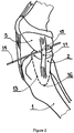
- a relatively simple, planar osteotomy 13 proposed in this invention, Figure 2 allows access to the plateau of the medial tibia condyle 2 while preserving the important stabilizer of the joint - the medial collateral ligament 11.
- a guide wire 14 is inserted across the joint in the craniomedial to caudolateral direction, passing medially to the inter-condylar eminence 15 of the tibia.
- the osteotomy is fully defined by that guide wire and its exit 16 from the tibia distally. The exit 16 is located distally to the insertion of the medial collateral ligament 11.
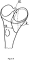
- the medial condyle 2 after the first osteotomy 13 can be flipped over, Figure 3 , to gain access to its proximal aspect and thus allow access for the second planar osteotomy 17, just below the subchondral bone of the condyle.
- the resurfacing implant 100 has a flat bottom surface 18 to be affixed to the condyle 2 and an upper surface 19.
- the upper surface 19 has a prominent concave recess 20 to receive the femoral condyle 5 for a stable articulation.
- the recess 20 has a radius of curvature 21 in the sagittal plane that matches approximately the radius of the curvature 22 of the femoral condyle, also in the sagittal plane.
- the contour 23 of the recess 20 in the frontal plane matches the shape 24 of the femoral condyle 5 in the frontal plane.
- the recess 20 in the upper surface of the implant 100 is shaped in the form that matches the shape of the tibial condyle covered by the meniscus.
- the implant 100 is provided with fixing means, e.g. at least two screw holes 25 and 26 outside the area of articulation that is limited to the recess 20.
- the upper surface, especially in the recess 20, may be highly polished and coated with hard, low-friction, abrasion resistant layer of e.g. ADLC or pyrolytic carbon.
- the lower, bone-facing surface of the implant 100 is coated for bony integration, by e.g. porous titanium and additionally by e.g. a layer of hydroxyapatite.
- the implant 100 could be manufactured by additive manufacturing and thus provided by pores for bone integration.
- the upper surface 19 is fully closed, machined, polished and coated.
- Fixation of the implant 100 to the medial condyle, Figure 5 is accomplished by placement of fixing means, e.g. at least two bone screws 27 and 28. This fixation is necessary so that bone ingrowth can proceed without movement at the interface. Further stabilization of the interface can be provided by protrusions, e.g. small spikes 29 protruding from the lower surface 18 of the implant 100. Alternatively, in special cases, such as poor cancellous bone stock, the implant 100 could be affixed to the tibia condyle by bone cement.
- the condyle, together with the implant 100 can be fixed back onto the proximal aspect 1 of the tibia. Fixation by screws 30 and 31 may suffice, Figure 6a , but a bone plate 32, Figure 6b , should be used for additional stability.
- Fixation of the condyle 2 to the tibia can be altered from its original position by slightly modifying its proximal-to-distal position to compensate for e.g. varus deformity. Additionally, the resurfaced tibial condyle can also be fixed back to the tibia by rotating it in the sagittal plane for added stability against caudal dislocation of the femoral medial condyle.
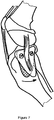
- Another use of the osteotomy as disclosed here is to emulate TPLO intervention, but only on the medial compartment, Figure 7 . This procedure could even be performed without the implant 100 if the condition of the medial compartment, including of the medial meniscus were satisfactory.
- An in-vitro test on a cadaver stifle has demonstrated the efficacy of such a reduced-morbidity TPLO on the stifle stability.
Landscapes
- Health & Medical Sciences (AREA)
- Orthopedic Medicine & Surgery (AREA)
- Physical Education & Sports Medicine (AREA)
- Cardiology (AREA)
- Oral & Maxillofacial Surgery (AREA)
- Transplantation (AREA)
- Engineering & Computer Science (AREA)
- Biomedical Technology (AREA)
- Heart & Thoracic Surgery (AREA)
- Vascular Medicine (AREA)
- Life Sciences & Earth Sciences (AREA)
- Animal Behavior & Ethology (AREA)
- General Health & Medical Sciences (AREA)
- Public Health (AREA)
- Veterinary Medicine (AREA)
- Prostheses (AREA)
- Surgical Instruments (AREA)
Priority Applications (6)
| Application Number | Priority Date | Filing Date | Title |
|---|---|---|---|
| EP19167259.1A EP3718512A1 (fr) | 2019-04-04 | 2019-04-04 | Prothèse de resurfaçage du condyle du tibia |
| CN202080022035.9A CN113613598A (zh) | 2019-04-04 | 2020-03-31 | 胫骨髁表面重修假体 |
| PCT/EP2020/059092 WO2020201268A1 (fr) | 2019-04-04 | 2020-03-31 | Prothèse de resurfaçage de condyle tibial |
| JP2021555458A JP2022525610A (ja) | 2019-04-04 | 2020-03-31 | 脛骨顆の表面再建プロテーゼ |
| EP20713912.2A EP3946168A1 (fr) | 2019-04-04 | 2020-03-31 | Prothèse de resurfaçage de condyle tibial |
| US17/600,661 US20220175540A1 (en) | 2019-04-04 | 2020-03-31 | Tibia condyle resurfacing prosthesis |
Applications Claiming Priority (1)
| Application Number | Priority Date | Filing Date | Title |
|---|---|---|---|
| EP19167259.1A EP3718512A1 (fr) | 2019-04-04 | 2019-04-04 | Prothèse de resurfaçage du condyle du tibia |
Publications (1)
| Publication Number | Publication Date |
|---|---|
| EP3718512A1 true EP3718512A1 (fr) | 2020-10-07 |
Family
ID=66092149
Family Applications (2)
| Application Number | Title | Priority Date | Filing Date |
|---|---|---|---|
| EP19167259.1A Withdrawn EP3718512A1 (fr) | 2019-04-04 | 2019-04-04 | Prothèse de resurfaçage du condyle du tibia |
| EP20713912.2A Pending EP3946168A1 (fr) | 2019-04-04 | 2020-03-31 | Prothèse de resurfaçage de condyle tibial |
Family Applications After (1)
| Application Number | Title | Priority Date | Filing Date |
|---|---|---|---|
| EP20713912.2A Pending EP3946168A1 (fr) | 2019-04-04 | 2020-03-31 | Prothèse de resurfaçage de condyle tibial |
Country Status (5)
| Country | Link |
|---|---|
| US (1) | US20220175540A1 (fr) |
| EP (2) | EP3718512A1 (fr) |
| JP (1) | JP2022525610A (fr) |
| CN (1) | CN113613598A (fr) |
| WO (1) | WO2020201268A1 (fr) |
Citations (4)
| Publication number | Priority date | Publication date | Assignee | Title |
|---|---|---|---|---|
| FR2747914A1 (fr) * | 1996-04-30 | 1997-10-31 | Raoult Andre | Prothese partielle du genou |
| US20050143831A1 (en) * | 2003-12-30 | 2005-06-30 | Medicinelodge, Inc. | Tibial condylar hemiplasty implants, anchor assemblies, and related methods |
| US20110015740A1 (en) * | 2009-07-14 | 2011-01-20 | Biomet Manufacturing Corp. | Pyrocarbon Orthopedic Implant |
| US20120330431A1 (en) * | 2010-03-09 | 2012-12-27 | Rolston Lindsey R | Device for unicompartmental knee arthroplasty |
Family Cites Families (17)
| Publication number | Priority date | Publication date | Assignee | Title |
|---|---|---|---|---|
| GB1509194A (en) * | 1974-04-22 | 1978-05-04 | Nat Res Dev | Endoprosthetic devices |
| US4166292A (en) * | 1977-09-08 | 1979-09-04 | Carbomedics, Inc. | Stress reinforced artificial joint prostheses |
| FR2593390B1 (fr) * | 1986-01-27 | 1991-09-06 | Epinette Jean Alain | Composant tibial de prothese unicompartimentaire du genou a implanter sans ciment |
| US5176710A (en) * | 1991-01-23 | 1993-01-05 | Orthopaedic Research Institute | Prosthesis with low stiffness factor |
| US20070100462A1 (en) * | 2001-05-25 | 2007-05-03 | Conformis, Inc | Joint Arthroplasty Devices |
| US5766257A (en) * | 1997-01-28 | 1998-06-16 | Implant Manufacturing And Testing Corporation | Artificial joint having natural load transfer |
| JPH11318960A (ja) * | 1998-05-12 | 1999-11-24 | Nissin Electric Co Ltd | 人工関節及びその製造方法 |
| US6299645B1 (en) * | 1999-07-23 | 2001-10-09 | William S. Ogden | Dove tail total knee replacement unicompartmental |
| US20040034432A1 (en) * | 2002-02-11 | 2004-02-19 | Dean Hughes | Mobile bearing tibial base prosthetic devices employing oxidized zirconium surfaces |
| EP2335653A1 (fr) * | 2009-12-16 | 2011-06-22 | Slobodan Tepic | Prothèse de hanche partielle |
| RU122016U1 (ru) * | 2012-03-26 | 2012-11-20 | Общество с ограниченной ответственностью "ОСТЕОМЕД-М" (ООО "ОСТЕОМЕД-М") | Эндопротез тазобедренного сустава |
| US8734523B2 (en) * | 2012-05-31 | 2014-05-27 | Howmedica Osteonics Corp. | Limited motion tibial bearing |
| US8845741B2 (en) * | 2012-09-27 | 2014-09-30 | Seavete LLC | Artificial joint components including integral magnetic fields configured to deflect wear debris particles |
| TWI456090B (zh) * | 2012-12-25 | 2014-10-11 | Univ Lunghwa Sci & Technology | 具有生物活性表面之鈦或鈦合金及其製作方法 |
| EP3302320B1 (fr) * | 2015-05-26 | 2022-11-16 | McAuliffe, Michael | Procédé de conception d'un dispositif chirurgical |
| CZ29005U1 (cs) * | 2015-10-27 | 2015-12-21 | Beznoska, S.R.O. | Nanostrukturované komponenta kloubních implantátů |
| CN108403264A (zh) * | 2018-05-14 | 2018-08-17 | 北京市春立正达医疗器械股份有限公司 | 多元化膝关节胫骨平台垫假体 |
-
2019
- 2019-04-04 EP EP19167259.1A patent/EP3718512A1/fr not_active Withdrawn
-
2020
- 2020-03-31 WO PCT/EP2020/059092 patent/WO2020201268A1/fr not_active Ceased
- 2020-03-31 CN CN202080022035.9A patent/CN113613598A/zh active Pending
- 2020-03-31 EP EP20713912.2A patent/EP3946168A1/fr active Pending
- 2020-03-31 JP JP2021555458A patent/JP2022525610A/ja active Pending
- 2020-03-31 US US17/600,661 patent/US20220175540A1/en active Pending
Patent Citations (4)
| Publication number | Priority date | Publication date | Assignee | Title |
|---|---|---|---|---|
| FR2747914A1 (fr) * | 1996-04-30 | 1997-10-31 | Raoult Andre | Prothese partielle du genou |
| US20050143831A1 (en) * | 2003-12-30 | 2005-06-30 | Medicinelodge, Inc. | Tibial condylar hemiplasty implants, anchor assemblies, and related methods |
| US20110015740A1 (en) * | 2009-07-14 | 2011-01-20 | Biomet Manufacturing Corp. | Pyrocarbon Orthopedic Implant |
| US20120330431A1 (en) * | 2010-03-09 | 2012-12-27 | Rolston Lindsey R | Device for unicompartmental knee arthroplasty |
Also Published As
| Publication number | Publication date |
|---|---|
| US20220175540A1 (en) | 2022-06-09 |
| WO2020201268A1 (fr) | 2020-10-08 |
| CN113613598A (zh) | 2021-11-05 |
| JP2022525610A (ja) | 2022-05-18 |
| EP3946168A1 (fr) | 2022-02-09 |
Similar Documents
| Publication | Publication Date | Title |
|---|---|---|
| EP1778559B1 (fr) | Dispositif modulaire pour sculpter la surface d'une articulation | |
| Barrack et al. | The use of a modular rotating hinge component in salvage revision total knee arthroplasty | |
| Delclaux et al. | Complications of radial head prostheses | |
| AU2006272871C1 (en) | Apparatus and method for sculpting the surface of a joint | |
| Ries et al. | Porous tantalum patellar augmentation: the importance of residual bone stock | |
| Anderson et al. | Constrained condylar knee without stem extensions for difficult primary total knee arthroplasty | |
| Ries | Primary arthroplasty for management of osteoporotic fractures about the knee | |
| Cross et al. | Results of custom-fit, noncemented, semiconstrained total elbow arthroplasty for inflammatory arthritis at an average of eighteen years of follow-up | |
| Friesenbichler et al. | Failure rate of a rotating hinge knee design due to yoke fracture of the hinged tibial insert: a retrospective data analysis and review of the literature | |
| Salazar Botero et al. | Surgical technique: about a new total and isoelastic wrist implant (Prosthelast®) | |
| Fink et al. | Results of elbow endoprostheses in patients with rheumatoid arthritis in correlation with previous operations | |
| Brigido et al. | Primary Zimmer trabecular metal total ankle replacement | |
| US20220175540A1 (en) | Tibia condyle resurfacing prosthesis | |
| Rajgopal et al. | Long term results of rotating hinge total knee arthroplasty in complex primary and revision cases | |
| Gaudot et al. | The Salto Talaris XT revision total ankle replacement system | |
| Watanabe et al. | Periprosthetic fracture of the tibia associated with osteolysis caused by failure of rotating patella in low-contact–stress total knee arthroplasty | |
| Nabergoj et al. | Options for glenoid reconstruction: graft vs. Metal vs. Combined | |
| Ericson et al. | Variation in the position of the elbow flexion axis after total joint replacement with three different prostheses | |
| Marinelli et al. | Anatomical monopolar press-fit radial head arthroplasty: surgical technique and expected outcomes | |
| Kani et al. | Update on the operative treatment of scapholunate instability for radiologists. II. Salvage procedures, total wrist arthrodesis, and total wrist arthroplasty | |
| Samijo et al. | Souter-Strathclyde total elbow arthroplasty: medium-term results | |
| Blackburn et al. | Hinge Implants | |
| Madanat et al. | Hinge implants | |
| Pfanner et al. | Primary Total Wrist Arthroplasty | |
| Grimm et al. | Fracture of an uncemented tantalum patellar component |
Legal Events
| Date | Code | Title | Description |
|---|---|---|---|
| PUAI | Public reference made under article 153(3) epc to a published international application that has entered the european phase |
Free format text: ORIGINAL CODE: 0009012 |
|
| STAA | Information on the status of an ep patent application or granted ep patent |
Free format text: STATUS: THE APPLICATION HAS BEEN PUBLISHED |
|
| AK | Designated contracting states |
Kind code of ref document: A1 Designated state(s): AL AT BE BG CH CY CZ DE DK EE ES FI FR GB GR HR HU IE IS IT LI LT LU LV MC MK MT NL NO PL PT RO RS SE SI SK SM TR |
|
| AX | Request for extension of the european patent |
Extension state: BA ME |
|
| STAA | Information on the status of an ep patent application or granted ep patent |
Free format text: STATUS: THE APPLICATION IS DEEMED TO BE WITHDRAWN |
|
| 18D | Application deemed to be withdrawn |
Effective date: 20210408 |