EP3726466A1 - Autonome level-identifikation anatomischer knöcherner strukturen in medizinischen 3d-bildern - Google Patents
Autonome level-identifikation anatomischer knöcherner strukturen in medizinischen 3d-bildern Download PDFInfo
- Publication number
- EP3726466A1 EP3726466A1 EP19169136.9A EP19169136A EP3726466A1 EP 3726466 A1 EP3726466 A1 EP 3726466A1 EP 19169136 A EP19169136 A EP 19169136A EP 3726466 A1 EP3726466 A1 EP 3726466A1
- Authority
- EP
- European Patent Office
- Prior art keywords
- anatomical
- components
- level
- levels
- anatomical components
- Prior art date
- Legal status (The legal status is an assumption and is not a legal conclusion. Google has not performed a legal analysis and makes no representation as to the accuracy of the status listed.)
- Pending
Links
Images

Classifications
-
- G—PHYSICS
- G06—COMPUTING OR CALCULATING; COUNTING
- G06T—IMAGE DATA PROCESSING OR GENERATION, IN GENERAL
- G06T7/00—Image analysis
- G06T7/10—Segmentation; Edge detection
- G06T7/11—Region-based segmentation
-
- G—PHYSICS
- G06—COMPUTING OR CALCULATING; COUNTING
- G06T—IMAGE DATA PROCESSING OR GENERATION, IN GENERAL
- G06T15/00—Three-dimensional [3D] image rendering
- G06T15/08—Volume rendering
-
- G—PHYSICS
- G06—COMPUTING OR CALCULATING; COUNTING
- G06T—IMAGE DATA PROCESSING OR GENERATION, IN GENERAL
- G06T7/00—Image analysis
- G06T7/0002—Inspection of images, e.g. flaw detection
- G06T7/0012—Biomedical image inspection
-
- G—PHYSICS
- G06—COMPUTING OR CALCULATING; COUNTING
- G06T—IMAGE DATA PROCESSING OR GENERATION, IN GENERAL
- G06T2207/00—Indexing scheme for image analysis or image enhancement
- G06T2207/10—Image acquisition modality
- G06T2207/10072—Tomographic images
-
- G—PHYSICS
- G06—COMPUTING OR CALCULATING; COUNTING
- G06T—IMAGE DATA PROCESSING OR GENERATION, IN GENERAL
- G06T2207/00—Indexing scheme for image analysis or image enhancement
- G06T2207/10—Image acquisition modality
- G06T2207/10072—Tomographic images
- G06T2207/10081—Computed x-ray tomography [CT]
-
- G—PHYSICS
- G06—COMPUTING OR CALCULATING; COUNTING
- G06T—IMAGE DATA PROCESSING OR GENERATION, IN GENERAL
- G06T2207/00—Indexing scheme for image analysis or image enhancement
- G06T2207/10—Image acquisition modality
- G06T2207/10072—Tomographic images
- G06T2207/10088—Magnetic resonance imaging [MRI]
-
- G—PHYSICS
- G06—COMPUTING OR CALCULATING; COUNTING
- G06T—IMAGE DATA PROCESSING OR GENERATION, IN GENERAL
- G06T2207/00—Indexing scheme for image analysis or image enhancement
- G06T2207/20—Special algorithmic details
- G06T2207/20084—Artificial neural networks [ANN]
-
- G—PHYSICS
- G06—COMPUTING OR CALCULATING; COUNTING
- G06T—IMAGE DATA PROCESSING OR GENERATION, IN GENERAL
- G06T2207/00—Indexing scheme for image analysis or image enhancement
- G06T2207/30—Subject of image; Context of image processing
- G06T2207/30004—Biomedical image processing
- G06T2207/30008—Bone
- G06T2207/30012—Spine; Backbone
Definitions
- the present disclosure generally relates to autonomous level identification of anatomical bony structures on three dimensional (3D) medical imagery, useful in particular for the field of computer assisted surgery, diagnostics, and surgical planning.
- Image guided or computer assisted surgery is a surgical procedure where the surgeon uses tracked surgical instruments in conjunction with preoperative or intraoperative images in order to indirectly guide the procedure.
- Image guided surgery can utilize images acquired intraoperatively, provided for example from computer tomography (CT) scanners.
- CT computer tomography
- Specialized computer systems can be used to process the CT images to develop three-dimensional models of the anatomy fragment subject to the surgery procedure.
- CNN convolutional neural network
- CNNs use a variation of feature detectors and/or multilayer perceptrons designed to require minimal preprocessing of input data.
- a publication " Hierarchical Segmentation and Identification of Thoracic Vertebra Using Learning-Based Edge Detection and Coarse-to-Fine Deformable Model” discloses a segmentation and identification of thoracic vertebrae based on learned bone-structure edge detectors and a coarse-to-fine deformable surface model to segment and identify vertebrae in 3D CT thoracic imagery.
- a discriminative classifier for object-specific edge detection is trained using steerable features and statistical shape models for 12 thoracic vertebrae are also learned.
- a coarse-to-fine, two-stage segmentation strategy is used: subregions of a vertebra first deforms together as a group; then vertebra mesh vertices in a smaller neighborhood move group-wise, to progressively drive the deformable model towards edge response maps by optimizing a probability cost function.
- Such an approach requires a dedicated edge detection algorithm and shape matching. The algorithm was tested only on thoracic spine and its accuracy varies based on the level and a number of vertebrae provided as an input.
- a computer-implemented method for fully-autonomous level identification of anatomical structures within a three-dimensional (3D) medical imagery comprising of: receiving the 3D medical imagery comprising a set of medical scan images of the anatomical structures; processing the set of medical scan images to perform an autonomous semantic segmentation of anatomical components and to store segmentation results; processing segmentation results by removing the false positives, and smoothing 3D surfaces of the generated anatomical components; determining morphological and spatial relationships of the anatomical components; processing the set of medical scan images using a convolutional neural network to autonomously assign an initial level type; assigning the determined level type to each group of anatomical components by combining the determined morphological and spatial relationships with the determined initial level type; assigning an ordinal identifier to each group of anatomical components to complement the assigned level type based on the relative distribution of the groups of anatomical components and their respective level types; and storing information about the assigned levels with their ordinal identifier.
- the anatomical structure can be a spine and the level type is a vertebral level type (C, T, L, S), to which an ordinal identifier (C1-C7, T1-T12, L1-L4, S1-S5) is assigned.
- C, T, L, S vertebral level type
- an ordinal identifier C1-C7, T1-T12, L1-L4, S1-S5
- the determined morphological relationships of anatomical components may be their size and bounding box.
- the determined spatial relationships of anatomical component may be their relative position and orientation.
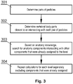
- the anatomical structure may be a spine, and determining morphological and spatial relationships among the anatomical components, may include in particular: determining pairs of pedicles; determining a vertebral body closest to or intersecting with each pair of pedicles; searching for anatomy parts intersecting with other components that were already assigned to the level; and repeating the previous steps for each level separately, excluding parts that were already assigned.
- the method may further comprise providing a virtual 3D model, including the identified levels, as an augmented reality image with some of the levels shown and some other levels hidden.
- the augmented reality image may further comprise implants corresponding to the levels shown.
- the method may further comprise providing a visual representation of identified levels as labels displayed in the surgical field as an augmented reality image, with some of the labels shown and some other labels hidden.
- a computer-implemented system comprising: at least one nontransitory processor-readable storage medium that stores at least one of processor-executable instructions or data; and at least one processor communicably coupled to at least one nontransitory processor-readable storage medium, wherein at least one processor is configured to perform the steps of the method of any of the previous claims.
- the invention relates to processing images of a bony structure, for example a spine (including, in particular, specific sections: cervical, thoracic, lumbar, sacrum), as well as to other structures having a plurality of multiple anatomical components.
- a spine including, in particular, specific sections: cervical, thoracic, lumbar, sacrum
- Other structures having a plurality of multiple anatomical components including, in particular, specific sections: cervical, thoracic, lumbar, sacrum
- the aim of this procedure is to identify levels of a bony structure, i.e. adjacent groups of similar components of the anatomical structure, for example the vertebrae.
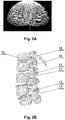
- Each vertebra can be considered as a level that comprises a plurality of anatomical parts, such as vertebral body 16, pedicles 15, transverse process 14, lamina 13, articular process 17, and spinous process 11.
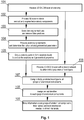
- a 3D scan volume is received, comprising a set of DICOM (Digital Imaging and Communications in Medicine) images of an anatomical bony structure, in this example - a spine, which is to be segmented, such as a set of CT (Computed Tomography) or MRI (Magnetic Resonance Imaging) scans.
- the set represents consecutive slices of the anatomical structure (such as one slice shown in Fig. 2A ).
- the received images are processed in step 102 to perform autonomous segmentation of the anatomical structure to determine plurality of anatomical components, such as vertebral body 16, pedicles 15, transverse processes 14, lamina 13, articular process 17, spinous process 11 and ribs 18, as shown in Fig. 2B .
- This can be done by employing a method for segmentation of images disclosed in a European patent application EP17195826 by the present applicant or any other method that outputs a 3D model of the anatomical structure which is semantically segmented, as shown in Fig. 2B .
- step 103 the segmentation results generated in step 102 are processed to remove false positives after segmentation, and to check whether some anatomy components are connected together (and upon detection, disconnecting them) and smoothing the surface of the components (including simplification of the geometry and filling holes in the geometry).
- vertebral bodies or other anatomical components of different levels may appear to be connected with each other after the segmentation.
- L5 and S1 vertebral bodies may be connected - in order to disconnect them, a combination of watershed and distance transform algorithms can be used to calculate separation lines in 3D.
- step 104 geometrical parameters for each anatomical component are determined, including, at least, their relative positions, size, bounding box edges, and preferably also orientation based on semantic segmentation results and the moment of inertia.
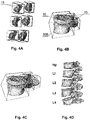
- each individual anatomical component is analyzed and assigned to a corresponding vertebral level by determining morphological and spatial relationships of the anatomical components. For example, this may be done by the procedure shown in Fig. 3 and supporting images of Figs. 4A-4D .
- the pairs of pedicles 15 are determined in step 301 (as shown in Fig. 4A ). It can be estimated which pedicles correspond to separate levels by their location in space and relation between all other anatomy components. Then, for each spinal level, the closest (or intersecting) vertebral body 16, determined in step 302 is assigned, that is touching at least one of the pedicles as shown in Fig. 4B . In step 303, further assignment of other anatomy components to levels, such as transverse process 14, articular process 17, spinous process 11 and/or lamina 13, can be conducted with the same approach based on anatomical knowledge and geometrical analysis, as shown in Fig. 4C . This process shall be repeated for each level separately in step 304, excluding anatomical components that were already picked by other levels.
- step 106 prediction by means of a neural network is performed in step 106, to assign an initial classification of vertebral level type.
- level types can be assigned: C (Cervical), T (Thoracic), L (lumbar), S (Sacral).
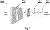
- a Convolutional Neural Network (CNN) architecture 500 can be used for this purpose.
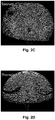
- the network performs vertebral type prediction on each DICOM slice, as shown in Fig 2C or 2D .
- the CNN 500 returns a probability map for each vertebral type class, for example, thoracic, sacrum or lumbar, in a way that the sum of individual probability components is always equal to one, and the biggest number explicitly suggests a predicted vertebral type.
- the left side of the network 500 is a contracting path, which includes a plurality of convolution blocks 501 stacked together.
- Each convolution block comprises of two convolutional layers with optional batch normalization layer between them, followed up by a pooling layer.
- the last convolution block is directly connected to a plurality of dense, fully-connected layers 502 stacked together.
- the last fully-connected layer 503, which is considered as a network output layer, represents all possible vertebral type classes, where each type has its own neuron, for example, 3 neurons for vertebral types thoracic, sacrum or lumbar.
- the convolution layers inside a block 501 can be of a standard kind, the dilated kind, or a combination thereof, with ReLU or leaky ReLU activation function attached.
- Each fully-connected layer 502 can be preceded by a dropout layer, and each fully-connected layer can have a ReLU or leaky ReLU activation function attached.
- Output layer 503 denotes a probability map for each output class (vertebral type) with a softmax activation function at the end.
- the number of layers and the number of filters within layers, as well as dropout rate for dropout layers, is subject to change, depending on the requirements of the application. Deeper networks with more filters typically give results of better quality. However, there is a point at which increasing the number of layers/filters does not result in significant improvement, but significantly increases the computation time, making such a large network impractical.
- step 107 Since the neural network model operates on individual slices and each level prepared in step 105 can be visible on plurality of slices, in step 107, the most frequent vertebral type class prediction is taken into consideration for each subvolume (spinal level) of DICOM scan. Therefore, in step 107, the level type (such as C, T, L or S) is assigned to each group of anatomical components by combining morphological and spatial relationships determined in step 105 with the type identification determined in step 106.
- the level type such as C, T, L or S
- step 108 final counting and identification of vertebral components take place. Based on the orientation of the patient anatomy in the volumetric scandata, neural network prediction, and overall distribution of levels' types, proper indices are assigned to form a full identification of levels with an ordinal identifier assigned. For example, counting of lumbar vertebrae may start from L5 (or L6) if sacrum is included in the scanset, or from L1 if thoracic spine is included in the scanset.
- an ordinal identifier is assigned in step 108 to each group of anatomical components represented by the level (C, T, L, S) determined in step 107, based on the anatomical structure and distribution of all the other levels - for example, the following identifiers may be assigned: C1-S5 or C1-C7, T1-T12, L1-L5, S1-S5.
- virtual 3D model output is provided in step 109, which can be used to conveniently display any configuration of identified levels (and other components of the patient anatomy, such as nerves or surgical instruments (such as implants 603, Fig. 6B ), to let the surgeon focus only on meaningful information that is necessary at a particular time during surgery.
- the model is useful for providing an augmented reality image display in computer assisted surgery systems, wherein the virtual 3D model of the anatomy 600 is displayed over the real anatomy, wherein the information on the segments can be used to conveniently hide some of the levels 601 of segments that are not relevant for the surgery and display only the levels of interest 602, as shown for example in Fig. 6A .
- the augmented reality image may be presented with labels 604 corresponding to the identified levels as shown in Fig. 6C .
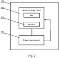
- the functionality described herein can be implemented in a computer system 700, such as shown in Fig. 7 .
- the system 700 may include at least one nontransitory processor-readable storage medium 710 that stores at least one of processor-executable instructions 715 or data; and at least one processor 720 communicably coupled to the at least one nontransitory processor-readable storage medium 710.
- the at least one processor 720 may be configured to (by executing the instructions 715) perform the procedure of Fig. 1 .
Landscapes
- Engineering & Computer Science (AREA)
- Physics & Mathematics (AREA)
- General Physics & Mathematics (AREA)
- Theoretical Computer Science (AREA)
- Computer Vision & Pattern Recognition (AREA)
- General Health & Medical Sciences (AREA)
- Health & Medical Sciences (AREA)
- Medical Informatics (AREA)
- Nuclear Medicine, Radiotherapy & Molecular Imaging (AREA)
- Radiology & Medical Imaging (AREA)
- Quality & Reliability (AREA)
- Computer Graphics (AREA)
- Apparatus For Radiation Diagnosis (AREA)
- Image Processing (AREA)
Priority Applications (3)
| Application Number | Priority Date | Filing Date | Title |
|---|---|---|---|
| EP19169136.9A EP3726466A1 (de) | 2019-04-15 | 2019-04-15 | Autonome level-identifikation anatomischer knöcherner strukturen in medizinischen 3d-bildern |
| US16/833,750 US20200327721A1 (en) | 2019-04-15 | 2020-03-30 | Autonomous level identification of anatomical bony structures on 3d medical imagery |
| US18/062,306 US20230360313A1 (en) | 2019-04-15 | 2022-12-06 | Autonomous level identification of anatomical bony structures on 3d medical imagery |
Applications Claiming Priority (1)
| Application Number | Priority Date | Filing Date | Title |
|---|---|---|---|
| EP19169136.9A EP3726466A1 (de) | 2019-04-15 | 2019-04-15 | Autonome level-identifikation anatomischer knöcherner strukturen in medizinischen 3d-bildern |
Publications (1)
| Publication Number | Publication Date |
|---|---|
| EP3726466A1 true EP3726466A1 (de) | 2020-10-21 |
Family
ID=66323673
Family Applications (1)
| Application Number | Title | Priority Date | Filing Date |
|---|---|---|---|
| EP19169136.9A Pending EP3726466A1 (de) | 2019-04-15 | 2019-04-15 | Autonome level-identifikation anatomischer knöcherner strukturen in medizinischen 3d-bildern |
Country Status (2)
| Country | Link |
|---|---|
| US (2) | US20200327721A1 (de) |
| EP (1) | EP3726466A1 (de) |
Cited By (2)
| Publication number | Priority date | Publication date | Assignee | Title |
|---|---|---|---|---|
| WO2023064957A1 (en) * | 2021-10-15 | 2023-04-20 | Surgalign Spine Technologies, Inc. | Systems, devices, and methods for level identification of three-dimensional anatomical images |
| US12154239B2 (en) | 2023-02-03 | 2024-11-26 | Rayhan Papar | Live surgical aid for brain tumor resection using augmented reality and deep learning |
Families Citing this family (23)
| Publication number | Priority date | Publication date | Assignee | Title |
|---|---|---|---|---|
| GB2536650A (en) | 2015-03-24 | 2016-09-28 | Augmedics Ltd | Method and system for combining video-based and optic-based augmented reality in a near eye display |
| EP3470006B1 (de) | 2017-10-10 | 2020-06-10 | Holo Surgical Inc. | Automatische segmentierung von dreidimensionalen knochenstrukturbildern |
| EP3445048B8 (de) | 2017-08-15 | 2026-01-14 | Augmedics, Inc. | Grafische benutzeroberfläche für ein chirurgisches navigationssystem zur bereitstellung eines bildes mit erweiterter realität während des betriebs |
| US12458411B2 (en) | 2017-12-07 | 2025-11-04 | Augmedics Ltd. | Spinous process clamp |
| US12521201B2 (en) | 2017-12-07 | 2026-01-13 | Augmedics Ltd. | Spinous process clamp |
| EP3787543A4 (de) | 2018-05-02 | 2022-01-19 | Augmedics Ltd. | Registrierung einer bezugsmarke für ein system der erweiterten realität |
| EP4710888A3 (de) | 2018-08-10 | 2026-03-25 | Augmedics Inc. | Computergestützte identifizierung einer geeigneten anatomischen struktur zur platzierung einer medizinischen vorrichtung während eines chirurgischen eingriffs |
| US11766296B2 (en) | 2018-11-26 | 2023-09-26 | Augmedics Ltd. | Tracking system for image-guided surgery |
| US11980506B2 (en) | 2019-07-29 | 2024-05-14 | Augmedics Ltd. | Fiducial marker |
| US12178666B2 (en) | 2019-07-29 | 2024-12-31 | Augmedics Ltd. | Fiducial marker |
| US11382712B2 (en) | 2019-12-22 | 2022-07-12 | Augmedics Ltd. | Mirroring in image guided surgery |
| US11389252B2 (en) | 2020-06-15 | 2022-07-19 | Augmedics Ltd. | Rotating marker for image guided surgery |
| US12239385B2 (en) | 2020-09-09 | 2025-03-04 | Augmedics Ltd. | Universal tool adapter |
| US12502163B2 (en) | 2020-09-09 | 2025-12-23 | Augmedics Ltd. | Universal tool adapter for image-guided surgery |
| EP4329660A1 (de) | 2021-04-30 | 2024-03-06 | Augmedics, Inc. | Grafische benutzeroberfläche für ein chirurgisches navigationssystem |
| CN117957566A (zh) | 2021-05-12 | 2024-04-30 | 增强医疗公司 | 用于分割解剖图像数据的系统、设备和方法 |
| EP4105887B1 (de) | 2021-06-15 | 2026-02-18 | Stryker European Operations Limited | Technik zur erzeugung von chirurgischen informationen aus intraoperativ und präoperativ erfassten bilddaten |
| US11896445B2 (en) | 2021-07-07 | 2024-02-13 | Augmedics Ltd. | Iliac pin and adapter |
| US12150821B2 (en) | 2021-07-29 | 2024-11-26 | Augmedics Ltd. | Rotating marker and adapter for image-guided surgery |
| US12475662B2 (en) | 2021-08-18 | 2025-11-18 | Augmedics Ltd. | Stereoscopic display and digital loupe for augmented-reality near-eye display |
| JP2025508791A (ja) | 2022-02-22 | 2025-04-10 | オーグメディックス インコーポレイテッド | 脊椎解析のためのシステム、デバイス、及び方法 |
| WO2023203521A1 (en) | 2022-04-21 | 2023-10-26 | Augmedics Ltd. | Systems and methods for medical image visualization |
| JP2025531829A (ja) | 2022-09-13 | 2025-09-25 | オーグメディックス リミテッド | 画像誘導医療介入のための拡張現実アイウェア |
-
2019
- 2019-04-15 EP EP19169136.9A patent/EP3726466A1/de active Pending
-
2020
- 2020-03-30 US US16/833,750 patent/US20200327721A1/en active Pending
-
2022
- 2022-12-06 US US18/062,306 patent/US20230360313A1/en not_active Abandoned
Non-Patent Citations (3)
| Title |
|---|
| ANA JIMÉNEZ-PASTOR ET AL: "Automatic localization and identification of vertebrae in spine CT scans by combining Deep Learning with morphological image processing techniques", EUROPEAN CONGRESS OF RADIOLOGY (ECR) 2018, 4 March 2018 (2018-03-04), XP055626761, Retrieved from the Internet <URL:https://quibim.com/wp-content/uploads/2018/03/3_ECR2018_AJP.pdf> [retrieved on 20190927] * |
| JUN MA ET AL.: "Hierarchical Segmentation and Identification of Thoracic Vertebra Using Learning-Based Edge Detection and Coarse-to-Fine Deformable Model", MEDICAL IMAGE COMPUTING AND COMPUTER-ASSISTED INTERVENTION, MICCAI, 2010, pages 19 - 27, XP047462949, DOI: doi:10.1007/978-3-642-15705-9_3 |
| YANG DONG ET AL: "Deep Image-to-Image Recurrent Network with Shape Basis Learning for Automatic Vertebra Labeling in Large-Scale 3D CT Volumes", 4 September 2017, ROBOCUP 2008: ROBOCUP 2008: ROBOT SOCCER WORLD CUP XII; [LECTURE NOTES IN COMPUTER SCIENCE; LECT.NOTES COMPUTER], SPRINGER INTERNATIONAL PUBLISHING, CHAM, PAGE(S) 498 - 506, ISBN: 978-3-319-10403-4, XP047502790 * |
Cited By (2)
| Publication number | Priority date | Publication date | Assignee | Title |
|---|---|---|---|---|
| WO2023064957A1 (en) * | 2021-10-15 | 2023-04-20 | Surgalign Spine Technologies, Inc. | Systems, devices, and methods for level identification of three-dimensional anatomical images |
| US12154239B2 (en) | 2023-02-03 | 2024-11-26 | Rayhan Papar | Live surgical aid for brain tumor resection using augmented reality and deep learning |
Also Published As
| Publication number | Publication date |
|---|---|
| US20200327721A1 (en) | 2020-10-15 |
| US20230360313A1 (en) | 2023-11-09 |
Similar Documents
| Publication | Publication Date | Title |
|---|---|---|
| US20230360313A1 (en) | Autonomous level identification of anatomical bony structures on 3d medical imagery | |
| Lessmann et al. | Iterative fully convolutional neural networks for automatic vertebra segmentation and identification | |
| Al Arif et al. | Fully automatic cervical vertebrae segmentation framework for X-ray images | |
| US10223610B1 (en) | System and method for detection and classification of findings in images | |
| CN111369576B (zh) | 图像分割模型的训练方法、图像分割方法、装置及设备 | |
| EP3718077B1 (de) | Dreidimensionales medizinisches bildanalyseverfahren und system zur identifizierung von wirbelfrakturen | |
| US11593943B2 (en) | RECIST assessment of tumour progression | |
| Suzani et al. | Fast automatic vertebrae detection and localization in pathological ct scans-a deep learning approach | |
| US20190201106A1 (en) | Identification and tracking of a predefined object in a set of images from a medical image scanner during a surgical procedure | |
| EP4292045A1 (de) | Systeme und verfahren zur automatisierten segmentierung patientenspezifischer anatomien für pathologiespezifische messungen | |
| US10635924B2 (en) | System and method for surgical guidance and intra-operative pathology through endo-microscopic tissue differentiation | |
| CN113822845A (zh) | 医学图像中组织结构的层级分割方法、装置、设备及介质 | |
| Wang et al. | Fully automatic intervertebral disc segmentation using multimodal 3D U-Net | |
| Wang et al. | Automatic vertebrae localization and identification by combining deep SSAE contextual features and structured regression forest | |
| CN113610746B (zh) | 一种图像处理方法、装置、计算机设备及存储介质 | |
| EP3107031A1 (de) | Verfahren, vorrichtung und system zur rückenbeschriftung | |
| Jimenez-Pastor et al. | Automated vertebrae localization and identification by decision forests and image-based refinement on real-world CT data | |
| CN120495295B (zh) | 基于图像识别的骨质疏松诊断方法 | |
| Manoila et al. | SmartMRI framework for segmentation of MR images using multiple deep learning methods | |
| CN120280132B (zh) | 基于深度学习的口腔颌面外科影像识别诊断方法及系统 | |
| Koh et al. | Automatic spinal canal detection in lumbar MR images in the sagittal view using dynamic programming | |
| EP4012650A1 (de) | Segmentierung einer anatomischen struktur in einem bild | |
| Damopoulos et al. | Automatic localization of the lumbar vertebral landmarks in CT images with context features | |
| Korez et al. | Segmentation of pathological spines in CT images using a two-way CNN and a collision-based model | |
| Zhu et al. | A novel method for 3D knee anatomical landmark localization by combining global and local features |
Legal Events
| Date | Code | Title | Description |
|---|---|---|---|
| PUAI | Public reference made under article 153(3) epc to a published international application that has entered the european phase |
Free format text: ORIGINAL CODE: 0009012 |
|
| STAA | Information on the status of an ep patent application or granted ep patent |
Free format text: STATUS: THE APPLICATION HAS BEEN PUBLISHED |
|
| AK | Designated contracting states |
Kind code of ref document: A1 Designated state(s): AL AT BE BG CH CY CZ DE DK EE ES FI FR GB GR HR HU IE IS IT LI LT LU LV MC MK MT NL NO PL PT RO RS SE SI SK SM TR |
|
| AX | Request for extension of the european patent |
Extension state: BA ME |
|
| RIN1 | Information on inventor provided before grant (corrected) |
Inventor name: MEJIA OROZCO, EDWING ISAAC Inventor name: TRZMIEL, MICHAL Inventor name: LUCIANO, CRISTIAN J. Inventor name: SIEMIONOW, KRIS B. Inventor name: LEWICKI, PAUL |
|
| STAA | Information on the status of an ep patent application or granted ep patent |
Free format text: STATUS: REQUEST FOR EXAMINATION WAS MADE |
|
| 17P | Request for examination filed |
Effective date: 20210421 |
|
| RBV | Designated contracting states (corrected) |
Designated state(s): AL AT BE BG CH CY CZ DE DK EE ES FI FR GB GR HR HU IE IS IT LI LT LU LV MC MK MT NL NO PL PT RO RS SE SI SK SM TR |
|
| RAP3 | Party data changed (applicant data changed or rights of an application transferred) |
Owner name: HOLO SURGICAL INC. |