EP3733221B1 - Implantierbare wirkstofffreisetzungsvorrichtung - Google Patents
Implantierbare wirkstofffreisetzungsvorrichtung Download PDFInfo
- Publication number
- EP3733221B1 EP3733221B1 EP18897786.2A EP18897786A EP3733221B1 EP 3733221 B1 EP3733221 B1 EP 3733221B1 EP 18897786 A EP18897786 A EP 18897786A EP 3733221 B1 EP3733221 B1 EP 3733221B1
- Authority
- EP
- European Patent Office
- Prior art keywords
- zinc
- alloy
- scaffold
- loaded device
- implantable drug
- Prior art date
- Legal status (The legal status is an assumption and is not a legal conclusion. Google has not performed a legal analysis and makes no representation as to the accuracy of the status listed.)
- Active
Links
Images

Classifications
-
- A—HUMAN NECESSITIES
- A61—MEDICAL OR VETERINARY SCIENCE; HYGIENE
- A61L—METHODS OR APPARATUS FOR STERILISING MATERIALS OR OBJECTS IN GENERAL; DISINFECTION, STERILISATION OR DEODORISATION OF AIR; CHEMICAL ASPECTS OF BANDAGES, DRESSINGS, ABSORBENT PADS OR SURGICAL ARTICLES; MATERIALS FOR BANDAGES, DRESSINGS, ABSORBENT PADS OR SURGICAL ARTICLES
- A61L31/00—Materials for other surgical articles, e.g. stents, stent-grafts, shunts, surgical drapes, guide wires, materials for adhesion prevention, occluding devices, surgical gloves, tissue fixation devices
- A61L31/02—Inorganic materials
- A61L31/022—Metals or alloys
-
- A—HUMAN NECESSITIES
- A61—MEDICAL OR VETERINARY SCIENCE; HYGIENE
- A61L—METHODS OR APPARATUS FOR STERILISING MATERIALS OR OBJECTS IN GENERAL; DISINFECTION, STERILISATION OR DEODORISATION OF AIR; CHEMICAL ASPECTS OF BANDAGES, DRESSINGS, ABSORBENT PADS OR SURGICAL ARTICLES; MATERIALS FOR BANDAGES, DRESSINGS, ABSORBENT PADS OR SURGICAL ARTICLES
- A61L27/00—Materials for grafts or prostheses or for coating grafts or prostheses
- A61L27/02—Inorganic materials
- A61L27/04—Metals or alloys
-
- A—HUMAN NECESSITIES
- A61—MEDICAL OR VETERINARY SCIENCE; HYGIENE
- A61L—METHODS OR APPARATUS FOR STERILISING MATERIALS OR OBJECTS IN GENERAL; DISINFECTION, STERILISATION OR DEODORISATION OF AIR; CHEMICAL ASPECTS OF BANDAGES, DRESSINGS, ABSORBENT PADS OR SURGICAL ARTICLES; MATERIALS FOR BANDAGES, DRESSINGS, ABSORBENT PADS OR SURGICAL ARTICLES
- A61L31/00—Materials for other surgical articles, e.g. stents, stent-grafts, shunts, surgical drapes, guide wires, materials for adhesion prevention, occluding devices, surgical gloves, tissue fixation devices
- A61L31/08—Materials for coatings
-
- A—HUMAN NECESSITIES
- A61—MEDICAL OR VETERINARY SCIENCE; HYGIENE
- A61L—METHODS OR APPARATUS FOR STERILISING MATERIALS OR OBJECTS IN GENERAL; DISINFECTION, STERILISATION OR DEODORISATION OF AIR; CHEMICAL ASPECTS OF BANDAGES, DRESSINGS, ABSORBENT PADS OR SURGICAL ARTICLES; MATERIALS FOR BANDAGES, DRESSINGS, ABSORBENT PADS OR SURGICAL ARTICLES
- A61L31/00—Materials for other surgical articles, e.g. stents, stent-grafts, shunts, surgical drapes, guide wires, materials for adhesion prevention, occluding devices, surgical gloves, tissue fixation devices
- A61L31/08—Materials for coatings
- A61L31/082—Inorganic materials
-
- A—HUMAN NECESSITIES
- A61—MEDICAL OR VETERINARY SCIENCE; HYGIENE
- A61L—METHODS OR APPARATUS FOR STERILISING MATERIALS OR OBJECTS IN GENERAL; DISINFECTION, STERILISATION OR DEODORISATION OF AIR; CHEMICAL ASPECTS OF BANDAGES, DRESSINGS, ABSORBENT PADS OR SURGICAL ARTICLES; MATERIALS FOR BANDAGES, DRESSINGS, ABSORBENT PADS OR SURGICAL ARTICLES
- A61L31/00—Materials for other surgical articles, e.g. stents, stent-grafts, shunts, surgical drapes, guide wires, materials for adhesion prevention, occluding devices, surgical gloves, tissue fixation devices
- A61L31/08—Materials for coatings
- A61L31/082—Inorganic materials
- A61L31/088—Other specific inorganic materials not covered by A61L31/084 or A61L31/086
-
- A—HUMAN NECESSITIES
- A61—MEDICAL OR VETERINARY SCIENCE; HYGIENE
- A61L—METHODS OR APPARATUS FOR STERILISING MATERIALS OR OBJECTS IN GENERAL; DISINFECTION, STERILISATION OR DEODORISATION OF AIR; CHEMICAL ASPECTS OF BANDAGES, DRESSINGS, ABSORBENT PADS OR SURGICAL ARTICLES; MATERIALS FOR BANDAGES, DRESSINGS, ABSORBENT PADS OR SURGICAL ARTICLES
- A61L31/00—Materials for other surgical articles, e.g. stents, stent-grafts, shunts, surgical drapes, guide wires, materials for adhesion prevention, occluding devices, surgical gloves, tissue fixation devices
- A61L31/08—Materials for coatings
- A61L31/10—Macromolecular materials
-
- A—HUMAN NECESSITIES
- A61—MEDICAL OR VETERINARY SCIENCE; HYGIENE
- A61L—METHODS OR APPARATUS FOR STERILISING MATERIALS OR OBJECTS IN GENERAL; DISINFECTION, STERILISATION OR DEODORISATION OF AIR; CHEMICAL ASPECTS OF BANDAGES, DRESSINGS, ABSORBENT PADS OR SURGICAL ARTICLES; MATERIALS FOR BANDAGES, DRESSINGS, ABSORBENT PADS OR SURGICAL ARTICLES
- A61L31/00—Materials for other surgical articles, e.g. stents, stent-grafts, shunts, surgical drapes, guide wires, materials for adhesion prevention, occluding devices, surgical gloves, tissue fixation devices
- A61L31/14—Materials characterised by their function or physical properties, e.g. injectable or lubricating compositions, shape-memory materials, surface modified materials
-
- A—HUMAN NECESSITIES
- A61—MEDICAL OR VETERINARY SCIENCE; HYGIENE
- A61L—METHODS OR APPARATUS FOR STERILISING MATERIALS OR OBJECTS IN GENERAL; DISINFECTION, STERILISATION OR DEODORISATION OF AIR; CHEMICAL ASPECTS OF BANDAGES, DRESSINGS, ABSORBENT PADS OR SURGICAL ARTICLES; MATERIALS FOR BANDAGES, DRESSINGS, ABSORBENT PADS OR SURGICAL ARTICLES
- A61L31/00—Materials for other surgical articles, e.g. stents, stent-grafts, shunts, surgical drapes, guide wires, materials for adhesion prevention, occluding devices, surgical gloves, tissue fixation devices
- A61L31/14—Materials characterised by their function or physical properties, e.g. injectable or lubricating compositions, shape-memory materials, surface modified materials
- A61L31/148—Materials at least partially resorbable by the body
-
- A—HUMAN NECESSITIES
- A61—MEDICAL OR VETERINARY SCIENCE; HYGIENE
- A61L—METHODS OR APPARATUS FOR STERILISING MATERIALS OR OBJECTS IN GENERAL; DISINFECTION, STERILISATION OR DEODORISATION OF AIR; CHEMICAL ASPECTS OF BANDAGES, DRESSINGS, ABSORBENT PADS OR SURGICAL ARTICLES; MATERIALS FOR BANDAGES, DRESSINGS, ABSORBENT PADS OR SURGICAL ARTICLES
- A61L31/00—Materials for other surgical articles, e.g. stents, stent-grafts, shunts, surgical drapes, guide wires, materials for adhesion prevention, occluding devices, surgical gloves, tissue fixation devices
- A61L31/14—Materials characterised by their function or physical properties, e.g. injectable or lubricating compositions, shape-memory materials, surface modified materials
- A61L31/16—Biologically active materials, e.g. therapeutic substances
-
- A—HUMAN NECESSITIES
- A61—MEDICAL OR VETERINARY SCIENCE; HYGIENE
- A61L—METHODS OR APPARATUS FOR STERILISING MATERIALS OR OBJECTS IN GENERAL; DISINFECTION, STERILISATION OR DEODORISATION OF AIR; CHEMICAL ASPECTS OF BANDAGES, DRESSINGS, ABSORBENT PADS OR SURGICAL ARTICLES; MATERIALS FOR BANDAGES, DRESSINGS, ABSORBENT PADS OR SURGICAL ARTICLES
- A61L2300/00—Biologically active materials used in bandages, wound dressings, absorbent pads or medical devices
- A61L2300/10—Biologically active materials used in bandages, wound dressings, absorbent pads or medical devices containing or releasing inorganic materials
- A61L2300/102—Metals or metal compounds, e.g. salts such as bicarbonates, carbonates, oxides, zeolites, silicates
-
- A—HUMAN NECESSITIES
- A61—MEDICAL OR VETERINARY SCIENCE; HYGIENE
- A61L—METHODS OR APPARATUS FOR STERILISING MATERIALS OR OBJECTS IN GENERAL; DISINFECTION, STERILISATION OR DEODORISATION OF AIR; CHEMICAL ASPECTS OF BANDAGES, DRESSINGS, ABSORBENT PADS OR SURGICAL ARTICLES; MATERIALS FOR BANDAGES, DRESSINGS, ABSORBENT PADS OR SURGICAL ARTICLES
- A61L2300/00—Biologically active materials used in bandages, wound dressings, absorbent pads or medical devices
- A61L2300/20—Biologically active materials used in bandages, wound dressings, absorbent pads or medical devices containing or releasing organic materials
- A61L2300/216—Biologically active materials used in bandages, wound dressings, absorbent pads or medical devices containing or releasing organic materials with other specific functional groups, e.g. aldehydes, ketones, phenols, quaternary phosphonium groups
-
- A—HUMAN NECESSITIES
- A61—MEDICAL OR VETERINARY SCIENCE; HYGIENE
- A61L—METHODS OR APPARATUS FOR STERILISING MATERIALS OR OBJECTS IN GENERAL; DISINFECTION, STERILISATION OR DEODORISATION OF AIR; CHEMICAL ASPECTS OF BANDAGES, DRESSINGS, ABSORBENT PADS OR SURGICAL ARTICLES; MATERIALS FOR BANDAGES, DRESSINGS, ABSORBENT PADS OR SURGICAL ARTICLES
- A61L2300/00—Biologically active materials used in bandages, wound dressings, absorbent pads or medical devices
- A61L2300/20—Biologically active materials used in bandages, wound dressings, absorbent pads or medical devices containing or releasing organic materials
- A61L2300/23—Carbohydrates
- A61L2300/236—Glycosaminoglycans, e.g. heparin, hyaluronic acid, chondroitin
-
- A—HUMAN NECESSITIES
- A61—MEDICAL OR VETERINARY SCIENCE; HYGIENE
- A61L—METHODS OR APPARATUS FOR STERILISING MATERIALS OR OBJECTS IN GENERAL; DISINFECTION, STERILISATION OR DEODORISATION OF AIR; CHEMICAL ASPECTS OF BANDAGES, DRESSINGS, ABSORBENT PADS OR SURGICAL ARTICLES; MATERIALS FOR BANDAGES, DRESSINGS, ABSORBENT PADS OR SURGICAL ARTICLES
- A61L2300/00—Biologically active materials used in bandages, wound dressings, absorbent pads or medical devices
- A61L2300/40—Biologically active materials used in bandages, wound dressings, absorbent pads or medical devices characterised by a specific therapeutic activity or mode of action
- A61L2300/41—Anti-inflammatory agents, e.g. NSAIDs
-
- A—HUMAN NECESSITIES
- A61—MEDICAL OR VETERINARY SCIENCE; HYGIENE
- A61L—METHODS OR APPARATUS FOR STERILISING MATERIALS OR OBJECTS IN GENERAL; DISINFECTION, STERILISATION OR DEODORISATION OF AIR; CHEMICAL ASPECTS OF BANDAGES, DRESSINGS, ABSORBENT PADS OR SURGICAL ARTICLES; MATERIALS FOR BANDAGES, DRESSINGS, ABSORBENT PADS OR SURGICAL ARTICLES
- A61L2300/00—Biologically active materials used in bandages, wound dressings, absorbent pads or medical devices
- A61L2300/40—Biologically active materials used in bandages, wound dressings, absorbent pads or medical devices characterised by a specific therapeutic activity or mode of action
- A61L2300/416—Anti-neoplastic or anti-proliferative or anti-restenosis or anti-angiogenic agents, e.g. paclitaxel, sirolimus
-
- A—HUMAN NECESSITIES
- A61—MEDICAL OR VETERINARY SCIENCE; HYGIENE
- A61L—METHODS OR APPARATUS FOR STERILISING MATERIALS OR OBJECTS IN GENERAL; DISINFECTION, STERILISATION OR DEODORISATION OF AIR; CHEMICAL ASPECTS OF BANDAGES, DRESSINGS, ABSORBENT PADS OR SURGICAL ARTICLES; MATERIALS FOR BANDAGES, DRESSINGS, ABSORBENT PADS OR SURGICAL ARTICLES
- A61L2300/00—Biologically active materials used in bandages, wound dressings, absorbent pads or medical devices
- A61L2300/40—Biologically active materials used in bandages, wound dressings, absorbent pads or medical devices characterised by a specific therapeutic activity or mode of action
- A61L2300/42—Anti-thrombotic agents, anticoagulants, anti-platelet agents
-
- A—HUMAN NECESSITIES
- A61—MEDICAL OR VETERINARY SCIENCE; HYGIENE
- A61L—METHODS OR APPARATUS FOR STERILISING MATERIALS OR OBJECTS IN GENERAL; DISINFECTION, STERILISATION OR DEODORISATION OF AIR; CHEMICAL ASPECTS OF BANDAGES, DRESSINGS, ABSORBENT PADS OR SURGICAL ARTICLES; MATERIALS FOR BANDAGES, DRESSINGS, ABSORBENT PADS OR SURGICAL ARTICLES
- A61L2300/00—Biologically active materials used in bandages, wound dressings, absorbent pads or medical devices
- A61L2300/60—Biologically active materials used in bandages, wound dressings, absorbent pads or medical devices characterised by a special physical form
- A61L2300/606—Coatings
-
- A—HUMAN NECESSITIES
- A61—MEDICAL OR VETERINARY SCIENCE; HYGIENE
- A61L—METHODS OR APPARATUS FOR STERILISING MATERIALS OR OBJECTS IN GENERAL; DISINFECTION, STERILISATION OR DEODORISATION OF AIR; CHEMICAL ASPECTS OF BANDAGES, DRESSINGS, ABSORBENT PADS OR SURGICAL ARTICLES; MATERIALS FOR BANDAGES, DRESSINGS, ABSORBENT PADS OR SURGICAL ARTICLES
- A61L2420/00—Materials or methods for coatings medical devices
- A61L2420/08—Coatings comprising two or more layers
-
- A—HUMAN NECESSITIES
- A61—MEDICAL OR VETERINARY SCIENCE; HYGIENE
- A61L—METHODS OR APPARATUS FOR STERILISING MATERIALS OR OBJECTS IN GENERAL; DISINFECTION, STERILISATION OR DEODORISATION OF AIR; CHEMICAL ASPECTS OF BANDAGES, DRESSINGS, ABSORBENT PADS OR SURGICAL ARTICLES; MATERIALS FOR BANDAGES, DRESSINGS, ABSORBENT PADS OR SURGICAL ARTICLES
- A61L2430/00—Materials or treatment for tissue regeneration
- A61L2430/30—Materials or treatment for tissue regeneration for muscle reconstruction
Definitions
- the embodiments relate to the field of interventional medical devices, and more particularly relates to an implantable drug-loaded device.
- the active drug is released around the device, and is absorbed and metabolized by the tissue.
- the active drug can exert its pesticide effect and play a role in inhibiting the cell proliferation.
- the device When the device carries the same amount of a drug, if the release rate of the active drug is lower, the content of the active drug accumulated in the tissue is smaller, resulting in a failure of inhibiting the cell proliferation around the device; and if the release rate of the active drug is higher, the content of the active drug accumulated in the tissue is larger, which easily inhibits the endothelialization of the lumen of an implanted section of the device and even causes necrosis of normal histocytes.
- the release rate of the active drug is constant, if fewer active drugs are loaded, the content of the active drugs accumulated in the tissue is smaller, which may lead to a failure of effectively inhibiting the cell proliferation around the device; and if more active drugs are loaded, the content of the active drugs accumulated in the tissue is larger, which may also lead to the inhibition to the endothelialization of the lumen of the implanted section of the device and even kill normal cells.
- longer release time of the active drug may cause higher toxicity to the tissue, so that an appropriate drug release rate should be selected.
- the lower the drug loading capacity the better.
- Zinc as a bio-corrodible material, can be used to prepare a fully absorbable device, and thus has gradually attracted people's attention. Corrosion occurs after implantation in the human body. Zinc corrosion products include metal ions and solid products. Previous studies have shown that the zinc corrosion products released during the zinc corrosion can inhibit the smooth muscle cell proliferation when reaching a certain concentration, thereby deducing that the luminal stenosis rate of the tissue around the device can be reduced. However, if excessive zinc is released during the zinc corrosion, it will kill smooth muscle cells, and even endothelial cells and other normal histocytes, eventually leading to tissue ulceration or necrosis at the implanting position. Documents WO 2017/117924 A1 and WO 2017/117923 A1 disclose an iron based stent comprising a Zn layer on which a coating layer comprising PLA and sirolimus is deposited.
- the zinc and the drug will work together on vascular tissues to synergistically achieve an effect of inhibiting vascular neointimal hyperplasia. If not controlled, the effect is easily enhanced to generate toxicity to cause adverse tissue reaction and even tissue necrosis. Therefore, it is necessary to design a reasonable drug release concentration for a zinc-containing drug-loaded device to ensure that after the zinc-containing implantable drug-loaded device is implanted in a human body, the zinc and the drug can synergistically work to achieve the objectives of effectively inhibiting the smooth muscle cell proliferation and maintaining an unobstructed lumen, and there will be no adverse reactions and even necrosis in the tissue.
- references to methods of treatment in the summary and detailed description of the invention in this description are to be interpreted as references to the compounds, pharmaceutical compositions and medicaments of the present invention for use in a method for treatment of the human (or animal) body by therapy.
- the present invention provides an implantable drug-loaded device as defined in claim 1.
- the zinc-containing matrix is a matrix formed by zinc or a zinc alloy.
- the zinc-containing matrix includes a body and zinc-containing portions attached to the body, and a material of the zinc-containing portions is pure zinc or a zinc alloy.
- the zinc alloy is an alloy formed by zinc and at least one of iron, magnesium, palladium, rhenium, gold, platinum, tantalum, and strontium; or the zinc alloy is an alloy formed by doping zinc with at least one of carbon, nitrogen, oxygen, sulfur, boron, and silicon.
- the zinc-containing matrix includes the body, and the zinc-containing portions attached to the body, and the material of the zinc-containing portions is the pure zinc or the zinc alloy, the zinc-containing portions at least partially cover the surface of the body, or the zinc-containing portions fill the inside of the body.
- the zinc-containing matrix includes the body, and the zinc-containing portions attached to the body, and the material of the zinc-containing portions is the zinc alloy, and the body is a metal body
- the zinc-containing portions are formed by reaction between the metal body and pure zinc.
- the active drug layer does not cover the zinc-containing portions at all, or the active drug layer at least partially covers the zinc-containing portions.
- the active drug layer includes an active drug and a carrier, and the active drug is dispersed in the carrier.
- the active drug is at least one of heparin, colchicine, rapamycin and a derivative thereof, paclitaxel and a derivative thereof, and hirudin and a derivative thereof.
- the carrier is at least one of a degradable polymer, a non-degradable polymer, and a copolymer formed by at least two of monomers that form the degradable polymer and monomers that form the non-degradable polymer.
- the degradable polymer is at least one of polylactic acid, polyglycolic acid, polycaprolactone, polysuccinate, and poly( ⁇ -hydroxybutyrate), and the non-degradable polymer is at least one of polystyrene, polytetrafluoroethylene, polymethyl methacrylate, polycarbonate, and polyethylene terephthalate.
- the body is made of a metal, and the metal is a degradable metal or a non-degradable metal.
- the degradable metal is pure iron, an iron alloy, pure magnesium, or a magnesium alloy.
- the non-degradable metal is 316L stainless steel, a cobalt-chromium alloy, a platinum-chromium alloy, pure titanium or a titanium alloy.
- the body is at least partially made of a polymer.
- the polymer is at least one of a degradable polymer, a non-degradable polymer, and a copolymer formed by at least two of monomers that form the degradable polymer and monomers that form the non-degradable polymer.
- the degradable polymer is at least one of polylactic acid, polyglycolic acid, polycaprolactone, polysuccinate, and poly( ⁇ -hydroxybutyrate), and the non-degradable polymer is at least one of polystyrene, polytetrafluoroethylene, polymethyl methacrylate, polycarbonate, and polyethylene terephthalate.
- the implantable drug-loaded device is a vascular scaffold, a biliary scaffold, an esophageal scaffold, a urethral scaffold, an airway scaffold, an andrology implant, a gynecological implant, or a vena cava filter.
- the above-mentioned implantable drug-loaded device includes the zinc-containing matrix and the active drug layer.
- the content p of the active drug in the zinc-containing matrix per unit area and the content p of the zinc corroded from the zinc-containing matrix per unit area in the Guaranteed grade acetic acid aqueous solution at the concentration of 8.3 vol.% within unit time are reasonably set, so that the zinc release rate and the content of the active drug are matched. Therefore, after the implantable device is implanted into a human body, the zinc and the drug work synergistically to effectively inhibit smooth muscle cell proliferation, and cannot kill cells and cause necrosis in tissues.
- the zinc-containing matrix is a lumen component or a component of other shapes, such as a component of a vena cava filter structure.
- the zinc-containing matrix is a matrix formed by pure zinc or a zinc alloy.
- the zinc-containing matrix is a matrix of a lumen structure formed by pure zinc.
- the zinc alloy is an alloy formed by zinc and at least one of iron, magnesium, palladium, rhenium, gold, platinum, tantalum, and strontium.
- the zinc alloy is an alloy formed by doping zinc with at least one of carbon, nitrogen, oxygen, sulfur, boron, and silicon.
- the zinc-containing matrix includes a body, and zinc-containing portions attached to the body.
- the body is a lumen component or a component of other shapes.
- the body may be made of a degradable metal or a non-degradable metal.
- the degradable metal is pure iron, an iron alloy, pure magnesium, or a magnesium alloy.
- the non-degradable metal is 316L stainless steel, a cobalt-chromium alloy, a platinum-chromium alloy, pure titanium or a titanium alloy.
- the body is at least partially made of a polymer.
- the polymer is at least one of a degradable polymer, a non-degradable polymer, and a copolymer formed by at least two of monomers that form the degradable polymer and monomers that form the non-degradable polymer.
- the degradable polymer is at least one of polylactic acid, polyglycolic acid, polycaprolactone, polysuccinate, and poly( ⁇ -hydroxybutyrate).
- the non-degradable polymer is at least one of polystyrene, polytetrafluoroethylene, polymethyl methacrylate, polycarbonate, and polyethylene terephthalate.
- the material of the body is not limited to the above-mentioned materials, and any material that provides a certain supporting effect and has good biocompatibility may be used to prepare the body.
- a material of the zinc-containing portion is pure zinc or a zinc alloy.
- the zinc alloy is an alloy formed by zinc and at least one of iron, magnesium, palladium, rhenium, gold, platinum, tantalum, and strontium.
- the zinc alloy is an alloy formed by doping zinc with at least one of carbon, nitrogen, oxygen, sulfur, boron, and silicon.
- the zinc-containing portions are of a layered structure, which may be a continuous layered structure or a discontinuous layered structure. Whether it is the continuous layered structure or the discontinuous layered structure, the zinc-containing portions may be uniform or non-uniform in thickness.
- the zinc-containing portions may also be structures which are of a regular or irregular dot shape, block shape, line shape, belt shape and the like, are distributed in a scattered manner on the surface of the body, and are formed of the zinc or the zinc alloy. Furthermore, the zinc-containing portions of the regular or irregular dot shape, block shape, line shape, belt shape and the like may be uniformly or non-uniformly distributed.
- the zinc-containing portions which are distributed in a scattered manner on the surface of the body and are of the regular or irregular dot shape, block shape, line shape, belt shape and the like may be equal or unequal in thickness.
- the zinc-containing portions at least partially cover the surface of the body.
- the surface of the body refers to an outer surface, an inner surface or a side surface of the body. If the body has a radiopaque structure which is filled with a radiopaque marker, an exposed area of the radiopaque marker is also considered as a part of the surface area of the body.
- the zinc-containing portions at least partially covering the surface of the body means that the zinc-containing portions completely cover the surface of the body or the zinc-containing portions only partially cover the surface of the body.
- the zinc-containing portions cover only the outer surface, or only the inner surface, or only the side surface.
- the zinc-containing portions may be disposed on the outer surface, the inner surface, or the side surface of the body at the same time, but the outer surface, the inner surface, or the side surface of the body are not completely covered, respectively.
- the zinc-containing portions fill the inside of the body.
- grooves or microholes are formed in the body, and the pure zinc or the zinc alloy fills the grooves or are embedded in the microholes to form the zinc-containing portions filling the inside of the body.
- the above-mentioned zinc alloy refers to a zinc-based alloy whose zinc content is dominant.
- the material of the body may not be the pure zinc or the zinc-based alloy.
- the material of the body may contain zinc, but the content of the zinc is small.
- an iron-based alloy or a magnesium-based alloy may contain a little of zinc.
- the material of the body may not contain zinc, either, such as a polymer body.
- the active drug layer is of a layered structure, which may be a continuous layered structure or a discontinuous layered structure.
- the active drug layer is disposed on the surface of the zinc-containing matrix.
- the zinc-containing matrix is a matrix formed by the pure zinc or the zinc alloy
- the active drug layer completely covers the surface of the zinc-containing matrix, or the active drug layer partially covers the surface of the zinc-containing matrix.
- the zinc-containing matrix includes the body, and the zinc-containing portions attached to the body, and the active drug layer is disposed on the body, the active drug layer completely covers the zinc-containing portions or the active drug layer at least partially covers the zinc-containing portions.
- the active drug layer includes an active drug and a carrier.
- the active drug is dispersed in the carrier.
- the active drug is at least one of heparin, colchicine, rapamycin and a derivative thereof, paclitaxel and a derivative thereof, and hirudin and a derivative thereof.
- the carrier is at least one of a degradable polymer, a non-degradable polymer, and a copolymer formed by at least two of monomers that form the degradable polymer and monomers that form the non-degradable polymer.
- the degradable polymer is at least one of polylactic acid, polyglycolic acid, polycaprolactone, polysuccinate, and poly( ⁇ -hydroxybutyrate), and the non-degradable polymer is at least one of polystyrene, polytetrafluoroethylene, polymethyl methacrylate, polycarbonate, and polyethylene terephthalate.
- the carrier is not limited to the above-mentioned substances. Any pharmacologically acceptable carrier that has film-forming property and may enable the active drug to be loaded on the zinc-containing matrix may be applied into the foregoing active drug layer.
- a pure zinc layer may be firstly formed on the surface of the metal body, and then the pure zinc layer and a partial region of the metal body are alloyed by thermal treatment to form the zinc alloy, i.e., the zinc-containing portions.
- a zinc-containing matrix 100 includes a metal body 10, and a pure zinc layer 20 covering the outer surface of the metal body 10. The zinc-containing matrix 100 is alloyed.
- the pure zinc layer 20 and a partial region of the metal body 10, which is close to the pure zinc layer 20, are alloyed to form a zinc alloy 30.
- the partial region of the metal body 10 refers to a region extending from the surface of the metal body 10, which is close to the pure zinc layer 20, in a radial direction toward a direction close to the metal body 10.
- the implantable drug-loaded device may control the release concentrations of zinc corrosion products and the active drug in a human body when it is implanted in the human body.
- the zinc and the drug work synergistically to inhibit smooth muscle cells, so that a lumen has a relatively good stenosis rate, and no cell apoptosis and tissue necrosis phenomena occur.
- the temperature of the guaranteed grade acetic acid aqueous solution at 8.3 vol.% is 25 °C, and the corrosion time is 20 minutes.
- the fact that when the zinc-containing matrix is a matrix formed by the zinc or the zinc alloy, the content ⁇ (ug•mm -2 ) of the active drug in the zinc-containing matrix per unit area means that the content of the active drug in the matrix formed by the zinc or the zinc alloy of the unit area is ⁇ (ug•mm -2 ).
- the fact that when the zinc-containing matrix includes the body, and the zinc-containing portions attached to the body, the content of the active drug is ⁇ ( ⁇ g•mm -2 ) means that the content of the active drug in the body of the unit area is p ( ⁇ g•mm -2 ).
- the surface area of the zinc-containing matrix refers to a total surface area of the zinc-containing matrix, i.e., a sum of the areas of the outer surface, the inner surface, and the side surface.
- the surface area of the zinc-containing matrix is a total surface area of the matrix formed of the pure zinc or the zinc alloy, or the surface area of the zinc-containing matrix refers to a total surface area of the body and the zinc-containing portions.
- the matrix formed by the pure zinc or the zinc alloy or the zinc-containing matrix is that a radiopaque structure is arranged on the body, and is filled with a radiopaque marker, an exposed area of the radiopaque marker is also considered as a part of the total surface area.
- the reason why the guaranteed grade acetic acid aqueous solution at 8.3 vol.% at 25 °C is selected to corrode the zinc-containing matrix of the zinc-containing drug-loaded device for 20 minutes is as follows. At the present, by commercially available drug-loaded devices, the peaks of both the drug release rate and the tissue drug concentration appear within 1 month after the devices are implanted. It is necessary to ensure that the content of the zinc corroded from the zinc-containing matrix per unit area of the zinc-containing drug-loaded device within unit time is p in vivo ( ⁇ g•mm -2 •min -1 ) which avoids cell apoptosis and tissue necrosis phenomena occurring in a tissue in an implanted section.
- a result of an animal experiment on the zinc-containing drug-loaded device shows that the content p in vivo ( ⁇ g•mm -2 •min -1 ) of the zinc corroded from the zinc-containing matrix per unit area of the device in an animal body within the unit time and the content p of the zinc corroded from the zinc-containing matrix in the Guaranteed grade acetic acid aqueous solution at the concentration of 8.3 vol.% at 25 °C within the unit time are very nearly the same, so that p may represent pin vivo within one month after the zinc-containing drug-loaded device is implanted into the human body.
- the zinc and the drug work synergistically to play a role of inhibiting the smooth muscle cell proliferation without causing the cell apoptosis and the tissue necrosis.
- the zinc-containing matrix is the matrix formed by the zinc alloy, or when the zinc-containing matrix includes the body, and the zinc-containing portions formed by the zinc alloy, regardless of the type of non-zinc elements in the zinc alloy, regardless of the content of the zinc and the content of the non-zinc elements in the zinc alloy, and regardless of a ratio of the active drug to the carrier in the active drug layer
- the effect that the zinc and the drug may work synergistically to effectively inhibit the smooth muscle cell proliferation, and would not kill cells to cause the tissue necrosis may be achieved, as long as the non-zinc elements in the zinc alloy are harmless to the human body, or the content of the non-zinc elements in the zinc alloy is not enough to generate toxic action on the human body, and as long as the value of p and the value of p satisfy 0.1 ⁇ 15p+ ⁇ 3.5 where p is greater than or equal to 0.005 and less than or equal to 0.14.
- the implantable drug-loaded device is a vascular scaffold, a biliary scaffold, an esophageal scaffold, a urethral scaffold, an airway scaffold, an andrology implant, a gynecological implant, or a vena cava filter.
- the implantable drug-loaded device is prepared by a method familiar to those of ordinary skill in the art.
- a laser cutting method is used to prepare the matrix formed by the pure zinc or the zinc alloy, and then a method such as spraying and soaking is used to form the active drug layer.
- the laser cutting method is used to prepare a body formed by a tubular product such as a degradable metal or a non-degradable metal, and then a method such as an electroplating method, a vapor deposition method and a vapor deposition method is used to form the zinc-containing portions on the body, and then prepare the active drug layer.
- a method such as an electroplating method, a vapor deposition method and a vapor deposition method is used to form the zinc-containing portions on the body, and then prepare the active drug layer.
- the implantable drug-loaded device is further described below through embodiments.
- the following embodiments use the following detection methods.
- Zinc is detected by X-ray photoelectron spectroscopy (XPS) or a scanning electron microscope (SEM). According to an actual position of the zinc in the zinc-containing matrix of the implantable drug-loaded device in the device, one section of a zinc-containing position of the implantable device is exposed by three methods, i.e., grinding after resin embedding, direct grinding and ion sputtering. Then, the section is observed by the XPS or the SEM. A square region is randomly selected in this section, and ingredients of the square region are detected through an accessory, i.e., an energy dispersion spectroscopy, of the XPS or the SEM, so as to determine this region as a constituent element containing the zinc and the zinc alloy.
- XPS X-ray photoelectron spectroscopy
- SEM scanning electron microscope
- the XPS is the ESCALAB 250Xi type XPS of Thermo Fisher Scientific, and the SEM is JSM6510 type SEM of Japan Electron Optics Laboratory.
- the zinc-containing matrix is of a regular shape, such as a cylindrical shape, dimensions may be detected directly by a macro test (a ruler and a micrometer caliper) or a micro test (a stereo microscope and the SEM), so as to calculate the surface area. If the zinc-containing matrix is of an irregular shape, micro-CT may be applied to scanning, so as to automatically calculate the surface area of the device.
- the corrosion rate of the zinc-containing portions of the implantable drug-loaded device is tested in the guaranteed grade acetic acid aqueous solution at 8.3 vol.% at 25 °C according to the following method.
- the active drug layer of the implantable drug-loaded device is eluted to expose the zinc-containing portions or expose the matrix formed of the pure zinc or the zinc alloy.
- the device is placed into the guaranteed grade acetic acid aqueous solution at 8.3 vol.% at 25 °C for 20 minutes. After the device is taken out, a fixed volume of acetic acid solution is used to fix the volume, and then digestion is performed.
- Atomic absorption spectroscopy (AAS) (e.g.
- the implantable drug-loaded device to be tested is put into a brown glass bottle, and an organic solvent that may dissolve the active drug is quantitatively added according to the active drug carried by it.
- the sample bottle with a fixed volume is placed in an ultrasonic instrument for 20 minutes. After the sample bottle subjected to the ultrasonic treatment is recovered to room temperature, an extracting solution is sucked with an injector, and is filtered with a 0.22 ⁇ m organic filter. The extracting solution is filtered into a 2mL sample injection bottle as a test sample solution which is tested by using a liquid chromatograph.
- the liquid chromatograph is Agilent 1260.
- the stenosis rate in the tissue surrounding the implantable drug-loaded device is confirmed by an animal implantation experiment.
- Zinc was uniformly plated on the surface of an iron-alloy vascular scaffold body having a surface area of 34.6 mm 2 by using an electroplating method, so as to form a zinc plated layer on the surface of the iron-alloy vascular scaffold body. Then, a rapamycin and polylactic acid mixture was uniformly sprayed to the surface of the zinc plated layer. After the surface was dried, a rapamycin-polylactic acid coating was formed on the surface of the zinc plated layer, thus obtaining the vascular scaffold of the present embodiment.
- the zinc plated layer in the present embodiment was the zinc-containing portions.
- the zinc-containing matrix was composed of the iron-alloy vascular scaffold body and the zinc plated layer.
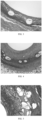
- the vascular scaffold in the present embodiment was implanted into a coronary vessel of a minipig, and an over-expanding ratio range was kept at 1.1:1 to 1.2:1 in the implantation process. Then, follow-up were carried out within three months after the implantation. During the follow-up, the vascular scaffold and vascular tissues around the vascular scaffold were taken out to perform pathologic analysis on the tissues around the vascular scaffold, and a pathological image was as shown in FIG. 3 .
- a pathologic analysis result showed that after the vascular scaffold provided by Embodiment 1 was implanted into the animal body for three months, no obvious smooth muscle cell proliferation phenomenon occurred in the tissues around the scaffold, endothelial cells of vascular tissues grew normally, and no histocyte necrosis occurred around a scaffold strut.
- the stenosis rate of the blood vessel was measured as 27% through the above-mentioned detection method.
- a pure iron vascular scaffold body having a surface area of 34.6 mm 2 was soaked into a zinc plating solution in half of a lengthwise direction.
- Zinc was uniformly plated by using an electroplating method, so as to form a zinc plated layer on half of the surface of the pure iron vascular scaffold body.
- the zinc-plated pure iron vascular scaffold body was placed in a vacuum furnace for thermal treatment at furnace chamber pressure of 20 Pa and thermal treatment temperature of 300 °C for 3 h. After the thermal treatment was completed, the sample was taken out, and was naturally cooled to room temperature.
- a rapamycin and polylactic acid mixture was uniformly sprayed to the surface of the scaffold body.
- a rapamycin-polylactic acid coating was formed on the surface of the scaffold body, thus obtaining the vascular scaffold of the present embodiment.
- a zinc-iron alloy layer covering the surface of the pure iron vascular scaffold body was formed.
- the zinc-iron alloy layer was the zinc-containing portions.
- the zinc-containing matrix was composed of the pure iron vascular scaffold body and the zinc-iron alloy layer.
- the vascular scaffold in the present embodiment was implanted into a coronary vessel of a minipig, and an over-expanding ratio range was kept at 1:1 to 1.2:1 in the implantation process. Then, follow-up were carried out within three months after the implantation. During the follow-up, the vascular scaffold and vascular tissues around the vascular scaffold were taken out to perform pathologic analysis on the tissues around the vascular scaffold.
- a pathologic analysis result showed that after the vascular scaffold provided by Embodiment 2 was implanted into the animal body for three months, no obvious smooth muscle cell proliferation phenomenon occurred in the tissues around the scaffold, endothelial cells of vascular tissues grew normally, and no histocyte necrosis occurred around a scaffold strut.
- the stenosis rate of the blood vessel was measured as 30% through the above-mentioned detection method.
- Zinc was uniformly plated on the surface of an iron-alloy vascular scaffold body having a surface area of 34.6 mm 2 by using an electroplating method, so as to form a zinc plated layer on the surface of the iron-alloy vascular scaffold body. Then, a rapamycin and polylactic acid mixture was uniformly sprayed to the surface of the zinc plated layer. After the surface was dried, a rapamycin-polylactic acid coating was formed on the surface of the zinc plated layer, thus obtaining the vascular scaffold of the present embodiment.
- the zinc plated layer in the present embodiment was the zinc-containing portions.
- the zinc-containing matrix was composed of the iron-alloy scaffold body, and the zinc plated layer.
- the vascular scaffold in the present embodiment was implanted into a coronary vessel of a minipig, and an over-expanding ratio range was kept at 1.1:1 to 1.2:1 in the implantation process. Then, follow-ups were carried out within three months after the implantation. During the follow-up, the vascular scaffold and vascular tissues around the vascular scaffold were taken out to perform pathologic analysis on the tissues around the vascular scaffold. A result showed that after the vascular scaffold provided by Embodiment 3 was implanted into the animal body for three months, no obvious smooth muscle cell proliferation phenomenon occurred in the tissues around the vascular scaffold, endothelial cells of vascular tissues grew normally, and no histocyte necrosis occurred around a scaffold strut. The stenosis rate of the blood vessel was measured as 32% through the above-mentioned detection method.
- a magnesium-alloy vascular scaffold body having a surface area of 34.6 mm 2 was soaked into a zinc plating solution in half of a lengthwise direction.
- Zinc was uniformly plated by using an electroplating method, so as to form a zinc plated layer on half of the surface of the magnesium-alloy vascular scaffold body.
- a rapamycin and polylactic acid mixture was uniformly sprayed to the surface of the magnesium-alloy vascular scaffold body.
- a rapamycin-polylactic acid coating was formed on the surface of the magnesium-alloy vascular scaffold body, and the zinc plated layer and the rapamycin-polylactic acid coating have no overlapping regions, thus obtaining the vascular scaffold of the present embodiment.
- the zinc plated layer in the present embodiment was the zinc-containing portions.
- the zinc-containing matrix was composed of the magnesium-alloy vascular scaffold body and the zinc plated layer.
- the vascular scaffold in the present embodiment was implanted into a coronary vessel of a minipig, and an over-expanding ratio range was kept at 1:1 to 1.2:1 in the implantation process. Then, follow-ups were carried out within three months after the implantation. During the follow-up, the scaffold and vascular tissues around the scaffold were taken out to perform pathologic analysis on the tissues around the scaffold. A result showed that after the vascular scaffold provided by Embodiment 4 was implanted into the animal body for three months, no obvious smooth muscle cell proliferation phenomenon occurred in the tissues around the vascular scaffold, endothelial cells of vascular tissues grew normally, and no histocyte necrosis occurred around a scaffold strut. The stenosis rate of the blood vessel was measured as 35% through the above-mentioned detection method.
- a zinc-iron alloy layer was electrically plated on the surface of a 316L stainless steel vascular scaffold having a surface area of 34.6 mm 2 .
- An atomic ratio of zinc to iron in the zinc-iron alloy layer was 7:3.
- a paclitaxel and polylactic acid mixture was uniformly sprayed to the zinc-iron alloy layer.
- the vascular scaffold of the present embodiment was obtained.
- the zinc-iron alloy layer in the present embodiment was the zinc-containing portions.
- the zinc-containing matrix was composed of the 316L stainless steel vascular scaffold body, and the zinc-iron alloy layer.
- the content p of the paclitaxel in the zinc-containing matrix per unit area was 0.025 ug•mm -2
- the vascular scaffold in the present embodiment was implanted into a coronary vessel of a minipig, and an over-expanding ratio range was kept at 1.1:1 to 1.2:1 in the implantation process. Then, follow-ups were carried out within three months after the implantation. During the follow-up, the vascular scaffold and vascular tissues around the vascular scaffold were taken out to perform pathologic analysis on the tissues around the vascular scaffold.
- a pathologic analysis result showed that after the vascular scaffold provided by Embodiment 5 was implanted into the animal body for three months, no obvious smooth muscle cell proliferation phenomenon occurred in the tissues around the scaffold, endothelial cells of vascular tissues grew normally, and no histocyte necrosis occurred around a scaffold strut.
- the stenosis rate of the blood vessel was measured as 36% through the above-mentioned detection method.
- a strut wall of a polylactic acid vascular scaffold body having a surface area of 34.6 mm 2 was provided with grooves, and the grooves in the surface of the polylactic acid vascular scaffold body were filled with pure zinc powder. Then, a rapamycin and polylactic acid mixture was uniformly sprayed to the surface of the polylactic acid vascular scaffold body. After the surface was dried, a rapamycin-polylactic acid coating was formed, thus obtaining the vascular scaffold of the present embodiment.
- the grooves of the polylactic acid vascular scaffold body were filled with the pure zinc powder to form the zinc-containing portions arranged on the polylactic acid vascular scaffold body.
- the vascular scaffold in the present embodiment was implanted into a coronary vessel of a minipig, and an over-expanding ratio range was kept at 1:1 to 1.2:1 in the implantation process. Then, follow-up were carried out within three months after the implantation. During the follow-up, the vascular scaffold and vascular tissues around the vascular scaffold were taken out to perform pathologic analysis on the tissues around the vascular scaffold.
- a pathologic analysis result showed that after the vascular scaffold provided by Embodiment 6 was implanted into the animal body for three months, no obvious smooth muscle cell proliferation phenomenon occurred in the tissues around the scaffold, endothelial cells of vascular tissues grew normally, and no histocyte necrosis occurred around a scaffold strut.
- the stenosis rate of the blood vessel was measured as 33% through the above-mentioned detection method.
- a rapamycin and polylactic acid mixture was uniformly sprayed to the outer surface of a pure zinc vascular scaffold matrix having a surface area of 34.6 mm 2 . After the outer surface was dried, a rapamycin-polylactic acid coating was formed on the outer surface of the pure zinc vascular scaffold matrix, thus obtaining the vascular scaffold of the present embodiment.
- the pure zinc vascular scaffold matrix was the zinc-containing matrix.
- the vascular scaffold in the present embodiment was implanted into a coronary vessel of a minipig, and an over-expanding ratio range was kept at 1.1:1 to 1.2:1 in the implantation process. Then, follow-ups were carried out within three months after the implantation. During the follow-up, the scaffold and vascular tissues around the scaffold were taken out to perform pathologic analysis on the tissues around the scaffold A result showed that after the vascular scaffold provided by Embodiment 7 was implanted into the animal body for three months, no obvious smooth muscle cell proliferation phenomenon occurred in the tissues around the vascular scaffold, endothelial cells of vascular tissues grew normally, and no histocyte necrosis occurred around a scaffold strut. The stenosis rate of the blood vessel was measured as 36% through the above-mentioned detection method.
- a rapamycin and polylactic acid mixture was uniformly sprayed to the surface of a zinc-alloy vascular scaffold matrix having a surface area of 34.6 mm 2 . After the surface was dried, a rapamycin-polylactic acid coating was formed on the surface of the zinc-alloy vascular scaffold matrix, thus obtaining the vascular scaffold of the present embodiment.
- the zinc-alloy vascular scaffold matrix was the zinc-containing matrix.
- the vascular scaffold in the present embodiment was implanted into a coronary vessel of a minipig, and an over-expanding ratio range was kept at 1:1 to 1.2:1 in the implantation process. Then, follow-up were carried out within three months after the implantation. During the follow-up, the vascular scaffold and vascular tissues around the vascular scaffold were taken out to perform pathologic analysis on the tissues around the vascular scaffold.
- a pathologic analysis result showed that after the vascular scaffold provided by Embodiment 8 was implanted into the animal body for three months, no obvious smooth muscle cell proliferation phenomenon occurred in the tissues around the scaffold, endothelial cells of vascular tissues grew normally, and no histocyte necrosis occurred around a scaffold strut.
- the stenosis rate of the blood vessel was measured as 32% through the above-mentioned detection method.
- a zinc-iron alloy layer was electrically plated on the surface of a 316L stainless steel vascular scaffold having a surface area of 34.6 mm 2 .
- An atomic ratio of zinc to iron in the zinc-iron alloy layer was 7:3.
- a paclitaxel and polylactic acid mixture was uniformly sprayed to the zinc-iron alloy layer.
- the vascular scaffold of the present embodiment was obtained.
- the zinc-iron alloy layer in the present embodiment was the zinc-containing portions.
- the zinc-containing matrix was composed of the stainless steel vascular scaffold body and the zinc-iron alloy layer.
- the vascular scaffold in the present comparative example was implanted into a coronary vessel of a minipig, and an over-expanding ratio range was kept at 1.1:1 to 1.2:1 in the implantation process. Then, follow-ups were carried out within three months after the implantation. During the follow-up, the vascular scaffold and vascular tissues around the vascular scaffold were taken out to perform pathologic analysis on the tissues around the vascular scaffold, and a pathological image was as shown in FIG. 4 .
- a pathologic analysis result showed that after the vascular scaffold provided by the comparative example 1 was implanted into the animal body for three months, no obvious smooth muscle cell proliferation phenomenon occurred in the tissues around the scaffold, endothelial cells of vascular tissues grew normally, and no histocyte necrosis occurred around a scaffold strut.
- the stenosis rate of the blood vessel was measured as 45%, which is obviously more than that of Embodiment 5.
- a pure iron vascular scaffold body having a surface area of 34.6 mm 2 was soaked into a zinc plating solution in half of a lengthwise direction.
- Zinc was uniformly plated by using an electroplating method, so as to form a zinc plated layer on the surface of half of a matrix of the pure iron vascular scaffold.
- the zinc-plated pure iron vascular scaffold body was placed in a vacuum furnace for thermal treatment at furnace chamber pressure of 20 Pa and thermal treatment temperature of 300 °C for 3 h. After the thermal treatment was completed, the sample was taken out, and was naturally cooled to room temperature.
- a rapamycin and polylactic acid mixture was uniformly sprayed to the surface of the scaffold body.
- a rapamycin-polylactic acid coating was formed on the surface of the scaffold body, thus obtaining the vascular scaffold of the present embodiment.
- a zinc-iron alloy layer covering the surface of the pure iron vascular scaffold body was formed.
- the zinc-iron alloy layer was the zinc-containing portions.
- the zinc-containing matrix was composed of the pure iron vascular scaffold body and the zinc-iron alloy layer.
- the vascular scaffold in the present comparative example was implanted into a coronary vessel of a minipig, and an over-expanding ratio range was kept at 1.1:1 to 1.2:1 in the implantation process. Then, follow-ups were carried out within one month after the implantation. During the follow-up, the vascular scaffold and vascular tissues around the vascular scaffold were taken out to perform pathologic analysis on the tissues around the vascular scaffold, and a pathological image was as shown in FIG. 5 .
- a pathologic analysis result showed that after the vascular scaffold provided by the comparative example 2 was implanted for one month, obvious intimal hyperplasia occurred around a scaffold strut, obvious luminal stenosis occurred in the blood vessel where the scaffold was placed, and obvious inflammatory cells appeared around the scaffold strut.
- the vascular scaffold of the comparative example 2 loads excessive drugs, resulting in necrocytosis in the tissues around the scaffold.
- Zinc was uniformly plated on the surface of an iron-alloy vascular scaffold body having a surface area of 34.6 mm 2 by using an electroplating method, so as to form a zinc plated layer on the surface of an iron-alloy vascular scaffold matrix. Then, a rapamycin and polylactic acid mixture was uniformly sprayed to the surface of the zinc plated layer. After the surface was dried, a rapamycin-polylactic acid coating was formed on the surface of the zinc plated layer, thus obtaining the vascular scaffold of the present embodiment.
- the zinc plated layer was the zinc-containing portions.
- the zinc-containing matrix was composed of the iron-alloy vascular scaffold body and the zinc plated layer.
- the vascular scaffold in the present comparative example was implanted into a coronary vessel of a minipig, and an over-expanding ratio range was kept at 1.1:1 to 1.2:1 in the implantation process. Then, follow-up were carried out within one month after the implantation. During the follow-up, the vascular scaffold and vascular tissues around the vascular scaffold were taken out to perform pathologic analysis on the tissues around the vascular scaffold. A result showed that after the vascular scaffold provided by the comparative example 3 was implanted for one month, obvious intimal hyperplasia occurred around a scaffold strut, obvious luminal stenosis occurred in the blood vessel where the scaffold was placed, and obvious inflammatory cells appeared around the scaffold strut.
- the vascular scaffold of the comparative example 3 causes necrocytosis in the tissues around the scaffold due to too fast corrosion of the zinc-containing portions.
Landscapes
- Health & Medical Sciences (AREA)
- Life Sciences & Earth Sciences (AREA)
- Veterinary Medicine (AREA)
- Public Health (AREA)
- General Health & Medical Sciences (AREA)
- Animal Behavior & Ethology (AREA)
- Epidemiology (AREA)
- Vascular Medicine (AREA)
- Surgery (AREA)
- Heart & Thoracic Surgery (AREA)
- Chemical & Material Sciences (AREA)
- Inorganic Chemistry (AREA)
- Medicinal Chemistry (AREA)
- Biomedical Technology (AREA)
- Engineering & Computer Science (AREA)
- Molecular Biology (AREA)
- Dermatology (AREA)
- Oral & Maxillofacial Surgery (AREA)
- Transplantation (AREA)
- Materials For Medical Uses (AREA)
- Pharmaceuticals Containing Other Organic And Inorganic Compounds (AREA)
Claims (14)
- Implantierbare arzneimittelbeladene Vorrichtung, die Folgendes umfasst:eine zinkhaltige Matrix und eine an die zinkhaltige Matrix angelagerte Arzneimittelwirkstoffschicht, wobei in der zinkhaltigen Matrix pro Flächeneinheit der Gehalt an einem Arzneimittelwirkstoff ρ mit der Einheit µg•mm-2 ist, der Gehalt an Zink, das aus der zinkhaltigen Matrix pro Flächeneinheit in einer wässrigen Lösung von Essigsäure garantierten Gütegrades bei 8,3 Vol.-% innerhalb einer Zeiteinheit korrodiert, p mit der Einheit µg•mm-2•min-1 ist, wobei der Gehalt an aus der zinkhaltigen Matrix pro Flächeneinheit korrodiertem Zink in der wässrigen Lösung von Essigsäure garantierten Gütegrades bei 8,3 Vol.-% bei 25°C durch ein Verfahren geprüft wird, das aus den folgenden Schritten besteht:- zunächst wird die Arzneimittelwirkstoffschicht der implantierbaren arzneimittelbeladenen Vorrichtung eluiert, um die zinkhaltigen Bereiche oder die aus dem reinen Zink oder der Zinklegierung gebildete Matrix freizulegen;- nach dem Wiegen wird die Vorrichtung 20 Minuten lang in die wässrige Lösung von Essigsäure garantierten Gütegrades bei 8,3 Vol.-% bei 25°C gelegt;- nach dem Herausnehmen der Vorrichtung wird ein festgelegtes Volumen an Essigsäurelösung zur Fixierung des Volumens verwendet, und dann wird ein Aufschluss durchgeführt;- die Zinkionenkonzentration in korrosiver Flüssigkeit wird mittels Atomabsorptionsspektroskopie geprüft;- der Gehalt des Zinks in der Lösung zu diesem Zeitpunkt wird berechnet, der dem Zinkgewichtsverlust der zinkhaltigen Bereiche oder der durch das reine Zink oder die Zinklegierung gebildeten Matrix entspricht;- schließlich wird der Gehalt durch den Flächeninhalt der Vorrichtung und die Korrosionszeit dividiert, um den Gehalt p des aus der zinkhaltigen Matrix korrodierten Zinks pro Flächeneinheit in der wässrigen Lösung von Essigsäure garantierten Gütegrades bei 8,3 Vol.-% innerhalb der Zeiteinheit zu erhalten;wobei p und p die folgende Beziehung erfüllen:
0,1≤15p+ρ≤3,5, worin p größer oder gleich 0,005 und kleiner oder gleich 0,14 ist. - Implantierbare arzneimittelbeladene Vorrichtung nach Anspruch 1, bei der die zinkhaltige Matrix eine aus Zink oder einer Zinklegierung gebildete Matrix ist oder die zinkhaltige Matrix einen Körper und an den Körper angelagerte zinkhaltige Bereiche umfasst, und
ein Material der zinkhaltigen Bereiche reines Zink oder eine Zinklegierung ist. - Implantierbare arzneimittelbeladene Vorrichtung nach Anspruch 2, bei der die Zinklegierung eine aus Zink und Eisen und/oder Magnesium und/oder Palladium und/oder Rhenium und/oder Gold und/oder Platin und/oder Tantal und/oder Strontium gebildete Legierung ist oder die Zinklegierung eine durch Dotierung von Zink mit Kohlenstoff und/oder Stickstoff und/oder Sauerstoff und/oder Schwefel und/oder Bor und/oder Silizium gebildete Legierung ist.
- Implantierbare arzneimittelbeladene Vorrichtung nach Anspruch 2, bei der dann, wenn die zinkhaltige Matrix den Körper und die an den Körper angelagerten zinkhaltigen Bereiche umfasst und das Material der zinkhaltigen Bereiche das reine Zink oder die Zinklegierung ist, die zinkhaltigen Bereiche zumindest teilweise die Oberfläche des Körpers bedecken oder die zinkhaltigen Bereiche das Innere des Körpers ausfüllen.
- Implantierbare arzneimittelbeladene Vorrichtung nach Anspruch 2, bei der dann, wenn die zinkhaltige Matrix den Körper und die an den Körper angelagerten zinkhaltigen Bereiche umfasst und das Material der zinkhaltigen Bereiche die Zinklegierung ist, der Körper ein Metallkörper ist und die zinkhaltigen Bereiche durch Reaktion zwischen dem Metallkörper und reinem Zink gebildet sind.
- Implantierbare arzneimittelbeladene Vorrichtung nach Anspruch 2, bei der dann, wenn die zinkhaltige Matrix den Körper und die an den Körper angelagerten zinkhaltigen Bereiche umfasst und das Material der zinkhaltigen Bereiche das reine Zink oder die Zinklegierung ist, die Arzneimittelwirkstoffschicht die zinkhaltigen Bereiche überhaupt nicht bedeckt oder die Arzneimittelwirkstoffschicht die zinkhaltigen Bereiche zumindest teilweise bedeckt.
- Implantierbare arzneimittelbeladene Vorrichtung nach Anspruch 1, bei der die Arzneimittelwirkstoffschicht einen Arzneimittelwirkstoff und einen Träger umfasst und der Arzneimittelwirkstoff in dem Träger dispergiert ist.
- Implantierbare arzneimittelbeladene Vorrichtung nach Anspruch 7, bei der der Arzneimittelwirkstoff Heparin und/oder Colchicin und/oder Rapamycin und/oder ein Derivat davon und/oder Paclitaxel und/oder ein Derivat davon und/oder Hirudin und/oder ein Derivat davon ist.
- Implantierbare arzneimittelbeladene Vorrichtung nach Anspruch 7, bei der der Träger ein abbaubares Polymer und/oder ein nicht abbaubares Polymer und/oder ein Copolymer ist, das aus mindestens zwei von Monomeren, die das abbaubare Polymer bilden, und Monomeren gebildet ist, die das nicht abbaubare Polymer bilden, und das abbaubare Polymer Polymilchsäure und/oder Polyglykolsäure und/oder Polycaprolacton und/oder Polysuccinat und/oder Poly(β-hydroxybutyrat) ist und das nicht abbaubare Polymer Polystyrol und/oder Polytetrafluorethylen und/oder Polymethylmethacrylat und/oder Polycarbonat und/oder Polyethylenterephthalat ist.
- Implantierbare arzneimittelbeladene Vorrichtung nach Anspruch 2, bei der der Körper aus einem Metall besteht und das Metall ein abbaubares Metall oder ein nicht abbaubares Metall ist.
- Implantierbare arzneimittelbeladene Vorrichtung nach Anspruch 10, bei der das abbaubare Metall reines Eisen, eine Eisenlegierung, reines Magnesium oder eine Magnesiumlegierung ist.
- Implantierbare arzneimittelbeladene Vorrichtung nach Anspruch 10, bei der das nicht abbaubare Metall Edelstahl 316L, eine Kobalt-Chrom-Legierung, eine Platin-Chrom-Legierung, reines Titan oder eine Titanlegierung ist.
- Implantierbare arzneimittelbeladene Vorrichtung nach Anspruch 2, bei der der Körper zumindest teilweise aus einem Polymer besteht, das Polymer ein abbaubares Polymer und/oder ein nicht abbaubares Polymer und/oder ein Copolymer ist, das aus mindestens zwei von Monomeren, die das abbaubare Polymer bilden, und Monomeren gebildet ist, die das nicht abbaubare Polymer bilden, das abbaubare Polymer Polymilchsäure und/oder Polyglykolsäure und/oder Polycaprolacton und/oder Polysuccinat und/oder Poly(β-hydroxybutyrat) ist und das nicht abbaubare Polymer Polystyrol und/oder Polytetrafluorethylen und/oder Polymethylmethacrylat und/oder Polycarbonat und/oder Polyethylenterephthalat ist.
- Implantierbare arzneimittelbeladene Vorrichtung nach Anspruch 1, wobei die implantierbare arzneimittelbeladene Vorrichtung ein Gefäßgerüst, ein biliäres Gerüst, ein Speiseröhrengerüst, ein Harnröhrengerüst, ein Atemwegsgerüst, ein andrologisches Implantat, ein gynäkologisches Implantat oder ein Hohlvenenfilter ist.
Applications Claiming Priority (2)
| Application Number | Priority Date | Filing Date | Title |
|---|---|---|---|
| CN201711461899.7A CN109966563B (zh) | 2017-12-28 | 2017-12-28 | 植入式载药器械 |
| PCT/CN2018/121678 WO2019128767A1 (zh) | 2017-12-28 | 2018-12-18 | 植入式载药器械 |
Publications (4)
| Publication Number | Publication Date |
|---|---|
| EP3733221A1 EP3733221A1 (de) | 2020-11-04 |
| EP3733221A4 EP3733221A4 (de) | 2021-10-06 |
| EP3733221B1 true EP3733221B1 (de) | 2023-09-13 |
| EP3733221C0 EP3733221C0 (de) | 2023-09-13 |
Family
ID=67063106
Family Applications (1)
| Application Number | Title | Priority Date | Filing Date |
|---|---|---|---|
| EP18897786.2A Active EP3733221B1 (de) | 2017-12-28 | 2018-12-18 | Implantierbare wirkstofffreisetzungsvorrichtung |
Country Status (4)
| Country | Link |
|---|---|
| US (1) | US20200330642A1 (de) |
| EP (1) | EP3733221B1 (de) |
| CN (1) | CN109966563B (de) |
| WO (1) | WO2019128767A1 (de) |
Families Citing this family (1)
| Publication number | Priority date | Publication date | Assignee | Title |
|---|---|---|---|---|
| US20220354488A1 (en) | 2021-05-10 | 2022-11-10 | Cilag Gmbh International | Absorbable surgical staples comprising sufficient structural properties during a tissue healing window |
Family Cites Families (15)
| Publication number | Priority date | Publication date | Assignee | Title |
|---|---|---|---|---|
| US20050267560A1 (en) * | 2000-02-03 | 2005-12-01 | Cook Incorporated | Implantable bioabsorbable valve support frame |
| AU2015252144A1 (en) * | 2005-04-05 | 2015-11-26 | Elixir Medical Corporation | Degradable implantable medical devices |
| CN101161299B (zh) * | 2006-10-09 | 2011-07-06 | 乐普(北京)医疗器械股份有限公司 | 孔洞及聚合物共载的药物释放结构及其制备方法 |
| US20080200421A1 (en) * | 2007-02-21 | 2008-08-21 | Zhao Jonathon Z | Coating for a medical device having an anti-thrombotic conjugate |
| CN101185779B (zh) * | 2007-12-19 | 2010-06-02 | 上海赢生医疗科技有限公司 | 一种药物缓释支架的制备方法 |
| CN102772831A (zh) * | 2012-08-20 | 2012-11-14 | 道淼科技(北京)有限公司 | 一种可降解载药支架 |
| CN104593650A (zh) * | 2013-11-01 | 2015-05-06 | 上海交通大学医学院附属第九人民医院 | 一种具有抗菌功能可降解吸收的镁锌铜合金及其应用 |
| CN104195369B (zh) * | 2014-08-21 | 2016-09-21 | 北京大学 | 一种Zn-Ca系锌合金及其制备方法与应用 |
| CN104857570B (zh) * | 2015-05-05 | 2017-03-29 | 乐普(北京)医疗器械股份有限公司 | 一种可降解锌基合金支架及其制备方法 |
| CN104888282B (zh) * | 2015-05-05 | 2017-11-24 | 乐普(北京)医疗器械股份有限公司 | 一种可降解锌基微孔载药支架及其制备方法 |
| CN106474545B (zh) * | 2015-08-28 | 2020-04-10 | 元心科技(深圳)有限公司 | 可吸收铁基合金植入医疗器械 |
| CN106606801B (zh) * | 2015-10-21 | 2019-04-16 | 北京大学 | 一种Zn-ZnO系锌合金及其制备方法与应用 |
| CN106806938B (zh) * | 2015-11-27 | 2020-04-14 | 先健科技(深圳)有限公司 | 可吸收铁基合金植入医疗器械 |
| CN106955374B (zh) * | 2016-01-08 | 2019-11-08 | 先健科技(深圳)有限公司 | 植入式器械 |
| CN106267378A (zh) * | 2016-10-10 | 2017-01-04 | 同济大学 | 一种可控降解的金属支架的制备方法 |
-
2017
- 2017-12-28 CN CN201711461899.7A patent/CN109966563B/zh active Active
-
2018
- 2018-12-18 US US16/957,975 patent/US20200330642A1/en active Pending
- 2018-12-18 EP EP18897786.2A patent/EP3733221B1/de active Active
- 2018-12-18 WO PCT/CN2018/121678 patent/WO2019128767A1/zh not_active Ceased
Also Published As
| Publication number | Publication date |
|---|---|
| CN109966563A (zh) | 2019-07-05 |
| CN109966563B (zh) | 2021-08-03 |
| EP3733221A1 (de) | 2020-11-04 |
| EP3733221C0 (de) | 2023-09-13 |
| EP3733221A4 (de) | 2021-10-06 |
| WO2019128767A1 (zh) | 2019-07-04 |
| US20200330642A1 (en) | 2020-10-22 |
Similar Documents
| Publication | Publication Date | Title |
|---|---|---|
| Park et al. | In vivo evaluation and characterization of a bio-absorbable drug-coated stent fabricated using a 3D-printing system | |
| EP3562526B1 (de) | Stent aus einer biologisch abbaubaren magnesiumlegierung mit einer magnesiumfluorid-beschichtung und einer organischen beschichtung | |
| US8927002B2 (en) | Stent with a coating or a basic body containing a lithium salt and use of lithium salts for prevention of restenosis | |
| RU2642254C2 (ru) | Рассасывающие стенты, которые содержат магниевые сплавы | |
| Ma et al. | Paclitaxel/sirolimus combination coated drug-eluting stent: in vitro and in vivo drug release studies | |
| EP2716307B1 (de) | Arzneimittelelutionsstent mit biologisch abbaubarer, mit elektrogepfropfter grundierung befestigter trennschicht | |
| EP2380528A1 (de) | Medikamentenausgabevorrichtung mit einer mikrobohrungsstruktur zur lagerung und freigabe mehrerer medikamente sowie verfahren zu ihrer herstellung | |
| Wang et al. | In vitro and in vivo assessment of the biocompatibility of an paclitaxel-eluting poly-l-lactide-coated Mg-Zn-Y-Nd alloy stent in the intestine | |
| Chen et al. | High nitrogen stainless steel drug-eluting stent-assessment of pharmacokinetics and preclinical safety in vivo | |
| EP3733221B1 (de) | Implantierbare wirkstofffreisetzungsvorrichtung | |
| CA2817450C (en) | Biocorrodible magnesium alloy implant | |
| US20120239140A1 (en) | Medical product comprising an active coating | |
| US10016775B2 (en) | Device for coating a stent and associated coating method and stent produced according to the method | |
| EP3650053B1 (de) | Implantierbare vorrichtung | |
| Karoussos et al. | Inorganic materials as drug delivery systems in coronary artery stenting | |
| US12414849B2 (en) | Implantable device | |
| US8920490B2 (en) | Endoprostheses | |
| CN111358603A (zh) | 一种药物洗脱器械及其制造方法 | |
| Lu et al. | In vivo and in vitro studies of biodegradable WE43 stent | |
| US20040176838A1 (en) | Medical device | |
| KR100613951B1 (ko) | 의료장치 | |
| JP2025524216A (ja) | 医療デバイス用のコーティング | |
| CN113491796A (zh) | 含锌医疗器械 | |
| HK1190647B (en) | Drug eluting stent with a biodegradable release layer attached with an electro-grafted primer coating | |
| HK1190647A (en) | Drug eluting stent with a biodegradable release layer attached with an electro-grafted primer coating |
Legal Events
| Date | Code | Title | Description |
|---|---|---|---|
| STAA | Information on the status of an ep patent application or granted ep patent |
Free format text: STATUS: THE INTERNATIONAL PUBLICATION HAS BEEN MADE |
|
| PUAI | Public reference made under article 153(3) epc to a published international application that has entered the european phase |
Free format text: ORIGINAL CODE: 0009012 |
|
| STAA | Information on the status of an ep patent application or granted ep patent |
Free format text: STATUS: REQUEST FOR EXAMINATION WAS MADE |
|
| 17P | Request for examination filed |
Effective date: 20200721 |
|
| AK | Designated contracting states |
Kind code of ref document: A1 Designated state(s): AL AT BE BG CH CY CZ DE DK EE ES FI FR GB GR HR HU IE IS IT LI LT LU LV MC MK MT NL NO PL PT RO RS SE SI SK SM TR |
|
| AX | Request for extension of the european patent |
Extension state: BA ME |
|
| DAV | Request for validation of the european patent (deleted) | ||
| DAX | Request for extension of the european patent (deleted) | ||
| A4 | Supplementary search report drawn up and despatched |
Effective date: 20210906 |
|
| RIC1 | Information provided on ipc code assigned before grant |
Ipc: A61L 31/16 20060101ALI20210831BHEP Ipc: A61L 31/14 20060101ALI20210831BHEP Ipc: A61L 31/10 20060101ALI20210831BHEP Ipc: A61L 31/08 20060101ALI20210831BHEP Ipc: A61L 31/02 20060101AFI20210831BHEP |
|
| GRAP | Despatch of communication of intention to grant a patent |
Free format text: ORIGINAL CODE: EPIDOSNIGR1 |
|
| STAA | Information on the status of an ep patent application or granted ep patent |
Free format text: STATUS: GRANT OF PATENT IS INTENDED |
|
| INTG | Intention to grant announced |
Effective date: 20230420 |
|
| GRAS | Grant fee paid |
Free format text: ORIGINAL CODE: EPIDOSNIGR3 |
|
| GRAA | (expected) grant |
Free format text: ORIGINAL CODE: 0009210 |
|
| STAA | Information on the status of an ep patent application or granted ep patent |
Free format text: STATUS: THE PATENT HAS BEEN GRANTED |
|
| AK | Designated contracting states |
Kind code of ref document: B1 Designated state(s): AL AT BE BG CH CY CZ DE DK EE ES FI FR GB GR HR HU IE IS IT LI LT LU LV MC MK MT NL NO PL PT RO RS SE SI SK SM TR |
|
| REG | Reference to a national code |
Ref country code: CH Ref legal event code: EP |
|
| REG | Reference to a national code |
Ref country code: DE Ref legal event code: R096 Ref document number: 602018057730 Country of ref document: DE |
|
| REG | Reference to a national code |
Ref country code: IE Ref legal event code: FG4D |
|
| U01 | Request for unitary effect filed |
Effective date: 20230927 |
|
| U07 | Unitary effect registered |
Designated state(s): AT BE BG DE DK EE FI FR IT LT LU LV MT NL PT SE SI Effective date: 20231005 |
|
| PG25 | Lapsed in a contracting state [announced via postgrant information from national office to epo] |
Ref country code: GR Free format text: LAPSE BECAUSE OF FAILURE TO SUBMIT A TRANSLATION OF THE DESCRIPTION OR TO PAY THE FEE WITHIN THE PRESCRIBED TIME-LIMIT Effective date: 20231214 |
|
| U20 | Renewal fee for the european patent with unitary effect paid |
Year of fee payment: 6 Effective date: 20231218 |
|
| PG25 | Lapsed in a contracting state [announced via postgrant information from national office to epo] |
Ref country code: RS Free format text: LAPSE BECAUSE OF FAILURE TO SUBMIT A TRANSLATION OF THE DESCRIPTION OR TO PAY THE FEE WITHIN THE PRESCRIBED TIME-LIMIT Effective date: 20230913 Ref country code: NO Free format text: LAPSE BECAUSE OF FAILURE TO SUBMIT A TRANSLATION OF THE DESCRIPTION OR TO PAY THE FEE WITHIN THE PRESCRIBED TIME-LIMIT Effective date: 20231213 Ref country code: HR Free format text: LAPSE BECAUSE OF FAILURE TO SUBMIT A TRANSLATION OF THE DESCRIPTION OR TO PAY THE FEE WITHIN THE PRESCRIBED TIME-LIMIT Effective date: 20230913 Ref country code: GR Free format text: LAPSE BECAUSE OF FAILURE TO SUBMIT A TRANSLATION OF THE DESCRIPTION OR TO PAY THE FEE WITHIN THE PRESCRIBED TIME-LIMIT Effective date: 20231214 |
|
| PG25 | Lapsed in a contracting state [announced via postgrant information from national office to epo] |
Ref country code: IS Free format text: LAPSE BECAUSE OF FAILURE TO SUBMIT A TRANSLATION OF THE DESCRIPTION OR TO PAY THE FEE WITHIN THE PRESCRIBED TIME-LIMIT Effective date: 20240113 |
|
| PG25 | Lapsed in a contracting state [announced via postgrant information from national office to epo] |
Ref country code: ES Free format text: LAPSE BECAUSE OF FAILURE TO SUBMIT A TRANSLATION OF THE DESCRIPTION OR TO PAY THE FEE WITHIN THE PRESCRIBED TIME-LIMIT Effective date: 20230913 |
|
| PG25 | Lapsed in a contracting state [announced via postgrant information from national office to epo] |
Ref country code: SM Free format text: LAPSE BECAUSE OF FAILURE TO SUBMIT A TRANSLATION OF THE DESCRIPTION OR TO PAY THE FEE WITHIN THE PRESCRIBED TIME-LIMIT Effective date: 20230913 Ref country code: RO Free format text: LAPSE BECAUSE OF FAILURE TO SUBMIT A TRANSLATION OF THE DESCRIPTION OR TO PAY THE FEE WITHIN THE PRESCRIBED TIME-LIMIT Effective date: 20230913 Ref country code: IS Free format text: LAPSE BECAUSE OF FAILURE TO SUBMIT A TRANSLATION OF THE DESCRIPTION OR TO PAY THE FEE WITHIN THE PRESCRIBED TIME-LIMIT Effective date: 20240113 Ref country code: ES Free format text: LAPSE BECAUSE OF FAILURE TO SUBMIT A TRANSLATION OF THE DESCRIPTION OR TO PAY THE FEE WITHIN THE PRESCRIBED TIME-LIMIT Effective date: 20230913 Ref country code: CZ Free format text: LAPSE BECAUSE OF FAILURE TO SUBMIT A TRANSLATION OF THE DESCRIPTION OR TO PAY THE FEE WITHIN THE PRESCRIBED TIME-LIMIT Effective date: 20230913 Ref country code: SK Free format text: LAPSE BECAUSE OF FAILURE TO SUBMIT A TRANSLATION OF THE DESCRIPTION OR TO PAY THE FEE WITHIN THE PRESCRIBED TIME-LIMIT Effective date: 20230913 |
|
| PG25 | Lapsed in a contracting state [announced via postgrant information from national office to epo] |
Ref country code: PL Free format text: LAPSE BECAUSE OF FAILURE TO SUBMIT A TRANSLATION OF THE DESCRIPTION OR TO PAY THE FEE WITHIN THE PRESCRIBED TIME-LIMIT Effective date: 20230913 |
|
| REG | Reference to a national code |
Ref country code: DE Ref legal event code: R097 Ref document number: 602018057730 Country of ref document: DE |
|
| PLBE | No opposition filed within time limit |
Free format text: ORIGINAL CODE: 0009261 |
|
| STAA | Information on the status of an ep patent application or granted ep patent |
Free format text: STATUS: NO OPPOSITION FILED WITHIN TIME LIMIT |
|
| REG | Reference to a national code |
Ref country code: CH Ref legal event code: PL |
|
| 26N | No opposition filed |
Effective date: 20240614 |
|
| PG25 | Lapsed in a contracting state [announced via postgrant information from national office to epo] |
Ref country code: MC Free format text: LAPSE BECAUSE OF FAILURE TO SUBMIT A TRANSLATION OF THE DESCRIPTION OR TO PAY THE FEE WITHIN THE PRESCRIBED TIME-LIMIT Effective date: 20230913 |
|
| PG25 | Lapsed in a contracting state [announced via postgrant information from national office to epo] |
Ref country code: MC Free format text: LAPSE BECAUSE OF FAILURE TO SUBMIT A TRANSLATION OF THE DESCRIPTION OR TO PAY THE FEE WITHIN THE PRESCRIBED TIME-LIMIT Effective date: 20230913 |
|
| REG | Reference to a national code |
Ref country code: IE Ref legal event code: MM4A |
|
| PG25 | Lapsed in a contracting state [announced via postgrant information from national office to epo] |
Ref country code: IE Free format text: LAPSE BECAUSE OF NON-PAYMENT OF DUE FEES Effective date: 20231218 |
|
| PG25 | Lapsed in a contracting state [announced via postgrant information from national office to epo] |
Ref country code: CH Free format text: LAPSE BECAUSE OF NON-PAYMENT OF DUE FEES Effective date: 20231231 |
|
| PG25 | Lapsed in a contracting state [announced via postgrant information from national office to epo] |
Ref country code: IE Free format text: LAPSE BECAUSE OF NON-PAYMENT OF DUE FEES Effective date: 20231218 Ref country code: CH Free format text: LAPSE BECAUSE OF NON-PAYMENT OF DUE FEES Effective date: 20231231 |
|
| U20 | Renewal fee for the european patent with unitary effect paid |
Year of fee payment: 7 Effective date: 20241218 |
|
| PG25 | Lapsed in a contracting state [announced via postgrant information from national office to epo] |
Ref country code: CY Free format text: LAPSE BECAUSE OF FAILURE TO SUBMIT A TRANSLATION OF THE DESCRIPTION OR TO PAY THE FEE WITHIN THE PRESCRIBED TIME-LIMIT; INVALID AB INITIO Effective date: 20181218 |
|
| PG25 | Lapsed in a contracting state [announced via postgrant information from national office to epo] |
Ref country code: HU Free format text: LAPSE BECAUSE OF FAILURE TO SUBMIT A TRANSLATION OF THE DESCRIPTION OR TO PAY THE FEE WITHIN THE PRESCRIBED TIME-LIMIT; INVALID AB INITIO Effective date: 20181218 |
|
| PG25 | Lapsed in a contracting state [announced via postgrant information from national office to epo] |
Ref country code: TR Free format text: LAPSE BECAUSE OF FAILURE TO SUBMIT A TRANSLATION OF THE DESCRIPTION OR TO PAY THE FEE WITHIN THE PRESCRIBED TIME-LIMIT Effective date: 20230913 |
|
| PGFP | Annual fee paid to national office [announced via postgrant information from national office to epo] |
Ref country code: GB Payment date: 20251229 Year of fee payment: 8 |
|
| U20 | Renewal fee for the european patent with unitary effect paid |
Year of fee payment: 8 Effective date: 20251217 |