EP3748363A1 - Verfahren zur hochdurchsatzanalyse von proteinen und anwendbare bibliothek dafür - Google Patents
Verfahren zur hochdurchsatzanalyse von proteinen und anwendbare bibliothek dafür Download PDFInfo
- Publication number
- EP3748363A1 EP3748363A1 EP19746592.5A EP19746592A EP3748363A1 EP 3748363 A1 EP3748363 A1 EP 3748363A1 EP 19746592 A EP19746592 A EP 19746592A EP 3748363 A1 EP3748363 A1 EP 3748363A1
- Authority
- EP
- European Patent Office
- Prior art keywords
- protein
- tag
- interest
- embryonic stem
- proteins
- Prior art date
- Legal status (The legal status is an assumption and is not a legal conclusion. Google has not performed a legal analysis and makes no representation as to the accuracy of the status listed.)
- Withdrawn
Links
Images



Classifications
-
- C—CHEMISTRY; METALLURGY
- C12—BIOCHEMISTRY; BEER; SPIRITS; WINE; VINEGAR; MICROBIOLOGY; ENZYMOLOGY; MUTATION OR GENETIC ENGINEERING
- C12N—MICROORGANISMS OR ENZYMES; COMPOSITIONS THEREOF; PROPAGATING, PRESERVING, OR MAINTAINING MICROORGANISMS; MUTATION OR GENETIC ENGINEERING; CULTURE MEDIA
- C12N15/00—Mutation or genetic engineering; DNA or RNA concerning genetic engineering, vectors, e.g. plasmids, or their isolation, preparation or purification; Use of hosts therefor
- C12N15/09—Recombinant DNA-technology
- C12N15/10—Processes for the isolation, preparation or purification of DNA or RNA
- C12N15/1034—Isolating an individual clone by screening libraries
- C12N15/1093—General methods of preparing gene libraries, not provided for in other subgroups
-
- C—CHEMISTRY; METALLURGY
- C12—BIOCHEMISTRY; BEER; SPIRITS; WINE; VINEGAR; MICROBIOLOGY; ENZYMOLOGY; MUTATION OR GENETIC ENGINEERING
- C12N—MICROORGANISMS OR ENZYMES; COMPOSITIONS THEREOF; PROPAGATING, PRESERVING, OR MAINTAINING MICROORGANISMS; MUTATION OR GENETIC ENGINEERING; CULTURE MEDIA
- C12N15/00—Mutation or genetic engineering; DNA or RNA concerning genetic engineering, vectors, e.g. plasmids, or their isolation, preparation or purification; Use of hosts therefor
- C12N15/09—Recombinant DNA-technology
- C12N15/10—Processes for the isolation, preparation or purification of DNA or RNA
- C12N15/1034—Isolating an individual clone by screening libraries
- C12N15/1086—Preparation or screening of expression libraries, e.g. reporter assays
-
- C—CHEMISTRY; METALLURGY
- C12—BIOCHEMISTRY; BEER; SPIRITS; WINE; VINEGAR; MICROBIOLOGY; ENZYMOLOGY; MUTATION OR GENETIC ENGINEERING
- C12N—MICROORGANISMS OR ENZYMES; COMPOSITIONS THEREOF; PROPAGATING, PRESERVING, OR MAINTAINING MICROORGANISMS; MUTATION OR GENETIC ENGINEERING; CULTURE MEDIA
- C12N15/00—Mutation or genetic engineering; DNA or RNA concerning genetic engineering, vectors, e.g. plasmids, or their isolation, preparation or purification; Use of hosts therefor
- C12N15/09—Recombinant DNA-technology
- C12N15/63—Introduction of foreign genetic material using vectors; Vectors; Use of hosts therefor; Regulation of expression
- C12N15/79—Vectors or expression systems specially adapted for eukaryotic hosts
- C12N15/85—Vectors or expression systems specially adapted for eukaryotic hosts for animal cells
-
- C—CHEMISTRY; METALLURGY
- C12—BIOCHEMISTRY; BEER; SPIRITS; WINE; VINEGAR; MICROBIOLOGY; ENZYMOLOGY; MUTATION OR GENETIC ENGINEERING
- C12N—MICROORGANISMS OR ENZYMES; COMPOSITIONS THEREOF; PROPAGATING, PRESERVING, OR MAINTAINING MICROORGANISMS; MUTATION OR GENETIC ENGINEERING; CULTURE MEDIA
- C12N5/00—Undifferentiated human, animal or plant cells, e.g. cell lines; Tissues; Cultivation or maintenance thereof; Culture media therefor
- C12N5/06—Animal cells or tissues; Human cells or tissues
- C12N5/0602—Vertebrate cells
- C12N5/0603—Embryonic cells ; Embryoid bodies
-
- C—CHEMISTRY; METALLURGY
- C12—BIOCHEMISTRY; BEER; SPIRITS; WINE; VINEGAR; MICROBIOLOGY; ENZYMOLOGY; MUTATION OR GENETIC ENGINEERING
- C12Q—MEASURING OR TESTING PROCESSES INVOLVING ENZYMES, NUCLEIC ACIDS OR MICROORGANISMS; COMPOSITIONS OR TEST PAPERS THEREFOR; PROCESSES OF PREPARING SUCH COMPOSITIONS; CONDITION-RESPONSIVE CONTROL IN MICROBIOLOGICAL OR ENZYMOLOGICAL PROCESSES
- C12Q1/00—Measuring or testing processes involving enzymes, nucleic acids or microorganisms; Compositions therefor; Processes of preparing such compositions
- C12Q1/68—Measuring or testing processes involving enzymes, nucleic acids or microorganisms; Compositions therefor; Processes of preparing such compositions involving nucleic acids
-
- C—CHEMISTRY; METALLURGY
- C40—COMBINATORIAL TECHNOLOGY
- C40B—COMBINATORIAL CHEMISTRY; LIBRARIES, e.g. CHEMICAL LIBRARIES
- C40B50/00—Methods of creating libraries, e.g. combinatorial synthesis
- C40B50/06—Biochemical methods, e.g. using enzymes or whole viable microorganisms
-
- G—PHYSICS
- G01—MEASURING; TESTING
- G01N—INVESTIGATING OR ANALYSING MATERIALS BY DETERMINING THEIR CHEMICAL OR PHYSICAL PROPERTIES
- G01N33/00—Investigating or analysing materials by specific methods not covered by groups G01N1/00 - G01N31/00
- G01N33/48—Biological material, e.g. blood, urine; Haemocytometers
- G01N33/50—Chemical analysis of biological material, e.g. blood, urine; Testing involving biospecific ligand binding methods; Immunological testing
- G01N33/68—Chemical analysis of biological material, e.g. blood, urine; Testing involving biospecific ligand binding methods; Immunological testing involving proteins, peptides or amino acids
-
- G—PHYSICS
- G01—MEASURING; TESTING
- G01N—INVESTIGATING OR ANALYSING MATERIALS BY DETERMINING THEIR CHEMICAL OR PHYSICAL PROPERTIES
- G01N33/00—Investigating or analysing materials by specific methods not covered by groups G01N1/00 - G01N31/00
- G01N33/48—Biological material, e.g. blood, urine; Haemocytometers
- G01N33/50—Chemical analysis of biological material, e.g. blood, urine; Testing involving biospecific ligand binding methods; Immunological testing
- G01N33/68—Chemical analysis of biological material, e.g. blood, urine; Testing involving biospecific ligand binding methods; Immunological testing involving proteins, peptides or amino acids
- G01N33/6803—General methods of protein analysis not limited to specific proteins or families of proteins
-
- G—PHYSICS
- G01—MEASURING; TESTING
- G01N—INVESTIGATING OR ANALYSING MATERIALS BY DETERMINING THEIR CHEMICAL OR PHYSICAL PROPERTIES
- G01N33/00—Investigating or analysing materials by specific methods not covered by groups G01N1/00 - G01N31/00
- G01N33/48—Biological material, e.g. blood, urine; Haemocytometers
- G01N33/50—Chemical analysis of biological material, e.g. blood, urine; Testing involving biospecific ligand binding methods; Immunological testing
- G01N33/68—Chemical analysis of biological material, e.g. blood, urine; Testing involving biospecific ligand binding methods; Immunological testing involving proteins, peptides or amino acids
- G01N33/6803—General methods of protein analysis not limited to specific proteins or families of proteins
- G01N33/6842—Proteomic analysis of subsets of protein mixtures with reduced complexity, e.g. membrane proteins, phosphoproteins, organelle proteins
-
- G—PHYSICS
- G01—MEASURING; TESTING
- G01N—INVESTIGATING OR ANALYSING MATERIALS BY DETERMINING THEIR CHEMICAL OR PHYSICAL PROPERTIES
- G01N2570/00—Omics, e.g. proteomics, glycomics or lipidomics; Methods of analysis focusing on the entire complement of classes of biological molecules or subsets thereof, i.e. focusing on proteomes, glycomes or lipidomes
Definitions
- the present disclosure relates to the field of biology, specifically to the field of proteomics research, and more specifically to a high-throughput protein analysis method and a suitable library thereof.
- the binding of an ovum to a sperm forms a pluripotent fertilized ovum that begins a life.
- a life individual with more than 200 different somatic cells is ultimately formed by embryonic development, and the process is extremely complicated.
- the cell In the development process from the fertilized ovum to a biological individual, the cell is always faced with choice: to maintain the existing identity and status or to transform into another identity and status.
- the maintenance and change of cell identity and status are controlled by the intrinsic genetic factors of the cell itself and also regulated by the environmental factors surrounding the cell.
- the interaction of intracellular and extracellular factors makes the fate of the cell variable and transformational.
- biomacromolecules including proteins.
- biomacromolecules including proteins.
- current research on the biomacromolecules lacks a system suitable for in vivo, real-time and dynamic research.
- a first aspect of the present disclosure is to provide a high-throughput protein analysis method, including: using a tagged semi-cloned mouse library to perform parallel indicator analysis on a plurality of different target proteins of interest with one or several tag protein antibodies.
- each semi-cloned mouse is a semi-cloned mouse obtained by culturing after injecting an androgenetic haploid embryonic stem cell into an ovum, or a sexually propagated progeny thereof.
- the androgenetic haploid embryonic stem cell contains a gene that expresses a fusion protein of a target protein of interest and a tag protein, and the semi-cloned mouse can express the fusion protein of the target protein of interest and the tag protein.
- a second aspect of the present disclosure is to further provide a tagged semi-cloned mouse library suitable for the aforementioned high-throughput protein analysis method and a method for constructing the same.
- each semi-cloned mouse is a semi-cloned mouse obtained by culturing after injecting an androgenetic haploid embryonic stem cell into an ovum, or a sexually propagated progeny thereof, and the androgenetic haploid embryonic stem cell contains a gene that expresses a fusion protein of the target protein of interest and the tag protein.
- the tagged semi-cloned mouse library of the present disclosure or the semi-cloned mouse from the library can be used in the fields of protein analysis, protein function research or drug research.
- a third aspect of the present disclosure is to further provide a tagged androgenetic haploid embryonic stem cell library suitable for the aforementioned high-throughput protein analysis method and a method for constructing the same.
- each androgenetic haploid embryonic stem cell contains a gene that expresses a fusion protein of a target protein of interest and a tag protein, and the semi-cloned mouse obtained by culturing after injecting the androgenetic haploid embryonic stem cell into an ovum can express the fusion protein of the target protein of interest and the tag protein.
- the tagged androgenetic haploid embryonic stem cell library of the present disclosure or the androgenetic haploid embryonic stem cell from the library can be used in the fields of protein analysis, protein function research, and drug research.
- the present disclosure provides a high-throughput protein analysis method, including: using a tagged semi-cloned mouse library to perform parallel indicator analysis on a plurality of different target proteins of interest with one or several tag protein antibodies.
- each semi-cloned mouse is a semi-cloned mouse obtained by culturing after injecting an androgenetic haploid embryonic stem cell into an ovum, or a sexually propagated progeny thereof
- the androgenetic haploid embryonic stem cell contains a gene that expresses a fusion protein of a target protein of interest and a tag protein
- the semi-cloned mouse can express the fusion protein of the target protein of interest and the tag protein.
- a high-throughput protein analysis method because it uses the tagged semi-cloned mouse library, and can perform simultaneous parallel research on a plurality of target proteins of interest in the library by only needing to use a limited number of universal tag protein antibodies corresponding to tags in the library. Not only does it not require the preparation of antibodies to the target protein of interest, but the same operation procedure can be used to perform simultaneously in vivo study of a plurality of target proteins of interest. This is different from the existing research method that needs to prepare antibodies of the target proteins of interest, the scientific research of proteins is greatly simplified, and the research efficiency is improved.
- the protein analysis method of the present disclosure does not need to prepare or use antibodies of target proteins of interest.
- the method changes the conventional in vivo research idea of proteins, and provides great convenience for drug screening, drug action mechanism analysis, drug metabolism and other researches. Due to that the same antibody is used for research, the antibody-antigen binding affinity is also consistent. Compared with the situation that different target protein antibodies are utilized for different target proteins of interest, when parallel comparison is performed, the tag protein antibody research is matured, the antibody of the present disclosure is stable in both sensitivity and specificity, and is stronger in reference.
- the androgenetic haploid embryonic stem cell of the present disclosure has the self-replication ability and pluripotency of stem cells, and can replace the sperm to bind with an oocyte to support the complete development of an embryo.
- the semi-cloned mouse of the present disclosure may be in the morphologies of various stages after injecting the androgenetic haploid embryonic stem cell into the ovum, including the morphology of the diploid embryonic stem cell, the morphology of the embryonic stage, and the morphology of each development and growth stage after the newborn.
- Indicator analysis performed on the target proteins of interest using tag protein antibodies mainly utilizes the antigen-antibody binding property between the tag protein antibodies and the tag proteins. Due to the fusion expression of the target protein and the tag protein, the tag protein antibodies can indicate the target proteins of interest.
- the existing immunoassay test methods utilizing an antigen-antibody specific binding reaction are all suitable for use in the indicator analysis performed on the target proteins of interest using the tag protein antibodies in the present disclosure, including but not limited to: western blot, immunofluorescence assay (IF), immunoprecipitation (IP), co-immunoprecipitation (Co-IP), chromatin immunoprecipitation (Chip-seq), RNA immunoprecipitation (RIP), cross-linked immunoprecipitation (CLIP), mass spectrometry MS, Elisa, tandem affinity purification technology, fluorescence resonance energy transfer technology, fusion reporter gene localization, etc.
- analysis samples may be taken from physiological slice samples, tissue samples, body fluid samples, in vitro cell samples, organ samples, etc. from semi-cloned mice of various morphologies.
- the protein analysis method of the present disclosure includes, but is not limited to, analysis of protein expression, protein spatiotemporal localization, protein-protein interaction, protein metabolism, protein DNA binding region, protein and RNA binding region, and the like.
- the expression situation of proteins can be determined by the western blot and the Elisa; the spatiotemporal localization of proteins can be performed by the immunofluorescence assay and the fusion reporter gene localization; the protein-protein interaction can be analyzed by the co-immunoprecipitation technology, the tandem affinity purification technology, the fluorescence resonance energy transfer technology, and the co-immunoprecipitation-mass spectrometry (Co-IP-MS); the protein metabolism is analyzed by the nuclear magnetic resonance (NMR), the mass spectrometry (MS), chromatography (HPLC, GC) and chromatography-mass spectrometry technology; the protein DNA binding region is analyzed by Chip-seq; the protein and RNA specific binding region is analyzed by RIP, CLIP, and RNA Western blot, and the physiological metabolic network of the proteins is comprehensively analyzed and studied by the above systems.
- the protein-protein interaction can be analyzed by the co-immunoprecipitation technology, the tandem affinity purification technology,
- the present disclosure can be utilized to analyze samples of various growth stages and various tissues from semi-cloned mice to understand the expression of proteins in various tissues of mice, the expression at a specific growth stage, or the expression at a certain growth stage. Therefore, it is considered that the protein analysis method of the present disclosure is suitable for in vivo, real-time, and dynamic analysis.
- protein analysis method of the present disclosure is not intended for the diagnosis or treatment of a disease.
- the protein analysis method of the present disclosure can be realized by utilizing a tagged semi-cloned mouse library.
- the target protein of interest expressed by each of the semi-cloned mice is expressed in fusion with the tag protein.
- Each semi-cloned mouse may be a semi-cloned mouse obtained by culturing after injecting an androgenetic haploid embryonic stem cell into an ovum, or may also be a sexually propagated progeny thereof.
- the androgenetic haploid embryonic stem cell used for constructing the semi-cloned mouse should contain a gene that expresses a fusion protein of the target protein of interest and the tag protein.
- the tagged androgenetic haploid embryonic stem cell may be taken as a donor of ICAHCI, a semi-cloned embryo is obtained by an ICAHCI method, and the semi-cloned embryo can be further cultured in a suitable mother mouse by an embryo transfer method to obtain a semi-cloned mouse.
- the protein in vivo analysis can be realized conveniently and quickly by only needing to select a semi-cloned mouse that can perform fusion expression on the fusion protein of the target protein to be studied and the tag protein.
- the protein analysis method of the present disclosure more preferably utilizes a tagged androgenetic haploid embryonic stem cell library to construct a semi-cloned mouse or a semi-cloned mouse library. Based on the optimized androgenetic haploid embryonic stem cell technology, the androgenetic haploid embryonic stem cells can still support the stable acquisition of semi-cloned mice after multiple rounds of in vitro genetic manipulation and long-term in vitro culture.
- the androgenetic haploid embryo stem cells suitable for expressing the fusion protein of the target protein and the tag protein only need to be selected from the library to be injected into the ovum before protein analysis, and it takes only one month to obtain the desired semi-cloned mouse or semi-cloned mouse library.
- the preparation time is short, the efficiency is high, the cage sites and time for breeding the mice are substantially saved, and the cost is greatly reduced.
- the tagged androgenetic haploid embryonic stem cell library is stored in the form of cells, and the mice can be obtained by ovum injection when needed, which greatly reduces the cost of animal breed conservation.
- the type of the target proteins of interest expressed in fusion with the tag proteins in the tagged semi-cloned mouse library or the tagged androgenetic haploid embryonic stem cell library is confirmed to constitute a target protein combination of interest.
- the tagged androgenetic haploid embryonic stem cells or tagged semi-cloned mice that can express the fusion protein of the target protein of interest and the tag protein in the target protein combination of interest are combined to constitute a tagged androgenetic haploid embryonic stem cell library or a tagged semi-cloned mouse library.
- each target protein of interest in the target protein combination of interest can be set as desired.
- the members of the target protein combination of interest are determined according to domain classification, functional classification, localization classification, signal pathway classification, disease pathway classification, and the like.
- Domain classification includes, but is not limited to, bromodomain family, death-domain family, PHD finger family, POU domain family, ring finger family, SET domain family, and the like.
- Functional classification includes, but is not limited to, cell adhesion, RNA binding, DNA repair, cell surface receptors, cytokines, cytokine receptors, transcription factors, inflammation-related factors, kinases, lipid transport metabolism-related factors, stress-related factors, apoptosis, nuclear receptors, cell cycle regulatory factors, heat shock proteins, growth factors, cell migration, and the like.
- Localization classification includes, but is not limited to, cytoplasm, nucleoli, nuclear membranes, centrosomes, Golgi apparatus, endoplasmic reticulums, mitochondria, ribosomes, cell membranes, lysosomes, and the like.
- Signal pathway classification includes, but is not limited to, Caspase family, IAP family, TRAF family, TNF receptor family, TNF ligand family, P53 signal pathway, DNA loss response pathway, cell cycle arrest pathway, Notch signal pathway, small GTPase protein signal pathway, Wnt signal pathway, and the like.
- Disease pathway classification includes, but is not limited to, cancer, immune system diseases, neurodegenerative diseases, circulation system diseases, metabolic disorder, infectious disease circulation system diseases, and the like.
- the construction of the tagged androgenetic haploid embryonic stem cell may include the following steps:
- step 1) genetic modification can be performed on the androgenetic haploid embryonic stem cells by using the existing technology.
- the genetic modification of the present disclosure may be introducing a tag protein gene in situ into a target protein of interest-coding gene already existing in the mouse androgenetic haploid embryonic stem cells; or introducing an exogenous target protein of interest-coding gene into the mouse androgenetic haploid embryonic stem cells and then introducing a tag protein gene in situ into the exogenous target protein of interest-coding gene; or directly introducing a tagged exogenous target protein of interest-coding gene into the mouse androgenetic haploid embryonic stem cells.
- Genetic modification can be accomplished using the existing gene targeting, homologous recombination and other technologies, including but not limited to, genetic manipulations based on ZFN (zinc finger nuclease), TALEN (transcriptional activation-like effector nuclease), and CRISPR/Cas9 (clustered regularly interspaced short palindromic repeat), and the like.
- gene targeting is performed on the androgenetic haploid embryonic stem cells to introduce a tag protein gene using a CRISPR-Cas9 technology-mediated gene-editing technology.
- PCR can be used for genotype identification to screen out the androgenetic haploid embryonic stem cells that can express the fusion protein of the target protein of interest and the tag protein.
- a plurality of pairs of primers is designed to determine the genotype according to the particular situation to be identified.
- the androgenetic haploid embryonic stem cells, which are correctly sequenced, can also be subjected to western-blot assay using tag protein antibodies to screen out the androgenetic haploid embryonic stem cells that can express the fusion protein of the target protein of interest and the tag protein.
- step 3 the passage and breed conservation of the androgenetic haploid embryonic stem cells can be carried out by the conventional cell passage and breed conservation methods. Haploid cells can be collected by flow cytometry.
- the tag protein is completely or partially exposed to the surface of the fusion protein. Whether the tag protein can be exposed to the designed fusion protein or not can be predicted by means of related simulation software such as OMIC Tools, I-TASSER, HHpred, RaptorX, IntFOLD, NAMD (NAnoscale Molecular Dynamics) and VMD
- a preferred method is to allow the tag protein to be located at the N-terminal and/or C-terminal of the target protein of interest. This method is suitable for most of the target proteins of interest. If it is found that placing the tag protein at the N-terminal or C-terminal of the target protein of interest cannot allow the antigenic determinant of the tag protein to be exposed, the tag protein can also be inserted into a suitable position in the target protein of interest so as to enable the antigenic determinant of the tag protein to be successfully exposed.
- the related design can be carried out by using OMIC Tools, I-TASSER, HHpred, RaptorX, IntFOLD, NAMD (NAnoscale Molecular Dynamics) and VMD software.
- the tag protein can be designed at the C-terminal of the target protein of interest, or the tag protein can be designed behind the N-terminal signal peptide.
- the N-terminal of the target protein of interest is fused with the tag protein by inserting the tag protein gene behind an initiation codon ATG of the target protein of interest and before the target protein of interest-coding gene; the C-terminal of the target protein of interest is fused with the tag protein by inserting the tag protein gene before a termination codon of the target protein of interest and behind the target protein of interest-coding gene.
- the tag protein gene can be inserted between a signal peptide-coding gene of the target protein of interest and the remaining coding genes of the target protein of interest.
- the tag protein used may be selected from one or more of the following: Flag, HA, Green Proteins (TurboGFP, TagGFP2, mUKG, Superfolder GFP, Emerald, EGFP, Monomeric Azami Green, mWasabi, Clover, mNeonGreen, NowGFP, mClover3), Red Proteins (TagRFP, TagRFP-T, RRvT, mRuby, mRuby2, mTangerine, mApple, mStrawberry, FusionRed, mCherry, mNectarine, mRuby3, mScarlet, mScarlet-I), Cyan Proteins (ECFP, Cerulean, mCerulean3, SCFP3A, CyPet, mTurquoise, mTurquoise2, TagCFP, mTFP1, monomeric Midoriishi-Cyan, Aquamarine), Yellow Proteins (TagYFP, EY
- tag proteins can follow the following principle that: the fluorescence resonance energy transfer technology and the fusion reporter gene localization can select Green Proteins, Red Proteins, Cyan Proteins, Yellow Proteins, Orange Proteins, SNAP-tag, CLIP-tag, ACP- Tag, MCP-tag, LumioTM tag, etc.
- RNA Western blot can select one or more of Flag, HA, Myc, His, GST, Strep, CBP, MBP, HaloTag, Avi-tag, TAP-tag, etc. according to the needs.
- the tag proteins When a plurality of tag proteins is selected at a label site, the tag proteins may be directly linked to each other or may be linked by a linker peptide. In order to facilitate multiple operations, a protein or polypeptide sequence and the like which can be digested by a specific enzyme can also be linked between the tag proteins.
- 3 ⁇ Flag-TEV-Avi, 3 ⁇ Flag-TEV-Avi and HA-TEV-Avi are respectively selected as labeled proteins of bromodomain proteins.
- the androgenetic haploid embryonic stem cells of the present disclosure are preferably selected from the H19 DMR and IG-DMR-knockout androgenetic haploid embryonic stem cells, i.e. DKO-AG-haESCs.
- H19 DMR refers to a differentially methylated region (DMR) in an H19 -Igf2 imprinted cluster.
- the specific location and sequence of H19 DMR can be determined by the existing methods such as methylation sequencing or homologous sequence analysis prediction. It is known that the human H19 DMR is located in the chromosome 11p15.5 region, and the mouse H19 DMR is located at the distal end of chromosome No. 7, between two genes of H19 and Igf2, at 2 kb to 4 kb upstream of the H19 gene.
- H19 DMR is in a methylated state on a paternal allele, resulting in the inability of a CTCF protein to bind to the methylated region, so that an enhancer at the downstream of H19 does not need to overcome the obstacle of CTCF, thereby enhancing the expression of upstream Igf2 and reducing the expression of H19.
- H19 DMR is in a demethylated state on a maternal allele, resulting in the ability of the CTCF protein to bind to the unmethylated region, so an enhancer at the downstream of H19 can only enhance the expression of H19, but cannot regulate the upstream Igf2.
- the enhancer at the downstream of H19 can up-regulate the expression of Igf2. Since the androgenetic haploid is from a paternal origin, it should theoretically be in a completely methylated state, but the study found that the methylation of the androgenetic haploid H19 DMR cultured in vitro is abnormally erased and the androgenetic haploid H19 DMR becomes in a demethylated state, resulting in abnormal up-regulation of H19 expression and down-regulation of Igf2 expression. Knockout of H19 DMR can correct the abnormal state of the up-regulation of H19 expression and down-regulation of Igf2 expression.
- IG-DMR refers to a differentially methylated region (DMR) in a Dlk-Dio3 imprinted cluster.
- the specific location and sequence of IG DMR can be determined by the existing methods such as methylation sequencing or homologous sequence analysis prediction. It is known that the mouse IG-DMR is located on chromosome No. 12, which is a 4.15 kb repeat sequence between Dlk1 and Gtl2 genes in the imprinted cluster, and the human IG-DMR is located on chromosome No. 14 (14q32.2).
- IG-DMR When IG-DMR is located in a paternal allele, DNA methylation occurs in this region, the gene Gtl2 and some mircroRNAs in the imprinted cluster are not expressed, but genes Rtll, Dlk1 and Dio3 are expressed. When it is in a maternal allele, this region does not undergo DNA methylation (demethylated state), so Gtl2 and some mircroRNAs are expressed, but genes Rtll, Dlk1 and Dio3 are not expressed.
- the tag proteins expressed in fusion with each target protein of interest are the same.
- a tag protein or a plurality of tag proteins may be used to label the target protein of interest, for example, the tag protein is expressed in fusion with the target protein of interest at the N-terminal or C-terminal, or different tag proteins are expressed in fusion with the target protein of interest at the N-terminal or C-terminal.
- each target protein of interest is expressed in fusion with the same tag proteins, so that each target protein of interest can be ensured to have the same tag proteins.
- the same tag protein expressed in fusion with each target protein of interest can simplify the parallel analysis operation and facilitate parallel analysis.
- the tag proteins expressed in fusion with each target protein of interest may also be different.
- the tag proteins in the library come from a combination consisting of a limited number of tag proteins
- the parallel analysis can also be performed on each target protein of interest on the basis of not preparing an antibody of the target protein of interest by only using an antibody of each tag protein in the combination consisting of the limited number of tag proteins.
- mice or androgenetic embryonic stem cells in the library provided in the present disclosure not only can the target protein of interest be analyzed, but also drug research can be performed.
- the mechanism of action of the drug is understood by studying the target protein of interest before and after the action of the drug.
- a drug having a specific effect can be screened out by high throughput by the change in the expression of each target protein of interest in the mice before and after the action of the drug.
- an in vivo study on drug metabolism is performed by detecting the change in the expression of the corresponding toxicological protein before and after the action of the drug.
- Trim21 is an E3 ubiquitinated ligase that is brought to its specific recognition epitope, i.e., an antigen, by binding to an Fc region of an antibody, to trigger a downstream protein degradation pathway, thereby specifically degrading an antigenic protein recognized by the antibody.
- the target protein expressed in fusion with the tag protein can be specifically, quickly and efficiently degraded by introducing the androgenetic embryonic stem cells in the library of the present disclosure into Trim21 and a tag protein-specific antibody, i.e, transiently transfecting or genomically integrating Trim21 and a tag protein-specific antibody DNA sequence.
- Trim21 and the tag protein antibody are conditionally expressed, such as the inducible promoter Tet-On/Off system-driven expression, Doxycycline/Tetracycline-mediated inducible, specific, efficient, and rapid degradation of the target protein can be achieved.
- An F0 generation heterozygous tag can also be knocked in the mouse in the library of the present disclosure, and then further an F2 progeny mouse in which a homozygous tag is knocked in is obtained by a mating method.
- Inducible degradation regulation of the target protein can be realized by enabling the homozygous F2 progeny mouse to mate with a tool mouse that the Trim21 and the tag protein antibody are expressed driven by a tissue-specific promoter or an inducible promoter Tet-On/Off system, thereby detecting the change in mouse phenotype and physiological indicators.
- the experimental methods, detection methods, and preparation methods disclosed in the present disclosure all employ conventional molecular biology, biochemistry, chromatin structure and analysis, analytical chemistry, cell culture, recombinant DNA technology in the existing technology, and conventional technologies in the related fields. These technologies are well described in the existing literature.
- the androgenetic haploid embryonic stem cell line is constructed according to the reported method. ( Yang, H., Shi, L., Wang, B.A., Liang, D., Zhong, C.,Liu, W., Nie, Y., Liu, J., Zhao, J., Gao, X. , et al. (2012). Generation of genetically modified mice by oocyte injection of androgenetic haploid embryonic stem cells. Cell 149, 605-617 ,).
- Methods Removing the cell nucleus of an MII ovum and injecting a corresponding sperm head into it.
- the mouse MII ovum was collected 14 hours after human chorionic gonadotropin (HCG) treatment and then enucleated by a Piezo needle in an HEPES-CZB culture solution containing 5 ug/ml cytochalasin B (CB). After enucleation, a single sperm head was injected into the cytoplasm of the ovum. Reconstructed embryos were cultured in a CZB culture solution for 1 hour and then transferred to an activation solution containing 1 mM Sr 2+ for activation.
- HCG human chorionic gonadotropin
- CB cytochalasin B
- a reconstructed embryo zona pellucida was digested for removal by an Acid Tyrode solution. Each was transferred into wells of a 96-well plate covered with a mouse fibroblast trophoblast and cultured in an ESC medium containing 20% knockout serum replacement (KSR), 1,500 U/ml LIF, 3M CHIR99021 and 1M PD0325901. After 4 to 5 days of culture, cell clones were trypsinized and passed to a 96-well plate covered with a fresh trophoblast. The cell culture was further expanded, and passed to a 48-well plate and further to a 6-well plate, and the daily cell maintenance was only in the 6-well plate.
- KSR knockout serum replacement
- haploid IN peak-shaped cells were sorted out by a flow sorter BD FACS AriaII and collected for subsequent culture to obtain the androgenetic haploid embryonic stem cell line.
- CRISPR-Cas9 plasmid the forward oligonucleotide strand and the reverse oligonucleotide strand of a synthesized sgRNA were annealed to obtain a double-stranded oligonucleotide strand (the sgRNA sequences in the present disclosure all refer to a forward oligonucleotide strand sequence of sgRNA), and then it was ligated to pX330-mCherry (Addgene #98750) digested with BbsI (New England Biolabs).
- left and right homologous arms were amplified from a genome containing a target protein of interest gene by synthesized left and right homologous arm amplification primers. If the target protein of interest is a mouse endogenous protein, the homologous arms can be amplified by using a mouse genome as a template. If the tag protein gene is a very small fragment, such as 20 to 70 bp, it can be prepared by synthesis and annealing of single-stranded DNA. If the tag protein gene is relatively long and cannot be directly synthesized, it can be constructed on a T vector or genetically synthesized, and then prepared by tag high-fidelity PCR amplification. The left and right homologous arm fragments, the tag protein gene fragment and the linearized T vector were ligated by using a seamless cloning kit to obtain the KI donor vector.
- the constructed corresponding plasmid and KI donor vector were transfected into the androgenetic haploid embryonic stem cells using Lipofectamine 2000 (Life Technologies) according to the instruction. After 12 hours of transfection, haploid cells were sorted out by flow sorter (FACSAriaII, BD Biosciences), and then laid down at a lower density. After 4 to 5 days of growth, monoclones were picked for subsequent line establishment. Finally, a tagged androgenetic embryonic stem cell line was obtained by the identification of a PCR sequencing method.
- the CRISPR-Cas9 technology-mediated gene-editing technology is a mature technology.
- the desired sgRNA Oligos can be designed online by using the CRISPR design website (http://crispr.mit.edu:8079/).
- a 25 to 40 bp genomic sequence near a pre-inserted tag protein site is selected for sgRNA design.
- Homologous arms with appropriate length (1 kb to 1.5 kb) were respectively selected at the upstream and downstream of the pre-inserted tag protein site, and amplification primers of left and right homologous arms were designed by using the online primer design website primer3 (http://primer3.ut.ee/) to amplify the left and right homologous arms to construct the KI donor vector.
- the androgenetic haploid embryonic stem cells were genetically edited by CRISPR-Cas9-mediated gene manipulation to obtain androgenetic haploid embryonic stem cells that can express the tagged target protein of interest.
- the androgenetic haploid embryonic stem cells that can express the target protein of interest can also be firstly constructed by the CRISPR-Cas9 technology-mediated gene-editing technology, and further edited into the tag protein.
- Intracytoplasmic tagged AG-haESCs injection To obtain semi-cloned (SC) embryos, the tagged AG-haESCs were treated with a medium containing 0.05 ⁇ g/ml colchicine for 8 h to synchronize cells to an M phase, followed by cytoplasmic injection. The digested AG-haESCs were washed 3 times with an HEPES-CZB culture solution, and then resuspended in a 3% (w/v) polyvinylpyrrolidone (PVP)-containing HEPES-CZB culture solution. Each cell nucleus of AG-haESCs at M phase was injected into the MII ovum by using a Piezo micromanipulator.
- PVP polyvinylpyrrolidone
- the reconstructed embryos were firstly cultured in a CZB culture solution for 1 h and then activated with a CB-free activation solution for 5 to 6 h. After activation, all reconstructed embryos were cultured in a KSOM culture solution at a temperature of 37°C under the condition of 5% CO 2 .
- ICAHCI embryos were cultured in the KSOM culture solution for 24 h to obtain 2-cell stage embryos.
- Cells to be assayed were lysed with a RIPA cell lysate containing a protein inhibitor (Cell Signaling Technology), and the protein concentration was assayed by a BCA protein concentration assay kit (Beyotime); a protein sample was separated by SDS/PAGE, and then transferred by a wet method onto a nitrocellulose membrane; the membrane was blocked with 5% skim milk powder/TBST for 1 hour at room temperature; a primary antibody was hybridized at a temperature of 4 degrees overnight; TBST was used for washing three times; a secondary antibody was hybridized for 1.5 hours at room temperature; the TBST was used for washing three times; and finally, color development was carried out with a color developing solution (Tanon), and photographing was performed by using a fully automatic chemiluminescence image analysis system (Tanon).
- a color developing solution Teanon
- Cells to be assayed were lysed with a TNE cell lysate containing a protein inhibitor (50 mM Tris-HCl (pH 7.5), 150 mM NaCl, 1% NP-40), and the protein concentration was assayed by a BCA protein concentration assay kit (Beyotime); the quantified cell lysate was pre-cleaned with an appropriate amount of magnetic beads at a temperature of 4 degrees for 1 hour; after the magnetic beads were removed, magnetic beads coupled with a tag antibody were added for a rotation reaction at a temperature of 4°C overnight; the magnetic beads were washed three times at a temperature of 4 degrees for 10 minutes by using the TNE cell lysate containing the protein inhibitor; an appropriate volume of 1 ⁇ SDS-PAGE protein loading buffer was added and boiled in a 100°C air bath for 10 minutes.
- a protein inhibitor 50 mM Tris-HCl (pH 7.5), 150 mM NaCl, 1% NP-40
- Beyotime BCA
- the protein samples after IP were separated by SDS/PAGE, and then were transferred onto a nitrocellulose membrane by a wet method; the membrane was blocked with 5% skim milk powder/TBST for 1 hour at room temperature; a primary antibody was hybridized at a temperature of 4 degrees overnight; TBST was used for washing three times; a secondary antibody was hybridized at room temperature for 1.5 hours; TBST was used for washing three times; and finally, color development was carried out with a color developing solution (Tanon), and photographing was performed by using a fully automatic chemiluminescence image analysis system (Tanon).
- Cells were fixed with formaldehyde, subjected to ultrasonication, purified by adding different antibodies and subjected to other steps. Finally, DNAs with a purified fragment size between 200 and 500 bp were used to construct a library. Each antibody corresponds to 10 7 cells.
- a qualified sample library in each group of constructed libraries produced 150 bp reads by Illumina NovaSeq, and the number of reads per group is at least more than 20 megabytes. The measured data was aligned to a mouse genome mm10, and the unique aligned reads was retained; reads aligned to multiple locations were randomly selected to retain the location with the best alignment results. A protein-enriched region (Peak) was obtained by using default parameters.
- the genomic DNA of a sample to be detected was extracted according to the genomic DNA extraction kit (Tiangen) process.
- Real-time quantitative PCR was accomplished with SYBR Green Realtime PCR Master Mix (TOYOBO), and a 20 ⁇ l reaction system was provided, wherein a genomic DNA template was diluted 10 times and 1 ⁇ l was added, and 40 cycles were amplified on a Bio-Rad CFX96 real-time quantitative PCR instrument.
- the copy number was calculated by the value of TAP-tag to the de-targeted endogenous genomic DNA value.
- the data was analyzed with the software of the CFX96 real-time quantitative PCR instrument.
- Brd4 has three isoforms in total, isoforms 1, 2, and 3 express 1401, 724, and 1402 amino acids, respectively ( Fig. 1A ), and the full-length protein isoform 3 was selected for labeling the N-terminal and the C-terminal in its corresponding genome respectively, to detect the labeling situation of the Brd4 protein by TAP-tag ( Fig. 1B ).
- the C-terminal TAP-tag will label the isoforms 1 and 3 at the same time; while the N-terminals of isoforms 1, 2, and 3 are the same, the N-terminal TAP-tag will label three isoforms at the same time.
- N-ATF 3 ⁇ Flag-TEV-Avi
- C-FTA 3 ⁇ Flag-TEV-Avi
- C-HTA HA-TEV-Avi
- Brd4-N-ATF, Brd4-C-FTA and Brd4-C-HTA targeting were respectively performed on the Brd4 genomic DNA on an androgenetic haploid stem cell line DKO-AG-haESCs.
- a template for homologous arm amplification was mouse genomic DNA.
- the correct cell line verified by sequencing was subjected to ICAHCI injection to obtain semi-cloned blastocysts, and the corresponding heterozygous diploid ES cell lines were established (Table 2 and Table 3).
- the TAP-tag-labeled genome copy number was detected by realtime PCR, two pairs of primers were designed for different TAP-tag sequences, and endogenous genomic DNA sequences at the Brd4 N-terminal and C-terminal were used as internal parameters for comparison.
- Each of the androgenetic haploid embryonic stem cells respectively corresponds to 2 to 4 strains of heterozygous ES cell line (with a symbol "#") established after ICAHCI, and NC represents untargeted androgenetic haploid embryonic stem cells.
- the results show that the TAP-tag copy number of the androgenetic haploid embryonic stem cells is about 1, and the TAP-tag copy number of the heterozygous ES cell line is about 0.5. It indicates that TAP-tag belongs to site-specific integration and there is no random insertion of a transgene (Table 4).
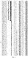
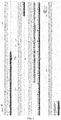
- the TAP-tag-labeled Brd4 expression quantity of the heterozygous ES cell line was indeed less than that of the androgenetic haploid embryonic stem cells, but both were expressed. From the point of expression quantity of heterozygous ES cells, the C-terminal TAP-tag was better. A strong extra band (about 150 kDa) was detected by using the Brd4 antibody, and a weak protein signal was detected only near 250 kDa. When the exposure is strong, it can be seen that the band size is changed with the existence of the TAP-tag label, and it indicates that the band is indeed the Brd4 protein.
- NC cells were completely negative control, and the Brd4-N-ATF cells could detect the 250 kDa large protein and 120 kDa small protein. Both NC and Brd4-N-ATF cells could be detected binding to the known binding protein CDK9 by Co-IP, but the binding efficiency was lower compared with the input of the total cell lysate before IP, and more proteins were bound under the LB1 lysate condition.
- the NC and Brd4-N-ATF cells could be detected binding to an H3 protein by Co-IP ( Fig. 7A ).
- the Brd4-C-FTA and Brd4-N-ATF androgenetic haploid embryonic stem cell lines could indeed obtain an endogenous Brd4 protein by IP with Flag antibody-coupled beads.
- Brd4-C-FTA with Flag and Brd4 antibodies there was only a 250 kDa large protein, and by detecting Brd4-N-ATF, there were a 250 kDa large protein and a 120 kDa small protein. Since the input of the Brd4 antibody was too high in the expression quantity of the heteroprotein, only the heteroprotein was detected.
- the Brd4-C-HTA androgenetic haploid embryonic stem cell line could indeed obtain an endogenous Brd4 protein by IP with HA antibody-coupled beads, and only a 250 kDa large protein was detected by the HA and Brd4 antibodies, respectively. From the view of the ratio of input, HA-beads had higher Brd4 binding efficiency compared with Flag-beads, it indicates that HTA tag was better. Both Brd4-C-FTA and Brd4-C-HTA Co-IP could detect binding to H3, and more proteins were bound under the LB1 lysate condition. Brd4-C-HTA bound more than Brd4-C-FTA, it indicates that HTA tag was better.
- Atad2b, Baz2b, Brd3, Cecr2, Bazlb and Pbrm1 in the bromodomain genes were subjected to C-HTA labeling on the androgenetic haploid stem cell line DKO-AG-haESCs, and sgRNA and homologous arms and primers thereof were conventionally designed to detect the protein expression level of the TAP-tag-labeled bromodomain gene.
- a 3 ⁇ Flag sequence was inserted at the N-terminal of a Phf7 endogenous genome of the androgenetic haploid embryonic stem cell ( Fig. 9A ), a Phf7-KI-Flag heterozygous mouse F0 was obtained by ICAHCI injection, and a Phf7-KI-Flag homozygous male mouse was obtained by mating between F1 heterozygous mice ( Fig. 9B ).
- Phf7-Flag was detected in different germ cells isolated from the Phf7-KI-Flag homozygous male mice ( Fig. 9C ).
- Phf7 was subjected to chip-seq detection by using the Flag antibody and compared with the results of H3K4me3 chip-seq and ubH2A Chip-seq on the exon/intron/intergenic region enrichment situation ( Fig. 9E ).
- the Venn diagram shows that peaks of Phf7 chip-seq and H3K4me3 chip-seq binding regions are highly coincident ( Fig. 9F ).
- Heatmap shows the signal distribution situation of ubH2A in H3K4me3&Phf7 common, H3K4me3 unique, and Phf7 unique results ( Fig. 9G ), and specifically counts the signal result value of ubH2A ( Fig. 9H ).
- the construction of Phf7-KI-Flag tag mice is free of the restriction of Phf7 antibody, and the functional study of endogenous Phf7 proteins in the tag mice can be completed by using the Flag antibody.
- Hspg2 antibody Since there is no good Hspg2 antibody on the market, in order to study the function of this gene, by considering that there is a signal peptide at the N-terminal of Hspg2 protein, a 3 ⁇ Flag sequence was inserted at the C-terminal of an Hspg2 endogenous genome of the androgenetic haploid embryonic stem cell, and an Hspg2-KI-Flag heterozygous mouse was obtained by ICAHCI injection.
- the sgRNA sequence of Hspg2-C-Flag (SEQ ID NO: 57): TCATAGGCACCCACCTGCCT
- Hspg2-KI-Flag heterozygous mouse embryos at embryonic E15.5 days were taken, and protein electrophoresis was performed on the whole embryo sample to detect the expression of Hspg2-Flag.
- the result shows that the C-terminal of the Hspg2 protein is successfully labeled ( Fig. 10 ).
Landscapes
- Life Sciences & Earth Sciences (AREA)
- Health & Medical Sciences (AREA)
- Engineering & Computer Science (AREA)
- Chemical & Material Sciences (AREA)
- Biomedical Technology (AREA)
- Genetics & Genomics (AREA)
- Molecular Biology (AREA)
- Organic Chemistry (AREA)
- Biotechnology (AREA)
- Bioinformatics & Cheminformatics (AREA)
- Wood Science & Technology (AREA)
- Zoology (AREA)
- Biochemistry (AREA)
- Physics & Mathematics (AREA)
- Microbiology (AREA)
- General Engineering & Computer Science (AREA)
- General Health & Medical Sciences (AREA)
- Biophysics (AREA)
- Immunology (AREA)
- Urology & Nephrology (AREA)
- Hematology (AREA)
- Bioinformatics & Computational Biology (AREA)
- Proteomics, Peptides & Aminoacids (AREA)
- Cell Biology (AREA)
- Medicinal Chemistry (AREA)
- Analytical Chemistry (AREA)
- Plant Pathology (AREA)
- Pathology (AREA)
- General Physics & Mathematics (AREA)
- Food Science & Technology (AREA)
- Crystallography & Structural Chemistry (AREA)
- Gynecology & Obstetrics (AREA)
- Developmental Biology & Embryology (AREA)
- Reproductive Health (AREA)
- General Chemical & Material Sciences (AREA)
- Chemical Kinetics & Catalysis (AREA)
- Peptides Or Proteins (AREA)
- Micro-Organisms Or Cultivation Processes Thereof (AREA)
- Investigating Or Analysing Biological Materials (AREA)
Applications Claiming Priority (2)
| Application Number | Priority Date | Filing Date | Title |
|---|---|---|---|
| CN201810096299.3A CN110095610A (zh) | 2018-01-31 | 2018-01-31 | 一种高通量蛋白质分析方法及其适用文库 |
| PCT/CN2019/071005 WO2019149040A1 (zh) | 2018-01-31 | 2019-01-09 | 一种高通量蛋白质分析方法及其适用文库 |
Publications (2)
| Publication Number | Publication Date |
|---|---|
| EP3748363A1 true EP3748363A1 (de) | 2020-12-09 |
| EP3748363A4 EP3748363A4 (de) | 2021-11-10 |
Family
ID=67442049
Family Applications (1)
| Application Number | Title | Priority Date | Filing Date |
|---|---|---|---|
| EP19746592.5A Withdrawn EP3748363A4 (de) | 2018-01-31 | 2019-01-09 | Verfahren zur hochdurchsatzanalyse von proteinen und anwendbare bibliothek dafür |
Country Status (5)
| Country | Link |
|---|---|
| US (1) | US20200181605A1 (de) |
| EP (1) | EP3748363A4 (de) |
| JP (1) | JP2021524016A (de) |
| CN (1) | CN110095610A (de) |
| WO (1) | WO2019149040A1 (de) |
Families Citing this family (2)
| Publication number | Priority date | Publication date | Assignee | Title |
|---|---|---|---|---|
| KR102320688B1 (ko) * | 2020-07-27 | 2021-11-01 | 서울대학교산학협력단 | Phf7의 히스톤 3 유비퀴틴화를 표적으로 하는 정자 크로마틴 응축 억제제 스크리닝 방법 |
| CN114107398B (zh) * | 2020-08-27 | 2023-11-03 | 中国科学院分子细胞科学卓越创新中心 | 一种牙本质发育不全的点突变动物模型构建方法及其应用 |
Family Cites Families (10)
| Publication number | Priority date | Publication date | Assignee | Title |
|---|---|---|---|---|
| WO2013143467A1 (zh) * | 2012-03-27 | 2013-10-03 | 中国科学院上海生命科学研究院 | 孤雄单倍体干细胞系及其制法和应用 |
| WO2013159313A1 (zh) * | 2012-04-26 | 2013-10-31 | 中国科学院动物研究所 | 一种动物胚胎干细胞系及其制备方法和应用 |
| US9535070B2 (en) * | 2012-11-09 | 2017-01-03 | Arizona Board Of Regents, A Body Corporate Of The State Of Arizona, Acting For And On Behalf Of Arizona State University | High throughput detection of fusion proteins |
| MX375361B (es) * | 2014-03-26 | 2025-03-06 | Univ Maryland | Edicion de genoma dirigida en cigotos de animales grandes domesticos. |
| EP3183580B1 (de) * | 2014-08-21 | 2025-03-26 | Eurofins DiscoverX Corporation | Homogener thermal-shift-ligandenbindungstest |
| CN105112446A (zh) * | 2015-06-25 | 2015-12-02 | 中国医学科学院基础医学研究所 | 使用单倍体干细胞高效建立遗传修饰动物模型的方法 |
| WO2017000302A1 (zh) * | 2015-07-02 | 2017-01-05 | 中国科学院上海生命科学研究院 | 一种孤雄单倍体胚胎干细胞及其制备与应用 |
| CN106676075B (zh) * | 2015-11-11 | 2020-06-26 | 中国科学院分子细胞科学卓越创新中心 | 一种孤雌单倍体胚胎干细胞及其制备与应用 |
| US20190134227A1 (en) * | 2016-04-18 | 2019-05-09 | The Board Of Regents Of The University Of Texas System | Generation of genetically engineered animals by crispr/cas9 genome editing in spermatogonial stem cells |
| CN106399311A (zh) * | 2016-09-07 | 2017-02-15 | 同济大学 | 用于Chip‑seq全基因组结合谱的内源蛋白标记的方法 |
-
2018
- 2018-01-31 CN CN201810096299.3A patent/CN110095610A/zh active Pending
-
2019
- 2019-01-09 WO PCT/CN2019/071005 patent/WO2019149040A1/zh not_active Ceased
- 2019-01-09 JP JP2020542301A patent/JP2021524016A/ja active Pending
- 2019-01-09 EP EP19746592.5A patent/EP3748363A4/de not_active Withdrawn
- 2019-10-31 US US16/670,813 patent/US20200181605A1/en not_active Abandoned
Also Published As
| Publication number | Publication date |
|---|---|
| CN110095610A (zh) | 2019-08-06 |
| US20200181605A1 (en) | 2020-06-11 |
| EP3748363A4 (de) | 2021-11-10 |
| WO2019149040A1 (zh) | 2019-08-08 |
| JP2021524016A (ja) | 2021-09-09 |
Similar Documents
| Publication | Publication Date | Title |
|---|---|---|
| Lasman et al. | Context-dependent functional compensation between Ythdf m6A reader proteins | |
| Mei et al. | H2AK119ub1 guides maternal inheritance and zygotic deposition of H3K27me3 in mouse embryos | |
| Xiong et al. | Ultrasensitive Ribo-seq reveals translational landscapes during mammalian oocyte-to-embryo transition and pre-implantation development | |
| Rom et al. | Regulation of CHD2 expression by the Chaserr long noncoding RNA gene is essential for viability | |
| Bultman et al. | Maternal BRG1 regulates zygotic genome activation in the mouse | |
| Van Der Weyden et al. | Loss of TSLC1 causes male infertility due to a defect at the spermatid stage of spermatogenesis | |
| Miles et al. | Transgenic pig carrying green fluorescent proteasomes | |
| Chen et al. | ZFP30 promotes adipogenesis through the KAP1-mediated activation of a retrotransposon-derived Pparg2 enhancer | |
| Miao et al. | Mediator complex component MED13 regulates zygotic genome activation and is required for postimplantation development in the mouse | |
| Chatzinikolaou et al. | ERCC1–XPF cooperates with CTCF and cohesin to facilitate the developmental silencing of imprinted genes | |
| US20040137572A1 (en) | Compositions and methods for generating conditional knockouts | |
| Brouze et al. | TENT5-mediated polyadenylation of mRNAs encoding secreted proteins is essential for gametogenesis in mice | |
| EP3748363A1 (de) | Verfahren zur hochdurchsatzanalyse von proteinen und anwendbare bibliothek dafür | |
| Li et al. | Remodelling bivalent chromatin is essential for mouse peri-implantation embryogenesis | |
| Kuales et al. | A resource for the transcriptional signature of bona fide trophoblast stem cells and analysis of their embryonic persistence | |
| Tan et al. | Pramel15 facilitates zygotic nuclear DNMT1 degradation and DNA demethylation | |
| Kubinyecz et al. | Maternal SMARCA5 is required for major ZGA in mouse embryos | |
| CN109022546A (zh) | 一种Nanos2启动子核心区域关键转录因子的验证方法 | |
| Kour et al. | Loss of function of ribosomal protein L13a blocks blastocyst formation and reveals a potential nuclear role in gene expression | |
| Bai et al. | Histone demethylase UTX is an essential factor for zygotic genome activation and regulates Zscan4 expression in mouse embryos | |
| CN117310176A (zh) | 一种rna结合模体蛋白用于诊断和治疗生育缺陷的应用 | |
| Edupuganti et al. | Integrator promotes the association of TFIID and RNA polymerase II to maintain pluripotency during development | |
| Chen et al. | Comparative maternal protein profiling of mouse biparental and uniparental embryos | |
| Gresakova et al. | Dual role of Fam208a during zygotic cleavage and early embryonic development | |
| Miura et al. | Transfer of spermatogenesis‐related cDNAs into eel testis germ‐somatic cell coculture pellets by electroporation: Methods for analysis of gene function |
Legal Events
| Date | Code | Title | Description |
|---|---|---|---|
| STAA | Information on the status of an ep patent application or granted ep patent |
Free format text: STATUS: THE INTERNATIONAL PUBLICATION HAS BEEN MADE |
|
| PUAI | Public reference made under article 153(3) epc to a published international application that has entered the european phase |
Free format text: ORIGINAL CODE: 0009012 |
|
| STAA | Information on the status of an ep patent application or granted ep patent |
Free format text: STATUS: REQUEST FOR EXAMINATION WAS MADE |
|
| 17P | Request for examination filed |
Effective date: 20200715 |
|
| AK | Designated contracting states |
Kind code of ref document: A1 Designated state(s): AL AT BE BG CH CY CZ DE DK EE ES FI FR GB GR HR HU IE IS IT LI LT LU LV MC MK MT NL NO PL PT RO RS SE SI SK SM TR |
|
| AX | Request for extension of the european patent |
Extension state: BA ME |
|
| DAV | Request for validation of the european patent (deleted) | ||
| DAX | Request for extension of the european patent (deleted) | ||
| RIC1 | Information provided on ipc code assigned before grant |
Ipc: C12Q 1/68 20180101ALI20210924BHEP Ipc: C12N 5/0735 20100101ALI20210924BHEP Ipc: C12N 15/85 20060101ALI20210924BHEP Ipc: G01N 33/68 20060101AFI20210924BHEP |
|
| A4 | Supplementary search report drawn up and despatched |
Effective date: 20211008 |
|
| STAA | Information on the status of an ep patent application or granted ep patent |
Free format text: STATUS: THE APPLICATION IS DEEMED TO BE WITHDRAWN |
|
| 18D | Application deemed to be withdrawn |
Effective date: 20220507 |