EP3760728B1 - Bewertung von zellen anhand der richtungsbewegungsfähigkeit. - Google Patents
Bewertung von zellen anhand der richtungsbewegungsfähigkeit. Download PDFInfo
- Publication number
- EP3760728B1 EP3760728B1 EP18907612.8A EP18907612A EP3760728B1 EP 3760728 B1 EP3760728 B1 EP 3760728B1 EP 18907612 A EP18907612 A EP 18907612A EP 3760728 B1 EP3760728 B1 EP 3760728B1
- Authority
- EP
- European Patent Office
- Prior art keywords
- cells
- movement
- cell
- neurons
- movement pattern
- Prior art date
- Legal status (The legal status is an assumption and is not a legal conclusion. Google has not performed a legal analysis and makes no representation as to the accuracy of the status listed.)
- Active
Links
Images




Classifications
-
- G—PHYSICS
- G01—MEASURING; TESTING
- G01N—INVESTIGATING OR ANALYSING MATERIALS BY DETERMINING THEIR CHEMICAL OR PHYSICAL PROPERTIES
- G01N33/00—Investigating or analysing materials by specific methods not covered by groups G01N1/00 - G01N31/00
- G01N33/48—Biological material, e.g. blood, urine; Haemocytometers
- G01N33/50—Chemical analysis of biological material, e.g. blood, urine; Testing involving biospecific ligand binding methods; Immunological testing
- G01N33/5005—Chemical analysis of biological material, e.g. blood, urine; Testing involving biospecific ligand binding methods; Immunological testing involving human or animal cells
- G01N33/5008—Chemical analysis of biological material, e.g. blood, urine; Testing involving biospecific ligand binding methods; Immunological testing involving human or animal cells for testing or evaluating the effect of chemical or biological compounds, e.g. drugs, cosmetics
- G01N33/502—Chemical analysis of biological material, e.g. blood, urine; Testing involving biospecific ligand binding methods; Immunological testing involving human or animal cells for testing or evaluating the effect of chemical or biological compounds, e.g. drugs, cosmetics for testing non-proliferative effects
- G01N33/5032—Chemical analysis of biological material, e.g. blood, urine; Testing involving biospecific ligand binding methods; Immunological testing involving human or animal cells for testing or evaluating the effect of chemical or biological compounds, e.g. drugs, cosmetics for testing non-proliferative effects on intercellular interactions
-
- G—PHYSICS
- G01—MEASURING; TESTING
- G01N—INVESTIGATING OR ANALYSING MATERIALS BY DETERMINING THEIR CHEMICAL OR PHYSICAL PROPERTIES
- G01N33/00—Investigating or analysing materials by specific methods not covered by groups G01N1/00 - G01N31/00
- G01N33/48—Biological material, e.g. blood, urine; Haemocytometers
- G01N33/50—Chemical analysis of biological material, e.g. blood, urine; Testing involving biospecific ligand binding methods; Immunological testing
- G01N33/5005—Chemical analysis of biological material, e.g. blood, urine; Testing involving biospecific ligand binding methods; Immunological testing involving human or animal cells
- G01N33/5008—Chemical analysis of biological material, e.g. blood, urine; Testing involving biospecific ligand binding methods; Immunological testing involving human or animal cells for testing or evaluating the effect of chemical or biological compounds, e.g. drugs, cosmetics
- G01N33/502—Chemical analysis of biological material, e.g. blood, urine; Testing involving biospecific ligand binding methods; Immunological testing involving human or animal cells for testing or evaluating the effect of chemical or biological compounds, e.g. drugs, cosmetics for testing non-proliferative effects
- G01N33/5029—Chemical analysis of biological material, e.g. blood, urine; Testing involving biospecific ligand binding methods; Immunological testing involving human or animal cells for testing or evaluating the effect of chemical or biological compounds, e.g. drugs, cosmetics for testing non-proliferative effects on cell motility
-
- G—PHYSICS
- G01—MEASURING; TESTING
- G01N—INVESTIGATING OR ANALYSING MATERIALS BY DETERMINING THEIR CHEMICAL OR PHYSICAL PROPERTIES
- G01N33/00—Investigating or analysing materials by specific methods not covered by groups G01N1/00 - G01N31/00
- G01N33/48—Biological material, e.g. blood, urine; Haemocytometers
- G01N33/50—Chemical analysis of biological material, e.g. blood, urine; Testing involving biospecific ligand binding methods; Immunological testing
- G01N33/5005—Chemical analysis of biological material, e.g. blood, urine; Testing involving biospecific ligand binding methods; Immunological testing involving human or animal cells
- G01N33/5008—Chemical analysis of biological material, e.g. blood, urine; Testing involving biospecific ligand binding methods; Immunological testing involving human or animal cells for testing or evaluating the effect of chemical or biological compounds, e.g. drugs, cosmetics
- G01N33/5044—Chemical analysis of biological material, e.g. blood, urine; Testing involving biospecific ligand binding methods; Immunological testing involving human or animal cells for testing or evaluating the effect of chemical or biological compounds, e.g. drugs, cosmetics involving specific cell types
- G01N33/5058—Neurological cells
-
- G—PHYSICS
- G01—MEASURING; TESTING
- G01N—INVESTIGATING OR ANALYSING MATERIALS BY DETERMINING THEIR CHEMICAL OR PHYSICAL PROPERTIES
- G01N33/00—Investigating or analysing materials by specific methods not covered by groups G01N1/00 - G01N31/00
- G01N33/48—Biological material, e.g. blood, urine; Haemocytometers
- G01N33/50—Chemical analysis of biological material, e.g. blood, urine; Testing involving biospecific ligand binding methods; Immunological testing
- G01N33/5005—Chemical analysis of biological material, e.g. blood, urine; Testing involving biospecific ligand binding methods; Immunological testing involving human or animal cells
- G01N33/5008—Chemical analysis of biological material, e.g. blood, urine; Testing involving biospecific ligand binding methods; Immunological testing involving human or animal cells for testing or evaluating the effect of chemical or biological compounds, e.g. drugs, cosmetics
- G01N33/5044—Chemical analysis of biological material, e.g. blood, urine; Testing involving biospecific ligand binding methods; Immunological testing involving human or animal cells for testing or evaluating the effect of chemical or biological compounds, e.g. drugs, cosmetics involving specific cell types
- G01N33/5073—Stem cells
Definitions
- the present invention relates to a cell-based assay system. More specifically, the present invention relates to a drug evaluation method using a cell-based assay system to obtain a movement pattern defined by directional movement ability of individual cells constituting a cell population of neurons derived from induced pluripotent stem cells as an index and its application
- SCZ and ASD genome analysis studies have identified a plurality of mutations in genes involved in neurodevelopment in both diseases, one of which is a mutation in RELN (for example, see NPL 3).
- the protein Reelin encoded by RELN is a large secreted protein and is essential for the formation of the layer structure of the developing brain.
- a RELN homozygous deletion mutation causes lissencephaly with developmental delay, and a decrease in Reelin has been reported to be associated with the onset of neurodevelopmental disorders (NPL 4).
- reeler mice which are Reln mutant mice, a disorder of brain layer structure and an abnormal behavior have been reported (for example, see NPL 5).
- JP 2013 039113 A describes a method of cell quality control based on cell displacement.
- JP 2014 179061 A describes a system for quality control of nerve cells based on movement analysis.
- the present invention relates to the embodiments as characterized in the claims.
- Reelin tyrosine hydroxylase (TH)-positive dopamine neurons have been reported, in researches using mice, to express Reelin only in a limited period before and after birth. Although Reelin is expressed only in such a limited period, abnormalities in dopamine neurons have been confirmed in Rein-mutated reeler mice.
- TH tyrosine hydroxylase
- the present inventors have advanced researches, focusing on Reelin, in order to elucidate the onset mechanism of SCZ and ASD and to establish a treatment method therefor. As one of the research results, the present inventors have succeeded in establishing a method for specifically and efficiently preparing dopamine neurons from iPS cells ( Japanese Patent Application No. 2017-82600 ).
- an object of the present invention is to provide a novel evaluation system that is particularly useful in researches on the onset mechanism and pathologic conditions of a central nervous system disease (for example, a mental disease), development of a therapeutic method, or the like.
- a central nervous system disease for example, a mental disease
- development of a therapeutic method or the like.
- neurospheres neurospheres
- cells dopamine neurons
- the movement pattern of healthy subject-derived cells is characteristic (the cells show a specific movement pattern) and that RELN-deficient mental disease patient-derived cells show a movement pattern different from that of the healthy subject-derived cells.
- the movement pattern defined by the movement characteristics (in particular, movement direction) of the individual cells constituting a cell population reflects the disease state, and indicates that an evaluation system using this movement pattern as an index is useful, for example, not only in basic researches (for example, elucidation of the disease mechanism), but also in understanding pathologic conditions and developing therapeutic drugs.
- an evaluation system using this movement pattern as an index is useful, for example, not only in basic researches (for example, elucidation of the disease mechanism), but also in understanding pathologic conditions and developing therapeutic drugs.
- normalization of the movement pattern demonstrates efficacy
- abnormalization of the movement pattern demonstrates toxicity.
- the normalization of the movement pattern can indicate the efficacy of rescuing endogenous Reelin.
- the coverage (target cells, uses/applications, etc.) of the above evaluation system focusing on the movement characteristics of cells is broad, i.e., the evaluation system can be widely used as an evaluation system for efficacy or toxicity, and its practical value and significance are extremely high. Further studies by the present inventors have also provided various findings useful and important for putting the above evaluation system into practical use.
- the present invention relates to the embodiments as characterized in the claims.
- the present invention provides a drug evaluation method used in the evaluation of the efficacy or toxicity of a test substance (hereinafter referred to also as “evaluation method of the present invention”).
- drug evaluation is used as a general term for evaluation of efficacy and toxicity. Therefore, the efficacy or toxicity of the test substance is evaluated in the present invention.
- toxicity should be interpreted in a broad sense, and includes, in addition to general toxicities (acute toxicity, subacute toxicity, and chronic toxicity), side effects, carcinogenicity, mutagenicity, and teratogenicity.
- the evaluation method of the present invention is an evaluation system utilizing neurons derived from induced pluripotent stem cells, involving preparing a specific cell population, and evaluating the efficacy or toxicity of a test substance using, as an index, a movement pattern defined by directional movement ability of individual cells constituting the cell population (which is associated with the degree (level) of variations in movement direction of the cells and the movement velocity (speed)).
- the studies by the present inventors have revealed that when attention is paid to the variations in movement of the individual cells constituting the cell population, the "movement pattern" that represents the movement characteristics of the cell population can be defined, and that the "movement pattern” is correlated with the normality (abnormality, in other words) of the cell population and is useful for evaluation of efficacy or toxicity (see Examples below).
- the evaluation method of the present invention includes the following steps (i) to (iii) of:
- step (i) a population of cells exhibiting movement ability is prepared, and culture is started in the presence of a test substance.
- a cell population constituted by cells exhibiting directional movement ability is used.
- the term "mixture (mix)" as used herein means that such cells are present in the cell population but are not major constituent cells. Therefore, the abundance (mixing rate) of such cells is, for example, 40% or less of the entire cell population, preferably 30% or less of the entire cell population, more preferably 20% or less of the entire cell population, still more preferably 10% of the entire cell population.
- Cells that exert a predetermined movement ability in the process of normal generation or formation, or regeneration of structures such as tissues and organs of the living body correspond to the "cells exhibiting directional movement ability" and are suitable as cells constituting the cell population in the method of the present invention, wherein the cells are neurons derived from induced pluripotent stem cells.
- the cells exhibiting directional movement ability include neurons (dopamine neurons, glutamatergic neurons, serotonin neurons, and GABAergic neurons), cardiomyocytes, and hematopoietic cells (leucocytes).
- the cell population may include two or more kinds of cells which are different in cell types. Further, it may include two or more kinds of cells which are different in degree of differentiation.
- Normal cells or abnormal cells can be used as the cells constituting the cell population.
- the "normal cell” is a cell that has no abnormality in relation with the efficacy (improvement in cell movement disorder and therapeutic effect based thereon) or toxicity evaluated by the method of the present invention.
- Typical examples of the normal cells are cells derived from healthy subjects.
- cells derived from persons not suffering from a disease against which the efficacy to be evaluated can exert a therapeutic effect, in other words, a disease of which the onset and progress are caused at least by cell movement disorder (hereinafter referred to as "target disease”) can also be used as the normal cells.
- target disease cell movement disorder
- abnormal cell is a cell that is in contrast to the normal cell, and refers to a cell that is no longer in its original state due to gene mutation, chromosomal abnormality, or the like.
- abnormal cells are cells having a genetic abnormality characteristic of the target disease, and cells derived from patients suffering from the target disease correspond to the abnormal cells.
- patient-derived cells include cells collected from patients or subcultured cells thereof, and differentiated cells obtained by inducing differentiation of patient-derived induced pluripotent stem (iPS) cells (iPS cells prepared using cells collected from patients).
- cells in which a genetic abnormality characteristic of the target disease is introduced for example, differentiated cells obtained by introducing a gene mutation into undifferentiated cells such as iPS cells by gene manipulation such as genome editing, and then inducing differentiation of the cells
- the target disease include neurodevelopmental disorders such as ASD and SCZ, and blood diseases developing leukocyte migration disorder
- examples of the genetic abnormality include deletion of Reelin gene, deletion of chromosome 3q29 region, and deletion of chromosome 22q11 region.
- the toxicity of therapeutic drugs or therapeutic drug candidates can also be evaluated (details will be described later), in addition to the evaluation of the efficacy for the purpose of screening (searching) for the therapeutic drug candidates.
- the method of the present invention has a unique characteristic that it can be used in both the efficacy evaluation system and the toxicity evaluation system by selecting the cells to be used. Details of each evaluation system will be described later.
- dopamine neurons prepared by the method including the following steps (1) to (3) are used as the cells in step (i).
- pluripotent stem cells are used.
- the "pluripotent stem cell” refers to a cell having both the potential for differentiating into all cells constituting the body (pluripotency), and the potential for producing daughter cells having the same differentiation potency via cell division (self-replication competence).
- the pluripotency can be evaluated by transplanting cells of an evaluation subject into a nude mouse, and testing the presence or absence of formation of teratoma containing each cell of the three germ layers (ectoderm, mesoderm, and endoderm).
- pluripotent stem cells examples include induced pluripotent stem cells (iPS cells).
- iPS cells are used.
- pluripotent stem cells are cells of mammals (for example, primates such as humans or chimpanzees, and rodents such as mice or rats), and particularly preferably, pluripotent stem cells are human cells. Therefore, in a most preferable embodiment, human iPS cells are used as pluripotent stem cells.
- iPS cell refers to a cell having pluripotency and self-replication competence, produced by reprogramming somatic cells (e.g., fibroblasts, skin cells, and lymphocytes), for example, through the introduction of initializing factors.
- somatic cells e.g., fibroblasts, skin cells, and lymphocytes
- Somatic cells used in the preparation of iPS cells are not particularly limited, and may be differentiated somatic cells or undifferentiated stem cells.
- iPS cells can be prepared by various methods reported so far. The application of iPS cell preparation methods which will be developed in the future is also contemplated. Examples of the cells available for preparing iPS cells (i.e.
- T cells include lymphocytes (T cells, B cells), fibroblasts, epithelial cells, endothelial cells, mucosal epithelial cells, mesenchymal cells, hematopoietic stem cells, adipose stem cells, dental pulp stem cells and neural stem cells.
- T cells lymphocytes
- B cells fibroblasts
- epithelial cells endothelial cells
- mucosal epithelial cells mesenchymal cells
- hematopoietic stem cells hematopoietic stem cells
- adipose stem cells adipose stem cells
- dental pulp stem cells and neural stem cells.
- the most fundamental technique of iPS cell preparation methods is to introduce four factors of Oct3/4, Sox2, Klf4, and c-Myc, which are transcription factors, into cells by using virus ( Takahashi K, Yamanaka S: Cell 126 (4), 663-676, 2006 ; Takahashi, K, et al.: Cell 131 (5), 861-72, 2007 ).
- the establishment of human iPS cells by introduction of four factors of Oct4, Sox2, Lin28, and Nonog has been reported ( Yu J, et al.: Science 318 (5858), 1917-1920, 2007 ).
- the establishment of iPS cells by introduction of three factors other than c-Myc Nakagawa M, et al.: Nat. Biotechnol.
- Cells in which transformation into iPS cells, i.e., initialization (reprogramming) has occurred can be selected by using, as an indicator, the expression of pluripotent stem cell markers (undifferentiated markers) such as Fbxo15, Nanog, Oct/4, Fgf-4, Esg-1, and Cript.
- pluripotent stem cell markers undifferentiated markers
- iPS cells can also be provided from, for example, the National University Corporation, Kyoto University or the Independent Administrative Institution, RIKEN BioResource Center.
- Pluripotent stem cells can be maintained in vitro by known methods. For example, when it is desired to provide highly safe cells (e.g. in a case where clinical application is considered), pluripotent stem cells are preferably maintained by serum-free culture using a serum alternative or by feeder-free cell culture. If a serum is used (or used in combination), autologous serum (i.e., recipient's serum) is preferably used. A serum alternative can be prepared by known methods (see, for example, WO 98/30679 ). Commercially available serum alternatives can also be used. Examples of the commercially available serum alternatives include KSR (manufactured by Invitrogen), Chemically-defined Lipid concentrated (manufactured by Gibco), and Glutamax (manufactured by Gibco).
- step (1) the pluripotent stem cells prepared as described above are cultured in the presence of a TGF- ⁇ family inhibitor, a GSK3 ⁇ inhibitor, and a BMP inhibitor. That is, the pluripotent stem cells are cultured using a medium to which a TGF- ⁇ family inhibitor, a GSK3 ⁇ inhibitor, and a BMP inhibitor are added. Step (1) aims to enhance the neuronal differentiation potency of the pluripotent stem cells.
- the medium to be used can be prepared using a medium used for culturing mammalian cells as a basal medium.
- a basal medium for example, a BME medium, a BGJb medium, a CMRL 1066 medium, a Glasgow MEM medium, an Improved MEM Zinc Option medium, an IMDM medium, a Medium 199 medium, an Eagle MEM medium, an ⁇ MEM medium, a DMEM medium, a Ham's medium, a Ham's F-12 medium, a RPMI 1640 medium, a Fischer's medium, a Neurobasal medium, and a mixed medium thereof can be used, and the basal medium is not particularly limited as long as the it can be used for culturing mammalian cells.
- a mixed medium of IMDM medium and Ham's F-12 medium can be used.
- a TGF- ⁇ family inhibitor, a GSK3 ⁇ inhibitor, and a BMP inhibitor are added to the medium.
- the TGF- ⁇ family inhibitor is a substance that inhibits TGF- ⁇ signaling through binding between TGF- ⁇ and a TGF- ⁇ receptor.
- the TGF- ⁇ inhibitor includes proteinaceous inhibitors and small molecule inhibitors. Examples of such proteinaceous inhibitors are anti-TGF- ⁇ neutralizing antibodies and anti-TGF- ⁇ receptor neutralizing antibodies.
- SB431542 (4-[4-(1,3-benzodioxol-5-yl)-5-(2-pyridinyl)-1H-imidazol-2-yl]-benzamide or its hydrate), SB202190 (4-(4-fluorophenyl)-2-(4-hydroxyphenyl)-5-(4-pyridyl)-1H-imidazole), SB505124 (GlaxoSmithKline), NPC30345, SD093, SD908, SD208 (Scios), LY2109761, LY364947, and LY580276 (Lilly Research Laboratories).
- SB431542 is used.
- the concentration of the TGF- ⁇ family inhibitor (amount to be added to the medium) is not particularly limited as long as the purpose of enhancing the neuronal differentiation potency of the pluripotent stem cells is achieved.
- concentration thereof is, for example, 0.5 ⁇ M to 20 ⁇ M, preferably 1 ⁇ M to 10 ⁇ M.
- the optimum concentration can be set through preliminary experiments. Instead of keeping the TGF- ⁇ family inhibitor concentration constant throughout the entire culture period, changes in TGF- ⁇ family inhibitor concentration, e.g., a stepwise increase in TGF- ⁇ family inhibitor concentration, may be provided.
- GSK3 ⁇ inhibitors examples include CHIR99021 (6-[[2-[[4-(2,4-dichlorophenyl)-5-(4-methyl-1H-imidazol-2-yl)-2-pyrimidinyl]amino]ethyl]amino]nicotinonitrile), SB-415286(3-[(3-chloro-4-hydroxyphenyl)amino]-4-(2-nitrophenyl)-1H-pyrrole-2,5-dione), SB-2167, Indirubin-3'-Monoxime, Kenpaullone, and BIO (6-bromoindirubin-3'-oxime).
- CHIR99021 is used.
- the concentration of the GSK3 ⁇ inhibitor (amount to be added to the medium) is not particularly limited as long as the purpose of enhancing the neuronal differentiation potency of the pluripotent stem cells is achieved.
- the concentration thereof is, for example, 0.5 ⁇ M to 20 ⁇ M, preferably 1 ⁇ M to 10 ⁇ M.
- the optimum concentration can be set through preliminary experiments. Instead of keeping the GSK3 ⁇ inhibitor concentration constant throughout the entire culture period, changes in GSK3 ⁇ inhibitor concentration, e.g., a stepwise increase in GSK3 ⁇ inhibitor concentration, may be provided.
- the BMP inhibitor is a substance that inhibits BMP signaling through binding between BMP (bone morphogenetic protein) and a BMP receptor (type I or type II).
- BMP bone morphogenetic protein
- the BMP inhibitor includes proteinaceous inhibitors and small molecule inhibitors. Examples of such proteinaceous inhibitors include natural inhibitors such as Noggin, chordin and follistatin.
- Examples of such small molecule inhibitors include Dorsomorphin (6-[4-(2-piperidin-1-ylethoxy)phenyl]-3-pyridin-4-ylpyrazolo[1,5-a]pyrimidine) and its derivatives, LDN-193189 (4-(6-(4-piperazin-1-yl)phenyl) pyrazolo[1,5-a]pyrimidin-3-yl) quinoline) and its derivatives. These compounds are commercially available (e.g., available from Sigma-Aldrich and Stemgent) and are readily available. Preferably, Dorsomorphin is used.
- the concentration of the BMP inhibitor (amount to be added to the medium) is not particularly limited as long as the purpose of enhancing the neuronal differentiation potency of the pluripotent stem cells is achieved.
- the concentration thereof is, for example, 0.5 ⁇ M to 20 ⁇ M, preferably 1 ⁇ M to 10 ⁇ M.
- the optimum concentration can be set through preliminary experiments. Instead of keeping the BMP inhibitor concentration constant throughout the entire culture period, changes in BMP inhibitor concentration, e.g., a stepwise increase in BMP inhibitor concentration, may be provided.
- the medium can contain other components.
- the components to be added include insulin, an iron source (e.g., transferrin), a mineral (e.g., sodium selenate), a saccharide (e.g., glucose), an organic acid (e.g., pyruvic acid or lactic acid), a serum protein (e.g., albumin), an amino acid (e.g., L-glutamine), a reducing agent (e.g., 2-mercaptoethanol), a vitamin (e.g., ascorbic acid or d-biotin), an antibiotic (e.g., streptomycin, penicillin or gentamicin), and a buffer (e.g., HEPES).
- an iron source e.g., transferrin
- a mineral e.g., sodium selenate
- a saccharide e.g., glucose
- an organic acid e.g., pyruvic acid or lactic acid
- a serum protein e.g.
- Pluripotent stem cells are usually subjected to adherent culture.
- the adherent culture is in contrast to suspension culture, and is typically two-dimensional culture (plane culture) under adherent conditions.
- Matrigel TM (BD) or the like may be used for three-dimensional culture.
- dishes, Petri dishes, tissue culture dishes, multi-dishes, microplates, microwell plates, multi-plates, multi-well plates, chamber slides, laboratory dishes, and the like can be used for adherent culture.
- a culture vessel coated with Matrigel TM (BD), poly-D-lysine, poly-L-lysine, collagen, gelatin, laminin, heparan sulfate proteoglycan, entactin, or a combination of two or more thereof.
- the pluripotent stem cells can be cultured under either condition, i.e., either in the presence or absence of feeder cells.
- the pluripotent stem cells may be cultured in the absence of feeder cells (feeder cell-free culture).
- feeder cells include MEFs (mouse embryonic fibroblasts), STO cells (mouse embryonic fibroblast cell line), and SNL cells (subclones of the STO cells).
- culture conditions such as culture temperature, CO 2 concentration, and O 2 concentration can be set as appropriate.
- the culture temperature is, for example, about 30 to 40°C, preferably about 37°C.
- the CO 2 concentration is, for example, about 1 to 10%, preferably about 5%.
- culture may be performed under normal oxygen partial pressure.
- the oxygen concentration in the case of the condition "under normal oxygen partial pressure” is typically about 18% to about 22%, though it may vary depending on other conditions (humidity, CO 2 concentration, and the like). The details of the condition "under normal oxygen partial pressure" will be described later.
- the period (culture period) of step (1) is 4 days or longer, specifically, for example, 4 days to 20 days, preferably 6 days to 14 days. If the culture period is too short, the neurosphere formation ability will be reduced.
- the cells may be subjected to subculture as necessary.
- the cells are collected at a stage where they are brought in a subconfluent or confluent state, a part of the cells is seeded in another culture vessel, and culture is continued.
- a cell dissociation solution or the like may be used for cell collection.
- proteases such as EDTA-trypsin, collagenase IV, and metalloprotease can be used alone or in an appropriate combination.
- Cell dissociation solutions with low cell toxicity are preferred.
- commercially available products such as DISPASE (EIDIA Co., Ltd.), TrypLE (Invitrogen), and Accutase (MILLIPORE) are available.
- the collected cells may be subjected to subculture after treatment with a cell strainer or the like so as to arrive at a dispersed (discrete) state.
- step (1) the neuronal differentiation potency of the pluripotent stem cells is enhanced. Increased neuronal differentiation potency can be confirmed by using as an index an increase in expression of nervous system markers (Sox2, nestin, Sox1, and the like) as compared with that before the start of step (1). Moreover, the expression of an undifferentiation marker may be utilized for evaluation of increased neuronal differentiation potency.
- nervous system markers Sox2, nestin, Sox1, and the like
- step (1) the cells after step (1) are once collected and then the process proceeds to the next culture (step (2)).
- the collection operation can be performed in the same manner as the collection operation during subculture.
- the cells obtained in step (1) are cultured in suspension in the presence of a TGF- ⁇ family inhibitor, a GSK3 ⁇ inhibitor, FGF8, and a hedgehog signal agonist and under normal oxygen partial pressure to form a neurosphere. That is, the cells after step (1) are cultured using a medium to which a TGF- ⁇ family inhibitor, a GSK3 ⁇ inhibitor, FGF8, and a hedgehog signal agonist are added and under the condition of normal oxygen partial pressure.
- Step (2) aims to induce differentiation along the neuron lineage.
- the features not specifically mentioned (usable basic medium, usable TGF- ⁇ family inhibitor, GSK3 ⁇ inhibitor, other components that can be added to the medium, and the like) are the same as those in step (1), and the explanations thereof are omitted.
- flasks, tissue culture flasks, dishes, Petri dishes, tissue culture dishes, multi-dishes, microplates, microwell plates, micropores, multi-plates, multi-well plates, chamber slides, laboratory dishes, tubes, trays, culture bags, roller bottles, and the like can be used for suspension culture.
- a culture vessel having a non-cell-adherent culture surface examples include culture vessels whose surfaces (culture surfaces) have been treated to be non-cell-adherent, and culture vessels whose surfaces (culture surfaces) have not undergone a treatment for improving the cell adhesiveness (for example, coating treatment with an extracellular matrix). It is only necessary to maintain the non-adherent state of the cells to the culture vessel in suspension culture.
- Static culture may be employed, or swirl culture or shaking culture may be employed.
- the TGF- ⁇ family inhibitor and the GSK3 ⁇ inhibitor are as described above. Also in this step, it is preferable to use SB431542 as the TGF- ⁇ family inhibitor and CHIR99021 as the GSK3 ⁇ inhibitor.
- the concentration of the TGF- ⁇ family inhibitor in the medium when SB431542 is used is, for example, 0.5 ⁇ M to 20 ⁇ M, preferably 1 ⁇ M to 10 ⁇ M.
- the concentration of the GSK3 ⁇ inhibitor in the medium when CHIR99021 is used is, for example, 0.5 ⁇ M to 20 ⁇ M, preferably 1 ⁇ M to 10 ⁇ M.
- FGF8 is a member of the fibroblast growth factor family. FGF8 is involved in the control of vertebrate brain formation and is required for regionalization to the midbrain. As long as the object of the present invention can be achieved, FGF8s derived from various mammals can be used. However, it is preferable to use FGF8 derived from the same origin (animal species) as that of the pluripotent stem cells to be used. Therefore, human FGF8 is preferably used when human pluripotent stem cells are used.
- the "human FGF8” means FGF8 having an amino acid sequence of FGF8 naturally expressed in the human body, and may be a recombinant.
- FGF8 As a typical amino acid sequence of human FGF8, the NCBI accession number: NP_006110.1 (fibroblast growth factor 8 isoform B precursor [Homo sapiens].) can be exemplified.
- concentration of FGF8 is not particularly limited as long as the purpose of inducing differentiation along the neuronal lineage is achieved, but is, for example, 1 ng/ml to 5 ⁇ g/ml, preferably 10 to 500 ng/ml, more preferably 50 to 400 ng/ml. The optimum concentration can be set through preliminary experiments.
- the hedgehog signal agonist is not particularly limited as long as it promotes a sonic hedgehog (SHH) signal.
- SHH sonic hedgehog
- purmorphamine (9-cyclohexyl-N-[4-(4-morpholinyl)phenyl]-2-(1-naphthalenyloxy)-9H-purin-6-amine) useful for inducing ventralization is preferably used.
- concentration of purmorphamine (amount added to the medium) is not particularly limited as long as the purpose of inducing differentiation along the neuron lineage is achieved, but is, for example, 1 ng/ml to 5 ⁇ g/ml, preferably 10 to 500 ng/ml, more preferably 50 to 400 ng/ml. The optimum concentration can be set through preliminary experiments.
- SAG N-methyl-N'-(3-pyridinylbenzyl)-N'-(3-chlorobenzo[b]thiophene-2-carbonyl)-1,4-diaminocyclohexane
- concentration of SAG used is not particularly limited as long as the purpose of inducing differentiation along the neuron lineage is achieved, but is, for example, 10 nM to 100 ⁇ M, preferably 100 nM to 10 ⁇ M, more preferably 100 nM to 2 ⁇ M.
- the optimum concentration can be set through preliminary experiments.
- the concentrations of the respective components be constant throughout the entire culture period, and the concentration(s) of a specific component (which may be two or more components) or all the components may change during the culture.
- the addition of FGF8 and hedgehog signal agonist is started on the second to sixth days of step (2). According to this condition, rapid stimulation to cells can be relieved.
- the addition of FGF8 and hedgehog signal agonist is started on the third to fifth days of step (2).
- LIF leukemia inhibitory factor
- a medium to which a leukemia inhibitory factor (LIF) is also added is preferably used.
- LIFs derived from various mammals can be used.
- LIF derived from the same origin (animal species) as that of the pluripotent stem cells to be used. Therefore, human LIF is preferably employed when human pluripotent stem cells are used.
- the concentration of the LIF is not particularly limited, but is, for example, 0.25 ng/ml to 1 ⁇ g/ml, preferably 1 ng/ml to 50 ng/ml, more preferably 5 ng/ml to 20 ng/ml. The optimum concentration can be set through preliminary experiments.
- a medium to which a bFGF (basic fibroblast growth factor) is also added is preferably used.
- the bFGF is also called FGF2.
- bFGFs derived from various mammals can be used.
- human bFGF is preferably employed when human pluripotent stem cells are used.
- the "human FGF2" means FGF2 having an amino acid sequence of FGF2 naturally expressed in the human body.
- the NCBI accession number: NP_001997.5 fibroblast growth factor 2 [Homo sapiens]
- the concentration of the bFGF is not particularly limited, but is, for example, 0.25 ng/ml to 1 ⁇ g/ml, preferably 1 ng/ml to 50 ng/ml, more preferably 3 ng/ml to 30 ng/ml. The optimum concentration can be set through preliminary experiments.
- a medium to which a ROCK inhibitor (Rho-associated coiled-coil forming kinase/Rho-binding kinase) (for example, Y-27632 or Fasudil (HA-1077)) is also added is preferably used.
- the concentration of Y-27632 used as the ROCK inhibitor is, for example, about 1 ⁇ M to about 50 ⁇ M. The optimum concentration can be set through preliminary experiments.
- the ROCK inhibitor strongly inhibits cell death when cells are in a dispersed state. Therefore, instead of using the ROCK inhibitor over the entire culture period of step (2), the cells may be treated in a medium containing the ROCK inhibitor only when seeding cells (i.e. at the start of culture) or when collecting and dispersing cells, for example, for subculture.
- a medium having a ciliary neurotrophic factor (CNTF), a brain-derived neurotrophic factor (BDNF), a neurotrophin 3 (NT-3), a fetal bovine serum, an N2 supplement, a B27 supplement, or the like added is used so that the use thereof is advantageous in differentiation induction along the neuron lineage.
- the N2 supplement is available from Gibco (product name N2 supplement (x100)) or the like
- the B27 supplement is available from Gibco (product name B27 supplement (x100)) or the like.
- any other component may be added to the medium as needed.
- the component which can be added include insulin, an iron source (e.g., transferrin), a mineral (e.g., sodium selenate), a saccharide (e.g., glucose), an organic acid (e.g., pyruvic acid or lactic acid), a serum protein (e.g., albumin), an amino acid (e.g., L-glutamine), a reducing agent (e.g., 2-mercaptoethanol), a vitamin (e.g., ascorbic acid or d-biotin), an antibiotic (e.g., streptomycin, penicillin, or gentamicin), and a buffer (e.g., HEPES).
- an iron source e.g., transferrin
- a mineral e.g., sodium selenate
- a saccharide e.g., glucose
- an organic acid e.g., pyruvic acid or lactic acid
- suspension culture is performed to form a neurosphere.
- Serum-free Floating culture of Embryoid Body-like aggregates with quick reaggregation SFEB method/SFEBq method; Watanabe et al., Nature Neuroscience 8,288-296 (2005 ), WO 2005/123902
- neurosphere method Reynolds BA and Weiss S., Science, USA, 1992 Mar 27; 255 (5052): 1707-10 ) and the like can be employed.
- step (2) is performed under normal oxygen partial pressure.
- the condition of a lowered oxygen concentration (low oxygen partial pressure/low oxygen concentration) may sometimes be used in consideration of the environment in the living body.
- the condition "under normal oxygen partial pressure” in the present invention is in contrast to such a special condition. That is, the condition "under normal oxygen partial pressure” is a condition in which the oxygen concentration is not intentionally adjusted.
- the oxygen concentration in the case of the condition "under normal oxygen partial pressure” is typically about 18% to about 22%, though it may vary depending on other conditions (humidity, coexisting CO 2 concentration, and the like).
- step (2) under normal oxygen partial pressure eliminates the need to set a special oxygen condition (typically, low oxygen environment) throughout the entire culture period (steps (1) to (3)), and can suppress induction of differentiation into unnecessary cells such as glial cells, and thus is a highly practical preparation method.
- a special oxygen condition typically, low oxygen environment
- culture temperature is, for example, about 30 to 40°C, preferably about 37°C.
- the CO 2 concentration is, for example, about 1 to 10%, preferably about 5%.
- the period of step (2) (culture period) is, for example, 7 days to 21 days, preferably 10 days to 16 days. If the culture period is too short or too long, the differentiation efficiency may be lowered.
- the formed neurosphere may be collected to dissociate, and then the dissociated cells may be subjected to further suspension culture. That is, subculture may be performed.
- the number of subcultures is preferably small, and the number of subcultures is set to 1 or 0 (that is, no subculture is performed).
- Such a small number of subcultures is advantageous in the preparation of dopamine neurons in a short period of time, and is considered to be also effective in avoiding promotion of unintended differentiation induction (for example, induction of differentiation into glial cells).
- since subculture is effective in improving the cell purity, it can be said that the subculture is optimally performed once.
- the subculture When subculture is performed once, the subculture is preferably performed on the sixth to tenth days from the start of step (2).
- the neurosphere formed by step (2) contains undifferentiated cells of the nervous system and undifferentiated cells of the midbrain system.
- the cells constituting the neurosphere are collected and induced differentiation into dopamine neurons.
- the neurosphere is directly (i.e. as a cell aggregate) induced into dopamine neurons.
- Media and culture conditions suitable for inducing differentiation into dopamine neurons are known.
- a protocol provided by ThermoFisher published on the ThermoFisher website) or the like can be referred to.
- differentiation into dopamine neurons is induced by adherent culture in a medium containing a ⁇ -secretase inhibitor, a neurotrophic factor, ascorbic acid, TGF- ⁇ 3 and cAMP or a cAMP analog.
- a ⁇ -secretase inhibitor used is N-[N-(3,5-difluorophenacetyl-L-alanyl)]-S-phenylglycine t-butyl ester
- the neurotrophic factor used is a brain-derived neurotrophic factor (BDNF) and a glial cell-derived neurotrophic factor (GDNF)
- the cAMP analog used is diptyryl cAMP.
- the neurosphere formed in step (2) is collected to dissociate (to form single cells), and the dissociated cells are seeded in a culture vessel and cultured.
- the collected neurosphere is subjected to adherent culture as a cell aggregate. In this case, normally, if culture is continued, some of cells migrate from the neurosphere to the surrounding, and dopamine neurons can be observed in the migrated cells.
- dishes Petri dishes, tissue culture dishes, multi-dishes, microplates, microwell plates, multi-plates, multi-well plates, chamber slides, laboratory dishes, and the like can be used in this adherent culture.
- Matrigel TM Matrigel TM
- poly-D-lysine poly-L-lysine
- collagen collagen
- gelatin laminin
- entactin a combination of two or more thereof.
- culture temperature is, for example, about 30 to 40°C, preferably about 37°C.
- the CO 2 concentration is, for example, about 1 to 10%, preferably about 5%.
- culture may be performed under normal oxygen partial pressure.
- the period (culture period) of step (3) is not particularly limited.
- the culture is performed for 5 days or longer, preferably 7 days or longer.
- culture for an excessively long period can cause exhaustion of cells, decrease in activity, cell death, or the like, differentiation/maturation generally progresses as the culture period is longer. Therefore, the culture period is, for example, set to 5 days to 21 days, although the upper limit of the culture period in this step is not particularly limited.
- the cells may be subjected to subculture as necessary. For example, the cells are collected at a stage where they are brought in a subconfluent or confluent state, a part of the cells is seeded in another culture vessel, and culture is continued.
- dopamine neurons are obtained.
- Dopamine neurons can be identified or confirmed using, as an index, expression of dopamine markers (tyrosine hydroxylase, dopamine transporter), FOXA2 as a midbrain marker, or the like, or by evaluation of the dopamine production ability.
- dopamine markers tyrosine hydroxylase, dopamine transporter
- FOXA2 as a midbrain marker, or the like
- Dopamine neurons can be obtained from pluripotent stem cells in about 21 days to 30 days, though it may vary depending on the type and state of the cells used and the culture conditions in each step.
- the prepared cell population is cultured in the presence of the test substance, and the culture conditions may be general conditions suitable for survival and proliferation of the cells to be used.
- special culture conditions may be adopted, for example, in the presence of a substance that inhibits cell movement ability (for example, a Rho signal stimulant).
- a scaffold material such as a hydrogel (an animal extracellular matrix extraction hydrogel, a protein hydrogel, a peptide hydrogel, or a polymer hydrogel) can also be adopted.
- a scaffold material such as a hydrogel (an animal extracellular matrix extraction hydrogel, a protein hydrogel, a peptide hydrogel, or a polymer hydrogel) can also be adopted.
- a scaffold material such as a hydrogel (an animal extracellular matrix extraction hydrogel, a protein hydrogel, a peptide hydrogel, or a polymer hydrogel) can also be adopted.
- a hydrogel an animal extracellular matrix extraction hydrogel, a protein hydrogel, a peptide hydrogel, or a polymer hydro
- the test substance in step (i) is not particularly limited. Various substances whose efficacy or toxicity is required to be evaluated can be test substances.
- organic or inorganic compounds having various molecular sizes can be used as the test substance. Examples of the organic compounds include nucleic acids, peptides, proteins, lipids (simple lipids, complex lipids (phosphoglycerides, sphingolipids, glycosyl glycerides, cerebrosides, etc.), prostaglandins, isoprenoids, terpenes, steroids, polyphenols, catechins, and vitamins (B 1, B2, B3, B5, B6, B7, B9, B12, C, A, D, E, etc.).
- test substances Existing or candidate components such as pharmaceuticals, nutritional foods, food additives, agricultural chemicals, and cosmetics are also preferred test substances.
- a plant extract, a cell extract, a culture supernatant, or the like may be used as the test substance.
- the test substance may be derived from natural products or obtained by synthesis. In the latter case, for example, an efficient assay system can be constructed using a combinatorial synthesis technique.
- the positions of the individual cells that have moved are measured.
- cells that have moved among the cells constituting the cell population i.e., cells that have moved from the positions at the start of culture to the surroundings are to be measured.
- the position of each of the cells is measured. It is not essential that all of the cells that have moved should be measured. However, in order to determine a high-quality movement pattern that provides more useful evaluation results, preferably 50% or more, more preferably 60% or more, still more preferably 70% or more, even more preferably 80% or more of the cells that have moved are to be measured.
- an optical microscope, a fluorescence microscope, a still camera, a video camera, and the like can be combined as appropriate to enable measurement over time.
- the positions of the cells can be measured using the label as an index.
- the label herein can include fluorescent labels such as GFP and RFP.
- the position of each of the cells is measured multiple times over time (usually, continuously along the time axis) so that a more suitable movement pattern can be determined for the evaluation of efficacy or toxicity. That is, the cell is tracked and its position is continuously measured and recorded. Thus, it becomes possible to grasp a change over time in cell movement direction.
- the interval at continuous measurement is, for example, several seconds to several hours, preferably several minutes to several tens of minutes (for example, 1 minute, 2 minutes, 3 minutes, 4 minutes, 5 minutes, 10 minutes, 15 minutes, 20 minutes, or 25 minutes).
- the measurement interval defines the quantity and quality of the obtained position information, and thus affects the quality of the movement pattern determined based on the position information.
- step (iii) a movement pattern of sufficiently high quality based on which efficacy or toxicity can be evaluated.
- Software such as Particle Tracker available from Fiji can be used for measurement or tracking over time.
- the timing of starting the measurement is not particularly limited, and the measurement may be started either before or after the cells start to move. Preferably, the measurement is started before or immediately after the cells start to move, so that the movement process can be recorded from the beginning.
- the timing of starting the measurement may be set depending on the elapsed time after the start of culture. For example, the measurement is started at the time when several hours have elapsed after the start of the culture.
- the time from the initial measurement to the final measurement (referred to also as “tracking time”) is also not particularly limited.
- the tracking time can be set within the range of 4 hours to 12 hours.
- Step (ii) involves analyzing the variations in movement of the respective cells from information on the measured positions, totalizing the analysis results, and determining a movement pattern of the cell population. For example, a distance ratio calculated in the following (A) is used for the analysis of the movement variations (see Fig. 1 ).
- the distance ratio calculated in (A) reflects the variation in movement direction.
- a smaller distance ratio value means a greater variation (fluctuation) in movement direction.
- the movement pattern is determined by totalizing the analysis results (in this case, distance ratio) for the individual cells. At the time of totalization, statistical processing (for example, an average value and a standard deviation or standard error is used) is usually performed. As a result of totalization, if the value of the distance ratio tends to be small, the movement pattern has a great variation (fluctuation) in movement direction, and if the value of the distance ratio tends to be large, the movement pattern has a small variation (fluctuation) in movement direction.
- step (i) By measuring the cell position over time in step (i) and calculating the distance ratio of (A) at each measurement time, it is possible to grasp a change over time in distance ratio (that is, variation in movement direction).
- an angle ⁇ obtained in the following (B) is used for the analysis of the movement variations (see Fig. 2 ).
- a measurement time interval t/n (where n is an integer of 2 or more) is set with respect to the measurement time t to measure the position of the cell, thereby determining the angle ⁇ between a position vector of the cell and a reference axis at each time point.
- a position vector that reflects the positional relationship of a cell at a certain time point is determined using the position of a reference cell at the start of measurement ((t/n) ⁇ 0) as a reference point, and the angle ⁇ with the reference axis is calculated and used.
- the position of the cell at the start of measurement is set as the reference point (at the start).
- the measurement time interval t/n (where n is an integer of 2 or more) is set with respect to the measurement time t, and the position vector of the cell is measured at each measurement ((t/n) ⁇ 1, (t/n) ⁇ 2, (t/n) ⁇ 3, ... (t/n) ⁇ n).
- the angle ⁇ between the position vector of the cell at each measurement and the reference axis (typically, a straight line passing through the reference point is used as the reference axis) is determined.
- the values of the angle ⁇ (n values can be obtained per cell) for the individual cells thus obtained are totalized for all the cells to be measured to create the frequency distribution of the angle ⁇ .
- the reference axis can be arbitrarily set.
- the above-obtained position vectors of the cell at the respective measurements that is, position vectors of the cell at (t/n) ⁇ 1, (t/n) ⁇ 2, (t/n) ⁇ 3, ... (t/n) ⁇ n) are summed, and the sum is divided by n to calculate an average position vector, which is used as the reference axis.
- the reference point and the reference axis are set for each cell.
- the measurement time interval is, for example, 10 seconds to 120 minutes. Further, the integer n that defines the measurement time interval has only to be determined in relation with the total measurement time (i.e., predetermined time t). For example, t is 1 hour to 240 hours, and 2 ⁇ n ⁇ 1000000.
- the frequency distribution of the angle ⁇ defines the movement pattern.
- the variations in angle ⁇ and movement direction of the cell show a positive correlation. Therefore, when the frequency is high in a range of small angles ( ⁇ ), the movement pattern has a small variation (fluctuation), and when the frequency is high in a range of large angles ( ⁇ ), the movement pattern has a great variation (fluctuation).
- a histogram may be created to determine the movement pattern based on the shape thereof.
- the variability of the angle ⁇ from the average value may be examined, and a median or average value of the variability may be used to determine the degree or tendency of the variation (fluctuation).
- a velocity calculated in the following (C) is used for the analysis of the movement variations.
- the velocity also reflects variations in movement of the cell.
- the deviation of the velocity at a certain time or the velocity change over time from the normal pattern indicates an abnormality in movement variation.
- the movement pattern can be expressed in the form of a table, a graph, a plot, or the like. For example, if a plurality of sections is provided for the range of distance ratio values and the degree of variation is associated with each of the sections using a number or symbol, the level of the variation in movement direction can also be expressed by the number or symbol.
- step (iii) subsequent to step (ii) the efficacy or toxicity of the test substance is determined based on the determined movement pattern.
- the method of the present invention can be used as an efficacy evaluation system or toxicity evaluation system. In the former case, the efficacy of the test substance is determined in this step, and in the latter case, the toxicity of the test substance is determined in this step. The characteristics of each of the evaluation systems will be described below.
- the evaluation method of the present invention is configured as a means for evaluating the efficacy of the test substance, that is, a drug evaluation system. According to this aspect, it becomes possible to screen for a pharmaceutical component or candidate therefor that is effective against various diseases of which the onset and progress are caused at least by cell migration disorder (target diseases).
- abnormal cells are used as the cells in step (i).
- the efficacy of the test substance is determined using normalization of the movement pattern determined in step (ii) as an index.
- the "normalization of the movement pattern” means improvement in abnormal movement pattern exhibited by a population of abnormal cells. When the movement pattern is normalized, it is closer to the movement pattern of the corresponding normal cells (usually, movement pattern with a small variation (fluctuation)).
- the effectiveness of the test substance is supported by the fact that the movement pattern determined in step (ii) (referred to as “movement pattern of the test group”) is closer to the movement pattern of the corresponding normal cells (usually, movement pattern with a small variation (fluctuation)), as compared with the movement pattern determined according to the same procedures except that the cells are cultured in the absence of the test substance (referred to as "movement pattern of the control group"), i.e., the movement pattern determined in step (ii) has approximated the movement pattern of the corresponding normal cells due to the presence (influence) of the test substance.
- movement pattern of the test group the movement pattern determined in step (ii) has approximated the movement pattern of the corresponding normal cells due to the presence (influence) of the test substance.
- the degree of normalization of the movement pattern reflects the degree of effectiveness (efficacy), and thus the degree or potency of effectiveness can also be determined based on the degree of normalization. It can be said that a test substance which has been confirmed to be effective is promising as a pharmaceutical component or candidate therefor for the disease caused by cell movement disorder.
- the "corresponding normal cell” is a cell of the same type, which is typically derived from a healthy subject.
- the abnormal cells are dopamine neurons derived from a patient suffering from schizophrenia
- the corresponding normal cells are dopamine neurons derived from a healthy subject (typically, dopamine neurons obtained by inducing differentiation of iPS cells produced from peripheral blood collected from a healthy subject).
- the abnormal cells are glutamatergic neurons derived from a patient suffering from Rett syndrome
- the corresponding normal cells are glutamatergic neurons derived from a healthy female.
- the evaluation method of the present invention is configured as a means for evaluating the toxicity of the test substance, that is, a toxicity evaluation system.
- a test substance for example, a pharmaceutical component or candidate therefor, a dietary supplement (supplement) or candidate therefor, or a food additive or candidate therefor
- a test substance for example, a pharmaceutical component or candidate therefor, a dietary supplement (supplement) or candidate therefor, or a food additive or candidate therefor
- a test substance for example, a pharmaceutical component or candidate therefor, a dietary supplement (supplement) or candidate therefor, or a food additive or candidate therefor
- step (iii) when normal cells are used, the toxicity of the test substance is determined using abnormalization of the movement pattern determined in step (ii) as an index.
- the abnormalization of the movement pattern means that the movement pattern changes, that is, changes into a movement pattern different from the normal movement pattern. Therefore, typically, the toxicity of the test substance is supported by the fact that, when the movement pattern determined in step (ii) (movement pattern of the test group) is compared with the movement pattern determined according to the same procedures except that the cells are cultured in the absence of the test substance (movement pattern of the control group), they are different from each other.
- the degree of change in movement pattern reflects the degree of toxicity, and thus the degree or potency of toxicity can also be determined based on the degree of difference.
- Appropriate cells are preferably selected as normal cells in consideration of the purpose of toxicity evaluation.
- neurons dopamine neurons, glutamatergic neurons, serotonin neurons, GABAergic neurons, etc. are selected.
- step (iii) when abnormal cells are used, the toxicity of the test substance is determined using further abnormalization of the movement pattern determined in step (ii) as an index.
- the "further abnormalization” means that the difference from the normal movement pattern becomes more remarkable, and when further abnormalization occurs, the movement pattern typically has a greater variation (fluctuation) in movement direction.
- the movement pattern determined in step (ii) moving pattern of the test group
- the movement pattern determined according to the same procedures except that the cells are cultured in the absence of the test substance movement pattern of the control group
- the movement pattern of the corresponding normal cells moving pattern of the normal group
- cells derived from a patient may be used as the abnormal cells. If diseased cells derived from a patient suffering from a specific disease are adopted as the abnormal cells, the evaluation system can be used to evaluate the toxicity of a therapeutic drug targeting the disease or a candidate therefor. Such an evaluation system is useful, for example, as a means for evaluating a side effect of a therapeutic drug or candidate therefor.
- the "movement pattern defined by variations in movement direction of individual cells constituting a cell population" used as an index in the drug evaluation method of the present invention reflects the state of the cells. Focusing on this point, the cell movement pattern is also useful for evaluating the quality (normality, abnormality, homogeneity, etc.) of the cell population. Therefore, the present invention also provides, as another aspect, a method for evaluating the quality of a cell population based on a cell-based assay system to obtain a movement pattern defined by directional movement ability of individual cells constituting a cell population as characterized in the claims.
- a healthy female control (CON1) was obtained from RIKEN BioResource Research Center (BRC). The absence of genomic abnormalities including RELN deletion was confirmed in advance.
- iPS cells from the RELN-deleted person were established, using episomal vectors, from peripheral lymphocytes in accordance with the previous report ( Okita, K. et al. A more efficient method to generate integration-free human iPS cells. Nature methods 8, 409-412 (2011 )).
- the established iPS cells were cultured on feeder cells (mitomycin-C-treated mouse embryonic fibroblasts: MEFs) using an iPS cell medium (DMEM/F12 containing 20% KSR, 2 mM L-glutamine, 0.1 mM non-essential amino acids, 2-mercaptoethanol, penicillin/streptomycin, and bFGF).
- iPS cell medium DMEM/F12 containing 20% KSR, 2 mM L-glutamine, 0.1 mM non-essential amino acids, 2-mercaptoethanol, penicillin/streptomycin, and bFGF.
- BD Matrigel TM
- an iPS cell medium MEF-conditioned medium exposed to feeder cells overnight was used.
- RELN-deleted isogenic iPS cells (Isogenic(- /-) and Isogenic(+/-)) were artificially produced from the healthy control (CON1).
- a Cas9 expression vector and an sgRNA expression vector were obtained from Addgene.
- the Cas9 expression vector and sgRNA expression vector were co-transfected using Lipofectamine 3000, and selection with puromycin was performed after 48 hours. After pretreatment with 10 ⁇ M Y-27632, the iPS cells were dispersed using TrypLE TM select (Thermo Fisher Scientific Inc.).
- the Cas9 expression vector and sgRNA expression vector were co-transfected using FuGENE (registered trademark) HD (Promega), and the cells were seeded on a 6-well plate coated with Matrigel TM (BD) at 1 ⁇ 10 6 cells/well. Selection with puromycin was performed after 24 hours.
- FuGENE registered trademark
- HD Matrigel TM
- Neurospheres were formed from the healthy subject iPS cells, RELN-deleted patient iPS cells, and RELN-deleted isogenic iPS cells in accordance with the following protocol.
- iPS cells were cultured in an iPS cell medium to which SB431542 (3 ⁇ M), CHIR99021 (3 ⁇ M), and dorsomorphin (3 ⁇ M) were added for 7 days (from Day 0 to Day 7).
- iPS cells which were dispersed by TrypLE TM select (Thermo Fisher Scientific Inc.) and caused to pass through a cell strainer were cultured in suspension in a neurosphere medium (a medium (MHM medium) obtained by adding, to DMEM/F12, 1 ⁇ N2 supplement, 0.6% glucose, penicillin/streptomycin, and 5 mM HEPES, to which 1 ⁇ B27 supplement, 20 ng/ml bFGF, 10 ng/ml human LIF, 10 ⁇ M Y27632, 3 ⁇ M CHIR99021, 2 ⁇ M SB431542, 100 ng/ml FGF8, and 1 ⁇ M purmorphamine were added) for 2 weeks to form neurospheres (Day 7 to Day 21). FGF8 and purmorphamine were added from Day 10. On Day 14, the neurospheres were collected and dispersed to form single cells, and then the cells were cultured in suspension again to form neurospheres again (secondary neurospheres).
- a neurosphere medium a medium (MHM medium) obtained by adding
- Secondary neurospheres (Day 21) were seeded one by one onto a Matrigel TM (BD) coated culture dish, and cultured in a medium for dopamine neurons (obtained by adding, to an MHM medium, B27 supplement, 10 ⁇ M DAPT, 20 ng/ml BDNF, 20 ng/GDNF, 0.2 mM ascorbic acid, 1 ng/ml TGF- ⁇ 3, and 0.5 mM dbcAMP).
- a video was shot with IncuCyte (registered trademark) (ESSEN BIOSCIENCE).
- IncuCyte registered trademark
- images were shot continuously every 15 minutes for a total of 4 hours, 48 hours to 52 hours after seeding, and analyzed using ImageJ (time-lapse analysis).
- the movement distance was calculated based on the XY coordinates at each shooting point. Differentiation into dopamine neurons was performed by a method obtained by improving the previously reported method ( Fujimori, K. et al. Modeling neurological diseases with induced pluripotent cells reprogrammed from immortalized lymphoblastoid cell lines. Molecular brain 9, 88 (2016 )).
- the movement direction was evaluated by the ratio between the total movement distance of a cell and the linear distance between two points, i.e., a start point and an end point.
- the movement direction was evaluated by the angle between the position vector of a cell and the reference axis (average position vector).
- the position vector measurement interval was set to 15 minutes.
- Some of the cells constituting neurospheres were labeled with GFP. Specifically, CellLight Reagent (Thermo Fisher) was used. The labeled neurospheres were subjected to a movement ability test, time-lapse observed (using IncuCyte), and analyzed by automatic particle software (Particle Tracker, Fiji). According to this method, there is no bias due to an artificial operation for automatic detection. Also, the XY coordinate axis values can be used to quantify the neuronal dynamics.
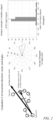
- the positions of dopamine neurons induced from the healthy subject iPS cells were tracked, and the movement pattern focusing especially on the directional property was evaluated according to the above "Evaluation 2 of movement method (vector and angle)" ( Fig. 3 ).
- the healthy subject-derived neurons showed a constant movement pattern (characterized by straightness) even in vitro. There was almost no difference in rule of the directional property among the individual cells.
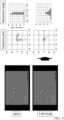
- the positions of dopamine neurons induced from the RELN-deleted patient iPS cells were tracked, and the movement pattern was evaluated according to the above "Evaluation 2 of movement method (vector and angle)" ( Fig. 4 ).
- the RELN-deleted patient-derived neurons showed a pattern in which the individual cells were different in movement direction, in contrast to the healthy subject-derived neurons. That is, an abnormality in the movement pattern could be detected.
- the movement patterns of the dopamine neurons induced from RELN-deleted isogenic iPS cells were also analyzed, and the movement pattern of the healthy subjects and the movement patterns of the RELN deletion (RELN-deleted patient and RELN-deleted isogenic (Isogenic(-/-) and Isogenic (+/-)) were compared with each other ( Fig. 5 ).
- the healthy subject shows a movement pattern with a small variation (fluctuation) in which the angle ⁇ is distributed in a narrow range (about 80% of the angle ⁇ falling within the range of-20° to +20°).
- the movement patterns of the RELN deletion have a great variation, with only about 45% to 65% of the angle ⁇ falling within the range of -20° to +20°.
- a measurement system for automatically detecting/quantifying movement patterns was constructed ( Fig. 6 ) and the effectiveness thereof was verified.
- the healthy subject-derived neurons, RELN-deleted patient-derived neurons, and RELN-deleted isogenic-derived neurons were subjected to trajectory plotting (12 hours) by automatic detection, and the movement patterns were compared and evaluated according to the above "Evaluation 1 of movement method (distance ratio)" ( Fig. 7 ).
- a clear decrease in distance ratio was observed in the RELN-deleted groups (RELN-del1(+/-) and 201B7(-/-)) as compared with the healthy subject (201B7(+/+)).
- the drug evaluation system of the present invention using the cell movement pattern as an index can be widely used and applied.
- the present invention can be expected to be utilized for: pathological researches on central nervous system diseases, basic researches on neurogenesis and circuit formation, development of central nervous system drugs, development of therapeutic methods/therapeutic drugs for spinal cord injury, peripheral neuropathy, and organ formation (for example, congenital heart disease), and prediction of the effects of drugs taken by a pregnant or nursing mother on the fetus/child brain and organ formation.
Landscapes
- Health & Medical Sciences (AREA)
- Life Sciences & Earth Sciences (AREA)
- Engineering & Computer Science (AREA)
- Biomedical Technology (AREA)
- Immunology (AREA)
- Hematology (AREA)
- Chemical & Material Sciences (AREA)
- Urology & Nephrology (AREA)
- Molecular Biology (AREA)
- Cell Biology (AREA)
- Toxicology (AREA)
- General Health & Medical Sciences (AREA)
- Microbiology (AREA)
- Tropical Medicine & Parasitology (AREA)
- Bioinformatics & Cheminformatics (AREA)
- Pathology (AREA)
- Food Science & Technology (AREA)
- Medicinal Chemistry (AREA)
- Physics & Mathematics (AREA)
- Analytical Chemistry (AREA)
- Biochemistry (AREA)
- Biotechnology (AREA)
- General Physics & Mathematics (AREA)
- Neurology (AREA)
- Neurosurgery (AREA)
- Developmental Biology & Embryology (AREA)
- Micro-Organisms Or Cultivation Processes Thereof (AREA)
- Measuring Or Testing Involving Enzymes Or Micro-Organisms (AREA)
- Medicines Containing Material From Animals Or Micro-Organisms (AREA)
- Investigating Or Analysing Biological Materials (AREA)
- Apparatus Associated With Microorganisms And Enzymes (AREA)
Claims (14)
- Arzneimittelbewertungsverfahren, das ein zellbasiertes Assaysystem verwendet, um ein Bewegungsmuster zu erhalten, das durch die Richtungsbewegungsfähigkeit einzelner Zellen, die eine Zellpopulation bilden, als Index definiert ist, wobei die Zellen Neuronen sind, die aus induzierten pluripotenten Stammzellen stammen.
- Verfahren nach Anspruch 1, umfassend die folgenden Schritte (i) bis (iii):(i) Herstellung einer Population von Zellen, die die Fähigkeit zur Bewegung aufweisen, Beginn der Kultur in Gegenwart einer Testsubstanz und anschließende Messung der Positionen der einzelnen Zellen, die sich im Laufe der Zeit bewegt haben,(ii) Analysieren der Richtungsbewegungsfähigkeit der jeweiligen Zellen aus Informationen über die gemessenen Positionen, Summieren der Analyseergebnisse und Bestimmen eines Bewegungsmusters der Zellpopulation, und(iii) Bestimmung der Wirksamkeit oder Toxizität der Testsubstanz auf der Grundlage des ermittelten Bewegungsmusters.
- Verfahren nach Anspruch 2, wobei ein im Folgenden (A) berechnetes Abstandsverhältnis, ein im Folgenden (B) ermittelter Winkel θ oder eine im Folgenden (C) berechnete Geschwindigkeit für die Analyse von Schritt (ii) verwendet wird:(A) Berechnung des Abstandsverhältnisses d(t)/D(t), wobei D(t) der Gesamtbewegungsabstand jeder Zelle zu einem Messzeitpunkt t und d(t) ein linearer Abstand zwischen zwei Punkten, d.h. einem Bewegungsstartpunkt und einem Bewegungsendpunkt, ist,(B) Einstellen eines Messzeitintervalls t/n, wobei n eine ganze Zahl von 2 oder mehr ist, in Bezug auf die Messzeit t, um eine Position jeder der Zellen zu messen, wodurch der Winkel θ zwischen einem Positionsvektor jeder der Zellen und einer Referenzachse zu jedem Zeitpunkt bestimmt wird, und(C) Berechnung der Geschwindigkeit D(t)/t, wobei D(t) die Gesamtbewegungsstrecke jeder der Zellen zum Messzeitpunkt t ist.
- Verfahren nach Anspruch 3, wobei ein durchschnittlicher Positionsvektor, der aus einer Bewegungstrajektorie jeder der Zellen bis zum Zeitpunkt t berechnet wird, als Referenzachse von (B) verwendet wird.
- Verfahren nach einem der Ansprüche 2 bis 4, wobei es sich bei den Zellen aus Schritt (i) um abnorme Zellen handelt und in Schritt (iii) die Wirksamkeit der Testsubstanz unter Verwendung der Normalisierung des Bewegungsmusters als Index bestimmt wird.
- Verfahren nach einem der Ansprüche 2 bis 4, wobei es sich bei den Zellen aus Schritt (i) um normale Zellen handelt und in Schritt (iii) die Toxizität der Testsubstanz unter Verwendung der Abnormalisierung des Bewegungsmusters als Index bestimmt wird.
- Verfahren nach einem der Ansprüche 2 bis 4, wobei es sich bei den Zellen aus Schritt (i) um abnorme Zellen handelt und in Schritt (iii) die Toxizität der Testsubstanz anhand einer weiteren Abnormalisierung des Bewegungsmusters als Index bestimmt wird.
- Verfahren nach Anspruch 5 oder 7, wobei die abnormen Zellen kranke Zellen mit genetischen Merkmalen einer Zielkrankheit sind.
- Verfahren nach Anspruch 8, wobei die erkrankten Zellen vom Patienten stammende oder gentechnisch hergestellte Zellen sind.
- Verfahren nach Anspruch 9, wobei die vom Patienten stammenden Zellen Zellen sind, die durch Induktion der Differenzierung von induzierten pluripotenten Stammzellen, die aus Patientenzellen hergestellt wurden, erhalten wurden.
- Verfahren nach einem der Ansprüche 1 bis 10, wobei die Neuronen Dopamin-Neuronen, glutamaterge Neuronen, Serotonin-Neuronen oder GABAerge Neuronen sind.
- Verfahren nach Anspruch 11, wobei die Dopamin-Neuronen durch das Verfahren hergestellt werden, das die folgenden Schritte (1) bis (3) umfasst:(1) Kultivierung pluripotenter Stammzellen in Gegenwart eines Inhibitors der TGF-β-Familie, eines GSK3β-Inhibitors und eines BMP-Inhibitors;(2) Suspensionskultivierung der in Schritt (1) erhaltenen Zellen in Gegenwart eines Inhibitors der TGF-β-Familie, eines GSK3β-Inhibitors, FGF8 und eines Hedgehog-Signalagonisten und unter normalem Sauerstoffpartialdruck zur Bildung einer Neurosphäre; und(3) Sammeln der Zellen, die die Neurosphäre bilden, um deren Differenzierung zu Dopamin-Neuronen zu induzieren, oder direkte Induktion der Neurosphäre zu Dopamin-Neuronen.
- Verfahren zur Bewertung der Qualität einer Zellpopulation, das ein zellbasiertes Assaysystem verwendet, um ein Bewegungsmuster zu erhalten, das durch die Richtungsbewegungsfähigkeit einzelner Zellen, die eine Zellpopulation bilden, als Index definiert ist, wobei die Zellen Neuronen sind, die von induzierten pluripotenten Stammzellen stammen.
- Verwendung eines automatischen Erkennungssystems zur automatischen Erkennung und/oder Quantifizierung von Bewegungsmustern im Verfahren nach einem der Ansprüche 1 bis 13.
Applications Claiming Priority (2)
| Application Number | Priority Date | Filing Date | Title |
|---|---|---|---|
| JP2018035758 | 2018-02-28 | ||
| PCT/JP2018/047146 WO2019167398A1 (ja) | 2018-02-28 | 2018-12-21 | 薬剤評価方法 |
Publications (3)
| Publication Number | Publication Date |
|---|---|
| EP3760728A1 EP3760728A1 (de) | 2021-01-06 |
| EP3760728A4 EP3760728A4 (de) | 2021-10-27 |
| EP3760728B1 true EP3760728B1 (de) | 2023-11-29 |
Family
ID=67804913
Family Applications (1)
| Application Number | Title | Priority Date | Filing Date |
|---|---|---|---|
| EP18907612.8A Active EP3760728B1 (de) | 2018-02-28 | 2018-12-21 | Bewertung von zellen anhand der richtungsbewegungsfähigkeit. |
Country Status (4)
| Country | Link |
|---|---|
| US (1) | US20200408743A1 (de) |
| EP (1) | EP3760728B1 (de) |
| JP (1) | JP7357366B2 (de) |
| WO (1) | WO2019167398A1 (de) |
Families Citing this family (1)
| Publication number | Priority date | Publication date | Assignee | Title |
|---|---|---|---|---|
| WO2022009663A1 (ja) * | 2020-07-06 | 2022-01-13 | 株式会社ニコン | 遊走性算出装置、遊走性評価方法およびコンピュータに遊走性評価方法を実行させるコンピュータプログラム |
Family Cites Families (15)
| Publication number | Priority date | Publication date | Assignee | Title |
|---|---|---|---|---|
| CA2277278A1 (en) | 1997-01-10 | 1998-07-16 | Life Technologies, Inc. | Embryonic stem cell serum replacement |
| WO2005123902A1 (ja) | 2004-06-18 | 2005-12-29 | Riken | 無血清浮遊培養による胚性幹細胞の神経分化誘導法 |
| US7940978B2 (en) * | 2007-06-05 | 2011-05-10 | General Electric Company | Automatic characterization of cellular motion |
| JP5228826B2 (ja) * | 2008-11-17 | 2013-07-03 | 大日本印刷株式会社 | 細胞画像解析装置および細胞画像解析方法 |
| JP5166360B2 (ja) * | 2009-06-23 | 2013-03-21 | 株式会社島津製作所 | マイクロ反応装置を用いた細胞運動評価方法 |
| JP2013039113A (ja) * | 2011-08-19 | 2013-02-28 | Nagoya Univ | 細胞品質管理方法及び細胞の生産方法 |
| JP6494903B2 (ja) * | 2013-02-14 | 2019-04-03 | ソニー株式会社 | 分析システム、分析プログラム及び分析方法 |
| TW201500549A (zh) * | 2013-06-19 | 2015-01-01 | Univ Taipei Medical | 促進幹細胞方向性移動之方法及裝置 |
| US10670583B2 (en) * | 2013-09-20 | 2020-06-02 | The General Hospital Corporation | Cell chemotaxis assays |
| ES2939807T3 (es) * | 2014-03-21 | 2023-04-27 | Fujifilm Cellular Dynamics Inc | Producción de neuronas dopaminérgicas del mesencéfalo y métodos para su utilización |
| JP7420334B2 (ja) * | 2015-06-25 | 2024-01-23 | 株式会社カネカ | 液体注入方法 |
| LU92771B1 (en) * | 2015-07-10 | 2017-01-30 | Univ Luxembourg | Long-term self-renewing neural stem cells |
| WO2017035315A1 (en) * | 2015-08-27 | 2017-03-02 | The Johns Hopkins University | Methods and device for phenotypic classification of cells based on migratory behavior |
| JP6441199B2 (ja) | 2015-10-23 | 2018-12-19 | 本田技研工業株式会社 | 内燃機関の制御装置 |
| JP6755579B2 (ja) | 2016-08-31 | 2020-09-16 | 大洋プラント株式会社 | ブレード傾倒機構および該ブレード傾倒機構を備えた風力エネルギー利用装置 |
-
2018
- 2018-12-21 EP EP18907612.8A patent/EP3760728B1/de active Active
- 2018-12-21 JP JP2020502824A patent/JP7357366B2/ja active Active
- 2018-12-21 US US16/976,586 patent/US20200408743A1/en not_active Abandoned
- 2018-12-21 WO PCT/JP2018/047146 patent/WO2019167398A1/ja not_active Ceased
Also Published As
| Publication number | Publication date |
|---|---|
| US20200408743A1 (en) | 2020-12-31 |
| JP7357366B2 (ja) | 2023-10-06 |
| WO2019167398A1 (ja) | 2019-09-06 |
| EP3760728A4 (de) | 2021-10-27 |
| JPWO2019167398A1 (ja) | 2021-03-11 |
| EP3760728A1 (de) | 2021-01-06 |
Similar Documents
| Publication | Publication Date | Title |
|---|---|---|
| US20240043811A1 (en) | Method of producing a mesodermal-lineage primitive streak cell | |
| JP7023820B2 (ja) | 多分化能細胞および多能性細胞の分化を方向付けることによって発生させる皮質介在ニューロンおよびその他のニューロン細胞 | |
| EP2582795B1 (de) | Verfahren zur auswahl menschlicher induzierter pluripotenter stammzellen | |
| KR102265024B1 (ko) | Fgfr3병의 예방 및 치료제 및 그 스크리닝 방법 | |
| EP3031908A1 (de) | Verfahren zur herstellung von dopaminergen neuronen | |
| EP3613848A1 (de) | Verfahren zur herstellung von dopaminergen neuronen | |
| KR20180101467A (ko) | 인간 다능성줄기세포로부터 시상하부 뉴런으로의 분화 유도 | |
| JP6856266B2 (ja) | Fgfr3病の予防および治療剤ならびにそのスクリーニング方法 | |
| EP3760728B1 (de) | Bewertung von zellen anhand der richtungsbewegungsfähigkeit. | |
| EP4357449A1 (de) | Verfahren zur herstellung einer zubereitung aus menschlichen pluripotenten stammzellen für hirnrindenzellen | |
| US20240318142A1 (en) | Method for producing renal interstitial progenitor cells, erythropoietin-producing cells, and method for producing renin-producing cells | |
| JP7090881B2 (ja) | 細胞内カルシウム動態評価系 | |
| JP2024541964A (ja) | 変異を含むドーパミン作動性ニューロン及びその使用方法 | |
| EP4092107A1 (de) | Verfahren zur herstellung pluripotenter stammzellen mit fähigkeit zur differenzierung in spezifische zellen und deren verwendung | |
| EP3950932A1 (de) | Verfahren zur herstellung pluripotenter stammzellen mit fähigkeit zur differenzierung in spezifische zellen und deren verwendung | |
| HK40104497A (en) | Method for producing cerebral cortical cell preparation derived from human pluripotent stem cells | |
| WO2025018366A1 (ja) | 腸管上皮細胞を作製する方法 | |
| Lyssiotis | Insight into the reprogramming of cell fate with small molecules |
Legal Events
| Date | Code | Title | Description |
|---|---|---|---|
| STAA | Information on the status of an ep patent application or granted ep patent |
Free format text: STATUS: THE INTERNATIONAL PUBLICATION HAS BEEN MADE |
|
| PUAI | Public reference made under article 153(3) epc to a published international application that has entered the european phase |
Free format text: ORIGINAL CODE: 0009012 |
|
| STAA | Information on the status of an ep patent application or granted ep patent |
Free format text: STATUS: REQUEST FOR EXAMINATION WAS MADE |
|
| 17P | Request for examination filed |
Effective date: 20200922 |
|
| AK | Designated contracting states |
Kind code of ref document: A1 Designated state(s): AL AT BE BG CH CY CZ DE DK EE ES FI FR GB GR HR HU IE IS IT LI LT LU LV MC MK MT NL NO PL PT RO RS SE SI SK SM TR |
|
| AX | Request for extension of the european patent |
Extension state: BA ME |
|
| DAV | Request for validation of the european patent (deleted) | ||
| DAX | Request for extension of the european patent (deleted) | ||
| A4 | Supplementary search report drawn up and despatched |
Effective date: 20210924 |
|
| RIC1 | Information provided on ipc code assigned before grant |
Ipc: C12N 5/10 20060101ALI20210920BHEP Ipc: C12N 5/0793 20100101ALI20210920BHEP Ipc: C12M 1/34 20060101ALI20210920BHEP Ipc: G01N 33/50 20060101ALI20210920BHEP Ipc: C12Q 1/02 20060101AFI20210920BHEP |
|
| STAA | Information on the status of an ep patent application or granted ep patent |
Free format text: STATUS: EXAMINATION IS IN PROGRESS |
|
| 17Q | First examination report despatched |
Effective date: 20220531 |
|
| GRAP | Despatch of communication of intention to grant a patent |
Free format text: ORIGINAL CODE: EPIDOSNIGR1 |
|
| STAA | Information on the status of an ep patent application or granted ep patent |
Free format text: STATUS: GRANT OF PATENT IS INTENDED |
|
| INTG | Intention to grant announced |
Effective date: 20230525 |
|
| GRAS | Grant fee paid |
Free format text: ORIGINAL CODE: EPIDOSNIGR3 |
|
| GRAA | (expected) grant |
Free format text: ORIGINAL CODE: 0009210 |
|
| STAA | Information on the status of an ep patent application or granted ep patent |
Free format text: STATUS: THE PATENT HAS BEEN GRANTED |
|
| AK | Designated contracting states |
Kind code of ref document: B1 Designated state(s): AL AT BE BG CH CY CZ DE DK EE ES FI FR GB GR HR HU IE IS IT LI LT LU LV MC MK MT NL NO PL PT RO RS SE SI SK SM TR |
|
| REG | Reference to a national code |
Ref country code: GB Ref legal event code: FG4D |
|
| REG | Reference to a national code |
Ref country code: CH Ref legal event code: EP |
|
| REG | Reference to a national code |
Ref country code: DE Ref legal event code: R096 Ref document number: 602018062039 Country of ref document: DE |
|
| REG | Reference to a national code |
Ref country code: IE Ref legal event code: FG4D |
|
| PGFP | Annual fee paid to national office [announced via postgrant information from national office to epo] |
Ref country code: GB Payment date: 20231207 Year of fee payment: 6 |
|
| PGFP | Annual fee paid to national office [announced via postgrant information from national office to epo] |
Ref country code: FR Payment date: 20231212 Year of fee payment: 6 Ref country code: DE Payment date: 20231205 Year of fee payment: 6 |
|
| REG | Reference to a national code |
Ref country code: LT Ref legal event code: MG9D |
|
| REG | Reference to a national code |
Ref country code: NL Ref legal event code: MP Effective date: 20231129 |
|
| PG25 | Lapsed in a contracting state [announced via postgrant information from national office to epo] |
Ref country code: GR Free format text: LAPSE BECAUSE OF FAILURE TO SUBMIT A TRANSLATION OF THE DESCRIPTION OR TO PAY THE FEE WITHIN THE PRESCRIBED TIME-LIMIT Effective date: 20240301 |
|
| PG25 | Lapsed in a contracting state [announced via postgrant information from national office to epo] |
Ref country code: IS Free format text: LAPSE BECAUSE OF FAILURE TO SUBMIT A TRANSLATION OF THE DESCRIPTION OR TO PAY THE FEE WITHIN THE PRESCRIBED TIME-LIMIT Effective date: 20240329 |
|
| PG25 | Lapsed in a contracting state [announced via postgrant information from national office to epo] |
Ref country code: LT Free format text: LAPSE BECAUSE OF FAILURE TO SUBMIT A TRANSLATION OF THE DESCRIPTION OR TO PAY THE FEE WITHIN THE PRESCRIBED TIME-LIMIT Effective date: 20231129 |
|
| PG25 | Lapsed in a contracting state [announced via postgrant information from national office to epo] |
Ref country code: ES Free format text: LAPSE BECAUSE OF FAILURE TO SUBMIT A TRANSLATION OF THE DESCRIPTION OR TO PAY THE FEE WITHIN THE PRESCRIBED TIME-LIMIT Effective date: 20231129 |
|
| PG25 | Lapsed in a contracting state [announced via postgrant information from national office to epo] |
Ref country code: LT Free format text: LAPSE BECAUSE OF FAILURE TO SUBMIT A TRANSLATION OF THE DESCRIPTION OR TO PAY THE FEE WITHIN THE PRESCRIBED TIME-LIMIT Effective date: 20231129 Ref country code: IS Free format text: LAPSE BECAUSE OF FAILURE TO SUBMIT A TRANSLATION OF THE DESCRIPTION OR TO PAY THE FEE WITHIN THE PRESCRIBED TIME-LIMIT Effective date: 20240329 Ref country code: GR Free format text: LAPSE BECAUSE OF FAILURE TO SUBMIT A TRANSLATION OF THE DESCRIPTION OR TO PAY THE FEE WITHIN THE PRESCRIBED TIME-LIMIT Effective date: 20240301 Ref country code: ES Free format text: LAPSE BECAUSE OF FAILURE TO SUBMIT A TRANSLATION OF THE DESCRIPTION OR TO PAY THE FEE WITHIN THE PRESCRIBED TIME-LIMIT Effective date: 20231129 Ref country code: BG Free format text: LAPSE BECAUSE OF FAILURE TO SUBMIT A TRANSLATION OF THE DESCRIPTION OR TO PAY THE FEE WITHIN THE PRESCRIBED TIME-LIMIT Effective date: 20240229 |
|
| REG | Reference to a national code |
Ref country code: AT Ref legal event code: MK05 Ref document number: 1636158 Country of ref document: AT Kind code of ref document: T Effective date: 20231129 |
|
| PG25 | Lapsed in a contracting state [announced via postgrant information from national office to epo] |
Ref country code: NL Free format text: LAPSE BECAUSE OF FAILURE TO SUBMIT A TRANSLATION OF THE DESCRIPTION OR TO PAY THE FEE WITHIN THE PRESCRIBED TIME-LIMIT Effective date: 20231129 |
|
| PG25 | Lapsed in a contracting state [announced via postgrant information from national office to epo] |
Ref country code: SE Free format text: LAPSE BECAUSE OF FAILURE TO SUBMIT A TRANSLATION OF THE DESCRIPTION OR TO PAY THE FEE WITHIN THE PRESCRIBED TIME-LIMIT Effective date: 20231129 Ref country code: RS Free format text: LAPSE BECAUSE OF FAILURE TO SUBMIT A TRANSLATION OF THE DESCRIPTION OR TO PAY THE FEE WITHIN THE PRESCRIBED TIME-LIMIT Effective date: 20231129 Ref country code: PL Free format text: LAPSE BECAUSE OF FAILURE TO SUBMIT A TRANSLATION OF THE DESCRIPTION OR TO PAY THE FEE WITHIN THE PRESCRIBED TIME-LIMIT Effective date: 20231129 Ref country code: NO Free format text: LAPSE BECAUSE OF FAILURE TO SUBMIT A TRANSLATION OF THE DESCRIPTION OR TO PAY THE FEE WITHIN THE PRESCRIBED TIME-LIMIT Effective date: 20240229 Ref country code: NL Free format text: LAPSE BECAUSE OF FAILURE TO SUBMIT A TRANSLATION OF THE DESCRIPTION OR TO PAY THE FEE WITHIN THE PRESCRIBED TIME-LIMIT Effective date: 20231129 Ref country code: LV Free format text: LAPSE BECAUSE OF FAILURE TO SUBMIT A TRANSLATION OF THE DESCRIPTION OR TO PAY THE FEE WITHIN THE PRESCRIBED TIME-LIMIT Effective date: 20231129 Ref country code: HR Free format text: LAPSE BECAUSE OF FAILURE TO SUBMIT A TRANSLATION OF THE DESCRIPTION OR TO PAY THE FEE WITHIN THE PRESCRIBED TIME-LIMIT Effective date: 20231129 |
|
| PG25 | Lapsed in a contracting state [announced via postgrant information from national office to epo] |
Ref country code: DK Free format text: LAPSE BECAUSE OF FAILURE TO SUBMIT A TRANSLATION OF THE DESCRIPTION OR TO PAY THE FEE WITHIN THE PRESCRIBED TIME-LIMIT Effective date: 20231129 |
|
| PG25 | Lapsed in a contracting state [announced via postgrant information from national office to epo] |
Ref country code: AT Free format text: LAPSE BECAUSE OF FAILURE TO SUBMIT A TRANSLATION OF THE DESCRIPTION OR TO PAY THE FEE WITHIN THE PRESCRIBED TIME-LIMIT Effective date: 20231129 Ref country code: CZ Free format text: LAPSE BECAUSE OF FAILURE TO SUBMIT A TRANSLATION OF THE DESCRIPTION OR TO PAY THE FEE WITHIN THE PRESCRIBED TIME-LIMIT Effective date: 20231129 |
|
| PG25 | Lapsed in a contracting state [announced via postgrant information from national office to epo] |
Ref country code: SK Free format text: LAPSE BECAUSE OF FAILURE TO SUBMIT A TRANSLATION OF THE DESCRIPTION OR TO PAY THE FEE WITHIN THE PRESCRIBED TIME-LIMIT Effective date: 20231129 |
|
| PG25 | Lapsed in a contracting state [announced via postgrant information from national office to epo] |
Ref country code: SM Free format text: LAPSE BECAUSE OF FAILURE TO SUBMIT A TRANSLATION OF THE DESCRIPTION OR TO PAY THE FEE WITHIN THE PRESCRIBED TIME-LIMIT Effective date: 20231129 Ref country code: SK Free format text: LAPSE BECAUSE OF FAILURE TO SUBMIT A TRANSLATION OF THE DESCRIPTION OR TO PAY THE FEE WITHIN THE PRESCRIBED TIME-LIMIT Effective date: 20231129 Ref country code: RO Free format text: LAPSE BECAUSE OF FAILURE TO SUBMIT A TRANSLATION OF THE DESCRIPTION OR TO PAY THE FEE WITHIN THE PRESCRIBED TIME-LIMIT Effective date: 20231129 Ref country code: IT Free format text: LAPSE BECAUSE OF FAILURE TO SUBMIT A TRANSLATION OF THE DESCRIPTION OR TO PAY THE FEE WITHIN THE PRESCRIBED TIME-LIMIT Effective date: 20231129 Ref country code: EE Free format text: LAPSE BECAUSE OF FAILURE TO SUBMIT A TRANSLATION OF THE DESCRIPTION OR TO PAY THE FEE WITHIN THE PRESCRIBED TIME-LIMIT Effective date: 20231129 Ref country code: DK Free format text: LAPSE BECAUSE OF FAILURE TO SUBMIT A TRANSLATION OF THE DESCRIPTION OR TO PAY THE FEE WITHIN THE PRESCRIBED TIME-LIMIT Effective date: 20231129 Ref country code: CZ Free format text: LAPSE BECAUSE OF FAILURE TO SUBMIT A TRANSLATION OF THE DESCRIPTION OR TO PAY THE FEE WITHIN THE PRESCRIBED TIME-LIMIT Effective date: 20231129 Ref country code: AT Free format text: LAPSE BECAUSE OF FAILURE TO SUBMIT A TRANSLATION OF THE DESCRIPTION OR TO PAY THE FEE WITHIN THE PRESCRIBED TIME-LIMIT Effective date: 20231129 |
|
| REG | Reference to a national code |
Ref country code: CH Ref legal event code: PL |
|
| PG25 | Lapsed in a contracting state [announced via postgrant information from national office to epo] |
Ref country code: PT Free format text: LAPSE BECAUSE OF FAILURE TO SUBMIT A TRANSLATION OF THE DESCRIPTION OR TO PAY THE FEE WITHIN THE PRESCRIBED TIME-LIMIT Effective date: 20240401 |
|
| PG25 | Lapsed in a contracting state [announced via postgrant information from national office to epo] |
Ref country code: LU Free format text: LAPSE BECAUSE OF NON-PAYMENT OF DUE FEES Effective date: 20231221 |
|
| PG25 | Lapsed in a contracting state [announced via postgrant information from national office to epo] |
Ref country code: MC Free format text: LAPSE BECAUSE OF FAILURE TO SUBMIT A TRANSLATION OF THE DESCRIPTION OR TO PAY THE FEE WITHIN THE PRESCRIBED TIME-LIMIT Effective date: 20231129 |
|
| REG | Reference to a national code |
Ref country code: BE Ref legal event code: MM Effective date: 20231231 |
|
| PG25 | Lapsed in a contracting state [announced via postgrant information from national office to epo] |
Ref country code: PT Free format text: LAPSE BECAUSE OF FAILURE TO SUBMIT A TRANSLATION OF THE DESCRIPTION OR TO PAY THE FEE WITHIN THE PRESCRIBED TIME-LIMIT Effective date: 20240401 Ref country code: MC Free format text: LAPSE BECAUSE OF FAILURE TO SUBMIT A TRANSLATION OF THE DESCRIPTION OR TO PAY THE FEE WITHIN THE PRESCRIBED TIME-LIMIT Effective date: 20231129 Ref country code: LU Free format text: LAPSE BECAUSE OF NON-PAYMENT OF DUE FEES Effective date: 20231221 |
|
| REG | Reference to a national code |
Ref country code: DE Ref legal event code: R097 Ref document number: 602018062039 Country of ref document: DE |
|
| PLBE | No opposition filed within time limit |
Free format text: ORIGINAL CODE: 0009261 |
|
| STAA | Information on the status of an ep patent application or granted ep patent |
Free format text: STATUS: NO OPPOSITION FILED WITHIN TIME LIMIT |
|
| REG | Reference to a national code |
Ref country code: IE Ref legal event code: MM4A |
|
| PG25 | Lapsed in a contracting state [announced via postgrant information from national office to epo] |
Ref country code: IE Free format text: LAPSE BECAUSE OF NON-PAYMENT OF DUE FEES Effective date: 20231221 |
|
| PG25 | Lapsed in a contracting state [announced via postgrant information from national office to epo] |
Ref country code: BE Free format text: LAPSE BECAUSE OF NON-PAYMENT OF DUE FEES Effective date: 20231231 |
|
| PG25 | Lapsed in a contracting state [announced via postgrant information from national office to epo] |
Ref country code: CH Free format text: LAPSE BECAUSE OF NON-PAYMENT OF DUE FEES Effective date: 20231231 |
|
| PG25 | Lapsed in a contracting state [announced via postgrant information from national office to epo] |
Ref country code: SI Free format text: LAPSE BECAUSE OF FAILURE TO SUBMIT A TRANSLATION OF THE DESCRIPTION OR TO PAY THE FEE WITHIN THE PRESCRIBED TIME-LIMIT Effective date: 20231129 |
|
| PG25 | Lapsed in a contracting state [announced via postgrant information from national office to epo] |
Ref country code: SI Free format text: LAPSE BECAUSE OF FAILURE TO SUBMIT A TRANSLATION OF THE DESCRIPTION OR TO PAY THE FEE WITHIN THE PRESCRIBED TIME-LIMIT Effective date: 20231129 Ref country code: IE Free format text: LAPSE BECAUSE OF NON-PAYMENT OF DUE FEES Effective date: 20231221 Ref country code: CH Free format text: LAPSE BECAUSE OF NON-PAYMENT OF DUE FEES Effective date: 20231231 Ref country code: BE Free format text: LAPSE BECAUSE OF NON-PAYMENT OF DUE FEES Effective date: 20231231 |
|
| 26N | No opposition filed |
Effective date: 20240830 |
|
| REG | Reference to a national code |
Ref country code: DE Ref legal event code: R119 Ref document number: 602018062039 Country of ref document: DE |
|
| PG25 | Lapsed in a contracting state [announced via postgrant information from national office to epo] |
Ref country code: FI Free format text: LAPSE BECAUSE OF FAILURE TO SUBMIT A TRANSLATION OF THE DESCRIPTION OR TO PAY THE FEE WITHIN THE PRESCRIBED TIME-LIMIT Effective date: 20231129 |
|
| PG25 | Lapsed in a contracting state [announced via postgrant information from national office to epo] |
Ref country code: CY Free format text: LAPSE BECAUSE OF FAILURE TO SUBMIT A TRANSLATION OF THE DESCRIPTION OR TO PAY THE FEE WITHIN THE PRESCRIBED TIME-LIMIT; INVALID AB INITIO Effective date: 20181221 |
|
| PG25 | Lapsed in a contracting state [announced via postgrant information from national office to epo] |
Ref country code: HU Free format text: LAPSE BECAUSE OF FAILURE TO SUBMIT A TRANSLATION OF THE DESCRIPTION OR TO PAY THE FEE WITHIN THE PRESCRIBED TIME-LIMIT; INVALID AB INITIO Effective date: 20181221 |
|
| GBPC | Gb: european patent ceased through non-payment of renewal fee |
Effective date: 20241221 |
|
| PG25 | Lapsed in a contracting state [announced via postgrant information from national office to epo] |
Ref country code: DE Free format text: LAPSE BECAUSE OF NON-PAYMENT OF DUE FEES Effective date: 20250701 |
|
| PG25 | Lapsed in a contracting state [announced via postgrant information from national office to epo] |
Ref country code: GB Free format text: LAPSE BECAUSE OF NON-PAYMENT OF DUE FEES Effective date: 20241221 |
|
| PG25 | Lapsed in a contracting state [announced via postgrant information from national office to epo] |
Ref country code: FR Free format text: LAPSE BECAUSE OF NON-PAYMENT OF DUE FEES Effective date: 20241231 |
|
| PG25 | Lapsed in a contracting state [announced via postgrant information from national office to epo] |
Ref country code: TR Free format text: LAPSE BECAUSE OF FAILURE TO SUBMIT A TRANSLATION OF THE DESCRIPTION OR TO PAY THE FEE WITHIN THE PRESCRIBED TIME-LIMIT Effective date: 20231129 |