EP3767587B1 - Détection de la présence des différents types de motifs fluorescents d'anticorps antinucléaires et dispositif associé - Google Patents
Détection de la présence des différents types de motifs fluorescents d'anticorps antinucléaires et dispositif associé Download PDFInfo
- Publication number
- EP3767587B1 EP3767587B1 EP19187348.8A EP19187348A EP3767587B1 EP 3767587 B1 EP3767587 B1 EP 3767587B1 EP 19187348 A EP19187348 A EP 19187348A EP 3767587 B1 EP3767587 B1 EP 3767587B1
- Authority
- EP
- European Patent Office
- Prior art keywords
- image
- fluorescence pattern
- subimage
- basis
- pattern types
- Prior art date
- Legal status (The legal status is an assumption and is not a legal conclusion. Google has not performed a legal analysis and makes no representation as to the accuracy of the status listed.)
- Active
Links
Images











Classifications
-
- G—PHYSICS
- G01—MEASURING; TESTING
- G01N—INVESTIGATING OR ANALYSING MATERIALS BY DETERMINING THEIR CHEMICAL OR PHYSICAL PROPERTIES
- G01N21/00—Investigating or analysing materials by the use of optical means, i.e. using sub-millimetre waves, infrared, visible or ultraviolet light
- G01N21/62—Systems in which the material investigated is excited whereby it emits light or causes a change in wavelength of the incident light
- G01N21/63—Systems in which the material investigated is excited whereby it emits light or causes a change in wavelength of the incident light optically excited
- G01N21/64—Fluorescence; Phosphorescence
- G01N21/645—Specially adapted constructive features of fluorimeters
- G01N21/6456—Spatial resolved fluorescence measurements; Imaging
-
- G—PHYSICS
- G01—MEASURING; TESTING
- G01N—INVESTIGATING OR ANALYSING MATERIALS BY DETERMINING THEIR CHEMICAL OR PHYSICAL PROPERTIES
- G01N15/00—Investigating characteristics of particles; Investigating permeability, pore-volume or surface-area of porous materials
- G01N15/10—Investigating individual particles
- G01N15/1023—Microstructural devices for non-optical measurement
-
- G—PHYSICS
- G01—MEASURING; TESTING
- G01N—INVESTIGATING OR ANALYSING MATERIALS BY DETERMINING THEIR CHEMICAL OR PHYSICAL PROPERTIES
- G01N21/00—Investigating or analysing materials by the use of optical means, i.e. using sub-millimetre waves, infrared, visible or ultraviolet light
- G01N21/62—Systems in which the material investigated is excited whereby it emits light or causes a change in wavelength of the incident light
- G01N21/63—Systems in which the material investigated is excited whereby it emits light or causes a change in wavelength of the incident light optically excited
- G01N21/64—Fluorescence; Phosphorescence
- G01N21/6486—Measuring fluorescence of biological material, e.g. DNA, RNA, cells
-
- G—PHYSICS
- G01—MEASURING; TESTING
- G01N—INVESTIGATING OR ANALYSING MATERIALS BY DETERMINING THEIR CHEMICAL OR PHYSICAL PROPERTIES
- G01N33/00—Investigating or analysing materials by specific methods not covered by groups G01N1/00 - G01N31/00
- G01N33/48—Biological material, e.g. blood, urine; Haemocytometers
- G01N33/50—Chemical analysis of biological material, e.g. blood, urine; Testing involving biospecific ligand binding methods; Immunological testing
- G01N33/53—Immunoassay; Biospecific binding assay; Materials therefor
- G01N33/569—Immunoassay; Biospecific binding assay; Materials therefor for microorganisms, e.g. protozoa, bacteria, viruses
- G01N33/56966—Animal cells
-
- G—PHYSICS
- G01—MEASURING; TESTING
- G01N—INVESTIGATING OR ANALYSING MATERIALS BY DETERMINING THEIR CHEMICAL OR PHYSICAL PROPERTIES
- G01N33/00—Investigating or analysing materials by specific methods not covered by groups G01N1/00 - G01N31/00
- G01N33/48—Biological material, e.g. blood, urine; Haemocytometers
- G01N33/50—Chemical analysis of biological material, e.g. blood, urine; Testing involving biospecific ligand binding methods; Immunological testing
- G01N33/53—Immunoassay; Biospecific binding assay; Materials therefor
- G01N33/575—Immunoassay; Biospecific binding assay; Materials therefor for cancer
- G01N33/5751—Immunoassay; Biospecific binding assay; Materials therefor for cancer of the skin, e.g. melanoma
-
- G—PHYSICS
- G01—MEASURING; TESTING
- G01N—INVESTIGATING OR ANALYSING MATERIALS BY DETERMINING THEIR CHEMICAL OR PHYSICAL PROPERTIES
- G01N33/00—Investigating or analysing materials by specific methods not covered by groups G01N1/00 - G01N31/00
- G01N33/48—Biological material, e.g. blood, urine; Haemocytometers
- G01N33/50—Chemical analysis of biological material, e.g. blood, urine; Testing involving biospecific ligand binding methods; Immunological testing
- G01N33/58—Chemical analysis of biological material, e.g. blood, urine; Testing involving biospecific ligand binding methods; Immunological testing involving labelled substances
- G01N33/582—Chemical analysis of biological material, e.g. blood, urine; Testing involving biospecific ligand binding methods; Immunological testing involving labelled substances with fluorescent label
-
- G—PHYSICS
- G06—COMPUTING OR CALCULATING; COUNTING
- G06N—COMPUTING ARRANGEMENTS BASED ON SPECIFIC COMPUTATIONAL MODELS
- G06N3/00—Computing arrangements based on biological models
- G06N3/02—Neural networks
- G06N3/04—Architecture, e.g. interconnection topology
- G06N3/045—Combinations of networks
-
- G—PHYSICS
- G06—COMPUTING OR CALCULATING; COUNTING
- G06N—COMPUTING ARRANGEMENTS BASED ON SPECIFIC COMPUTATIONAL MODELS
- G06N3/00—Computing arrangements based on biological models
- G06N3/02—Neural networks
- G06N3/04—Architecture, e.g. interconnection topology
- G06N3/0464—Convolutional networks [CNN, ConvNet]
-
- G—PHYSICS
- G06—COMPUTING OR CALCULATING; COUNTING
- G06N—COMPUTING ARRANGEMENTS BASED ON SPECIFIC COMPUTATIONAL MODELS
- G06N3/00—Computing arrangements based on biological models
- G06N3/02—Neural networks
- G06N3/08—Learning methods
-
- G—PHYSICS
- G06—COMPUTING OR CALCULATING; COUNTING
- G06N—COMPUTING ARRANGEMENTS BASED ON SPECIFIC COMPUTATIONAL MODELS
- G06N3/00—Computing arrangements based on biological models
- G06N3/02—Neural networks
- G06N3/08—Learning methods
- G06N3/09—Supervised learning
-
- G—PHYSICS
- G06—COMPUTING OR CALCULATING; COUNTING
- G06T—IMAGE DATA PROCESSING OR GENERATION, IN GENERAL
- G06T7/00—Image analysis
- G06T7/0002—Inspection of images, e.g. flaw detection
- G06T7/0012—Biomedical image inspection
-
- G—PHYSICS
- G06—COMPUTING OR CALCULATING; COUNTING
- G06T—IMAGE DATA PROCESSING OR GENERATION, IN GENERAL
- G06T7/00—Image analysis
- G06T7/10—Segmentation; Edge detection
- G06T7/11—Region-based segmentation
-
- G—PHYSICS
- G06—COMPUTING OR CALCULATING; COUNTING
- G06T—IMAGE DATA PROCESSING OR GENERATION, IN GENERAL
- G06T7/00—Image analysis
- G06T7/70—Determining position or orientation of objects or cameras
-
- G—PHYSICS
- G01—MEASURING; TESTING
- G01N—INVESTIGATING OR ANALYSING MATERIALS BY DETERMINING THEIR CHEMICAL OR PHYSICAL PROPERTIES
- G01N15/00—Investigating characteristics of particles; Investigating permeability, pore-volume or surface-area of porous materials
- G01N15/10—Investigating individual particles
- G01N2015/1006—Investigating individual particles for cytology
-
- G—PHYSICS
- G01—MEASURING; TESTING
- G01N—INVESTIGATING OR ANALYSING MATERIALS BY DETERMINING THEIR CHEMICAL OR PHYSICAL PROPERTIES
- G01N2469/00—Immunoassays for the detection of microorganisms
- G01N2469/10—Detection of antigens from microorganism in sample from host
-
- G—PHYSICS
- G06—COMPUTING OR CALCULATING; COUNTING
- G06T—IMAGE DATA PROCESSING OR GENERATION, IN GENERAL
- G06T2207/00—Indexing scheme for image analysis or image enhancement
- G06T2207/10—Image acquisition modality
- G06T2207/10064—Fluorescence image
-
- G—PHYSICS
- G06—COMPUTING OR CALCULATING; COUNTING
- G06T—IMAGE DATA PROCESSING OR GENERATION, IN GENERAL
- G06T2207/00—Indexing scheme for image analysis or image enhancement
- G06T2207/20—Special algorithmic details
- G06T2207/20084—Artificial neural networks [ANN]
-
- G—PHYSICS
- G06—COMPUTING OR CALCULATING; COUNTING
- G06T—IMAGE DATA PROCESSING OR GENERATION, IN GENERAL
- G06T2207/00—Indexing scheme for image analysis or image enhancement
- G06T2207/30—Subject of image; Context of image processing
- G06T2207/30004—Biomedical image processing
- G06T2207/30024—Cell structures in vitro; Tissue sections in vitro
Definitions
- the invention relates to a method and a device for detecting respective potential presences of respective different cellular fluorescence pattern types on a biological cell substrate comprising human epithelioma cells (HEp cells), wherein the fluorescence pattern types comprise different antinuclear antibody fluorescence pattern types.
- the invention further relates to a method for detecting potential presences of different cellular fluorescence pattern types on a biological cell substrate comprising human epithelioma cells by means of digital image processing and a computing unit, a data network device, a computer program product and a data carrier signal therefor.
- a biological substrate containing human epithelioma cells For the purposes of medical diagnostics and in particular for the purpose of detecting autoantibodies in a liquid patient sample such as blood or blood serum, it is a known method to incubate a biological substrate containing human epithelioma cells with the liquid patient sample or the diluted liquid patient sample. Depending on the presence of certain primary autoantibodies in the patient sample, these primary antibodies bind to different areas of the cells mentioned. The biological cell substrate is then further incubated with secondary antibodies that are labeled with a so-called fluorescent dye. Such secondary antibodies can in turn bind to the primary antibodies that are bound to the cell substrate.
- the aforementioned fluorescent dye fluoresces, so that in a corresponding fluorescence image the primary antibodies of the patient sample, which are bound to the cells of the cell substrate and to which secondary antibodies with the fluorescent dye are bound, can be localized as a fluorescence pattern in a fluorescence image.
- different specific fluorescence patterns arise.
- autoantibodies which are directed against the cell nuclei or the antigens of the cell nucleus
- antinuclear antibody fluorescence patterns arise.
- ANA fluorescence patterns or an ANA pattern.
- IIFT indirect immunofluorescence
- human epithelial cells or human epithelioma cells the task is to recognize and classify the resulting different types of fluorescence patterns.
- a single pattern does not necessarily have to be present in a fluorescence image; several patterns can also be present at the same time.
- Antibodies against nuclear antigens are directed against various nuclear components (biochemical substances of the cell nucleus). These include nucleic acids, nuclear proteins and ribonucleoproteins.
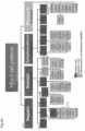
- the different resulting antinuclear antibody fluorescence pattern types are divided into different pattern types.
- One approach to this can be found on the website www.anapatterns.org, where, among other things, different nuclear patterns are listed and differentiated.
- An example classification according to this scheme can be found in the Figure 24 illustrated.
- the document DE102006027516A1 discloses a method using an indicator dye as a so-called counterstain and a fluorescent dye to show antibody-specific binding to a substrate. Relevant structures, such as cells in the mitotic stage, are identified in the counterstain. Individual images of the fluorescent images are then extracted for such relevant structures and fed to a classifier.
- the HEp-2 cell as target for multiparametric autoantibody analyses: automation and standardisation LaboratoriumsMedizin, vol. 35, no. 6, 2011, pp. 351-361 , also discloses a method in which detection of fluorescence patterns is carried out by means of pattern recognition in fluorescence images of a single color channel.
- the object of the present invention is to provide a method by means of digital image processing which automatically detects respective potential presences of respective different antinuclear antibody fluorescence pattern types in a fluorescence image.
- the object according to the invention is achieved by the proposed method according to claim 1, the proposed computing unit according to claim 6, the data network device according to claim 7, the proposed computer program product according to claim 8 and the proposed data carrier signal according to claim 9.
- the method comprises different steps.
- the cell substrate is incubated with a liquid patient sample which potentially contains primary antibodies.
- the liquid patient sample is diluted patient blood, particularly preferably diluted blood serum of the patient.
- the cell substrate is further incubated with a first fluorescent dye.
- the cell substrate is further incubated with secondary antibodies which are labelled with a second fluorescent dye.
- the cell substrate is further irradiated with excitation radiation.
- a first image is captured which represents a colouring of the cell substrate by the first fluorescent dye.
- a second image is captured which represents a colouring of the cell substrate by the second fluorescent dye.
- the coloring of the cell substrate with the first fluorescent dye Due to the coloring of the cell substrate with the first fluorescent dye, a coloring of the cell substrate in the first image results, which makes it possible to optically recognize different cellular areas in relation to their position in the first image.
- the first image is preferably spatially aligned in the same way as the second image.
- Such a first fluorescent dye binds nonspecifically to cellular areas from the presence of primary antibodies in a patient sample and thus allows a fundamental recognition of different cell areas in the first image.
- the secondary antibodies can bind to primary antibodies that originate from the liquid patient sample and are bound to specific areas of the cell substrate or cell nucleus, depending on the presence of different types of primary antibodies, different antibody fluorescence patterns can be present in the second image and then recognized.
- respective image segments are further detected, each representing at least one mitotic cell of sufficient quality, on the basis of the first image, wherein a mitotic cell has sufficient quality when it is in metaphase.
- Sub-images of the first image and corresponding sub-images of the second image are then selected on the basis of the detected image segments, each representing at least one mitotic cell (MZX, MZY) of sufficient quality.
- the respective actual presences of respective cellular fluorescence pattern types are further detected by means of a convolutional neural network on the basis of the selected sub-images of the first image and the selected sub-images of the second image.
- respective confidence measures are determined for the respective actual presences of the respective fluorescence pattern types by means of the convolutional neural network on the basis of the selected partial images of the first image and the selected partial images of the second image, whereby for a respective partial image tuple, which has a partial image of the first image and a corresponding partial image of the second image, respective partial image confidence measures for respective actual partial image presences of respective cellular fluorescence pattern types are determined by means of the convolutional neural network, so that the convolutional neural network evaluates mutually corresponding partial images of different color channels simultaneously.
- the respective confidence measures for the respective actual presences of the respective fluorescence pattern types are determined on the basis of the partial image confidence measures.
- processing such a large overall image with a large number of HEp cells would represent a large degree of freedom in terms of abstract image information. If the convolutional neural network were to be trained with such large overall fluorescence images during a training phase, the amount and variance of abstract image information would possibly be too large for the convolutional neural network to converge sufficiently to a state in the training phase to enable reliable detection of different fluorescence pattern types or classes of fluorescence patterns.
- firstly certain image segments are detected, each of which represents at least one mitotic cell, in order to then select those partial images of the first image and those partial images of the second image which have a corresponding image segment with at least one mitotic cell.
- a mitotic cell is also referred to as a mitotic cell.
- the image segments are smaller in their spatial extent than the partial images.
- the convolutional neural network only processes a partial image of the second image with antibody-specific fluorescent dye staining and the corresponding partial image of the first image must be processed and not an entire first image and an entire second image at once. Therefore, the convolutional neural network can be trained to the size of such partial images and only has to evaluate those partial images with a limited number of HEp cells and thus only a limited amount of abstract image information. Furthermore, the proposed method is particularly advantageous because only those partial images are selected which also contain a mitotic cell. Because the partial images contain a mitotic cell, they are particularly suitable for differentiating different nuclear patterns, since in order to detect certain nuclear patterns, not only so-called interphase cells have to be considered with regard to their coloration by the fluorescent dye, but also mitotic cells. This allows differentiation between certain types of fluorescence pattern. By ensuring that a partial image has an image segment with a mitotic cell, the quality of the detection of the respective antinuclear antibody fluorescence pattern types is increased.
- the proposed method is particularly advantageous because the convolutional neural network, by ensuring that the partial images have corresponding segments with at least one mitotic cell for detecting the respective presence of the respective fluorescence pattern types, can also detect a possible staining of a metaphase plate of a mitotic cell in order to distinguish between different antinuclear antibody fluorescence pattern types.
- the convolutional neural network not only processes the selected partial image of the second image, which has a patient-specific coloring with the second fluorescent dye, but also a corresponding partial image of the first image, which has a patient-unspecific coloring of the cell substrate with the first fluorescent dye, the convolutional neural network can rely on localization information of cell areas or cell nucleus areas that are present in the first partial image and at the same time also detect the actual patient-specific fluorescence pattern in the partial images of the second image. The convolutional neural network therefore simultaneously evaluates corresponding partial fluorescence images of different color channels.
- a mitotic cell has sufficient quality or is valid if it is in a correct or valid mitotic stage.
- a correct or valid mitotic stage is the so-called metaphase.
- a mitotic cell that is not in a metaphase is therefore an incorrect or invalid mitotic cell.
- the respective confidence measures are output.
- the convolutional neural network has an output layer which generates a respective feature map for a respective cellular fluorescence pattern type, in particular for a respective partial image tuple, wherein the convolutional neural network further determines a respective confidence measure, in particular for a respective partial image tuple, on the basis of a respective feature map.
- a partial image tuple has a partial image of the first image and a corresponding partial image of the second image.
- the method further comprises: segmenting the first image into image segments of different segment classes, wherein preferably at least a first segment class represents a cell stage of a cell and wherein preferably at least a second segment class represents a cell region within a cell, further determining a brightness value for at least one fluorescence pattern type based on one or more image segments of at least one specific segment class, and further verifying the confidence measure of the fluorescence pattern type based on the brightness value of the fluorescence pattern type.
- the confidence measure is verified on the basis of the brightness value and depending on a threshold value specified by a user.
- the method further comprises: dividing the second image into a set of partial images according to a predetermined division scheme, selecting partial images of the second image on the basis of the detected image segments and further selecting corresponding partial images of the first image and detecting respective actual presences of respective cellular fluorescence pattern types by means of a convolutional neural network on the basis of the selected partial images of the second image and on the basis of the selected partial images of the first image.
- a device for detecting respective potential presences of respective different cellular fluorescence pattern types on a biological cell substrate comprising human epithelioma cells by means of digital Image processing.
- the device comprises: a holding device for the biological substrate which has been incubated with a liquid patient sample which potentially contains primary antibodies, further with a first fluorescent dye and further with secondary antibodies which are labelled with a second fluorescent dye.
- the device further comprises: at least one image capture unit for capturing a first image which represents a colouring of the cell substrate by the first fluorescent dye and for capturing a second image which represents a colouring of the cell substrate by the second dye.
- the device is characterized by at least one computing unit which is designed to detect respective image segments on the basis of the first image, each representing at least one mitotic cell of sufficient quality, wherein a mitotic cell has sufficient quality when it is in metaphase, further to select partial images of the first image and corresponding partial images of the second image on the basis of the detected image segments, each representing at least one mitotic cell (MZX, MZY) of sufficient quality, and further to detect respective actual presences of respective cellular fluorescence pattern types by means of a convolutional neural network on the basis of the selected partial images of the first image and the selected partial images of the second image.
- at least one computing unit which is designed to detect respective image segments on the basis of the first image, each representing at least one mitotic cell of sufficient quality, wherein a mitotic cell has sufficient quality when it is in metaphase, further to select partial images of the first image and corresponding partial images of the second image on the basis of the detected image segments, each representing at least one mitotic cell (MZX, MZY) of sufficient quality, and
- the computing unit is further designed to determine respective confidence measures for the respective actual presences of the respective fluorescence pattern types by means of the convolutional neural network on the basis of the selected partial images of the first image and the selected partial images of the second image, wherein for a respective partial image tuple which has a partial image of the first image and a corresponding partial image of the second image, respective partial image confidence measures for respective actual partial image presences of respective cellular fluorescence pattern types are determined by means of the convolutional neural network, so that the convolutional neural network evaluates mutually corresponding partial images of different color channels simultaneously.
- the computing unit is further designed to determine the respective confidence measures for the respective actual presences of the respective fluorescence pattern types on the basis of the partial image confidence measures.
- a computing unit which is designed to receive a first image in the course of digital image processing, which represents a coloration of a biological cell substrate by a first fluorescent dye, wherein the biological cell substrate comprises human epithelioma cells, and to receive a second image, which represents a coloration of the biological cell substrate by a second fluorescent dye.
- the computing unit is characterized in that it is further designed to detect respective image segments on the basis of the first image, which each represent at least one mitotic cell of sufficient quality, wherein a mitotic cell then has sufficient quality when it is in metaphase, to select partial images of the first image and corresponding partial images of the second image on the basis of the detected image segments, which each represent at least one mitotic cell (MZX, MZY) of sufficient quality, and to detect respective actual presences of respective cellular fluorescence pattern types by means of a convolutional neural network on the basis of the selected partial images of the first image and the selected partial images of the second image.
- MZX, MZY mitotic cell
- the computing unit is further designed to determine respective confidence measures for the respective actual presences of the respective fluorescence pattern types by means of the convolutional neural network on the basis of the selected partial images of the first image and the selected partial images of the second image, wherein for a respective partial image tuple which has a partial image of the first image and a corresponding partial image of the second image, respective partial image confidence measures for respective actual partial image presences of respective cellular fluorescence pattern types are determined by means of the convolutional neural network, so that the convolutional neural network evaluates mutually corresponding partial images of different color channels simultaneously.
- the computing unit is further designed to determine the respective confidence measures for the respective actual presences of the respective fluorescence pattern types on the basis of the partial image confidence measures.
- a data network device which has at least one data interface for receiving a first image, which represents a coloration of a biological cell substrate by a first fluorescent dye, wherein the biological cell substrate comprises human epithelioma cells, and for receiving a second image, which represents a coloration of the biological cell substrate by a second fluorescent dye.
- the data network device also has a computing unit according to the invention.
- a computer program product which comprises instructions which, when the program is executed by a computer, cause the computer to carry out a method for digital image processing, comprising: receiving a first image which represents a coloration of a biological cell substrate by a first fluorescent dye, wherein the biological cell substrate comprises human epithelioma cells, receiving a second image which represents a coloration of the biological cell substrate by a second fluorescent dye, detecting respective image segments which each represent at least one mitotic cell of sufficient quality on the basis of the first image, wherein a mitotic cell has sufficient quality when it is in metaphase, selecting partial images of the first image and corresponding partial images of the second image on the basis of the detected image segments, each representing at least one mitotic cell of sufficient quality, and detecting respective actual presences of respective ones of the cellular fluorescence pattern types by means of a convolutional neural network on the basis of the selected partial images of the first image and the selected partial images of the second image.
- the method is characterized by the steps: determining respective confidence measures for the respective actual presences of the respective fluorescence pattern types by means of the convolutional neural network on the basis of the selected partial images of the first image and the selected partial images of the second image, wherein for a respective partial image tuple which has a partial image of the first image and a corresponding partial image of the second image, respective partial image confidence measures for respective actual partial image presences of respective cellular fluorescence pattern types are determined by means of the convolutional neural network, so that the convolutional neural network evaluates mutually corresponding partial images of different color channels simultaneously, and finally determining the respective confidence measures for the respective actual presences of the respective fluorescence pattern types on the basis of the partial image confidence measures.
- a data carrier signal which transmits the computer program product is also proposed.
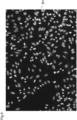
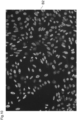
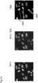
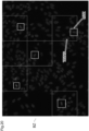
- the Figure 8 shows a fluorescence pattern image B superimposed from two color channels, which has a gray coloration representing a coloration of a cell substrate in a so-called red channel as a first color channel by a first fluorescent dye and also a superimposed coloration of the cell substrate in a so-called green channel as a second color channel by a second fluorescent dye.
- the cell substrate shown here comprising HEp cells, with a first fluorescent dye, preferably propidium iodide, certain cell areas are colored, as shown in image B1 in Figure 9
- the HEp cells are preferably so-called HEp2 cells.
- the second fluorescent dye is preferably fluorescein isothiocyanate (FITC).
- the coloring by the first fluorescent dye in the first image B1 allows a fundamental recognition of cell structures or cell areas. Furthermore, a differentiated pattern-like coloring of cell areas in the second image B2 of the green channel allows a differentiated recognition of the presence of fluorescence pattern types.
- the first image B1 and the second image B2 can be captured by means of at least one image capture unit in the form of one or more cameras K1 and K2 of a device V1 from Figure 6 captured.
- the images B1 and B2 can be spatially aligned with one another before further processing so that certain image areas of the first image B1 with their corresponding pixel indices correspond to certain image areas of the second image with the same pixel indices.
- the device V1 with regard to optical components and optical filters, to capture the two images B1 and B2 by means of a single image capture unit or a single camera which has a sensor which can capture images in the two color channels separately from one another, for example a color sensor.
- the different antinuclear antibody fluorescence pattern types include homogeneous patterns (AC-1), speckled patterns (AC-2, 4, 5), a centromere pattern (AC-3), nuclear dot patterns (AC-6,7), nucleolar patterns (AC-8, 9,10), as well as the nuclear peripheral fluorescence pattern type (AC-11,12).
- a further fluorescence pattern type class is the so-called negative class (AC-0) in the case of a patient sample without specific primary antinuclear antibodies.
- Image B from the Figure 8 is an exemplary fluorescence image for which the second image B2 from the green channel in the Figure 10 has a presence of both the homogeneous pattern (AC-1) and finely or coarsely speckled patterns (AC-4, AC-5).
- AC-1 homogeneous pattern
- AC-4, AC-5 finely or coarsely speckled patterns
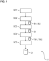
- a next step SC3 the respective image segments, each representing at least one mitotic cell, are detected on the basis of the first image or the first image information BI1. This results in so-called segmentation information SI.
- a step SC5 the respective actual presences of the respective antinuclear antibody fluorescence pattern types are then detected using a convolutional neural network CNN based on the selected partial images of the first image and the selected partial images of the second image.
- the detection result is then output as detection information DI.
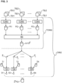
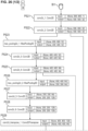
- Processing then takes place over several layers L1,..., LP of a convolutional neural network CNN1, where each layer L1,..., LP has at least one convolutional layer.
- a pooling layer PL or a pooling layer function PL which preferably performs a so-called global average pooling, to reduce a feature map FM1 to a single scalar value or an averaged value, which is preferably also called a logit, as a value LO1.
- the logit values LO1,.., LON of the N classes or fluorescence pattern types are then each individually subjected to a so-called sigmoid function SF in order to determine a respective prediction value or a respective confidence measure Pn with index n for a respective class n based on the respective feature map FMn with index n.
- This structure of the convolutional neural network therefore results in the advantage that there is a separate detection channel for each individual fluorescence pattern type and that not only a single pattern or a single pattern type can be detected as present, but also several pattern types at the same time.
- the part TCNN1 of the convolutional neural network CNN1 which is used to determine the confidence measures Pn based on the partial images TB11, TB12, can be applied separately to respective partial image tuples TB11, TB12, consisting of a first partial image of the first color channel TB11 and a corresponding second partial image of the second color channel TB12, as will be explained in detail later.
- the Figure 2 further shows further steps by means of which, on the basis of the confidence measures or prediction values, which can be summarized as prediction information PI1, and by means of which detection results or detection values D11,..., D1N can be determined, which in turn can be summarized as detection information DI1.
- a confidence measure Pn can then be calculated using a threshold function TS1 using a threshold value that can preferably be specified by a user or a threshold value that can be applied to other
- the Convolutional Neural Network CNN1 from Figure 2 has a partial convolutional neural network TCNN1, which will be used later in the Figure 3 applies.
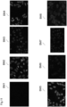
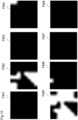
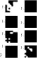
- the Figure 15 shows exemplary feature maps FM1,..., FM8 for the different fluorescence pattern types or example patterns, which are shown in Figure 11 and examples of the corresponding fluorescence pattern types from the Figure 24 with the corresponding labels.
- the feature map FM2 represents an activation with respect to a homogeneous pattern for the partial images TB11 and TB12 from the Figure 14
- the degree of brightness in the feature map FM2 indicates a probable presence of a homogeneous fluorescence pattern type in the partial image TB12.
- the feature map FM5 represents a probable presence of finely speckled or coarsely speckled patterns in the corresponding partial image TB12.
- the feature maps FM1,..., FM8 from the Figure 15 were originally determined with a resolution of 8x8 pixels and then interpolated to the resolution of 512x512 pixels of the second sub-image TB12.
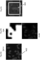
- the valid mitotic cells VMZ are the cells which are responsible for the detection of the homogeneous pattern according to the Figure 16c in the superimposed feature map OLM2. Even if in principle the interphase cells, which are in the superimposed feature map OLM5 from the Figure 16b were selected, a homogeneous staining may be present, but this is covered by the speckled staining of these interphase cells, so that a detection of a homogeneous pattern on these interphase cells alone from the Figure 16b in the superimposed feature map OLM5 cannot be done safely.
- the homogeneous pattern staining or the homogeneous fluorescence pattern type can advantageously be recognized precisely on these mitotic cells, as can be seen by a joint consideration of the superimposed feature map OLM2 from the Figure 16c and the segmented partial image TBS from the Figure 14 becomes apparent.
- the Figure 3 illustrates further advantageous embodiments of the method according to the invention.
- a partial image confidence measure PI corresponds to a vector P j with index j.
- This embodiment of the method according to the invention is advantageous because a convolutional neural network CNN2 does not simultaneously extract all information of an entire first and an entire second image B1, B2 from the Figures 9 and 10 all at once, but the partial image tuples can be evaluated individually in separate, separate processing paths.
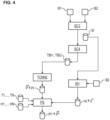
- the Figure 5 shows details of step SC3 from the Figure 1 for detecting image segments, each of which represents at least one mitotic cell of sufficient quality, based on the first image BI1.
- This is done using a segmentation convolutional neural network SEG-CNN.
- the segmentation convolutional neural network SEG-CNN has steps or layers LA1, ..., LAQ, which in turn can be understood as a convolutional neural network.
- the segmentation is therefore done using a another convolutional neural network SEG-CNN, which differs from the convolutional neural network CNN2 for detecting the respective actual presence of each of the cellular fluorescence pattern types using a convolutional neural network based on the selected partial images of the first image and the selected partial images of the second image.
- the division of the segmentation task and the detection task into two different convolutional neural networks is advantageous because the convolutional neural network for detection does not also have to be trained for the purpose of segmentation and thus the convolutional neural network can be trained particularly specifically for the purpose of segmentation.
- the first image e.g. image B1 from the Figure 9 , or the corresponding image information BI1 is fed to a Convolutional Neural Network CNNS for the purpose of segmentation.
- the segmentation convolutional neural network SEG-CNN is shown, which segments an input image using several layers LA1, LAQ.
- the image data BI1 is normalized to a value range from 0 to 1.
- a so-called downscaling also takes place in an optional step DWS.
- a first image of the image data BI1 has a dimensionality of 2400 ⁇ 1700 pixels. This first image can then be reduced to a size of 800 ⁇ 800 pixels by so-called downsampling.
- the further processing steps are set out here in detail for the example case in which a downscaling DWS of the first image was carried out to a size of 800 ⁇ 800 pixels.
- the segmentation convolutional neural network SEG-CNN can also be dimensioned in such a way that it can process the first image with a size of 2400x1700 pixels without downscaling.
- the convolutional neural network CNNS has several layers LA1,..., LAQ, each of these layers having at least one convolutional layer.
- the activation maps M1, ..., MS each preferably have the same size or resolution of preferably 800x800 pixels as the first image or the first image data BI1 after the downscaling DWS.
- Corresponding individual pixels PI1,...PIS of the activation maps M1,...MS each indicate the degree to which the pixel of the first image B1 or the first image information BI1 located at the same location belongs to the corresponding class.
- the x and y axis curves shown here indicate respective indices for the corresponding pixel positions within the probability maps PM1, ..., PMS.
- the probability maps PM1, ..., PMS preferably have the same size or resolution of preferably 800x800 pixels as the first image or the first image data BI1 after the downscaling DWS and as the activation maps M1, ..., MS.
- the corresponding pixel values of the corresponding pixel position PIX1 are linked across all probability maps PM1,..., PMS using an Argmax function.
- the segmentation map SM preferably has the same size or resolution of preferably 800 ⁇ 800 pixels as the first image or the first image data BI1 after the downscaling DWS, as do the activation maps M1, ..., MS and the probability maps PM1, ..., PMS.
- a so-called upscaling of the segmentation map SM from preferably 800x800 pixels back to the original image resolution of the image B1, for example 2400 x 1700 pixels, can then preferably be carried out in a preferably carried out step UP.
- the segmentation map SM can then be further processed to merge partial areas or segment areas using digital image processing in an image processing step BV using standard image processing measures such as eroding, delation and/or contour matching in order to then arrive at a modified segmentation map SM', which can be output as segmentation information SI.
- standard image processing measures such as eroding, delation and/or contour matching
- Such information with a segmentation SI is in the Figure 12a shown.
- the Figure 5 illustrates how the segmentation of the first image into image segments of different segment classes can be carried out, whereby the segment classes are as in the Figure 12b illustrated.
- at least a first segment class represents a cell stage of a cell and furthermore at least a second segment class represents a cell region within a cell.
- the segmentation information SI can then be used to determine at least one brightness value for at least one fluorescence pattern type based on one or more image segments of at least one specific segment class and to then verify at least one confidence measure of a fluorescence pattern type based on at least one such brightness value.
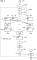
- the Figure 5 the possibility of extracting at least one brightness value for at least one fluorescence pattern type from the second image B2 based on the second image B2 and based on the segment information SI. This is done in a step SH for determining brightness.
- Brightness values determined in this way h ⁇ h 1 ⁇ h N brightness information HI can then be provided.
- the partial convolutional neural network TCNN2 uses those partial images or partial image information TBI1, TBI2 that was determined on the basis of the first image B1 using the segmentation step SC3 and the segmentation information SI obtained from it, as well as the second image B2.
- the partial convolutional neural network TCNN2 therefore uses those partial images that were selected from the first image B1 and the second image B2 using the segmentation step SC3 and the selection step SC4.
- a test step PS the confidence measure information PI is then verified on the basis of the brightness information Hl.
- the threshold values T1,..., TN are specified.
- the threshold values H1,..., HN are specified.
- the threshold values T1,..., TN can be specified by a user in order to influence the evaluation.
- the threshold values H1,..., HN can be specified by a user in order to influence the evaluation.
- the threshold values H1,..., HN are individual threshold values for each fluorescence pattern type.
- the procedure proposed here is advantageous because the confidence measures are initially determined on the basis of the first image and the second image or the corresponding partial images in which a convolutional neural network determines the confidence measures PI, but now brightness values HI are also explicitly determined from the second image or the entire second image, in particular without a reduction to partial images, in order to verify the confidence measure or the confidence measures PI.
- This enables even more precise detection of fluorescence pattern types.
- One advantage here is in particular that the entire second image B2 is considered with regard to brightness values in corresponding segment classes, so that not only the brightness of cells or segment areas of a individual partial images are considered, but also segment areas outside of selected partial images are taken into account.
- the Figure 2 shows preprocessing steps VS, VS1, VS2, which can be used for preprocessing the first partial image TB11 and the second partial image TB12.
- the first preprocessing step VS1 the first partial image TB11 and the second partial image TB12 are convolved as input feature maps with preferably eight different convolution kernels, whereby no striding is carried out and whereby the dimensionality of the partial images TB11, TB12 is maintained with an exemplary dimensionality of 512x512 image pixels.
- a so-called batch normalization is then carried out, whereby the number of feature maps, here for example eight feature maps, is retained.
- the Figure 25 shows an example of the first layer L1 from the Figure 2
- the preprocessing step VS2 is shown in dashed lines, since it is not part of this layer L1 but is only placed upstream of it.
- the feature maps from the preprocessing step VS2 are forwarded to the processing steps PS1, PS2, PS4.
- Each of these processing steps PS1, PS2, PS4 processes all the feature maps received from the step VS2 separately.
- a so-called max pooling is performed with a striding factor of 2 for each of the feature maps.
- step PS5 the 8 feature maps from step PS3 are concatenated with the 8 feature maps from step PS2.
- step PS6 the 16 incoming feature maps are again convolved with 11 kernels without striding.
- a so-called activation takes place, preferably in the form of a RELU activation.
- a so-called dropout with a dropout factor of 20% preferably occurs during the training phase. This dropout does not occur during the classification phase.
- step PS10 a convolution with 11 kernels without striding is performed.
- step PS10 The results of the feature maps or the 11 feature maps from step PS10 are added element by element with the 11 feature maps from step PS4 in step PS11, so that step PS11 generates 11 feature maps. No striding takes place here.
- step PS12 a so-called batch normalization takes place.
- a dropout preferably occurs during the training phase with a dropout factor of, for example, 20%.
- the last layer LP generates 56 (fifty-six) different such feature maps, in which case in a Figure 2
- the segmentation convolutional neural network SEG-CNN is shown, which uses the layers LA1, LAQ to segment the first image B1 or the image information BI1.
- the so-called downscaling takes place in an optional step DWS.
- the first image in the form of the image data BI1 has a dimensionality of 2400 ⁇ 1700 pixels.
- This first image can then be reduced to a size of 800 ⁇ 800 pixels by so-called downsampling.
- the further processing steps from the Figure 26 are explained in detail for the exemplary case that a downscaling DWS of the first image to a size of 800 ⁇ 800 pixels was carried out.
- the Convolutional Neural Network CNNS from the Figure 26 can also be dimensioned in such a way that it can process the first image with a size of 2400x1700 pixels without downscaling.
- step PS21 has a dimension of 800 ⁇ 800 pixels and the output variable of step PS21 also has a dimension of 800 ⁇ 800 pixels means that no striding occurs.
- the dimensionality of the input variable is indicated as "input” in brackets using the two numbers after the "None” specification.
- the dimensionality of the output variable is indicated as "output” in brackets using the two numbers after the "None” specification.
- the first image or image data BI1 is processed into the feature maps M1,.., MS, as already described in the Figure 5 illustrated.
- step PS29 performs a so-called deconvolution or transposed convolution.
- next steps either perform a folding, as already described in the description of the Figure 25 or a max pooling or a concatenation, as already described in the description of Figure 25 explained.
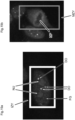
- FIG. 17b magnifies an interphase cell IZX as well as a mitotic cell MZX. It is clearly visible that the metaphase plate of the mitotic cell MZX is not significantly stained, i.e. the mitotic cell with its chromosomes in the metaphase plate is not stained. This information allows us to distinguish whether a so-called fine or coarse speckled pattern (AC-4, AC-5) is present here or whether perhaps a homogeneous pattern (AC-1) is present.
- AC-4, AC-5 fine or coarse speckled pattern
- AC-1 homogeneous pattern
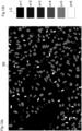
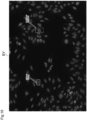
- the Figure 18 shows another example BY of an image in the second fluorescence channel or related to the second fluorescent dye highlighting a mitotic cell MZY as well as an interphase cell IZY.
- the Figure 20 shows another example of a fluorescence image BZ, in which a partial image area TBZ12 has a mitotic cell MZZ.
- the Figure 21 shows the exemplary partial image TBZ11 as the first partial image of the first fluorescent dye and the second partial image TBZ12, which corresponds to the first partial image TBZ11, from the second fluorescent color channel. Furthermore, the Figure 21 the corresponding segmented image TBZS.
- the method according to the invention has been described here by preparing in a step SC2 from the Figure 1 the first and the second image are captured. Therefore, a method for detecting respective potential presences of respective different cellular fluorescence pattern types on a biological cell substrate comprising human epithelioma cells by means of digital image processing is proposed, which begins with the step of capturing the first image and the second image.
- a method for digital image processing can also be carried out correspondingly and in an analogous manner, in which such a first image and such a second image are provided or received in a corresponding step and in which the further steps SC3, SC4, SC5 are then carried out.
- the Figure 7A a proposed computing unit R, which receives a corresponding first image or a first image information BI1 as well as a corresponding second image or a corresponding second image information BI2 in the form of a preferably data signal SIG via an interface DS2.
- the computing unit R can then output the determined confidence measures PI, the verified confidence measures PI* and/or the detection information DI preferably via an output interface AS, in particular to a display unit AE.
- This information can preferably also be output via a data interface DS3 in the form of a data network interface in the form of a signal SI3.
- the Figure 7B shows a proposed data network device which receives the images or the image information B11, BI2 via an interface S4 and a corresponding data signal SIG1.
- the data network device also preferably has an internal data bus IDB which connects a previously described computing unit R to preferably a memory unit MEM.
- the Figure 7C illustrates a proposed computer program product CPP, which comprises instructions which, when the program is executed by a computer, cause the computer to carry out the proposed method for digital image processing.
- This computer program product can, for example, be transmitted in the proposed manner via a data signal SIG2 can be provided or transmitted. This transmission can be made to a computer CO or to a data point DSX of the computer CO.
- a computing unit R is designed to receive the first fluorescence image in the form of digital image data BI1.
- the computing unit R is also designed to receive the second fluorescence image in the form of digital image data BI2.
- the computing unit R is also designed to detect respective image segments on the basis of the first image, each of which represents at least one mitotic cell, to select partial images of the first image and corresponding partial images of the second image on the basis of the detected image segments, and to detect respective actual presences of respective cellular fluorescence pattern types using a convolutional neural network on the basis of the selected partial images of the first image and the selected partial images of the second image.
- aspects have been described in connection with a device, it is understood that these aspects also represent a description of the corresponding methods, so that a block or a component of a device can also be understood as a corresponding method step or as a feature of a method step. Analogously, aspects that have been described in connection with or as a method step also represent a description of a corresponding block or detail or feature of a corresponding device.
- embodiments of the invention can implement the computing unit R or the data network device DV in hardware and/or in software.
- An implementation of a computing unit R mentioned here can take place here as at least one computing unit or by several computing units in a network.
- the implementation can be carried out using a digital storage medium, for example a floppy disk, a DVD, a Blu-ray disc, a CD, a ROM, a PROM, an EPROM, an EEPROM or a FLASH memory, a hard disk or another magnetic or optical memory on which electronically readable control signals are stored, which can interact or interact with a programmable hardware component in such a way that the respective method is carried out.
- the digital storage medium can therefore be machine- or computer-readable.
- Some embodiments thus comprise a data carrier that has electronically readable control signals that are capable of interacting with a programmable computer system or a programmable hardware component such that one of the methods described herein is carried out.
- embodiments or parts of the embodiments of the present invention may be implemented as a program, firmware, computer program or computer program product with a program code or as data, wherein the program code or the data is or are effective to carry out one of the methods or a part of a procedure when the program runs on a processor or a programmable hardware component.
- a specialist can use a so-called open source deep learning library called "Keras".
- Keras open source deep learning library
- the specified system or the method according to the invention was tested on 161 different patient samples, whereby the samples were diluted in a linear dilution series starting from 1:40. This means that the patient samples were diluted and incubated in a linearly increasing sequence starting from 1:40. In this case, samples were diluted in steps of either 1:40, 1:80, 1:640, 1:1280 or 1:40, 1:80, 1:160, 1:320, 1:640, 1:1280, 1:2560, 1:5120, 1:10240.
- the system is in no way limited to specified dilution series.
- a pattern was declared present by an expert for a specific sample if it was recognized as present in any dilution or fluorescence image for the specific sample. The pattern did not have to be recognized by the expert in all dilutions or all fluorescence images for the specific sample to be declared present.
- a pattern was declared negative (showing no pattern) by an expert for a specific sample if it was identified as negative (showing no pattern) in all dilutions or in all fluorescence images for the specific sample.
- a pattern was detected as generally present by the method according to the invention with respect to a specific sample if it was detected as present in any dilution or any fluorescence image with respect to the specific sample.
- the pattern did not have to be detected by the method according to the invention in all dilutions or all fluorescence images with respect to the specific sample in order to be generally detected as present.
- a pattern was detected as negative (having no pattern) by the method according to the invention with respect to a specific sample if it was detected as negative (having no pattern) in all dilutions or all fluorescence images with respect to the specific sample.
- both patterns will be output for this sample.
- the convolutional neural network for detecting the presence of patterns used a total of 5949 sub-image tuples for training, of which 25% of the respective pattern types were used for separate validation.
- a total of 6463 images were available for the convolutional neural network for segmentation, of which 4847 were in the actual training set and 1616 in a validation set.
Landscapes
- Engineering & Computer Science (AREA)
- Health & Medical Sciences (AREA)
- Life Sciences & Earth Sciences (AREA)
- Physics & Mathematics (AREA)
- General Physics & Mathematics (AREA)
- Theoretical Computer Science (AREA)
- General Health & Medical Sciences (AREA)
- Biomedical Technology (AREA)
- Molecular Biology (AREA)
- Immunology (AREA)
- Chemical & Material Sciences (AREA)
- Hematology (AREA)
- Urology & Nephrology (AREA)
- Pathology (AREA)
- Computer Vision & Pattern Recognition (AREA)
- Analytical Chemistry (AREA)
- Biochemistry (AREA)
- Artificial Intelligence (AREA)
- Nuclear Medicine, Radiotherapy & Molecular Imaging (AREA)
- Software Systems (AREA)
- Mathematical Physics (AREA)
- Biophysics (AREA)
- Computational Linguistics (AREA)
- Data Mining & Analysis (AREA)
- Evolutionary Computation (AREA)
- Computing Systems (AREA)
- General Engineering & Computer Science (AREA)
- Cell Biology (AREA)
- Microbiology (AREA)
- Food Science & Technology (AREA)
- Medicinal Chemistry (AREA)
- Biotechnology (AREA)
- Medical Informatics (AREA)
- Radiology & Medical Imaging (AREA)
- Quality & Reliability (AREA)
- Zoology (AREA)
- Tropical Medicine & Parasitology (AREA)
- Virology (AREA)
- Dispersion Chemistry (AREA)
- Image Analysis (AREA)
Claims (9)
- Procédé de détection de présences potentielles respectives de différents types de motifs de fluorescence cellulaire respectifs sur un substrat cellulaire biologique (SU) comportant des cellules d'épithéliome humain,les types de motifs de fluorescence cellulaire comprenant plusieurs types différents de motifs de fluorescence d'anticorps antinucléaires,le procédé comportant les étapes suivantes :- faire incuber le substrat cellulaire (SU) avec un échantillon de patient liquide qui contient potentiellement des anticorps primaires, en outre avec un premier colorant fluorescent et en outre avec des anticorps secondaires qui sont marqués avec un deuxième colorant fluorescent,- capturer une première image (B1), qui représente une coloration du substrat cellulaire par le premier colorant fluorescent, et capturer une deuxième image (B2), qui représente une coloration du substrat cellulaire par le deuxième colorant fluorescent,- détecter des segments d'image respectifs (VMZ), qui représentent chacun au moins une cellule mitotique (MZX, MZY) de qualité suffisante, sur la base de la première image (B1), une cellule mitotique présentant une qualité suffisante lorsqu'elle est en métaphase,- sélectionner des images partielles (TB11) de la première image (B1) et des images partielles correspondantes (TB12) de la deuxième image (B2) sur la base des segments d'image détectés (VMZ), qui représentent chacun au moins une cellule mitotique (MZX, MZY) de qualité suffisante, et- détecter des présences réelles respectives des types de motifs de fluorescence cellulaire respectifs à l'aide d'un réseau neuronal convolutif (CNN2) sur la base des images partielles sélectionnées (TB11) de la première image (B1) et des images partielles sélectionnées (TB12) de la deuxième image (B2),caractérisé par- déterminer des mesures de confiance respectives (PI, PI*) pour les présences réelles respectives des types de motifs de fluorescence respectifs à l'aide du réseau neuronal convolutif (CNN2) sur la base des images partielles sélectionnées (TB11) de la première image (B1) et des images partielles sélectionnées (TB12) de la deuxième image (B2),- une mesure de confiance d'image partielle respective (PI1) pour les présences d'images partielles réelles respectives de types de motifs de fluorescence cellulaire respectifs étant déterminée pour un uplet d'images partielles respectif au moyen du réseau neuronal convolutif (CNN2), un uplet d'images partielles comportant une image partielle (TB11) de la première image (B1) et une image partielle correspondante (TB12) de la deuxième image (B2) et une mesure de confiance d'image partielle correspondant à un vecteur de mesures de confiance, un type de motif de fluorescence respectif correspondant à une mesure de confiance respective,- le réseau neuronal convolutif évaluant simultanément l'image partielle de la première image (B1) et l'image partielle correspondante de la deuxième image (B2),- et déterminer des mesures de confiance respectives pour les présences réelles respectives des types de motifs de fluorescence respectifs sur la base des mesures de confiance d'image partielle.
- Procédé selon la revendication 1,le réseau neuronal convolutif (CNN2) comportant une couche de sortie (LP, AVS) qui génère une carte de caractéristiques respective (FM1,..., FMN) pour un type de motif de fluorescence cellulaire respectif,et le réseau neuronal convolutif (CNN2) déterminant une mesure de confiance respective (P1,..., PN) sur la base d'une carte de caractéristiques respective (FM1,..., FMN).
- Procédé selon la revendication 1,
ledit procédé comprenant en outre les étapes suivantes- segmenter la première image (B1) en segments d'image de différentes classes de segments,- déterminer au moins une valeur de luminosité (Hl) pour au moins un type de motif de fluorescence sur la base d'un ou plusieurs segments d'image d'au moins une classe de segments déterminée et- vérifier la mesure de confiance (PI, PI*) pour la présence réelle dudit au moins un type de motif de fluorescence sur la base de la valeur de luminosité (HI) dudit au moins un type de motif de fluorescence. - Procédé selon la revendication 3,
la vérification de la mesure de confiance (PI, PI*) étant effectuée sur la base de la valeur de luminosité (Hl) et en fonction d'une valeur seuil (H1,..., HN) pouvant être spécifiée par un utilisateur. - Procédé selon la revendication 1,
ledit procédé comportant en outre les étapes suivantes- diviser la deuxième image (B2) en un ensemble d'images partielles selon un schéma de division spécifié,- sélectionner des images partielles (TB12) de la deuxième image (B2) sur la base des segments d'image détectés et sélectionner des images partielles correspondantes (TB11) de la première image (B1)- et détecter des présences réelles respectives de types de motifs de fluorescence cellulaire respectifs au moyen du réseau neuronal convolutif (CNN2) sur la base des images partielles sélectionnées (TB12) de la deuxième image (B2) et sur la base des images partielles sélectionnées (TB11) de la première image (B1). - Unité de calcul (R) destinée à détecter les présences potentielles respectives de différents types de motifs de fluorescence cellulaire respectifs, les types de motifs de fluorescence cellulaire comprenant plusieurs types différents de motifs de fluorescence d'anticorps antinucléaires, laquelle unité de calcul est conçue pour, au cours d'un traitement d'image numérique,- recevoir une première image (B1), qui représente une coloration d'un substrat cellulaire biologique par un premier colorant fluorescent, le substrat cellulaire biologique comportant des cellules d'épithéliome humain,- recevoir une deuxième image (B2), qui représente une coloration du substrat cellulaire biologique par un deuxième colorant fluorescent,- détecter, sur la base de la première image (B1), des segments d'image respectifs (VMZ), qui représentent chacun au moins une cellule mitotique de qualité suffisante, une cellule mitotique présentant une qualité suffisante lorsqu'elle est en métaphase,- sélectionner, sur la base des segments d'image détectés (VMZ), des images partielles (TB11) de la première image (B1) et des images partielles correspondantes (TB12) de la deuxième image (B2), chacune d'elles représentant au moins une cellule mitotique (MZX, MZY) de qualité suffisante, et- détecter, sur la base des images partielles sélectionnées (TB11) de la première image (B1) et des images partielles sélectionnées (TB12) de la deuxième image (B2), des présences réelles respectives des types de motifs de fluorescence cellulaire respectifs à l'aide d'un réseau neuronal convolutif (CNN2),
caractérisée en ce que l'unité de calcul est en outre conçue pour- déterminer des mesures de confiance respectives (PI, PI*) pour les présences réelles respectives des types de motifs de fluorescence respectifs à l'aide du réseau neuronal convolutif (CNN2) sur la base des images partielles sélectionnées (TB11) de la première image (B1) et des images partielles sélectionnées (TB12) de la deuxième image (B2),- déterminer, pour un uplet d'images partielles respectif, une mesure de confiance d'image partielle respective (PI1) pour des présences d'images partielles réelles respectives de types de motifs de fluorescence cellulaire respectifs à l'aide du réseau neuronal convolutif (CNN2), un uplet d'images partielles comportant une image partielle (TB11) de la première image (B1) et une image partielle correspondante (TB12) de la deuxième image (B2) et une mesure de confiance d'image partielle correspondant à un vecteur de mesures de confiance, un type de motif de fluorescence respectif correspondant à une mesure de confiance respective,- le réseau neuronal convolutif évaluant simultanément l'image partielle de la première image (B1) et l'image partielle correspondante de la deuxième image (B2),- et déterminer des mesures de confiance respectives pour les présences réelles respectives des types de motifs de fluorescence respectifs sur la base des mesures de confiance d'images partielles. - Dispositif de réseau de données (DV), comportantau moins une interface de données (DS4) destinée à recevoir une première image (B1), qui représente une coloration d'un substrat cellulaire biologique par un premier colorant fluorescent, le substrat cellulaire biologique comportant des cellules d'épithéliome humain, et recevoir une deuxième image (B2), qui représente une coloration du substrat cellulaire biologique par un deuxième colorant fluorescent,caractérisé par une unité de calcul (R) selon la revendication 6.
- Produit-programme informatique (CPP), comprenant des instructions qui, lorsque le programme est exécuté par un ordinateur (CO), amènent celui-ci à mettre en oeuvre un procédé de traitement d'images numériques destiné à détecter des présences potentielles respectives de différents types de motifs de fluorescence cellulaire respectifs,
les types de motifs de fluorescence cellulaire comprenant plusieurs types différents de motifs de fluorescence d'anticorps antinucléaires :ledit procédé comportant les étapes suivantes- recevoir une première image (B1), qui représente une coloration d'un substrat cellulaire biologique par un premier colorant fluorescent, le substrat cellulaire biologique comportant des cellules d'épithéliome humain,- recevoir une deuxième image (B2), qui représente une coloration du substrat cellulaire biologique par un deuxième colorant fluorescent,- détecter des segments d'image respectifs (VMZ), qui représentent chacun au moins une cellule mitotique (MZX, MZY) de qualité suffisante, sur la base de la première image (B1), une cellule mitotique présentant une qualité suffisante lorsqu'elle est en métaphase,- sélectionner des images partielles (TB11) de la première image (B1) et des images partielles correspondantes (TB12) de la deuxième image (B2) sur la base des segments d'image détectés (VMZ), qui représentent chacun au moins une cellule mitotique (MZX, MZY) de qualité suffisante, et- détecter des présences réelles respectives des types de motifs de fluorescence cellulaire respectifs à l'aide d'un réseau neuronal convolutif (CNN2) sur la base des images partielles sélectionnées (TB11) de la première image (B1) et des images partielles sélectionnées (TB12) de la deuxième image (B2),caractérisé par les étapes suivantes- déterminer des mesures de confiance respectives (PI, PI*) pour les présences réelles respectives des types de motifs de fluorescence respectifs à l'aide du réseau neuronal convolutif (CNN2) sur la base des images partielles sélectionnées (TB11) de la première image (B1) et des images partielles sélectionnées (TB12) de la deuxième image (B2),- déterminer, pour un uplet d'images partielles respectif, une mesure de confiance d'image partielle respective (PI1) pour des présences d'images partielles réelles respectives de types de motifs de fluorescence cellulaire respectifs à l'aide du réseau neuronal convolutif (CNN2), un uplet d'images partielles comportant une image partielle (TB11) de la première image (B1) et une image partielle correspondante (TB12) de la deuxième image (B2), et une mesure de confiance d'image partielle correspondant à un vecteur de mesures de confiance, un type de motif de fluorescence respectif correspondant à une mesure de confiance respective,- le réseau neuronal convolutif évaluant simultanément l'image partielle de la première image (B1) et l'image partielle correspondante de la deuxième image (B2),- et déterminer des mesures de confiance respectives pour les présences réelles respectives des types de motifs de fluorescence respectifs sur la base des mesures de confiance d'image partielle. - Signal porteur de données (SIG2), lequel transmet le produit-programme informatique (CPP) selon la revendication 8.
Priority Applications (3)
| Application Number | Priority Date | Filing Date | Title |
|---|---|---|---|
| EP19187348.8A EP3767587B1 (fr) | 2019-07-19 | 2019-07-19 | Détection de la présence des différents types de motifs fluorescents d'anticorps antinucléaires et dispositif associé |
| US16/946,733 US11367187B2 (en) | 2019-07-19 | 2020-07-02 | Method for detecting the presence of different antinuclear antibody fluorescence pattern types and apparatus for this purpose |
| CN202010677726.4A CN112240878B (zh) | 2019-07-19 | 2020-07-14 | 用于检测不同抗核抗体荧光图案类型的存在的方法和装置 |
Applications Claiming Priority (1)
| Application Number | Priority Date | Filing Date | Title |
|---|---|---|---|
| EP19187348.8A EP3767587B1 (fr) | 2019-07-19 | 2019-07-19 | Détection de la présence des différents types de motifs fluorescents d'anticorps antinucléaires et dispositif associé |
Publications (2)
| Publication Number | Publication Date |
|---|---|
| EP3767587A1 EP3767587A1 (fr) | 2021-01-20 |
| EP3767587B1 true EP3767587B1 (fr) | 2024-10-23 |
Family
ID=67438205
Family Applications (1)
| Application Number | Title | Priority Date | Filing Date |
|---|---|---|---|
| EP19187348.8A Active EP3767587B1 (fr) | 2019-07-19 | 2019-07-19 | Détection de la présence des différents types de motifs fluorescents d'anticorps antinucléaires et dispositif associé |
Country Status (3)
| Country | Link |
|---|---|
| US (1) | US11367187B2 (fr) |
| EP (1) | EP3767587B1 (fr) |
| CN (1) | CN112240878B (fr) |
Families Citing this family (8)
| Publication number | Priority date | Publication date | Assignee | Title |
|---|---|---|---|---|
| EP3712618B1 (fr) * | 2019-03-18 | 2023-10-04 | Euroimmun Medizinische Labordiagnostika AG | Procédé de détection d'une liaison d'anticorps d'un échantillon de patient à l'adn double brin à l'aide des cellules de crithidia luciliae et microscopie par fluorescence |
| EP3876193A1 (fr) | 2020-03-02 | 2021-09-08 | Euroimmun Medizinische Labordiagnostika AG | Procédé de traitement des images permettant d'afficher des cellules d'une pluralité d'images complètes |
| EP3971827B1 (fr) | 2020-09-17 | 2023-12-27 | Euroimmun Medizinische Labordiagnostika AG | Procédé de détection d'une liaison d'anticorps d'un échantillon de patient à l'adn double brin à l'aide des cellules de crithidia luciliae et microscopie par fluorescence |
| EP4016081B1 (fr) * | 2020-12-21 | 2023-02-22 | Euroimmun Medizinische Labordiagnostika AG | Procédé et dispositif de détection d'une présence d'un type de motif fluorescent sur une section d'organes au moyen d'une microscopie immunofluorescente |
| RU2764705C1 (ru) | 2020-12-22 | 2022-01-19 | Общество с ограниченной ответственностью «Аби Продакшн» | Извлечение нескольких документов из единого изображения |
| EP4030340B1 (fr) * | 2021-01-19 | 2023-11-01 | EUROIMMUN Medizinische Labordiagnostika AG | Procédé de détection des présences de différents types de motifs fluorescents antinucléaires d'anticorps sans contre-marquage et dispositif associé |
| JP7834528B2 (ja) * | 2022-03-24 | 2026-03-24 | 株式会社東芝 | 微小粒子計測システム、および、微小粒子計測方法 |
| EP4345775B1 (fr) * | 2022-09-30 | 2025-08-06 | Euroimmun Medizinische Labordiagnostika AG | Procédé de détection d'au moins un motif fluorescent sur une image immunofluorescente d'un substrat de cellule biologique |
Family Cites Families (14)
| Publication number | Priority date | Publication date | Assignee | Title |
|---|---|---|---|---|
| DE102006027516B4 (de) * | 2006-06-09 | 2021-10-07 | Euroimmun Medizinische Labordiagnostika Ag | Verfahren zur Optimierung der automatischen Fluoreszenzmustererkennung in der Immundiagnostik |
| EP1921552A1 (fr) | 2006-11-13 | 2008-05-14 | Euroimmun Medizinische Labordiagnostika AG | Procédé destiné à assurer l'authenticité et la qualité de diagnostiques acquis visuellement par des tests d'immunofluorescence indirecte via un réseau |
| US8160364B2 (en) * | 2007-02-16 | 2012-04-17 | Raytheon Company | System and method for image registration based on variable region of interest |
| WO2010140024A1 (fr) * | 2009-06-03 | 2010-12-09 | Institut National De La Sante Et De La Recherche Medicale (Inserm) | Procédés de diagnostic et de traitement d'une maladie rénale chez un individu |
| US20150032671A9 (en) * | 2010-07-23 | 2015-01-29 | General Electric Company | Systems and methods for selecting and analyzing particles in a biological tissue |
| US8639013B2 (en) * | 2011-08-17 | 2014-01-28 | General Electric Company | System and methods for generating a brightfield image using fluorescent images |
| WO2014177700A1 (fr) * | 2013-05-02 | 2014-11-06 | Universite D'aix-Marseille | Procédé d'immunofluorescence indirecte pur détecter des auto-anticorps anti-nucléaires |
| US9536304B2 (en) * | 2013-08-30 | 2017-01-03 | Dairy Quality Inc. | Determining pathogens based on an image of somatic cells in a fluid sample |
| JP6703824B2 (ja) * | 2015-11-30 | 2020-06-03 | シスメックス株式会社 | 細胞選択方法、細胞検出方法、細胞選択装置、および細胞検出装置 |
| WO2017212055A1 (fr) * | 2016-06-10 | 2017-12-14 | F. Hoffmann-La Roche Ag | Système de simulation d'images en champ lumineux |
| EP3591406A1 (fr) | 2018-07-06 | 2020-01-08 | Euroimmun Medizinische Labordiagnostika AG | Dispositif et procédé de détection des anticorps |
| EP3712618B1 (fr) * | 2019-03-18 | 2023-10-04 | Euroimmun Medizinische Labordiagnostika AG | Procédé de détection d'une liaison d'anticorps d'un échantillon de patient à l'adn double brin à l'aide des cellules de crithidia luciliae et microscopie par fluorescence |
| JP7475848B2 (ja) * | 2019-11-29 | 2024-04-30 | シスメックス株式会社 | 細胞解析方法、細胞解析装置、細胞解析システム、及び細胞解析プログラム、並びに訓練された人工知能アルゴリズムの生成方法、生成装置、及び生成プログラム |
| JP7545202B2 (ja) * | 2019-11-29 | 2024-09-04 | シスメックス株式会社 | 細胞解析方法、細胞解析装置、細胞解析システム、及び細胞解析プログラム |
-
2019
- 2019-07-19 EP EP19187348.8A patent/EP3767587B1/fr active Active
-
2020
- 2020-07-02 US US16/946,733 patent/US11367187B2/en active Active
- 2020-07-14 CN CN202010677726.4A patent/CN112240878B/zh active Active
Non-Patent Citations (13)
| Title |
|---|
| "Precision Medicine - Role ofAutoantibodies in the Prediction and Care of Autoimmune Diseases Report on the 14th Dresden Symposium on Autoantibodies, September 10-13, 2019", 1 January 2019, PABST SCIENCE PUBLISHERS, article RICO HIEMANN ET AL: "Artificial intelligence for automated pattern recognition", pages: 415 - 416, XP055864591 * |
| CASCIO DONATO ET AL: "Deep Convolutional Neural Network for HEp-2 Fluorescence Intensity Classification", APPLIED SCIENCES, vol. 9, no. 3, 26 January 2019 (2019-01-26), pages 408, XP055864499, DOI: 10.3390/app9030408 * |
| CONRAD KARSTEN: "14 th DRESDEN SYMPOSIUM ON AUTOANTIBODIES CALL FOR ABSTRACTS -DEADLINE JUNE 10, 2019", 10 June 2019 (2019-06-10), pages 1 - 2, XP055886226, Retrieved from the Internet <URL:chrome-extension://efaidnbmnnnibpcajpcglclefindmkaj/viewer.html?pdfurl=https%3A%2F%2Fwww.gfid-ev.de%2Fformulare%2F14DSA_abstract.pdf&clen=207066&chunk=true> * |
| DIVYA BS ET AL: "HEp-2 cell classification using artificial neural network approach", 2016 23RD INTERNATIONAL CONFERENCE ON PATTERN RECOGNITION (ICPR), IEEE, 4 December 2016 (2016-12-04), pages 84 - 89, XP033085564, DOI: 10.1109/ICPR.2016.7899613 * |
| EGERER KARL ET AL: "Automated evaluation of autoantibodies on human epithelial-2 cells as an approach to standardize cell-based immunofluorescence tests", ARTHRITIS RESEARCH AND THERAPY, BIOMED CENTRAL, LONDON, GB, vol. 12, no. 2, 9 March 2010 (2010-03-09), pages R40, XP021085168, ISSN: 1478-6354, DOI: 10.1186/AR2949 * |
| GAO ZHIMIN ET AL: "HEp-2 Cell Image Classification with Deep Convolutional Neural Networks", FACULTY OF ENGINEERING AND INFORMATION SCIENCES -PAPERS: PART B, 1 January 2017 (2017-01-01), pages 1 - 32, XP055864511, Retrieved from the Internet <URL:https://ro.uow.edu.au/cgi/viewcontent.cgi?referer=https://www.google.com/&httpsredir=1&article=1154&context=eispapers1> [retrieved on 20211123] * |
| GUPTA KRATI ET AL: "CNN based Mitotic HEp-2 Cell Image Detection :", PROCEEDINGS OF THE 11TH INTERNATIONAL JOINT CONFERENCE ON BIOMEDICAL ENGINEERING SYSTEMS AND TECHNOLOGIES, 1 January 2018 (2018-01-01), pages 167 - 174, XP055864490, ISBN: 978-989-7583-07-0, Retrieved from the Internet <URL:https://pdfs.semanticscholar.org/7921/a9e39138d63d69e479c24bd5a718a564db82.pdf?_ga=2.78673044.1673975225.1637658002-1613779853.1637242792> DOI: 10.5220/0006721501670174 * |
| INFANTINO M ET AL: "The burden of the variability introduced by the HEp-2 assay kit and the CAD system in ANA indirect immunofluorescence test", IMMUNOLOGIC RESEARCH, HUMANA PRESS, INC, US, vol. 65, no. 1, 25 July 2016 (2016-07-25), pages 345 - 354, XP036222202, ISSN: 0257-277X, [retrieved on 20160725], DOI: 10.1007/S12026-016-8845-3 * |
| ORAIBI ZAKARIYA A ET AL: "Learning Local and Deep Features for Efficient Cell Image Classification Using Random Forests", 2018 25TH IEEE INTERNATIONAL CONFERENCE ON IMAGE PROCESSING (ICIP), IEEE, 7 October 2018 (2018-10-07), pages 2446 - 2450, XP033454860, DOI: 10.1109/ICIP.2018.8451287 * |
| PETTER STRANDMARK ET AL: "HEp-2 staining pattern classification", PATTERN RECOGNITION (ICPR), 2012 21ST INTERNATIONAL CONFERENCE ON, IEEE, 11 November 2012 (2012-11-11), pages 33 - 36, XP032329262, ISBN: 978-1-4673-2216-4 * |
| RICO HIEMANN1 ET AL: "Die HEp-2-Zelle als Target fur multiparametrische Autoantikorperanalytik - Automatisierung und Standardisierung1) The HEp-2 cell as target for multiparametric autoantibody analyses:automation and standardisation", J LAB MED, vol. 35, no. 6, 1 January 2011 (2011-01-01), pages 351 - 361, XP055864482 * |
| WAN SHENG ET AL: "Confnet: Predict with Confidence", 2018 IEEE INTERNATIONAL CONFERENCE ON ACOUSTICS, SPEECH AND SIGNAL PROCESSING (ICASSP), IEEE, 15 April 2018 (2018-04-15), pages 2921 - 2925, XP033401070, DOI: 10.1109/ICASSP.2018.8461745 * |
| XI JIA ET AL: "Deep convolutional neural network based HEp-2 cell classification", 2016 23RD INTERNATIONAL CONFERENCE ON PATTERN RECOGNITION (ICPR), IEEE, 4 December 2016 (2016-12-04), pages 77 - 80, XP033085562, DOI: 10.1109/ICPR.2016.7899611 * |
Also Published As
| Publication number | Publication date |
|---|---|
| US11367187B2 (en) | 2022-06-21 |
| EP3767587A1 (fr) | 2021-01-20 |
| CN112240878B (zh) | 2023-05-26 |
| CN112240878A (zh) | 2021-01-19 |
| US20210019883A1 (en) | 2021-01-21 |
Similar Documents
| Publication | Publication Date | Title |
|---|---|---|
| EP3767587B1 (fr) | Détection de la présence des différents types de motifs fluorescents d'anticorps antinucléaires et dispositif associé | |
| EP3712618B1 (fr) | Procédé de détection d'une liaison d'anticorps d'un échantillon de patient à l'adn double brin à l'aide des cellules de crithidia luciliae et microscopie par fluorescence | |
| EP3971827B1 (fr) | Procédé de détection d'une liaison d'anticorps d'un échantillon de patient à l'adn double brin à l'aide des cellules de crithidia luciliae et microscopie par fluorescence | |
| Chen et al. | Deep learning based automatic immune cell detection for immunohistochemistry images | |
| DE102017100396A1 (de) | Auf Straßenszenenbilder angewendete Klassifikatoren für hohe und niedrige Wiedergabetreue | |
| DE60226043T2 (de) | Verfahren für quantitative video-mikroskopie und vorrichtung und programm zur durchführung des verfahrens | |
| DE102018114005A1 (de) | Materialprüfung von optischen Prüflingen | |
| EP4016082B1 (fr) | Procédé et dispositif de détection d'une présence d'un type de motif fluorescent sur une section d'organe par microscopie immunofluorescente | |
| EP2400458B1 (fr) | Procédé et dispositif de segmentation de cellules biologiques dans une image | |
| DE112019005143T5 (de) | System zur co-registrierung medizinischer bilder unter verwendung eines klassifikators | |
| JP6924761B2 (ja) | 異なる取得性質を持つ画像を分離するシステム及び方法 | |
| DE102020126554A1 (de) | Mikroskopiesystem und verfahren zum überprüfen von eingabedaten | |
| DE102010063965A1 (de) | Vorrichtung und Verfahren zur Bestimmung von Objekten einer Farbaufnahme | |
| EP4030340B1 (fr) | Procédé de détection des présences de différents types de motifs fluorescents antinucléaires d'anticorps sans contre-marquage et dispositif associé | |
| DE102022121543A1 (de) | Mikroskopiesystem und Verfahren zur Qualitätsüberprüfung eines maschinell gelernten Bildverarbeitungsmodells | |
| EP4345658B1 (fr) | Procédé et dispositif de détection d'une présence d'un motif fluorescent sur une image immunofluorescente d'un substrat de cellule biologique | |
| EP2440920B1 (fr) | Localisation d'une région saine d'un frottis sanguin | |
| DE102022131444A1 (de) | Verfahren zum Identifizieren von Analyten in einer Bildfolge | |
| EP3948649B1 (fr) | Masquage d'objets contenus dans une image | |
| EP4345775B1 (fr) | Procédé de détection d'au moins un motif fluorescent sur une image immunofluorescente d'un substrat de cellule biologique | |
| DE102023123114A1 (de) | Verfahren und Vorrichtung zur semantischen Segmentierung und Clutter-Erkennung | |
| DE102022209113A1 (de) | Training von instanzsegmentierungsalgorithmen mit partiell annotierten bildern | |
| DE102022201258A1 (de) | Verfahren zur Separierung von Objekten | |
| EP4625019A1 (fr) | Procédé de détermination d'une mesure de focalisation d'une image de microscope | |
| DE102024211449A1 (de) | Steuereinheit zum Segmentieren wenigstens eines in einer Probe vorhandenen roten Blutkörperchens in einer Vorrichtung |
Legal Events
| Date | Code | Title | Description |
|---|---|---|---|
| PUAI | Public reference made under article 153(3) epc to a published international application that has entered the european phase |
Free format text: ORIGINAL CODE: 0009012 |
|
| STAA | Information on the status of an ep patent application or granted ep patent |
Free format text: STATUS: THE APPLICATION HAS BEEN PUBLISHED |
|
| AK | Designated contracting states |
Kind code of ref document: A1 Designated state(s): AL AT BE BG CH CY CZ DE DK EE ES FI FR GB GR HR HU IE IS IT LI LT LU LV MC MK MT NL NO PL PT RO RS SE SI SK SM TR |
|
| AX | Request for extension of the european patent |
Extension state: BA ME |
|
| STAA | Information on the status of an ep patent application or granted ep patent |
Free format text: STATUS: REQUEST FOR EXAMINATION WAS MADE |
|
| 17P | Request for examination filed |
Effective date: 20210720 |
|
| RBV | Designated contracting states (corrected) |
Designated state(s): AL AT BE BG CH CY CZ DE DK EE ES FI FR GB GR HR HU IE IS IT LI LT LU LV MC MK MT NL NO PL PT RO RS SE SI SK SM TR |
|
| TPAC | Observations filed by third parties |
Free format text: ORIGINAL CODE: EPIDOSNTIPA |
|
| STAA | Information on the status of an ep patent application or granted ep patent |
Free format text: STATUS: EXAMINATION IS IN PROGRESS |
|
| 17Q | First examination report despatched |
Effective date: 20220214 |
|
| GRAP | Despatch of communication of intention to grant a patent |
Free format text: ORIGINAL CODE: EPIDOSNIGR1 |
|
| STAA | Information on the status of an ep patent application or granted ep patent |
Free format text: STATUS: GRANT OF PATENT IS INTENDED |
|
| INTG | Intention to grant announced |
Effective date: 20240619 |
|
| P01 | Opt-out of the competence of the unified patent court (upc) registered |
Free format text: CASE NUMBER: APP_41124/2024 Effective date: 20240711 |
|
| GRAS | Grant fee paid |
Free format text: ORIGINAL CODE: EPIDOSNIGR3 |
|
| GRAA | (expected) grant |
Free format text: ORIGINAL CODE: 0009210 |
|
| STAA | Information on the status of an ep patent application or granted ep patent |
Free format text: STATUS: THE PATENT HAS BEEN GRANTED |
|
| AK | Designated contracting states |
Kind code of ref document: B1 Designated state(s): AL AT BE BG CH CY CZ DE DK EE ES FI FR GB GR HR HU IE IS IT LI LT LU LV MC MK MT NL NO PL PT RO RS SE SI SK SM TR |
|
| REG | Reference to a national code |
Ref country code: GB Ref legal event code: FG4D Free format text: NOT ENGLISH |
|
| REG | Reference to a national code |
Ref country code: CH Ref legal event code: EP |
|
| REG | Reference to a national code |
Ref country code: DE Ref legal event code: R096 Ref document number: 502019012332 Country of ref document: DE |
|
| REG | Reference to a national code |
Ref country code: IE Ref legal event code: FG4D Free format text: LANGUAGE OF EP DOCUMENT: GERMAN |
|
| REG | Reference to a national code |
Ref country code: LT Ref legal event code: MG9D |
|
| REG | Reference to a national code |
Ref country code: NL Ref legal event code: MP Effective date: 20241023 |
|
| PG25 | Lapsed in a contracting state [announced via postgrant information from national office to epo] |
Ref country code: NL Free format text: LAPSE BECAUSE OF FAILURE TO SUBMIT A TRANSLATION OF THE DESCRIPTION OR TO PAY THE FEE WITHIN THE PRESCRIBED TIME-LIMIT Effective date: 20241023 |
|
| PG25 | Lapsed in a contracting state [announced via postgrant information from national office to epo] |
Ref country code: NL Free format text: LAPSE BECAUSE OF FAILURE TO SUBMIT A TRANSLATION OF THE DESCRIPTION OR TO PAY THE FEE WITHIN THE PRESCRIBED TIME-LIMIT Effective date: 20241023 |
|
| PG25 | Lapsed in a contracting state [announced via postgrant information from national office to epo] |
Ref country code: HR Free format text: LAPSE BECAUSE OF FAILURE TO SUBMIT A TRANSLATION OF THE DESCRIPTION OR TO PAY THE FEE WITHIN THE PRESCRIBED TIME-LIMIT Effective date: 20241023 Ref country code: IS Free format text: LAPSE BECAUSE OF FAILURE TO SUBMIT A TRANSLATION OF THE DESCRIPTION OR TO PAY THE FEE WITHIN THE PRESCRIBED TIME-LIMIT Effective date: 20250223 Ref country code: PT Free format text: LAPSE BECAUSE OF FAILURE TO SUBMIT A TRANSLATION OF THE DESCRIPTION OR TO PAY THE FEE WITHIN THE PRESCRIBED TIME-LIMIT Effective date: 20250224 |
|
| PG25 | Lapsed in a contracting state [announced via postgrant information from national office to epo] |
Ref country code: FI Free format text: LAPSE BECAUSE OF FAILURE TO SUBMIT A TRANSLATION OF THE DESCRIPTION OR TO PAY THE FEE WITHIN THE PRESCRIBED TIME-LIMIT Effective date: 20241023 |
|
| PG25 | Lapsed in a contracting state [announced via postgrant information from national office to epo] |
Ref country code: BG Free format text: LAPSE BECAUSE OF FAILURE TO SUBMIT A TRANSLATION OF THE DESCRIPTION OR TO PAY THE FEE WITHIN THE PRESCRIBED TIME-LIMIT Effective date: 20241023 |
|
| PG25 | Lapsed in a contracting state [announced via postgrant information from national office to epo] |
Ref country code: ES Free format text: LAPSE BECAUSE OF FAILURE TO SUBMIT A TRANSLATION OF THE DESCRIPTION OR TO PAY THE FEE WITHIN THE PRESCRIBED TIME-LIMIT Effective date: 20241023 |
|
| PG25 | Lapsed in a contracting state [announced via postgrant information from national office to epo] |
Ref country code: NO Free format text: LAPSE BECAUSE OF FAILURE TO SUBMIT A TRANSLATION OF THE DESCRIPTION OR TO PAY THE FEE WITHIN THE PRESCRIBED TIME-LIMIT Effective date: 20250123 |
|
| PG25 | Lapsed in a contracting state [announced via postgrant information from national office to epo] |
Ref country code: GR Free format text: LAPSE BECAUSE OF FAILURE TO SUBMIT A TRANSLATION OF THE DESCRIPTION OR TO PAY THE FEE WITHIN THE PRESCRIBED TIME-LIMIT Effective date: 20250124 Ref country code: LV Free format text: LAPSE BECAUSE OF FAILURE TO SUBMIT A TRANSLATION OF THE DESCRIPTION OR TO PAY THE FEE WITHIN THE PRESCRIBED TIME-LIMIT Effective date: 20241023 |
|
| PG25 | Lapsed in a contracting state [announced via postgrant information from national office to epo] |
Ref country code: PL Free format text: LAPSE BECAUSE OF FAILURE TO SUBMIT A TRANSLATION OF THE DESCRIPTION OR TO PAY THE FEE WITHIN THE PRESCRIBED TIME-LIMIT Effective date: 20241023 |
|
| PG25 | Lapsed in a contracting state [announced via postgrant information from national office to epo] |
Ref country code: RS Free format text: LAPSE BECAUSE OF FAILURE TO SUBMIT A TRANSLATION OF THE DESCRIPTION OR TO PAY THE FEE WITHIN THE PRESCRIBED TIME-LIMIT Effective date: 20250123 |
|
| PG25 | Lapsed in a contracting state [announced via postgrant information from national office to epo] |
Ref country code: SM Free format text: LAPSE BECAUSE OF FAILURE TO SUBMIT A TRANSLATION OF THE DESCRIPTION OR TO PAY THE FEE WITHIN THE PRESCRIBED TIME-LIMIT Effective date: 20241023 |
|
| PG25 | Lapsed in a contracting state [announced via postgrant information from national office to epo] |
Ref country code: DK Free format text: LAPSE BECAUSE OF FAILURE TO SUBMIT A TRANSLATION OF THE DESCRIPTION OR TO PAY THE FEE WITHIN THE PRESCRIBED TIME-LIMIT Effective date: 20241023 |
|
| PG25 | Lapsed in a contracting state [announced via postgrant information from national office to epo] |
Ref country code: EE Free format text: LAPSE BECAUSE OF FAILURE TO SUBMIT A TRANSLATION OF THE DESCRIPTION OR TO PAY THE FEE WITHIN THE PRESCRIBED TIME-LIMIT Effective date: 20241023 |
|
| PG25 | Lapsed in a contracting state [announced via postgrant information from national office to epo] |
Ref country code: RO Free format text: LAPSE BECAUSE OF FAILURE TO SUBMIT A TRANSLATION OF THE DESCRIPTION OR TO PAY THE FEE WITHIN THE PRESCRIBED TIME-LIMIT Effective date: 20241023 |
|
| REG | Reference to a national code |
Ref country code: DE Ref legal event code: R097 Ref document number: 502019012332 Country of ref document: DE |
|
| PG25 | Lapsed in a contracting state [announced via postgrant information from national office to epo] |
Ref country code: SK Free format text: LAPSE BECAUSE OF FAILURE TO SUBMIT A TRANSLATION OF THE DESCRIPTION OR TO PAY THE FEE WITHIN THE PRESCRIBED TIME-LIMIT Effective date: 20241023 |
|
| PG25 | Lapsed in a contracting state [announced via postgrant information from national office to epo] |
Ref country code: CZ Free format text: LAPSE BECAUSE OF FAILURE TO SUBMIT A TRANSLATION OF THE DESCRIPTION OR TO PAY THE FEE WITHIN THE PRESCRIBED TIME-LIMIT Effective date: 20241023 |
|
| PG25 | Lapsed in a contracting state [announced via postgrant information from national office to epo] |
Ref country code: IT Free format text: LAPSE BECAUSE OF FAILURE TO SUBMIT A TRANSLATION OF THE DESCRIPTION OR TO PAY THE FEE WITHIN THE PRESCRIBED TIME-LIMIT Effective date: 20241023 |
|
| PLBE | No opposition filed within time limit |
Free format text: ORIGINAL CODE: 0009261 |
|
| STAA | Information on the status of an ep patent application or granted ep patent |
Free format text: STATUS: NO OPPOSITION FILED WITHIN TIME LIMIT |
|
| PG25 | Lapsed in a contracting state [announced via postgrant information from national office to epo] |
Ref country code: SE Free format text: LAPSE BECAUSE OF FAILURE TO SUBMIT A TRANSLATION OF THE DESCRIPTION OR TO PAY THE FEE WITHIN THE PRESCRIBED TIME-LIMIT Effective date: 20241023 |
|
| 26N | No opposition filed |
Effective date: 20250724 |
|
| PGFP | Annual fee paid to national office [announced via postgrant information from national office to epo] |
Ref country code: DE Payment date: 20250722 Year of fee payment: 7 |
|
| PGFP | Annual fee paid to national office [announced via postgrant information from national office to epo] |
Ref country code: GB Payment date: 20250724 Year of fee payment: 7 |
|
| PGFP | Annual fee paid to national office [announced via postgrant information from national office to epo] |
Ref country code: FR Payment date: 20250723 Year of fee payment: 7 |
|
| REG | Reference to a national code |
Ref country code: CH Ref legal event code: H13 Free format text: ST27 STATUS EVENT CODE: U-0-0-H10-H13 (AS PROVIDED BY THE NATIONAL OFFICE) Effective date: 20260224 |
|
| PG25 | Lapsed in a contracting state [announced via postgrant information from national office to epo] |
Ref country code: LU Free format text: LAPSE BECAUSE OF NON-PAYMENT OF DUE FEES Effective date: 20250719 |
|
| REG | Reference to a national code |
Ref country code: BE Ref legal event code: MM Effective date: 20250731 |
|
| PG25 | Lapsed in a contracting state [announced via postgrant information from national office to epo] |
Ref country code: BE Free format text: LAPSE BECAUSE OF NON-PAYMENT OF DUE FEES Effective date: 20250731 |
|
| PG25 | Lapsed in a contracting state [announced via postgrant information from national office to epo] |
Ref country code: CH Free format text: LAPSE BECAUSE OF NON-PAYMENT OF DUE FEES Effective date: 20250731 |