EP3768333B1 - Substitut de type greffon osseux et procédé de fabrication associé - Google Patents
Substitut de type greffon osseux et procédé de fabrication associé Download PDFInfo
- Publication number
- EP3768333B1 EP3768333B1 EP19714744.0A EP19714744A EP3768333B1 EP 3768333 B1 EP3768333 B1 EP 3768333B1 EP 19714744 A EP19714744 A EP 19714744A EP 3768333 B1 EP3768333 B1 EP 3768333B1
- Authority
- EP
- European Patent Office
- Prior art keywords
- bgs
- bone
- cement
- gelatin
- mixture
- Prior art date
- Legal status (The legal status is an assumption and is not a legal conclusion. Google has not performed a legal analysis and makes no representation as to the accuracy of the status listed.)
- Active
Links
Images








Classifications
-
- A—HUMAN NECESSITIES
- A61—MEDICAL OR VETERINARY SCIENCE; HYGIENE
- A61L—METHODS OR APPARATUS FOR STERILISING MATERIALS OR OBJECTS IN GENERAL; DISINFECTION, STERILISATION OR DEODORISATION OF AIR; CHEMICAL ASPECTS OF BANDAGES, DRESSINGS, ABSORBENT PADS OR SURGICAL ARTICLES; MATERIALS FOR BANDAGES, DRESSINGS, ABSORBENT PADS OR SURGICAL ARTICLES
- A61L27/00—Materials for grafts or prostheses or for coating grafts or prostheses
- A61L27/02—Inorganic materials
- A61L27/025—Other specific inorganic materials not covered by A61L27/04 - A61L27/12
-
- A—HUMAN NECESSITIES
- A61—MEDICAL OR VETERINARY SCIENCE; HYGIENE
- A61L—METHODS OR APPARATUS FOR STERILISING MATERIALS OR OBJECTS IN GENERAL; DISINFECTION, STERILISATION OR DEODORISATION OF AIR; CHEMICAL ASPECTS OF BANDAGES, DRESSINGS, ABSORBENT PADS OR SURGICAL ARTICLES; MATERIALS FOR BANDAGES, DRESSINGS, ABSORBENT PADS OR SURGICAL ARTICLES
- A61L27/00—Materials for grafts or prostheses or for coating grafts or prostheses
- A61L27/02—Inorganic materials
- A61L27/12—Phosphorus-containing materials, e.g. apatite
-
- A—HUMAN NECESSITIES
- A61—MEDICAL OR VETERINARY SCIENCE; HYGIENE
- A61L—METHODS OR APPARATUS FOR STERILISING MATERIALS OR OBJECTS IN GENERAL; DISINFECTION, STERILISATION OR DEODORISATION OF AIR; CHEMICAL ASPECTS OF BANDAGES, DRESSINGS, ABSORBENT PADS OR SURGICAL ARTICLES; MATERIALS FOR BANDAGES, DRESSINGS, ABSORBENT PADS OR SURGICAL ARTICLES
- A61L27/00—Materials for grafts or prostheses or for coating grafts or prostheses
- A61L27/28—Materials for coating prostheses
- A61L27/30—Inorganic materials
- A61L27/32—Phosphorus-containing materials, e.g. apatite
-
- A—HUMAN NECESSITIES
- A61—MEDICAL OR VETERINARY SCIENCE; HYGIENE
- A61L—METHODS OR APPARATUS FOR STERILISING MATERIALS OR OBJECTS IN GENERAL; DISINFECTION, STERILISATION OR DEODORISATION OF AIR; CHEMICAL ASPECTS OF BANDAGES, DRESSINGS, ABSORBENT PADS OR SURGICAL ARTICLES; MATERIALS FOR BANDAGES, DRESSINGS, ABSORBENT PADS OR SURGICAL ARTICLES
- A61L27/00—Materials for grafts or prostheses or for coating grafts or prostheses
- A61L27/40—Composite materials, i.e. containing one material dispersed in a matrix of the same or different material
- A61L27/44—Composite materials, i.e. containing one material dispersed in a matrix of the same or different material having a macromolecular matrix
- A61L27/46—Composite materials, i.e. containing one material dispersed in a matrix of the same or different material having a macromolecular matrix with phosphorus-containing inorganic fillers
-
- A—HUMAN NECESSITIES
- A61—MEDICAL OR VETERINARY SCIENCE; HYGIENE
- A61L—METHODS OR APPARATUS FOR STERILISING MATERIALS OR OBJECTS IN GENERAL; DISINFECTION, STERILISATION OR DEODORISATION OF AIR; CHEMICAL ASPECTS OF BANDAGES, DRESSINGS, ABSORBENT PADS OR SURGICAL ARTICLES; MATERIALS FOR BANDAGES, DRESSINGS, ABSORBENT PADS OR SURGICAL ARTICLES
- A61L27/00—Materials for grafts or prostheses or for coating grafts or prostheses
- A61L27/50—Materials characterised by their function or physical properties, e.g. injectable or lubricating compositions, shape-memory materials, surface modified materials
- A61L27/54—Biologically active materials, e.g. therapeutic substances
-
- A—HUMAN NECESSITIES
- A61—MEDICAL OR VETERINARY SCIENCE; HYGIENE
- A61L—METHODS OR APPARATUS FOR STERILISING MATERIALS OR OBJECTS IN GENERAL; DISINFECTION, STERILISATION OR DEODORISATION OF AIR; CHEMICAL ASPECTS OF BANDAGES, DRESSINGS, ABSORBENT PADS OR SURGICAL ARTICLES; MATERIALS FOR BANDAGES, DRESSINGS, ABSORBENT PADS OR SURGICAL ARTICLES
- A61L27/00—Materials for grafts or prostheses or for coating grafts or prostheses
- A61L27/50—Materials characterised by their function or physical properties, e.g. injectable or lubricating compositions, shape-memory materials, surface modified materials
- A61L27/56—Porous materials, e.g. foams or sponges
-
- A—HUMAN NECESSITIES
- A61—MEDICAL OR VETERINARY SCIENCE; HYGIENE
- A61L—METHODS OR APPARATUS FOR STERILISING MATERIALS OR OBJECTS IN GENERAL; DISINFECTION, STERILISATION OR DEODORISATION OF AIR; CHEMICAL ASPECTS OF BANDAGES, DRESSINGS, ABSORBENT PADS OR SURGICAL ARTICLES; MATERIALS FOR BANDAGES, DRESSINGS, ABSORBENT PADS OR SURGICAL ARTICLES
- A61L27/00—Materials for grafts or prostheses or for coating grafts or prostheses
- A61L27/50—Materials characterised by their function or physical properties, e.g. injectable or lubricating compositions, shape-memory materials, surface modified materials
- A61L27/58—Materials at least partially resorbable by the body
-
- B—PERFORMING OPERATIONS; TRANSPORTING
- B33—ADDITIVE MANUFACTURING TECHNOLOGY
- B33Y—ADDITIVE MANUFACTURING, i.e. MANUFACTURING OF THREE-DIMENSIONAL [3D] OBJECTS BY ADDITIVE DEPOSITION, ADDITIVE AGGLOMERATION OR ADDITIVE LAYERING, e.g. BY 3D PRINTING, STEREOLITHOGRAPHY OR SELECTIVE LASER SINTERING
- B33Y10/00—Processes of additive manufacturing
-
- B—PERFORMING OPERATIONS; TRANSPORTING
- B33—ADDITIVE MANUFACTURING TECHNOLOGY
- B33Y—ADDITIVE MANUFACTURING, i.e. MANUFACTURING OF THREE-DIMENSIONAL [3D] OBJECTS BY ADDITIVE DEPOSITION, ADDITIVE AGGLOMERATION OR ADDITIVE LAYERING, e.g. BY 3D PRINTING, STEREOLITHOGRAPHY OR SELECTIVE LASER SINTERING
- B33Y70/00—Materials specially adapted for additive manufacturing
- B33Y70/10—Composites of different types of material, e.g. mixtures of ceramics and polymers or mixtures of metals and biomaterials
-
- B—PERFORMING OPERATIONS; TRANSPORTING
- B33—ADDITIVE MANUFACTURING TECHNOLOGY
- B33Y—ADDITIVE MANUFACTURING, i.e. MANUFACTURING OF THREE-DIMENSIONAL [3D] OBJECTS BY ADDITIVE DEPOSITION, ADDITIVE AGGLOMERATION OR ADDITIVE LAYERING, e.g. BY 3D PRINTING, STEREOLITHOGRAPHY OR SELECTIVE LASER SINTERING
- B33Y80/00—Products made by additive manufacturing
-
- A—HUMAN NECESSITIES
- A61—MEDICAL OR VETERINARY SCIENCE; HYGIENE
- A61L—METHODS OR APPARATUS FOR STERILISING MATERIALS OR OBJECTS IN GENERAL; DISINFECTION, STERILISATION OR DEODORISATION OF AIR; CHEMICAL ASPECTS OF BANDAGES, DRESSINGS, ABSORBENT PADS OR SURGICAL ARTICLES; MATERIALS FOR BANDAGES, DRESSINGS, ABSORBENT PADS OR SURGICAL ARTICLES
- A61L2300/00—Biologically active materials used in bandages, wound dressings, absorbent pads or medical devices
- A61L2300/40—Biologically active materials used in bandages, wound dressings, absorbent pads or medical devices characterised by a specific therapeutic activity or mode of action
- A61L2300/404—Biocides, antimicrobial agents, antiseptic agents
- A61L2300/406—Antibiotics
-
- A—HUMAN NECESSITIES
- A61—MEDICAL OR VETERINARY SCIENCE; HYGIENE
- A61L—METHODS OR APPARATUS FOR STERILISING MATERIALS OR OBJECTS IN GENERAL; DISINFECTION, STERILISATION OR DEODORISATION OF AIR; CHEMICAL ASPECTS OF BANDAGES, DRESSINGS, ABSORBENT PADS OR SURGICAL ARTICLES; MATERIALS FOR BANDAGES, DRESSINGS, ABSORBENT PADS OR SURGICAL ARTICLES
- A61L2300/00—Biologically active materials used in bandages, wound dressings, absorbent pads or medical devices
- A61L2300/40—Biologically active materials used in bandages, wound dressings, absorbent pads or medical devices characterised by a specific therapeutic activity or mode of action
- A61L2300/41—Anti-inflammatory agents, e.g. NSAIDs
-
- A—HUMAN NECESSITIES
- A61—MEDICAL OR VETERINARY SCIENCE; HYGIENE
- A61L—METHODS OR APPARATUS FOR STERILISING MATERIALS OR OBJECTS IN GENERAL; DISINFECTION, STERILISATION OR DEODORISATION OF AIR; CHEMICAL ASPECTS OF BANDAGES, DRESSINGS, ABSORBENT PADS OR SURGICAL ARTICLES; MATERIALS FOR BANDAGES, DRESSINGS, ABSORBENT PADS OR SURGICAL ARTICLES
- A61L2300/00—Biologically active materials used in bandages, wound dressings, absorbent pads or medical devices
- A61L2300/40—Biologically active materials used in bandages, wound dressings, absorbent pads or medical devices characterised by a specific therapeutic activity or mode of action
- A61L2300/412—Tissue-regenerating or healing or proliferative agents
- A61L2300/414—Growth factors
-
- A—HUMAN NECESSITIES
- A61—MEDICAL OR VETERINARY SCIENCE; HYGIENE
- A61L—METHODS OR APPARATUS FOR STERILISING MATERIALS OR OBJECTS IN GENERAL; DISINFECTION, STERILISATION OR DEODORISATION OF AIR; CHEMICAL ASPECTS OF BANDAGES, DRESSINGS, ABSORBENT PADS OR SURGICAL ARTICLES; MATERIALS FOR BANDAGES, DRESSINGS, ABSORBENT PADS OR SURGICAL ARTICLES
- A61L2300/00—Biologically active materials used in bandages, wound dressings, absorbent pads or medical devices
- A61L2300/40—Biologically active materials used in bandages, wound dressings, absorbent pads or medical devices characterised by a specific therapeutic activity or mode of action
- A61L2300/416—Anti-neoplastic or anti-proliferative or anti-restenosis or anti-angiogenic agents, e.g. paclitaxel, sirolimus
-
- A—HUMAN NECESSITIES
- A61—MEDICAL OR VETERINARY SCIENCE; HYGIENE
- A61L—METHODS OR APPARATUS FOR STERILISING MATERIALS OR OBJECTS IN GENERAL; DISINFECTION, STERILISATION OR DEODORISATION OF AIR; CHEMICAL ASPECTS OF BANDAGES, DRESSINGS, ABSORBENT PADS OR SURGICAL ARTICLES; MATERIALS FOR BANDAGES, DRESSINGS, ABSORBENT PADS OR SURGICAL ARTICLES
- A61L2400/00—Materials characterised by their function or physical properties
- A61L2400/12—Nanosized materials, e.g. nanofibres, nanoparticles, nanowires, nanotubes; Nanostructured surfaces
-
- A—HUMAN NECESSITIES
- A61—MEDICAL OR VETERINARY SCIENCE; HYGIENE
- A61L—METHODS OR APPARATUS FOR STERILISING MATERIALS OR OBJECTS IN GENERAL; DISINFECTION, STERILISATION OR DEODORISATION OF AIR; CHEMICAL ASPECTS OF BANDAGES, DRESSINGS, ABSORBENT PADS OR SURGICAL ARTICLES; MATERIALS FOR BANDAGES, DRESSINGS, ABSORBENT PADS OR SURGICAL ARTICLES
- A61L2430/00—Materials or treatment for tissue regeneration
- A61L2430/02—Materials or treatment for tissue regeneration for reconstruction of bones; weight-bearing implants
Definitions
- the invention concerns a method of making a synthetic, biodegradable, bone graft comprising a three-dimensional porous structure; and a formulation of constituents for use in said method.
- Bone defects caused by bone tumour, diseases and trauma are a great challenge to orthopaedic surgery and material scientists. Such bone defects result in a void in the bone structure. Bone generally can regenerate completely but, unless the defect space is very small it requires some form of scaffold to support it during growth. The repair of bone defects therefore requires a void filling material that must be capable of supporting bone regeneration at the defective site. Bone tissue regeneration requires bioactive scaffolds, or bone graft substitutes, to guide functional osteoblastic cells that will deposit new bone matrix for bone formation. The porous structure of the scaffold, which mimics bone structure, is crucial to the success of bone regeneration.
- the most suitable material for a bone graft is bone tissue harvested from the patients themselves. This is referred to as an autologous or autogenous bone graft.
- an autologous or autogenous bone graft there are significant challenges in obtaining an autologous bone graft from the patient. Firstly, the available donor sites within a patient from which an autologous bone graft may be harvested are limited. In addition, the harvesting of the autologous bone graft requires additional surgery. It has been shown that such surgery can cause morbidity at the donor site.
- Allograft bone grafts represent an alternative and they can be stored in bone banks for later use, unfortunately however, the availability of allograft bone grafts is not only limited but they also carry the potential risk of disease transmission by viruses or bacteria.
- an autologous bone graft As an alternative to an autologous bone graft, it is known to use materials of a biological origin to create a bone graft substitute, typically in a process referred to as xenogenic bone grafting. Substrates such as bovine porous demineralized bone or demineralized bone matrix (DBM) may be used. However, these materials also present a risk of disease transfer as well as an immunological response when implanted in the patient. Deprotonation and defatting having been employed to reduce the antigenic response, but this has been shown to be at the cost of the osteo-inductive capacity of the material.
- DBM demineralized bone matrix
- BGS Synthetic, calcium-based bone graft substitutes
- bioactive glass ceramics such as bioactive glass ceramics, calcium phosphate ceramics (hydroxyapatite (HA), beta-tricalcium phosphate (TCP) or biphasic calcium phosphate) are currently available for clinical use.
- HA hydroxyapatite
- TCP beta-tricalcium phosphate
- biphasic calcium phosphate are currently available for clinical use.
- These materials are biocompatible and osteoconductive.
- the materials have been shown to guide the regenerating bone tissue from the periphery of the defect toward the centre of the graft to fill and regenerate the defect, although the full success rate of these materials in terms of regeneration has yet to be achieved. This is partially because connective pores and porous structures are essential for effective bone formation, however, ceramics are generally brittle, and so these materials do not lend themselves to providing porous structures without compromising their mechanical properties.
- hydroxyapatite ceramics are non-biodegradable: ⁇ -tricalcium phosphate (Beta-TCP) shows very little absorption at 3-4 years following implantation in clinical trials.
- Non- or slow-biodegradable bone grafts potentially risk the generation of stress forces due to the differences in the mechanical properties between the graft and host bone tissue and this may result in a fracture.
- residual non-biodegradable bone grafts in the body can be a source of bacterium biofilm formation in the event of infection.
- coral It is known to use coral as a bone graft substitute. Hard corals are formed by innumerable individual polyps that are cemented together by the calcium carbonate 'skeletons' they secrete. Coral has a structure that is similar to the structure of cancellous bone. Coral is also biocompatible, as well as osteoconductive and resorbable, it also allows for the complete regeneration of bone structure.
- commercially available coral is restricted in many parts of the world, it is in limited supply, it is very slow to grow, and as a result it is very expensive.
- US Patent 8,936,638 uses a technique to farm coral in a special environment with added nutrients, such as silicium, calcium silicate, magnesium and phosphate to enhance bone formation once the coral is harvested and processed. However, it is a very long process before the farmed coral is ready for application.
- US 2004/245668 describes a method for forming a three-dimensional porous scaffold, comprising (i) preparing a dry powder mixture comprising tetracalcium phosphate, calcium carbonate and mono-calcium phosphate or orthophosphoric acid free of uncombined water, (ii) preparing a fluid component, (iii) dispensing the solid and fluid component separately from each other whereby upon dispensing a mixture is formed that reacts to form a calcium phosphate composition.
- the place of deposition is controlled in three dimensions in order to form a porous scaffold with controlled macroporosity, which is done layer by layer.
- 3D printing also called additive manufacturing (AM) and Rapid Prototyping
- AM additive manufacturing
- FDM fused deposition modelling
- the three-dimensional printer is preferably a bioprinter.
- a bioprinter comprises a syringe that pushes a mixture through a nozzle under controlled conditions to build up a three-dimensional structure.
- Bioprinting uses equipment such as a 3D bio-plotter, by injecting a paste, gel, cement or slurry from a nozzle which then hardens to form a 3D structure.
- a 3D bioprinter can be used to print porous scaffolds with selected mechanical properties.
- a 3D bioprinter can be used to print porous scaffolds with selected mechanical properties.
- calcium carbonate can be obtained from numerous sources and may be used in the method according to the invention.
- calcium carbonate may be naturally occurring or synthetically manufactured.
- said calcium carbonate is obtained from a natural source such as, but not limited to, cuttlefish bone.
- the use of cuttlefish bone provides a cheap and non-exhaustive source of substrate material that provides chemical and structural properties similar to that of bone and coral when combined with the binding material.
- said calcium carbonate is provided as a powder.
- the relative amount of calcium carbonate is important for determining the biodegradation profile of the bone graft substitute, with it found that the higher the amount of calcium carbonate as a proportion of the end product the faster the biodegradation. Further, and importantly, the relative amount of calcium carbonate is imperative to the mechanical strength of the resultant bone graft substitute, with insufficient amounts leading to disintegration of the product. Conversely, when the amount of calcium carbonate present in excessive amounts the cement cannot be printed or leads to defective graft formation. Therefore, in even yet a further preferred embodiment, the amount of calcium carbonate is provided in the range 10-60% by weight of the cement, including every 0.1% therebetween. It has been found within this range the cement can be easily printed and leads to a mechanically stable resultant bone graft substitute. More ideally, the amount of calcium carbonate is provided in the range 20-50% by weight of the cement , including every 0.1% therebetween.
- the calcium carbonate may be in the form of micro or nanoparticles.
- micro or nanoparticles refers to particles of calcium carbonate less than 1000 microns or less than 1 micron in diameter, respectively.
- said microparticles are in the range of 1-30 ⁇ m, including every 1 ⁇ m integer therebetween
- nanoparticles are used. The utilisation of nanoparticles has been found to increase the mechanical strength of the 3D-BGS. More preferably, the nanoparticles are in the range of about 1-999nm in diameter, yet more preferably 10-999nm, including every 1nm integer therebetween. More preferably still, said nanoparticles are in the range of about 10-500nm, including every 1nm integer therebetween. Most ideally, said nanoparticles are in the range of about 50-250nm, including every 1nm integer therebetween.
- binding material refers to a material having at least a first component or chemical compound and at least a second component or chemical compound that react together when in the presence of solution/water that enables the calcium carbonate particles to bind to each other and to solidify and form a porous 3D-BGS.
- the binding material is able to bind the calcium carbonate and provide for improved mechanical strength of the 3D-BGS.
- This is particularly beneficial for a bone graft substitute as fragility is significantly reduced whilst maintaining the beneficial properties of biodegradability.
- the risk of damage to the structure, inside or outside, the body is significantly reduced.
- the binding material comprises tetracalcium phosphate and calcium hydrogen phosphate.
- TTCP can react with calcium hydrogen phosphate to form hydroxyapatite without the formation of acids or bases as by-products of the reaction, for example, according to the following equations: Ca 4 (PO 4 ) 2 O+CaHPO 4 ⁇ Ca 5 (PO 4 ) 3 OH+2H 2 O 3Ca 4 (PO 4 ) 2 O+6CaHPO 4 ⁇ H 2 O ⁇ 2Ca 9 (PO 4 )(PO 4 )OH+13H 2 O 3Ca 4 (PO 4 ) 2 O+6 CaHPO 4 ⁇ 2Ca 9 (HPO 4 )(PO 4 ) 5 OH+H 2 O
- the binding material comprising the first and second components is provided in the range 10-50% by weight of the cement, including every 0.1% therebetween.
- the amount of calcium carbonate is provided in the range 25-40% by weight of the cement, including every 0.1% therebetween.
- the first and second components are provided in solid form and are mixed and ground together in relative amounts wherein the ratio of the first component to the second component is a range of about 0.5 to 5.5, more preferably a ratio of about 1 to 1.5. It has been found that the relative ratios of the first and second components are important to the mechanical strength and structural integrity of the bone graft substitute, which are variable, given the desired strength varies according to the intended purpose of the bone graft.
- the mixture In order to enable exudation or injection from the bioprinter the mixture must be capable of flowing.
- the product is in the form of a paste or slurry.
- the mixture may also be termed a colloid as it exhibits properties in between a solid and a liquid.
- part (a) above may further comprise mixing of an additive wherein said additive is any compound that affects viscosity and so ensures the mixed product has suitable fluidity to flow but not so much that it fails to hold shape when exuded or compressed. This also affects mechanical strength and setting time and so viscosity has to be controlled with this in mind.
- said additive is selected from the group comprising: gelatin, collagen, cellulose, self-assembling peptides or a bioink. Most ideally, said additive is gelatin and thus also satisfies the requirement of being a gelling agent.
- said additive is provided in a solution or solvent, most ideally in water.
- the relative proportion of additive is important for the fluidity of the product. Accordingly, in yet a further preferred embodiment said additive is provided in the range 10-50% by weight of the cement, including every 0.1% therebetween. More ideally, the amount of additive is provided in the range 25-50% by weight of the cement, including every 0.1% therebetween. Most ideally, the amount of additive is provided in the range 25-40% by weight of the cement, including every 0.1% there between.
- the powdered mixture of calcium carbonate, tetracalcium phosphate and calcium hydrogen phosphate is combined with gelatin, which is provided in a solution or solvent in an amount of 30-36% by weight of the cement or slurry.
- the mixing in step (a), and/or step (b), with the gelatin is undertaken at a temperature of between 25 and 40o, including every 0.1oC integer therebetween. Even more preferably the temperature range is between 30-37oC, including every 0.1oC integer therebetween. This ensures that the mixture is in the optimised thermal window for ensuring viscosity whilst also ensuring that, once deposited, setting occurs quickly thereby maintaining the desired shape of the exuded, compressed or printed structure.
- the method includes providing a platform onto which the 3D-BGS is printed wherein the temperature of the platform is selected such that the reaction speed of the first and second components is reduced whilst also ensuring that the gelatin cools to facilitate the provision of the optimised and preferred shape to the structure.
- the temperature range for the platform is between 2.5°-C-10°-C, including every 0.1oC integer therebetween.
- the printing of step (c) comprises depositing the mixture from step (b) layer by layer to produce 3D-BGS wherein each layer is ideally deposited at an angle with respect to the preceding layer to generate a porous structure, most ideally said angle is about 90o between each layer.
- the resulting 3D-BGS is formed having a porous structure in which a 3D matrix of pores are interconnected with each other. These pores form interconnected tunnels creating a structure like cancellous bone.
- step (c) is controlled to produce desired pore sizes in the 3D-BGS product wherein said pore sizes are selected according to the intended use.
- step (c) produces pore sizes between 1-1000 ⁇ m, including every 1 ⁇ m integer therebetween.
- the pore sizes are between 200-750 ⁇ m, including every 1 ⁇ m integer therebetween.
- step (c) further comprises printing onto a substrate comprising a support material wherein said cement is deposited in a manner such that said support material is incorporated, partially or fully, into the 3D-BGS.
- said support material may further enhance the mechanical properties of the 3D-BGS, such as further improved mechanical strength, which may be appropriate in circumstances wherein the bone graft is used in tissues or bones of the body that are subjected to high mechanical stress.
- support materials include, but are not limited to, carbon nanotubes, natural or synthetic polymer micro-fibres such as poly-L-lactide (PLA) or silk fibroin.
- a bone graft substitute that releases an active agent such that when incorporated into the body the active agent is released into the surrounding tissue.
- active agents include pharmaceutical agents and drugs in particular small drug molecules as well as biomacromolecules, biological agents including growth/maturation factors and agents known to promote bone and cartilage generation, reduce or remove risk of infection, or prevent abnormal cell growth, such as, but not limited to, growth factors (e.g. bone morphogenic protein, BMPs; PDGF, FGF, EGF etc.), antibiotics (e.g. gentamycin and vancomycin for osteomyelitis) antiinflammatory/anti-bone resorption agents (e.g. bisphosphonates), steroidal drugs, anti-cancer therapeutics (e.g.
- growth factors e.g. bone morphogenic protein, BMPs; PDGF, FGF, EGF etc.
- antibiotics e.g. gentamycin and vancomycin for osteomyelitis
- antiinflammatory/anti-bone resorption agents e.g. bisphosphonates
- the active agent may be incorporated into the cement, slurry or product prior to the printing step, for example, via its incorporation into the mixture of step (a).
- the method does not require the need for extreme temperatures or setting agents, there is no detriment to the activity of the active agent during the manufacturing process.
- the active agent may be incorporated into the cement, slurry or product during or following step (b) or (c).
- 3D-BGS may be coated with HA post formation.
- HA may be fabricated using a simulated body fluid (SBF), which is a solution with an ion concentration close to that of human blood plasma.
- SBF simulated body fluid
- the 3D-BGS is immersed in the SBF solvent at a temperature of approximately 37°C.
- the ion concertation of the SBF solvent is Na + 142.0, K + 5.0, Ca 2+ 2.5, Mg 2+ 1.5, Cl - 147.8, HCO 3 - 4.2, HPO 4 2- 1.0, SO 4 2- 0.5 (in mM).
- the SBF is buffered at pH 7.4 by a buffer e.g.
- tris -hydroxymethyl-aminomethane Tris, 50 mM
- hydrochloric acid having a ratio of 1M-HCl 40 ml/L and (CH 2 OH) 3 CNH 2 6.057g/L.
- the 3D-BGS remains immersed in the SBF for period of between 1 day and 14 days. More specifically, the period of immersion may be varied to control to the degree of HA transformation between 1 and 7 days, including every 0.5 day therebetween.
- a mixture for making a three-dimensional bone graft scaffold comprising i) calcium carbonate ; ii) a binding material wherein the binding material comprises tetracalcium phosphate (Ca4PO4)2O; TTCP) and calcium hydrogen phosphate (CaHPO4) that react in the presence of gelatin to provide a cement or slurry, and iii) gelatin provided in a solution or solvent at a concentration whereby the amount of gelling agent when in the mixture is in the range of 30-36% by weight of the mixture.
- the binding material comprises tetracalcium phosphate (Ca4PO4)2O; TTCP) and calcium hydrogen phosphate (CaHPO4) that react in the presence of gelatin to provide a cement or slurry
- TTCP tetracalcium phosphate
- CaHPO4 calcium hydrogen phosphate
- said mixture comprises calcium carbonate in an amount provided in the range 10-60% by weight of the cement, including every 0.1% therebetween. It has been found at this amount the cement can be easily printed and lead to a mechanically stable resultant bone graft substitute. More ideally, the amount of calcium carbonate is provided in the range 20-50% by weight of the cement, including every 0.1% therebetween.
- 3D-BGS three-dimensional bone graft substitute
- HA hydroxyapatite
- said calcium carbonate is provided in an amount in the range 10-60% by weight, including every 0.1% therebetween. It has been found at this amount the cement can be easily printed and lead to a mechanically stable resultant bone graft substitute. More ideally, the amount of calcium carbonate is provided in the range 20-50% by weight, including every 0.1% therebetween.
- any feature disclosed herein may be replaced by an alternative feature serving the same or a similar purpose.
- Gelatin from bovine skin was supplied by Sigma-Aldrich; Gelatin powder was supplied by Merk KGaA Germany; hMSC was isolated from bone from patient with informed consent and ethic approval by South-Wales Research Ethic Committee (REC reference:12/WA/0029); Alamarblue and LIVE/DEAD ® Viability/Cytotoxicity Kit was supplied by Thermal Fisher-Life Technology; TTCP(tetracalcium phosphate) was supplied by Shanghai Rebone Biomaterials Co., Ltd; Calcium hydrogen phosphate was supplied by Shanghai Rebone Biomaterials Co., Ltd; Calcium Carbonate (99%) was supplied by Sigma-Aldrich.
- hMSC isolated from the bone of the patients were seeded into T75 flasks and keep them in incubator until about 75% of the flask was covered by cells and cells were cultured into next passage. The media were replaced every 3 days. After 10 days, the cells were seeded on the scaffold which had been washed by PBS twice and immersed in alpha MEM overnight for Alamarblue assay and Live/Dead staining.
- the surface structure of coralline hydroxyapatite/calcium carbonate (CHACC) and 3D BGS before and after incorporating hMSCs for 2 weeks were observed by SEM.
- the materials were fixed in 4% glutaraldehyde in 0.1M PBS, dehydrated in a serial of ethanol, the ethanol was replaced by 50% and 100% hexamethlydisilizane, air-dried, sputter-coated with gold and observed by SEM.
- the wound was stitched subcutaneously then the fully layer of skin and cleaned with povidone iodine to avoid infection.
- Analgesia was used for two days. The procedure was approved by local ethical committee at Tongji Medical School, Huazhong University of Science and Technology.
- the rats were euthanized by schedule 1 procedure and the autopsies were harvested and fixed immediately in 4% glutaraldehyde 0.1 M PBS. Then the materials were serially dehydrated in ethanol, embedded in resin. Decalcified semi-thin sections were produced for toluidine blue staining and ultrathin sections for transmission electron microscopy.
- the wounds were cleaned with povidone iodine to avoid infection.
- Analgesia was used for two days. The procedure was approved by local ethical committee at Tongji Medical School, Huazhong University of Science and Technology. At 1, 2 and 3 months after operation, the rats were euthanized by schedule 1 procedure and the left distal femur containing the implants were harvested and fixed immediately in neutral 10% formalin.
- the autopsies were scanned by a MicroCT to illustrate the bone defect regeneration; then serially dehydrated in ethanol, embedded in resin. Decalcified semi-thin sections were produced for toluidine blue staining and ultrathin sections for transmission electron microscopy.
- the bone defects were scanned by a MicroCT (Scanco VivaCT40).
- the data set was collecting using 70kV voltage, 21 ⁇ m layer thickness and 200ms scanning speed.
- the gross images were obtained from the sawed samples.
- TEM For TEM, selected areas of blocks were sawn out, re-embedded in LR White resin and 100 nm sections were cut with a glass knife on an Ultracut E ultramicrotome and collected onto 300 mesh nickel grids. All sections were stained with uranyl acetate and lead citrate. For TEM, sections were examined in a Philips CM12 TEM (FEI U. K. Ltd. UK) at 80kV and images captured with a Megaview III camera and AnalySIS software (Soft Imaging System GmbH, Germany).
- cement with 10-14% gelatin concentration was fluid; cements with 15-19% were more viscous and worked well during the entire duration; cements with 20-25% gelatin were sufficiently fluid but found to have limited duration of use only for the first 2 minutes.
- the results are shown in tables 2, 3 and 4.
- Cement feasibility test of gelatin percentage by weight versus temperature was tested. 33%-36% percentage by weight of gelatin works well at all temperatures. The result is shown in table 5.
- Cement setting time is 40 minutes at 37oC, 1 hour at room temperature and 2 hours at 4oC.
- Gelatin solvent is fluid when its temperature is above 30oC, so viscosity was assessed from 20oC to 30oC. As shown in figure 2 the viscosity changes at about 22oC, thus gelatin with 15% concentration changes from fluid to gel at this temperature ( figure 2 ).
- 1 mol TTCP and 1 mol CaHPO 4 should react in water and form hydroxyapatite.
- FTIR analysis of grafts is shown figure 4 and 5 .
- the red line shows the spectrum of 3D-BGS and the black line shows the spectrum of standard HA sample.
- the black line shows 3D-BGS with 50% CaCOs in powder system soaked in water for 48 hours, whilst the red line shows standard HA and green line shows TTCP and blue line shows CaHPO 4 .
- the reference samples of FTIR spectrum show absorptions at v1-963 cm -1 , v3-1036 and 1095 cm -1 , v4-568 and 600 cm -1 are due to PO 3 -4 ions, OH - groups lay at 630 cm -1 .
- the reference samples of FTIR spectrum show absorptions at v1-963 cm -1 , v3-1036 and 1095 cm -1 , v4-568 and 600 cm -1 are due to PO 3 -4 ions, OH - groups lay at 630 cm -1 .
- the reference samples of FTIR spectrum show absorptions at v3 peak of 1453.7 cm -1 , v2 peak of 853.8 cm -1 , v1 peak of 1083.8 cm -1 and v4 peaks of 699.2 and 712.2 cm -1 corresponding to CO 3 2- .
- Figure 6 show the results of relative fluorescence unit on Alamarblue assay and statistics analysis of hMSC seeded on scaffolds with 35%, 42% and 28% CaCOs by weight at 7 days and 14 days. There is not much difference between the 3 groups in day 7, but there is a difference between 35% and 42% group in day 14. Compared with day 7 the relative fluorescence units obtained has increased by day 14.
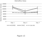
- Figure 13 show the results of relative fluorescence unit on Alamarblue assay and statistics analysis of hMSC seeded on 3D-BGS, CHACC and tissue culture plates. Live/Dead staining showed in Figure 13 indicated that the cell viability was around 92.7 ⁇ 2.8%, which indicates that 3D-BGS is nontoxic to BMSC cells (ISO 10993-5).
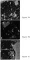
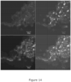
- the confocal microscopy of hMSCs stained with Live/Dead dye is shown in figures 7 and 14 .
- the blue fluorescence shows all the cells, the green fluorescence shows the living cells, the red fluorescence shows the dead cells.
- the 3D-BGS scaffolds were implanted juxtapositionally between rat tibia and tibialis anterior muscle to observe soft tissue reaction on 3D-BGS in comparison with gelatin sponge.
- Gelatin is one of additive materials in 3D-BGS.
- Fig 9C The TEM observation confirmed the finding by light microscopy, as in control gelatin sponge implantation, macrophagic responses were observed with fibroblast infiltration for tissue regeneration ( Fig 9C ); whereas within the patch of bone-like tissue in the 3D-BGS group, typical osteocyte-like cells with calcified Lacuna and canaliculi-like structure were observed ( Fig 9D ).
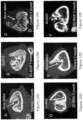
- FIG 10 MicroCT scans at 1, 2 and 3 months after gelatin sponge and 3D-BGS implantations are shown in Fig 10 .
- the ⁇ 3.5 mm bone defects in gelatin sponge implantation group remained non-union ( Fig 10 A, C and E ).
- Fig 10A Even though there were reactive callus formation in the bone marrow cavity at 1-month post operation ( Fig 10A ), the calluses were remodelled at 2 ( Fig 10C ) and 3 months ( Fig 10E ).
- 3D-BGS completely integrated with host compact bone tissue whereas trabecular bone formed within the porous structure of 3D-BGS ( Fig 10D ).
- large part of 3D-BGS within the bone marrow cavity was degraded via callus remodelling and the remaining 3D-BGS joined the compact bone of the femur ( Fig 10F ).
- FIG. 16 The integration of new bone and 3D-BGS materials illustrated by light and backscatter SEM microscopy at 3 months after implantation are shown in Figure 16 .
- the dark coloured area shows implanted 3D-BGS, light grey areas are new trabecular bone formation, and the blue areas are toluidine blue stained cells in bone marrow (containing lipid droplets), blood vessels and bone tissue.
- the 3D-BGS implants are completely integrated and firmly bond to new trabecular bone tissue.
- the majority of 3D-BGS was degraded and replaced by new bone formation.
- High magnification of [A] shows a large particle of biodegrading 3D-BGS within new formed trabecular bone tissue.
- the scattered small Toluidine blue stained dots are osteocytes; whereas the large Toluidine Blue areas are bone marrow (containing lipid droplets) and new blood vessels.
- C High magnification of the marked area in [B]. Toluidine blue stained dots with tails are osteocytes; whereas the large Toluidine Blue area is new blood vessel.
- D The high electron density crystalline showing by backscatter demonstrated where the 3D-BGS particles are. The white arrows indicated osteocytes lacunae. The osteocytes are closely integrated with 3D-BGS.
- Micro-illustration of SEM backscatter, Energy-dispersive X-ray spectroscopy (EDS) map and element analysis of cross section of 3D-BGS at 1, 2 and 3 months post implantation are shown in Figure 17 .
- the high crystalline areas are 3D-BGS implants in which the areas reduced over three months (A, B, and C).
- the EDS mapping showed the calcium (A1, B1 and C1) and phosphate (A2, B2, and C2) composition of the implants and bone tissue.
- X-ray radiography illustrated that the comparison of 5 bone graft materials implanted in ⁇ 5 mm bone defects of Bama mini pigs at day 1, 1 month, 3 months and 6 months post implantation are shown in Figure 18 .
- Gelatin Sponge (1) implantation between 1 day and 3 months with bone formation in the defects at 6 months.
- Two 3D-BGS formula implants (2 and 3) were healed with clear sign of biodegradation (similar density to bone without gaps) in 3 months.
- Pure non-porous hydroxyapatite (4) was healed but not degraded.
- Swine allograft (5) was healed but there was still gaps in the bone defect area in 3 months but fully recovered at 6 months.
- 3D-BGS 3D-printed bone graft substitute
- 3D-BGS was produced and optimised by testing different formulas of compositions to ensure the purged paste had appropriate viscosity and could retain structural integrity after printing. Parameters of printer were adjusted and the biomimetic 3D-BGS with various pore size were fabricated. The products were tested by FTIR spectrum to characterise the composition.
- the 3D-BGS cytotoxicity was tested by using human mesenchymal stem cells (hMSCs), and further implanted in rat tissue and bone defects to test in vivo biocompatibility, osteogenic capacity and potential biodegradation.
- Results of FTIR spectrum showed that the 3D-BGS produced by this technique is a mixture of hydroxyapatite (HA) and calcium carbonate (CC).
- the cytotoxicity test showed that the scaffold is nontoxic and the cells could attach and grow on the material. No infection or adverse tissue reaction were observed after 3D-BGS implantation in comparison to clinically applied gelatin sponge. Interestingly, there were patches of bone-like tissue formation after 3D-BGS implantation juxtapositionally between tibia and tibialis anterior muscle.
Landscapes
- Health & Medical Sciences (AREA)
- Chemical & Material Sciences (AREA)
- Life Sciences & Earth Sciences (AREA)
- Medicinal Chemistry (AREA)
- Public Health (AREA)
- General Health & Medical Sciences (AREA)
- Veterinary Medicine (AREA)
- Animal Behavior & Ethology (AREA)
- Epidemiology (AREA)
- Transplantation (AREA)
- Dermatology (AREA)
- Oral & Maxillofacial Surgery (AREA)
- Engineering & Computer Science (AREA)
- Inorganic Chemistry (AREA)
- Materials Engineering (AREA)
- Manufacturing & Machinery (AREA)
- Composite Materials (AREA)
- Civil Engineering (AREA)
- Ceramic Engineering (AREA)
- Structural Engineering (AREA)
- Biomedical Technology (AREA)
- Molecular Biology (AREA)
- Dispersion Chemistry (AREA)
- Materials For Medical Uses (AREA)
Claims (13)
- Procédé de fabrication d'un substitut de type greffon osseux synthétique biodégradable comprenant :(a) le mélange de carbonate de calcium et d'un matériau liant pour fournir un mélange pulvérulent, ledit matériau liant comprenant du phosphate de tétracalcium (Ca4(PO4)2O ; TTCP) et de l'hydrogénophosphate de calcium (CaHPO4) qui réagissent en présence de gélatine pour fabriquer un ciment ou une pâte ;(b) le mélange du mélange pulvérulent de la partie (a) avec de la gélatine pour fabriquer un ciment ou une pâte, ladite gélatine étant fournie dans une solution ou un solvant en une quantité de 30 à 36 % en poids du ciment ou de la pâte ;(c) ledit mélange à l'étape (a) et/ou à l'étape (b) avec de la gélatine étant effectué à une température de 25°C à 40°C ; et(d) l'impression d'un substitut de type greffon osseux tridimensionnel (3D-BGS) à partir du ciment ou de la pâte de la partie (b) sur une plate-forme à l'aide d'une imprimante tridimensionnelle, la température de la plate-forme étant de 2,5°C à 10°C.
- Procédé selon l'une quelconque des revendications précédentes, ledit carbonate de calcium étant fourni dans la plage de 10 à 60 % ou de 20 à 50 % ou de 25 à 50 % en poids du ciment, y compris tous les 0,1 % entre les deux.
- Procédé selon l'une quelconque des revendications précédentes, ledit matériau liant comprenant les premier et second composants fournis dans la plage de 10 à 50 % en poids du ciment, y compris tous les 0,1 % entre les deux.
- Procédé selon la revendication 3, lesdits premier et second composants étant fournis sous forme solide et étant mélangés et broyés ensemble en les quantités relatives, le rapport du premier composant au second composant étant dans la plage d'environ 0,5 à 5,5 ou 1 à 1,5.
- Procédé selon l'une quelconque des revendications précédentes, ladite partie (a) comprenant en outre le mélange d'un additif, ledit additif étant tout composé qui influe sur la viscosité et garantit ainsi que le produit mélangé comporte une fluidité appropriée à l'écoulement mais pas jusqu'au point de ne pas réussir à conserver sa forme lorsqu'il est exsudé.
- Procédé selon la revendication 5, ledit additif étant fourni dans une solution ou un solvant.
- Procédé selon l'une quelconque des revendications précédentes, ladite impression de l'étape (d) comprenant le dépôt du mélange de l'étape (b) couche par couche pour produire du 3D-GBS, chaque couche étant idéalement déposée suivant un angle par rapport à la couche précédente de manière à générer une structure poreuse.
- Procédé selon l'une quelconque des revendications précédentes, ladite impression de l'étape (d) étant commandée de manière à produire les tailles de pores souhaitées dans le produit de 3D-GBS, lesdites tailles de pores étant choisies selon l'utilisation envisagée.
- Procédé selon l'une quelconque des revendications précédentes, ladite étape (d) comprenant en outre l'impression sur un substrat comprenant un matériau support, ledit ciment étant déposé de manière à ce que ledit matériau support soit incorporé, partiellement ou complètement, dans le 3D-GBS.
- Procédé selon l'une quelconque des revendications précédentes comprenant en outre l'incorporation d'un agent actif dans le ciment, la pâte ou le produit avant ou suite à l'étape d'impression.
- Procédé selon la revendication 10, ledit agent actif étant un agent pharmaceutique, des facteurs de croissance, un antibiotique, un agent de résorption anti-inflammatoire/anti-os, ou des agents thérapeutiques anti-cancer.
- Mélange destiné à fabriquer un échafaudage de greffon osseux comprenant :i) du carbonate de calcium ;ii) un matériau liant, ledit matériau liant comprenant du phosphate de tétracalcium (Ca4(P04)20 ; TTCP) et de l'hydrogénophosphate de calcium (CaHPO4) qui réagissent en présence de gélatine pour fabriquer un ciment ou une pâte ; etiii) de la gélatine fournie dans une solution ou un solvant à une concentration grâce à laquelle la quantité d'agent gélifiant, lorsqu'il se trouve dans le mélange, est dans la plage de 30 à 36 % en poids du mélange.
- Mélange selon la revendication 12, ledit mélange comprenant du carbonate de calcium en une quantité fournie dans la plage de 10 à 60 % en poids, y compris tous les 0,1 % entre les deux.
Applications Claiming Priority (2)
| Application Number | Priority Date | Filing Date | Title |
|---|---|---|---|
| GBGB1804594.8A GB201804594D0 (en) | 2018-03-22 | 2018-03-22 | Bonegraft substituteand method of manufacture |
| PCT/GB2019/050795 WO2019180439A1 (fr) | 2018-03-22 | 2019-03-21 | Substitut de type greffon osseux et procédé de fabrication associé |
Publications (3)
| Publication Number | Publication Date |
|---|---|
| EP3768333A1 EP3768333A1 (fr) | 2021-01-27 |
| EP3768333C0 EP3768333C0 (fr) | 2024-12-11 |
| EP3768333B1 true EP3768333B1 (fr) | 2024-12-11 |
Family
ID=62068117
Family Applications (1)
| Application Number | Title | Priority Date | Filing Date |
|---|---|---|---|
| EP19714744.0A Active EP3768333B1 (fr) | 2018-03-22 | 2019-03-21 | Substitut de type greffon osseux et procédé de fabrication associé |
Country Status (5)
| Country | Link |
|---|---|
| US (1) | US12011515B2 (fr) |
| EP (1) | EP3768333B1 (fr) |
| CN (1) | CN111886030B (fr) |
| GB (1) | GB201804594D0 (fr) |
| WO (1) | WO2019180439A1 (fr) |
Families Citing this family (6)
| Publication number | Priority date | Publication date | Assignee | Title |
|---|---|---|---|---|
| JP7289500B2 (ja) * | 2019-01-09 | 2023-06-12 | 国立研究開発法人日本原子力研究開発機構 | 炭酸アパタイトの製造方法 |
| CN110665057B (zh) * | 2019-11-07 | 2021-10-01 | 上海健康医学院 | 一种羟基磷灰石/plga双层支架的制备方法 |
| CN112843332B (zh) * | 2020-11-04 | 2022-11-29 | 华东数字医学工程研究院 | 骨修复材料及其制备方法和应用 |
| CN113387615A (zh) * | 2021-06-30 | 2021-09-14 | 成都百年贝雅医疗科技有限公司 | 一种dlp生物陶瓷打印浆料 |
| US20230407248A1 (en) * | 2022-06-10 | 2023-12-21 | Novum Coral, Inc. | Methods of increasing growth of corals using a bioceramic |
| KR102931217B1 (ko) * | 2023-04-04 | 2026-02-27 | 주식회사 네오바이오텍 | 골이식재 표면에 저결정성 인산칼슘을 코팅하는 방법 |
Citations (2)
| Publication number | Priority date | Publication date | Assignee | Title |
|---|---|---|---|---|
| EP0395187B1 (fr) * | 1989-04-28 | 1996-01-24 | Interpore International | Biomatériaux revêtus et leur procédé de fabrication |
| EP3031441B1 (fr) * | 2013-08-06 | 2021-02-03 | Kuraray Noritake Dental Inc. | Composition de phosphate de calcium durcissable pour matériel dentaire |
Family Cites Families (9)
| Publication number | Priority date | Publication date | Assignee | Title |
|---|---|---|---|---|
| US20040254668A1 (en) * | 2003-06-16 | 2004-12-16 | Jang Bor Z. | Macro-porous hydroxyapatite scaffold compositions and freeform fabrication method thereof |
| DE202004005420U1 (de) | 2004-03-31 | 2005-08-18 | Technische Universität Dresden | Modifizierter Calciumphosphatzement |
| GB2424223C (en) * | 2005-03-07 | 2011-02-02 | Massachusetts Inst Technology | Biomaterial. |
| CN101366971A (zh) | 2008-10-15 | 2009-02-18 | 苗军 | 可注射防水型磷酸钙骨水泥 |
| CN104771785B (zh) * | 2015-04-03 | 2017-06-20 | 周宏志 | 具有神经多肽诱导成骨活性的骨修复材料的制造方法 |
| CN105107023A (zh) * | 2015-07-01 | 2015-12-02 | 李亚屏 | 一种骨移植用可降解多孔复合支架材料 |
| CN105056298B (zh) * | 2015-08-25 | 2018-01-02 | 华南理工大学 | 一种具有表面大孔的多孔磷酸钙微球材料、制备方法及应用 |
| CN105749337A (zh) | 2016-03-29 | 2016-07-13 | 西北工业大学 | 一种分层分量载药人工骨支架的制备方法 |
| CN107469152B (zh) * | 2017-09-29 | 2020-10-09 | 深圳市艾科赛龙科技股份有限公司 | 一种骨组织修复复合材料及其制备方法和由其制备得到的骨组织修复结构体 |
-
2018
- 2018-03-22 GB GBGB1804594.8A patent/GB201804594D0/en not_active Ceased
-
2019
- 2019-03-21 EP EP19714744.0A patent/EP3768333B1/fr active Active
- 2019-03-21 WO PCT/GB2019/050795 patent/WO2019180439A1/fr not_active Ceased
- 2019-03-21 CN CN201980020743.6A patent/CN111886030B/zh active Active
- 2019-03-21 US US16/981,643 patent/US12011515B2/en active Active
Patent Citations (2)
| Publication number | Priority date | Publication date | Assignee | Title |
|---|---|---|---|---|
| EP0395187B1 (fr) * | 1989-04-28 | 1996-01-24 | Interpore International | Biomatériaux revêtus et leur procédé de fabrication |
| EP3031441B1 (fr) * | 2013-08-06 | 2021-02-03 | Kuraray Noritake Dental Inc. | Composition de phosphate de calcium durcissable pour matériel dentaire |
Also Published As
| Publication number | Publication date |
|---|---|
| CN111886030A (zh) | 2020-11-03 |
| CN111886030B (zh) | 2024-07-05 |
| EP3768333C0 (fr) | 2024-12-11 |
| GB201804594D0 (en) | 2018-05-09 |
| US20210106719A1 (en) | 2021-04-15 |
| EP3768333A1 (fr) | 2021-01-27 |
| WO2019180439A1 (fr) | 2019-09-26 |
| US12011515B2 (en) | 2024-06-18 |
Similar Documents
| Publication | Publication Date | Title |
|---|---|---|
| EP3768333B1 (fr) | Substitut de type greffon osseux et procédé de fabrication associé | |
| Jahan et al. | Composite biopolymers for bone regeneration enhancement in bony defects | |
| EP2059269B1 (fr) | Préparation présentant une stabilité de phases et de sédimentation, déformable plastiquement, avec formation intrinsèque de pores, par exemple pour combler des défauts osseux ou s'utilisant comme matériau de substitution osseuse, et procédé pour la produire | |
| CN101163512B (zh) | 包含多层结构的软骨样和骨软骨的替代物 | |
| EP2173273B1 (fr) | Matériau composite de réparation des os | |
| Schnettler et al. | Calcium phosphate-based bone substitutes | |
| Munhoz et al. | Use of collagen/chitosan sponges mineralized with hydroxyapatite for the repair of cranial defects in rats | |
| US20190255218A1 (en) | Methods of manufacturing crosslinkable 3d printed biomaterial-based implants | |
| Rahimnejad et al. | Biomaterials in bone and mineralized tissue engineering using 3D printing and bioprinting technologies | |
| Kucko et al. | Current advances in hydroxyapatite-and β-tricalcium phosphate-based composites for biomedical applications: a review | |
| Cardoso et al. | In vivo approach of calcium deficient hydroxyapatite filler as bone induction factor | |
| KR101427305B1 (ko) | 골 이식재 및 그의 제조방법 | |
| Amin et al. | Bioceramic scaffolds | |
| US20240058131A1 (en) | A method of making an individual 3d printed ceramic bioresorbable bone implant for use in traumatology and orthopedics | |
| NL2007850C2 (en) | Injectable calcium phosphate cement comprising glucono-delta-lactone. | |
| Zhang et al. | Constructing a 3D-printable, bioceramic sheathed articular spacer assembly for infected hip arthroplasty | |
| Yuan et al. | Experimental study of natural hydroxyapatite/chitosan composite on reconstructing bone defects | |
| ES2357191T3 (es) | Material compuesto poroso, correspondiente proceso de preparación y su uso para realizar dispositivo para ingeniería tisular. | |
| WO2024067485A1 (fr) | Échafaudage dérivé de mousse de ciment d'oxychlorure de magnésium (mocf) injectable pour le traitement de lésions osseuses ostéoporotiques | |
| KR20230108930A (ko) | 인산칼슘계 골 이식재 조성물 | |
| US7767226B2 (en) | Calcium sulfate based nanoparticles | |
| Sa et al. | Fabrication of hybrid scaffolds by polymer deposition system and its in-vivo evaluation with a rat tibial defect model | |
| Golafshan | Towards 3D Fabrication of MgP-based Mechanically Competent Implants for Bone Regeneration | |
| Chowdhury et al. | Introduction to biomedical polymer and composites | |
| JPH04242658A (ja) | 生体移植材とその製造方法 |
Legal Events
| Date | Code | Title | Description |
|---|---|---|---|
| STAA | Information on the status of an ep patent application or granted ep patent |
Free format text: STATUS: UNKNOWN |
|
| STAA | Information on the status of an ep patent application or granted ep patent |
Free format text: STATUS: THE INTERNATIONAL PUBLICATION HAS BEEN MADE |
|
| PUAI | Public reference made under article 153(3) epc to a published international application that has entered the european phase |
Free format text: ORIGINAL CODE: 0009012 |
|
| STAA | Information on the status of an ep patent application or granted ep patent |
Free format text: STATUS: REQUEST FOR EXAMINATION WAS MADE |
|
| 17P | Request for examination filed |
Effective date: 20200930 |
|
| AK | Designated contracting states |
Kind code of ref document: A1 Designated state(s): AL AT BE BG CH CY CZ DE DK EE ES FI FR GB GR HR HU IE IS IT LI LT LU LV MC MK MT NL NO PL PT RO RS SE SI SK SM TR |
|
| AX | Request for extension of the european patent |
Extension state: BA ME |
|
| DAV | Request for validation of the european patent (deleted) | ||
| DAX | Request for extension of the european patent (deleted) | ||
| STAA | Information on the status of an ep patent application or granted ep patent |
Free format text: STATUS: EXAMINATION IS IN PROGRESS |
|
| 17Q | First examination report despatched |
Effective date: 20220913 |
|
| RAP1 | Party data changed (applicant data changed or rights of an application transferred) |
Owner name: 3D-BGS (UK) LTD |
|
| REG | Reference to a national code |
Ref country code: DE Ref legal event code: R079 Free format text: PREVIOUS MAIN CLASS: A61L0027020000 Ipc: A61L0027540000 Ref document number: 602019063332 Country of ref document: DE |
|
| GRAP | Despatch of communication of intention to grant a patent |
Free format text: ORIGINAL CODE: EPIDOSNIGR1 |
|
| STAA | Information on the status of an ep patent application or granted ep patent |
Free format text: STATUS: GRANT OF PATENT IS INTENDED |
|
| RIC1 | Information provided on ipc code assigned before grant |
Ipc: B33Y 10/00 20150101ALI20240918BHEP Ipc: B33Y 70/10 20200101ALI20240918BHEP Ipc: B33Y 80/00 20150101ALI20240918BHEP Ipc: A61L 27/58 20060101ALI20240918BHEP Ipc: A61L 27/46 20060101ALI20240918BHEP Ipc: A61L 27/56 20060101ALI20240918BHEP Ipc: A61L 27/54 20060101AFI20240918BHEP |
|
| INTG | Intention to grant announced |
Effective date: 20240930 |
|
| GRAS | Grant fee paid |
Free format text: ORIGINAL CODE: EPIDOSNIGR3 |
|
| GRAA | (expected) grant |
Free format text: ORIGINAL CODE: 0009210 |
|
| STAA | Information on the status of an ep patent application or granted ep patent |
Free format text: STATUS: THE PATENT HAS BEEN GRANTED |
|
| AK | Designated contracting states |
Kind code of ref document: B1 Designated state(s): AL AT BE BG CH CY CZ DE DK EE ES FI FR GB GR HR HU IE IS IT LI LT LU LV MC MK MT NL NO PL PT RO RS SE SI SK SM TR |
|
| REG | Reference to a national code |
Ref country code: GB Ref legal event code: FG4D |
|
| REG | Reference to a national code |
Ref country code: CH Ref legal event code: EP |
|
| REG | Reference to a national code |
Ref country code: DE Ref legal event code: R096 Ref document number: 602019063332 Country of ref document: DE |
|
| REG | Reference to a national code |
Ref country code: IE Ref legal event code: FG4D |
|
| U01 | Request for unitary effect filed |
Effective date: 20250103 |
|
| U07 | Unitary effect registered |
Designated state(s): AT BE BG DE DK EE FI FR IT LT LU LV MT NL PT RO SE SI Effective date: 20250115 |
|
| U20 | Renewal fee for the european patent with unitary effect paid |
Year of fee payment: 7 Effective date: 20250120 |
|
| PG25 | Lapsed in a contracting state [announced via postgrant information from national office to epo] |
Ref country code: HR Free format text: LAPSE BECAUSE OF FAILURE TO SUBMIT A TRANSLATION OF THE DESCRIPTION OR TO PAY THE FEE WITHIN THE PRESCRIBED TIME-LIMIT Effective date: 20241211 |
|
| PG25 | Lapsed in a contracting state [announced via postgrant information from national office to epo] |
Ref country code: ES Free format text: LAPSE BECAUSE OF FAILURE TO SUBMIT A TRANSLATION OF THE DESCRIPTION OR TO PAY THE FEE WITHIN THE PRESCRIBED TIME-LIMIT Effective date: 20241211 |
|
| PG25 | Lapsed in a contracting state [announced via postgrant information from national office to epo] |
Ref country code: NO Free format text: LAPSE BECAUSE OF FAILURE TO SUBMIT A TRANSLATION OF THE DESCRIPTION OR TO PAY THE FEE WITHIN THE PRESCRIBED TIME-LIMIT Effective date: 20250311 |
|
| PG25 | Lapsed in a contracting state [announced via postgrant information from national office to epo] |
Ref country code: GR Free format text: LAPSE BECAUSE OF FAILURE TO SUBMIT A TRANSLATION OF THE DESCRIPTION OR TO PAY THE FEE WITHIN THE PRESCRIBED TIME-LIMIT Effective date: 20250312 |
|
| PG25 | Lapsed in a contracting state [announced via postgrant information from national office to epo] |
Ref country code: RS Free format text: LAPSE BECAUSE OF FAILURE TO SUBMIT A TRANSLATION OF THE DESCRIPTION OR TO PAY THE FEE WITHIN THE PRESCRIBED TIME-LIMIT Effective date: 20250311 |
|
| PG25 | Lapsed in a contracting state [announced via postgrant information from national office to epo] |
Ref country code: SM Free format text: LAPSE BECAUSE OF FAILURE TO SUBMIT A TRANSLATION OF THE DESCRIPTION OR TO PAY THE FEE WITHIN THE PRESCRIBED TIME-LIMIT Effective date: 20241211 |
|
| PG25 | Lapsed in a contracting state [announced via postgrant information from national office to epo] |
Ref country code: PL Free format text: LAPSE BECAUSE OF FAILURE TO SUBMIT A TRANSLATION OF THE DESCRIPTION OR TO PAY THE FEE WITHIN THE PRESCRIBED TIME-LIMIT Effective date: 20241211 |
|
| PG25 | Lapsed in a contracting state [announced via postgrant information from national office to epo] |
Ref country code: IS Free format text: LAPSE BECAUSE OF FAILURE TO SUBMIT A TRANSLATION OF THE DESCRIPTION OR TO PAY THE FEE WITHIN THE PRESCRIBED TIME-LIMIT Effective date: 20250411 |
|
| PG25 | Lapsed in a contracting state [announced via postgrant information from national office to epo] |
Ref country code: SK Free format text: LAPSE BECAUSE OF FAILURE TO SUBMIT A TRANSLATION OF THE DESCRIPTION OR TO PAY THE FEE WITHIN THE PRESCRIBED TIME-LIMIT Effective date: 20241211 |
|
| PG25 | Lapsed in a contracting state [announced via postgrant information from national office to epo] |
Ref country code: CZ Free format text: LAPSE BECAUSE OF FAILURE TO SUBMIT A TRANSLATION OF THE DESCRIPTION OR TO PAY THE FEE WITHIN THE PRESCRIBED TIME-LIMIT Effective date: 20241211 |
|
| PG25 | Lapsed in a contracting state [announced via postgrant information from national office to epo] |
Ref country code: MC Free format text: LAPSE BECAUSE OF FAILURE TO SUBMIT A TRANSLATION OF THE DESCRIPTION OR TO PAY THE FEE WITHIN THE PRESCRIBED TIME-LIMIT Effective date: 20241211 |
|
| PLBE | No opposition filed within time limit |
Free format text: ORIGINAL CODE: 0009261 |
|
| STAA | Information on the status of an ep patent application or granted ep patent |
Free format text: STATUS: NO OPPOSITION FILED WITHIN TIME LIMIT |
|
| REG | Reference to a national code |
Ref country code: CH Ref legal event code: L10 Free format text: ST27 STATUS EVENT CODE: U-0-0-L10-L00 (AS PROVIDED BY THE NATIONAL OFFICE) Effective date: 20251022 |
|
| REG | Reference to a national code |
Ref country code: CH Ref legal event code: H13 Free format text: ST27 STATUS EVENT CODE: U-0-0-H10-H13 (AS PROVIDED BY THE NATIONAL OFFICE) Effective date: 20251023 |
|
| 26N | No opposition filed |
Effective date: 20250912 |
|
| PG25 | Lapsed in a contracting state [announced via postgrant information from national office to epo] |
Ref country code: CH Free format text: LAPSE BECAUSE OF NON-PAYMENT OF DUE FEES Effective date: 20250331 |
|
| PG25 | Lapsed in a contracting state [announced via postgrant information from national office to epo] |
Ref country code: IE Free format text: LAPSE BECAUSE OF NON-PAYMENT OF DUE FEES Effective date: 20250321 |
|
| PGFP | Annual fee paid to national office [announced via postgrant information from national office to epo] |
Ref country code: GB Payment date: 20260308 Year of fee payment: 8 |
|
| U20 | Renewal fee for the european patent with unitary effect paid |
Year of fee payment: 8 Effective date: 20260313 |