EP3777904A1 - Osteotroper knochenersatz - Google Patents
Osteotroper knochenersatz Download PDFInfo
- Publication number
- EP3777904A1 EP3777904A1 EP19192028.9A EP19192028A EP3777904A1 EP 3777904 A1 EP3777904 A1 EP 3777904A1 EP 19192028 A EP19192028 A EP 19192028A EP 3777904 A1 EP3777904 A1 EP 3777904A1
- Authority
- EP
- European Patent Office
- Prior art keywords
- autoclave
- strontium
- fluorine
- osteotropic
- bone
- Prior art date
- Legal status (The legal status is an assumption and is not a legal conclusion. Google has not performed a legal analysis and makes no representation as to the accuracy of the status listed.)
- Granted
Links
Images


Classifications
-
- A—HUMAN NECESSITIES
- A61—MEDICAL OR VETERINARY SCIENCE; HYGIENE
- A61L—METHODS OR APPARATUS FOR STERILISING MATERIALS OR OBJECTS IN GENERAL; DISINFECTION, STERILISATION OR DEODORISATION OF AIR; CHEMICAL ASPECTS OF BANDAGES, DRESSINGS, ABSORBENT PADS OR SURGICAL ARTICLES; MATERIALS FOR BANDAGES, DRESSINGS, ABSORBENT PADS OR SURGICAL ARTICLES
- A61L27/00—Materials for grafts or prostheses or for coating grafts or prostheses
- A61L27/02—Inorganic materials
- A61L27/12—Phosphorus-containing materials, e.g. apatite
-
- C—CHEMISTRY; METALLURGY
- C01—INORGANIC CHEMISTRY
- C01B—NON-METALLIC ELEMENTS; COMPOUNDS THEREOF; METALLOIDS OR COMPOUNDS THEREOF NOT COVERED BY SUBCLASS C01C
- C01B25/00—Phosphorus; Compounds thereof
- C01B25/16—Oxyacids of phosphorus; Salts thereof
- C01B25/26—Phosphates
- C01B25/32—Phosphates of magnesium, calcium, strontium, or barium
-
- C—CHEMISTRY; METALLURGY
- C04—CEMENTS; CONCRETE; ARTIFICIAL STONE; CERAMICS; REFRACTORIES
- C04B—LIME, MAGNESIA; SLAG; CEMENTS; COMPOSITIONS THEREOF, e.g. MORTARS, CONCRETE OR LIKE BUILDING MATERIALS; ARTIFICIAL STONE; CERAMICS; REFRACTORIES; TREATMENT OF NATURAL STONE
- C04B35/00—Shaped ceramic products characterised by their composition; Ceramics compositions; Processing powders of inorganic compounds preparatory to the manufacturing of ceramic products
- C04B35/01—Shaped ceramic products characterised by their composition; Ceramics compositions; Processing powders of inorganic compounds preparatory to the manufacturing of ceramic products based on oxide ceramics
- C04B35/447—Shaped ceramic products characterised by their composition; Ceramics compositions; Processing powders of inorganic compounds preparatory to the manufacturing of ceramic products based on oxide ceramics based on phosphates, e.g. hydroxyapatite
-
- A—HUMAN NECESSITIES
- A61—MEDICAL OR VETERINARY SCIENCE; HYGIENE
- A61L—METHODS OR APPARATUS FOR STERILISING MATERIALS OR OBJECTS IN GENERAL; DISINFECTION, STERILISATION OR DEODORISATION OF AIR; CHEMICAL ASPECTS OF BANDAGES, DRESSINGS, ABSORBENT PADS OR SURGICAL ARTICLES; MATERIALS FOR BANDAGES, DRESSINGS, ABSORBENT PADS OR SURGICAL ARTICLES
- A61L2430/00—Materials or treatment for tissue regeneration
- A61L2430/02—Materials or treatment for tissue regeneration for reconstruction of bones; weight-bearing implants
Definitions
- the invention relates to a method for producing an osteotropic bone substitute material from a starting material which essentially comprises portlandite, quick lime, aragonite, calcite, for example skeletons of lime-encrusting algae, and / or apatite, for example as hydroxyapatite, e.g. made from vertebrate bones or synthetically produced.
- the invention also relates to an osteotropic bone material which was produced in accordance with the method according to the invention.
- the cited starting materials can also be understood to mean materials which have a structure analogous to the starting materials, but are synthetically produced.
- hydroxyapatite bone substitute materials made from natural bone, both autologous, allogeneic and xenogeneic, corals, algae or also fully synthetically produced hydroxyapatite bone substitute materials, which are used as bone implants or as bone substitute material in the context of bone augmentation, only have osteoconductive properties.
- the previous calcium phosphate or hydroxyapatite bone substitute materials offer a suitable biocompatible surface for the direct growth of bone tissue on the implant surface, but they do not directly stimulate new bone formation in the immediate bone environment of the implant.
- Hydroxyapatite or calcium phosphate materials are basically only osteoconductive. They allow bones to grow, but do not themselves stimulate the proliferation or differentiation of cells that develop bone, such as osteoblasts or their precursors. Under direct growth within the meaning of the invention In particular, growth can be understood without an "intermediate tissue layer" between the implant surface and bone tissue.
- bone substitute materials or organic substances such as collagens or organic molecules have been supplemented with further proteins, peptides or other molecules.
- growth factors such as various BMPs, IGF1 / 2, FGF or the like, or serum products.
- such a bone substitute is only osteoconductive and not osteoinductive.
- the invention is therefore based on the object of specifying a method for producing a bone substitute material and a bone substitute material itself which has long-lasting, locally limited osteotropic properties.
- this object is achieved by a method for producing an osteotropic bone substitute material with the features of claim 1 and an osteotropic bone substitute material with the features of claim 15.
- a starting material which essentially comprises portlandite, quick lime, aragonite, calcite and / or apatite, for example as hydroxyapatite.
- the starting material is made with a Strontium, fluorine and / or gallium source placed in an autoclave, wherein when using a starting material which essentially portlandite, quicklime, aragonite; Calcite has a source of phosphate being introduced.
- a starting material which consists essentially of apatite a phosphate source can also be introduced, but this is not absolutely necessary.
- water is added to the autoclave as a component of a solvent.
- the pH is then set in the autoclave in a range above 7 at room temperature and normal pressure.
- the closed and filled autoclave is then heated for at least one hour and then cooled.
- the contents of the autoclave are cleaned of the residues of the strontium, fluoride and / or gallium source. The same applies to the source of phosphorus, if used.
- strontium, fluoride and / or gallium ions which are incorporated into an apatite structure, have osteodinductive or osteotropic or antiresorptive properties.
- the process according to the invention is essential to the invention in order to incorporate the strontium, fluoride and / or gallium ions accordingly in apatite, in particular in the hydroxyapatite structure, and not merely to add them.
- the accumulated substances could become detached.
- a phosphate source For a successful conversion of the raw materials from calcium carbonate materials. so in particular portlandite, quick lime, aragonite, calcite, in apatite or hydroxylapatite structure, the presence of a phosphate source is essential, and various phosphate salts can be used.
- a phosphate source When using an apatite material as the starting material, for example vertebrate bones, the presence of a phosphate source is not absolutely necessary but is advantageous. This in particular prevents the existing structure from being weakened during the process.
- the closed and filled autoclave is heated to at least 30 ° C., preferably above 190 ° C. This can be done in an oven or a heating module. It is also possible to have a heating module integrated with the autoclave. At these temperatures, a sufficiently good conversion of the starting material into doped apatite is achieved, in addition, as already described, the ions mentioned being incorporated and connected to the crystal lattice of the apatite. The lower the temperature of the oven, the slower the reaction that takes place in the autoclave.
- a heating cabinet or the like can also be referred to as an oven in the context of the method according to the invention. It is essential here that the device can maintain a desired temperature between 30 and several hundred degrees constant over a longer period of time.
- the contents of the autoclave can preferably be cleaned mechanically with a filter device. This is the case when the largest possible or sparingly soluble sources are used.
- the contents of the autoclave are cleaned after cleaning, in particular mechanical, until a pH value of below 8 is reached.
- a basic solution in particular an ammonia solution, can be used to set the pH before closing the autoclave in a range of over 7.
- an ammonium dihydrogen phosphate solution or other phosphate compound can be used for this.
- the introduction of a strontium, fluorine and / or gallium source in excess with respect to the starting material is preferred. This advantageously also applies to the phosphate source. In other words, a large reservoir of the corresponding materials is made available, so that good incorporation into the crystal lattice of the apatite can be achieved with a high degree of certainty.
- the corresponding unused Starting materials of the strontium, fluorine and / or gallium source and the phosphorus source are removed from the material produced. This means that the material is cleaned.
- ammonium dihydrogen phosphate or diammonium phosphate as a source of phosphorus, this agent can also be used to set the desired pH. It has also been found that the use of ammonium dihydrogen phosphate dissolved in water is particularly easy to dose and an optimal reaction environment can be achieved in the autoclave.
- Calcium and phosphorus atoms are preferably present in the total mixture in the autoclave in a ratio of at most 10: 5. Such a ratio has been found to be advantageous for the osteotropic properties of the resulting bone substitute material, since the corresponding osteotropic, i.e. osteoinductive and / or antiresorptively active substances are thus present in a ratio which enables particularly good results.
- Vertebrate or mammalian bones can be used as the apatite.
- Mammalian bones for example from cattle or pigs, already provide apatite with a considerable proportion of hydroxyl and carbonate apatite.
- aragonite or calcite calcite algae skeletons or other calcium carbonate materials in burned, unfired and / or chemically processed form can also be used as the starting material. If vertebrate or mammalian bones are used, these are advantageously subjected to a pyrolytic or chemical maceration beforehand, that is to say the removal of immunogenic material.
- the strontium, fluorine and / or gallium source is removed from the resulting after heating in the autoclave and subsequent cooling osteotropic bone substitute material is separated by cleaning it. It is preferred here if an easily washable or sparingly water-soluble material is used as the strontium, fluorine and / or gallium source.
- an easily washable material has the advantage that it can be easily washed out when the produced osteotropic bone substitute material is washed and thus the osteotropic bone substitute material can be cleaned more easily.
- a sparingly soluble, in particular coarsely crystalline, material offers the advantage that sufficient strontium, fluorine and / or gallium ions are available during the conversion process in the autoclave, but cleaning can then be carried out particularly easily, for example mechanically.
- the resulting osteotropic bone substitute material can be cleaned particularly easily if the strontium, fluorine and / or gallium source is added to the autoclave as a solid in a container.
- the container is designed in such a way that it enables the exchange of ions from the strontium, fluorine and / or gallium source with the solvent that is present in the autoclave, but the solids are retained. This makes the final cleaning of the resulting osteotropic bone substitute material much easier.
- the starting material is subjected to a pyrolytic treatment and / or chemical cleaning before or after being introduced into the autoclave and / or the contents of the autoclave after the process has been completed.
- the pyrolytic treatment or the chemical cleaning process have the advantage that any proteins or other organic foreign substances that may be present are removed from the osteotropic bone substitute material produced, so that interactions when implanting the bone substitute material in a human body are minimized or excluded.
- the invention also relates to an osteotropic bone substitute material which has been produced by the method according to the invention described above.
- the osteotropic bone substitute material which essentially has apatite, has strontium, fluorine and / or gallium ions in its crystal lattice. These develop osteotropic properties after implantation in an animal or human body.
- small components can also be used calcium phosphate, calcite, aragonite and tricalcium phosphate may be present.
- the osteotropic bone substitute material according to the invention produced according to the method according to the invention can be used, for example, to produce dimensionally stable blocks by also integrating cylindrical dental implants or other metallic objects. These blocks can be implanted in a jawbone as future dental implant carriers and used immediately after a healing phase of 3 to 6 months without a further second operation. Since the resulting osteotropic bone substitute material has a powder-like shape, the dimensionally stable blocks that are made from it can be made to measure and thus adapted to bone defects, such as a tooth gap.
- the bone substitute material By embedding the strontium, fluorine and / or gallium ions according to the invention, the bone substitute material develops a local osteotropic effect without further addition, which mainly and essentially only takes place in the implant site.
- the bone substitute material according to the invention therefore has no systemic effect.
- Another advantage is that the bone substitute material according to the invention develops its osteoinductive or osteotropic effects over the entire time it remains in the implant site and only ends as soon as it is replaced by autochthonous bone tissue. In this way, faster and more lasting bone healing of a bone defect is achieved.
- osteotropic bone substitute material can be the stabilization of osteoporotic, traumatic and / or malignancy-related vertebral fractures or sintering fractures.
- Another possibility is to use the material produced according to the invention as a starting material for 3-D printing processes.
- Teflon vessel A 150 ml Teflon vessel is used for this. This is filled with the following substances: burned algae material 19.822 g Ammonium dihydrogen phosphate 26.38 g Strontium fluoride 2.212 g Potassium fluoride 2.212 g Ammonia solution (25%) 50 ml deionized H 2 O 50 ml
- a skeleton of lime-encrusting algae is used as a starting material as an example for aragonite.
- CO 2 -containing starting materials it is usually advantageous if these are fired, in which case it is desirable to maintain the external structure of the material.
- the method according to the invention can also be applied to an apatite material, for example from vertebrate bones, as the starting material, the presence of a phosphate source having proven to be advantageous on the one hand for introducing the osteotropic ions on the other hand, but also for maintaining the structure of the starting material.
- the algae skeleton is burned out before it is added to the Teflon vessel so that any foreign proteins, egg whites or the like are removed. Furthermore, ammonium dihydrogen phosphate, strontium fluoride, potassium fluoride and a 25 percent ammonia solution are added. Deionized water is also added.
- ammonium dihydrogen phosphate serves as a source of phosphate, although other sources of phosphate are also possible, as described.
- Strontium fluoride is used as a source of strontium as well as a source of fluorine.
- Potassium fluoride is also used as a source of fluoride.
- Teflon vessel After the substances have been placed in the Teflon vessel and any gas evolution has been waited for, it is closed.
- the Teflon vessel is then placed in an autoclave, for example in a pressure digestion vessel.
- This vessel is preferably made of stainless steel.
- the lid is then screwed tightly, creating an autoclave.
- the correspondingly tightly closed pressure digestion vessel is then placed in a preheated heating cabinet or heating block which has a temperature of 190 ° C.
- the pressure digestion vessel remains in the heating cabinet for 5 days, the temperature of 190 ° C. being maintained. After this time has elapsed, the heating cabinet is switched off. The pressure digestion vessel then slowly cools down in the heating cabinet or heating block. This takes about a day.
- the pressure digestion vessel and the Teflon vessel are opened and the resulting osteotropic bone substitute material is cleaned.
- the osteotropic bone substitute material is applied to a filter paper with water and washed.
- cleaning processes for example rinsing processes, are carried out until a pH value of below 8 is reached.
- the osteotropic bone substitute material is then returned to the heating cabinet, but only dried at 40 ° C.
- the osteotropic bone substitute material is then ready for further use. It can then, for example, be brought into desired shapes and sterilized.
- the new osteotropic bone substitute material which is produced in accordance with the method according to the invention, is explained in more detail or the osteoinductive one based on test results Proven effectiveness.
- the results show that the bone replacement material according to the invention should additionally stimulate the local formation of new bone in the bone defect, since in human bone cells the most important bone growth marker, alkaline phosphatase, is stimulated.
- Primary human osteoblastic bone cells are therefore suitable as a sensitive test system prior to clinical use of new bone substitute materials in vivo in order to examine the effect of a new type of bone substitute material on human bone cell metabolism.
- the comparative results are expressed in% of the control +/- standard deviation.
- the activity of the alkaline phosphatase in the medium supernatant after the culture of the human bone cells in the presence of the various materials was examined. An aliquot of the cell culture nutrient medium used, alone without cells, served as a control, since residues of the serum contained in the culture medium always remain in the holes of the culture medium despite serum-free rinsing before the cells were exposed to the materials. The serum always contains small amounts of alkaline phosphatase.
- the new BioOss1 was not yet available for this first attempt. Cells alone BioOss® Algipore® NA1 NA2 243 +/- 9% 216 +/- 3% 240 +/- 8% 291 +/- 9% 443 +/- 15%
- BioOss® rather reduces the activity of the enzyme alkaline phosphatase, which is indispensable for the mineral and bone formation of human bone cells, while the bone substitute material according to the invention causes a highly significant increased activity of the alkaline phosphatase secreted by human bone cells.
- the bone-specific alkaline phosphatase as the most important osteoblast marker protein is therefore stimulated by the bone substitute material according to the invention to a very significantly greater extent in the human bone cell model than when the conventional materials are present.
- the different potency of NA1 and NA2 on alkaline phosphatase activity can be traced back to an increased fluoride and strontium content in NA2 compared to NA1.
- the protein content in the individual "holes", i.e. reaction chambers, of the multi-hole plates used was analyzed.
- a triton extract from the respective holes was used to determine the protein content according to the BCA method.
- a reaction chamber would therefore be synonymous with a decrease in the number of cells that are adherent to the cell culture soil or to the bone substitute (apoptotic, i.e. dead cells, do not remain adherent and are washed away before the addition of Triton).
- the results are again given in% of the control +/- standard deviation.
- Cells alone BioOss® Algipore® NA1 NA2 96 +/- 3% 108 +/- 4% 103 +/- 3% 103 +/- 3% 103 +/- 3% 103 +/- 3%
- the BioOss® material previously used as a bone replacement material in dentistry or maxillofacial surgery, consists of the natural hydroxyapatite material from the inorganic bone tissue of cattle.
- a partial exchange of calcium ions in the hydroxylapatite crystal with strontium and fluoride ions can also be carried out in a controlled manner in a natural calcium phosphate crystal lattice. Calcium and phosphorus atoms should be present in the total mixture of the autoclave weighed in at a maximum ratio of 10: 5.
- the materials already tested above are also used in the same experiment in order to enable a classification of the effectiveness of all the materials produced with the method according to the invention in a parallel test approach.
- the alkaline phosphatase activity in the cell culture supernatant was measured after the cells had been incubated with the new material for 24 hours.
- BioOss® material does not have a significantly stimulating effect on the alkaline phosphatase activity in the cell culture supernatants compared to human bone cells without contact to a bone substitute material
- BioOss® material BioOss1 pretreated by the method according to the invention almost doubles the alkaline phosphatase activity .
- the method according to the invention is therefore also suitable for activating commercially available bovine hydroxylapatite material and gives the previously only osteoconductive BioOss® material osteoinductive properties.
- alkaline phosphatase activities were again measured in the cell culture supernatants in the reaction chambers.
- Alkaline phosphatase ⁇ /u> Cells alone BioOss® BioOss1 Algipore® NA1 NA2 566 +/- 32% 488 +/- 14% 708 +/- 18% 683 +/- 31% 683 +/- 61% 867 +/- 63%
- the bone substitute materials NA2 and BioOss1 "activated" by the method according to the invention prove to be significantly more effective in terms of stimulating the alkaline phosphatase activity than the commercially available bone substitutes BioOss® and Algipore®.
- the NA1 material treated in an initial process shows an effect comparable to that of the commercially available Algipore® bone substitute material.
- every bone substitute material "activated" by the production method according to the invention proves to the previous products to be in terms of stimulation superior to alkaline phosphatase of the standard osteoblastic bone growth marker
- the production method according to the invention is therefore suitable both during a conversion process from a calcium carbonate or a mixture of portlandite, quicklime, and calcite into an apatite material, as well as directly from an existing hydroxyapatite material, an activated bone substitute material by inserting activating ions such as strontium ions, in the crystal lattice.
- a material which has osteoinductive or osteotropic or also antiresorptive properties can in particular be regarded as activated within the scope of the invention.
- a material is created which has long-acting and locally limited osteoinductive or osteotropic properties and is outstandingly suitable as an implant material in bone tissue.
Landscapes
- Chemical & Material Sciences (AREA)
- Health & Medical Sciences (AREA)
- Engineering & Computer Science (AREA)
- Organic Chemistry (AREA)
- Inorganic Chemistry (AREA)
- Ceramic Engineering (AREA)
- Animal Behavior & Ethology (AREA)
- Epidemiology (AREA)
- Veterinary Medicine (AREA)
- Dermatology (AREA)
- Medicinal Chemistry (AREA)
- Oral & Maxillofacial Surgery (AREA)
- Transplantation (AREA)
- Public Health (AREA)
- Life Sciences & Earth Sciences (AREA)
- General Health & Medical Sciences (AREA)
- Manufacturing & Machinery (AREA)
- Structural Engineering (AREA)
- Materials Engineering (AREA)
- Materials For Medical Uses (AREA)
Abstract
Description
- Die Erfindung betrifft ein Verfahren zum Herstellen eines osteotropen Knochenersatzmaterials aus einem Ausgangsmaterial, welches im Wesentlichen Portlandit, Ätzkalk, Aragonit, Kalzit, beispielsweise Skelette kalkinkrustierender Algen, und/oder Apatit, beispielsweise als Hydroxylapatit, z.B. aus Wirbeltierknochen oder synthetisch hergestellt, aufweist. Ferner betrifft die Erfindung ein osteotropes Knochenmaterial, welches entsprechend des erfindungsgemäßen Verfahrens hergestellt wurde.
- Im Rahmen der Erfindung können unter den angeführten Ausgangsmaterialien auch Materialien verstanden werden, welche einen zu den Ausgangsmaterialien analoge Struktur aufweisen, jedoch synthetisch hergestellt sind.
- Viele zurzeit verwendete Knochenersatzmaterialien aus natürlichem Knochen, sowohl autolog, allogen als auch xenogen, Korallen, Algen oder auch vollsynthetisch hergestellte Hydroxylapatit-Knochenersatzmaterialien, welche als Knochenimplantat beziehungsweise als Knochenersatzmaterial im Rahmen von Knochenaugmentationen verwendet werden, haben nur osteokonduktive Eigenschaften. Dies bedeutet, dass die bisherigen Calciumphosphat oder Hydroxylapatit-Knochenersatzmaterialien zwar eine geeignete biokompatible Oberfläche zum direkten Aufwachsen von Knochengewebe auf der Implantatoberfläche bieten, jedoch stimulieren sie nicht direkt die Knochenneubildung in der direkten Knochenumgebung des Implantats. Hydroxylapatit oder Calciumphosphatmaterialien sind grundsätzlich nur osteokonduktiv. Sie erlauben also das Aufwachsen von Knochen, stimulieren selbst aber nicht die Proliferation oder Ausdifferenzierung von knochenanbauvornehmenden Zellen, wie Osteoblasten oder deren Vorläufern. Unter direkten Aufwachsen im Sinne der Erfindung kann insbesondere das Aufwachsen ohne "Zwischen-Gewebeschicht" zwischen Implantatoberfläche und Knochengewebe verstanden werden.
- Um eine osteoinduktive Wirkung zu entfalten werden bisher Knochenersatzmaterialien oder auch organische Substanzen, wie Kollagene oder organische Moleküle, mit weiteren Proteinen, Peptiden oder anderen Molekülen ergänzt. Beispiele hierfür sind Wachstumsfaktoren beispielsweise diverse BMPs, IGF1/2, FGF o.ä., oder Serumprodukte. Hierbei besteht die Problematik, dass diese Substanzen häufig unklare Zeiträume im Implantat beziehungsweise am Implantatort verweilen, da sie meist schnell aus dem Implantatlager weggespült oder abgebaut werden. Dadurch liegen die osteoinduktiven Eigenschaften zum einen nur kurzzeitig und nicht wirklich dosierbar vor, zum anderen sind auch systemische Wirkungen im gesamten Organismus möglich, die in der Regel nicht bezweckt sind.
- Beispiele für Herstellungsverfahren für ein derartiges Knochenersatzmaterial gehen aus den europäischen Patenten
EP 230 570 B1 EP 028 074 B1 - Jedoch ist ein derartiges Knochenersatzmittel wie bereits beschrieben, lediglich osteokonduktiv und nicht osteoinduktiv.
- Der Erfindung liegt daher die Aufgabe zugrunde, ein Verfahren zum Herstellen eines Knochenersatzmaterials und ein Knochenersatzmaterial selbst anzugeben, welches langwirksame, örtlich begrenzte osteotrope Eigenschaften aufweist.
- Diese Aufgabe wird erfindungsgemäß durch ein Verfahren zum Herstellen eines osteotropen Knochenersatzmaterials mit den Merkmalen des Anspruches 1 sowie ein osteotropes Knochenersatzmaterial mit den Merkmalen des Anspruches 15 gelöst.
- Vorteilhafte Ausführungsformen der Erfindung sind in den Unteransprüchen angegeben.
- Entsprechend der Erfindung ist vorgesehen, dass ein Ausgangsmaterial verwendet wird, welches im Wesentlichen Portlandit, Ätzkalk, Aragonit, Kalzit und/oder Apatit beispielsweise als Hydroxylapatit aufweist. Das Ausgangsmaterial wird mit einer Strontium-, Fluor- und/oder Galliumquelle in einen Autoklav eingebracht, wobei beim Verwenden eines Ausgangsmaterial, welches im Wesentlichen Portlandit, Ätzkalk, Aragonit; Kalzit aufweist eine Phosphatquelle eingebracht wird. Bei einem Ausgangsmaterial, welches im Wesentlichen aus Apatit besteht, kann ebenfalls eine Phosphatquelle eingebracht werden, dies ist jedoch nicht zwingend erforderlich. Zusätzlich wird Wasser als Bestandteil eines Lösungsmittels in den Autoklav zugegeben. Anschließend wird der pH-Wert bei Raumtemperatur und Normaldruck in dem Autoklav in einem Bereich über 7 eingestellt. Dann wird der geschlossene und gefüllte Autoklav für mindestens eine Stunde erhitzt und anschließend abgekühlt. Schlussendlich wird der Inhalt des Autoklavs von den Resten der Strontium-, Fluorid- und/oder Galliumquelle gereinigt. Selbiges gilt auch für die Phosphorquelle, sofern eingesetzt.
- Entsprechend der Erfindung wurde erkannt, dass unter anderem Strontium, Fluorid- und/oder Galliumionen, welche in eine Apatitstruktur eingebaut werden, osteodinduktive beziehungsweise osteotrope oder antiresorptive Eigenschaften aufweisen.
- Wesentlich bei der Erfindung ist hierbei das erfindungsgemäße Verfahren, um die Strontium- Fluorid- und/oder Galliumionen entsprechend in Apatit, insbesondere Hydroxylapatitstruktur, einzubauen und nicht lediglich anzulagern. Bei einer rein oberflächlichen Anlagerung könnte ein Ablösen der angelagerten Substanzen auftreten. Durch das erfindungsgemäße Verfahren ist es jedoch möglich, die oben angesprochenen Ionen, die osteotrope, also osteoinduktive, antiresorptive und/oder dem Knochengewebsaufbau förderliche Eigenschaften aufweisen, derart mit der entstehenden Apatitstruktur zu verbinden, dass diese nicht ohne weiteres herausgelöst werden können.
- Für eine erfolgreiche Umwandlung der Ausgangsmaterialien aus Kalziumkarbonatmaterialien. also insbesondere Portlandit, Ätzkalk, Aragonit, Kalzit, in Apatit beziehungsweise Hydroxylapatitstruktur ist das Vorhandensein einer Phosphatquelle wesentlich, wobei verschiedene Phosphatsalze eingesetzt werden können. Bei der Verwendung eines Apatitmateriales als Ausgangsmaterial, beispielsweise Wirbeltierknochen, ist das Vorhandensein einer Phosphatquelle nicht zwingend notwendig aber vorteilhaft. Hierdurch wird insbesondere vorgebeugt, dass die vorhandene Struktur während des Verfahrens geschwächt wird.
- Besonders gute Ergebnisse konnten entsprechend den Untersuchungen, welche der Erfindung zugrunde liegen, erreicht werden, wenn der geschlossene und gefüllte Autoklav bei mindestens 30°C bevorzugt über 190°C, erhitzt wird. Dies kann in einem Ofen oder ein Heizmodul erfolgen. Es ist auch möglich ein Heizmodul integriert mit dem Autoklav vorzusehen. Bei diesen Temperaturen wird eine ausreichend gute Umwandlung des Ausgangsmaterials in dotierten-Apatit erreicht, wobei zusätzlich, wie bereits beschrieben, die genannten Ionen eingelagert und mit dem Kristallgitter des Apatits verbunden werden. Je geringer die Temperatur des Ofens, um so langsamer läuft die im Autoklav ablaufende Reaktion ab.
- Als Ofen im Sinne des erfindungsgemäßen Verfahrens kann auch ein Wärmeschrank oder dergleichen bezeichnet werden. Wesentlich hierbei ist, dass das Gerät eine gewünschte Temperatur zwischen 30 und mehreren hundert Grad konstant über einen längeren Zeitraum halten kann.
- Zum Reinigen des Inhalts des Autoklavs von Resten der Strontium-, Fluor-, und/oder Galliumquelle sowie der Phosphorquelle, sind verschiedene Verfahren möglich. Je nach Auswahl der Quellen kann der Inhalt des Autoklavs bevorzugt mechanisch mit einer Filtervorrichtung gereinigt werden. Dies ist der Fall, wenn möglichst große beziehungsweise schwer lösliche Quellen verwendet werden.
- Für eine gute Gewebsverträglichkeit des erfindungsgemäßen Knochenersatzmaterials ist es vorteilhaft, wenn der Inhalt des Autoklavs nach einer, insbesondere mechanischen, Reinigung bis zum Erreichen eines pH-Wertes von unter 8 gereinigt wird.
- Zum Einstellen des pH-Wertes vor dem Verschließen des Autoklavs in einem Bereich von über 7 kann eine basische Lösung, insbesondere eine Ammoniaklösung, verwendet werden. Beispielsweise kann hierfür eine Ammoniumdihydrogenphosphatlösung oder auch andere Phosphatverbindung verwendet werden.
- Bevorzugt wird die Einbringung einer Strontium-, Fluor- und/oder Galliumquelle im Überschuss in Bezug auf das Ausgangsmaterial. Dies gilt vorteilhafterweise auch für die Phosphatquelle. Anders ausgedrückt, es wird ein großes Reservoir der entsprechenden Materialien zur Verfügung gestellt, so dass mit hoher Sicherheit ein guter Einbau in das Kristallgitter des Apatites erreicht werden kann. Im abschließenden zuvor beschriebenen Reinigungsschritt werden die entsprechenden nichtverbrauchten Ausgangsstoffe der Strontium-, Fluor- und/oder Galliumquelle sowie der Phosphorquelle aus dem hergestellten Material entfernt. Dies bedeutet, dass das Material gereinigt wird.
- Bei der Verwendung von zum Beispiel Ammoniumdihydrogenphosphat oder Diammoniumphosphat als Phosphorquelle, kann dieses Mittel zugleich zum Einstellen des gewünschten pH-Wertes verwendet werden. Auch hat sich herausgestellt, dass die Verwendung von Ammoniumdihydrogenphosphat aufgelöst in Wasser besonders einfach zu dosieren ist und eine optimale Reaktionsumgebung in dem Autoklav erreicht werden kann.
- Bevorzugt liegen Calcium- und Phosphor-Atome in der Gesamtmischung in der Autoklave höchstens im Verhältnis 10:5 vor. Ein derartiges Verhältnis hat sich als vorteilhaft für die osteotropen Eigenschaften des entstehenden Knochenersatzmaterials herausgestellt, da so die entsprechenden osteotropen also osteoinduktiven und/oder antiresorptiv wirksamen Stoffe in einem Verhältnis vorliegen, welches besonders gute Ergebnisse ermöglicht.
- Als Apatit können beispielsweise Wirbeltier- oder Säugetierknochen verwendet werden. Säugetierknochen, beispielsweise von Rindern oder Schweinen, liefern bereits Apatit mit einem erheblichen Anteil von Hydroxyl- und Carbonat-Apatit. Dem gegenüber kann als Ausgangsmaterial auch Aragonit oder Kalzit Kalkalgenskelette oder andere Calziumkarbonat-Materialien in gebrannter, ungebrannter und/oder chemisch aufbereiteter Form eingesetzt werden. Bei dem Verwenden von Wirbeltier- oder Säugetierknochen werden diese vorteilhafterweise zuvor einer pyrolytischen oder chemischen Mazeration, das heißt dem Entfernen von immunogenem Material, unterzogen.
- Grundsätzlich wird ein längeres Erhitzen des geschlossenen und gefüllten Autoklavs bevorzugt. Es wurde entsprechend der Erfindung festgestellt, dass nach 1 bis 4 Tagen sehr gute Ergebnisse des entstehenden osteotropen Knochenersatzmaterials erreicht werden, so dass ein noch längeres Erhitzen nicht zwingend zu deutlich besseren Ergebnissen führt.
- Wie bereits beschrieben, wird die Strontium-, Fluor- und/oder Galliumquelle nach dem Erhitzen im Autoklav und dem anschließenden Abkühlen von dem entstehenden osteotropen Knochenersatzmaterial abgetrennt, indem dieses gereinigt wird. Hierbei ist es bevorzugt, wenn als Strontium-, Fluor- und/oder Galliumquelle ein leicht auswaschbares oder schwer wasserlösliches Material verwendet wird. Die Verwendung eines leicht auswaschbaren Materials hat den Vorteil, dass dieses beim Waschen des hergestellten osteotropen Knochenersatzmaterials leicht ausgewaschen und somit das osteotrope Knochenersatzmaterial leichter gereinigt werden kann. Alternativ bietet ein schwer lösliches, insbesondere grobkristallines, Material den Vorteil, dass während des Umwandlungsprozesses im Autoklav ausreichend Strontium-, Fluor- und/oder Galliumionen zur Verfügung stehen, jedoch eine Reinigung anschließend besonders leicht, beispielsweise auch mechanisch erfolgen kann.
- Ein besonders leichtes Reinigen des entstandenen osteotropen Knochenersatzmaterials kann dann erreicht werden, wenn die Strontium-, Fluor- und/oder Galliumquelle als Feststoff in einem Behältnis, in den Autoklav zugegeben werden. Hierbei ist das Behältnis derart ausgebildet, dass es den Austausch von Ionen der Strontium-, Fluor- und/oder Galliumquelle mit dem Lösungsmittel, welches im Autoklav vorhanden ist, ermöglicht, jedoch die Feststoffe zurückgehalten werden. Dies erleichtert das abschließende Reinigen des entstandenen osteotropen Knochenersatzmaterials deutlich.
- Bevorzugt ist es, wenn das Ausgangsmaterial vor oder nach dem Einbringen in den Autoklav und/oder der Inhalt des Autoklavs nach dem Ablauf des Verfahrens einer pyrolytischen Behandlung und/oder einer chemischen Reinigung unterzogen wird. Die pyrolytische Behandlung beziehungsweise das chemische Reinigungsverfahren haben den Vorteil, dass eventuell vorhandene Proteine oder andere organische Fremdstoffe aus dem hergestellten osteotropen Knochenersatzmaterial entfernt werden, so dass Wechselwirkungen beim Implantieren des Knochenersatzmaterials in einen menschlichen Körper minimiert beziehungsweise ausgeschlossen werden.
- Ferner betrifft die Erfindung ein osteotropes Knochenersatzmaterial, welches nach dem vorstehend beschriebenen erfindungsgemäßen Verfahren hergestellt wurde. Das osteotrope Knochenersatzmaterial, welches im Wesentlich Apatit aufweist, weist Strontium-, Fluor- und/oder Galliumionen in seinem Kristallgitter auf. Diese entfalten osteotrope Eigenschaften nach dem Implantieren in einen tierischen oder menschlichen Körper. Neben Apatit, insbesondere Hydroxylapatit, können auch geringe Bestandteile von Calziumphosphat, Calcit, Aragonit und Tricalziumphosphat vorhanden sein.
- Das gemäß dem erfindungsgemäßen Verfahren hergestellte erfindungsgemäße osteotrope Knochenersatzmaterial kann beispielsweise zum Herstellen von formstabilen Blöcken verwendet werden, indem auch Zahnzylinderimplantate oder sonstige metallische Gegenstände integriert sind. Diese Blöcke können als zukünftige Zahnimplantatträger in einen Kieferknochen implantiert werden und sofort und ohne einen weiteren Zweiteingriff nach einer Heilungsphase von 3 bis 6 Monaten verwendet werden. Da das entstehende osteotrope Knochenersatzmaterial eine pulverähnliche Form aufweist, können die formstabilen Blöcke, welche aus diesem hergestellt werden, nach Maß angefertigt werden und somit auf Knochendefekte angepasst werden, wie beispielsweise eine Zahnlücke.
- Durch das erfindungsgemäße Einbetten der Strontium-, Fluor- und/oder Galliumionen entfaltet das Knochenersatzmaterial ohne weitere Zugabe eine örtliche osteotrope Wirkung, welche hauptsächlich und im Wesentlichen nur im Implantatlager stattfindet. Damit hat das erfindungsgemäße Knochenersatzmaterial keine systemische Wirkung. Ein weiterer Vorteil ist, dass das erfindungsgemäße Knochenersatzmaterial seine osteoinduktiven bzw. osteotropen Wirkungen über die Gesamtzeit des Verbleibs im Implantatlager entfaltet und erst beendet, sobald es von autochtonem Knochengewebe ersetzt wird. So wird eine schnellere und nachhaltigere Knochenheilung eines Knochendefektes erreicht.
- Ein anderes Anwendungsbeispiel für das erfindungsgemäße osteotrope Knochenersatzmaterial kann die Stabilisierung bei osteoporotischen, traumatischen und/oder malignombedingten Wirbelbrüchen oder Sinterungsfrakturen sein. Eine weitere Möglichkeit ist, das erfindungsgemäß hergestellte Material als Ausgangsmaterial für 3-D-Druckverfahren einzusetzen.
- Die Erfindung wird nachfolgend anhand von schematisch exemplarischen Ausführungsbeispielen mit Bezugnahme auf die Figuren noch näher erläutert. Hierbei zeigt
- Figuren 1 bis 6
- Ergebnisse der Vergleichsversuche
- Im Folgenden wird eine beispielsweise Herstellung des erfindungsgemäßen osteotropen Knochenersatzmaterials entsprechend dem erfindungsgemäßen Verfahren beschrieben.
- Hierfür wird ein 150 ml fassendes Teflongefäß verwendet. Dieses wird mit folgenden Substanzen gefüllt:
gebranntes Algenmaterial 19,822 g Ammoniumdihydrogenphosphat 26,38 g Strontiumfluorid 2,212 g Kaliumfluorid 2,212 g Ammoniaklösung (25%ig) 50 ml deionisiertes H2O 50 ml - Als Ausgangsstoff wird als ein Beispiel für Aragonit ein Skelett von kalkinkrustierenden Algen verwendet. Bei CO2-haltigen Ausgangsmaterialien ist es meist vorteilhaft, wenn diese gebrannt werden, hierbei ist eine Erhaltung der äußeren Struktur des Materials wünschenswert. Wie beschrieben, kann das erfindungsgemäße Verfahren aber auch auf ein Apatitmaterial zum Beispiel aus Wirbeltierknochen als Ausgangsmaterial, angewendet werden, wobei sich die Anwesenheit einer Phosphatquelle als vorteilhaft einerseits für die Einbringung der osteotropen Ionen andererseits aber auch für den Erhalt der Struktur des Ausgangsmaterials erwiesen hat.
- Das Algenskelett wird vor der Zugabe in das Teflongefäß ausgebrannt, damit etwaige Fremdproteine, Eiweiße oder dergleichen entfernt werden. Ferner wird Ammoniumdihydrogenphosphat, Strontiumfluorid, Kaliumfluorid und eine 25-prozentige Ammoniaklösung zugegeben. Zusätzlich wird auch deionisiertes Wasser zugefügt.
- Die entsprechenden Gewichte beziehungsweise Volumina der zugeführten Stoffe können der Tabelle entnommen werden.
- Hierbei dient das Ammoniumdihydrogenphosphat als Phosphatquelle, wobei wie beschrieben auch andere Phosphatquellen möglich sind. Strontiumfluorid wird als Strontiumquelle wie auch als Fluorquelle verwendet. Kaliumfluorid wird ebenfalls als Fluoridquelle eingesetzt.
- Nachdem die Substanzen in das Teflongefäß eingebracht wurden und eine eventuelle Gasentwicklung abgewartet wurde, wird dieses verschlossen. Anschließend wird das Teflongefäß in einen Autoklaven beispielsweise in ein Druckaufschlussgefäß eingesetzt. Dieses Gefäß ist bevorzugt aus Edelstahl. Anschließend wird der Deckel fest verschraubt, so dass ein Autoklav entsteht.
- Das entsprechend fest verschlossene Druckaufschlussgefäß wird anschließend in einen vorgeheizten Wärmeschrank beziehungsweise einen Heizblock eingesetzt, welcher eine Temperatur von 190°C aufweist.
- Das Druckaufschlussgefäß verbleibt in dem Wärmeschrank für 5 Tage, wobei die Temperatur von 190°C aufrechterhalten wird. Nach Ablauf dieser Zeit wird der Wärmeschrank abgeschaltet. Das Druckaufschlussgefäß kühlt dann im Wärmeschrank beziehungsweise im Heizblock langsam ab. Dies nimmt etwa einen Tag in Anspruch.
- Nach vollständigem Abkühlen wird das Druckaufschlussgefäß und das Teflongefäß geöffnet und das entstandene osteotrope Knochenersatzmaterial gereinigt. Hierfür wird das osteotrope Knochenersatzmaterial mit Wasser auf ein Filterpapier aufgebracht und gewaschen. Es werden mehrere Reinigungsvorgänge, beispielsweise Spühlvorgänge, durchgeführt, bis sich ein pH-Wert von unter 8 einstellt.
- Anschließend wird das osteotrope Knochenersatzmaterial erneut in den Wärmeschrankt eingebracht, jedoch nur bei 40°C getrocknet.
- Danach liegt das osteotrope Knochenersatzmaterial zur weiteren Verwendung bereit. Es kann dann beispielsweise in gewünschte Formen gebracht und sterilisiert werden.
- Im Folgenden wird die Wirkung des neuen osteotropen Knochenersatzmaterials, welches entsprechend dem erfindungsgemäßen Verfahren hergestellt wird, näher erläutert beziehungsweise anhand von Versuchsergebnissen die osteoinduktive Wirksamkeit belegt. Die Ergebnisse zeigen, dass durch das erfindungsgemäße Knochenersatzmaterial die lokale Knochenneubildung im Knochendefekt zusätzlich stimuliert werden sollte, da bei menschlichen Knochenzellen der wichtigste Knochenanbaumarker, die alkalische Phosphatase, stimuliert wird.
- Es wurden in vitro Versuche mit primären menschlichen osteoblastären Knochenzellen durchgeführt, um den direkten Einfluss des erfindungsgemäßen osteotropen Knochenersatzmaterials im Kontakt zu primären menschlichen Zellen zu untersuchen. Dies wurde auch durchgeführt, um schädliche Einflüsse des erfindungsgemäßen osteotropen Knochenersatzmaterials auf menschliche Knochenzellen nicht zu übersehen, die mit einem Zelltod einher gehen würden.
- Primäre menschliche osteoblastäre Knochenzellen sind vor einem klinischen Einsatz neuer Knochenersatzmaterialien in vivo daher als sensibles Testsystem geeignet, um die Wirkung eines neuartigen Knochenersatzmaterials auf den humanen Knochenzellstoffwechselzu untersuchen.
- Eine Zelle in vitro - so auch eine primäre menschliche Knochenzelle - hat vier mögliche Reaktionsweisen:
- keinerlei Reaktion
- Apoptose (Zelltod)
- vermehrte / verminderte Zellteilung
- vermehrte / verminderte Produktion von differenzierten Zellprodukten (zum Beispiel bei Knochenzellen alkalische Phosphatase), die für den Aufbau neuen Knochengewebes und für die Mineralisierung und Hydroxylapatitbildung in vivo notwendig ist..
- Zum Durchführen der Versuche wurden Kokulturen von primären menschlichen Knochenzellen verwendet. Zum Vergleich der Wirkung wurden kommerziell verfügbare Knochenersatzmaterialien sowie osteotropes Knochenersatzmaterial, welches entsprechend dem erfindungsgemäßen Verfahren hergestellt wurde, verwendet. Bei den in vitro Experimenten wurden folgende Knochenersatzstoffe hinsichtlich ihrer Wirkung auf primäre menschliche Knochenzellen vergleichend ausgetestet:
- 1. BioOss® (bovines Knochen-Granulat, kommerziell von Geistlich Biomaterials vertrieben)
- 2. Algipore® (basierend auf
EP 230 570 B1 - 3. Neues Algipore 1 (= NA1) erfindungsgemäßes osteotropes Knochenersatzmaterial
- 4. Neues Algipore 2 (= NA2) erfindungsgemäßes osteotropes Knochenersatzmaterial
- 5. Neues BioOss1 (BioOss®, welches dem erfindungsgemäßen Verfahren unterzogen wurde)
- Es wurden folgende Bestimmungsmethoden zur Untersuchung des Einflusses der obigen Materialien auf Zellfunktionen von primären menschlichen Knochenzellen ausgewählt:
- a. Alkalische Phosphatase
- b. Zellprotein
- Die vergleichenden Ergebnisse werden in % der Kontrolle +/- Standardabweichung ausgedrückt. Zunächst wurde die Aktivität der alkalischen Phosphatase im Mediumüberstand nach Kultur der menschlichen Knochenzellen in Anwesenheit der verschiedenen Materialien untersucht. Als Kontrolle diente ein Aliquot des verwendeten Zellkultur-Nährmediums alleine ohne Zellen, da in den Löchern der Kulturplatten stets Reste des im Kulturmediums enthaltenen Serums trotz serumfreien Spülens vor der Exposition der Zellen zu den Materialien zurück bleiben. Das Serum enthält stets auch geringe Mengen alkalische Phosphatase. Bei diesem ersten Versuch stand das neue BioOss1 noch nicht zur Verfügung.
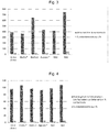
Zellen alleine BioOss® Algipore® NA1 NA2 243+/-9 % 216+/-3 % 240+/-8 % 291+/-9 % 443+/-15 % - Diese Ergebnisse sind in
Fig. 1 graphisch verdeutlicht. - BioOss® vermindert eher die Aktivität des für die Mineral- und Knochenbildung menschlicher Knochenzellen unverzichtbaren Enzyms alkalische Phosphatase, während das erfindungsgemäße Knochenersatzmaterial eine hochsignifikant gesteigerte Aktivität der von menschlichen Knochenzellen sezernierten alkalischen Phosphatase bewirkt.
- Die knochenspezifische alkalische Phosphatase als das wichtigste Osteoblasten-Marker Protein wird also durch das erfindungsgemäße Knochenersatzmaterial höchst signifikant stärker im menschlichen Knochenzellmodell stimuliert, als bei der Anwesenheit der herkömmlichen Materialien. Die unterschiedliche Wirkstärke von NA1 und NA2 auf die alkalische Phosphataseaktivität ist zurückführbar auf einen erhöhten Fluorid und Strontiumgehalt in NA2 gegenüber NA1.
- Bei der Messung der Aktivität der alkalischen Phosphatase im Zellkulturüberstand ist zu berücksichtigen, dass auch in vivo die Mineral- bzw. Knochenbildung extrazellulär durch die Sekretion der alkalischen Phosphatase in das osteoblastäre Mikromilieu erfolgt.
- Als einen Hinweis auf eine Wirkung der Knochenersatzmaterialien auf die Zellteilung wurde der Proteingehalt in den einzelnen "Löchern", das heißt Reaktionskammern, der verwendeten Multilochplatten analysiert. Dazu wurde ein Tritonextrakt aus den jeweiligen Löchern für die Bestimmung des Proteingehaltes nach dem BCA-Verfahren verwendet. Je höher der Proteingehalt in den einzelnen Löchern, umso mehr Zellmaterial, welches Proteinmaterial entspricht, muss in den Löchern vorhanden sein. Dies bedeutet, dass sich die Zellzahl erhöht hat, da eine Knochenzelle stets eine ähnliche Größe aufweist und Proteine nicht in größerem Ausmaß intrazellulär in Knochenzellen gespeichert werden. Eine Abnahme des Proteingehaltes in einem Zellkulturloch, also einer Reaktionskammer, wäre also gleichbedeutend mit einer Abnahme der Zellzahl, die adhärent am Zellkulturboden oder auf den Knochenersatzstoffen sitzen (apoptotische, d.h. abgestorbene Zellen, bleiben nicht adhärent und werden vor der Triton-Zugabe weggespült). Die Ergebnisse werden wieder in % der Kontrolle +/- Standardabweichung angegeben.
Zellen alleine BioOss® Algipore® NA1 NA2 96+/-3 % 108+/-4 % 103+/-3 % 103+/-3 % 103+/-3 % - Diese Ergebnisse sind in
Fig. 2 graphisch verdeutlicht. - Es gibt keinen Unterschied zwischen den untersuchten Materialien auf den Zellproteingehalt in den Zellkulturlöchern. Daher beeinflussen die Materialien die Zellteilung nicht, aber auch den Zelltod nicht, da apoptotisch degenerierende Zellen nicht adhärent bleiben, sondern sich ablösen und vor dem Assay-Verfahren weggespült würden.
- Diese Beobachtungen deuten darauf hin, dass die nach erfindungsgemäßen Verfahren gewonnenen Materialien einen sehr positiven Effekt auf die alkalische Phosphatase Aktivität menschlicher Knochenzellen haben.
- Diese Beobachtung ist vereinbar mit einem sehr günstigen und nachhaltigen, die Knochenmineralbildung in vivo stimulierenden Effekt des erfindungsgemäßen Knochenersatzmaterials, ohne dass die Zellteilung beeinflusst, also stimuliert oder gehemmt wird.
- Das bisher als Knochenersatzmaterial in der Zahnheilkunde oder Kieferchirurgie eingesetzte Material BioOss® besteht aus dem natürlichen Hydroxylapatitmaterial des anorganischen Knochengewebes von Rindern. Mit dem erfindungsgemäßen Verfahren kann auch bei einem natürlichen Calciumphosphat Kristallgitter ein teilweiser Austausch von Calciumionen im Hydroxylapatitkristall mit Strontium- und Fluoridionen kontrolliert durchgeführt werden. Hierbei sollten Calcium- und Phosphor-Atome in der Gesamtmischung der Autoklaveneinwaage höchstens im Verhältnis 10:5 vorliegen.
- In dem folgenden Experiment mit primären humanen osteoblastären Zellen wird die Wirkung des konventionellen BioOss® Materials parallel zu den Wirkungen des durch das erfindungsgemäßen Verfahren aktivierten BioOss1 Materials auf die alkalische Phosphatase Aktivität untersucht.
- Zugleich werden auch die oben bereits getesteten Materialien im gleichen Experiment eingesetzt, um eine Einordnung der Wirksamkeiten aller mit dem erfindungsgemäßen Verfahren hergestellten Materialien in einem parallelen Versuchsansatz zu ermöglichen. Es wurde auch hier die alkalische Phosphatase Aktivität im Zellkulturüberstand nach einer Inkubationszeit der Zellen mit dem neuen Material von 24 Stunden gemessen.
- Dieses Experiment wurde wieder mit primären menschlichen Knochenzellen eines anderen Individuums als bei den ersten Experimenten (1. Versuch) durchgeführt.
Alkalische Phosphatase Zellen alleine BioOss® BioOss1 Algipore® NA1 NA2 393+/-19 % 368+/-16 % 645+/-20 % 431 +/-24 % 418+/-9 % 747+/-51 % - Diese Ergebnisse sind in
Fig. 3 graphisch verdeutlicht.Zellteilung / Zellprotein Zellen alleine BioOss® BioOss1 Algipore® NA1 NA2 96+/-5 % 106+/-5 % 97+/-3 % 93+/-6 % 98+/-3 % 107+/-5 % - Diese Ergebnisse sind in
Fig. 4 graphisch verdeutlicht. - Während das konventionelle BioOss® Material keine signifikant stimulierende Wirkung auf die alkalische Phosphatase Aktivität in den Zellkulturüberständen im Vergleich zu humanen Knochenzellen ohne Kontakt zu einem Knochenersatzmaterial hat, bewirkt das durch das erfindungsgemäße Verfahren vorbehandelte BioOss® Material (BioOss1) fast eine Verdopplung der alkalischen Phosphatase Aktivität. Daher eignet sich das erfindungsgemäße Verfahren auch zur Aktivierung von kommerziell verfügbarem bovinen Hydroxylapatit-Material und verleiht dem bisher nur osteokonduktiven BioOss® Material osteoinduktive Eigenschaften.
- Im Übrigen reproduziert dieses Experiment die Ergebnisse des oben bereits analysierten ersten Versuchs: Kommerzielles BioOss® zeigt keine wesentliche Stimulation der Alkalische Phosphatase, während das mit dem erfindungsgemäßen Verfahren hergestellte BioOss1 eine fast doppelt so starke Aktivierung der Alkalische Phosphatase-Aktivität zeigt. Auch die mit dem erfindungsgemäßen Verfahren hergestellten "Algenhydroxylapatitmaterialen" NA1 und insbesondere NA2 zeigen eine noch potentere Stimulation der Alkalische Phosphatase-Aktivität.
- In diesem vergleichenden Experiment ergibt sich erneut kein sicherer Hinweis auf eine signifikant unterschiedliche Wirkung der getesteten Knochenersatzmaterialien auf die zelluläre Gesamt-Proteinproduktion in den Zellüberständen in den Reaktionskammern. (siehe
Fig 4 ) - In einem weiteren Ansatz werden alle Experimente nochmals mit anderen primären humanen osteoblastären Zellen eines dritten gesunden Spenders wiederholt und die Ergebnisse konsistenter Weise reproduziert. Es werden hier also nochmals die Wirkungen aller bisher nach dem erfindungsgemäßen Verfahren hergestellten Materialien parallel in einem weiteren Versuchsansatz reproduziert und mit den Wirkungen kommerziell verfügbarer Knochenersatzstoffe verglichen.
- Die alkalische Phosphatase Aktivitäten wurden wieder in den Zellkulturüberständen in den Reaktionskammern gemessen.
Alkalische Phosphatase Zellen alleine BioOss® BioOss1 Algipore® NA1 NA2 566+/-32 % 488+/-14 % 708+/-18 % 683+/-31 % 683+/-61 % 867+/-63 % - Diese Ergebnisse sind in
Fig. 5 graphisch verdeutlicht.Zellteilung / Zellprotein Zellen alleine BioOss® BioOss1 Algipore® NA1 NA2 96+/-5 % 113+/-4 % 98+/-5 % 103+/-3 % 100+/-7 % 102+/-3 % - Diese Ergebnisse sind in
Fig. 6 graphisch verdeutlicht. - Auch in diesem Experiment erweisen sich die durch das erfindungsgemäße Verfahren "aktivierten" Knochenersatzmaterialien NA2 und BioOss1 als signifikant stärker wirksam hinsichtlich der Stimulation der alkalischen Phosphatase Aktivität als die kommerziell verfügbaren Knochenersatzstoffe BioOss® und Algipore®. Das in einem initialen Prozess behandelte NA1 Material zeigt eine vergleichbare Wirkung wie das kommerziell erhältliche Algipore® Knochenersatzmaterial.
- Somit erweist sich jedes durch das erfindungsgemäße Herstellungsverfahren "aktivierte" Knochenersatzmaterial den bisherigen Produkten als hinsichtlich der Stimulation des osteoblastären Standard-Knochenanbaumarkers alkalische Phosphatase überlegen
- Das erfindungsgemäße Herstellungsverfahren ist also geeignet, sowohl während eines Umsetzungsprozess aus einem Kalziumkarbonat beziehungsweise einer Mischung aus Portlandit, Ätzkalk, und Kalzit in ein Apatitmaterial, als auch direkt aus einem bestehendem Hydroxylapatitmaterial, ein aktiviertes Knochenersatzmaterial durch das Einfügen von aktivierenden Ionen, wie beispielsweise Strontiumionen, in das Kristallgitter herzustellen. Als aktiviert im Rahmen der Erfindung kann insbesondere ein Material angesehen werden, welches osteoinduktive beziehungsweise osteotrope oder auch antiresorptive Eigenschaften besitzt.
- Somit wird mit dem erfindungsgemäßen Verfahren beziehungsweise mit dem durch das erfindungsgemäße Verfahren hergestellten osteotropen Knochenersatzmaterial ein Material geschaffen, welches langwirkende und örtlich begrenzte osteoinduktive beziehungsweise osteotrope Eigenschaften aufweist und sich hervorragend als Implantatmaterial im Knochengewebe eignet.
| NA1 | NA2 | BioOss1 | |
| Ausgangsmaterial [g] | 7.21 | 7.51 | 0.5 |
| Dauer [h:min] | 116.9666667 | 221.75 | 116 |
| Ammoniumhydrogenphosphat [g] | 10 | 10.001 | 10.0521 |
| Kaliumfluorid [g] | 0.4 | 0.821 | 1,996 |
| SrF2 [g] | 0.35 | 0.688 | 0.821 |
| Ammoniaklösung 25% [g] | 6g | 50,278 | 18.183 |
| H2O | auf 75% aufgefüllt | auf 75% aufgefüllt | auf 75% aufgefüllt |
| Autoklavvolumen [ml] | 57.7 | 57.7 | 57.7 |
Claims (15)
- Verfahren zum Herstellen eines osteotropen Knochenersatzmaterials aus einem Ausgangsmaterial, welches im Wesentlichen Portlandit, Ätzkalk, Aragonit; Kalzit und/oder Apatit, insbesondere Hydroxylapatit, aufweist,
wobei das Ausgangsmaterial mit einer Strontium-; Fluor- und/oder Galliumquelle in einen Autoklav eingebracht wird,
wobei beim Verwenden eines Ausgangsmaterials, welches im Wesentlichen Portlandit, Ätzkalk, Aragonit; Kalzit aufweist eine Phosphatquelle eingebracht wird,
wobei H2O als Bestandteil eines Lösungsmittels in den Autoklav zugegeben wird, wobei der pH-Wert in dem Autoklav in einem Bereich über 7 eingestellt wird,
wobei der geschlossene und gefüllte Autoklav für mindestens 1 Stunde erhitzt und anschließend abgekühlt wird, und
wobei anschließend der Inhalt des Autoklavs von Resten der Phosphor-, Strontium-, Fluor- und/oder Galliumquelle gereinigt wird. - Verfahren nach Anspruch 1,
dadurch gekennzeichnet,
dass der geschlossene und gefüllte Autoklav auf mindestens 30 Grad Celsius bevorzugt auf über 190 Grad Celsius erhitzt wird. - Verfahren nach Anspruch 1 oder 2,
dadurch gekennzeichnet,
dass der Inhalt des Autoklavs mechanisch mit einer Filtervorrichtung gereinigt wird. - Verfahren nach einem der Ansprüche 1 bis 3,
dadurch gekennzeichnet,
dass der Inhalt des Autoklavs mit H2O bis zum Erreichen eines pH-Wertes von bevorzugt kleiner als 8 gewaschen wird. - Verfahren nach einem der Ansprüche 1 bis 4,
dadurch gekennzeichnet,
dass zum Einstellen des pH-Wertes in dem Autoklav in einem Bereich über 7 eine basische Lösung, insbesondere eine Ammoniak-Lösung, verwendet wird. - Verfahren nach einem der Ansprüche 1 bis 5,
dadurch gekennzeichnet,
dass die Strontium-, Fluor- und/oder Galliumquelle im Überschuss in Bezug auf das Ausgangsmaterial eingebracht wird. - Verfahren nach einem der Ansprüche 1 bis 6,
dadurch gekennzeichnet,
dass als Phosphorquelle Ammoniumdihydrogenphosphat oder Diammoniumphosphat zugegeben wird. - Verfahren nach Anspruch 7,
dadurch gekennzeichnet,
dass Calcium und Phosphoratome in der Gesamtmischung höchstens im Verhältnis 10:5 (Atomverhältnis) vorliegen. - Verfahren nach einem der Ansprüche 1 bis 8,
dadurch gekennzeichnet,
dass Portlandit, Ätzkalk, Aragonit und/oder Kalzit in Form von gebrannten, ungebrannten und/oder chemisch aufbereiteten biogenen Skeletten verwendet werden. - Verfahren nach einem der Ansprüche 1 bis 8
dadurch gekennzeichnet,
dass als Apatit Wirbeltierknochen nach pyrolytischer oder chemischer Mazeration verwendet werden. - Verfahren nach einem der Ansprüche 1 bis 10,
dadurch gekennzeichnet,
dass der geschlossene und gefüllte Autoklav für mindestens 12 Stunde erhitzt wird. - Verfahren nach einem der Ansprüche 1 bis 11,
dadurch gekennzeichnet,
dass als Strontium-, Fluor- und/oder Galliumquelle ein leicht auswaschbares oder schwer wasserlösliches Material verwendet wird. - Verfahren nach einem der Ansprüche 1 bis 12,
dadurch gekennzeichnet,
dass die Strontium-, Fluor- und/oder Galliumquelle als Feststoff in einem Behältnis in den Autoklav zugegeben wird, und
dass das Behältnis den Austausch von Ionen der Strontium-, Fluor- und/oder Galliumquelle mit dem Lösungsmittel ermöglicht, wobei Feststoffe zurückgehalten werden. - Verfahren nach einem der Ansprüche 1 bis 13,
dadurch gekennzeichnet,
dass das Ausgangsmaterial vor oder nach dem Einbringen in den Autoklav und/oder der Inhalt des Autoklavs einer pyrolytischen Behandlung und/oder einem chemischen Reinigungsverfahren unterzogen wird. - Osteotropes Knochenersatzmaterial, insbesondere nach einem Verfahren nach einem der Ansprüche 1 bis 14, welches im Wesentlichen Apatit aufweist, wobei Strontium-, Fluor- und/oder Galliumionen in das Kristallgitter des Apatits eingebaut sind.
Priority Applications (9)
| Application Number | Priority Date | Filing Date | Title |
|---|---|---|---|
| ES19192028T ES2922210T3 (es) | 2019-08-16 | 2019-08-16 | Sustitución osteotrópica de hueso |
| PL19192028.9T PL3777904T3 (pl) | 2019-08-16 | 2019-08-16 | Osteotropowy substytut kości |
| EP19192028.9A EP3777904B1 (de) | 2019-08-16 | 2019-08-16 | Osteotroper knochenersatz |
| DK19192028.9T DK3777904T3 (da) | 2019-08-16 | 2019-08-16 | Osteotrop knogleerstatning |
| CA3143623A CA3143623A1 (en) | 2019-08-16 | 2020-08-14 | Osteotropic bone replacement |
| JP2021572842A JP7596315B2 (ja) | 2019-08-16 | 2020-08-14 | 骨親和性骨補填品 |
| CN202080044666.0A CN115151281A (zh) | 2019-08-16 | 2020-08-14 | 促骨性骨修复 |
| PCT/EP2020/072884 WO2021032628A1 (de) | 2019-08-16 | 2020-08-14 | Osteotroper knochenersatz |
| US17/619,952 US20220370681A1 (en) | 2019-08-16 | 2020-08-14 | Osteotropic bone replacement |
Applications Claiming Priority (1)
| Application Number | Priority Date | Filing Date | Title |
|---|---|---|---|
| EP19192028.9A EP3777904B1 (de) | 2019-08-16 | 2019-08-16 | Osteotroper knochenersatz |
Publications (2)
| Publication Number | Publication Date |
|---|---|
| EP3777904A1 true EP3777904A1 (de) | 2021-02-17 |
| EP3777904B1 EP3777904B1 (de) | 2022-04-13 |
Family
ID=67659183
Family Applications (1)
| Application Number | Title | Priority Date | Filing Date |
|---|---|---|---|
| EP19192028.9A Active EP3777904B1 (de) | 2019-08-16 | 2019-08-16 | Osteotroper knochenersatz |
Country Status (9)
| Country | Link |
|---|---|
| US (1) | US20220370681A1 (de) |
| EP (1) | EP3777904B1 (de) |
| JP (1) | JP7596315B2 (de) |
| CN (1) | CN115151281A (de) |
| CA (1) | CA3143623A1 (de) |
| DK (1) | DK3777904T3 (de) |
| ES (1) | ES2922210T3 (de) |
| PL (1) | PL3777904T3 (de) |
| WO (1) | WO2021032628A1 (de) |
Cited By (1)
| Publication number | Priority date | Publication date | Assignee | Title |
|---|---|---|---|---|
| EP4574180A1 (de) | 2023-12-19 | 2025-06-25 | Andreas Haas | Modifiziertes knochenersatzmaterial |
Families Citing this family (1)
| Publication number | Priority date | Publication date | Assignee | Title |
|---|---|---|---|---|
| WO2021240539A1 (en) * | 2020-05-25 | 2021-12-02 | SHREY, Dhawan | Ion-substituted bovine hydroxyapatite for bone regeneration |
Citations (4)
| Publication number | Priority date | Publication date | Assignee | Title |
|---|---|---|---|---|
| EP0028074A1 (de) | 1979-10-26 | 1981-05-06 | AMP INCORPORATED (a New Jersey corporation) | Drahtverbindungsapparat |
| EP0230570A1 (de) | 1985-12-03 | 1987-08-05 | Rolf Prof. Dr. Dr. Ewers | Poröses Hydroxylapatit-Material |
| EP2228080A1 (de) * | 2009-03-03 | 2010-09-15 | Graftys | Galliierte Calciumphosphat-Biomaterialien |
| CN101928136A (zh) * | 2010-07-16 | 2010-12-29 | 崔顺玉 | 氟化羟磷灰石制备方法及其用途 |
Family Cites Families (9)
| Publication number | Priority date | Publication date | Assignee | Title |
|---|---|---|---|---|
| DE3709897A1 (de) * | 1987-03-26 | 1988-10-06 | Ewers Rolf | Verfahren zur herstellung eines hydroxylapatitmaterials |
| GB9808189D0 (en) * | 1998-04-17 | 1998-06-17 | Royal Free Hosp School Med | Bone implant |
| DE19950113A1 (de) * | 1999-10-18 | 2001-05-03 | Jordanova Spassova Margarita | Tricalciumphosphat-haltiges Hydroxylapatit-Material |
| US6881227B2 (en) * | 1999-11-30 | 2005-04-19 | Margarita Jordanova-Spassova | Hydroxylapatite material containing tricalcium phosphate with microporous structure |
| DE10225420A1 (de) * | 2002-06-07 | 2003-12-24 | Sanatis Gmbh | Strontium-Apatit-Zement-Zubereitungen, die daraus gebildeten Zemente und die Verwendung davon |
| TW201200471A (en) * | 2010-06-21 | 2012-01-01 | Cheng-Chei Wu | The preparation of fluoridated hydroxyapatites and their applications |
| TW201200172A (en) * | 2010-06-21 | 2012-01-01 | Cheng-Chei Wu | The fluoridated hydroxyapatite composites having an action of enhancing the biological activity of human osteoblast cells, a process for the preparation thereof, and a pharmaceutical composition comprising them |
| EA035416B1 (ru) * | 2015-08-06 | 2020-06-10 | Гринбоун Орто С.Р.Л. | Большие трехмерные каркасы, выполненные из активного гидроксиапатита, полученного биоморфным превращением природных структур, и способ их получения |
| CN105712737A (zh) * | 2016-01-29 | 2016-06-29 | 云南省第一人民医院 | 一种骨修复用多孔锶掺杂羟基磷灰石材料的制备方法 |
-
2019
- 2019-08-16 PL PL19192028.9T patent/PL3777904T3/pl unknown
- 2019-08-16 ES ES19192028T patent/ES2922210T3/es active Active
- 2019-08-16 EP EP19192028.9A patent/EP3777904B1/de active Active
- 2019-08-16 DK DK19192028.9T patent/DK3777904T3/da active
-
2020
- 2020-08-14 CA CA3143623A patent/CA3143623A1/en active Pending
- 2020-08-14 WO PCT/EP2020/072884 patent/WO2021032628A1/de not_active Ceased
- 2020-08-14 CN CN202080044666.0A patent/CN115151281A/zh active Pending
- 2020-08-14 US US17/619,952 patent/US20220370681A1/en active Pending
- 2020-08-14 JP JP2021572842A patent/JP7596315B2/ja active Active
Patent Citations (4)
| Publication number | Priority date | Publication date | Assignee | Title |
|---|---|---|---|---|
| EP0028074A1 (de) | 1979-10-26 | 1981-05-06 | AMP INCORPORATED (a New Jersey corporation) | Drahtverbindungsapparat |
| EP0230570A1 (de) | 1985-12-03 | 1987-08-05 | Rolf Prof. Dr. Dr. Ewers | Poröses Hydroxylapatit-Material |
| EP2228080A1 (de) * | 2009-03-03 | 2010-09-15 | Graftys | Galliierte Calciumphosphat-Biomaterialien |
| CN101928136A (zh) * | 2010-07-16 | 2010-12-29 | 崔顺玉 | 氟化羟磷灰石制备方法及其用途 |
Non-Patent Citations (2)
| Title |
|---|
| DATABASE WPI Week 201127, Derwent World Patents Index; AN 2011-B19964, XP002797327 * |
| P. MELNIKOV ET AL: "Gallium-containing hydroxyapatite for potential use in orthopedics", MATERIALS CHEMISTRY AND PHYSICS, vol. 117, no. 1, 1 September 2009 (2009-09-01), Switzerland, Taiwan, Republic of China, pages 86 - 90, XP055497554, ISSN: 0254-0584, DOI: 10.1016/j.matchemphys.2009.05.046 * |
Cited By (2)
| Publication number | Priority date | Publication date | Assignee | Title |
|---|---|---|---|---|
| EP4574180A1 (de) | 2023-12-19 | 2025-06-25 | Andreas Haas | Modifiziertes knochenersatzmaterial |
| WO2025131925A1 (de) | 2023-12-19 | 2025-06-26 | Andreas Haas | Modifiziertes knochenersatzmaterial |
Also Published As
| Publication number | Publication date |
|---|---|
| EP3777904B1 (de) | 2022-04-13 |
| US20220370681A1 (en) | 2022-11-24 |
| ES2922210T3 (es) | 2022-09-09 |
| PL3777904T3 (pl) | 2022-08-22 |
| CA3143623A1 (en) | 2021-02-25 |
| JP2023503755A (ja) | 2023-02-01 |
| WO2021032628A1 (de) | 2021-02-25 |
| JP7596315B2 (ja) | 2024-12-09 |
| DK3777904T3 (da) | 2022-07-04 |
| CN115151281A (zh) | 2022-10-04 |
Similar Documents
| Publication | Publication Date | Title |
|---|---|---|
| EP0141004B1 (de) | Knochenersatzmaterial auf der Basis natürlicher Knochen | |
| EP0193588B1 (de) | Carbonatapatit enthaltendes mittel und die verwendung von carbonatapatit für implantate | |
| DE69621595T2 (de) | Knochentransplantat-verbindungen | |
| DE69922312T2 (de) | Keramikmaterial zur Osteoinduktion enthaltend Mikroporen an der Öberfläche von Makroporen | |
| DE69111021T2 (de) | Flexible Membrane hergestellt aus organischer Knochenmatrix zum Ausbessern und Wiederherstellen von Knochen. | |
| EP1206292B1 (de) | Resorbierbares knochenersatz- und knochenaufbaumaterial | |
| EP0475189B1 (de) | Implantatmaterial | |
| DE69430069T2 (de) | Förmige materialien auf der basis von länglichen knochenteilchen und verfahren zu ihrer herstellung | |
| DE60020526T2 (de) | Osteogene pastenzusammensetzungen und ihre verwendung | |
| DE3038047C2 (de) | Füllmaterial | |
| DE69733292T2 (de) | Verfahren zur in vitro Herstellung von Knochen | |
| EP0657178B1 (de) | Verfahren zur Herstellung von Implantatkeramikmaterial, insbesonders von Hydroxylapatit aufweisenden Implantatkeramikmaterial | |
| EP1227851B1 (de) | Resorbierbares knochen-implantatmaterial sowie verfahren zur herstellung desselben | |
| DE69427947T2 (de) | Verwendung von teilchen eines bioverträglichen und bioresorbierbaren kalziumsalzes als wirkstoff in der herstellung eines arzneimittels zur lokalen behandlung von entkalkungskrankheiten | |
| DE69928412T2 (de) | Knochenreparaturgemisch | |
| EP0605799A1 (de) | Hohlendoprothesen mit Knochenwachstumsfördernder Füllung | |
| DE69615859T2 (de) | Ersatz für knochentransplantat und seine herstellung | |
| DE68918043T2 (de) | Knochenersatzmaterial und Knochenzement. | |
| DE10061704A1 (de) | Verfahren und Vorrichtung zur Herstellung von biologischem Gewebe in einer Wachstumskammer | |
| EP3777904B1 (de) | Osteotroper knochenersatz | |
| EP1124593B1 (de) | Verfahren zur präparation von knochenmaterial | |
| DE2756256A1 (de) | Hilfsmittel zum bedecken und/oder ausfuellen von knochendefekten und verfahren zur herstellung desselben | |
| EP1732618B1 (de) | Verfahren zur herstellung eines knochen-implantatmaterials | |
| DE19917696A1 (de) | Mittel für die Herstellung biologischer Teile mit einem Wirkstoffkomplex und für diesen geeigneten Trägermaterialien | |
| EP3231453B1 (de) | Bone-sialopreotein(bsp)-funktionalisierte knochenersatzkörper |
Legal Events
| Date | Code | Title | Description |
|---|---|---|---|
| PUAI | Public reference made under article 153(3) epc to a published international application that has entered the european phase |
Free format text: ORIGINAL CODE: 0009012 |
|
| STAA | Information on the status of an ep patent application or granted ep patent |
Free format text: STATUS: REQUEST FOR EXAMINATION WAS MADE |
|
| 17P | Request for examination filed |
Effective date: 20200622 |
|
| AK | Designated contracting states |
Kind code of ref document: A1 Designated state(s): AL AT BE BG CH CY CZ DE DK EE ES FI FR GB GR HR HU IE IS IT LI LT LU LV MC MK MT NL NO PL PT RO RS SE SI SK SM TR |
|
| AX | Request for extension of the european patent |
Extension state: BA ME |
|
| STAA | Information on the status of an ep patent application or granted ep patent |
Free format text: STATUS: EXAMINATION IS IN PROGRESS |
|
| 17Q | First examination report despatched |
Effective date: 20210419 |
|
| GRAP | Despatch of communication of intention to grant a patent |
Free format text: ORIGINAL CODE: EPIDOSNIGR1 |
|
| STAA | Information on the status of an ep patent application or granted ep patent |
Free format text: STATUS: GRANT OF PATENT IS INTENDED |
|
| INTG | Intention to grant announced |
Effective date: 20220126 |
|
| GRAS | Grant fee paid |
Free format text: ORIGINAL CODE: EPIDOSNIGR3 |
|
| GRAA | (expected) grant |
Free format text: ORIGINAL CODE: 0009210 |
|
| STAA | Information on the status of an ep patent application or granted ep patent |
Free format text: STATUS: THE PATENT HAS BEEN GRANTED |
|
| AK | Designated contracting states |
Kind code of ref document: B1 Designated state(s): AL AT BE BG CH CY CZ DE DK EE ES FI FR GB GR HR HU IE IS IT LI LT LU LV MC MK MT NL NO PL PT RO RS SE SI SK SM TR |
|
| REG | Reference to a national code |
Ref country code: GB Ref legal event code: FG4D Free format text: NOT ENGLISH |
|
| REG | Reference to a national code |
Ref country code: CH Ref legal event code: EP |
|
| REG | Reference to a national code |
Ref country code: DE Ref legal event code: R096 Ref document number: 502019004038 Country of ref document: DE |
|
| REG | Reference to a national code |
Ref country code: IE Ref legal event code: FG4D Free format text: LANGUAGE OF EP DOCUMENT: GERMAN |
|
| REG | Reference to a national code |
Ref country code: AT Ref legal event code: REF Ref document number: 1482899 Country of ref document: AT Kind code of ref document: T Effective date: 20220515 |
|
| REG | Reference to a national code |
Ref country code: FI Ref legal event code: FGE |
|
| REG | Reference to a national code |
Ref country code: DK Ref legal event code: T3 Effective date: 20220701 |
|
| REG | Reference to a national code |
Ref country code: NL Ref legal event code: FP |
|
| REG | Reference to a national code |
Ref country code: SE Ref legal event code: TRGR |
|
| REG | Reference to a national code |
Ref country code: LT Ref legal event code: MG9D |
|
| REG | Reference to a national code |
Ref country code: NO Ref legal event code: T2 Effective date: 20220413 |
|
| REG | Reference to a national code |
Ref country code: ES Ref legal event code: FG2A Ref document number: 2922210 Country of ref document: ES Kind code of ref document: T3 Effective date: 20220909 |
|
| PG25 | Lapsed in a contracting state [announced via postgrant information from national office to epo] |
Ref country code: PT Free format text: LAPSE BECAUSE OF FAILURE TO SUBMIT A TRANSLATION OF THE DESCRIPTION OR TO PAY THE FEE WITHIN THE PRESCRIBED TIME-LIMIT Effective date: 20220816 Ref country code: LT Free format text: LAPSE BECAUSE OF FAILURE TO SUBMIT A TRANSLATION OF THE DESCRIPTION OR TO PAY THE FEE WITHIN THE PRESCRIBED TIME-LIMIT Effective date: 20220413 Ref country code: HR Free format text: LAPSE BECAUSE OF FAILURE TO SUBMIT A TRANSLATION OF THE DESCRIPTION OR TO PAY THE FEE WITHIN THE PRESCRIBED TIME-LIMIT Effective date: 20220413 Ref country code: GR Free format text: LAPSE BECAUSE OF FAILURE TO SUBMIT A TRANSLATION OF THE DESCRIPTION OR TO PAY THE FEE WITHIN THE PRESCRIBED TIME-LIMIT Effective date: 20220714 Ref country code: BG Free format text: LAPSE BECAUSE OF FAILURE TO SUBMIT A TRANSLATION OF THE DESCRIPTION OR TO PAY THE FEE WITHIN THE PRESCRIBED TIME-LIMIT Effective date: 20220713 |
|
| PG25 | Lapsed in a contracting state [announced via postgrant information from national office to epo] |
Ref country code: RS Free format text: LAPSE BECAUSE OF FAILURE TO SUBMIT A TRANSLATION OF THE DESCRIPTION OR TO PAY THE FEE WITHIN THE PRESCRIBED TIME-LIMIT Effective date: 20220413 Ref country code: LV Free format text: LAPSE BECAUSE OF FAILURE TO SUBMIT A TRANSLATION OF THE DESCRIPTION OR TO PAY THE FEE WITHIN THE PRESCRIBED TIME-LIMIT Effective date: 20220413 Ref country code: IS Free format text: LAPSE BECAUSE OF FAILURE TO SUBMIT A TRANSLATION OF THE DESCRIPTION OR TO PAY THE FEE WITHIN THE PRESCRIBED TIME-LIMIT Effective date: 20220813 |
|
| REG | Reference to a national code |
Ref country code: DE Ref legal event code: R097 Ref document number: 502019004038 Country of ref document: DE |
|
| PG25 | Lapsed in a contracting state [announced via postgrant information from national office to epo] |
Ref country code: SM Free format text: LAPSE BECAUSE OF FAILURE TO SUBMIT A TRANSLATION OF THE DESCRIPTION OR TO PAY THE FEE WITHIN THE PRESCRIBED TIME-LIMIT Effective date: 20220413 Ref country code: SK Free format text: LAPSE BECAUSE OF FAILURE TO SUBMIT A TRANSLATION OF THE DESCRIPTION OR TO PAY THE FEE WITHIN THE PRESCRIBED TIME-LIMIT Effective date: 20220413 Ref country code: RO Free format text: LAPSE BECAUSE OF FAILURE TO SUBMIT A TRANSLATION OF THE DESCRIPTION OR TO PAY THE FEE WITHIN THE PRESCRIBED TIME-LIMIT Effective date: 20220413 Ref country code: EE Free format text: LAPSE BECAUSE OF FAILURE TO SUBMIT A TRANSLATION OF THE DESCRIPTION OR TO PAY THE FEE WITHIN THE PRESCRIBED TIME-LIMIT Effective date: 20220413 Ref country code: CZ Free format text: LAPSE BECAUSE OF FAILURE TO SUBMIT A TRANSLATION OF THE DESCRIPTION OR TO PAY THE FEE WITHIN THE PRESCRIBED TIME-LIMIT Effective date: 20220413 |
|
| PLBE | No opposition filed within time limit |
Free format text: ORIGINAL CODE: 0009261 |
|
| STAA | Information on the status of an ep patent application or granted ep patent |
Free format text: STATUS: NO OPPOSITION FILED WITHIN TIME LIMIT |
|
| 26N | No opposition filed |
Effective date: 20230116 |
|
| PG25 | Lapsed in a contracting state [announced via postgrant information from national office to epo] |
Ref country code: MC Free format text: LAPSE BECAUSE OF FAILURE TO SUBMIT A TRANSLATION OF THE DESCRIPTION OR TO PAY THE FEE WITHIN THE PRESCRIBED TIME-LIMIT Effective date: 20220413 Ref country code: AL Free format text: LAPSE BECAUSE OF FAILURE TO SUBMIT A TRANSLATION OF THE DESCRIPTION OR TO PAY THE FEE WITHIN THE PRESCRIBED TIME-LIMIT Effective date: 20220413 |
|
| PG25 | Lapsed in a contracting state [announced via postgrant information from national office to epo] |
Ref country code: SI Free format text: LAPSE BECAUSE OF FAILURE TO SUBMIT A TRANSLATION OF THE DESCRIPTION OR TO PAY THE FEE WITHIN THE PRESCRIBED TIME-LIMIT Effective date: 20220413 |
|
| P01 | Opt-out of the competence of the unified patent court (upc) registered |
Effective date: 20230613 |
|
| PG25 | Lapsed in a contracting state [announced via postgrant information from national office to epo] |
Ref country code: CY Free format text: LAPSE BECAUSE OF FAILURE TO SUBMIT A TRANSLATION OF THE DESCRIPTION OR TO PAY THE FEE WITHIN THE PRESCRIBED TIME-LIMIT Effective date: 20220413 |
|
| PG25 | Lapsed in a contracting state [announced via postgrant information from national office to epo] |
Ref country code: MK Free format text: LAPSE BECAUSE OF FAILURE TO SUBMIT A TRANSLATION OF THE DESCRIPTION OR TO PAY THE FEE WITHIN THE PRESCRIBED TIME-LIMIT Effective date: 20220413 Ref country code: HU Free format text: LAPSE BECAUSE OF FAILURE TO SUBMIT A TRANSLATION OF THE DESCRIPTION OR TO PAY THE FEE WITHIN THE PRESCRIBED TIME-LIMIT; INVALID AB INITIO Effective date: 20190816 |
|
| PG25 | Lapsed in a contracting state [announced via postgrant information from national office to epo] |
Ref country code: MT Free format text: LAPSE BECAUSE OF FAILURE TO SUBMIT A TRANSLATION OF THE DESCRIPTION OR TO PAY THE FEE WITHIN THE PRESCRIBED TIME-LIMIT Effective date: 20220413 |
|
| PG25 | Lapsed in a contracting state [announced via postgrant information from national office to epo] |
Ref country code: BG Free format text: LAPSE BECAUSE OF FAILURE TO SUBMIT A TRANSLATION OF THE DESCRIPTION OR TO PAY THE FEE WITHIN THE PRESCRIBED TIME-LIMIT Effective date: 20220413 |
|
| PG25 | Lapsed in a contracting state [announced via postgrant information from national office to epo] |
Ref country code: BG Free format text: LAPSE BECAUSE OF FAILURE TO SUBMIT A TRANSLATION OF THE DESCRIPTION OR TO PAY THE FEE WITHIN THE PRESCRIBED TIME-LIMIT Effective date: 20220413 |
|
| PGFP | Annual fee paid to national office [announced via postgrant information from national office to epo] |
Ref country code: LU Payment date: 20250820 Year of fee payment: 7 Ref country code: NL Payment date: 20250821 Year of fee payment: 7 |
|
| PGFP | Annual fee paid to national office [announced via postgrant information from national office to epo] |
Ref country code: FI Payment date: 20250820 Year of fee payment: 7 Ref country code: ES Payment date: 20250917 Year of fee payment: 7 |
|
| PGFP | Annual fee paid to national office [announced via postgrant information from national office to epo] |
Ref country code: DK Payment date: 20250821 Year of fee payment: 7 Ref country code: DE Payment date: 20250827 Year of fee payment: 7 |
|
| PGFP | Annual fee paid to national office [announced via postgrant information from national office to epo] |
Ref country code: NO Payment date: 20250820 Year of fee payment: 7 |
|
| PGFP | Annual fee paid to national office [announced via postgrant information from national office to epo] |
Ref country code: TR Payment date: 20250808 Year of fee payment: 7 Ref country code: PL Payment date: 20250806 Year of fee payment: 7 Ref country code: IT Payment date: 20250829 Year of fee payment: 7 |
|
| PGFP | Annual fee paid to national office [announced via postgrant information from national office to epo] |
Ref country code: BE Payment date: 20250820 Year of fee payment: 7 Ref country code: GB Payment date: 20250822 Year of fee payment: 7 |
|
| PGFP | Annual fee paid to national office [announced via postgrant information from national office to epo] |
Ref country code: FR Payment date: 20250821 Year of fee payment: 7 Ref country code: AT Payment date: 20250819 Year of fee payment: 7 |
|
| PGFP | Annual fee paid to national office [announced via postgrant information from national office to epo] |
Ref country code: SE Payment date: 20250821 Year of fee payment: 7 Ref country code: CH Payment date: 20250901 Year of fee payment: 7 |
|
| PGFP | Annual fee paid to national office [announced via postgrant information from national office to epo] |
Ref country code: IE Payment date: 20250821 Year of fee payment: 7 |