EP3785603A1 - Procédé, appareil et système de détection par rétinophotographie sur la base de l'apprentissage automatique - Google Patents
Procédé, appareil et système de détection par rétinophotographie sur la base de l'apprentissage automatique Download PDFInfo
- Publication number
- EP3785603A1 EP3785603A1 EP19792203.2A EP19792203A EP3785603A1 EP 3785603 A1 EP3785603 A1 EP 3785603A1 EP 19792203 A EP19792203 A EP 19792203A EP 3785603 A1 EP3785603 A1 EP 3785603A1
- Authority
- EP
- European Patent Office
- Prior art keywords
- features
- fundus image
- classification model
- classification
- region
- Prior art date
- Legal status (The legal status is an assumption and is not a legal conclusion. Google has not performed a legal analysis and makes no representation as to the accuracy of the status listed.)
- Granted
Links
Images


Classifications
-
- A—HUMAN NECESSITIES
- A61—MEDICAL OR VETERINARY SCIENCE; HYGIENE
- A61B—DIAGNOSIS; SURGERY; IDENTIFICATION
- A61B3/00—Apparatus for testing the eyes; Instruments for examining the eyes
- A61B3/10—Objective types, i.e. instruments for examining the eyes independent of the patients' perceptions or reactions
- A61B3/12—Objective types, i.e. instruments for examining the eyes independent of the patients' perceptions or reactions for looking at the eye fundus, e.g. ophthalmoscopes
-
- G—PHYSICS
- G06—COMPUTING OR CALCULATING; COUNTING
- G06F—ELECTRIC DIGITAL DATA PROCESSING
- G06F18/00—Pattern recognition
- G06F18/20—Analysing
- G06F18/24—Classification techniques
-
- G—PHYSICS
- G06—COMPUTING OR CALCULATING; COUNTING
- G06V—IMAGE OR VIDEO RECOGNITION OR UNDERSTANDING
- G06V10/00—Arrangements for image or video recognition or understanding
- G06V10/40—Extraction of image or video features
- G06V10/46—Descriptors for shape, contour or point-related descriptors, e.g. scale invariant feature transform [SIFT] or bags of words [BoW]; Salient regional features
- G06V10/462—Salient features, e.g. scale invariant feature transforms [SIFT]
-
- G—PHYSICS
- G06—COMPUTING OR CALCULATING; COUNTING
- G06V—IMAGE OR VIDEO RECOGNITION OR UNDERSTANDING
- G06V10/00—Arrangements for image or video recognition or understanding
- G06V10/40—Extraction of image or video features
- G06V10/46—Descriptors for shape, contour or point-related descriptors, e.g. scale invariant feature transform [SIFT] or bags of words [BoW]; Salient regional features
- G06V10/462—Salient features, e.g. scale invariant feature transforms [SIFT]
- G06V10/464—Salient features, e.g. scale invariant feature transforms [SIFT] using a plurality of salient features, e.g. bag-of-words [BoW] representations
-
- G—PHYSICS
- G06—COMPUTING OR CALCULATING; COUNTING
- G06V—IMAGE OR VIDEO RECOGNITION OR UNDERSTANDING
- G06V30/00—Character recognition; Recognising digital ink; Document-oriented image-based pattern recognition
- G06V30/10—Character recognition
- G06V30/18—Extraction of features or characteristics of the image
- G06V30/18143—Extracting features based on salient regional features, e.g. scale invariant feature transform [SIFT] keypoints
- G06V30/18152—Extracting features based on a plurality of salient regional features, e.g. "bag of words"
-
- G—PHYSICS
- G06—COMPUTING OR CALCULATING; COUNTING
- G06V—IMAGE OR VIDEO RECOGNITION OR UNDERSTANDING
- G06V40/00—Recognition of biometric, human-related or animal-related patterns in image or video data
- G06V40/10—Human or animal bodies, e.g. vehicle occupants or pedestrians; Body parts, e.g. hands
- G06V40/18—Eye characteristics, e.g. of the iris
- G06V40/193—Preprocessing; Feature extraction
-
- A—HUMAN NECESSITIES
- A61—MEDICAL OR VETERINARY SCIENCE; HYGIENE
- A61B—DIAGNOSIS; SURGERY; IDENTIFICATION
- A61B3/00—Apparatus for testing the eyes; Instruments for examining the eyes
- A61B3/10—Objective types, i.e. instruments for examining the eyes independent of the patients' perceptions or reactions
- A61B3/14—Arrangements specially adapted for eye photography
Definitions
- the present invention relates to the field of medical image recognition technology, and specifically to a method, apparatus and system for detecting a fundus image based on machine learning.
- the deep learning technology can detect a certain feature of a fundus image more accurately.
- a deep learning model is trained using a large number of samples having a feature of macular holes, and macular hole detection is performed on the fundus image by using the trained model.
- These technologies are often limited to the detection of a single feature or a few associated features, and cannot accurately detect other features.
- the eye is a very fine and complex organ in the human body and contains a wide variety of features, and the features are often greatly different.
- the detection results are difficult to converge with the use of the existing detection technologies, and the detection results are inaccurate accordingly.
- a model is trained for detecting each feature or features, which not only requires a large number of samples, but also sharply increases the amount of calculation in the presence of numerous features, resulting in a decrease in the detection efficiency.
- a method for detecting a fundus image based on machine learning including:
- an apparatus for detecting a fundus image based on machine learning including:
- an electronic device including at least one processor; and a memory connected to the at least one processor by communication; wherein the memory stores instructions executable by the one processor, and the instructions are executed by the at least one processor to cause the at least one processor to implement the method for detecting a fundus image based on machine learning in the first aspect.
- a computer storage medium storing instructions thereon that, when running on a computer, causing the computer to implement the method for detecting a fundus image in the first aspect.
- a computer program product including instructions, when running on a computer, causing the computer to implement the method for detecting a fundus image in the first aspect.
- a system for detecting a fundus image based on machine learning including:
- the entire region of the fundus image to be detected is for the detection of features from the first feature set with high saliency, and the specific region of the fundus image is simultaneously detected for features of the second feature set with low saliency, so that the two classification models do not interfere with each other and work clearly, and each region can be accurately classified to determine whether it contains relevant features; and the detection result is finally obtained by judgment in combination with the classification results of the two features to improve the accuracy of the final result, thereby achieving simultaneous analysis of features of multiple categories and multiple saliencies, with higher efficiency.
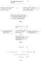
- An embodiment of the present invention provides a method for detecting a fundus image based on machine learning, which can be executed by a computer or a server. As shown in Fig. 1 , the method may include the following steps:
- the first features may include a large area of abnormal tissue or structure within the fundus, large spots within the fundus, and the like, such as image features related to lesions such as leopard fundus, fundus white spots, and fundus laser spots.
- the first features are detected using a machine learning algorithm.
- the classification model should be trained using a large number of fundus image samples having various first features, so that the classification model has a certain capability of classification.
- the first classification model may be a single-classification model or a multi-classification model. If it is a single classification model, the output result is two classes, that is, containing or not containing the first features; if it is a multi-classification model, the output result is multiple classes, that is, not containing any first feature or the class of the contained first feature.
- Steps S12 and S13 are preferably executed synchronously or executed in any order.
- the second feature should be interpreted as a detailed feature, and the saliency of the first feature is greater than that of the second feature.
- the chromatic aberration, contrast, grayscale, or area of the second features is smaller than that of the first features.
- the first classification model and the second classification model both detect the second features, but the first classification model is less sensitive to the second features, while the second classification model is more sensitive.
- the second features are in the specific region.
- the specific region includes at least one of an optic disc region, a macular region, a blood vessel region and a retinal region, and may also be one region or a plurality of regions within a set range.
- the specific region is an optic disc region
- the second features include a specific feature such as shape abnormality of the optic disc, color abnormality of the optic disc, or abnormality of the optic nerve; alternatively, the specific region is macular, and the second feature includes a specific feature such as macular structural abnormality, or macular shape abnormality; alternatively, the specific region is a blood vessel region, and the second features include a specific feature such as color abnormality of the blood vessel, trend abnormality of the blood vessel, shape abnormality of the central vein, or shape abnormality of the branch vein; alternatively, the specific region is a retinal region, and the second features include small abnormal points such as color abnormal points, irregular points, or reduction of the retinal region.
- the second features may also include features of other details in the fundus, such
- the second features are processed using a machine learning algorithm.
- the corresponding classification model should be trained using a large number of fundus image samples having various second features, so that the classification model has a certain capability of classification.
- a plurality of second classification models are used for parallel classification detection for different specific regions, and each of the second classification models independently outputs a classification result.
- three-second classification models are used: the classification model A is used for an optic disc region and detects whether the optic disc region contains specific features related to the optic disc, for example, features of various optic disc lesions such as papilledema, papillitis, and optic atrophy; the classification model B is used for a macular region and detects whether the macular region contains specific features related to the macula, for example, features of various macular lesions such as macular holes, macular edema, and cartographic atrophy of the macular region; and the classification model C is used for a blood vessel region and detects whether the blood vessel region contains specific features related to the blood vessel, for example, features of various blood vessel lesions such as vitreous hemorrhage, choroidal hemangioma, central vein occlusion, and branch vein occlusion.
- the second classification model may be configured to output binary classification results to indicate the presence or absence of the second features of the fundus image.
- the second classification models may be configured to output multi-classification results to indicate that the fundus image does not contain any second feature, or the specific class of the contained second features.
- the output of the multi-classification results or the single-classification results may be determined according to whether the various specific classes obtained by the second classification model conflict.
- S14 determining a analysis result at least according to the classification results of the first classification model and the second classification model.
- This step can completely follow the classification results of the two classification models, that is, directly output their classification results, or judge the classification results of the two classification models to obtain the final analysis result.
- the so-called judgment refers to determine a comprehensive result according to the combination of the classification results output by the first classification model and the second classification model.
- the final result may be inconsistent with the classification results output by the two classification models.
- the classification results of the first classification model and the second classification model are classified through a machine learning algorithm, and the classification result is used as the final result.
- a decision module is introduced in this step, and this model is also a classification model, specifically a binary-classification model or a multi-classification model. It should be noted that the input data of the decision module is label information rather than the fundus image, and the content of the label information is whether it has any first feature and second feature, or the specific classes of the first features and the second features.
- the classification result output by the classification model is generally a numerical value, specifically confidence information or probability expressed by 0-1.
- the value output by the classification model can be used as the final result, the value can also be further judged, and a corresponding result is determined based on the value.
- sample data should be used to train the decision module so that it has a certain classification ability.
- the sample data should include information about whether it has any first feature and second feature and its corresponding label or include information about the specific classes of the first features and the second features and its corresponding label.
- the entire region of the fundus image to be detected is detected by the first features having high saliency, and the specific region of the fundus image is simultaneously detected by the second features having low saliency so that the two classification models do not interfere with each other and work clearly, and each region can be accurately classified to determine whether it contains relevant features; and the result is finally obtained by judgment in combination with the classification results of the two features to improve the accuracy of the final result, thereby achieving simultaneous analysis of features of multiple categories and multiple saliencies, with higher efficiency.
- the fundus photos taken by an image shooter are very different in quality, the photos are often overexposed, gray, and blurry, which greatly increases the difficulty of machine learning judgment.
- the quality of images is analysed to screen qualified images, which further ensures the accuracy of image analysis.
- the fundus image may be subjected to any one or any combination analysis of stain/ bright spot, exposure, sharpness, light leakage, and local shadow.
- a plurality of images to be detected are weighted and averaged to obtain an average image, and whether the average image has pixels exceeding a preset brightness range is then judged; when the average image has pixels exceeding the preset brightness range, it is confirmed that the image to be detected has stains/ bright spots.
- the detection of stains or bright spots can be completed.
- the image to be detected is binarized to obtain a preset region in the image; a mask based on the boundary of the preset region is generated; the mask is fused with the image to be detected; the average color brightness of the image after fusion is calculated and compared with a preset color brightness threshold the degree of light leakage of the image to be detected is confirmed according to the comparison result.
- the degree of light leakage is greater than a preset value, it can be confirmed that the fundus image has light leakage.
- a histogram of any color channel in the image to be detected is counted; the number of pixels smaller than a preset pixel value is counted; whether the number of pixels smaller than the preset pixel value is less than a preset number is judged; and when the number of pixels smaller than the preset pixel value is less than the preset number, it is confirmed that the image to be detected has a local shadow.
- a high-frequency component of the image to be detected is extracted; an amount of information of the high-frequency component is calculated, and the sharpness of the image to be detected is confirmed based on the amount of information of the high-frequency component.
- the image to be detected is converted into a gray image; a root mean square of a histogram of the gray image is counted and the exposure of the image to be detected is confirmed based on the root mean square.
- the detection result of the image may be affected and may be inaccurate. Therefore, to ensure the detection accuracy of the image, the image having the above quality defects may be removed before the classification operation.
- an embodiment of the present invention further provides a method for detecting a fundus image. As shown in Fig. 2 , the method includes the following steps:
- the third classification model may be configured to output a binary classification result to indicate the presence or absence of the third features of the fundus image.
- the third classification model may be configured to output a multi-classification result to indicate that the fundus image does not contain any third feature, or the specific class of the contained third features.
- step S25 determining a detection result according to the classification results of the first classification model, the second classification model, and the third classification model.
- the classification result of the third features is further added here, so that the detection result is more accurate.
- the input data of the decision model is whether it has any first feature, second feature, and third feature, or the specific classes of the first features, the second features, and the third features.
- the above various classification models may be implemented by a convolutional neural network.
- the basic units of the convolutional neural network include convolutional layers, activation function (ReLu) layers, and pooling layers.
- the convolutional layers screen-specific image features, the activation function layers nonlinearly process the screened features by using a ReLu activation function, and the pooling layers extract the strongest information at different locations using max pooling.
- Batch normalization may be used during information extraction to improve the capacity of the network while preventing gradient dispersion in the process of training the network.
- the features in the fundus image can be extracted and finally output by fully connected layers and an output layer (softmax).
- the number of network layers of each module varies from 15 to 100 layers based on the classes of fundus features that need to be detected.
- the convolutional neural network may be implemented as the following structure: input layer - C1 - BN1 - R1 - P1 - C2 - BN2 - R2 - P2 - C3 - BN3 - R3 - P3 - C4 - BN4 - R4 - P4 - C5- BN5 - R5 - P5 - FC1 - FC2 - SoftMax.
- C represents a convolutional layer (e.g., C1, C2, C3, C4, C5)
- BN represents a batch normalization layer (e.g., BN1, BN2, BN3, BN4, BN5)
- R represents a function activation layer (e.g., R1, R2, R3, R4, R5)
- P represents a pooling layer (e.g., P1, P2, P3, P4, P5)
- the fully connected layers include FC1 and FC2, and SoftMax provides an output.
- the convolutional neural network used in the present embodiment is not limited to the structure of the convolutional neural network described above, and other neural network structures satisfying the present embodiment are also applicable.
- the sizes of hidden layers of the neural network can be changed according to the saliency of the features, the hidden layers being from the input to the output. Specifically, small hidden layers are used for the features having high saliency, while large hidden layers are used for the features having low saliency.
- the maximum hidden layer of the convolutional network for the second feature and third feature set having low saliency is larger than the maximum hidden layer of the convolutional network for the first features.
- the maximum hidden layer of the network is required to be small, for example, less than 200 ⁇ 200, facilitating feature extraction.
- the output of the maximum hidden layer should be kept large, for example, more than 300 ⁇ 300, ensuring that fine fundus sub-features such as small exudation points and bleeding points can be extracted.
- the output of the hidden layer with a maximum size is determined by the image input layer, the convolutional layers, and the pooling layers together, and is implemented in various ways, and details are not described herein again.
- the detecting apparatus includes an acquiring module 10, configured to acquire a fundus image to be detected a first classification module 20, configured to classify the entire region of the fundus image to determine whether the fundus image contains any first feature at least one-second classification model 30, configured to classify a specific region in the fundus image to determine whether the fundus image contains any second feature, wherein the saliency of the first features is greater than that of the second features; and a decision module 40, configured to determine a detection result at least according to the classification results of the first classification model and the second classification model.
- the plurality of second classification models are respectively configured to classify different specific regions and output second classification results, the second classification results being used to indicate whether the fundus image contains second features related to the specific regions.
- the first classification model and the second classification model are both multi-classification models, and the classification results thereof are used to indicate whether the fundus image contains the first features and the second features, and the specific classes of the first features and the second features.
- the detecting apparatus further includes:
- the third classification model is a multi-classification model, and the classification result thereof is used to indicate whether the fundus image contains any third feature and the specific class of the third features.
- the specific region includes at least one of an optic disc region, a macular region, a blood vessel region, and a retinal region.
- the first features, the second features, and the third features are all fundus lesion features.
- An electronic device may be a server or a terminal. As shown in Fig. 5 , a controller is included, the controller includes one or more processors 41 and a memory 42, and one processor 43 is taken as an example in Fig. 5 .
- the electronic device may further include an input apparatus 43 and an output apparatus 44.
- the processor 41, the memory 42, the input apparatus 43, and the output apparatus 44 may be connected by a bus or other means, exemplified by a bus in Fig. 4 .
- the processor 41 may be a Central Processing Unit (CPU).
- the processor may be other general-purpose processors, such as Digital Signal Processor (DSP), Application Specific Integrated Circuit (ASIC), Field-Programmable Gate Array (FPGA) or other programmable logic devices, such as discrete gate or transistor logic device, a discrete hardware component or other chips, or a combination of the various chips.
- DSP Digital Signal Processor
- ASIC Application Specific Integrated Circuit
- FPGA Field-Programmable Gate Array
- the general-purpose processor may be a microprocessor or any conventional processor, etc.
- the memory 42 can be used for storing non-transitory software programs, non-transitory computer-executable programs, and modules.
- the processor 41 runs the non-transitory software programs, instructions, and modules stored in the memory 42 to execute various function applications of the server and data processing, that is, to implement the method for detecting a fundus image in the above method embodiments.
- the memory 42 may include a program storage region and a data storage region.
- the program storage region may store an operating system, and an application program required by at least one function.
- the data storage region may store data created according to the use of processing apparatuses for the server.
- memory 42 may include a high-speed random access memory, and may also include a non-transitory memory, for example, at least one magnetic disk storage device, a flash memory, or other non-transitory solid-state storage devices.
- the memory 42 may alternatively include memories remotely disposed relative to the processor 41, and these remote memories may be connected to a network connecting apparatus through a network. Examples of the network include, but are not limited to, the Internet, an intranet, a local area network, a mobile communications network, or a combination thereof.
- the input apparatus 43 may receive input digit or character information, and generate a key signal input related to the user setting and function control of the processing apparatuses of the server.
- the output apparatus 44 may include a display device such as a display screen.
- One or more modules are stored in the memory 42, and when executed by one or more processors 41, implement the method as shown in Fig. 1 or 2 .
- An embodiment of the present invention further provides a system for analysing a fundus image based on machine learning.
- the system includes an image acquiring apparatus 100, configured to acquire a fundus image.
- the image acquiring apparatus may be plural.
- the image acquiring apparatus 100 is a fundus shooting device in each hospital, or a fundus shooting device of an individual user.
- the fundus analysis system further includes a cloud server 200.
- An apparatus for analysing a fundus image for executing the method for analysing a fundus image is provided in the cloud server 200.
- the cloud server 200 communicates with the image acquiring apparatus 100, for example, in the form of wireless communication, or wired communication.
- the fundus image acquired by the image acquiring apparatus 100 is uploaded to the cloud server 200, an electronic device executes the method for analysing a fundus image to obtain a analysis result, and an output apparatus outputs the analysis result.
- the output apparatus 300 may be a display device, or a printing device for printing in the form of a report, or a user terminal device, such as a mobile phone, tablet, or personal computer.
- the embodiments of the present invention may be provided as a method, a system, or a computer program product. Therefore, the present invention may be in the form of a full hardware embodiment, a full software embodiment, or an embodiment combining software and hardware. Besides, the present invention may be in the form of a computer program product implemented on one or more computer available storage media (including but not limited to a disk memory, a CD-ROM, an optical memory, and the like) including computer available program codes.
- These computer program instructions may also be stored in a computer-readable memory that can guide a computer or other programmable data processing equipment to work in a specific manner, so that the instructions stored in the computer-readable memory generate a product including an instruction device, where the instruction device implements functions specified in one or more processes in the flowcharts and/or one or more blocks in the block diagrams.
- These computer program instructions may also be loaded into a computer or other programmable data processing equipment, so that a series of operation steps are performed on the computer or other programmable data processing device to generate processing implemented by a computer, and instructions executed on the computer or other programmable data processing equipment provide steps for implementing functions specified in one or more processes in the flowcharts and/or one or more blocks in the block diagrams.
Landscapes
- Engineering & Computer Science (AREA)
- Health & Medical Sciences (AREA)
- Life Sciences & Earth Sciences (AREA)
- Physics & Mathematics (AREA)
- Theoretical Computer Science (AREA)
- General Physics & Mathematics (AREA)
- Computer Vision & Pattern Recognition (AREA)
- Multimedia (AREA)
- General Health & Medical Sciences (AREA)
- Ophthalmology & Optometry (AREA)
- Veterinary Medicine (AREA)
- Heart & Thoracic Surgery (AREA)
- Surgery (AREA)
- Public Health (AREA)
- Molecular Biology (AREA)
- Biophysics (AREA)
- Biomedical Technology (AREA)
- Animal Behavior & Ethology (AREA)
- Medical Informatics (AREA)
- Data Mining & Analysis (AREA)
- Bioinformatics & Computational Biology (AREA)
- Evolutionary Biology (AREA)
- Evolutionary Computation (AREA)
- General Engineering & Computer Science (AREA)
- Bioinformatics & Cheminformatics (AREA)
- Artificial Intelligence (AREA)
- Human Computer Interaction (AREA)
- Eye Examination Apparatus (AREA)
- Image Analysis (AREA)
Applications Claiming Priority (2)
| Application Number | Priority Date | Filing Date | Title |
|---|---|---|---|
| CN201810387484.8A CN108577803B (zh) | 2018-04-26 | 2018-04-26 | 基于机器学习的眼底图像检测方法、装置及系统 |
| PCT/CN2019/084210 WO2019206209A1 (fr) | 2018-04-26 | 2019-04-25 | Procédé, appareil et système de détection par rétinophotographie sur la base de l'apprentissage automatique |
Publications (3)
| Publication Number | Publication Date |
|---|---|
| EP3785603A1 true EP3785603A1 (fr) | 2021-03-03 |
| EP3785603A4 EP3785603A4 (fr) | 2021-06-23 |
| EP3785603B1 EP3785603B1 (fr) | 2023-08-02 |
Family
ID=63610363
Family Applications (1)
| Application Number | Title | Priority Date | Filing Date |
|---|---|---|---|
| EP19792203.2A Active EP3785603B1 (fr) | 2018-04-26 | 2019-04-25 | Procédé, appareil et système de détection par rétinophotographie sur la base de l'apprentissage automatique |
Country Status (3)
| Country | Link |
|---|---|
| EP (1) | EP3785603B1 (fr) |
| CN (1) | CN108577803B (fr) |
| WO (1) | WO2019206209A1 (fr) |
Cited By (1)
| Publication number | Priority date | Publication date | Assignee | Title |
|---|---|---|---|---|
| TWI921190B (zh) | 2025-05-14 | 2026-04-01 | 明志科技大學 | 用於眼底影像的影像處理系統及影像處理方法 |
Families Citing this family (19)
| Publication number | Priority date | Publication date | Assignee | Title |
|---|---|---|---|---|
| CN108577803B (zh) * | 2018-04-26 | 2020-09-01 | 上海鹰瞳医疗科技有限公司 | 基于机器学习的眼底图像检测方法、装置及系统 |
| CN109528155B (zh) * | 2018-11-19 | 2021-07-13 | 复旦大学附属眼耳鼻喉科医院 | 一种适用于高度近视并发开角型青光眼的智能筛查系统及其建立方法 |
| CN111374632B (zh) * | 2018-12-29 | 2023-05-26 | 张国明 | 视网膜病变检测方法、装置及计算机可读存储介质 |
| CN109859836B (zh) * | 2019-01-15 | 2022-03-22 | 上海鹰瞳医疗科技有限公司 | 医疗图像识别方法和设备 |
| CN109993731A (zh) * | 2019-03-22 | 2019-07-09 | 依未科技(北京)有限公司 | 一种眼底病变分析方法及装置 |
| CN109961848B (zh) * | 2019-04-02 | 2022-03-22 | 北京鹰瞳科技发展股份有限公司 | 黄斑图像分类方法和设备 |
| CN110428421A (zh) * | 2019-04-02 | 2019-11-08 | 上海鹰瞳医疗科技有限公司 | 黄斑图像区域分割方法和设备 |
| CN110021052B (zh) * | 2019-04-11 | 2023-05-30 | 北京百度网讯科技有限公司 | 用于生成眼底图像生成模型的方法和装置 |
| CN111242920A (zh) * | 2020-01-10 | 2020-06-05 | 腾讯科技(深圳)有限公司 | 一种生物组织图像检测方法、装置、设备及介质 |
| CN111291667A (zh) * | 2020-01-22 | 2020-06-16 | 上海交通大学 | 细胞视野图的异常检测方法及存储介质 |
| CN111696100A (zh) * | 2020-06-17 | 2020-09-22 | 上海鹰瞳医疗科技有限公司 | 基于眼底影像确定吸烟程度的方法及设备 |
| CN113947124B (zh) * | 2020-06-30 | 2025-04-25 | 中移(成都)信息通信科技有限公司 | 眼底彩色图像分类模型训练方法及眼底彩色图像分类方法 |
| CN112190227B (zh) * | 2020-10-14 | 2022-01-11 | 北京鹰瞳科技发展股份有限公司 | 眼底相机及其使用状态检测方法 |
| CN112905828B (zh) * | 2021-03-18 | 2023-06-16 | 西北大学 | 一种结合显著特征的图像检索器、数据库及检索方法 |
| CN113344894B (zh) * | 2021-06-23 | 2024-05-14 | 依未科技(北京)有限公司 | 眼底豹纹斑特征提取及特征指数确定的方法和装置 |
| CN113361487B (zh) * | 2021-07-09 | 2024-09-06 | 无锡时代天使医疗器械科技有限公司 | 异物检测方法、装置、设备及计算机可读存储介质 |
| CN114494195B (zh) * | 2022-01-26 | 2024-06-04 | 南通大学 | 用于眼底图像分类的小样本注意力机制并行孪生方法 |
| CN117315761A (zh) * | 2023-01-09 | 2023-12-29 | 华东师范大学 | 一种便携式智能眼底病灶检测装置及病灶图像检测方法 |
| CN117372284B (zh) * | 2023-12-04 | 2024-02-23 | 江苏富翰医疗产业发展有限公司 | 眼底图像处理方法及系统 |
Family Cites Families (13)
| Publication number | Priority date | Publication date | Assignee | Title |
|---|---|---|---|---|
| JP5387147B2 (ja) * | 2009-06-03 | 2014-01-15 | 日本電気株式会社 | 病理画像診断システム、病理画像処理方法、病理画像診断プログラム |
| TWI578977B (zh) * | 2011-04-07 | 2017-04-21 | 香港中文大學 | 視網膜圖像分析裝置 |
| CN102722735A (zh) * | 2012-05-24 | 2012-10-10 | 西南交通大学 | 一种融合全局和局部特征的内镜图像病变检测方法 |
| US20140314288A1 (en) * | 2013-04-17 | 2014-10-23 | Keshab K. Parhi | Method and apparatus to detect lesions of diabetic retinopathy in fundus images |
| US9462945B1 (en) * | 2013-04-22 | 2016-10-11 | VisionQuest Biomedical LLC | System and methods for automatic processing of digital retinal images in conjunction with an imaging device |
| EP4057215A1 (fr) * | 2013-10-22 | 2022-09-14 | Eyenuk, Inc. | Systèmes et procédés d'analyse automatisée d'images rétiniennes |
| CN107209933A (zh) * | 2014-08-25 | 2017-09-26 | 新加坡科技研究局 | 用于评估视网膜图像以及从视网膜图像获得信息的方法和系统 |
| CN104301585A (zh) * | 2014-09-24 | 2015-01-21 | 南京邮电大学 | 一种运动场景中特定种类目标实时检测方法 |
| CN105411525B (zh) * | 2015-11-10 | 2017-05-31 | 广州河谷互动医疗科技有限公司 | 一种眼底照片图像智能获取识别系统 |
| CN107346409B (zh) * | 2016-05-05 | 2019-12-17 | 华为技术有限公司 | 行人再识别方法和装置 |
| CN107146231B (zh) * | 2017-05-04 | 2020-08-07 | 季鑫 | 视网膜图像出血区域分割方法、装置和计算设备 |
| CN108596895B (zh) * | 2018-04-26 | 2020-07-28 | 上海鹰瞳医疗科技有限公司 | 基于机器学习的眼底图像检测方法、装置及系统 |
| CN108577803B (zh) * | 2018-04-26 | 2020-09-01 | 上海鹰瞳医疗科技有限公司 | 基于机器学习的眼底图像检测方法、装置及系统 |
-
2018
- 2018-04-26 CN CN201810387484.8A patent/CN108577803B/zh active Active
-
2019
- 2019-04-25 EP EP19792203.2A patent/EP3785603B1/fr active Active
- 2019-04-25 WO PCT/CN2019/084210 patent/WO2019206209A1/fr not_active Ceased
Cited By (1)
| Publication number | Priority date | Publication date | Assignee | Title |
|---|---|---|---|---|
| TWI921190B (zh) | 2025-05-14 | 2026-04-01 | 明志科技大學 | 用於眼底影像的影像處理系統及影像處理方法 |
Also Published As
| Publication number | Publication date |
|---|---|
| CN108577803B (zh) | 2020-09-01 |
| CN108577803A (zh) | 2018-09-28 |
| WO2019206209A1 (fr) | 2019-10-31 |
| EP3785603B1 (fr) | 2023-08-02 |
| EP3785603A4 (fr) | 2021-06-23 |
Similar Documents
| Publication | Publication Date | Title |
|---|---|---|
| EP3785603B1 (fr) | Procédé, appareil et système de détection par rétinophotographie sur la base de l'apprentissage automatique | |
| US11501428B2 (en) | Method, apparatus and system for detecting fundus image based on machine learning | |
| US11410277B2 (en) | Method and device for blurring image background, storage medium and electronic apparatus | |
| US9076198B2 (en) | Information processing apparatus, information processing system, information processing method, program and recording medium | |
| CN109697719B (zh) | 一种图像质量评估方法、装置及计算机可读存储介质 | |
| CN106920219B (zh) | 物品缺陷检测方法、图像处理系统与计算机可读记录介质 | |
| CN101599175B (zh) | 确定拍摄背景发生改变的检测方法及图像处理设备 | |
| WO2018104819A1 (fr) | Procédé et système d'évaluation de la qualité d'un échantillon de sperme | |
| KR20180065889A (ko) | 타겟의 검측 방법 및 장치 | |
| CN112115803B (zh) | 口罩状态提醒方法、装置及移动终端 | |
| WO2018224838A1 (fr) | Procédé et appareil destinés au traitement des images rétiniennes | |
| EP3293672A1 (fr) | Identification de limite de particules | |
| CN113313189A (zh) | 一种遮挡检测方法、装置及电子设备 | |
| CN110351549A (zh) | 屏幕显示状态检测方法、装置、终端设备及可读存储介质 | |
| CN114240978A (zh) | 基于自适应形态学的细胞边缘分割方法和装置 | |
| CN114359545B (zh) | 一种图像区域识别的方法及装置、电子设备 | |
| CN116433671B (zh) | 一种基于图像识别的胶体金检测方法、系统和存储介质 | |
| WO2019211896A1 (fr) | Système de détection de gaze et procédé de détection de gaze | |
| CN110533673B (zh) | 一种信息获取方法、装置、终端及介质 | |
| US8571279B2 (en) | Spot quantification apparatus, spot quantification method, and recording medium | |
| CN118348016B (zh) | 一种led面板缺陷检测方法、装置及电子设备 | |
| CN111935480B (zh) | 一种用于图像获取装置的检测方法及相关装置 | |
| CN114972712B (zh) | 一种目标检测方法、设备及计算机可读存储介质 | |
| CN114119588B (zh) | 一种训练眼底黄斑病变区域检测模型的方法、装置及系统 | |
| CN116681654A (zh) | 图像检测方法、装置、设备和介质 |
Legal Events
| Date | Code | Title | Description |
|---|---|---|---|
| STAA | Information on the status of an ep patent application or granted ep patent |
Free format text: STATUS: THE INTERNATIONAL PUBLICATION HAS BEEN MADE |
|
| PUAI | Public reference made under article 153(3) epc to a published international application that has entered the european phase |
Free format text: ORIGINAL CODE: 0009012 |
|
| STAA | Information on the status of an ep patent application or granted ep patent |
Free format text: STATUS: REQUEST FOR EXAMINATION WAS MADE |
|
| 17P | Request for examination filed |
Effective date: 20201117 |
|
| AK | Designated contracting states |
Kind code of ref document: A1 Designated state(s): AL AT BE BG CH CY CZ DE DK EE ES FI FR GB GR HR HU IE IS IT LI LT LU LV MC MK MT NL NO PL PT RO RS SE SI SK SM TR |
|
| AX | Request for extension of the european patent |
Extension state: BA ME |
|
| A4 | Supplementary search report drawn up and despatched |
Effective date: 20210521 |
|
| RIC1 | Information provided on ipc code assigned before grant |
Ipc: A61B 3/14 20060101AFI20210517BHEP Ipc: G06K 9/62 20060101ALI20210517BHEP |
|
| DAV | Request for validation of the european patent (deleted) | ||
| DAX | Request for extension of the european patent (deleted) | ||
| STAA | Information on the status of an ep patent application or granted ep patent |
Free format text: STATUS: EXAMINATION IS IN PROGRESS |
|
| 17Q | First examination report despatched |
Effective date: 20220201 |
|
| REG | Reference to a national code |
Ref country code: DE Free format text: PREVIOUS MAIN CLASS: A61B0003140000 Ref country code: DE Ref legal event code: R079 Ref document number: 602019034138 Country of ref document: DE Free format text: PREVIOUS MAIN CLASS: A61B0003140000 Ipc: G06V0010460000 |
|
| GRAP | Despatch of communication of intention to grant a patent |
Free format text: ORIGINAL CODE: EPIDOSNIGR1 |
|
| STAA | Information on the status of an ep patent application or granted ep patent |
Free format text: STATUS: GRANT OF PATENT IS INTENDED |
|
| RIC1 | Information provided on ipc code assigned before grant |
Ipc: G06F 18/24 20230101ALN20230201BHEP Ipc: A61B 3/14 20060101ALN20230201BHEP Ipc: G06V 30/18 20220101ALI20230201BHEP Ipc: G06V 10/46 20220101AFI20230201BHEP |
|
| INTG | Intention to grant announced |
Effective date: 20230217 |
|
| RIC1 | Information provided on ipc code assigned before grant |
Ipc: G06F 18/24 20230101ALN20230208BHEP Ipc: A61B 3/14 20060101ALN20230208BHEP Ipc: G06V 30/18 20220101ALI20230208BHEP Ipc: G06V 10/46 20220101AFI20230208BHEP |
|
| GRAS | Grant fee paid |
Free format text: ORIGINAL CODE: EPIDOSNIGR3 |
|
| GRAA | (expected) grant |
Free format text: ORIGINAL CODE: 0009210 |
|
| STAA | Information on the status of an ep patent application or granted ep patent |
Free format text: STATUS: THE PATENT HAS BEEN GRANTED |
|
| AK | Designated contracting states |
Kind code of ref document: B1 Designated state(s): AL AT BE BG CH CY CZ DE DK EE ES FI FR GB GR HR HU IE IS IT LI LT LU LV MC MK MT NL NO PL PT RO RS SE SI SK SM TR |
|
| P01 | Opt-out of the competence of the unified patent court (upc) registered |
Effective date: 20230623 |
|
| REG | Reference to a national code |
Ref country code: GB Ref legal event code: FG4D |
|
| REG | Reference to a national code |
Ref country code: CH Ref legal event code: EP |
|
| REG | Reference to a national code |
Ref country code: DE Ref legal event code: R096 Ref document number: 602019034138 Country of ref document: DE |
|
| REG | Reference to a national code |
Ref country code: IE Ref legal event code: FG4D |
|
| REG | Reference to a national code |
Ref country code: LT Ref legal event code: MG9D |
|
| REG | Reference to a national code |
Ref country code: NL Ref legal event code: MP Effective date: 20230802 |
|
| REG | Reference to a national code |
Ref country code: AT Ref legal event code: MK05 Ref document number: 1595668 Country of ref document: AT Kind code of ref document: T Effective date: 20230802 |
|
| PG25 | Lapsed in a contracting state [announced via postgrant information from national office to epo] |
Ref country code: GR Free format text: LAPSE BECAUSE OF FAILURE TO SUBMIT A TRANSLATION OF THE DESCRIPTION OR TO PAY THE FEE WITHIN THE PRESCRIBED TIME-LIMIT Effective date: 20231103 |
|
| PG25 | Lapsed in a contracting state [announced via postgrant information from national office to epo] |
Ref country code: IS Free format text: LAPSE BECAUSE OF FAILURE TO SUBMIT A TRANSLATION OF THE DESCRIPTION OR TO PAY THE FEE WITHIN THE PRESCRIBED TIME-LIMIT Effective date: 20231202 |
|
| PG25 | Lapsed in a contracting state [announced via postgrant information from national office to epo] |
Ref country code: SE Free format text: LAPSE BECAUSE OF FAILURE TO SUBMIT A TRANSLATION OF THE DESCRIPTION OR TO PAY THE FEE WITHIN THE PRESCRIBED TIME-LIMIT Effective date: 20230802 Ref country code: RS Free format text: LAPSE BECAUSE OF FAILURE TO SUBMIT A TRANSLATION OF THE DESCRIPTION OR TO PAY THE FEE WITHIN THE PRESCRIBED TIME-LIMIT Effective date: 20230802 Ref country code: PT Free format text: LAPSE BECAUSE OF FAILURE TO SUBMIT A TRANSLATION OF THE DESCRIPTION OR TO PAY THE FEE WITHIN THE PRESCRIBED TIME-LIMIT Effective date: 20231204 Ref country code: NO Free format text: LAPSE BECAUSE OF FAILURE TO SUBMIT A TRANSLATION OF THE DESCRIPTION OR TO PAY THE FEE WITHIN THE PRESCRIBED TIME-LIMIT Effective date: 20231102 Ref country code: NL Free format text: LAPSE BECAUSE OF FAILURE TO SUBMIT A TRANSLATION OF THE DESCRIPTION OR TO PAY THE FEE WITHIN THE PRESCRIBED TIME-LIMIT Effective date: 20230802 Ref country code: LV Free format text: LAPSE BECAUSE OF FAILURE TO SUBMIT A TRANSLATION OF THE DESCRIPTION OR TO PAY THE FEE WITHIN THE PRESCRIBED TIME-LIMIT Effective date: 20230802 Ref country code: LT Free format text: LAPSE BECAUSE OF FAILURE TO SUBMIT A TRANSLATION OF THE DESCRIPTION OR TO PAY THE FEE WITHIN THE PRESCRIBED TIME-LIMIT Effective date: 20230802 Ref country code: IS Free format text: LAPSE BECAUSE OF FAILURE TO SUBMIT A TRANSLATION OF THE DESCRIPTION OR TO PAY THE FEE WITHIN THE PRESCRIBED TIME-LIMIT Effective date: 20231202 Ref country code: HR Free format text: LAPSE BECAUSE OF FAILURE TO SUBMIT A TRANSLATION OF THE DESCRIPTION OR TO PAY THE FEE WITHIN THE PRESCRIBED TIME-LIMIT Effective date: 20230802 Ref country code: GR Free format text: LAPSE BECAUSE OF FAILURE TO SUBMIT A TRANSLATION OF THE DESCRIPTION OR TO PAY THE FEE WITHIN THE PRESCRIBED TIME-LIMIT Effective date: 20231103 Ref country code: FI Free format text: LAPSE BECAUSE OF FAILURE TO SUBMIT A TRANSLATION OF THE DESCRIPTION OR TO PAY THE FEE WITHIN THE PRESCRIBED TIME-LIMIT Effective date: 20230802 Ref country code: AT Free format text: LAPSE BECAUSE OF FAILURE TO SUBMIT A TRANSLATION OF THE DESCRIPTION OR TO PAY THE FEE WITHIN THE PRESCRIBED TIME-LIMIT Effective date: 20230802 |
|
| PG25 | Lapsed in a contracting state [announced via postgrant information from national office to epo] |
Ref country code: PL Free format text: LAPSE BECAUSE OF FAILURE TO SUBMIT A TRANSLATION OF THE DESCRIPTION OR TO PAY THE FEE WITHIN THE PRESCRIBED TIME-LIMIT Effective date: 20230802 |
|
| PG25 | Lapsed in a contracting state [announced via postgrant information from national office to epo] |
Ref country code: ES Free format text: LAPSE BECAUSE OF FAILURE TO SUBMIT A TRANSLATION OF THE DESCRIPTION OR TO PAY THE FEE WITHIN THE PRESCRIBED TIME-LIMIT Effective date: 20230802 |
|
| PG25 | Lapsed in a contracting state [announced via postgrant information from national office to epo] |
Ref country code: SM Free format text: LAPSE BECAUSE OF FAILURE TO SUBMIT A TRANSLATION OF THE DESCRIPTION OR TO PAY THE FEE WITHIN THE PRESCRIBED TIME-LIMIT Effective date: 20230802 Ref country code: RO Free format text: LAPSE BECAUSE OF FAILURE TO SUBMIT A TRANSLATION OF THE DESCRIPTION OR TO PAY THE FEE WITHIN THE PRESCRIBED TIME-LIMIT Effective date: 20230802 Ref country code: ES Free format text: LAPSE BECAUSE OF FAILURE TO SUBMIT A TRANSLATION OF THE DESCRIPTION OR TO PAY THE FEE WITHIN THE PRESCRIBED TIME-LIMIT Effective date: 20230802 Ref country code: EE Free format text: LAPSE BECAUSE OF FAILURE TO SUBMIT A TRANSLATION OF THE DESCRIPTION OR TO PAY THE FEE WITHIN THE PRESCRIBED TIME-LIMIT Effective date: 20230802 Ref country code: DK Free format text: LAPSE BECAUSE OF FAILURE TO SUBMIT A TRANSLATION OF THE DESCRIPTION OR TO PAY THE FEE WITHIN THE PRESCRIBED TIME-LIMIT Effective date: 20230802 Ref country code: SK Free format text: LAPSE BECAUSE OF FAILURE TO SUBMIT A TRANSLATION OF THE DESCRIPTION OR TO PAY THE FEE WITHIN THE PRESCRIBED TIME-LIMIT Effective date: 20230802 |
|
| REG | Reference to a national code |
Ref country code: DE Ref legal event code: R097 Ref document number: 602019034138 Country of ref document: DE |
|
| PLBE | No opposition filed within time limit |
Free format text: ORIGINAL CODE: 0009261 |
|
| STAA | Information on the status of an ep patent application or granted ep patent |
Free format text: STATUS: NO OPPOSITION FILED WITHIN TIME LIMIT |
|
| 26N | No opposition filed |
Effective date: 20240503 |
|
| PG25 | Lapsed in a contracting state [announced via postgrant information from national office to epo] |
Ref country code: SI Free format text: LAPSE BECAUSE OF FAILURE TO SUBMIT A TRANSLATION OF THE DESCRIPTION OR TO PAY THE FEE WITHIN THE PRESCRIBED TIME-LIMIT Effective date: 20230802 |
|
| PG25 | Lapsed in a contracting state [announced via postgrant information from national office to epo] |
Ref country code: BG Free format text: LAPSE BECAUSE OF FAILURE TO SUBMIT A TRANSLATION OF THE DESCRIPTION OR TO PAY THE FEE WITHIN THE PRESCRIBED TIME-LIMIT Effective date: 20230802 |
|
| PG25 | Lapsed in a contracting state [announced via postgrant information from national office to epo] |
Ref country code: MC Free format text: LAPSE BECAUSE OF FAILURE TO SUBMIT A TRANSLATION OF THE DESCRIPTION OR TO PAY THE FEE WITHIN THE PRESCRIBED TIME-LIMIT Effective date: 20230802 |
|
| PG25 | Lapsed in a contracting state [announced via postgrant information from national office to epo] |
Ref country code: MC Free format text: LAPSE BECAUSE OF FAILURE TO SUBMIT A TRANSLATION OF THE DESCRIPTION OR TO PAY THE FEE WITHIN THE PRESCRIBED TIME-LIMIT Effective date: 20230802 Ref country code: BG Free format text: LAPSE BECAUSE OF FAILURE TO SUBMIT A TRANSLATION OF THE DESCRIPTION OR TO PAY THE FEE WITHIN THE PRESCRIBED TIME-LIMIT Effective date: 20230802 |
|
| REG | Reference to a national code |
Ref country code: CH Ref legal event code: PL |
|
| PG25 | Lapsed in a contracting state [announced via postgrant information from national office to epo] |
Ref country code: LU Free format text: LAPSE BECAUSE OF NON-PAYMENT OF DUE FEES Effective date: 20240425 |
|
| GBPC | Gb: european patent ceased through non-payment of renewal fee |
Effective date: 20240425 |
|
| REG | Reference to a national code |
Ref country code: BE Ref legal event code: MM Effective date: 20240430 |
|
| PG25 | Lapsed in a contracting state [announced via postgrant information from national office to epo] |
Ref country code: LU Free format text: LAPSE BECAUSE OF NON-PAYMENT OF DUE FEES Effective date: 20240425 |
|
| PG25 | Lapsed in a contracting state [announced via postgrant information from national office to epo] |
Ref country code: BE Free format text: LAPSE BECAUSE OF NON-PAYMENT OF DUE FEES Effective date: 20240430 |
|
| PG25 | Lapsed in a contracting state [announced via postgrant information from national office to epo] |
Ref country code: GB Free format text: LAPSE BECAUSE OF NON-PAYMENT OF DUE FEES Effective date: 20240425 |
|
| PG25 | Lapsed in a contracting state [announced via postgrant information from national office to epo] |
Ref country code: FR Free format text: LAPSE BECAUSE OF NON-PAYMENT OF DUE FEES Effective date: 20240430 |
|
| PG25 | Lapsed in a contracting state [announced via postgrant information from national office to epo] |
Ref country code: GB Free format text: LAPSE BECAUSE OF NON-PAYMENT OF DUE FEES Effective date: 20240425 Ref country code: FR Free format text: LAPSE BECAUSE OF NON-PAYMENT OF DUE FEES Effective date: 20240430 Ref country code: BE Free format text: LAPSE BECAUSE OF NON-PAYMENT OF DUE FEES Effective date: 20240430 Ref country code: CH Free format text: LAPSE BECAUSE OF NON-PAYMENT OF DUE FEES Effective date: 20240430 |
|
| PG25 | Lapsed in a contracting state [announced via postgrant information from national office to epo] |
Ref country code: IE Free format text: LAPSE BECAUSE OF NON-PAYMENT OF DUE FEES Effective date: 20240425 |
|
| PGFP | Annual fee paid to national office [announced via postgrant information from national office to epo] |
Ref country code: DE Payment date: 20250424 Year of fee payment: 7 |
|
| PGFP | Annual fee paid to national office [announced via postgrant information from national office to epo] |
Ref country code: IT Payment date: 20250428 Year of fee payment: 7 |
|
| PG25 | Lapsed in a contracting state [announced via postgrant information from national office to epo] |
Ref country code: CY Free format text: LAPSE BECAUSE OF FAILURE TO SUBMIT A TRANSLATION OF THE DESCRIPTION OR TO PAY THE FEE WITHIN THE PRESCRIBED TIME-LIMIT; INVALID AB INITIO Effective date: 20190425 |
|
| PG25 | Lapsed in a contracting state [announced via postgrant information from national office to epo] |
Ref country code: HU Free format text: LAPSE BECAUSE OF FAILURE TO SUBMIT A TRANSLATION OF THE DESCRIPTION OR TO PAY THE FEE WITHIN THE PRESCRIBED TIME-LIMIT; INVALID AB INITIO Effective date: 20190425 |
|
| PGFP | Annual fee paid to national office [announced via postgrant information from national office to epo] |
Ref country code: CZ Payment date: 20260227 Year of fee payment: 8 |