EP3785665A1 - Greffon d'endoprothèse aortique ascendant - Google Patents
Greffon d'endoprothèse aortique ascendant Download PDFInfo
- Publication number
- EP3785665A1 EP3785665A1 EP18915822.3A EP18915822A EP3785665A1 EP 3785665 A1 EP3785665 A1 EP 3785665A1 EP 18915822 A EP18915822 A EP 18915822A EP 3785665 A1 EP3785665 A1 EP 3785665A1
- Authority
- EP
- European Patent Office
- Prior art keywords
- stent
- stent graft
- graft
- proximal end
- branch
- Prior art date
- Legal status (The legal status is an assumption and is not a legal conclusion. Google has not performed a legal analysis and makes no representation as to the accuracy of the status listed.)
- Withdrawn
Links
- 0 CC(CCCCCCCNN)*S[C@](C)* Chemical compound CC(CCCCCCCNN)*S[C@](C)* 0.000 description 2
- SYAJGEMCELAZNM-UHFFFAOYSA-N CNCC1C2C1C2CC=C Chemical compound CNCC1C2C1C2CC=C SYAJGEMCELAZNM-UHFFFAOYSA-N 0.000 description 1
Images




Classifications
-
- A—HUMAN NECESSITIES
- A61—MEDICAL OR VETERINARY SCIENCE; HYGIENE
- A61F—FILTERS IMPLANTABLE INTO BLOOD VESSELS; PROSTHESES; DEVICES PROVIDING PATENCY TO, OR PREVENTING COLLAPSING OF, TUBULAR STRUCTURES OF THE BODY, e.g. STENTS; ORTHOPAEDIC, NURSING OR CONTRACEPTIVE DEVICES; FOMENTATION; TREATMENT OR PROTECTION OF EYES OR EARS; BANDAGES, DRESSINGS OR ABSORBENT PADS; FIRST-AID KITS
- A61F2/00—Filters implantable into blood vessels; Prostheses, i.e. artificial substitutes or replacements for parts of the body; Appliances for connecting them with the body; Devices providing patency to, or preventing collapsing of, tubular structures of the body, e.g. stents
- A61F2/02—Prostheses implantable into the body
- A61F2/24—Heart valves ; Vascular valves, e.g. venous valves; Heart implants, e.g. passive devices for improving the function of the native valve or the heart muscle; Transmyocardial revascularisation [TMR] devices; Valves implantable in the body
- A61F2/2412—Heart valves ; Vascular valves, e.g. venous valves; Heart implants, e.g. passive devices for improving the function of the native valve or the heart muscle; Transmyocardial revascularisation [TMR] devices; Valves implantable in the body with soft flexible valve members, e.g. tissue valves shaped like natural valves
-
- A—HUMAN NECESSITIES
- A61—MEDICAL OR VETERINARY SCIENCE; HYGIENE
- A61F—FILTERS IMPLANTABLE INTO BLOOD VESSELS; PROSTHESES; DEVICES PROVIDING PATENCY TO, OR PREVENTING COLLAPSING OF, TUBULAR STRUCTURES OF THE BODY, e.g. STENTS; ORTHOPAEDIC, NURSING OR CONTRACEPTIVE DEVICES; FOMENTATION; TREATMENT OR PROTECTION OF EYES OR EARS; BANDAGES, DRESSINGS OR ABSORBENT PADS; FIRST-AID KITS
- A61F2/00—Filters implantable into blood vessels; Prostheses, i.e. artificial substitutes or replacements for parts of the body; Appliances for connecting them with the body; Devices providing patency to, or preventing collapsing of, tubular structures of the body, e.g. stents
- A61F2/02—Prostheses implantable into the body
- A61F2/04—Hollow or tubular parts of organs, e.g. bladders, tracheae, bronchi or bile ducts
- A61F2/06—Blood vessels
- A61F2/07—Stent-grafts
-
- A—HUMAN NECESSITIES
- A61—MEDICAL OR VETERINARY SCIENCE; HYGIENE
- A61F—FILTERS IMPLANTABLE INTO BLOOD VESSELS; PROSTHESES; DEVICES PROVIDING PATENCY TO, OR PREVENTING COLLAPSING OF, TUBULAR STRUCTURES OF THE BODY, e.g. STENTS; ORTHOPAEDIC, NURSING OR CONTRACEPTIVE DEVICES; FOMENTATION; TREATMENT OR PROTECTION OF EYES OR EARS; BANDAGES, DRESSINGS OR ABSORBENT PADS; FIRST-AID KITS
- A61F2/00—Filters implantable into blood vessels; Prostheses, i.e. artificial substitutes or replacements for parts of the body; Appliances for connecting them with the body; Devices providing patency to, or preventing collapsing of, tubular structures of the body, e.g. stents
- A61F2/02—Prostheses implantable into the body
- A61F2/24—Heart valves ; Vascular valves, e.g. venous valves; Heart implants, e.g. passive devices for improving the function of the native valve or the heart muscle; Transmyocardial revascularisation [TMR] devices; Valves implantable in the body
- A61F2/2412—Heart valves ; Vascular valves, e.g. venous valves; Heart implants, e.g. passive devices for improving the function of the native valve or the heart muscle; Transmyocardial revascularisation [TMR] devices; Valves implantable in the body with soft flexible valve members, e.g. tissue valves shaped like natural valves
- A61F2/2418—Scaffolds therefor, e.g. support stents
-
- A—HUMAN NECESSITIES
- A61—MEDICAL OR VETERINARY SCIENCE; HYGIENE
- A61F—FILTERS IMPLANTABLE INTO BLOOD VESSELS; PROSTHESES; DEVICES PROVIDING PATENCY TO, OR PREVENTING COLLAPSING OF, TUBULAR STRUCTURES OF THE BODY, e.g. STENTS; ORTHOPAEDIC, NURSING OR CONTRACEPTIVE DEVICES; FOMENTATION; TREATMENT OR PROTECTION OF EYES OR EARS; BANDAGES, DRESSINGS OR ABSORBENT PADS; FIRST-AID KITS
- A61F2/00—Filters implantable into blood vessels; Prostheses, i.e. artificial substitutes or replacements for parts of the body; Appliances for connecting them with the body; Devices providing patency to, or preventing collapsing of, tubular structures of the body, e.g. stents
- A61F2/82—Devices providing patency to, or preventing collapsing of, tubular structures of the body, e.g. stents
- A61F2/86—Stents in a form characterised by the wire-like elements; Stents in the form characterised by a net-like or mesh-like structure
- A61F2/89—Stents in a form characterised by the wire-like elements; Stents in the form characterised by a net-like or mesh-like structure the wire-like elements comprising two or more adjacent rings flexibly connected by separate members
-
- A—HUMAN NECESSITIES
- A61—MEDICAL OR VETERINARY SCIENCE; HYGIENE
- A61F—FILTERS IMPLANTABLE INTO BLOOD VESSELS; PROSTHESES; DEVICES PROVIDING PATENCY TO, OR PREVENTING COLLAPSING OF, TUBULAR STRUCTURES OF THE BODY, e.g. STENTS; ORTHOPAEDIC, NURSING OR CONTRACEPTIVE DEVICES; FOMENTATION; TREATMENT OR PROTECTION OF EYES OR EARS; BANDAGES, DRESSINGS OR ABSORBENT PADS; FIRST-AID KITS
- A61F2/00—Filters implantable into blood vessels; Prostheses, i.e. artificial substitutes or replacements for parts of the body; Appliances for connecting them with the body; Devices providing patency to, or preventing collapsing of, tubular structures of the body, e.g. stents
- A61F2/02—Prostheses implantable into the body
- A61F2/04—Hollow or tubular parts of organs, e.g. bladders, tracheae, bronchi or bile ducts
- A61F2/06—Blood vessels
- A61F2002/065—Y-shaped blood vessels
-
- A—HUMAN NECESSITIES
- A61—MEDICAL OR VETERINARY SCIENCE; HYGIENE
- A61F—FILTERS IMPLANTABLE INTO BLOOD VESSELS; PROSTHESES; DEVICES PROVIDING PATENCY TO, OR PREVENTING COLLAPSING OF, TUBULAR STRUCTURES OF THE BODY, e.g. STENTS; ORTHOPAEDIC, NURSING OR CONTRACEPTIVE DEVICES; FOMENTATION; TREATMENT OR PROTECTION OF EYES OR EARS; BANDAGES, DRESSINGS OR ABSORBENT PADS; FIRST-AID KITS
- A61F2/00—Filters implantable into blood vessels; Prostheses, i.e. artificial substitutes or replacements for parts of the body; Appliances for connecting them with the body; Devices providing patency to, or preventing collapsing of, tubular structures of the body, e.g. stents
- A61F2/02—Prostheses implantable into the body
- A61F2/04—Hollow or tubular parts of organs, e.g. bladders, tracheae, bronchi or bile ducts
- A61F2/06—Blood vessels
- A61F2002/065—Y-shaped blood vessels
- A61F2002/067—Y-shaped blood vessels modular
-
- A—HUMAN NECESSITIES
- A61—MEDICAL OR VETERINARY SCIENCE; HYGIENE
- A61F—FILTERS IMPLANTABLE INTO BLOOD VESSELS; PROSTHESES; DEVICES PROVIDING PATENCY TO, OR PREVENTING COLLAPSING OF, TUBULAR STRUCTURES OF THE BODY, e.g. STENTS; ORTHOPAEDIC, NURSING OR CONTRACEPTIVE DEVICES; FOMENTATION; TREATMENT OR PROTECTION OF EYES OR EARS; BANDAGES, DRESSINGS OR ABSORBENT PADS; FIRST-AID KITS
- A61F2250/00—Special features of prostheses classified in groups A61F2/00 - A61F2/26 or A61F2/82 or A61F9/00 or A61F11/00 or subgroups thereof
- A61F2250/0014—Special features of prostheses classified in groups A61F2/00 - A61F2/26 or A61F2/82 or A61F9/00 or A61F11/00 or subgroups thereof having different values of a given property or geometrical feature, e.g. mechanical property or material property, at different locations within the same prosthesis
- A61F2250/0039—Special features of prostheses classified in groups A61F2/00 - A61F2/26 or A61F2/82 or A61F9/00 or A61F11/00 or subgroups thereof having different values of a given property or geometrical feature, e.g. mechanical property or material property, at different locations within the same prosthesis differing in diameter
-
- A—HUMAN NECESSITIES
- A61—MEDICAL OR VETERINARY SCIENCE; HYGIENE
- A61F—FILTERS IMPLANTABLE INTO BLOOD VESSELS; PROSTHESES; DEVICES PROVIDING PATENCY TO, OR PREVENTING COLLAPSING OF, TUBULAR STRUCTURES OF THE BODY, e.g. STENTS; ORTHOPAEDIC, NURSING OR CONTRACEPTIVE DEVICES; FOMENTATION; TREATMENT OR PROTECTION OF EYES OR EARS; BANDAGES, DRESSINGS OR ABSORBENT PADS; FIRST-AID KITS
- A61F2250/00—Special features of prostheses classified in groups A61F2/00 - A61F2/26 or A61F2/82 or A61F9/00 or A61F11/00 or subgroups thereof
- A61F2250/0058—Additional features; Implant or prostheses properties not otherwise provided for
- A61F2250/0096—Markers and sensors for detecting a position or changes of a position of an implant, e.g. RF sensors, ultrasound markers
- A61F2250/0097—Visible markings, e.g. indicia
-
- A—HUMAN NECESSITIES
- A61—MEDICAL OR VETERINARY SCIENCE; HYGIENE
- A61F—FILTERS IMPLANTABLE INTO BLOOD VESSELS; PROSTHESES; DEVICES PROVIDING PATENCY TO, OR PREVENTING COLLAPSING OF, TUBULAR STRUCTURES OF THE BODY, e.g. STENTS; ORTHOPAEDIC, NURSING OR CONTRACEPTIVE DEVICES; FOMENTATION; TREATMENT OR PROTECTION OF EYES OR EARS; BANDAGES, DRESSINGS OR ABSORBENT PADS; FIRST-AID KITS
- A61F2250/00—Special features of prostheses classified in groups A61F2/00 - A61F2/26 or A61F2/82 or A61F9/00 or A61F11/00 or subgroups thereof
- A61F2250/0058—Additional features; Implant or prostheses properties not otherwise provided for
- A61F2250/0096—Markers and sensors for detecting a position or changes of a position of an implant, e.g. RF sensors, ultrasound markers
- A61F2250/0098—Markers and sensors for detecting a position or changes of a position of an implant, e.g. RF sensors, ultrasound markers radio-opaque, e.g. radio-opaque markers
Definitions
- the present invention relates to a medical device, more particularly, to a stent graft for ascending aorta for treating ascending aortic dissection involving aortic sinus, coronary artery openings, aortic valve and aortic aneurysm, aortic valve stenosis or insufficiency with aortic aneurysm and the like.
- Aortic dissection is one of the most serious diseases involving aorta.
- High-speed and high-pressure blood in the aorta flows into the outside of the tunica media or the junction of the tunica media and the tunica adventitia through the tearing opening of the tunica intima on the aortic wall, and thus the true and false cavities of the aorta are formed and expaned along the aortic axis, thereby forming aortic dissection and leading to aortic rupture or branch artery ischemia (e.g. myocardial infarction, cerebral infarction, internal organs or lower extremity ischemia), etc.
- aortic rupture or branch artery ischemia e.g. myocardial infarction, cerebral infarction, internal organs or lower extremity ischemia
- Aortic dissection with proximal tearing opening in the ascending aorta accounts for 65% of all aortic dissections.
- Aortic dissection with proximal tearing opening in the aortic arch accounts for 5% of all aortic dissections.
- Aortic dissection with proximal tearing opening in the descending aorta accounts for about 30% of all aortic dissections.
- Aortic aneurysm refers to pathological expansion of the aortawhich exceeds 50% of the diameter of the normal blood vessel.
- aortic sinus ascending aorta, aortic arch, descending thoracic aorta, thoracic-abdominal aorta, and abdominal aorta.
- Rupture of the aortic aneurysm can cause sudden death.
- the aortic aneurysm in the ascending aorta typically involves aortic sinus, which results in deformation of the aortic annulus and aortic valve insufficiency due to separation of the valve leaflet, thereby leading to heart failure due to long-term effects.
- aortic dissection and aortic aneurysm in which the proximal tearing opening and the aortic aneurysms are in the descending aorta, aortic arch and the middle 1/3 of the ascending aorta, they can be treated by implanting a stent graft covered by an artificial vascular membrane to isolate the proximal tearing opening, thereby shrinking the false cavity and aneurysm due to thrombosis and expanding the true cavity of the dissection.
- this treatment does not need to perform thoracotomy, blood transfusion, and extracorporeal circulation, and thus it is simple, minimally invasive, and effective.
- a stent graft for ascending aorta for performing minimally invasive endovascular treatment for aortic root diseases, comprising a stent; a cover attachment at least partially covering the stent; a valve leaflet for reconstructing aortic valves, which is positioned at the proximal end of the stent and attached to the cover attachment and the stent.
- the stent graft has two windows aligning with two openings of the coronary artery.
- the stent graft as described herein has the valve leaflet for reconstructing aortic valves, which are configured to allow blood to flow from the left ventricle to the aorta and prevent blood from flowing from the aorta back to the left ventricle. Therefore, for the aortic lesions involving aortic root, such stent graft can be implanted into aortic annulus to ascending aortic root by minimally invasive endovascular surgery to repair the ascending aorta lesions and reconstruct the aortic valves.
- the stent comprises a main stent for implanting to the ascending aorta and a first branch stent at the distal end of the main stent, for implanting to the branch arteries.
- the branch arteries comprise, but not limited to, innominate artery, left common carotid artery and left subclavian artery.
- the stent graft can be restricted to an appropriate position in the ascending aorta by the first branch stent during minimally invasive intervention surgery.
- the stent graft is restricted to a position to allow the proximal end of the stent graft to accurately anchor to the aortic annulus and allow the coronary windows of the stent graft to accurately align with the coronary openings, such that the aortic annulus and coronary artery can be accurately reconstructed, and tearing opening at the proximal end of the aortic dissection or arterial aneurysm can be isolated, thereby shrinking the false cavity of the aortic dissection or arterial aneurysm due to thrombosis for treatment of diseases.
- the stent graft as described herein can treat both branch artery and ascending aorta on the aortic arch by performing minimally invasive endovascular surgery one time.
- the integrated reconstruction of ascending aorta and aortic arch has high stability and low risk for lack of alignment with coronary windows and delayed endoleak due to movement of stent graft in short and long term, compared with combined stent graft.
- the stent graft has a generally cylindrical shape in its radially expanded state, with a length from about 35 to 155mm.
- the diameter of the proximal end ranges from 22 to 48mm.
- the diameter of the distal end ranges from 30 to 50mm.
- the diameter of the main stent may keep constant from the proximal end to the distal end along the axis thereof.
- the diameter of the main stent may be increased and then decreased from the proximal end to the distal end along the axis thereof.
- the main stent may have a section in trumpet-like structure at the proximal end.
- the maximum diameter of the main stent ranges from 28 to 50mm.
- the stent graft as described herein can be anchored on an appropriate position upon implantation due to its shape in radially expanded state.
- the first branch stent has a generally cylindrical shape with the diameter of proximal end from 6 to 16mm and the diameter of distal end from 10 to 26mm, in the case that the stent graft is in radially expanded state.
- the first branch stent has a trumpet-like structure, that is to say, the diameter of the first branch stent can be gradually increased from the proximal end to the distal end along its axis.
- the configuration that the diameter of the distal end of the first branch stent is larger than that of the proximal end is beneficial to anchor the stent graft to an appropriate position in the branch artery of the aortic arch after implantation and prevent endoleaks from distal sources.
- the stent graft further comprises an inner stent.
- a part of the first branch stent and the inner stent are arranged within the main stent side by side.
- the distal end of the inner stent is generally in the same plane as that of the main stent.
- the distal end of the inner stent goes beyond that of the main stent.
- the gap between the distal end of the main stent, the peripheral wall of the first branch stent, the peripheral wall of the inner stent and the inner wall of the main stent is sealed by a sealing element.
- the configuration that the branch stents are arranged within the main stent can allow the main stent to well attach to the vessel wall and anchor to an appropriate position, which will exhibit great efficiency for the diseases involving middle region of the ascending aorta, such as aortic aneurysm.
- the stent further comprises a second brach stent adjacent to the first branch stent, for facing or entering into aortic arch.
- the stent graft can be restricted to an appropriate position by using the second branch stent in addition to the first branch stent.
- the stent graft can be firmly anchored to the ascending aorta to avoid movement or endoleak from the distal end.
- use of the stent graft as described herein can achieve complete endovascular cure by performing minimally invasive endovascular surgery one time.
- the stent graft as described herein has high stability and low risk for lack of alignment with coronary windows and delayed endoleak due to movement of stent graft in short and long term, compared with combined stent graft.
- the second branch stent can act as a connection section for implantation of a stent graft to reconstruct the distal end of the aortic arch and the descending aorta.
- the second branch stent facing or entering into the aortic arch provides a connecting section for further implantation of the stent graft to reconstruct the distal end of the aortic arch and the descending aorta.
- the second branch stent in the case that the stent graft is in a radially expanded state, has a generally cylindrical shape with the diameter of the proximal end from 25 to 42 mm and the diameter of the distal end from 25 to 50mm. The diameter of the second branch stent is gradually increased from the proximal end to the distal end along its axis.
- the configuration that the diameter of the distal end of the second branch stent is larger than that of the proximal end is beneficial to anchor to an appropriate position in the aorta upon implantation, for avoiding endoleak from distal source.
- the two windows for aliging with two coronary arteries are provided adjacent to the proximal end of the main stent.
- the windows are 5 to 50mm away from the proximal end of the stent.
- the coronary blood supply can be preserved by providing such windows.
- each of the horizontally opposite edges of each window is provided with a marker for alignment during implantation of the stent graft.
- the X-ray developed markers on the front and rear edges of the windows are overlapped at the position of the coronary artery tangent to ensure that the windows are aligned with the coronary artery openings.
- each of of the horizontally opposite sides of at least one of middle part of the main stent and the part close to the distal end of the main stent is provided with a marker for alignment during implantation of the stent graft.
- the accuracy of alignment can be enhanced by using a plurality of markers at various positions in the stent graft.
- the stent graft in the case that the stent graft is in a radially expanded state, has a section in drum-like shape, which is close to the proximal end.
- the distance between the proximal edge of the drum and the proximal edge of the stent ranges from 0 to 50mm, and the maximum protrusion of the drum along the radial direction of the stent is between 3 and 15mm, and the height of the drum is between 10 and 30mm.
- sufficient space for aortic valves can be provided at the proximal end of the stent graft to prevent the aortic valves from occluding the windows of the coronary artery.
- such a shape is also beneficial to attach the proximal end of the stent graft to the aortic root, which facilitates reconstruction of the ascending aorta.
- the outer surface of the proximal end of the stent graft has a trumpet-like shape, which means the diameter of the proximal end is grandually decreased.
- Such trumpet-like shape has a length from 0 to 40mm and a diameter at the proximal end from 22 to 48mm.
- the stent graft as described herein can be used to treat aortic root disease, such as ascending aortic dissection and aortic aneurysm involving aortic sinus, coronary artery openings, aortic valves, aortic valve stenosis or insufficiency with aortic aneurysm or/and aortic dissection and the like, through minimally invasive endovascular treatment.
- aortic root disease such as ascending aortic dissection and aortic aneurysm involving aortic sinus, coronary artery openings, aortic valves, aortic valve stenosis or insufficiency with aortic aneurysm or/and aortic dissection and the like.
- cylindrical shape comprises cylindrical shape and the shapes similar to the cylindrical shape.
- phrase “generally in the same plane” means to describe that the objects are in the same plane or generally in the same plane.
- Figure 1 shows a front-view schematic diagram of the stent graft for ascending aorta according to one embodiment as described herein.
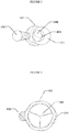
- Figure 2 shows a top-view schematic diagram of the stent graft according to one embodiment as described herein.
- Figure 3 shows a bottom-view schematic diagram of the stent graft according to one embodiment as described herein.
- the bottom is the proximal side (end) and the top is the distal side (end).
- the stent graft according to one embodiment as described herein will be described as below by referring to Figures 1 to 3 .
- the stent graft for ascending aorta can comprise: a stent 100, a cover attachment 200 covering the stent 100, a valve leaflet 300 for reconstructing aortic valves, which are positioned at the proximal end of the stent 100 and attached to the cover attachment 200 and the stent 100, and two windows 401 and 402 aligning with two coronary arteries.
- the valve leaflet 300 may be sutured to the cover attachment 200 and the stent 100, such that the valve leaflet 300 allows blood to flow from left ventricle to aorta and prevent blood from flowing from aorta back to left ventricle when the stent graft is implanted into aorta vessel.
- the stent 100 covered by the cover attachment 200 is shown in Figures.
- the stent 100 acts as a supporting structure or framework of the stent graft, which may be made of stent wires, in particular, may be knitted by metal wires, or may be a self-expandable nickle-titanium stent.
- the cover attachment 200 can be artificial vascular membrane.
- the stent 100 and the cover attachment 200 can be sutured together as a whole by non-absorbable suture, or can be formed together as a whole by thermocompression or adheresion.
- the valve leaflet 300 can be a biological valve made of biological materials such as bovine pericardium, pig pericardium, donkey pericardium and the like.
- the number of the valve leaflet can be three, each of which forms a "pocket" structure by suturing to the cover attachment and the stent, thereby forming three aortic sinuses.
- the stent graft for ascending aorta can have two windows 401 and 402 aligning with two openings of the coronary arteries, which are close to the proximal end of the stent 100.
- the proximal edges of the windows are 5-500mm away from the proximal end of the stent 100.
- the proximal edges of the windows are 15mm, or 25mm, or 35mm away from the proximal end of the stent 100.
- the distance between the proximal edges of the windows and the proximal end of the stent 100 can be set according to the structure of the patient's arteral vessels.
- the proximal edge of the window 401 is 25mm away from the proximal end of the stent 100 and the proximal edges of the window 402 is 27.5mm away from the proximal end of the stent 100, such that the coronary blood supply can be preserved.
- the windows can be formed prior to surgery, or can be formed during surgery, i.e., a part of cover attachment, which aligns with the coronary arteries is removed to allow the inside of the stent to be in blood communication with the coronary arteries.
- the cover attachment 200 can partially cover the stent 100, i.e., the area of the stent 100 which aligns with the coronary arteries is not covered by the cover attachment 200, thereby allowing the inside of the stent to be in blood communication with the coronary arteries.
- each of the horizontally opposite edges of each window (e.g., front and rear edges facing surgeons) is provided with a marker for alignment during implantation of the stent graft.
- the X-ray developed markers on the front and rear edges of the windows are overlapped at the position of the coronary artery tangent to ensure that the windows are aligned with the coronary artery openings.
- each of of the horizontally opposite sides (e.g., front and rear sides facing surgeons) of at least one of middle part (e.g., the part 17-67mm away from the proximal end of the main stent) of the main stent and the part (e.g., the part 0-20mm away from the distal end of the main stent) close to the distal end of the main stent is provided with a marker for alignment during implantation of the stent graft.
- the accuracy of alignment can be enhanced by using a plurality of markers at various positions in the stent graft.
- the markers can be made of any materials that are X-ray developed, such as nickle-titanium alloy and the like. And the markers can be sutured or adhered to the above positions of the stent.
- the stent 100 may comprise a main stent 101 for implanting into the ascending aorta and a first branch stent 102 positioned at the distal end of the main stent 101, for implanting into the branch artery, and a second branch stent 103 adjacent to the first branch stent 102 and for facing or entering into the aortic arch.
- the branch artery comprises any one of innominate artery, left common carotid artery, and left subclavian artery. This embodiment takes the innominate artery as an example. The skilled in the art should understand that the present invention is not limited to this and any changes can be made according to the patient's situation. This embodiment is also suitable for other branch arteries.
- the stent graft for ascending aorta is anchored to an appropriate position of the ascending aorta by the first and the second branch stents 102 and 103, but the present invention is not limited to this.
- the stent graft for ascending aorta may be anchored to an appropriate position of the ascending aorta by other ways, for example, hangnails on the stent graft for locating or anchoring the stent graft. Therefore, in other embodiments, the stent 100 may merely comprise the main stent 101, or may comprise the main stent 101 and the first branch stent 102.
- the main stent has a length from about 35 to 155mm, for example, 57mm.
- the first branch stent has a length from about 45 to 105mm, for example, 80mm.
- the second branch stent 103 has a length from about 40 to 80mm, for example 45mm.
- the skilled in the art should understand that the present invention is not limited to this.
- the length of the main stent and the branch stents can be appropriately set according to the positions of the aorta lesion and the features of the aorta lesion.
- the main stent 101 may have a length of 85mm.
- the first branch stent 102 may have a length of 45mm.
- the second branch stent 103 may have a length of 40mm.
- the main stent 101 may have a length of 45mm.
- the first branch stent 102 may have a length of 105mm.
- the second branch stent 103 may have a length of 80mm.
- the present invention will not make any specific limitation on the length of the main stent, the first branch stent and the second branch stent.
- the main stent 101 is in cylindrical or generally cylindrical shape, whose diameter can keep constant in axial direction, or can be gradually increased and then gradually decreased.
- the main stent 101 may have a section of trumpet-like structure at the proximal end.
- the diameter D1 of the main stent at the proximal end ranges from about 24 to 48mm, such as 30mm.
- the diameter D2 at the distal end ranges from about 30 to 50mm, such as 40mm.
- the maximum part has a diameter from 28 to 50mm, such as 46mm.
- the skilled in the art should understand that all the values as described herein are illustrative.
- the diameters of both ends of the main stent 101 can be set according to the patient's specific situation, which are not specifically limited herein.
- the first branch stent 102 can be in cylindrical or generally cylindrical shape, for example, can be in trumpet-like shape.
- the diameter of the first branch stent 102 can be gradually increased from the proximal end to the distal end along the axial direction.
- the diameter d11 of the first branch stent 102 at the proximal end ranges from about 6 to 16mm, such as 10mm.
- the diameter d12 at the distal end ranges from about 10 to 26mm, such as 18mm. It should be noticed that, the diameters of both ends of the first branch stent 102 can be set according to the patient's specific situation, which are not specifically limited herein.
- the first branch stent 102 can be in any shape that allows the first branch stent 102 to attach to the vessel wall.
- the ratio of the diameter d11 at the proximal end of the first branch stent 102 to the diameter D2 at the distal end of the main stent 101 ranges from 1:1 to 1:6, such as 1:4.
- the configuration that the diameter at the distal end of the first branch stent 102 is larger than that at the proximal end is beneficial to anchor to an appropriate position in the branch artery of the aortic arch upon implantation, thereby avoiding endoleak from distal source.
- the second branch stent 103 can be in cylindrical or generally cylindrical shape, and the diameter thereof is gradually increased from the proximal end to the distal end along the axial direction.
- the diameter d21 at the proximal end ranges from about 25 to 42mm, such as 35mm.
- the diameter d22 at the distal end ranges from about 25 to 50mm, such as 40mm.
- the skilled in the art should understand that all the values as described herein are illustrative.
- the diameters of both ends of the second branch stent 103 can be set according to the patient's specific situation, which are not specifically limited herein.
- the ratio of the diameter d21 at the proximal end of the second branch stent 103 to the diameter D2 at the distal end of the main stent 101 ranges from about 1:4 to 1:1, such as 3:4.
- the first branch stent 102 can enter into innominate artery 800 and thus the stent graft for can be restricted to an appropriate position in the ascending aorta.
- the proximal end of the stent graft can be accurately anchored to aortic annulus.
- the coronary windows 401 and 402 on the stent graft can accurately align with the openings 901 and 902 of the coronary artery.
- the tearing opening at the proximal end of the dissection or aortic aneurysm can be isolated, so as to shrink the false cavity of the dissection or aortic aneurysm due to thrombosis.
- the aortic diseases involving both branch artery and ascending aorta on the aortic arch can be treated by using the stent graft as described herein through minimally invasive intervention surgery one time.
- Such integrated reconstruction of the ascending aorta-aortic arch has high stability and low risk for lack of alignment of coronary windows and delayed endoleak due to movement of stent graft in short and long term, compared with combined stent graft.
- the stent graft for ascending aorta can be restricted to an appropriate position of the ascending aorta by using the second branch stent 103 in addition to the first branch stent 102.
- the stent graft can be firmly anchored to the ascending aorta to avoid displacement along the aortic axis or endoleak from distal end.
- the stent graft for the ascending aorta as described herein can achieve complete endovascular cure by performing minimally invasive endovascular surgery one time.
- the stent graft for the ascending aorta as described herein has high stability and low risk for lack of alignment of coronary windows and delayed endoleak due to movement of stent graft in short and long term, compared with the combined stent graft.
- the second branch stent 103 facing or entering into the aortic arch provides a joint for further implantation of the stent graft to reconstruct the distal end of the aortic arch and the descending aorta.
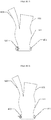
- Figure 5 shows a front-view schematic diagram of the stent graft with a drum-like proximal end according to another embodiment as described herein, in which the height of the drum is marked as H.
- Figure 6 shows another front-view schematic diagram of the stent graft with a drum-like proximal end as shown in Figure 5 , in which the protrusion of the drum is marked as h.
- the stent wires and the cover attachment are not shown in Figures 5 and 6 .
- the stent graft as shown in Figures 5 and 6 is different from that as shown in Figures 1 to 3 in that a section of the main stent 101, which is close to the proximal end is in a drum-like shape, for matching the shape of the aortic sinus.
- the proximal edge of the drum is 0-50mm away from the proximal end of the stent graft, and the height H of the drum ranges from 10 to 30mm.
- the maximum protrusion h of the drum along the radial direction of the stent ranges from 3 to 15mm.
- H and h can be set as needed, for example, can be set according to the anatomical structure of the ascending aorta of the patient, such that sufficient space for movement of aortic valves can be provided at the proximal end of the stent graft to prevent the aortic valves from occluding the windows of the coronary artery, and the space for preserving coronary openings is provided for the purpose of endovascularly re-implanting aortic valves.
- such a shape is also beneficial to attach the proximal end of the stent graft to the aortic root, which enhances isolation efficiency of the stent graft for tearing opening in the aortic sinus and facilitates the reconstruction of the ascending aorta.
- the stent covered by artificial vessel membrane plays a role in isolation of aortic lesions (tearing opening in dissection or aortic aneurysm), which can be cylindrical type or branched type.
- the branched type stent graft has two branches at the distal end, one is a long leg (i.e., the first branch stent 102) for placing in brachiocephalic artery (i.e., innominate artery), and the other is a short leg (i.e., the second branch stent 103) facing the aortic arch, for ease of further connection of stent graft to reconstruct aortic arch.
- the biological aortic valves are sutured to the proximal end of the stent for reconstructing aortic valves.
- the windows on the artificial vessel membrane which correspond to the coronary artery are used for preserving coronary blood supply.
- Figure 7 is a perspective schematic diagram of the stent graft according to another embodiment as described herein.
- Figure 8 shows a top-view schematice diagram of the stent graft as shown in Figure 7 upon cutting off the first branch stent outside of the main stent.
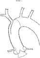
- Figure 9 illustrates that the stent graft as shown in Figure 7 is implanted into the ascending aorta vessel. For clarity, the stent wires and the cover attachment are not shown in Figures 7 to 9 .
- the stent graft can comprise a main stent 201, a first branch stent 202 and an inner stent 203.
- the skilled in the art should understand that the stent graft further comprises artificial vessel membranes covering the stents, although they are not shown.
- a part of the first branch stent 202 and the inner stent 203 are arranged within the main stent 201 side by side.
- the distal end of the inner stent 203 is flush with the distal end of the main stent 201, i.e., both of them are generally in the same plane.
- the distal end of the inner stent 203 goes beyond the distal end of the main stent 201.
- the gap between the peripheral wall of the first branch stent 202, the peripheral wall of the inner stent 203 and the inner-peripheral wall of the main stent 201 is sealed by a sealing element, such that the blood cannot flow through the gap among these three stents.
- the valve leaflet 700 for reconstructing the aortic valves are positioned at the proximal end of the main stent 201.
- Two windows 601 and 602 for aligning with two coronary arteries are provided close to the proximal end of the main stent 201.
- the first branch stent 202 goes into innominate artery 800.
- the branch stent 202 and the inner stent 203 are sleeved within the main stent 201, such that the main stent 201 can be well attached to the vessel wall, and the proximal end of the stent graft can be accurately anchored to the aortic annulus, and the coronary windows 601 and 602 on the stent graft can accurately align with the openings 901 and 902 on the coronary artery.
- the tearing opening in proximal dissection or aneurysm can be isolated, thereby shrinking the falsing cavity and aneurysm due to thrombosis.
- the main stent 201 can be well attached to the vessel wall, thereby resulting in excellent efficiency for the lesion involving the middle region of the ascending aorta, such as aneurysm.
- Healthy adult domestic pigs 60-65 kg were used to perform animal experiments.
- CT angiography (CTA) of the aorta in the domestic pigs was performed.
- the stent graft for ascending aorta was made, comprising: biological aortic valves made of bovine pericardium and sutured to the proximal end, two windows close to the proximal end for preserving the left and right coronary artery openings.
- the distal end of the stent graft was branched with a long leg (i.e., the first branch stent) for placing in the brachiocephalic artery and a short leg (i.e., the second branch stent, since the pig's ascending aorta segment was very short, the length of the short leg was less than 2cm) in which only one opening facing the aortic arch was preserved.
- the main body of the stent graft was in a cylindrical structure. Under general anesthesia, a small incision was used to dissect the apex of the pig's heart (see Figure 10A ).
- the stent graft was introduced through apex. After ensuring that the axial and circumferential position can well preserve the coronary arteries (see Figure 10D ), the stent graft was released. The position of the stent graft, the function of the aortic valves and the coronary artery were evaluated by angiography in combination with esophageal ultrasonography (see Figures 10E to 10J ).
- the stent graft for ascending aorta can be used to minimally invasively reconstruct the aortic root (including the aortic valves) and the ascending aorta, and keep the left and right the coronary arteries, thereby providing a joint for further reconstruction of the aortic arch and descending aorta.
- the stent graft for ascending aorta was suitable for treating the following diseases: ascending aortic dissection and aortic aneurysm involving aortic sinus, coronary artery openings, aortic valves, severe aortic valve insufficiency or severe stenosis with ascending aortic aneurysm, etc..
- Figure 11 illustrates that the stent graft with a trumpet-like proximal end according to another embodiment as described herein is implanted into the ascending aorta vessel and left ventricular outflow tract.
- This embodiment is different from that as shown in Figures 1 to 4 in that a trumpet-like structure is added to the proximal end of the stent graft.
- the proximal end of this stent graft is in a trumpet-like structure, which is 0-40mm away from the proximal edge of the valve leaflet, i.e., suitable for going into left ventricular outflow tract, and the diameter of the proximal end of the trumpet-like structure ranges from 22 to 48mm.
- a trumpet-like structure can be attached to the proximal end of the stent graft of the embodiments as shown in Figures 5-6 and 7-9 .
- the trumpet-like structure is suitable for inserting into the left ventricular outflow tract so as to anchor the stent therein.
- the skilled in the art should understand that the present invention is not limited to the trumpet-like structure. Any structure whose diameter at the proximal end is large and gradually decreased can be used.
Landscapes
- Health & Medical Sciences (AREA)
- Engineering & Computer Science (AREA)
- Biomedical Technology (AREA)
- Cardiology (AREA)
- Heart & Thoracic Surgery (AREA)
- Transplantation (AREA)
- Oral & Maxillofacial Surgery (AREA)
- Vascular Medicine (AREA)
- Life Sciences & Earth Sciences (AREA)
- Animal Behavior & Ethology (AREA)
- General Health & Medical Sciences (AREA)
- Public Health (AREA)
- Veterinary Medicine (AREA)
- Pulmonology (AREA)
- Gastroenterology & Hepatology (AREA)
- Prostheses (AREA)
Applications Claiming Priority (3)
| Application Number | Priority Date | Filing Date | Title |
|---|---|---|---|
| CN201820600490.2U CN209253227U (zh) | 2018-04-25 | 2018-04-25 | 升主动脉支架移植物 |
| CN201810379341.2A CN109091271A (zh) | 2018-04-25 | 2018-04-25 | 升主动脉支架移植物 |
| PCT/CN2018/116151 WO2019205599A1 (fr) | 2018-04-25 | 2018-11-19 | Greffon d'endoprothèse aortique ascendant |
Publications (2)
| Publication Number | Publication Date |
|---|---|
| EP3785665A1 true EP3785665A1 (fr) | 2021-03-03 |
| EP3785665A4 EP3785665A4 (fr) | 2022-01-19 |
Family
ID=68293741
Family Applications (1)
| Application Number | Title | Priority Date | Filing Date |
|---|---|---|---|
| EP18915822.3A Withdrawn EP3785665A4 (fr) | 2018-04-25 | 2018-11-19 | Greffon d'endoprothèse aortique ascendant |
Country Status (4)
| Country | Link |
|---|---|
| US (1) | US20210244525A1 (fr) |
| EP (1) | EP3785665A4 (fr) |
| JP (1) | JP2021522037A (fr) |
| WO (1) | WO2019205599A1 (fr) |
Cited By (2)
| Publication number | Priority date | Publication date | Assignee | Title |
|---|---|---|---|---|
| US11324583B1 (en) | 2021-07-06 | 2022-05-10 | Archo Medical LTDA | Multi-lumen stent-graft and related surgical methods |
| WO2025030162A1 (fr) * | 2023-08-02 | 2025-02-06 | Mavericks Endo, Inc. | Dispositifs de réparation aortique, et systèmes et méthodes associés |
Families Citing this family (6)
| Publication number | Priority date | Publication date | Assignee | Title |
|---|---|---|---|---|
| EP3941392B1 (fr) | 2019-03-20 | 2025-04-23 | Inqb8 Medical Technologies, LLC | Implant de dissection aortique |
| CN115300182A (zh) * | 2021-09-30 | 2022-11-08 | 上海御瓣医疗科技有限公司 | 一种带有显影结构的二尖瓣瓣膜支架 |
| USD1016289S1 (en) * | 2022-01-18 | 2024-02-27 | Sutherland Cardiovascular Ltd. | Aortic root replacement graft |
| CN115006053B (zh) * | 2022-08-09 | 2022-11-15 | 北京华脉泰科医疗器械股份有限公司 | 一体式术中支架 |
| WO2025101642A2 (fr) * | 2023-11-08 | 2025-05-15 | W. L. Gore & Associates, Inc. | Systèmes d'endoprothèse ramifiée et procédés |
| CN120000392B (zh) * | 2025-04-21 | 2025-07-11 | 四川国屹医疗科技有限公司 | 一种多分支覆膜支架 |
Family Cites Families (20)
| Publication number | Priority date | Publication date | Assignee | Title |
|---|---|---|---|---|
| US5628783A (en) * | 1991-04-11 | 1997-05-13 | Endovascular Technologies, Inc. | Bifurcated multicapsule intraluminal grafting system and method |
| US6200339B1 (en) * | 1999-02-23 | 2001-03-13 | Datascope Investment Corp. | Endovascular split-tube bifurcated graft prosthesis and an implantation method for such a prosthesis |
| US7226474B2 (en) * | 2000-05-01 | 2007-06-05 | Endovascular Technologies, Inc. | Modular graft component junctions |
| US6682537B2 (en) * | 2001-12-20 | 2004-01-27 | The Cleveland Clinic Foundation | Apparatus and method for capturing a wire in a blood vessel |
| CN2817766Y (zh) * | 2005-03-02 | 2006-09-20 | 郭伟 | 用于升主动脉及主动脉弓腔内修复术的分体式支架型血管 |
| CN100362971C (zh) * | 2005-11-16 | 2008-01-23 | 程英升 | 贲门支架 |
| CN101152109A (zh) * | 2006-09-27 | 2008-04-02 | 黄方炯 | 主动脉弓三侧支支架血管 |
| CN101352376A (zh) * | 2008-09-02 | 2009-01-28 | 中国人民解放军第二军医大学 | 带锚定脚的主动脉瓣膜支架 |
| CN101601618A (zh) * | 2009-07-15 | 2009-12-16 | 郭伟 | 一种主动脉血管支架 |
| US20120116498A1 (en) * | 2010-11-05 | 2012-05-10 | Chuter Timothy A | Aortic valve prostheses |
| CN102641164B (zh) * | 2011-02-16 | 2015-08-26 | 舒畅 | 分支型主动脉支架血管系统 |
| EP2836159B1 (fr) * | 2012-04-12 | 2018-07-18 | Sanford Health | Débranchement et endoprothèse de vaisseau principal |
| WO2014093473A1 (fr) * | 2012-12-14 | 2014-06-19 | Kelly Patrick W | Endoprothèses vasculaires combinées à double cylindre et débranchement |
| US9744032B2 (en) * | 2013-03-11 | 2017-08-29 | Cook Medical Technologies Llc | Endoluminal prosthesis comprising a valve and an axially extendable segment |
| US9808364B2 (en) * | 2013-03-11 | 2017-11-07 | Cook Medical Technologies Llc | Systems and methods for maintaining perfusion of branch vessels |
| US20170319359A1 (en) * | 2013-04-16 | 2017-11-09 | Manish Mehta | Endovascular docking apparatus and method |
| US10034784B2 (en) * | 2013-04-17 | 2018-07-31 | Gilbert H. L. Tang | Heart valve and endovascular graft components and method for delivery |
| WO2014188412A2 (fr) * | 2013-05-23 | 2014-11-27 | Endospan Ltd. | Système de greffon d'endoprothèse d'aorte ascendante |
| US9693860B2 (en) * | 2014-12-01 | 2017-07-04 | Medtronic, Inc. | Segmented transcatheter valve prosthesis having an unsupported valve segment |
| ES2921535T3 (es) * | 2015-06-18 | 2022-08-29 | Ascyrus Medical Llc | Injerto aórtico ramificado |
-
2018
- 2018-11-19 JP JP2021508038A patent/JP2021522037A/ja active Pending
- 2018-11-19 WO PCT/CN2018/116151 patent/WO2019205599A1/fr not_active Ceased
- 2018-11-19 US US17/050,634 patent/US20210244525A1/en not_active Abandoned
- 2018-11-19 EP EP18915822.3A patent/EP3785665A4/fr not_active Withdrawn
Cited By (3)
| Publication number | Priority date | Publication date | Assignee | Title |
|---|---|---|---|---|
| US11324583B1 (en) | 2021-07-06 | 2022-05-10 | Archo Medical LTDA | Multi-lumen stent-graft and related surgical methods |
| US12496182B2 (en) | 2021-07-06 | 2025-12-16 | Archo Medical LTDA | Multi-lumen stent-graft and related surgical methods |
| WO2025030162A1 (fr) * | 2023-08-02 | 2025-02-06 | Mavericks Endo, Inc. | Dispositifs de réparation aortique, et systèmes et méthodes associés |
Also Published As
| Publication number | Publication date |
|---|---|
| US20210244525A1 (en) | 2021-08-12 |
| JP2021522037A (ja) | 2021-08-30 |
| WO2019205599A1 (fr) | 2019-10-31 |
| EP3785665A4 (fr) | 2022-01-19 |
Similar Documents
| Publication | Publication Date | Title |
|---|---|---|
| EP3785665A1 (fr) | Greffon d'endoprothèse aortique ascendant | |
| US20230000647A1 (en) | Heart valve and endovascular graft components and method for delivery | |
| US11918451B2 (en) | Vascular repair devices and methods of use | |
| US11779454B2 (en) | Vascular prosthesis with crimped adapter and methods of use | |
| US11801129B2 (en) | Vascular prosthesis with fenestration ring and methods of use | |
| US10485651B2 (en) | Thoracic aorta stent graft | |
| US10010402B2 (en) | Thoracic aortic covered stent | |
| US7799072B2 (en) | Apparatus and methods for repairing the function of a diseased valve and method for making same | |
| ES2580028T3 (es) | Rama de un injerto de cánula de desramificación | |
| CN109091271A (zh) | 升主动脉支架移植物 | |
| US20230149149A1 (en) | Bifurcated vascualr stent and methods of manufacture | |
| EP4674379A1 (fr) | Dispositif d'aide à la reconstruction d'artère branche et système de reconstruction d'aorte de première branche s-c | |
| CN112914792A (zh) | 适于治疗升主动脉根部疾病的移植物 | |
| CN119970302B (zh) | 一种覆膜支架系统 | |
| CN209253227U (zh) | 升主动脉支架移植物 | |
| CN215535332U (zh) | 适于治疗升主动脉根部疾病的移植物 | |
| TW201927258A (zh) | 主動脈剝離及動脈瘤的治療系統及使用方法 | |
| CN212630955U (zh) | 模块式腹主动脉分叉型覆膜支架 | |
| RU2789589C1 (ru) | Способ хирургического лечения сердечной недостаточности в эксперименте. | |
| EP4327777A1 (fr) | Greffon d'endoprothèse ramifié ayant une endoprothèse de support | |
| US20230149189A1 (en) | Bifurcated vascualr stent and methods of manufacture |
Legal Events
| Date | Code | Title | Description |
|---|---|---|---|
| STAA | Information on the status of an ep patent application or granted ep patent |
Free format text: STATUS: THE INTERNATIONAL PUBLICATION HAS BEEN MADE |
|
| PUAI | Public reference made under article 153(3) epc to a published international application that has entered the european phase |
Free format text: ORIGINAL CODE: 0009012 |
|
| STAA | Information on the status of an ep patent application or granted ep patent |
Free format text: STATUS: REQUEST FOR EXAMINATION WAS MADE |
|
| 17P | Request for examination filed |
Effective date: 20201123 |
|
| AK | Designated contracting states |
Kind code of ref document: A1 Designated state(s): AL AT BE BG CH CY CZ DE DK EE ES FI FR GB GR HR HU IE IS IT LI LT LU LV MC MK MT NL NO PL PT RO RS SE SI SK SM TR |
|
| AX | Request for extension of the european patent |
Extension state: BA ME |
|
| DAV | Request for validation of the european patent (deleted) | ||
| DAX | Request for extension of the european patent (deleted) | ||
| A4 | Supplementary search report drawn up and despatched |
Effective date: 20211222 |
|
| RIC1 | Information provided on ipc code assigned before grant |
Ipc: A61F 2/24 20060101ALI20211216BHEP Ipc: A61F 2/89 20130101ALI20211216BHEP Ipc: A61F 2/06 20130101ALI20211216BHEP Ipc: A61F 2/07 20130101AFI20211216BHEP |
|
| STAA | Information on the status of an ep patent application or granted ep patent |
Free format text: STATUS: THE APPLICATION IS DEEMED TO BE WITHDRAWN |
|
| 18D | Application deemed to be withdrawn |
Effective date: 20220723 |