EP3786181A1 - Polymerer rekombinanter iga-antikörper und verwendung davon - Google Patents
Polymerer rekombinanter iga-antikörper und verwendung davon Download PDFInfo
- Publication number
- EP3786181A1 EP3786181A1 EP20196341.0A EP20196341A EP3786181A1 EP 3786181 A1 EP3786181 A1 EP 3786181A1 EP 20196341 A EP20196341 A EP 20196341A EP 3786181 A1 EP3786181 A1 EP 3786181A1
- Authority
- EP
- European Patent Office
- Prior art keywords
- antibody
- iga
- type
- protein
- amino acid
- Prior art date
- Legal status (The legal status is an assumption and is not a legal conclusion. Google has not performed a legal analysis and makes no representation as to the accuracy of the status listed.)
- Withdrawn
Links
Images







Classifications
-
- C—CHEMISTRY; METALLURGY
- C12—BIOCHEMISTRY; BEER; SPIRITS; WINE; VINEGAR; MICROBIOLOGY; ENZYMOLOGY; MUTATION OR GENETIC ENGINEERING
- C12N—MICROORGANISMS OR ENZYMES; COMPOSITIONS THEREOF; PROPAGATING, PRESERVING, OR MAINTAINING MICROORGANISMS; MUTATION OR GENETIC ENGINEERING; CULTURE MEDIA
- C12N15/00—Mutation or genetic engineering; DNA or RNA concerning genetic engineering, vectors, e.g. plasmids, or their isolation, preparation or purification; Use of hosts therefor
- C12N15/01—Preparation of mutants without inserting foreign genetic material therein; Screening processes therefor
-
- A—HUMAN NECESSITIES
- A61—MEDICAL OR VETERINARY SCIENCE; HYGIENE
- A61K—PREPARATIONS FOR MEDICAL, DENTAL OR TOILETRY PURPOSES
- A61K39/00—Medicinal preparations containing antigens or antibodies
- A61K39/395—Antibodies; Immunoglobulins; Immune serum, e.g. antilymphocytic serum
-
- A—HUMAN NECESSITIES
- A61—MEDICAL OR VETERINARY SCIENCE; HYGIENE
- A61P—SPECIFIC THERAPEUTIC ACTIVITY OF CHEMICAL COMPOUNDS OR MEDICINAL PREPARATIONS
- A61P31/00—Antiinfectives, i.e. antibiotics, antiseptics, chemotherapeutics
-
- A—HUMAN NECESSITIES
- A61—MEDICAL OR VETERINARY SCIENCE; HYGIENE
- A61P—SPECIFIC THERAPEUTIC ACTIVITY OF CHEMICAL COMPOUNDS OR MEDICINAL PREPARATIONS
- A61P31/00—Antiinfectives, i.e. antibiotics, antiseptics, chemotherapeutics
- A61P31/04—Antibacterial agents
-
- A—HUMAN NECESSITIES
- A61—MEDICAL OR VETERINARY SCIENCE; HYGIENE
- A61P—SPECIFIC THERAPEUTIC ACTIVITY OF CHEMICAL COMPOUNDS OR MEDICINAL PREPARATIONS
- A61P31/00—Antiinfectives, i.e. antibiotics, antiseptics, chemotherapeutics
- A61P31/10—Antimycotics
-
- A—HUMAN NECESSITIES
- A61—MEDICAL OR VETERINARY SCIENCE; HYGIENE
- A61P—SPECIFIC THERAPEUTIC ACTIVITY OF CHEMICAL COMPOUNDS OR MEDICINAL PREPARATIONS
- A61P31/00—Antiinfectives, i.e. antibiotics, antiseptics, chemotherapeutics
- A61P31/12—Antivirals
-
- A—HUMAN NECESSITIES
- A61—MEDICAL OR VETERINARY SCIENCE; HYGIENE
- A61P—SPECIFIC THERAPEUTIC ACTIVITY OF CHEMICAL COMPOUNDS OR MEDICINAL PREPARATIONS
- A61P31/00—Antiinfectives, i.e. antibiotics, antiseptics, chemotherapeutics
- A61P31/12—Antivirals
- A61P31/14—Antivirals for RNA viruses
- A61P31/16—Antivirals for RNA viruses for influenza or rhinoviruses
-
- A—HUMAN NECESSITIES
- A61—MEDICAL OR VETERINARY SCIENCE; HYGIENE
- A61P—SPECIFIC THERAPEUTIC ACTIVITY OF CHEMICAL COMPOUNDS OR MEDICINAL PREPARATIONS
- A61P33/00—Antiparasitic agents
-
- C—CHEMISTRY; METALLURGY
- C07—ORGANIC CHEMISTRY
- C07K—PEPTIDES
- C07K16/00—Immunoglobulins [IG], e.g. monoclonal or polyclonal antibodies
- C07K16/06—Immunoglobulins [IG], e.g. monoclonal or polyclonal antibodies from serum
- C07K16/065—Purification, fragmentation
-
- C—CHEMISTRY; METALLURGY
- C07—ORGANIC CHEMISTRY
- C07K—PEPTIDES
- C07K16/00—Immunoglobulins [IG], e.g. monoclonal or polyclonal antibodies
- C07K16/18—Immunoglobulins [IG], e.g. monoclonal or polyclonal antibodies against material from animals or humans
-
- C—CHEMISTRY; METALLURGY
- C12—BIOCHEMISTRY; BEER; SPIRITS; WINE; VINEGAR; MICROBIOLOGY; ENZYMOLOGY; MUTATION OR GENETIC ENGINEERING
- C12N—MICROORGANISMS OR ENZYMES; COMPOSITIONS THEREOF; PROPAGATING, PRESERVING, OR MAINTAINING MICROORGANISMS; MUTATION OR GENETIC ENGINEERING; CULTURE MEDIA
- C12N15/00—Mutation or genetic engineering; DNA or RNA concerning genetic engineering, vectors, e.g. plasmids, or their isolation, preparation or purification; Use of hosts therefor
- C12N15/09—Recombinant DNA-technology
-
- C—CHEMISTRY; METALLURGY
- C12—BIOCHEMISTRY; BEER; SPIRITS; WINE; VINEGAR; MICROBIOLOGY; ENZYMOLOGY; MUTATION OR GENETIC ENGINEERING
- C12N—MICROORGANISMS OR ENZYMES; COMPOSITIONS THEREOF; PROPAGATING, PRESERVING, OR MAINTAINING MICROORGANISMS; MUTATION OR GENETIC ENGINEERING; CULTURE MEDIA
- C12N15/00—Mutation or genetic engineering; DNA or RNA concerning genetic engineering, vectors, e.g. plasmids, or their isolation, preparation or purification; Use of hosts therefor
- C12N15/09—Recombinant DNA-technology
- C12N15/10—Processes for the isolation, preparation or purification of DNA or RNA
- C12N15/1034—Isolating an individual clone by screening libraries
- C12N15/1086—Preparation or screening of expression libraries, e.g. reporter assays
-
- C—CHEMISTRY; METALLURGY
- C12—BIOCHEMISTRY; BEER; SPIRITS; WINE; VINEGAR; MICROBIOLOGY; ENZYMOLOGY; MUTATION OR GENETIC ENGINEERING
- C12N—MICROORGANISMS OR ENZYMES; COMPOSITIONS THEREOF; PROPAGATING, PRESERVING, OR MAINTAINING MICROORGANISMS; MUTATION OR GENETIC ENGINEERING; CULTURE MEDIA
- C12N15/00—Mutation or genetic engineering; DNA or RNA concerning genetic engineering, vectors, e.g. plasmids, or their isolation, preparation or purification; Use of hosts therefor
- C12N15/09—Recombinant DNA-technology
- C12N15/63—Introduction of foreign genetic material using vectors; Vectors; Use of hosts therefor; Regulation of expression
- C12N15/66—General methods for inserting a gene into a vector to form a recombinant vector using cleavage and ligation; Use of non-functional linkers or adaptors, e.g. linkers containing the sequence for a restriction endonuclease
-
- A—HUMAN NECESSITIES
- A61—MEDICAL OR VETERINARY SCIENCE; HYGIENE
- A61K—PREPARATIONS FOR MEDICAL, DENTAL OR TOILETRY PURPOSES
- A61K39/00—Medicinal preparations containing antigens or antibodies
- A61K2039/505—Medicinal preparations containing antigens or antibodies comprising antibodies
-
- A—HUMAN NECESSITIES
- A61—MEDICAL OR VETERINARY SCIENCE; HYGIENE
- A61K—PREPARATIONS FOR MEDICAL, DENTAL OR TOILETRY PURPOSES
- A61K39/00—Medicinal preparations containing antigens or antibodies
- A61K2039/505—Medicinal preparations containing antigens or antibodies comprising antibodies
- A61K2039/507—Comprising a combination of two or more separate antibodies
-
- C—CHEMISTRY; METALLURGY
- C07—ORGANIC CHEMISTRY
- C07K—PEPTIDES
- C07K16/00—Immunoglobulins [IG], e.g. monoclonal or polyclonal antibodies
- C07K16/08—Immunoglobulins [IG], e.g. monoclonal or polyclonal antibodies against material from viruses
- C07K16/10—RNA viruses
- C07K16/102—Coronaviridae (F)
Definitions
- the present invention relates to a polymeric IgA-type recombinant antibody and use thereof.
- Priority is claimed on Japanese Patent Application No. 2014-148328, filed on July 18, 2014 , the content of which is incorporated herein by reference.
- antibodies are high levels of antigen-binding activity and specificity in which the antibodies strongly bind only to target antigens and do not bind to others.
- Antibodies have functional activities such as neutralizing activity and the like due to a combination of the antigen-binding activity and specificity, and thus have been widely used in the fields of bio-industry including therapeutic antibodies, diagnostic drugs, biological research tools, etc.
- IgG isotypes such as IgM, IgA, IgD, IgE, and the like in the case of antibodies, and each has different in vivo functions.
- IgG is the main antibody present in vivo in blood, but the main antibody in mucus or secretory fluid covering the mucous epithelium is IgA, which is in turn known to serve as a front-line in a biological defense mechanism against mucosal infections (for example, see Non-patent Literature 1).
- antibodies such as IgA and IgM are known to function in vivo by forming polymers such as dimers and pentamers with antibodies having the same variable region.
- polymeric IgA is known to be present in secretory fluids such as colostrum and saliva, and sera from patients with multiple myeloma.
- Monomeric, dimeric, trimeric and tetrameric IgA are found to be included at various ratios in sera from patients with multiple myeloma. Also, the dimers and tetramers are reported to be included in secretory fluids. However, little is known about the biological significance of this polymeric antibody.
- Non-patent Literature 1 Woof J. M. and Russell M.W., Structure and function relationships in IgA, Mucosal immunology, 4(6), 590-597, 2011
- an object of the present invention is to provide a polymeric IgA-type recombinant antibody.
- Another object of the present invention is to provide a medicine containing the polymeric IgA-type recombinant antibody as an active ingredient.
- Still another object of the present invention is to provide a method of producing a polymeric IgA-type antibody.
- Yet another object of the present invention is to provide a method of improving antigen-binding activity of the antibody.
- the present invention provides the following.
- a polymeric IgA-type recombinant antibody can be provided.
- a medicine containing the polymeric IgA-type recombinant antibody as an active ingredient can be provided.
- a method of producing a polymeric IgA-type antibody can be provided.
- a method of improving the antigen-binding activity of the antibody can be provided.
- IgA is mainly present as a monomeric IgA that has a molecular weight of approximately 170,000, and IgAl is a main component.
- a dimeric IgA is a dimeric IgA that is produced by plasma cells present in the mucosal lamina intestinal, and represents a molecule in which a heavy chain, a light chain and a J chain are present at a ratio of 4:4:1. In the dimeric IgA, IgA2 accounts for approximately half.
- dimeric or higher IgA recognized by a polymeric Ig receptor is often generally referred to as a polymeric IgA. That is, as a complex in which a heavy chain, a light chain and a J chain are present at a composition ratio of 4:4:1, a dimeric IgA in which two IgA molecules are associated via an antibody J-chain protein (a joining chain) is a main ingredient, and the term "polymeric IgA" is used both when it includes a secretory component protein (SC protein) and when it does not include an SC protein.
- SC protein secretory component protein
- cases in which a polymeric IgA (a complex in which a heavy chain, a light chain and a J chain are present at a composition ratio of 4:4:1) secreted by plasma cells is referred to as a "polymeric IgA”
- cases in which a S-IgA (a complex in which a heavy chain, a light chain, a J chain and SC are present at a composition ratio of 4:4:1:1) secreted by mucosal epithelial cells is referred to as a "polymeric IgA”
- cases in which it could refer to either are occasionally found, and thus these cases are not strictly distinguished.
- a component having a higher molecular weight than the dimeric IgA is detected based on the mobility in electrophoresis and the behaviors in gel filtration chromatography, and is often referred to as a polymeric IgA since the component is expected to be dimeric or higher. However, since the component is not distinguished from aggregates, a specific molecular structure of the component is not known.
- pIgR expressed on cell membranes in the basal side of mucosal epithelial cells is a type I transmembrane protein that belongs to an immunoglobulin superfamily, and is composed of an extracellular domain region, a transmembrane region, and an intracytoplasmic region.
- pIgR specifically recognizes and binds to a dimeric/polymeric Ig molecule including a J chain, which are produced by plasma cells exsiting in the mucosal lamina intestinal, and is transferred to an apical side in a state in which the pIgR is bound to the dimeric/polymeric Ig molecule after incorporation into epithelial cells.
- a cleavage between the extracellular domain region and the transmembrane region of the pIgR is needed to release the dimeric/polymeric Ig molecule from epithelial cells into a surface of the mucosa, and the dimeric/polymeric Ig molecule is secreted as S-IgA into the lumenal mucosal layer after the cleavage by a proteolytic enzyme in the epithelial cells.
- S-IgA secretory component protein
- the SC protein is a highly glycosylated polypeptide that corresponds to an extracellular domain region of pIgR and having a molecular weight of approximately 70 kDa.
- the SC protein has five immunoglobulin-like domains from the N-terminus thereof, which are sequentially named D1 to D5.
- D1 to D3 are required to bind to the dimeric IgA.
- D1 has a structure similar to complementarity-determining regions (CDRs) of variable regions of immunoglobulins, and thus plays an important role in binding to the dimeric IgA. Thr27-Thr33 in CDR1 of D1 is involved in this interaction.
- the J chain is a polypeptide chain having a molecular weight of 15 kDa, has an N-linked sugar chain, and is folded to form an immunoglobulin structure.
- the J chains have very high homology in terms of the primary structure and are cross-reactivly detected as antigen with species-specific antibody, and thus the basic characteristics of the J chains are considered to be conserved among organisms.
- the J chain is essential for the dimeric IgA to interact with pIgR.
- S-IgA refers to a dimeric or higher IgA complex that is secreted by mucosal epithelial cells and includes an SC protein.
- secretory dimeric IgA S-dIgA
- a heavy chain, a light chain, a J chain and SC are present at a composition ratio of 4:4:1:1.
- a polymeric antibody having a secretory component (SC) protein in a molecule thereof is referred to as a secretory antibody. Also, the polymeric antibody is often indicated as follows.
- an antibody has a secretory component (SC) protein in a molecule thereof
- SC secretory component
- An antibody having no SC in a molecule thereof is not marked with "S-.”
- an association state of a molecule is indicated by abbreviating the molecule before IgA.
- a monomer is marked with "m”
- a dimer is marked with “d”
- a trimer is marked with "t”
- a tetramer is marked with "q”
- a tetramer or higher polymer is marked with "p.”
- a monomeric antibody and a recombinant monomeric antibody may be referred to as a "monomer” without particular distinction.
- a dimeric antibody, a secretory dimeric antibody, a recombinant dimeric antibody, and a recombinant secretory dimeric antibody may be referred to as a "dimer” without particular distinction.
- a secretory trimeric antibody and a recombinant secretory trimeric antibody may be referred to as a "trimer” without particular distinction.
- a secretory tetrameric antibody and a recombinant secretory tetrameric antibody may be referred to as a "tetramer” without particular distinction.
- a tetrameric or higher secretory antibody and a tetrameric or higher recombinant secretory polymeric antibody may be referred to as a "polymer” without particular distinction.
- the present invention provides a polymeric IgA-type recombinant antibody.
- the polymeric IgA-type recombinant antibody according to this embodiment may further include polymerized recombinant IgA antibody converted from originally a non-IgA type by means of genetic recombination.
- Non-IgA-type antibodies may, for example, include non-IgA-type antibodies such as human antibodies, non-human mammal-derived antibodies, rodent-derived antibodies, bird-derived antibodies, and the like, but the present invention is not limited thereto.
- a class of antibodies is not particularly limited, and may, for example, include IgG-type, IgM-type, IgE-type, IgD-type, IgY-type antibodies, etc.
- an IgG-type antibody may be converted into an IgA type by transplanting a variable region of an IgG-type antibody into a backbone framework of an IgA-type antibody.
- the IgG-type antibody may be converted into an IgA type by transplanting only a CDR region of the IgG-type antibody into a CDR region of the IgA-type antibody.
- a subclass of IgA may be either an IgAl type or an IgA2 type.
- immunoglobulin molecules it is known that there are allotypes with genetically distinct antigenicity found among individuals within the same species. In many cases, the allotypes are often caused by mutation of one to several amino acids in a constant region of the immunoglobulin molecule.
- IgA2m(1) and IgA2m(2) are found in human IgA2, and it is reported that there is a third allotype called IgA2(n).
- IgA2 may be any one of the allotypes.
- the polymeric IgA-type recombinant antibody according to this embodiment preferably includes a secretory component (SC) protein in a molecule thereof.
- SC secretory component
- the polymeric IgA-type recombinant antibody is preferably a dimer or higher, more preferably a trimer or higher, and further preferably a tetramer or higher.
- the present inventors have first succeeded in efficiently producing a polymeric IgA-type recombinant antibody.
- the IgA-type recombinant antibody is industrially applicable.
- an amino acid residue at position 458 of a constant region of a heavy chain is preferably an amino acid residue derived from hydrophobic amino acids.
- a ratio of trimeric/tetrameric antibodies may be significantly increased when the amino acid residue at position 458 of the heavy chain constant region is the amino acid residue derived from hydrophobic amino acids.
- the hydrophobic amino acids may include isoleucine (I), leucine (L), methionine (M), tryptophan (W), and glycine (G).
- isoleucine is preferred because isoleucine has high activity to further increase a ratio of trimeric/tetrameric antibodies.
- the polymeric IgA-type recombinant antibody according to this embodiment may be a mixture of dimeric, trimeric and tetrameric antibodies. Also, a monomer may be mixed.
- the polymeric IgA-type recombinant antibody according to this embodiment includes a tetramer at a content of 20 mol% or more of the total IgA. The content of the tetramer is preferably greater than or equal to 30 mol%, more preferably greater than or equal to 40 mol%, further preferably greater than or equal to 50 mol%, and particularly preferably greater than or equal to 60 mol% of the total IgA.
- the ratio of monomeric, dimeric, trimeric/tetrameric antibodies in the polymeric IgA-type recombinant antibody may, for example, be measured by size exclusion chromatography, as will be described later in examples. Peaks of the trimer and tetramer may not be separated in some cases upon measurement by the size exclusion chromatography, as will be described later in examples.
- the polymeric IgA-type recombinant antibody according to this embodiment preferably includes the trimer/tetramer (trimer or tetramer) at a content of 20 mol% or more, more preferably 30 mol% or more, further preferably 40 mol% or more, particularly preferably 50 mol% or more, and most preferably 60 mol% or more of the total IgA.
- the dimeric IgA has a molecular weight of 300 to 400 kDa.
- the trimeric IgA has a molecular weight of 500 to 600 kDa.
- the tetrameric IgA has a molecular weight of 700 to 800 kDa.
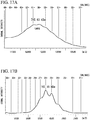
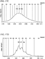
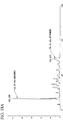
- the molecular weights of IgA may be measured by mass spectrometry and the like under mild iontophoresis conditions which are, for example, achieved by lowering a degree of vacuum in the vicinity of an iontophoresis port, as will be described later in examples.
- the antibody may be a chimera of an IgA-type antibody and a non-IgA-type antibody.
- the IgA-type antibody refers to an antibody having an amino acid sequence which is at least partially derived from an IgA-type antibody. That is, the IgA-type antibody may also be a protein with which a typical anti-IgA polyclonal antibody reacts.
- the present inventors have found that the polymeric antibodies have higher antigen-binding activity or neutralizing activity against antigens than the monomeric antibodies.
- the present invention provides a method of quantifying components of a polymeric IgA-type antibody, which includes the step of performing mass spectrometry using each of peptides having an amino acid sequence set forth in SEQ ID NOs: 70 to 97 as an internal standard. Also, when a stable isotope-labeled IgA antibody is produced by adding stable isotope-labeled amino acids into IgA antibody-secreting cells and culturing the IgA antibody-secreting cells using the method disclosed in Taga Y., et al., Stable isotope-labeled collagen: a novel and versatile tool for quantitative collagen analyses using mass spectrometry, J. Proteome Res.
- the stable isotope-labeled IgA antibody may be used as an internal standard.
- the polymeric IgA-type antibody is preferably derived from a human being.
- the components may include an IgAl antibody heavy chain, an IgA2 antibody heavy chain, a ⁇ -type antibody light chain, a ⁇ -type antibody light chain, a J chain, and an SC.
- the present invention provides standards for quantifying components of the polymeric IgA-type antibody, which includes a set of peptides having amino acid sequences set forth in SEQ ID NOs: 70 to 97.
- the present invention provides a medicine containing the polymeric IgA-type recombinant antibody as an active ingredient.
- the medicine according to this embodiment is preferably used for treatment or prevention of infections.
- the infections may include infections caused by pathogens such as parasites, bacteria, fungi, viruses, abnormal prions, etc.
- IgA A main antibody in mucus or secretory fluid covering the mucous epithelium is IgA, but IgA is not practically used as an antibody medicine although the IgA is known to serve as a front-line in a biological defense mechanism against mucosal infections.
- the present inventors have conducted research to develop a intranasal vaccine using a whole inactivated influena virus vaccine as a next-generation influenza vaccine, characterized by safe and simple inoculation of an inactivated whole virion of influenza virus as vaccine antigen. So far, in addition to basic research using animals, clinical research with recruited healthy adult volunteers shows good results, and the research is entering a clinical development stage for the purpose of practical application.
- the present inventors have found that among IgA antibodies, which are considered to play an important role in protection against viral infections in respiratory mucosa of subjects who received a nasal inactivated influenza vaccine, polymeric antibodies larger than dimers are present and have a higher influenza virus-neutralizing activity than monomeric and dimeric antibodies.
- the present inventors first succeeded in efficiently producing a polymeric IgA-type recombinant antibody, and have found that the polymeric IgA-type recombinant antibody has higher influenza virus-neutralizing activity and HA protein-binding activity than monomeric and dimeric antibodies.
- the medicine according to this embodiment is usefully used as a therapeutic or prophylactic agent against mucosal infections such as influenza, RS virus infection, severe acute respiratory syndrome (SARS), Middle Eastern respiratory syndrome (MERS), acquired immune deficiency syndrome (AIDS), etc.
- mucosal infections such as influenza, RS virus infection, severe acute respiratory syndrome (SARS), Middle Eastern respiratory syndrome (MERS), acquired immune deficiency syndrome (AIDS), etc.
- the polymeric IgA-type recombinant antibody may bind to and neutralize influenza virus, RS virus, etc. even when used in a small amount.
- the medicine according to this embodiment could be an antibody medicine for respiratory administration that may be applied as a prophylactic or therapeutic agent against the infections, an in vitro diagnostic agent, an antibody for research use, etc.
- the medicine according to this embodiment may be administered to a subject at high risk of being infected by viruses causing the infections.
- the medicine may be administered to a patient whose morbidity in the infections is identified for a therapeutic purpose and the purpose of preventing the virus from spreading.
- the medicine according to this embodiment may be administered after the medicine is prepared into formulations such as powders, liquids, etc.
- formulations such as powders, liquids, etc.
- a nasal excipient usually contained in nasal sprays for allergic rhinitis already available on the market may be added to the medicine according to this embodiment.
- the medicine according to this embodiment may be administered by spraying it onto the nasal mucosa, administered by inhalation into the lower respiratory tract using a nebulizer, etc.
- the administration by spraying onto the nasal mucosa may, for example, be performed in the same manner as for the nasal whole particle-inactivated influenza vaccine as disclosed in Ainai A, et al., Intranasal vaccination with an inactivated whole influenza virus vaccine induces strong antibody responses in serum and nasal mucus of healthy adults., Hum Vaccin Immunother. 9(9), 1962-1970, 2013 .
- the therapeutic or prophylactic agent of this embodiment When the therapeutic or prophylactic agent of this embodiment is administered by spraying it onto the nasal mucosa, for example, the therapeutic or prophylactic agent may be sprayed into both nostrils at an amount of 250 ⁇ L per nostril. Also, an amount of the administered antibody may be in a range of several hundred ⁇ g to several mg per inoculation (500 ⁇ L). For spraying, for example, a spray device used for nasal whole inactivated influenza virus vaccine may be used. Also, the therapeutic or prophylactic agent may be sprayed twice (morning and evening) to 4 times (once every 6 hours) a day. The administration period may, for example, be one week.
- a generally used aerosol-type inhaler may be used.
- An amount of the inhaled antibody may, for example, be several mg to several tens of mg per inhalation.
- the antibody may be inhaled approximately twice a day (morning and evening).
- the administration period may, for example, be one week.
- the medicine according to this embodiment may be administered to a human being, or, for example, a domestic animal such as a horse, a cow, a goat, a sheep, a pig, etc.; a pet such as a dog, a cat, etc.; a primate such as a chimpanzee, a gorilla, a cynomolgus monkey, etc.; a rodent such as a mouse, a rat, a guinea pig, etc.
- a domestic animal such as a horse, a cow, a goat, a sheep, a pig, etc.
- a pet such as a dog, a cat, etc.
- a primate such as a chimpanzee, a gorilla, a cynomolgus monkey, etc.
- a rodent such as a mouse, a rat, a guinea pig, etc.
- the IgA heavy chain, the IgA light chain, the J chain, the secretory component protein preferably have amino acid sequences derived from a target animal (a target animal type).
- the target animal type means that the constant regions of the IgA heavy chain and the IgA light chain coding for the polymeric antibody have amino acid sequences of constant regions of an IgA heavy chain and an IgA light chain of the target animal.
- the animal type targeted by the J chain and the secretory component protein means that the J chain and the secretory component protein have amino acid sequences of a J chain and a secretory component protein of the target animal.
- the amino acid sequences of the IgA heavy chain, the IgA light chain, the J chain, and the secretory component protein may include mutations as long as the amino acid sequences have a desired antigen-binding activity.
- the present invention provides a method of producing a polymeric IgA-type antibody, which includes the step of coexpressing an IgA-type antibody heavy-chain protein, an antibody light-chain protein, an antibody J-chain protein, and a secretory component protein in a single cell.
- the polymeric IgA-type antibody may be a recombinant antibody.
- pIgR expressed on cell membranes in the basal side of mucosal epithelial cells specifically recognizes and binds to a J chain protein in dimeric/polymeric Ig molecules produced by plasma cells present in the mucosal lamina intestinal, and incorporates dimeric/polymeric Ig molecules combind with the J chain protein into cells. Even after incorporation into epithelial cells, the pIgR is transferred to an apical side in a state in which the pIgR is bound to the dimeric/polymeric Ig molecule.
- a cleavage between an extracellular domain region and a transmembrane region of the pIgR occurs so that the pIgR is released from the inside of the epithelial cells into a surface of the mucosa.
- the IgA-type antibody heavy-chain protein, the antibody light-chain protein, the antibody J-chain protein, and the secretory component protein may not be coexpressed in vivo in a single cell.
- the present inventors have first succeeded in unexpectedly producing a polymeric IgA-type antibody by coexpressing a secretory component protein in a single cell together with an IgA-type antibody heavy-chain protein, an antibody light-chain protein, and an antibody J-chain protein.
- the polymeric IgA-type antibody is industrially applicable.
- the antibody J-chain protein and the secretory component protein preferably have amino acid sequences derived from an animal species as a target.
- the antibody J-chain protein and the secretory component protein may be chimeras having amino acid sequences derived from a plurality of animal species.
- the IgA-type antibody heavy-chain protein may be obtained by converting an originally non-IgA-type antibody into an IgA type by means of genetic recombination.
- the non-IgA-type antibody is not particularly limited, and may, for example, include a non-IgA-type human antibody, a non-human mammal-derived antibody, a rodent-derived antibody, a bird-derived antibody, etc.
- a class of antibodies is not particularly limited, and may, for example, include IgG-type, IgM-type, IgE-type, IgD-type, IgY-type antibodies, etc.
- the IgG type antibody may be converted into an IgA type by transplantation of the variable region of the IgG type antibody into the backbone framework of the IgA-type antibody.
- the conversion from IgG type antibody to IgA type antibody could be performed by the graft of only CDR region of the IgG type antibody into the corresponding region of IgA type antibody.
- a subclass of IgA may be an IgAl type or an IgA2 type.
- Host cells may include mammalian cells, insect cells, etc.
- the mammalian cells may include 293F cells, CHO cells, CHO YA7 cells, etc., and the CHO YA7 cells are particularly preferred.
- the insect cells may include an Sf9 cell line, an Sf21 cell line, etc.
- the present invention provides a method of producing a polymeric IgA-type antibody, which includes the step of coexpressing an IgA-type antibody heavy-chain protein, an antibody light-chain protein, an antibody J-chain protein, a secretory component protein, a p180 protein, and an SF3b4 protein in a single cell.
- the polymeric IgA-type antibody may be a recombinant antibody.
- An amino acid sequence of the p180 protein is set forth in SEQ ID NO: 98, and a base sequence coding for the p180 protein is set forth in SEQ ID NO: 99.
- an amino acid sequence of the SF3b4 protein is set forth in SEQ ID NO: 100, and a base sequence coding for the SF3b4 protein is set forth in SEQ ID NO: 101.
- Formation of polysomes on the endoplasmic reticulum membrane within the corresponding cell may be promoted by expressing the p180 protein and the SF3b4 protein in the cell.
- the polysome refers to one molecule of mRNA bound to a plurality of ribosomes present on the endoplasmic reticulum membrane within the cell.
- the protein synthesis ability may be enhanced, thereby improving production efficiency of a target protein.
- Suitable host cells are as described above.
- the human p180 protein is a type I transmembrane protein present in the endoplasmic reticulum, and is composed of a short intracytoplasmic region, a transmembrane region, and a cytoplasmic domain region.
- the cDNA sequence has a domain that is highly conserved among species in the vicinity of the N terminus of the cytoplasmic domain and shows very strong basicity (at positions 27 to 197 of SEQ ID NO: 98), followed by a basic repetitive sequence, and it is also known that there are at least three types of molecular species whose repeat numbers are 54, 26, and 14 ( Ogawa-Goto K. et al., An endoplasmic reticulum protein, p180, is highly expressed in human cytomegalovirus-permissive cells and interacts with the tegument protein encoded by UL48, J. Virol., 76 (5), 2350-2362, 2002 .).
- a p180 protein having any repeat number may be used, a p180 protein having a repeat number of 54 is preferred.
- the C-terminal side of the p180 protein forms a coiled-coil domain, and, in the coiled-coil domain, a 945 th to 1,540 th region set forth in SEQ ID NO: 98 interacts with the SF3b4 protein to play an important role in promoting the polysome formation ( Ueno T. et al., Regulation of polysome assembly on the endoplasmic reticulum by a coiled-coil protein, p180, Nucleic Acids Res., 40 (7), 3006-3017, 2012 .).
- mRNA precursor transcribed from intracellular DNA is converted into mature mRNA by removing an intron moiety by splicing.
- This process is performed with a macrocomplex consisting of small nuclear RNA (snRNA) and proteins, which is referred to as a spliceosome.
- snRNA small nuclear RNA
- proteins which is referred to as a spliceosome.
- snRNPs small nuclear ribo-nucleoprotein complexes
- the SF3b4 protein is a constituent of U2-snRNP, and has an RNA-binding domain.
- the present inventors have found that the SF3b4 protein predominantly increases in a membrane fraction containing endoplasmic reticulum in the cytoplasm, and the SF3b4 protein bound to mRNA also interacts with a coiled-coil domain of the p180 protein to promote localization of mRNA to the endoplasmic reticulum, resulting in enhanced abilities to synthesize or secrete proteins.
- mRNA transcribed from the corresponding nucleic acid interacts with the SF3b4 protein or the p180 protein, or the mRNA interacts with the SF3b4 protein, and the SF3b4 protein then interacts with a coiled-coil domain of the p180 protein, thereby promoting the localization of the mRNA to the endoplasmic reticulum to enhance the abilities to synthesize or secrete the target protein in these cells.
- the p180 protein may be a protein consisting of an amino acid sequence set forth in SEQ ID NO: 98, a protein consisting of an amino acid sequence having a sequence identity of 80% or more, preferably 85% or more, more preferably 90% or more, and particularly preferably 95% or more to the amino acid sequence set forth in SEQ ID NO: 98, and having a function of promoting formation of polysomes on the intracellular endoplasmic reticulum membrane, a protein consisting of an amino acid sequence having a sequence similarity of 80% or more, preferably 85% or more, more preferably 90% or more, and particularly preferably 95% or more to the amino acid sequence set forth in SEQ ID NO: 98, and having a function of promoting formation of polysomes on the intracellular endoplasmic reticulum membrane, a protein consisting of an amino acid sequence in which one to several amino acids are deleted, substituted or added in the amino acid sequence set forth in SEQ ID NO: 98, and having a function of promoting formation
- the SF3b4 protein may be a protein consisting of an amino acid sequence set forth in SEQ ID NO: 100, a protein consisting of an amino acid sequence having a sequence identity of 80% or more, preferably 85% or more, more preferably 90% or more, and particularly preferably 95% or more to the amino acid sequence set forth in SEQ ID NO: 100, and having an ability to synthesize or secrete proteins as a target product when expressed in cells, which is identical to that of the protein consisting of the amino acid sequence set forth in SEQ ID NO: 100, a protein consisting of an amino acid sequence having a sequence similarity of 80% or more, preferably 85% or more, more preferably 90% or more, and particularly preferably 95% or more to the amino acid sequence set forth in SEQ ID NO: 100, and having an ability to promote synthesis or secretion of proteins as a target product when expressed in cells, which is identical to that of the protein consisting of the amino acid sequence set forth in SEQ ID NO: 100, a protein consisting of an amino acid sequence in
- the present invention provides a method of producing a polymeric IgA-type antibody, which includes the step of expressing an IgA-type antibody heavy-chain protein, an antibody light-chain protein, an antibody J-chain protein, and a secretory component protein in a CHO YA7 cell line (name of depository authority: National Institute of Technology and Evaluation, Patent Microorganisms Depositary (NPMD), address of depository authority: #122, 2-5-8 Kazusakamatari, Kisarazu-shi, Chiba 292-0818, Japan, deposit date: February 13, 2013, accession number: NITE BP-01535 ).
- the polymeric IgA-type antibody may be a recombinant antibody.
- the CHO YA7 cell line is a cell line established by the present inventors, and constitutively expresses the p180 protein and the SF3b4 protein in cells. Therefore, the polymeric IgA-type antibody may be efficiently produced using the production method according to this embodiment.
- the present invention provides a method of producing a polymeric IgA-type antibody, which includes the step of coexpressing an IgA-type antibody heavy-chain protein, an antibody light-chain protein, an antibody J-chain protein, and a secretory component protein in a single cell.
- the step is performed by transfecting an expression vector for expressing the IgA-type antibody heavy-chain protein, the antibody light-chain protein, the antibody J-chain protein and the secretory component protein into the cell, the expression vector has a cis-element, which an RNA-binding protein recognizes, binds to or interacts with, downstream from a promoter and also upstream from an initiation codon of nucleic acids coding for the IgA-type antibody heavy-chain protein, the antibody light-chain protein, the antibody J-chain protein, or the secretory component protein.
- the polymeric IgA-type antibody may be a recombinant antibody.
- the cis-element preferably includes one to several base sequences consisting of a sequence motif GAN 1 -(X) n -ACN 2 (where n is an integer ranging from 3 to 6, and N 1 and N 2 are each independently one of A, T, G, and C).
- the present inventors have found that, when the cis-element is present in a 5'-'untranslated region of a mature mRNA, an RRM protein that recognizes the corresponding cis-element binds to the corresponding cis-element, and enhances transport/localization of mRNA onto a membrane of the endoplasmic reticulum that is a site of protein synthesis, resulting in increased translation efficiency.
- the polymeric IgA-type recombinant antibody may be more efficiently produced using the production method according to this embodiment.
- Suitable host cells are as described above.
- the cis-element may be a nucleic acid fragment that consists of a base sequence set forth in any one selected from SEQ ID NOs: 21 to 23, a base sequence in which one to several bases are deleted, substituted or added in the base sequence set forth in any one selected from SEQ ID NOs: 21 to 23, and which the RNA-binding protein recognizes, binds to or interacts with, a base sequence having an identity of 80% or more, preferably 85% or more, more preferably 90% or more, and particularly preferably 95% or more to the base sequence set forth in any one selected from SEQ ID NOs: 21 to 23, and which the RNA-binding protein recognizes, binds to or interacts with, or a base sequence hybridizable under a stringent condition with a nucleic acid consisting of a base sequence complementary to a nucleic acid consisting of the base sequence set forth in any one selected from SEQ ID NOs: 21 to 23, and which the RNA-binding protein recognizes, binds to or interacts with.
- the number of bases to be deleted, substituted or added may, for example, be in a range of 1 to 30, for example in a range of 1 to 15, for example in a range of 1 to 10, and for example in a range of 1 to 5.
- the number of amino acids to be deleted, substituted or added may, for example, be in a range of 2 to 40, for example in a range of 2 to 30, for example in a range of 2 to 20, for example in a range of 2 to 10, for example in a range of 2 to 7, for example in a range of 2 to 5, for example 5, for example 4, for example 3, and for example 2.
- the identity of an amino acid sequence refers to an identity between two target amino acid sequences, and is indicated by a percentage (%) of matched amino acid residues in an optimal alignment of amino acid sequences constructed using a mathematical algorithm known in the related art.
- the identity of the amino acid sequence may be determined by visual inspection and mathematical calculation, and may be calculated using a homology search program (for example, BLAST, FASTA), a sequence alignment program (for example, ClustalW), genetic information-processing software (for example, GENETYX (registered trademark)), etc. known in the related art.
- the similarity of an amino acid sequence refers to a similarity between two target amino acid sequences, and is indicated by a percentage (%) of matched amino acid residues and amino acid residues showing similarity in an optimal alignment of amino acid sequences constructed using a mathematical algorithm known in the related art.
- amino acids belonging to the same groups are, for example, referred to as similar amino acid residues in groups such as aromatic amino acids (Phe, Tyr, and Trp), hydrophobic amino acids (Ala, Leu, Ile, Val, Gly, Pro, Met, Phe, and Trp), aliphatic amino acids (Ala, Leu, Ile, and Val), polar amino acids (Asn and Gln), basic amino acids (Lys, Arg, and His), acidic amino acids (Asp and Glu), amino acids containing a hydroxyl group (Ser and Thr), and amino acids with a small side chain (Gly, Ala, Ser, Thr, and Met).
- amino acid residue showing this similarity is expected to have no influence on the phenotypes of proteins.
- the similarity of the amino acid sequence may be determined by visual inspection and mathematical calculation, and may be calculated using a homology search program (for example, BLAST, PSI-BLAST, and HMMER), genetic information-processing software (for example, GENETYX (registered trademark)), etc. known to those skilled in the related art.
- a homology search program for example, BLAST, PSI-BLAST, and HMMER
- genetic information-processing software for example, GENETYX (registered trademark)
- the similarity of the amino acid sequence may be specifically calculated under a default setting condition (Unit size to compare is set to 2) by executing Protein vs. Protein Global Homology using a GENETYX (registered trademark) network version (ver. 11.1.3; GENETYX CORPORATION).
- the identity of a base sequence refers to an identity of two target base sequences, and is indicated by a percentage (%) of matched nucleic acids in an optimal alignment of base sequences constructed using a mathematical algorithm known in the related art.
- a representative computer program for calculating the identity of a base sequence may include a Wisconsin Package version 10.0 program "GAP” ( Devereux, et al., 1984, Nucl. Acids Res., 12: 387 ) of the Genetics Computer Group (GCG; Madison, Wisconsin), a BLASTN program (version 2.2.7) available through the National Medical Library website: http://www.ncbi.nlm.nih.gov/blast/bl2seq/bls.html, or an UW-BLAST 2.0 algorithm.
- GAP Wisconsin Package version 10.0 program "GAP” ( Devereux, et al., 1984, Nucl. Acids Res., 12: 387 ) of the Genetics Computer Group (GCG; Madison, Wisconsin), a BLASTN program (version 2.2.7) available through the National Medical Library website: http://www.ncbi.nlm.nih.gov/blast/bl2seq/bls.html, or an UW-BLAST 2.0 algorithm.
- hybridization may be performed by incubating target sequences at 55 to 70°C for several hours to overnight in a hybridization buffer including 5 ⁇ SSC (a composition of 20 ⁇ SSC: 3 M sodium chloride, 0.3 M citric acid solution, pH 7.0), N-lauroylsarcosine at 0.1% by weight, SDS at 0.02% by weight, a blocking reagent for hybridization of nucleic acids at 2% by weight, and formamide at 50%.
- a washing buffer used for washing after the incubation is preferably a 1 ⁇ SSC solution containing SDS at 0.1% by weight, more preferably a 0.1 ⁇ SSC solution containing SDS at 0.1% by weight.
- the cis-element may have an effect of improving an expression or secretion level of the polymeric IgA-type antibody.
- the method according to this embodiment it is possible to prepare a polymeric antibody 26 times to 35 or more times as efficiently as in the conventional methods.
- the monoclonal tetrameric antibody (rS-qIgA) against influenza viruses prepared by such a method has an antigen-binding activity higher than that of the monomeric antibody, and shows 100 or more times the neutralizing activity.
- the present invention provides a method of improving the antigen-binding activity or neutralizing activity of the antibody, which includes the step of making an antibody into a polymeric IgA type.
- the step of making an antibody into a polymeric IgA type preferably includes the step of coexpressing an IgA-type antibody heavy-chain protein having a heavy-chain variable region of the antibody, a light-chain protein of the antibody, an antibody J-chain protein, and a secretory component protein in a single cell.
- the antigen binding activity of an antibody refers to the ability of an antibody to bind to a target molecule per se, but the specificity refers to the ability to not bind to substances other than the target molecule, and a site that exists only in the target molecule is required as a recognition site (an epitope) to ensure high specificity.
- the antigen-binding activity and specificity are generated due to the diversity of sequences of variable regions of the antibody molecules, it is difficult to predict the antigen-binding activity and specificity to the target molecule from the sequences of the variable regions, and antibodies having high antigen-binding activity and specificity need to be cloned from antibody gene in antibody-producing cells generated in a living subject such as a mouse, or a library of artificially prepared variable regions using any methods. Also, since both the specificity and the antigen-binding activity independently depend on the sequences of the variable regions, it is very difficult to improve the antigen-binding activity in an artificial manner without a change in the specificity of the obtained antibody, that is, without a change in the epitope to be recognized.
- the polymeric IgA-type antibody may be produced by coexpressing the IgA-type antibody heavy-chain protein, the antibody light-chain protein, the antibody J-chain protein, and the secretory component protein in a single cell. Suitable host cells are as described above.
- the present inventors have found that the antigen-binding activity or neutralizing activity of the antibody may be improved by making a monomeric antibody into a polymer.
- the virus-neutralizing activity per 1 mole of the antibody may be improved by a factor of 100 or more, compared to the monomeric antibody, by making the monomeric antibody into a polymer.
- Antibodies to be improved in the antigen-binding activity or neutralizing activity are not particularly limited, and may, for example, include a human antibody, a non-human mammal-derived antibody, a rodent-derived antibody, a bird-derived antibody, etc.
- a class of antibodies is not particularly limited, and may, for example, include IgG-type, IgM-type, IgE-type, IgA-type, IgD-type, and IgY-type antibodies, etc.
- the antigen-binding activity or neutralizing activity may be improved without a change in specificity of pre-existing antibodies using the method according to this embodiment.
- the method according to this embodiment may be applied by binding a variable region of the interesting antibody to a constant region of the IgA-type antibody to convert the antibody into an IgA type by means of genetic recombination.
- an IgG-type monoclonal antibody may be converted into an IgA-type monoclonal antibody by transplanting a variable region of the IgG-type monoclonal antibody to the backbone framework of an IgA-type antibody.
- the IgG-type monoclonal antibody may be converted into an IgA type by transplanting only CDR region of the antibody to be improved in the antigen-binding activity into the CDR region of the IgA-type antibody.
- a subclass of IgA may be an IgAl type or an IgA2 type.
- the method according to this embodiment is highly versatile as technology capable of converting an IgG-type monoclonal antibody, which has a variable region that recognizes the same epitope, into a high-binding type and a high-activity type. Therefore, the method may be widely applied to products using monoclonal antibodies such as antibodies for medicines, diagnostic antibodies used for immunochromatography, immunohistochemistry, ELISA, etc., and other antibodies for research applications, etc.
- a whole inactivated virion vaccine for a highly pathogenic avian influenza virus A/H5N1 was nasally inoculated into healthy adults twice at a three-week interval (250 ⁇ L per nostril; a total dose of 500 ⁇ L).
- An inactivated whole particle vaccine containing 45 ⁇ g of hemagglutinin (HA) was used as the vaccine.
- Peripheral blood was collected after 7 days of the second vaccination, and peripheral blood lymphocytes were collected using a blood cell separation solution, Lymphoprep (trademark) (AXIS-SHIELD).
- Isolation of antibody-producing plasma cells induced in peripheral blood by intranasal vaccination was performed using FACS Aria (BD Bioscience).
- a cell population of cell surface markers CD2 - , CD3 - , CD4 - , CD10 - , CD20 - , IgD - , CD19 low , CD27 high , and CD38 high was collected as antibody-producing plasma cells, and separated and collected as single cells.
- Single antibody-producing plasma cells were collected in a 96-well plate in which 9 ⁇ L of sterile water including 45 ng of carrier RNA was dispensed into each well.
- Preparation of cDNA was performed based on the article of T. Tiller et al.
- isotypes of an antibody heavy chain isolated in each well were determined by Real-time PCR.
- TaqMan probes and primers were prepared for each of constant regions of IgG, IgA, and IgM.
- the TaqMan probes for IgG, IgA, and IgM were labeled with FAM, HEX, and Cy5, respectively.
- Analysis was performed using LightCycler 480 (F. Hoffmann-La Roche, Ltd.) using QuantiTect Multiplex PCR NoROX Master Mix (QIAGEN N.V.).
- Amplification of an antibody variable region gene was performed based on the article of T. Tiller et al. (J Immunol Methods, 329, 112-24, 2008 ). Specifically, a mixture including 11.5 ⁇ L of HotStarTaq DNA polymerase (QIAGEN N.V.), dNTP mix, and a primer set for amplifying each of the antibody variable region genes was added to 1 ⁇ L of the prepared cDNA, and subjected to the first PCR reaction. Also, the second PCR reaction was performed using a primer set designed further inside for each of the genes included in 1 ⁇ L of the resulting PCR product.
- HotStarTaq DNA polymerase QIAGEN N.V.
- dNTP mix dNTP mix
- a primer set for amplifying each of the antibody variable region genes was added to 1 ⁇ L of the prepared cDNA, and subjected to the first PCR reaction. Also, the second PCR reaction was performed using a primer set designed further inside for each of the genes included in
- amplification was performed under conditions of one cycle of 95°C for 15 minutes, 43 cycles of 94°C for 30 seconds, 58°C for 20 seconds and 72°C for 60 seconds, and one cycle of 72°C for 2 minutes. Also, the base sequence analysis (sequencing) of PCR products was performed using a conventional method.
- PCR of an antibody variable region gene was performed according to the instructions using PrimeSTAR (registered trademark) MAX DNA Polymerase (TaKaRa).
- the above-described first PCR product was used as a template, and a pair of primers suitable for a locus to be amplified were selected as the primers based on the sequencing results of the above-described second PCR product.
- the PCR conditions were 25 cycles at 98°C for 10 seconds, 55°C for 5 seconds, and 72°C for 10 seconds.
- the PCR product was purified according to the instructions using MonoFas (registered trademark) DNA Purification Kit I (GL Sciences Inc.), and eluted in 30 ⁇ L of Buffer C.
- the purified PCR product was digested with restriction enzymes under suitable conditions using AgeI-HF (all chains), and SalI-HF (a heavy chain), BsiWI (a ⁇ chain) or XhoI (a ⁇ chain) (commercially available from NEB Co.) in a total volume of 30 ⁇ L.
- Expression vectors ⁇ 1 HC (a heavy chain), ⁇ LC (a ⁇ chain), and ⁇ LC (a ⁇ chain) corresponding to the respective chains were also enzymatically digested with the same combination of restriction enzymes.
- the restriction enzyme product was purified according to the instructions using MonoFas (registered trademark) DNA Purification Kit I (GL Sciences Inc.), and eluted in 20 ⁇ L of Buffer C.
- Ligation of DNA digested with the restriction enzymes was performed in a total volume of 10 ⁇ L according to the instructions using a DNA ligation kit ⁇ Mighty Mix> (TaKaRa). Competent Quick DH5 ⁇ (TOYOBO) was transformed with 10 ⁇ L of the ligation products by heating the competent cells at 42°C. Plasmid extraction was performed according to the instructions using a Pure Yield (trademark) plasmid miniprep system (Promega Corporation).
- a sequencing reaction of the extracted plasmid was performed according to the instructions using a BigDye (registered trademark) Terminator v3.1 Cycle sequencing kit (Life Technologies Inc.).
- the reaction product was purified according to the instructions using BigDye XTerminator (trademark) kit (Life Technologies Inc.), and sequenced using an Applied Biosystems 3130 genetic analyzer (Life Technologies Inc.). Alignment analysis of the read sequence and the sequence of the second PCR product was performed to select a sample holding the most consensus sequence.
- An Expi293 (trademark) expression system (Life Technologies Inc.) was used according to the instructions to produce a recombinant antibody. As one example, a 30 mL system will be described below.
- Expi293F cells subcultured had a density of more than 3.0 ⁇ 10 6 cells/mL and survival rate of more than 95%, and the cells were not aggregated.
- the number of cells was adjusted to 2.9 ⁇ 10 6 cells/mL using an Expi293 expression medium warmed at 37°C. 25.5 mL of the prepared cell suspension was transferred to a disposable Erlenmeyer flask equipped with a vent filter cap, returned to an incubator for cell culture set to 37°C and 8% CO 2 , and cultured by shaking at 125 rpm.
- 30 ⁇ g of plasmid DNA (15 ⁇ g each of a heavy chain and a light chain of IgG) was added to 1.5 mL of an Opti-MEM I medium.
- ExpiFectamine 293 reagent 80 ⁇ L of an ExpiFectamine 293 reagent was added to 1.5 mL of a separately prepared Opti-MEM I medium. After a DNA solution and an ExpiFectamine solution were incubated at room temperature for 5 minutes, a total volume of the DNA solution was added to an ExpiFectamine solution, and then incubated at room temperature for 20 to 30 minutes. After a transfection mix was added to the cells, the cells were returned to an incubator for cell culture set to 37°C and 8% CO 2 , and cultured by shaking at 125 rpm.
- the antibody was eluted with a 5-column volume of 0.1 M glycine-HCl (pH 3.0), and the resulting eluate was neutralized with 1 M Tris-HCl (pH 9.0).
- the column was re-equilibrated with a 10-column volume of PBS.
- a concentration of the antibody was measured using NanoDrop (Thermo Scientific Ltd.).
- the antibody was concentrated according to the instructions using an Amicon (registered trademark) ultra-centrifugal filter device (Millipore Corporation).
- Amicon registered trademark
- a Zeba Desalt spin column the buffer of the eluate was replaced with a phosphate buffer (PB, pH 7.4) according to the instructions. After the buffer replacement, the concentration was measured using NanoDrop.
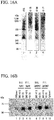
- Fig. 1 is an image showing results of the SDS-PAGE. It was confirmed that the antibody clones G2, H10, D11, F9, F11, H5, and C1 were converted to IgG1 types. The expression levels varied depending on the clones.
- IgA-type recombinant antibody was prepared using the following method.
- the IgG1 antibodies prepared in Experimental Example 1 are not particularly limited, and antibodies having known sequences may be made into IgA types using the following method.
- An expression vector ⁇ 1 HC containing a constant region gene of the IgAl antibody was prepared. PCR of the constant region gene of the IgAl antibody was performed according to the instructions using PrimeSTAR (registered trademark) MAX DNA Polymerase (TaKaRa).
- the constant region gene of the IgAl antibody was amplified using pFUSE-CHIg-hA1 (InvivoGen Ltd.) as a template.
- the PCR conditions were 30 cycles at 98°C for 10 seconds, 55°C for 5 seconds, and 72°C for 30 seconds.
- the PCR product was purified according to the instructions using MonoFas (registered trademark) DNA Purification Kit I (GL Sciences Inc.), and eluted in 30 ⁇ L of Buffer C.
- the purified PCR product and a ⁇ 1 HC plasmid were digested with restriction enzymes XhoI and HindIII-HF (commercially available from NEB Co.) at 37°C in a total volume of 30 ⁇ L.
- the restriction enzyme-digested products were purified according to the instructions using MonoFas (registered trademark) DNA Purification Kit I (GL Sciences Inc.), and eluted in 20 ⁇ L of Buffer C. Ligation of the restriction enzyme-digested DNAs was performed in a total volume of 10 ⁇ L according to the instructions using a DNA ligation kit ⁇ Mighty Mix> (TaKaRa). 10 ⁇ L of the ligation product was warmed at 42° to be transformed into Competent Quick DH 5 ⁇ .
- Plasmid extraction was performed according to the instructions using a Pure Yield (trademark) Plasmid Miniprep System (Promega Corporation).
- a sequencing reaction of the extracted plasmid was performed according to the instructions using a BigDye (registered trademark) Terminator v3.1 Cycle sequencing kit (Life Technologies Inc.).
- the reaction products were purified according to the instructions using a BigDye XTerminator (trademark) kit (Life Technologies Inc.), and sequenced using an Applied Biosystems 3130 genetic analyzer (Life Technologies Inc.).
- PCR of the antibody variable region gene was performed according to the instructions using PrimeSTAR (registered trademark) MAX DNA Polymerase (TaKaRa). Using the antibody gene cloned into the ⁇ 1 HC expression vector as a template, a reverse primer was replaced with a primer for the ⁇ 1 HC expression vector, and the PCR conditions were 25 cycles at 98°C for 10 seconds, 55°C for 5 seconds, and 72°C for 5 seconds.
- the PCR product was purified according to the instructions using MonoFas (registered trademark) DNA Purification Kit I (GL Sciences Inc.), and eluted in 30 ⁇ L of Buffer C.
- the purified PCR product and an ⁇ 1 HC expression vector were digested with restriction enzymes under suitable conditions using AgeI-HF and NheI-HF (commercially available from NEB Co.) in a total volume of 30 ⁇ L.
- the restriction enzyme-digested product was purified according to the instructions using MonoFas (registered trademark) DNA Purification Kit I (GL Sciences Inc.), and eluted in 20 ⁇ L of Buffer C.
- Ligation of the restriction enzyme-digested DNAs was performed in a total volume of 10 ⁇ L according to the instructions using a DNA ligation kit ⁇ Mighty Mix> (TaKaRa). 10 ⁇ L of the ligation product was warmed at 42°C to be transformed into Competent Quick DH 5 ⁇ (TOYOBO).
- Plasmid extraction was performed according to the instructions using a Pure Yield (trademark) Plasmid Miniprep System (Promega Corporation).
- a sequencing reaction of the extracted plasmid was performed according to the instructions using a BigDye (registered trademark) Terminator v3.1 Cycle sequencing kit (Life Technologies Inc.).
- the reaction product was purified according to the instructions using a BigDye XTerminator (trademark) kit (Life Technologies Inc.), and sequenced using an Applied Biosystems 3130 genetic analyzer (Life Technologies Inc.). Based on the sequencing results, it was confirmed that antibody variable region gene cloned into ⁇ 1 HC expression vector was identical to the antibody gene cloned into the ⁇ 1 HC expression vector.
- An Expi293 (trademark) expression system (Life Technologies Inc.) was used according to the instructions to produce a recombinant antibody. As one example, a 30 mL system will be described below.
- Expi293F cells subcultured had a density of more than 3.0 ⁇ 10 6 cells/mL and survival rate of more than 95%, and the cells were not aggregated.
- the number of cells was adjusted to 2.9 ⁇ 10 6 cells/mL using an Expi293 expression medium warmed at 37°C. 25.5 mL of the prepared cell suspension was transferred to an disposable Erlenmeyer flask equipped with a vent filter cap, returned to an incubator for cell culture set to 37°C and 8% CO 2 , and cultured by shaking at 125 rpm.
- 30 ⁇ g of plasmid DNA (15 ⁇ g each of a heavy chain and a light chain of IgAl) was added to 1.5 mL of an Opti-MEM I medium.
- ExpiFectamine 293 reagent 80 ⁇ L of an ExpiFectamine 293 reagent was added to 1.5 mL of a separately prepared Opti-MEM I medium. After a DNA solution and an ExpiFectamine solution were incubated at room temperature for 5 minutes, a total volume of the DNA solution was added to an ExpiFectamine solution, and then incubated at room temperature for 20 to 30 minutes. After a transfection mix was added to the cells, the cells were returned to an incubator for cell culture set to 37°C and 8% CO 2 , and cultured by shaking at 125 rpm.
- the antibody was eluted with a 5-column volume of 0.1 M glycine-HCl (pH 3.0), and the resulting eluate was neutralized with 1 M Tris-HCl (pH 9.0).
- the column was re-equilibrated with a 10-column volume of PBS.
- a concentration of the antibody was measured using NanoDrop (Thermo Scientific Ltd.).
- the antibody was concentrated according to the instructions using an Amicon (registered trademark) ultra-centrifugal filter device (Millipore Corporation).
- Amicon registered trademark
- a Zeba Desalt spin column the buffer of the eluate was replaced with a phosphate buffer (PB, pH 7.4) according to the instructions. After the buffer replacement, the concentration was measured using NanoDrop.
- SDS polyacrylamide gel electrophoresis SDS-PAGE
- NuPAGE registered trademark
- Bis-Tris gel Life Technologies Inc.
- Fig. 2 is an image showing results of the SDS-PAGE. It was confirmed that the antibody clones B12, D11, F9, F11, and H5 were converted to IgAl types.
- a dimeric IgAl-type antibody was prepared using the IgAl antibody expression construct prepared in Experimental Example 2.
- An artificial gene in which an XhoI cleavage site and a Kozak sequence were added to the 5' side of a coding sequence (CDS) of the J chain (GenBank accession no. NM_144646) and a NotI cleavage site was added to the 3' side of the CDS was synthesized for the antibody J chain using an artificial gene synthesis service (Operon Biotechnologies Inc.).
- a J chain gene was digested with restriction enzymes under suitable conditions using XhoI and NotI-HF (commercially available from NEB Co.).
- the restriction enzyme-digested product was cloned into a pCXSN vector (a mammalian cell expression vector composed of a CMV promoter and SV40 polyA) digested with the same restriction enzymes to obtain pCXSN-hJC which was a plasmid for expressing the antibody J chain.
- pCXSN vector a mammalian cell expression vector composed of a CMV promoter and SV40 polyA
- An Expi293 (trademark) expression system (Life Technologies Inc.) was used according to the instructions to produce a dimeric IgAl-type antibody. As one example, a 30 mL system will be described below.
- Expi293F cells subcultured had a density of more than 3.0 ⁇ 10 6 cells/mL and survival rate of more than 95%, and the cells were not aggregated.
- the number of cells was adjusted to 2.9 ⁇ 10 6 cells/mL using an Expi293 expression medium warmed at 37°C. 25.5 mL of the prepared cell suspension was transferred to a disposable Erlenmeyer flask equipped with a vent filter cap, returned to an incubator for cell culture set to 37°C and 8% CO 2 , and cultured by shaking at 125 rpm.
- Plasmid DNA (a J chain-expressed group: 12 ⁇ g each of a heavy chain and a light chain of IgAl, and 6 ⁇ g of a J chain; and a J chain-unexpressed group: 15 ⁇ g each of the heavy chain and light chain of IgAl) was added to 1.5 mL of an Opti-MEM I medium.
- ExpiFectamine 293 reagent 80 ⁇ L of an ExpiFectamine 293 reagent was added to 1.5 mL of a separately prepared Opti-MEM I medium. After a DNA solution and an ExpiFectamine solution were incubated at room temperature for 5 minutes, a total volume of the DNA solution was added to an ExpiFectamine solution, and then incubated at room temperature for 20 to 30 minutes. After a transfection mix was added to the cells, the cells were returned to an incubator for cell culture set to 37°C and 8% CO 2 , and cultured by shaking at 125 rpm.
- the antibody was eluted with a 5-column volume of 0.1 M glycine-HCl (pH 3.0), and the resulting eluate was neutralized with 1 M Tris-HCl (pH 9.0).
- the column was re-equilibrated with a 10-column volume of PBS.
- a concentration of the antibody was measured using NanoDrop (Thermo Scientific Ltd.).
- the antibody was concentrated according to the instructions using an Amicon (registered trademark) ultra-centrifugal filter device (Millipore Corporation).
- Amicon registered trademark
- a Zeba Desalt spin column the buffer of the eluate was replaced with a phosphate buffer (PB, pH 7.4) according to the instructions. After the buffer replacement, the concentration was measured using NanoDrop.
- BN-PAGE blue native polyacrylamide gel electrophoresis
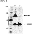
- Fig. 3 is an image showing results of the BN-PAGE. It was confirmed that a band of the monomeric IgA type recombinant antibody was observed in the J chain-unexpressed group (-J), whereas, in addition to a monomer band, a band of the dimeric IgA type recombinant antibody was observed in the J chain-expressed group (+J).
- a polymeric IgAl-type antibody was prepared using the IgAl antibody expression construct prepared in Experimental Example 2.
- the artificial DNA fragment in which an XhoI cleavage site and a Kozak sequence were added to the 5' side of 185 th to 2005 th residues of a polymeric immunoglobulin receptor (GenBank accession no. NM_002644) and a HindIII cleavage site, a thrombin cleavage site, a histidine tag, and a NotI cleavage site were added to the 3' side of the 185 th to 2005 th residues were designed and two artificial DNA fragments (i.e., a 5'-side fragment and a 3'-side fragment) were synthesized so as to overlapped each other using GeneArt (registered trademark) Strings (trademark) DNA fragments (Life Technologies Inc.).
- Overlap PCR in which a PrimeSTAR (registered trademark) MAX DNA Polymerase (TaKaRa) was used was performed using the two synthesized DNA fragments as templates to amplify a gene fragment coding for the secretory component (SC) (SEQ ID NO: 25). Then, the amplified gene fragment was digested with restriction enzymes XhoI and NotI, and cloned into a pCXSN vector to obtain a secretory component expression plasmid pCXSN-hSC-HisTag.
- inverse PCR was performed using the pCXSN-hSC-HisTag as a template to prepare pCXSN-hSC expressing only the secretory component, from which the HindIII cleavage site, the thrombin cleavage site, and the histidine tag added to the 3' side were removed.
- the polymeric antibody was able to be prepared using either plasmid as the secretory component.
- An Expi293 (trademark) expression system (Life Technologies Inc.) was used according to the instructions to produce a polymeric IgAl-type antibody. As one example, a 30 mL system will be described below.
- Expi293F cells subcultured had a density of more than 3.0 ⁇ 10 6 cells/mL and survival rate of more than 95%, and the cells were not aggregated.
- the number of cells was adjusted to 2.9 ⁇ 10 6 cells/mL using an Expi293 expression medium warmed at 37°C. 25.5 mL of the prepared cell suspension was transferred to a disposable Erlenmeyer flask equipped with a vent filter cap, returned to an incubator for cell culture set to 37°C and 8% CO 2 , and cultured by shaking at 125 rpm.
- Plasmid DNA (12 ⁇ g each of a heavy chain and a light chain of IgAl, and 6 ⁇ g each of a J chain and a secretory component) was added to 1.5 mL of an Opti-MEM I medium.
- ExpiFectamine 293 reagent 80 ⁇ L of an ExpiFectamine 293 reagent was added to 1.5 mL of a separately prepared Opti-MEM I medium. After a DNA solution and ExpiFectamin solution were incubated at room temperature for 5 minutes, a total volume of the DNA solution was added to an ExpiFectamine solution, and then incubated at room temperature for 20 to 30 minutes. After a transfection mix was added to the cells, the cells were returned to an incubator for cell culture set to 37°C and 8% CO 2 , and cultured by shaking at 125 rpm.
- the antibody was eluted with a 5-column volume of 0.1 M glycine-HCl (pH 3.0), and the resulting eluate was neutralized with 1 M Tris-HCl (pH 9.0).
- the column was re-equilibrated with a 10-column volume of PBS.
- a concentration of the antibody was measured using NanoDrop (Thermo Scientific Ltd.).
- the antibody was concentrated according to the instructions using an Amicon (registered trademark) ultra-centrifugal filter device (Millipore Corporation).
- Amicon registered trademark
- a Zeba Desalt spin column the buffer of the eluate was replaced with a phosphate buffer (PB, pH 7.4) according to the instructions. After the buffer replacement, the concentration was measured using NanoDrop.
- the concentrated polymeric IgAl-type antibody was fractionated by gel filtration chromatography using AKTA explorer10 (GE Healthcare). Superose6 10/300 GL (GE Healthcare) was used as the column. Dulbecco's phosphate-buffered saline (DPBS) was flushed according to the following protocol: a flow rate of 0.5 mL/min, a column equilibration of 1.5 column volume, and an elution of 0.5 mL per fraction (a total column volume of 1.5).
- DPBS Dulbecco's phosphate-buffered saline
- Fig. 4 is a chart of gel filtration chromatography, and an image showing results of the BN-PAGE for the antibodies contained in fractions 19 to 32. It was revealed that tetramers, dimers and monomers were included in the expressed IgA 1-type antibody. This result was the first report that a tetrameric IgA-type recombinant antibody was prepared.
- a human IgG1 antibody against influenza viruses was prepared in Experimental Example 1, and an IgAl antibody having the same variable region as the corresponding IgG1 antibody was prepared in Experimental Example 2. Also, polymeric IgAl-type antibodies were prepared in Experimental Examples 3 and 4. Then, the influenza virus-neutralizing activity was examined using these antibodies.
- the neutralizing activity of each of the prepared antibodies was quantified by measuring the minimum neutralization concentration using a microneutralization test. Two-fold serial dilutions of a sample were prepared, and mixed with a virus solution of 100 TCID 50 (100 times a 50% tissue culture infective dose), and the resulting mixture was then incubated at 37°C for 30 minutes. Thereafter, this mixture was added to MDCK cells (a cell line derived from a canine kidney), and cultured for 4 days. Then, a value obtained by dividing a concentration of the sample by the maximum dilution ratio of the sample, in which a cytopathic effect by influenza viruses was not able to be determined under a microscope, was used as the minimum neutralization concentration. It meant that the lower the minimum neutralization concentration was, the higher the virus-neutralizing activity was.
- influenza virus-neutralizing activity was measured using monomeric IgG1-type antibody and monomeric IgAl-type antibody of the antibody clones G2, H10, D11, F9, F11, H5, B12, and C1.
- An A/H5N1 strain (clade 2.1) and an A/H1N1 strain were used as the viruses.
- influenza virus-neutralizing activity was measured in the same manner as described above using the monomeric and dimeric IgAl-type antibodies of the antibody clones D11, F9, F11, H5 and B12.
- An A/H5N1 strain (clade 2.1) and an A/H1N1 strain were used as the viruses.
- influenza virus-neutralizing activity was measured in the same manner as described above using the monomeric, dimeric and tetrameric IgAl-type antibodies of the antibody clones F9, F11 and H5.
- An A/H5N1 strain (clade 2.1) was used as the virus.
- Table 3 It meant that the lower the minimum neutralization concentration was, the higher the virus-neutralizing activity was.
- the neutralizing activity ratios of the monomeric IgAl-type antibody, the dimeric IgAl-type antibody and the tetrameric IgAl-type antibody were indicated when it was assumed that the virus-neutralizing activity of the monomeric IgG1-type antibody was set to 1 as shown in Figs. 5A and 5B .
- An A/H5N1 strain (clade 2.1) was used as the virus.
- Fig. 5A is a graph illustrating the neutralizing activity ratios of the monomeric IgAl-type antibody, the dimeric IgAl-type antibody, and the tetrameric IgAl-type antibody per unit amount of proteins when it is assumed that the virus-neutralizing activity of the monomeric IgG1-type antibody is set to 1.
- Fig. 5B is a graph illustrating the neutralizing activity ratios of the monomeric IgAl-type antibody, the dimeric IgAl-type antibody, and the tetrameric IgAl-type antibody per unit number of molecules when it is assumed that the virus-neutralizing activity of the monomeric IgG1-type antibody is set to 1.
- the light chain of the clone F11 was a ⁇ chain.
- the light chain of the clone H5 was a ⁇ chain.
- a recombinant HA protein (1 ⁇ g/mL, derived from an A/H5N1 strain) was added to a 96-well half plate, stationarily incubated at 4°C overnight, and then blocked.
- Cis-element #1 set forth in SEQ ID NO: 22 was introduced downstream from a promoter of pCXSN-hJC which was an expression plasmid for the above-described antibody J chain protein and upstream from an initiation codon of the J chain protein to obtain pCXSN-cis#1-hJC. Based on the genetic sequence analysis, it was confirmed that the cis-element was inserted in the correct direction.
- cis-element #2 set forth in SEQ ID NO: 23 was introduced downstream from a promoter of pCXSN-hSC which was an expression plasmid for the above-described secretory component and upstream from an initiation codon of the secretory component protein to obtain pCXSN-cis#2-hSC. Based on the genetic sequence analysis, it was confirmed that the cis-element was inserted in the correct direction.
- CHO YA7 cells which are a cell line coexpressing the p180 protein and the SF3b4 protein, and 1 ⁇ 10 5 control CHO cells were transfected with 0.5 ⁇ g of each of four expression plasmids, for example, the IgAl antibody heavy chain expression plasmid and the full-length light chain expression plasmid, both of which were constructed in Experimental Example 2, pCXSN-cis#1-hJC, and pCXSN-cis#2-hSC, using a lipofection method.
- expression plasmids for example, the IgAl antibody heavy chain expression plasmid and the full-length light chain expression plasmid, both of which were constructed in Experimental Example 2, pCXSN-cis#1-hJC, and pCXSN-cis#2-hSC, using a lipofection method.
- Fig. 6 is an image showing results of the BN-PAGE.
- Lanes 1 and 2 show the results of expressing the polymeric IgA-type antibody in the CHO cells
- lanes 3 and 4 show the results of expressing the polymeric IgA-type antibody in the CHO YA7 cells.
- lanes 1 and 3 show the results obtained using the expression plasmid for the antibody J-chain protein having no cis-element, and the expression plasmid for the secretory component having no cis-elements as the controls
- lanes 2 and 4 show the results obtained using the expression plasmid for the antibody J-chain protein having the cis-element, and the expression plasmid for the secretory component having the cis-element.
- An arrow indicates a band of the tetrameric IgA-type antibody.
- the above-described results show that the polymeric IgA-type antibody can be expressed and secreted with high efficiency by coexpressing the p180 protein and the SF3b4 protein or using the expression plasmid having the cis-element.
- DNA fragments coding for amino acid sequences in which the amino acid sequences of the heavy chain and light chain variable regions of the amino acid sequence (PDB ID: 2HWZ) of the anti-RS virus F protein antibody registered in the public database RCSB PDB were codon-optimized for human beings, were artificially synthesized.
- the DNA fragment coding for the synthesized heavy chain variable region was cloned into the above-described ⁇ 1 HC, and the DNA fragment coding for the light chain variable region was cloned into the above-described ⁇ LC. Subsequently, the polymer of the anti-RS virus F protein antibody was expressed and purified in the same manner as in Experimental Example 4.
- a DNA fragment coding for an amino acid sequence, in which a trimerization sequence and a coding sequence of a tag for purification (a foldon sequence for trimerization of bacteriophage T4 fibritin, a thrombin cleavage site (RSRSLVPRGSPGSGYIPEAPRDGQAYVRKDGEWVLLSTFL, SEQ ID NO: 26), a Strep-tag (registered trademark) II sequence for purifications of proteins (WSHPQFEK, SEQ ID NO: 27), and a 6 ⁇ His tag sequence (see Stevens J.
- An Expi293 (trademark) expression system (Life Technologies Inc.) was used according to the instructions to produce a recombinant viral glycoprotein. As one example, a 30 mL system will be described below.
- Expi293F cells subcultured had a density of more than 30 ⁇ 10 6 cells/mL and survival rate of more than 95%, and the cells were not aggregated.
- the number of cells was adjusted to 2.9 ⁇ 10 6 cells/mL using an Expi293 expression medium warmed at 37°C. 25.5 mL of the prepared cell suspension was transferred to a disposable Erlenmeyer flask equipped with a vent filter cap, returned to an incubator for cell culture set to 37°C and 8% CO 2 , and cultured by shaking at 125 rpm. 30 ⁇ g of plasmid DNA was added to 1.5 mL of an Opti-MEM I medium.
- ExpiFectamine 293 reagent 80 ⁇ L of an ExpiFectamine 293 reagent was added to 1.5 mL of a separately prepared Opti-MEM I medium. After a DNA solution and an ExpiFectamin soultion were incubated at room temperature for 5 minutes, a total volume of the DNA solution was added to an ExpiFectamine solution, and then incubated at room temperature for 20 to 30 minutes. After a transfection mix was added to the cells, the cells were returned to an incubator for cell culture set to 37°C and 8% CO 2 , and cultured by shaking at 125 rpm.
- ExpiFectamine 293 Transfection Enhancer 1 150 ⁇ L of ExpiFectamine 293 Transfection Enhancer 1 and 1.5 mL of ExpiFectamine 293 Transfection Enhancer 2 were added.
- the cells were returned to an incubator for cell culture set to 37°C and 8% CO 2 , and cultured by shaking at 125 rpm. A supernatant was collected after 4 to 6 days of the transfection.
- Buffer W 100 mM Tris-HCl, 150 mM NaCl, and 1 mM EDTA, pH 8.0
- Buffer E 100 mM Tris-HCl, 150 mM NaCl, 1 mM EDTA, and 2.5 mM desthiobiotin, pH 8.0
- Buffer R 100 mM Tris-HCl, 150 mM NaCl, 1 mM EDTA, and 1 mM HABA, pH 8.0
- the viral glycoprotein was eluted with 3 CV of the Buffer E.
- the column was then washed three times with 5 CV of the Buffer R, and equilibrated twice with 4 CV of the Buffer W.
- the protein was concentrated according to the instructions using an Amicon (registered trademark) ultra-centrifugal filter device. The concentration of the purified protein was determined by measuring the absorbance using NanoDrop (Thermo Scientific Ltd.).
- Reactivity of a polymer of the anti-RS virus F protein antibody was examined using ELISA. First, 50 ⁇ L of a recombinant F protein (1 ⁇ g/mL) was immobilized on a 96-well half plate overnight at 4°C, and then blocked. Thereafter, two-fold serial dilutions of the anti-RS virus F protein antibody sample were reacted at room temperature for 2 hours. Subsequently, after the antibody sample was washed with PBST, an HRP-labeled goat anti-human IgA antibody (30,000 times) (BETHYL LABORATORIES) was reacted at room temperature for an hour.
- Genes of heavy chain constant regions of IgA2 allotypes were cloned. Specifically, a base sequence of each of a gene coding for a heavy chain constant region of IgA2m1 ( ⁇ 2m1 HC; accession no.: J00221), a gene coding for a heavy chain constant region of IgA2m2 ( ⁇ 2m2 HC; accession no.: M60192 and AJ012264), and a gene coding for a heavy chain constant region of IgA2(n) ( ⁇ 2(n) HC; accession no.: S71043) was obtained, codon-optimized for human beings, and then artificially synthesized based on the sequences registered in the public database IMGT/LIGM-DB.
- Codons for the last amino acid, alanine, of the variable region and the first amino acid, serine, of the constant region were changed to form a NheI cleavage site.
- the synthetic sequence digested with NheI and HindIII was cloned into a vector in which the above-described ⁇ 1 HC had been digested with NheI and HindIII.
- the antibody variable region gene of the above-described anti-influenza virus antibody clone B12 was subjected to PCR using PrimeSTAR (registered trademark) MAX DNA Polymerase.
- PrimeSTAR registered trademark
- MAX DNA Polymerase a method for cloning of the antibody gene into the ⁇ 1 HC expression vector was used.
- a subcloning method using conventional restriction enzymes such as AgeI and NheI was used.
- Fig. 8A shows representative results of the IgAl-type, IgA2m1-type, IgA2m2-type, and IgA2(n)-type antibodies on the chromatogram obtained by size exclusion chromatography.
- Fig. 8B is a graph illustrating results of calculating peak area ratios of the trimeric/tetrameric, dimeric, and monomeric antibodies based on the results of Fig. 8A .
- IgAl, IgA2m1, IgA2m2, and IgA2(n) all the IgA isotypes and allotypes reported so far could form polymers. Also, it was found that the IgAl and respective IgA2 allotypes had different tendencies to be polymerized. In particular, it was revealed that the IgA2m2-type antibody had a high ability to form a polymer.
- a sequence spanning from CH1 to CH2 of IgAl HC (an ⁇ 1 segment: spanning from the N-terminus of the constant region to G342) was PCR-amplified using a PrimeSTAR (registered trademark) MAX DNA Polymerase and the above-described ⁇ 1 HC expression vector as a template.
- a sequence spanning from CH3 to the C-terminus of IgA2m2 HC (an ⁇ 2m2 segment: spanning from N343 to the C-terminus of the constant region) was PCR-amplified using the ⁇ 2m2 HC expression vector as a template.
- variable region of the anti-influenza virus antibody clone B 12 was cloned into the IgA1A2m2 chimeric HC expression vector in the same manner as in Experimental Example 10. Also, the polymeric antibody was expressed and purified in the same manner as in Experimental Example 4. In addition, the abundance ratios of the antibodies having monomeric, dimeric, trimeric/tetrameric structures were analyzed using size exclusion chromatography in the same manner as in Experimental Example 10.
- Fig. 9 is a graph illustrating the results. As a result, it was revealed that the polymerization was promoted when the sequence spanning from the CH3 to the C-terminus of the heavy chain of the IgAl antibody was exchanged with the IgA2m2-derived sequence.
- the fragments IgAl H-NRE (SEQ ID NO: 28) and IgA2m2-NRE (SEQ ID NO: 29) of the constant region in which a restriction enzyme site was added to have no amino acid substitutions in the constant region were synthesized via artificial gene synthesis (Genscript). Thereafter, the IgA1H-NRE and IgA2m2-NRE were digested with restriction enzymes NheI and HindIII to prepare fragments for ligation.
- the expression plasmid (pIgA1H) of the IgAl-type heavy chain of the above-described anti-influenza virus antibody clone B 12, and the expression plasmid (pIgA2m2H) of the IgA2m2-type heavy chain of the anti-influenza virus antibody clone B12 were digested with the restriction enzymes NheI and HindIII to prepare fragments for ligation in the same manner. The respective fragments were ligated to prepare plasmids pIgA1H-NRE and pIgA2m2H-NRE.
- the IgA1/IgA2m2 chimeric heavy chain expression vector was constructed as follows. A fragment obtained by digesting pIgA2m2H-NRE with Mfel and HindIII was ligated to pIgA1H-NRE digested with Mfel and HindIII to construct a mutant pIgA1H/pIgA2m2-chimera in which a region of amino acids at positions 342 to 472 of the heavy chain of IgAl was replaced with an IgA2m2 type.
- Fig. 10A is a schematic diagram showing the structures of the respective mutants.
- cis#1-hJC ORF and cis#2-hSC ORF were excised from the pCXSN-cis#1-hJC and pCXSN-cis#2-hSC prepared in Example 5.
- the cis#1-hJC ORF and cis#2-hSC ORF were ligated between a promoter and poly A in an expression unit consisting of a human EF1 promoter and BGH polyA to construct expression vectors pEF-cis-hJC/Zeo and pEF/cis-hSC/Zeo, respectively.
- J NQ asparagine
- SC NQ glutamine
- fragments coding for J NQ and SC NQ were prepared by artificial gene synthesis (Genscript) (SEQ ID NOs: 30 and 31).
- J chain N59 was replaced with Q.
- SC N83, N90, N135, N186, N421, N469, and N499 were all replaced with Q.
- each of the synthesized fragments was digested with XhoI and NotI, and then ligated with the pCXSN-cis#1-hJC or pCXSN-cis#2-hSC digested with the same restriction enzymes XhoI and NotI, respectively, to construct pCXSN-cis#1-hJC NQ and pCXSN-cis#2-hSC NQ .
- the expression vectors for p180 and SF3b4 were introduced into CHO-K1 cells according to the method disclosed in International Publication No. WO 2014/157429 , and the drug selection using hygromycin was performed to prepare CHO-K1 cell line 1E26 stably expressing p180 and SF3b4.
- a technique for enhancing gene expression by introducing an expression vector for two expression-enhancing factors (p180 and SF3b4) into cells is also referred to as the "spERt Technology.”
- pEF-cis-hJC/Zeo and pEF/cis-hSC/Zeo were introduced into CHO-K1 (1E26) cells, and drug selection was performed using Zeocin (400 ⁇ g/mL) to establish a CHO-K1 strain C23 stably expressing hJC and hSC.
- the pCXSN-cis#1-hJC NQ and pCXSN-cis#2-hSC NQ were introduced into CHO-K1 (1E26) cells, and drug selection was performed using Zeocin (400 ⁇ g/mL) to establish a CHO-K1 strain C452 stably expressing hJC NQ and hSC NQ .
- C23 cells were transfected with the light chain expression vector (pIgA-LC) of the above-described anti-influenza virus antibody clone B 12 and the IgA heavy chain expression vector of the anti-influenza virus antibody clone B 12 using a lipofection method.
- the three above-described expression vectors (pIgA1H, pIgA2m2, and pIgA1H/pIgA2m2-chimera) were used as the IgA heavy chain expression vectors.
- each of the cells was cultured for 72 hours in DMEM supplemented with 5% fetal bovine serum, and 10 ⁇ L each of the culture supernatants was then subjected to native polyacrylamide gel electrophoresis (BN-PAGE: 3-12%, Invitrogen) in the same manner as in Experimental Example 3 to separate proteins, and the separated proteins were transferred to a PVDF membrane, and then detected using a peroxidase-labeled anti-human IgA antibody (Bethyl Co.).
- BN-PAGE 3-12%, Invitrogen
- Fig. 10B it was confirmed that the IgAl polymer and the IgA2m2 polymer were detected in the culture supernatants of the C23 cells ( Fig. 10B , Lanes 2 and 3, respectively), and the IgA polymeric antibody was able to be efficiently produced by introducing the genes of the heavy chain and light chain of the IgA antibody into the C23 cells.
- amino acid residues that differed between an amino acid sequence spanning from CH3 to the C-terminus of the IgAl heavy chain and a corresponding amino acid sequence of the IgA2m2 heavy chain corresponding to the amino acid sequence were five residues: a 411 th residue, a 428 th residue, a 451 st residue, a 458 th residue, and a 467 th residue, the following analysis was performed.
- Mutants having an amino acid substitution in the constant region (Fc region) of the heavy chain were prepared. More specifically, inverse PCR was performed using an ⁇ 2m2 HC expression vector as a template and using a PrimeSTAR (registered trademark) MAX DNA Polymerase and primers designed to introduce an amino acid substitution in the constant region (Fc region) of the heavy chain (Y411F, E428D, M451L, I458V, or A467V).
- PrimeSTAR registered trademark
- the 5'-terminus of the amplified PCR product was phosphorylated using a T4 polynucleotide kinase, and ligated in a circular form using a T4 DNA ligase.
- E. coli was transformed with the reaction product, and plasmids were extracted to check whether mutations causing the substitution of amino acids were introduced via sequencing.
- the prepared mutant is named "IgA2m2 Y411F.”
- the amino acid tyrosine (Y) corresponding to the 411 th residue in the constant region of IgA2m2 is replaced with phenylalanine (F). This nomenclature equally applies to the other mutants.
- Fig. 11 is a graph illustrating the examination results. As a result, it was revealed that, when I at position 458 of the IgA2m2 antibody was replaced with V, the polymerization was decreased, resulting in an increased ratio of the monomer.
- mutants having an amino acid substitution in the Fc region were prepared and analyzed in the same manner as in Experimental Example 13.
- the prepared mutant is named "IgAl F411Y.”
- the amino acid phenylalanine (F) corresponding to the 411 th residue in the constant region of IgAl is replaced with tyrosine (Y). This nomenclature equally applies to the other mutants.
- Fig. 12 is a graph illustrating the examination results. As a result, it was revealed that, when V at position 458 of the IgAl antibody was replaced with I, the polymerization was remarkably promoted.
- V458 mutant expression vector was constructed, as follows.
- V458I SEQ ID NOs: 32 and 33
- V458A SEQ ID NOs: 34 and 35
- V458W SEQ ID NOs: 36 and 37
- V458C SEQ ID NOs: 38 and 39
- V458D SEQ ID NOs: 40 and 41
- V458E SEQ ID NOs: 42 and 43
- V458F SEQ ID NOs: 44 and 45
- V458G SEQ ID NOs: 46 and 47
- V458H SEQ ID NOs: 48 and 49
- V458K SEQ ID NOs: 50 and 51
- V458L SEQ ID NOs: 52 and 53
- the respective linkers were ligated to the vector to construct pIgA1H-NRE-V458I, pIgA1H-NRE-V458A, pIgA1H-NRE-V458W, pIgA1H-NRE-V458C, pIgA1H-NRE-V458D, plgA1H-NRE-V458E, plgA1H-NRE-V458F, pIgA1H-NRE-V458G, pIgA1H-NRE-V458H, pIgA1H-NRE-V458K, pIgA1H-NRE-V458L, pIgA1H-NRE-V458M, pIgA1H-NRE-V458N, pIgA1H-NRE-V458P, pIgA1H-NRE-V458Q,
- C23 cells were transfected with a variety of the prepared V458 mutant expression vectors and the light chain expression vector (pIgA-LC) of the above-described anti-influenza virus antibody clone B 12 using a lipofection method.
- the transfected cells were cultured for 72 hours in DMEM supplemented with 5% fetal bovine serum, and 10 ⁇ L of each of the culture supernatants was then subjected to BN-PAGE and Western blotting analysis in the same manner as in Experimental Example 3 to evaluate an ability to produce the polymeric antibody.
- Fig. 13 is an image showing the results of Western blotting.
- the polymers were detected when the 458 th residue was replaced with I, L, M, W, G, and Y.
- a shift toward a side of the polymer was observed when the 458 th residue was replaced with I.
- the 458 th residue was replaced with amino acids other than the above amino acids, almost no formation of the polymer was observed. From these results, it was revealed that the 458 th amino acid residue played an important role in formation of the polymer, and polymerization of the antibody was remarkably promoted when the 458 th residue was a hydrophobic amino acid. Also, a strong activity to promote the polymerization was observed when the 458 th residue was isoleucine.
- Experimental Example 15 The same examination as in Experimental Example 15 was performed using Expi293F cells instead of the C23 cells. Specifically, the same experiments as in Experimental Example 13 were performed using the expression plasmids pIgA1H-NRE-V458I, pIgA1H-NRE-V458A, pIgA1H-NRE-V458W, pIgA1H-NRE-V458E, pIgA1H-NRE-V458G, pIgA1H-NRE-V458K, and pIgA1H-NRE-V458L prepared in Experimental Example 15.
- Fig. 14 is a graph illustrating the examination results. As a result, it was revealed that the polymerization was remarkably promoted when V at position 458 of the IgAl antibody was replaced with I.
- mutants in which the 458 th residue was replaced were prepared, and the role of the mutantions in polymerization was examined.
- inverse PCR was performed using the ⁇ 2m1 HC expression vector as a template and using a PrimeSTAR (registered trademark) MAX DNA Polymerase and primers designed to introduce an amino acid substitution (V458I) in the constant region (Fc region) of the heavy chain.
- the 5'-terminus of the amplified PCR product was phosphorylated using a T4 polynucleotide kinase, and ligated in a circular form using a T4 DNA ligase. Then, E. coli was transformed with the reaction product, and plasmids were extracted to check whether a mutation causing the desired amino acid substitution was introduced via sequencing. For ⁇ 2(n) HC, a mutant having an amino acid mutation at the 458 th residue was prepared in the same manner. The mutants prepared in Experimental Examples 14 and 13 were used for the ⁇ 1HC and ⁇ 2m2 HC, respectively.
- Fig. 15 is a graph illustrating the examination results. As a result, remarkable polymerization-promoting activity was observed in the IgAl and all the IgA2 allotypes when the 458 th amino acid residue was substituted to I.
- Tetrameric IgA antibodies were prepared using the above-described spERt Technology, and analyzed according to the following method.
- a J/SC stably expressing CHO strain C23 was transfected with the light chain expression vector pIgA-LC of the anti-influenza virus antibody clone F11 and the IgAl heavy chain expression vector pIgA1-HC of clone F11 or the IgA2m2 heavy chain expression vector pIgA2m2-HC of clone F11 using a lipofection method.
- a CHO strain C78 stably expressing the IgAl heavy chain and light chain and a CHO strain C179 stably expressing the IgA2m2 heavy chain and light chain were established through drug selection using puromycin (10 ⁇ g/mL).
- CHO-K1(1E26) cells were transfected with the above-described pCXSN-cis#1-hJC NQ and pCXSN-cis#2-hSC NQ , the light chain expression vector pIgA-LC of the anti-influenza virus antibody clone F11, and the IgA1 heavy chain expression vector pIgA1-HC of clone F11 or the IgA2m2 heavy chain expression vector pIgA2m2-HC of clone F11 using a lipofection method, and drug selection was performed using Zeocin (400 ⁇ g/mL) and puromycin (10 ⁇ g/mL) to establish a CHO-K1 strain C104 stably expressing the IgAl heavy and light chains, hJC NQ and hSC NQ and a CHO-K1 strain C117 stably expressing the IgA2m2 heavy and light chains, hJC NQ and hSC NQ .
- Each of the above-described cell lines C78, C179, C104 and C117 was cultured for 7 days in DMEM supplemented with 5% fetal bovine serum, a culture medium was made clear using low-speed centrifugation and filtration using a filter having a pore size of 0.45 ⁇ m, and the IgA antibody was then purified with a CaptureSelect human Fc affinity matrix (commercially available from Life Technologies Inc.) in the same manner as in Experimental Examples 1 to 4.
- a CaptureSelect human Fc affinity matrix commercially available from Life Technologies Inc.
- the crudely purified IgA fraction was subjected to solvent substitution with PBS(-) using Vivaspin 20 (commercially available from GE Healthcare) to prepare IgA 1 and IgA 2 m 2 polymeric antibody solutions.
- the yields of the antibody were approximately 0.404 mg for C78, 0.712 mg for C179, approximately 0.298 mg for C104, and approximately 0.179 mg for C117 per 80 mL of the cell suspension. Also, the yield of the antibody could be raised by one to two orders of magnitude by performing suspension culture after serum-free conditioning of each of the cells, or checking culture conditions, a culture medium, etc.
- the neutralizing activity of each of the prepared antibodies was determined by measuring the minimum neutralization concentration using a microneutralization test. Two-fold serial dilutions of an antibody sample were prepared, and mixed with a virus solution of 100 TCID 50 (100 times a 50% tissue culture infective dose), and the resulting mixture was then incubated at 37°C for 30 minutes. Thereafter, this mixture was added to MDCK cells, and cultured for 5 days. Then, a value obtained by dividing a concentration of the sample by the maximum dilution ratio of the sample, in which a cytopathic effect by influenza viruses was not observed under a microscope, was used as the minimum neutralization concentration.
- a vaccine-producing strain A/X-179A H1N1pdm09
- X-179A a laboratory strain A/Puerto Rico/8/34
- PR8 a laboratory strain A/Puerto Rico/8/34
- the minimum neutralization concentrations of the IgAl and IgA2m2 antibodies prepared from CHO cells with respect to X-179A were 0.44, 0.22, 0.63, and 0.22 ⁇ g/mL, the values of which were substantially the same as the minimum neutralization concentrations of the IgAl and IgA2m2 antibodies prepared in the Expi293 cells.
- the minimum neutralization concentrations of the IgAl antibody with respect to the A/Puerto Rico/8/34 were 4.42 ⁇ g/mL, the values of which were substantially the same as the minimum neutralization concentrations of the IgAl antibody prepared in the Expi 293 cells.
- a CHO-K1 cell line 1C17 stably expressing p180 and SF3b4 was established in the same manner as in the method disclosed in Experimental Example 12.
- the cell lines C23, CHO-K1 and 1C17 were gradually conditioned under serum-free suspension culture conditions to establish cell lines C23S, CHO-K1S, and 1C17S. Also, expression vectors pIgA1-cis-HC and pIgA2m2-cis-HC in which cis # 2 disclosed in International Publication No.
- WO 2014/157429 was inserted into pIgA1-HC or pIgA2m2-HC of the anti-influenza virus antibody clones B12 and clone F11, and an expression vector pIgA-cis-LC in which cis#1 was inserted into pIgA-LC of the anti-influenza virus antibody clone B12 and clone F11 were constructed.
- CHO-K1S cells were transfected with the pIgA-LC and the pIgA1-HC or pIgA2m2-HC.
- 1C17S cells were transfected with the pIgA-cis-LC and the pIgA1-cis-HC or pIgA2m2-cis-HC.
- the culture supernatant was analyzed through SDS-PAGE and Western blotting.
- Fig. 16B is an image showing the results.
- the term “293posi” represents a positive control of the polymeric IgA fraction prepared in the Expi293 cells as in Example 4, and the term "IgAposi" represents the standard of IgA.
- CHO-K1S cells were transfected with the above-described pIgA-LC, pEF-cis-hJC/Zeo, pEF/cis-hSC/Zeo, and pIgA1-HC or pIgA2m2-HC using a lipofection method.
- 1C17S cells were transfected with the above-described pIgA-cis-LC, pEF-cis-hJC/Zeo, pEF/cis-hSC/Zeo, and pIgA1-cis-HC or pIgA2m2-cis-HC.
- C23 cells were transfected with the pIgA-cis-LC and the pIgA1-cis-HC or pIgA2m2-cis-HC.
- Fig. 16C a positive band of the antibody against a human alpha chain was strongly detected in a range of 720 kDa or more for both of the clones B12 and F11 in the 1C17S cells and C23 cells, and an amount of the produced polymeric IgAl antibody increased from 26 times to 35 times or more ( Fig. 16 C , Lanes 1 to 3 and 7 to 9).
- an amount of the produced polymeric IgA2m2 antibody increased remarkably increased in the 1C17S cells and C23 cells.
- an expression of the polymeric IgA2m2 was hardly detected in the CHO-K1S cells that were a parent strain of these cells ( Fig. 16C , Lanes 4 to 6).
- the molecular weights of the polymeric (tetrameric) fraction components of the IgAl and IgA2m2 prepared in the same manner as in Experimental Example 4 were measured using a mass spectrometer.
- an untagged SC was prepared for IgAl using the pCXSN-hSC.
- a tagged SC was expressed for IgA2m2 using the pCXSN-hSC-HisTag to prepare each of the polymers.
- This sample was diluted 5 or 10 times with 50 mM or 100 mM ammonium acetate, introduced into a quadrupole-time-of-flight mass spectrometer maXis II (commercially available from Bruker Daltonics) using a nanoion source (commercially available from Advion, "TriVersa NanoMate”), and then analyzed under the following conditions: ionization: ESI positive (high mass option), ion spray voltage: 1.4 to 1.8 kV, and ion source temperature: 80°C.
- Figs. 17A to 17D show examples of ion peaks corresponding to the tetramer measured under mild iontophoresis conditions using the high mass option and the monomer measured under nearly normal iontophoresis conditions.
- a peak at an average molecular weight of 745.63 kDa corresponding to the tetramer and the like was detected in the polymeric fraction of IgAl using mild iontophoresis.
- a peak at 745.18 kDa corresponding to the tetramer and the like was detected in the polymeric fraction of IgA2m2 using mild iontophoresis.
- the ion peaks at 49.00 kDa and 49.21 kDa were detected from the analysis of the IgAl polymeric fraction. Also, the ion peaks at 57.28 kDa, 23.03 kDa and 354.49 kDa were detected in the IgA2m2 polymeric fraction.
- the average molecular weights obtained from the respective samples are listed in Table 8.
- the molecular weight of the polymeric region was measured using a MALDI-TOF mass spectrometer TOF/TOF 5800 system (commercially available from AB Sciex) equipped with an HM3 interaction module (commercially available from CovalX).
- the sample was directly analyzed, and a peak at 162.63 kDa corresponding to the monomer and a peak at 734.93 kDa corresponding to the tetramer were detected, as shown in Fig. 18A .
- a peak at 576.82 kDa was also detected, as shown in Fig. 18B .
- the peak at 576.82 kDa was believed to represent a trimeric type since the mass difference from the peak at 734.93 kDa corresponding to the tetramer was approximately 158 kDa.
- the peptides predicted to be generated by trypsin from amino acid sequences of the constant regions of each of the heavy chains ( ⁇ 1, ⁇ 2m1, ⁇ 2m2, and ⁇ 2n) and each of light chains ( ⁇ type and ⁇ type) of human IgA were compared to select candidates for IgAl heavy chain ( ⁇ 1)-specific sequences, IgA2 heavy chain ( ⁇ 2m1, ⁇ 2m2, and ⁇ 2n)-specific sequences, IgA1/IgA2 consensus sequences, and ⁇ and ⁇ type-specific sequences of the light chain.
- the consensus sequences covering most of the subtypes were selected based on the information on the amino acid sequences registered in the public database IMGT.
- the monomers of IgAl and IgA2m2 prepared in the same manner as in Experimental Example 2 were digested with trypsin according to the following procedure, and then subjected to a high-performance liquid chromatography-mass spectrometer (LC-MS) to select sequences having good ion intensity from the candidate sequences, and stable isotope-labeled peptides in which lysine or arginine located at the C-terminus of each peptide was stable isotopically labeled ( 13 C 6 15 N 4 -Lys or 13 C 6 15 N 4 -Arg) was synthesized via outsourcing (AnyGen Co., Ltd.).
- the selected amino acid sequences are listed in Table 9.
- the human nasal mucosa-derived IgA dimeric fraction and the recombinant IgAl polymeric fraction were analyzed using LC-MS.
- the human nasal mucosa-derived IgA dimeric fraction was prepared in the same manner as in the method disclosed in Suzuki T., et al., Relationship of the quaternary structure of human secretory IgA to neutralization of influenza virus, PNAS 112 (25), 7809-7817, 2015 .
- the recombinant IgAl polymeric fraction was prepared in the same manner as in Experimental Example 4. For these samples, the subunit ratio of the IgA polymeric fraction was evaluated using LC-MS after trypsin digestion, as follows.
- Tris-HCl (pH 7.6) and CaCl 2 were added to 10 ⁇ g of the IgA sample at concentrations of 100 mM and 1 mM, respectively, and 0.05 nmol of stable isotope-labeled peptides of each of the subunits were added as internal standards.
- DTT commercially available from Wako
- iodoacetamide commercially available from Wako
- an alkylation reaction of free SH groups was performed at room temperature for 30 minutes while shielding light.
- trypsin commercially available from Promega Corporation; "Sequencing Grade Modified Trypsin, Frozen"
- a degradation reaction was performed at 37°C for 16 hours.
- Formic acid was added so that formic acid amounted for 1% of the resulting degradation solution, which was then used as a measurement sample.
- the prepared peptide solution was analyzed using LC-MS (an LC part: Prominence manufactured by Shimadzu Corporation, and an MS part: maXis II manufactured by Bruker Daltonics). Separation was performed using an Ascentis Express C18 column (commercially available from Supleco; having a particle diameter of 5 ⁇ m, a diameter of 2.1 mm, and a length of 150 mm) at a column temperature of 25°C and a flow rate of 0.5 mL/min under the following gradient conditions:
- the separated peptide components were detected using the mass spectrometer under the following conditions. Ionization: ESI positive, ion spray voltage: 4.5 kV, and ion source temperature: 200°C.
- Fig. 19A shows the analysis results of the human-derived IgA dimeric fraction.
- the ratio of the IgAl and IgA2m2 in the heavy chain was approximately 5:1
- the ratio of the ⁇ type and ⁇ type in the light chain was approximately 1:2, indicating there are mixtures of subunits.
- the ratio of the heavy chain and light chain as a whole was calculated to be approximately 1:1.
- the J chain and SC were clearly present at low quantities, and the ratio to the heavy chain and light chain was calculated to be approximately 1:4.
- Fig. 19B shows the analysis results of the recombinant IgAl polymeric fraction.
- the ⁇ type of the light chain was detected at a ratio close to 1:1 with respect to the IgAl heavy chain.
- the ratios of the J chain and SC to the IgAl heavy chain were approximately 1:7 and approximately 1:8, respectively.
- a polymeric IgA-type recombinant antibody can be provided.
- a medicine containing the polymeric IgA-type recombinant antibody as an active ingredient can be provided.
- a method of producing the polymeric IgA-type antibody can be provided.
- a method of improving the antigen-binding activity of the antibody can be provided.
Landscapes
- Health & Medical Sciences (AREA)
- Life Sciences & Earth Sciences (AREA)
- Chemical & Material Sciences (AREA)
- Genetics & Genomics (AREA)
- Organic Chemistry (AREA)
- Engineering & Computer Science (AREA)
- General Health & Medical Sciences (AREA)
- Bioinformatics & Cheminformatics (AREA)
- Medicinal Chemistry (AREA)
- General Engineering & Computer Science (AREA)
- Zoology (AREA)
- Biotechnology (AREA)
- Biomedical Technology (AREA)
- Wood Science & Technology (AREA)
- Molecular Biology (AREA)
- Biophysics (AREA)
- Biochemistry (AREA)
- Veterinary Medicine (AREA)
- Microbiology (AREA)
- Public Health (AREA)
- Animal Behavior & Ethology (AREA)
- Pharmacology & Pharmacy (AREA)
- Physics & Mathematics (AREA)
- Plant Pathology (AREA)
- Nuclear Medicine, Radiotherapy & Molecular Imaging (AREA)
- Chemical Kinetics & Catalysis (AREA)
- General Chemical & Material Sciences (AREA)
- Immunology (AREA)
- Oncology (AREA)
- Communicable Diseases (AREA)
- Virology (AREA)
- Proteomics, Peptides & Aminoacids (AREA)
- Epidemiology (AREA)
- Mycology (AREA)
- Crystallography & Structural Chemistry (AREA)
- Bioinformatics & Computational Biology (AREA)
- Tropical Medicine & Parasitology (AREA)
- Pulmonology (AREA)
- Peptides Or Proteins (AREA)
- Preparation Of Compounds By Using Micro-Organisms (AREA)
Applications Claiming Priority (3)
| Application Number | Priority Date | Filing Date | Title |
|---|---|---|---|
| JP2014148328 | 2014-07-18 | ||
| EP15822054.1A EP3170839B1 (de) | 2014-07-18 | 2015-07-21 | Polymerer rekombinanter iga-antikörper und verwendung davon |
| PCT/JP2015/070742 WO2016010161A1 (ja) | 2014-07-18 | 2015-07-21 | 多量体IgA型遺伝子組換え抗体及びその利用 |
Related Parent Applications (2)
| Application Number | Title | Priority Date | Filing Date |
|---|---|---|---|
| EP15822054.1A Division EP3170839B1 (de) | 2014-07-18 | 2015-07-21 | Polymerer rekombinanter iga-antikörper und verwendung davon |
| EP15822054.1A Division-Into EP3170839B1 (de) | 2014-07-18 | 2015-07-21 | Polymerer rekombinanter iga-antikörper und verwendung davon |
Publications (1)
| Publication Number | Publication Date |
|---|---|
| EP3786181A1 true EP3786181A1 (de) | 2021-03-03 |
Family
ID=55078646
Family Applications (2)
| Application Number | Title | Priority Date | Filing Date |
|---|---|---|---|
| EP20196341.0A Withdrawn EP3786181A1 (de) | 2014-07-18 | 2015-07-21 | Polymerer rekombinanter iga-antikörper und verwendung davon |
| EP15822054.1A Active EP3170839B1 (de) | 2014-07-18 | 2015-07-21 | Polymerer rekombinanter iga-antikörper und verwendung davon |
Family Applications After (1)
| Application Number | Title | Priority Date | Filing Date |
|---|---|---|---|
| EP15822054.1A Active EP3170839B1 (de) | 2014-07-18 | 2015-07-21 | Polymerer rekombinanter iga-antikörper und verwendung davon |
Country Status (4)
| Country | Link |
|---|---|
| US (1) | US10925962B2 (de) |
| EP (2) | EP3786181A1 (de) |
| JP (1) | JP6564777B2 (de) |
| WO (1) | WO2016010161A1 (de) |
Families Citing this family (10)
| Publication number | Priority date | Publication date | Assignee | Title |
|---|---|---|---|---|
| RU2671477C2 (ru) * | 2016-12-30 | 2018-10-31 | Федеральное государственное бюджетное образовательное учреждение высшего образования "Московский государственный университет имени М.В. Ломоносова" (МГУ) | РЕКОМБИНАНТНАЯ ПЛАЗМИДНАЯ ДНК pBiPr-ABIgA2m1F16-ht ДЛЯ ПОЛУЧЕНИЯ РЕКОМБИНАНТНОГО ИММУНОГЛОБУЛИНА А ИЗОТИПА IGA2m1 |
| BR112021014276A2 (pt) * | 2019-01-22 | 2021-09-28 | Genentech, Inc. | Anticorpos iga isolados, moléculas de fusão igg-iga isoladas, ácido nucleico isolado, célula hospedeira, método para produzir um anticorpo, para tratar um indivíduo, para aumentar a expressão de dímeros, trímeros ou tetrâmeros, para aumentar a produção de polímeros, para aumentar a produção de dímeros, para aumentar a produção de um polímero, para diminuir a produção de polímeros, para aumentar a expressão transitória de um anticorpo, para expressar dímeros de moléculas de fusão, para expressar dímeros, trímeros ou tetrâmeros, para purificar um anticorpo, para purificar um estado oligomérico de um anticorpo, composição farmacêutica e uso do anticorpo |
| CN114096559B (zh) | 2019-03-27 | 2026-03-20 | 蒂嘉特克斯公司 | 工程化iga抗体及其使用方法 |
| CN115190888A (zh) * | 2020-01-24 | 2022-10-14 | 麦克法兰布奈特医疗研究与公共健康研究所有限公司 | 检测肠道屏障功能障碍和/或肝硬化 |
| TW202202514A (zh) * | 2020-03-23 | 2022-01-16 | 日商東興藥品工業股份有限公司 | 多聚體IgA抗體之製造法及多重特異性多聚體IgA抗體 |
| JP2023535677A (ja) * | 2020-07-10 | 2023-08-21 | バイオモレキュラー・ホールディングス・エルエルシー | 四面体抗体 |
| US20220033851A1 (en) * | 2020-08-03 | 2022-02-03 | Roger B. Swartz | mRNA, episomal and genomic integrated lentiviral and gammaretroviral vector expression of dimeric immunoglobulin A and polymeric immunoglobulin A to Enable Mucosal and Hematological Based Immunity/Protection via Gene Therapy for Allergens, viruses, HIV, bacteria, pneumonia, infections, pathology associated proteins, systemic pathologies, cancer, toxins and unnatural viruses. CAR engineered and non-CAR engineered immune cell expression of dimeric immunoglobulin A and polymeric immunoglobulin A. |
| CA3229487A1 (en) * | 2021-08-20 | 2023-02-23 | Adam WAICKMAN | Iga monoclonal antibodies for treating flavivirus infection |
| US20240425570A1 (en) * | 2021-09-17 | 2024-12-26 | The Board Of Trustees Of The University Of Illinois | Chimeric secretory component polypeptides and uses thereof |
| CN118995809B (zh) * | 2023-05-17 | 2026-03-31 | 清华大学 | 一种用于制备IgA双聚体的方法 |
Citations (2)
| Publication number | Priority date | Publication date | Assignee | Title |
|---|---|---|---|---|
| JP2014148328A (ja) | 2013-01-31 | 2014-08-21 | Yoshino Kogyosho Co Ltd | 詰め替え容器 |
| WO2014157429A1 (ja) | 2013-03-26 | 2014-10-02 | 株式会社 ニッピ | タンパク質の製造方法 |
Family Cites Families (3)
| Publication number | Priority date | Publication date | Assignee | Title |
|---|---|---|---|---|
| WO1995004081A1 (en) * | 1993-07-30 | 1995-02-09 | Oravax, Inc. | MONOCLONAL IgA ANTIBODY AGAINST RESPIRATORY SYNCYTIAL VIRUS |
| US6063905A (en) * | 1997-01-07 | 2000-05-16 | Board Of Regents, The University Of Texas System | Recombinant human IGA-J. chain dimer |
| EP2791171A1 (de) * | 2011-12-16 | 2014-10-22 | Synthon Biopharmaceuticals B.V. | Expression sekretorischer iga-antikörper in wasserlinse |
-
2015
- 2015-07-21 US US15/326,569 patent/US10925962B2/en active Active
- 2015-07-21 JP JP2016534515A patent/JP6564777B2/ja active Active
- 2015-07-21 WO PCT/JP2015/070742 patent/WO2016010161A1/ja not_active Ceased
- 2015-07-21 EP EP20196341.0A patent/EP3786181A1/de not_active Withdrawn
- 2015-07-21 EP EP15822054.1A patent/EP3170839B1/de active Active
Patent Citations (3)
| Publication number | Priority date | Publication date | Assignee | Title |
|---|---|---|---|---|
| JP2014148328A (ja) | 2013-01-31 | 2014-08-21 | Yoshino Kogyosho Co Ltd | 詰め替え容器 |
| WO2014157429A1 (ja) | 2013-03-26 | 2014-10-02 | 株式会社 ニッピ | タンパク質の製造方法 |
| EP2990476A1 (de) * | 2013-03-26 | 2016-03-02 | Nippi, Incorporated | Verfahren zur proteinherstellung |
Non-Patent Citations (24)
| Title |
|---|
| "GenBank", Database accession no. NM_002644 |
| AINAI A ET AL.: "Intranasal vaccination with an inactivated whole influenza virus vaccine induces strong antibody responses in serum and nasal mucus of healthy adults", HUM VACCIN IMMUNOTHER, vol. 9, no. 9, 2013, pages 1962 - 1970, XP055433271, DOI: 10.4161/hv.25458 |
| AOYAMA K ET AL: "Separation of different molecular forms of mouse IgA and IgM monoclonal antibodies by high-performance liquid chromatography on spherical hydroxyapatite beads", JOURNAL OF IMMUNOLOGICAL METHODS, ELSEVIER SCIENCE PUBLISHERS B.V.,AMSTERDAM, NL, vol. 162, no. 2, 18 June 1993 (1993-06-18), pages 201 - 210, XP023974805, ISSN: 0022-1759, [retrieved on 19930618], DOI: 10.1016/0022-1759(93)90385-K * |
| BICH C. ET AL.: "Reactivity and applications of new amine reactive cross-linkers for mass spectrometric detection of protein-protein complexes", ANAL. CHEM., vol. 82, 2010, pages 172 - 179, XP055667431, DOI: 10.1021/ac901651r |
| DEVEREUX ET AL., NUCL. ACIDS RES., vol. 12, 1984, pages 387 |
| HIROSHI KIYONO, MUCOSAL IMMUNOLOGY, vol. 4, no. 6, 2010, pages 590 - 597 |
| J M WOOF ET AL: "Structure and function relationships in IgA", MUCOSAL IMMUNOLOGY, vol. 4, no. 6, 21 September 2011 (2011-09-21), pages 590 - 597, XP055088695, ISSN: 1933-0219, DOI: 10.1038/mi.2011.39 * |
| JOHANSEN F-E ET AL: "Recombinant expression of polymeric IgA: incorporation of J chain and secretory component of human origin", EUROPEAN JOURNAL OF IMMUNOLOGY, WILEY VCH, WEINHEIM, vol. 29, no. 5, 1 May 1999 (1999-05-01), pages 1701 - 1708, XP002331960, ISSN: 0014-2980, DOI: 10.1002/(SICI)1521-4141(199905)29:05<1701::AID-IMMU1701>3.0.CO;2-Z * |
| LIU YS ET AL.: "Complete covalent structure of a human IgAl immunoglobulin", SCIENCE, vol. 193, 1976, pages 1017 - 20 |
| LUELLAU E ET AL: "ANTIGEN BINDING PROPERTIES OF PURIFIED IMMUNOGLOBULIN A AND RECONSTITUTED SECRETORY IMMUNOGLOBULIN A ANTIBODIES", JOURNAL OF BIOLOGICAL CHEMISTRY, AMERICAN SOCIETY FOR BIOCHEMISTRY AND MOLECULAR BIOLOGY, vol. 271, no. 27, 5 July 1996 (1996-07-05), pages 16300 - 16309, XP002028581, ISSN: 0021-9258, DOI: 10.1074/JBC.271.27.16300 * |
| MCLELLAN J. S. ET AL.: "Structure of respiratory syncytial virus fusion glycoprotein in the postfusion conformation reveals preservation of neutralizing epitopes", J. VIROL., vol. 85, no. 15, 2011, pages 7788 - 7796, XP002710047, DOI: 10.1128/JVI.00555-11 |
| MICHETTI P ET AL: "Production and use of monoclonal IgA antibodies complexed with recombinant secretory component for passive mucosal protection", ADVANCES IN EXPERIMENTAL MEDICINE AND BIOLOGY; [ADVANCES IN EXPERIMENTAL MEDICINE AND BIOLOGY], SPRINGER, US, vol. 310, 1 January 1991 (1991-01-01), pages 183 - 185, XP002081162, ISSN: 0065-2598 * |
| OGAWA-GOTO K. ET AL.: "An endoplasmic reticulum protein, p180, is highly expressed in human cytomegalovirus-permissive cells and interacts with the tegument protein encoded by UL48", J. VIROL., vol. 76, no. 5, 2002, pages 2350 - 2362 |
| S. LONGET ET AL: "Human Plasma-derived Polymeric IgA and IgM Antibodies Associate with Secretory Component to Yield Biologically Active Secretory-like Antibodies", JOURNAL OF BIOLOGICAL CHEMISTRY, vol. 288, no. 6, 18 December 2012 (2012-12-18), pages 4085 - 4094, XP055354168, ISSN: 0021-9258, DOI: 10.1074/jbc.M112.410811 * |
| SORENSEN VIGDIS ET AL: "Structural requirements for incorporation of J chain into human IgM and IgA", INTERNATIONAL IMMUNOLOGY, OXFORD UNIVERSITY PRESS|, vol. 12, no. 1, 1 January 2000 (2000-01-01), pages 19 - 27, XP002430237, ISSN: 0953-8178, DOI: 10.1093/INTIMM/12.1.19 * |
| STEVENS J. ET AL.: "Structure of the uncleaved human H1 hemagglutinin from the extinct 1918 influenza virus", SCIENCE, vol. 303, 2004, pages 1866 - 1870 |
| STUBBE H ET AL: "Polymeric IgA is superior to monomeric IgA and IgG carrying the same variable domain in preventing Clostridium difficile toxin A damaging of T84 monolayers", THE JOURNAL OF IMMUNOLOGY, WILLIAMS & WILKINS CO, vol. 164, no. 4, 15 February 2000 (2000-02-15), pages 1952 - 1960, XP009107500, ISSN: 0022-1767 * |
| SUZUKI T. ET AL.: "Relationship of the quaternary structure of human secretory IgA to neutralization of influenza virus", PNAS, vol. 112, no. 25, 2015, pages 7809 - 7817 |
| T. TILLER ET AL., J IMMUNOL METHODS, vol. 329, 2008, pages 112 - 24 |
| TAGA Y. ET AL.: "Stable isotope-labeled collagen: a novel and versatile tool for quantitative collagen analyses using mass spectrometry", J. PROTEOME RES., vol. 13, no. 8, 2014, pages 3671 - 3678 |
| UENO T. ET AL.: "Regulation of polysome assembly on the endoplasmic reticulum by a coiled-coil protein, p180", NUCLEIC ACIDS RES., vol. 40, no. 7, 2012, pages 3006 - 3017, XP055291787, DOI: 10.1093/nar/gkr1197 |
| VAERMAN J P ET AL: "Homogenous IgA monomers, dimers, trimers and tetramers from the same IgA myeloma serum", IMMUNOLOGICAL INVESTIGATIONS, INFORMA HEALTHCARE, US, vol. 24, no. 4, 1 January 1995 (1995-01-01), pages 631 - 641, XP009501644, ISSN: 0882-0139, DOI: 10.3109/08820139509066863 * |
| WOOF J. M.RUSSELL M.W.: "Structure and function relationships in IgA", MUCOSAL IMMUNOLOGY, vol. 4, no. 6, 2011, pages 590 - 597, XP055088695, DOI: 10.1038/mi.2011.39 |
| XIANYING A. CUI ET AL: "p180 Promotes the Ribosome-Independent Localization of a Subset of mRNA to the Endoplasmic Reticulum", PLOS BIOLOGY, vol. 10, no. 5, 29 May 2012 (2012-05-29), pages e1001336, XP055291786, DOI: 10.1371/journal.pbio.1001336 * |
Also Published As
| Publication number | Publication date |
|---|---|
| EP3170839A1 (de) | 2017-05-24 |
| WO2016010161A1 (ja) | 2016-01-21 |
| US10925962B2 (en) | 2021-02-23 |
| EP3170839A4 (de) | 2018-01-03 |
| US20170340732A1 (en) | 2017-11-30 |
| JPWO2016010161A1 (ja) | 2017-06-01 |
| EP3170839B1 (de) | 2020-10-28 |
| JP6564777B2 (ja) | 2019-08-21 |
Similar Documents
| Publication | Publication Date | Title |
|---|---|---|
| EP3170839B1 (de) | Polymerer rekombinanter iga-antikörper und verwendung davon | |
| US11926657B2 (en) | Neutralizing anti-influenza binding molecules and uses thereof | |
| US20200299364A1 (en) | Binding molecules directed against influenza hemagglutinin and uses thereof | |
| CA3175533A1 (en) | Antibodies against sars-cov-2 and methods of using the same | |
| US20250049939A1 (en) | Novel flt3 antibodies and antibody-drug-conjugates based thereon, therapeutic methods and uses thereof in combination with tyrosine kinase inhibitors | |
| CN117460833A (zh) | 表达抗ror1/抗cd3双特异性抗体的溶瘤病毒 | |
| KR20150135231A (ko) | 인간 메타뉴모바이러스에 특이적인 인간 항체 또는 그의 항원 결합성 단편 | |
| CN103030693A (zh) | 高致病性禽流感的中和分子及其制备方法 | |
| TW202411247A (zh) | 針對流感神經胺酸酶的廣泛中和抗體 | |
| CN110437332B (zh) | 一种抗病毒感染的sftsv蛋白结合分子 | |
| WO2016010160A1 (ja) | 抗インフルエンザウイルス抗体及びその利用 | |
| HK40069730A (en) | Neutralizing anti-influenza binding molecules and uses thereof | |
| US20220340644A1 (en) | Influenza neutralizing antibodies and their uses | |
| KR20250017193A (ko) | 감염성 장애의 치료 방법에 사용하기 위한 부티로필린(btn) 3a 활성화 항체 | |
| HK40061503B (zh) | 中和抗流感结合分子及其用途 | |
| HK1250725B (zh) | 中和抗流感结合分子及其用途 | |
| HK1253092B (en) | Neutralizing anti-influenza binding molecules and uses thereof |
Legal Events
| Date | Code | Title | Description |
|---|---|---|---|
| PUAI | Public reference made under article 153(3) epc to a published international application that has entered the european phase |
Free format text: ORIGINAL CODE: 0009012 |
|
| STAA | Information on the status of an ep patent application or granted ep patent |
Free format text: STATUS: THE APPLICATION HAS BEEN PUBLISHED |
|
| AC | Divisional application: reference to earlier application |
Ref document number: 3170839 Country of ref document: EP Kind code of ref document: P |
|
| AK | Designated contracting states |
Kind code of ref document: A1 Designated state(s): AL AT BE BG CH CY CZ DE DK EE ES FI FR GB GR HR HU IE IS IT LI LT LU LV MC MK MT NL NO PL PT RO RS SE SI SK SM TR |
|
| STAA | Information on the status of an ep patent application or granted ep patent |
Free format text: STATUS: REQUEST FOR EXAMINATION WAS MADE |
|
| 17P | Request for examination filed |
Effective date: 20210825 |
|
| RBV | Designated contracting states (corrected) |
Designated state(s): AL AT BE BG CH CY CZ DE DK EE ES FI FR GB GR HR HU IE IS IT LI LT LU LV MC MK MT NL NO PL PT RO RS SE SI SK SM TR |
|
| STAA | Information on the status of an ep patent application or granted ep patent |
Free format text: STATUS: EXAMINATION IS IN PROGRESS |
|
| 17Q | First examination report despatched |
Effective date: 20240123 |
|
| 17Q | First examination report despatched |
Effective date: 20240124 |
|
| STAA | Information on the status of an ep patent application or granted ep patent |
Free format text: STATUS: THE APPLICATION HAS BEEN WITHDRAWN |
|
| 18W | Application withdrawn |
Effective date: 20240319 |