EP3803400B1 - Procédé in vitro pour le diagnostic ou la détection d'infections mycobactériennes non tuberculeuses - Google Patents
Procédé in vitro pour le diagnostic ou la détection d'infections mycobactériennes non tuberculeuses Download PDFInfo
- Publication number
- EP3803400B1 EP3803400B1 EP19728095.1A EP19728095A EP3803400B1 EP 3803400 B1 EP3803400 B1 EP 3803400B1 EP 19728095 A EP19728095 A EP 19728095A EP 3803400 B1 EP3803400 B1 EP 3803400B1
- Authority
- EP
- European Patent Office
- Prior art keywords
- mycobacterium
- avium
- caused
- och
- ntm
- Prior art date
- Legal status (The legal status is an assumption and is not a legal conclusion. Google has not performed a legal analysis and makes no representation as to the accuracy of the status listed.)
- Active
Links
Images



Classifications
-
- G—PHYSICS
- G01—MEASURING; TESTING
- G01N—INVESTIGATING OR ANALYSING MATERIALS BY DETERMINING THEIR CHEMICAL OR PHYSICAL PROPERTIES
- G01N33/00—Investigating or analysing materials by specific methods not covered by groups G01N1/00 - G01N31/00
- G01N33/48—Biological material, e.g. blood, urine; Haemocytometers
- G01N33/50—Chemical analysis of biological material, e.g. blood, urine; Testing involving biospecific ligand binding methods; Immunological testing
- G01N33/53—Immunoassay; Biospecific binding assay; Materials therefor
- G01N33/569—Immunoassay; Biospecific binding assay; Materials therefor for microorganisms, e.g. protozoa, bacteria, viruses
- G01N33/56911—Bacteria
- G01N33/5695—Mycobacteria
-
- C—CHEMISTRY; METALLURGY
- C07—ORGANIC CHEMISTRY
- C07K—PEPTIDES
- C07K14/00—Peptides having more than 20 amino acids; Gastrins; Somatostatins; Melanotropins; Derivatives thereof
- C07K14/195—Peptides having more than 20 amino acids; Gastrins; Somatostatins; Melanotropins; Derivatives thereof from bacteria
- C07K14/35—Peptides having more than 20 amino acids; Gastrins; Somatostatins; Melanotropins; Derivatives thereof from bacteria from Mycobacteriaceae (F)
-
- G—PHYSICS
- G01—MEASURING; TESTING
- G01N—INVESTIGATING OR ANALYSING MATERIALS BY DETERMINING THEIR CHEMICAL OR PHYSICAL PROPERTIES
- G01N33/00—Investigating or analysing materials by specific methods not covered by groups G01N1/00 - G01N31/00
- G01N33/48—Biological material, e.g. blood, urine; Haemocytometers
- G01N33/50—Chemical analysis of biological material, e.g. blood, urine; Testing involving biospecific ligand binding methods; Immunological testing
- G01N33/68—Chemical analysis of biological material, e.g. blood, urine; Testing involving biospecific ligand binding methods; Immunological testing involving proteins, peptides or amino acids
- G01N33/6863—Cytokines, i.e. immune system proteins modifying a biological response such as cell growth proliferation or differentiation, e.g. TNF, CNF, GM-CSF, lymphotoxin, MIF or their receptors
-
- G—PHYSICS
- G01—MEASURING; TESTING
- G01N—INVESTIGATING OR ANALYSING MATERIALS BY DETERMINING THEIR CHEMICAL OR PHYSICAL PROPERTIES
- G01N2800/00—Detection or diagnosis of diseases
- G01N2800/52—Predicting or monitoring the response to treatment, e.g. for selection of therapy based on assay results in personalised medicine; Prognosis
Definitions
- the present invention is defined by the appended claims and relates to the medical and/or veterinary field. More specifically, the present invention refers to an in vitro method (hereinafter method of the invention) for detecting immune response against species of non-tuberculous mycobacteria (NTM). Consequently, it can be used in the diagnosis of NTM infections.
- the method of the invention can be used for the diagnosis of diseases directly caused by NTM infections for example: pulmonary infections, lymphadenitis and skin and soft tissue infections. Specifically, it can be used to identify NTM infections in children diagnosed with cystic fibrosis, in patients suffering from chronic respiratory diseases, including chronic obstructive pulmonary disease (COPD) and, among others, in immunosuppressed adults.
- COPD chronic obstructive pulmonary disease
- the method of the invention can be used for the differential diagnosis of NTM infections vs. tuberculosis (TB) infections in children with lymphadenopathy. Moreover, the method of the invention can be also used for differentiating NTM immune response from latent Mycobacterium tuberculosis infection (LTBI), allowing a more accurate diagnosis of LTBI, particularly in the pediatric population.
- LTBI latent Mycobacterium tuberculosis infection
- NTM can cause a broad range of infections. Although over 150 different species of NTM have been described, pulmonary infections are most commonly due to Mycobacterium avium complex (MAC), Mycobacterium kansasii, and Mycobacterium abscesses. NTM infections are also of particular importance in patients with chronic respiratory diseases (cystic fibrosis, COPD, etc.).
- MAC Mycobacterium avium complex
- COPD chronic respiratory diseases
- NTM can cause infections in other localizations.
- NTM cause four main clinical syndromes: lymphadenitis, skin and soft tissue infection, pulmonary diseases (predominantly in children with underlying pulmonary conditions), and disseminated disease (in immune-compromised children).
- NTM infections diseases directly caused by NTM infections.
- pulmonary infections, lymphadenitis and skin and soft tissue infections are the most commonly described diseases, which are caused by NTM infections. Between them, it could be highlighted: i) The colonization/infection of the airway respiratory tract by NTM in children diagnosed with cystic fibrosis and ii) NTM infection of patients suffering chronic respiratory diseases and immunosupressed patients.
- TST tuberculin skin tests
- the patient is frequently considered as infected and is treated as a LTBI patient, especially in non BCG-vaccinated children.
- the inventors of the present invention have observed that a large number of patients are not properly diagnosed as having LTBI, because some of these patients are responding to the TST due to cross-reaction with previous sensitization with NTM. Consequently, there is a real need of identifying adequately the NTM infections in order to administrate the adequate therapy, avoiding unnecessary treatments due to false positive TST results.
- WO02/066985 discloses that the invariant lipid core of a Mycobacterium avium GPL derived from the cell membrane is used as an active ingredient in an immunological assay to distinguish between tuberculosis and NTM infections, in particular infections by Mycobacterium avium complex (MAC), which will influence the choice of treatment.
- MAC Mycobacterium avium complex
- Kitada et al. 2005 (Clin Diagn Lab Immunol 12(1):44-51 ) discloses that the GPL core antigen from nontuberculous mycobacteria (NTM) Mycobacterium avium complex (MAC) is used as antigen in an enzyme immunoassay to diagnose pulmonary MAC disease.
- NTM nontuberculous mycobacteria
- MAC Mycobacterium avium complex
- the inventors of the present invention by using precisely M . avium nsGPLs antigens, have been able to identify non only infections caused by M. avium but also infections caused by any NTM; in particular by any of the following bacterial species selected from the list consisting of: Mycobacterium abcessus, Mycobacterium chimaera, Mycobacterium fortuitum, Mycobacterium intracellulare, Mycobacterium malmoense, Mycobacterium xenopi, Mycobacterium chelonae, Mycobacterium farcinogenes, Mycobacterium Habana, Mycobacterium hominis, Mycobacterium paratuberculosis, Mycobacterium peregrinun, Mycobacterium porcinum, Mycobacterium senegalense, Mycobacterium smegmatis, Mycobacterium scrofulaceum and Mycobacterium simiae; more particularly by any of the following bacterial species selected from the list consisting of: Mycobacterium ab
- tables 1 and 5 to 7 clearly show positive results obtained after stimulation with non-serovar specific Glycopeptidelipids (nsGPLs) such as ns4GPL or ns8GPL, or Mycobacterium smegmatis. It is noted that both chemical structures, ns4GPL or ns8GPL, are chemically identical. It is further noted that, as shown in figures 1 to 7 , stimulation with such non-serovar specific Glycopeptidelipids have provided positive results for Mycobacterium abcessus, Mycobacterium avium, Mycobacterium chimaera, Mycobacterium intracellulare, Mycobacterium malmoense and Mycobacterium xenopi.
- nsGPLs non-serovar specific Glycopeptidelipids
- the present invention gives rise to a versatile tool for the identification of infections caused by any NTM, not only by M. avium.
- This technical effect has been unexpectedly achieved in the present invention, as shown in tables 1 and 5 to 7, by using, precisely, nsGPLs of Formula I derived from M. avium.
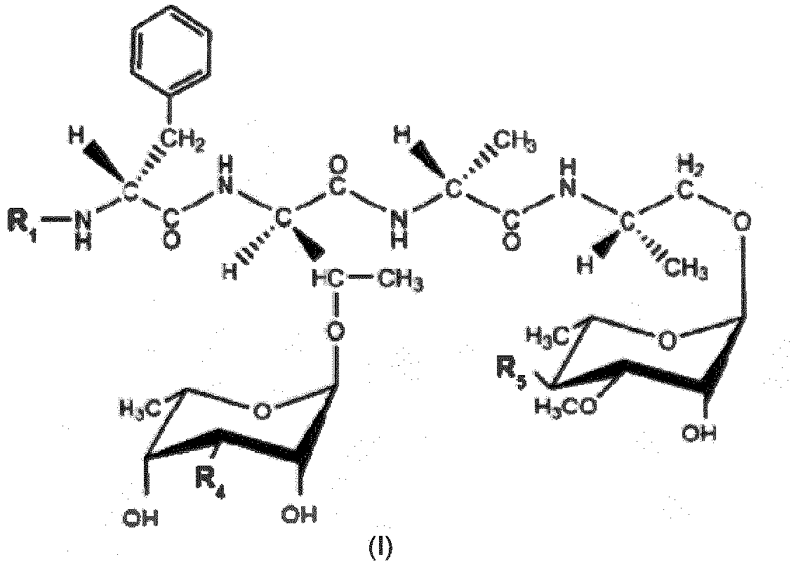
- the present invention as defined by the appended claims, is focused on detecting infections caused not only by M . avium but caused by any NTM, using the antigen of Formula I, or an antigen comprising the chemical structure of formula I, or any isomer thereof:
- R 1 is
- R 3 is an alkyl or alkenyl chain having from 1 to 34 carbon atoms.
- identification of infections caused by any NTM in particular by any of the following bacterial species selected from the list consisting of: Mycobacterium abcessus, Mycobacterium chimaera, Mycobacterium fortuitum, Mycobacterium intracellulare, Mycobacterium malmoense, Mycobacterium xenopi, Mycobacterium chelonae, Mycobacterium farcinogenes, Mycobacterium Habana, Mycobacterium hominis, Mycobacterium paratuberculosis, Mycobacterium peregrinun, Mycobacterium porcinum, Mycobacterium senegalense, Mycobacterium smegmatis, Mycobacterium scrofulaceum and Mycobacterium simiae; is not only achieved by antigens consisting or comprising formula I, but by any nsGPLs of any of the following bacteria: Mycobacterium abcessus, Mycobacterium chimaera, Mycobacterium fortuitum
- an alternative aspect disclosed herein but not part of the invention is focused on detecting infections caused by any NTM, using as an antigen any nsGPLs, or an antigen comprising such nsGPLs, of any of the following bacteria selected from the group consisting of: Mycobacterium abcessus, Mycobacterium chimaera, Mycobacterium fortuitum, Mycobacterium intracellulare, Mycobacterium malmoense, Mycobacterium xenopi or Mycobacterium smegmatis.
- such nsGPLs are capable of detecting infections caused by any of the following bacterial species selected from the list consisting of: Mycobacterium abcessus, Mycobacterium chimaera, Mycobacterium fortuitum, Mycobacterium intracellulare, Mycobacterium malmoense, Mycobacterium xenopi, Mycobacterium chelonae, Mycobacterium farcinogenes, Mycobacterium Habana, Mycobacterium hominis, Mycobacterium paratuberculosis, Mycobacterium peregrinun, Mycobacterium porcinum, Mycobacterium senegalense, Mycobacterium smegmatis, Mycobacterium scrofulaceum and Mycobacterium simiae; more particularly by any of the following bacterial species selected from the list consisting of: Mycobacterium abcessus, Mycobacterium avium, Mycobacterium chimaera, Mycobacterium fortuitum, Mycobacterium intracellular
- R 3 is an alkenyl chain having 34 carbon atoms
- R 4 is -OCH 3
- R 5 is -OCH 3 .
- the Formula I comprises a lipid chain C34:1 (preferably with a double bond between the carbon 10 and 11) and the carbohydrate moieties are respectively 3-O-Me-6-deoxytalose and 3,4-di-O-Me- ⁇ -L-Rhap.
- the method of the invention for the diagnosis of NTM infections can be applied in the following situations:
- M. avium non-serovar glycopeptidolipid (nsGPLs) is used as unique NTM antigens, which are totally specific of NTM, as candidates to create a test for the diagnosis or identification of NTM infections.
- the method of the invention is based on the detection of cytokines produced by blood T-cells after NTM antigen stimulation.
- nsGPLs were extracted and further analyzed using thin layer chromatography (TLC) and gas chromatography-mass spectrometry (GC-MS) to confirm their purity.
- nsGPLs in particular M . avium nsGPLs, are valid antigens to detect immune response against any NTM species.
- nsGPLs in particular M . avium nsGPLs, described in the invention are capable of acting as stimulating antigen, which in turn are capable of diagnosing the NTM infections.
- the diagnostic method of the invention will help to prescribe adequate treatments. It will also reduce unnecessary therapies with the consequent benefits preventing antibiotic resistance, reducing unnecessary prophylaxis for LTBI, and also reducing discomfort in the patient that have to receive unnecessary or inadequate therapy.
- the first embodiment of the present invention refers to the use of M . avium non-serovar specific GLP antigens of Formula (I) (see above), for the in vitro diagnosis or detection of infections caused not only by M . avium but caused by any NTM.
- nsGPL non-serovar specific GLP antigen, or an antigen comprising such nsGPL, obtained from a bacteria selected from any of the group consisting of: Mycobacterium abcessus, Mycobacterium chimaera, Mycobacterium fortuitum, Mycobacterium intracellulare, Mycobacterium malmoense, Mycobacterium xenopi or Mycobacterium smegmatis.
- an antigen of Formula II or an antigen comprising the chemical structure of formula II, or any isomer thereof.
- the second embodiment of the present invention refers to the use of M . avium non-serovar specific GLP antigens of Formula (I) (see above), for in vitro differentiating between infections caused not only by M. avium but caused by any NTM, from latent tuberculosis infection.
- nsGPL non-serovar specific GLP antigen, or an antigen comprising such nsGPL, obtained from a bacterium selected from any of the group consisting of: Mycobacterium abcessus, Mycobacterium chimaera, Mycobacterium fortuitum, Mycobacterium intracellulare, Mycobacterium malmoense, Mycobacterium xenopi or Mycobacterium smegmatis.
- an antigen of Formula II or an antigen comprising the chemical structure of formula II, or any isomer thereof.
- the third embodiment of the present invention as defined by the appended claims refers to the use of M . avium non-serovar specific GLP antigens of Formula (see above), for deciding whether to administer a medical treatment to a subject suspected of suffering from an infection caused not only by M . avium but caused by any NTM.
- nsGPL non-serovar specific GLP antigen, or an antigen comprising such nsGPL, obtained from a bacteria selected from any of the group consisting of: Mycobacterium abcessus, Mycobacterium chimaera, Mycobacterium fortuitum, Mycobacterium intracellulare, Mycobacterium malmoense, Mycobacterium xenopi or Mycobacterium smegmatis.
- an antigen of Formula II or an antigen comprising the chemical structure of formula II, or any isomer thereof.
- the fourth embodiment of the present invention refers to an in vitro method for detecting or diagnosing infections caused not only by M. avium but caused by any NTM, said method comprising: a) incubating a biological sample obtained from the subject with any of the antigens mentioned in any of embodiments one to three above, i.e. M. avium non-serovar specific GLP antigens of Formula (I), b) quantifying the secreted level of at least one cytokine, c) wherein if the cytokine level quantified in step (b) is higher than the reference control level, it is indicative that the subject is suffering from an infection caused not only by M. avium but caused by any NTM.
- the fifth embodiment of the present invention refers to an in vitro method for differentiating between infections caused not only by M. avium but caused by any NTM from latent tuberculosis infection, said method comprising: a) Incubating a biological sample obtained from the subject with any of the antigens mentioned in any of embodiments one to three above, i.e. M.avium non-serovar specific GLP antigen of Formula (I), b) quantifying the secreted level of at least one cytokine, c) wherein if the cytokine level quantified in step (b) is higher than the reference control level, it is indicative that the subject is suffering from an infection caused not only by M. avium but caused by any NTM, and a latent tuberculosis infection can be discarded when conventional IGRAs test is negative.
- the sixth embodiment of the present invention refers to an in vitro method for deciding whether to administer a medical treatment to a subject suspected of suffering from an infection caused not only by M . avium but caused by any NTM, said method comprising: a) Incubating a biological sample obtained from the subject with any of the antigens mentioned in any of embodiments one to three above, i.e. M.avium non-serovar specific GLP antigen of Formula (I) b) quantifying the secreted level of at least one cytokine, and c) wherein if the cytokine level quantified in step (b) is higher than the reference control level, it is indicative that the subject is suffering from an infection caused not only by M . avium but caused by any NTM and a medical treatment directed to NTM could be applied.
- the NTM infection is identified in subjects suffering from: lymphadenitis, skin and soft tissue infection, chronic pulmonary diseases, disseminated disease in immune compromised patients.
- the NTM infection is caused by a mycobacteria selected the group comprising: Mycobacterium avium, Mycobacterium intracellulare, Mycobacterium scrofulaceum, Mycobacterium abscessus, Mycobacterium chelonae, Mycobacterium chimaera Mycobacterium smegmatis, Mycobacterium peregrinum, Mycobacterium simiae, Mycobacterium fortuitum, Mycobacterium marinum, Mycobacterium paratuberculosis, Mycobacterium farcinogenes, Mycobacterium lepraemurium, Mycobacterium porcinum and Mycobacterium senegalens, among others.
- a mycobacteria selected the group comprising: Mycobacterium avium, Mycobacterium intracellulare, Mycobacterium scrofulaceum, Mycobacterium abscessus, Mycobacterium chelonae, Mycobacterium chimaera Mycobacterium sme
- any of the following bacteria selected from the group consisting of: Mycobacterium abcessus, Mycobacterium chimaera, Mycobacterium fortuitum, Mycobacterium intracellulare, Mycobacterium malmoense, Mycobacterium xenopi or Mycobacterium smegmatis.
- such nsGPLs are capable of detecting infections caused by any of the following bacterial species selected from the list consisting of: Mycobacterium abcessus, Mycobacterium chimaera, Mycobacterium fortuitum, Mycobacterium intracellulare, Mycobacterium malmoense, Mycobacterium xenopi, Mycobacterium chelonae, Mycobacterium farcinogenes, Mycobacterium Habana, Mycobacterium hominis, Mycobacterium paratuberculosis, Mycobacterium peregrinun, Mycobacterium porcinum, Mycobacterium senegalense, Mycobacterium smegmatis, Mycobacterium scrofulaceum and Mycobacterium simiae; still more particularly by any of the following bacterial species selected from the list consisting of: Mycobacterium abcessus, Mycobacterium avium, Mycobacterium chimaera, Mycobacterium fortuitum, Mycobacterium intra
- the cytokine or combination of cytokines are selected from the group comprising: IFN-gamma, TNF-alpha, IP-10, IL-10, IL-1RA, GM-CSF, IL-13, IL-17, IL-5, IL-2, IL-22 and IL-32.
- the biological sample is selected from: whole blood, saliva, bronchoalveolar fluid, cerebrospinal fluid, purified peripheral blood mononuclear cells (PBMCs) or biopsies.
- PBMCs peripheral blood mononuclear cells
- the subject is a human or an animal.
- the methods used for quantifying the secretion of cytokines in a biological sample are well known in the art.
- any immunological method such as but not limited to ELISA, multiplex strategies, ELISPOT, immunochromatography techniques, proteomic methods, Western blotting, FACS, or radioimmunoassays may be applicable to the present invention.
- said methods comprise contacting the biological sample with a binding partner capable of selectively interacting with the biomarkers present in the biological sample.
- the binding partner may be an antibody that may be polyclonal or monoclonal, preferably monoclonal.
- the binding partner may be an aptamer.
- Polyclonal antibodies or a fragment thereof can be raised according to known methods by administering the appropriate antigen or epitope to a host animal selected, e.g., from pigs, cows, horses, rabbits, goats, sheep, and mice, among others.
- a host animal selected, e.g., from pigs, cows, horses, rabbits, goats, sheep, and mice, among others.
- Various adjuvants known in the art can be used to enhance antibody production.
- antibodies useful in practicing the invention can be polyclonal, monoclonal antibodies are preferred.
- Monoclonal antibodies or a fragment thereof can be prepared and isolated using any technique that provides for the production of antibody molecules by continuous cell lines in culture.
- Techniques for production and isolation include but are not limited to the hybridoma technique originally described by Kohler and Milstein (1975); the human B-cell hybridoma technique (Cote et al., 1983); and the EBV-hybridoma technique (Cole et al. 1985).
- techniques described for the production of single chain antibodies can be adapted to produce anti-cytokine, single chain antibodies.
- Antibodies useful in practicing the present invention also include anti-cytokine fragments including but not limited to F(ab')2 fragments, which can be generated by pepsin digestion of an intact antibody molecule, and Fab fragments, which can be generated by reducing the disulfide bridges of the F(ab')2 fragments.
- Fab and/or scFv expression libraries can be constructed to allow rapid identification of fragments having the desired specificity to cytokine.
- phage display of antibodies may be used.
- single-chain Fv (scFv) or Fab fragments are expressed on the surface of a suitable bacteriophage, e.g., M13.
- spleen cells of a suitable host e.g., mouse
- a suitable host e.g., mouse
- the coding regions of the VL and VH chains are obtained from those cells that are producing the desired antibody against the protein. These coding regions are then fused to a terminus of a phage sequence.
- a suitable carrier e.g., bacteria
- the phage displays the antibody fragment. Phage display of antibodies may also be provided by combinatorial methods known to those skilled in the art.
- Antibody fragments displayed by a phage may then be used as part of an immunoassay.
- the binding partner may be an aptamer.
- Aptamers are a class of molecule that represents an alternative to antibodies in term of molecular recognition.
- Aptamers are oligonucleotide or oligopeptide sequences with the capacity to recognize virtually any class of target molecules with high affinity and specificity.
- Such ligands may be isolated through Systematic Evolution of Ligands by Exponential enrichment (SELEX) of a random sequence library, as described in Tuerk C. 1997.
- the random sequence library is obtainable by combinatorial chemical synthesis of DNA. In this library, each member is a linear oligomer, eventually chemically modified, of a unique sequence. Possible modifications, uses and advantages of this class of molecules have been reviewed in Jayasena S.D., 1999.
- Peptide aptamers consist of conformationally constrained antibody variable regions displayed by a platform protein, such as E. coli Thioredoxin A, that are selected from combinatorial libraries by two hybrid methods (Colas et al, 1996).
- the binding partners such as antibodies or aptamers, may be labelled with a detectable molecule or substance, such as a fluorescent molecule, a radioactive molecule or any others labels known in the art. Labels are known in the art that generally provide (either directly or indirectly) a signal.
- the term "labelled", with regard to the antibody is intended to encompass direct labelling of the antibody or aptamer by coupling (i.e., physically linking) a detectable substance, such as a radioactive agent or a fluorophore (e.g. fluorescein isothiocyanate (FITC) or phycoerythrin (PE) or Indocyanine (Cy5)) to the antibody or aptamer, as well as indirect labelling of the probe or antibody by reactivity with a detectable substance.
- a detectable substance such as a radioactive agent or a fluorophore (e.g. fluorescein isothiocyanate (FITC) or phycoerythrin (PE) or Indocyanine (Cy5)
- FITC fluorescein isothiocyanate
- PE phycoerythrin
- Indocyanine Indocyanine
- radioactive molecules include but are not limited radioactive atom for scintigraphic studies such as 1123, 1124, In111, Re 186, Re188.
- the aforementioned assays generally involve the binding of the binding partner (ie. antibody or aptamer) to a solid support.
- Solid supports which can be used in the practice of the invention include substrates such as nitrocellulose (e. g., in membrane or microtiter well form); polyvinylchloride (e. g., sheets or microtiter wells); polystyrene latex (e.g., beads or microtiter plates); polyvinylidine fluoride; diazotized paper; nylon membranes; activated beads, magnetically responsive beads, plastic or glass (e.g.
- an ELISA method can be used, wherein the wells of a microtiter plate are coated with a set of antibodies which recognize said cytokine(s). A biological sample containing or suspected of containing said cytokine(s) is then added to the coated wells. After a period of incubation sufficient to allow the formation of antibody-antigen complexes, the plate(s) can be washed to remove unbound moieties and a detectably labelled secondary binding molecule added. The secondary binding molecule is allowed to react with any captured sample marker protein, the plate washed and the presence of the secondary binding molecule detected using methods well known in the art. In one embodiment, an Enzyme-linked immunospot (ELISpot) method may be used.
- ELISpot Enzyme-linked immunospot
- the biological sample is sent to the laboratory and then the PBMCS are isolated and incubated with the antigens there. Typically, the biological sample is transferred to a plate which has been coated with the desired anti-cytokine capture antibodies. Revelation is carried out with biotinylated secondary Abs and standard colorimetric or fluorometric detection methods such as streptavidin-alkaline phosphatase and NBT-BCIP and the spots counted.
- biotinylated secondary Abs and standard colorimetric or fluorometric detection methods such as streptavidin-alkaline phosphatase and NBT-BCIP and the spots counted.
- the bead may be a cytometric bead for use in flow cytometry.
- Such beads may for example correspond to BD TM Cytometric Beads commercialized by BD Biosciences (San Jose, California).
- cytometric beads may be suitable for preparing a multiplexed bead assay.
- a multiplexed bead assay such as, for example, the BD TM Cytometric Bead Array, is a series of spectrally discrete beads that can be used to capture and quantify soluble antigens.
- beads are labelled with one or more spectrally distinct fluorescent dyes, and detection is carried out using a multiplicity of photodetectors, one for each distinct dye to be detected.
- a number of methods of making and using sets of distinguishable beads have been described in the literature.
- beads distinguishable by size wherein each size bead is coated with a different target-specific antibody (see e.g. Fulwyler and McHugh, 1990, Methods in Cell Biology 33:613-629 ), beads with two or more fluorescent dyes at varying concentrations, wherein the beads are identified by the levels of fluorescence dyes (see e.g. European Patent No. 0 126,450 ), and beads distinguishably labelled with two different dyes, wherein the beads are identified by separately measuring the fluorescence intensity of each of the dyes (see e.g. U.S. patent Nos. 4,499,052 and 4,717,655 ).
- One-dimensional and two-dimensional arrays for the simultaneous analysis of multiple antigens by flow cytometry are available commercially.
- Examples of one-dimensional arrays of singly dyed beads distinguishable by the level of fluorescence intensity include the BD TM Cytometric Bead Array (CBA) (BD Biosciences, San Jose, Calif.) and Cyto-Plex TM Flow Cytometry microspheres (Duke Scientific, Palo Alto, Calif).
- An example of a two-dimensional array of beads distinguishable by a combination of fluorescence intensity (five levels) and size (two sizes) is the QuantumPlex(TM) microspheres (Bangs Laboratories, Fisher, Ind.).
- the beads may be labelled with any fluorescent compound known in the art such as e.g. FITC (FL1), PE (FL2), fluorophores for use in the blue laser (e.g. PerCP, PE-Cy7, PE-Cy5, FL3 and APC or Cy5, FL4), fluorophores for use in the red, violet or UV laser (e.g. Pacific blue, pacific orange).
- FL1 fluorescent compound
- PE FL2
- fluorophores for use in the blue laser e.g. PerCP, PE-Cy7, PE-Cy5, FL3 and APC or Cy5, FL4
- fluorophores for use in the red, violet or UV laser e.g. Pacific blue, pacific orange
- bead is a magnetic bead for use in magnetic separation. Magnetic beads are known to those of skill in the art.
- the magnetic bead is preferably made of a magnetic material selected from the group consisting of metals (e.g. iron, cobalt and nickel), an alloy thereof and an oxide thereof.
- bead is bead that is dyed and magnetized.
- the incubation of the biological sample with the mycobacteria antigens is performed at the point of care locations such as physicians' offices, clinics, or outpatient facilities. Once incubation is complete, the requirement for fresh and active cells no longer exists. Cytokines are stable and, thus, the biological sample can be stored, frozen or shipped without special conditions. Accordingly, in one embodiment, the biological sample is collected in suitable container (e.g. collection tube) containing the mycobacteria antigen or a plurality of mycobacteria antigens.
- the incubation step may be from 5 to 48 hours, more preferably 5 to 36 hours and even more preferably 12 to 24 hours or a time period in between.
- the incubation time is 5 hours, 6 hours, 7 hours, 8 hours, 9 hours, 10 hours, 11 hours, 12 hours, 13 hours, 14 hours, 15 hours, 16 hours, 17 hours, 18 hours, 19 hours, 20 hours, 21 hours, 22 hours, 23 hours, 24 hours, 26 hours, 30 hours, 36 hours, 42 hours, or 48 hours.
- Example 6 and example 8 offer clear evidence that M . smegmatis nsGPLs antigens, can also be used to identify non only infections caused by M. smegmatis but also infections caused by any NTM.
- M . smegmatis nsGPLs antigens can also be used to identify non only infections caused by M. smegmatis but also infections caused by any NTM.
- Example 1 Method for obtaining the nsGPLs :
- M. avium complex colonies were scraped from the surface of Middlebrook 7H11 agar plates containing oleic acid-albumin-dextrose-catalase (OADC) enrichment, and were transferred aseptically to sterile screw-capped glass culture tubes (20 x 120 mm) containing 5 ml of methanol (CH 3 OH) followed by the addition of 10 ml of chloroform (CHCl 3 ) for a final ratio of CHCl 3 -CH 3 OH (2:1 v/v) and incubated at 37°C overnight. After this incubation period, samples were centrifuged for 5 min at 11,500 x g at 4°C to obtain supernatants (organic extractions containing total lipids).
- OADC oleic acid-albumin-dextrose-catalase
- total lipids were treated with mild alkali (an equal volume of 0.2 N sodium hydroxide (NaOH) in CH 3 OH at 37 °C for 35 min) followed by neutralization using 100 microliters of glacial acetic acid (2 to 3 drops, check pH to be 7.0).
- Mild alkali hydrolysis destroys alkalilabile non-specific acylglycerols, thus serving as a purification step for the alkaline resistant GPLs.
- the alkali-treated lipids were further partitioned between CHCl 3 -CH 3 OH (2:1 v/v) and H 2 O at a ratio of 1:1 (v/v) to remove generated sodium acetate salts. This 'washing' step was repeated twice.
- the final chloroform layer (bottom phase) containing the total GPLs fraction was transferred to a pre-weight tube and dried down under nitrogen to avoid lipid oxidation. Dried samples were kept at -20°C under nitrogen until further use. This allows obtaining total GPLs (nsGPLs + ssGPLs).
- SEPPAK Classic Silica long body cartridges (Waters Corporation, Milford, MA, USA) were fitted with 5 ml glass syringes, which acted as reservoirs for the eluting solvents.
- the SEPPAK cartridges were equilibrated with CHCl 3 (5 ml x 3 times) followed by the application of total GPLs dissolved in 0.5 ml CHC1 3 .
- the cartridge was then irrigated with 3 ml of CHCl 3 followed by 2, 10, 25, 35 and 50% CH 3 OH in CHCl 3 . Finally, the cartridge was washed several times with 5ml CH 3 OH.
- nsGPLs were removed with CHCl 3 and with 2% CH 3 OH in CHCl 3 .
- the total ssGPL population was collected in 20-35% CH 3 OH eluate. Fractions were examined for purity by TLC in CHCl 3 -CH 3 OH-H 2 O (60:35 : 8. v/v/v).
- nsGPLs or ssGPLs 150 mg were dissolved in 50% aqueous methanol [using pyrogen-free distilled water (1:1 v/v)] containing 1 N sodium hydroxide (NaOH) and 1 M sodium borohydrate (NaBH4) (a.k.a.
- the upper phases (aqueous layer) obtained from each wash were combined, concentrated using a speed-ac and desalted using Sephadex G-25 to obtain the reduced oligosaccharide. This technique destroys the peptide into single ammino acids. It is difficult only obtain the glycopeptide. Conversely, it is possible to purify the lipopeptide.
- CPT tubes Becton Dickinson Diagnostics, Franklin Lakes, NJ
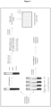
- blood collection tubes in which the antigens have been previously pre-fixed (see figure 1 ).
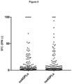
- the first approach is based on the enzyme-linked immunospot assay (ELISPOT). Briefly, PBMCs are stimulated with the selected antigens (ss4GPLs and nsGPLs), as well as, positive and reference controls, in a pre-coated well plate. After an overnight incubation, the plate is washed and developed, and the production of IFN-gamma is measured by counting the spot forming cells (SFC).
- ELISPOT enzyme-linked immunospot assay
- the second approach is based on the stimulation of whole blood in blood collection tubes in which the antigens have already been fixed.
- the antigens used are ss4GPLs and nsGPLs, as well as a reference and positive control. After an overnight incubation, plasma is harvested, and afterwards the production of IFN-gamma and other cytokines is detected.
- Example 4 Percentage of positives obtained after stimulation with ss4GPLs or nsGPLs per patient group using ELISPOT in-house technology. A total of 109 and 63 patients were stimulated with ss4GPLs and nsGPLs respectively. Patients were classified in six different groups: (i) healthy controls (individuals with negative TST and IGRAs, no adenopathies and normal X-ray). (ii) NTM positive culture (patients with a NTM confirmed culture). (iii) Adenitis not culture confirmed (patients with adenopathies and no NTM culture confirmation/negative culture).
- LTBI (individuals with positives TST and IGRAs, coming from contact tracing or LTBI screening studies, without adenopathies and normal X-ray).
- Active TB patients with culture and/or PCR Mycobacterium tuberculosis confirmation).
- Study group non-BCG vaccinated individuals with a positive TST and a negative IGRA, coming from contact tracing studies or LTBI screenings, without adenopathies and normal X-ray).
- PBMCs were isolated from each individual and stimulated with ss4GPLs and/or nsGPLs. The presence of sensitized T-cells against the antigens was analyzed by means of an ELISPOT in-house detecting spot-forming cells secreting IFN-gamma.
- any healthy control showed a positive ELISPOT result after stimulation with both of the antigens.
- a positive NTM culture isolation were analyzed, a total of 76.9% (20/26) and 77.8% (14/18) positive results were obtained after ss4GPLs and/or nsGPLs stimulation respectively.
- the type of mycobacteria isolated Patients with not culture confirmed adenopathies presented 48.1% (13/27) and 77.8% (7/9) of positive results for ss4GPLs and nsGPLs.
- the response to ss4GPLs and nsGPLs in LTBI individuals were 8.7% (2/23) and 14.3% (2/14) respectively.
- Example 5 Percentage of positives obtained after stimulation with nsGPLs per patient group using in-tube technology.
- a total of 14 patients were stimulated in-tube with 10 ⁇ g of M. avium nsGPLs (Table 2).
- Patients were classified into three groups: (i) healthy controls (individuals with negative TST and IGRAs, no adenopathies and normal X-ray).
- NTM positive culture patients with a NTM confirmed culture).
- Adenitis not culture confirmed patients with adenopathies and no NTM culture confirmation/negative culture).
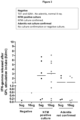
- Whole blood was directly incubated in-tube with nsGPLs. The amount of IFN-gamma released (IU/ml) was measured by ELISA.
- the one healthy individual tested by this technology presented a negative response when stimulated with nsGPLs.
- patients with a positive NTM culture isolation and not culture confirmed adenopathies were analyzed, a total of 77.8% (7/9) and 50% (2/4) positive results were obtained respectively.
- the total amount of IFN-gamma released detailed per patient group after nsGPLs stimulation is represented in Figure 2 .
- Example 6 Percentage of positive results obtained after stimulation with nsGPLs of Mycobacterium smegmatis.
- NTM positive culture patients with a NTM confirmed culture.
- Adenitis not culture confirmed patients with adenopathies and no NTM culture confirmation/negative culture).
- Example 7 Stimulation with peptidic fraction of the nsGPL.
- the inventors of this invention performed a series of experiments in which PBMCs were not only stimulated with the whole molecule of ss4GPLs and nsGPLs, but also with the peptidic fraction.
- the procedure followed was the same as described in the Example 2 and illustrated in Figure 1 .
- the peptidic fraction (D-Phe-D- allo -Thr-D-Ala-L-Alanol) was synthesized by JPT Innovative Peptide Solutions (JPT Peptide Technologies GmbH - Volmerstrasse 5 - 12489 Berlin).
- a total of 10 patients (2 healthy controls and 8 patients with a NTM culture confirmed) were tested by ELISPOT with the peptidic fraction of nsGPLs. Such as it can be seen in the Table 4, none of the patients included responded to the peptides.
- Example 8 Percentage of positive results obtained after stimulation with ss4GPL, ns4GPL, ns8GPL, total lipid and mixing ss4GPL and ns4GPL of Mycobacterium avium, and nsGPL of Mycobacterium smegmatis.
- nsGPL obtained from different M.avium serovar In order to determine whether or not the immunogenicity of the total lipids, nsGPL obtained from different M.avium serovar, or even from other species, the inventors of the present invention performed a series of experiments in which PBMCs were stimulated with the whole molecule of ss4GPLs, and nsGPLs obtained from ss4 and ss8 M. avium strains, total lipid, a mix of ss4 and ns4 and nsGPL from M. smegmatis. A total of 50 patients were tested for the antigens by the ELISPOT in-house technology (Table 5).
- Patients were classified into three groups: (i) healthy controls (individuals with negative TST and IGRAs, no adenopathies and normal X-ray), (ii) patients diagnosed with LTBI infection by TST and IGRAs, (iii) NTM positive culture (patients with a NTM confirmed culture), (iv) adenitis not culture confirmed (patients with adenopathies and no NTM culture confirmation/negative culture), (v) Active TB (patients with culture and/or PCR Mycobacterium tuberculosis confirmation), (vi) Study group (non-BCG vaccinated individuals with a positive TST and a negative IGRA, coming from contact tracing studies or LTBI screenings, without adenopathies and normal X-ray).
- ns4GPL Percentage of positives obtained after stimulation with ss4GPL, ns4GPL per patient group.
- Example 9 Plausability that the evidence presented in example 8 encompasses further NTM bacterial species.
- NTM species have similarities with the nsGPL structure of M . avium ( Schorey JS and Sweet L. The mycobacteria glycopeptidolipds: structure, function and their role in pathogenesis. Glycobiology 2008, 18:832-841 ; and Chatterjee D and Khoo KH. The surface glycopeptidolipids of mycobacteria: structures and biological properties. Cellular and Molecular Life Sciences 2001, 58: 2018-2042 ) In addition, these two NTM species have the same nsGPL structure than M. xenopi and M . abcessus, which both are experimentally detected as demonstrated in example 8. Therefore, we consider plausible that these two NTM species could be detected by our test.
- NTM species have similarities with the nsGPL structure of M. avium ( Schorey JS and Sweet L. The mycobacteria glycopeptidolipds: structure, function and their role in pathogenesis. Glycobiology 2008, 18:832-841 ; and Chatterjee D and Khoo KH. The surface glycopeptidolipids of mycobacteria: structures and biological properties. Cellular and Molecular Life Sciences 2001, 58: 2018-2042 ). In addition, these four NTM species have the same nsGPL structure than M. fortuitum that is experimentally detected as shown in example 8. Therefore, we consider plausible that these four NTM species could be detected by our test.
- M. hominis and M. paratuberculosis are members of the M.avium complex.
- M . scrofulaceum was also considered member of the M . avium complex.
- M. avium and M . intracellulare are easily detected as shown in example 8. Therefore, we consider plausible that these three NTM species could be detected as defined in the claims.
- nsGPLs from both of these two NTM species are identical to the nsGPL structure of M. avium (2). Therefore, we consider plausible that these two NTM species could be detected by our test.
Landscapes
- Health & Medical Sciences (AREA)
- Life Sciences & Earth Sciences (AREA)
- Chemical & Material Sciences (AREA)
- Immunology (AREA)
- Molecular Biology (AREA)
- Engineering & Computer Science (AREA)
- Hematology (AREA)
- Biomedical Technology (AREA)
- Urology & Nephrology (AREA)
- Medicinal Chemistry (AREA)
- Cell Biology (AREA)
- General Health & Medical Sciences (AREA)
- Biochemistry (AREA)
- Pathology (AREA)
- Organic Chemistry (AREA)
- Physics & Mathematics (AREA)
- Analytical Chemistry (AREA)
- Proteomics, Peptides & Aminoacids (AREA)
- Biotechnology (AREA)
- General Physics & Mathematics (AREA)
- Microbiology (AREA)
- Food Science & Technology (AREA)
- Virology (AREA)
- Gastroenterology & Hepatology (AREA)
- Biophysics (AREA)
- Genetics & Genomics (AREA)
- Tropical Medicine & Parasitology (AREA)
- Measuring Or Testing Involving Enzymes Or Micro-Organisms (AREA)
- Medicines Containing Antibodies Or Antigens For Use As Internal Diagnostic Agents (AREA)
- Measuring And Recording Apparatus For Diagnosis (AREA)
Claims (12)
- Utilisation d'antigène GPL non-spécifique de sérovar de Mycobacterium avium (glycopeptidolipide non-sérovar, nsGPL) de formule (I)
 dans laquelle R1 est
 dans laquelle R3 est une chaîne alkyle ou alcényle ayant de 1 jusqu'à 34 atomes de carbone ;dans laquelle R4 est -OH ou -OCH3 ; etdans laquelle R5 est -OH ou -OCH3 ;pour le diagnostic ou la détection in vitro d'infections causées non seulement par Mycobacterium avium mais causées par une quelconque mycobactérie non tuberculeuse sélectionnée du groupe consistant en : Mycobacterium abcessus, Mycobacterium chimaera, Mycobacterium intracellulare, Mycobacterium malmoense et Mycobacterium xenopi.
 - Utilisation d'antigène GPL non-spécifique de sérovar de Mycobacterium avium de formule (I)
 dans laquelle R1 est
 dans laquelle R3 est une chaîne alkyle ou alcényle ayant de 1 jusqu'à 34 atomes de carbone ;dans laquelle R4 est -OH ou -OCH3 ; etdans laquelle R5 est -OH ou -OCH3 ;pour la différenciation in vitro entre infections causées non seulement par Mycobacterium avium mais causées par une quelconque mycobactérie non tuberculeuse sélectionnée du groupe consistant en : Mycobacterium abcessus, Mycobacterium chimaera, Mycobacterium intracellulare, Mycobacterium malmoense et Mycobacterium xenopi, à partir d'une infection tuberculeuse latente.
 - Utilisation d'antigène GPL non-spécifique de sérovar de Mycobacterium avium de formule (I)
 dans laquelle R1 est
 dans laquelle R3 est une chaîne alkyle ou alcényle ayant de 1 jusqu'à 34 atomes de carbone ;dans laquelle R4 est -OH ou -OCH3 ; etdans laquelle R5 est -OH ou -OCH3 ;pour décider si administrer un traitement médical à un sujet soupçonné de souffrir d'une infection causée non seulement par Mycobacterium avium mais causée par une quelconque mycobactérie non tuberculeuse sélectionnée du groupe consistant en : Mycobacterium abcessus, Mycobacterium chimaera, Mycobacterium intracellulare, Mycobacterium malmoense et Mycobacterium xenopi.
 - Utilisation selon l'une quelconque des revendications 1 à 3, dans laquelle l'infection à mycobactéries non tuberculeuses est identifiée chez des sujets souffrant de : lymphadénite, infection de peau et de tissus mous, maladies pulmonaires chroniques et maladies disséminées chez des patients immunodéficients.
- Utilisation selon l'une quelconque des revendications 1 à 4, dans laquelle, dans la formule I, R3 est une chaîne alcényle ayant 34 atomes de carbone, R4 est -OCH3 et R5 est -OCH3.
- Procédé in vitro pour détecter ou diagnostiquer des infections causées non seulement par Mycobacterium avium mais causées par une quelconque mycobactérie non tuberculeuse sélectionnée du groupe consistant en : Mycobacterium abcessus, Mycobacterium chimaera, Mycobacterium intracellulare, Mycobacterium malmoense et Mycobacterium xenopi, ledit procédé comprenant :a) incuber un échantillon biologique, sous la condition qu'il contienne des leucocytes, obtenu du sujet avec l'antigène GPL non-spécifique de sérovar de Mycobacterium avium de formule (I)
 dans laquelle R1 est
 dans laquelle R3 est une chaîne alkyle ou alcényle ayant de 1 jusqu'à 34 atomes de carbone ;dans laquelle R4 est -OH ou -OCH3 ; etdans laquelle R5 est -OH ou -OCH3 ;b) quantifier le niveau sécrété d'au moins une cytokine,c) dans lequel si le niveau de cytokine quantifié dans l'étape (b) est plus élevé que le niveau de contrôle de référence, cela est révélateur que le sujet souffre d'une infection causée non seulement par Mycobacterium avium mais causée par une quelconque mycobactérie non tuberculeuse sélectionnée du groupe consistant en : Mycobacterium abcessus, Mycobacterium chimaera, Mycobacterium intracellulare, Mycobacterium malmoense et Mycobacterium xenopi.
 - Procédé in vitro pour différencier entre les infections causées non seulement par Mycobacterium avium mais causée par une quelconque mycobactérie non tuberculeuse sélectionnée du groupe consistant en : Mycobacterium abcessus, Mycobacterium chimaera, Mycobacterium intracellulare, Mycobacterium malmoense et Mycobacterium xenopi, d'une infection tuberculeuse latente, ledit procédé comprenant :a) incuber un échantillon biologique, sous la condition qu'il contienne des leucocytes, obtenu du sujet avec l'antigène GPL non-spécifique de sérovar de Mycobacterium avium de formule (I)
 dans laquelle R1 est
 dans laquelle R3 est une chaîne alkyle ou alcényle ayant de 1 jusqu'à 34 atomes de carbone ;dans laquelle R4 est -OH ou -OCH3 ; etdans laquelle R5 est -OH ou -OCH3 ;b) quantifier le niveau sécrété d'au moins une cytokine,c) dans lequel si le niveau de cytokine quantifié dans l'étape (b) est plus élevé que le niveau de contrôle de référence, cela est révélateur que le sujet souffre d'une infection causée non seulement par Mycobacterium avium mais causée par une quelconque mycobactérie non tuberculeuse sélectionnée du groupe consistant en : Mycobacterium abcessus, Mycobacterium chimaera, Mycobacterium intracellulare, Mycobacterium malmoense et Mycobacterium xenopi, et une infection tuberculeuse latente peut être rejetée lorsque le test IGRA conventionnel est négatif.
 - Procédé in vitro pour décider si administrer un traitement médical à un sujet soupçonné de souffrir d'une infection causée non seulement par Mycobacterium avium mais causée par une quelconque mycobactérie non tuberculeuse sélectionnée du groupe consistant en : Mycobacterium abcessus, Mycobacterium chimaera, Mycobacterium intracellulare, Mycobacterium malmoense et Mycobacterium xenopi, ledit procédé comprenant :a) incuber un échantillon biologique, sous la condition qu'il contienne des leucocytes, obtenu du sujet avec l'antigène GPL non-spécifique de sérovar de Mycobacterium avium de formule (I)
 dans laquelle R1 est
 dans laquelle R3 est une chaîne alkyle ou alcényle ayant de 1 jusqu'à 34 atomes de carbone ;dans laquelle R4 est -OH ou -OCH3 ; etdans laquelle R5 est -OH ou -OCH3 ;b) quantifier le niveau sécrété d'au moins une cytokine, etc) dans lequel si le niveau de cytokine quantifié dans l'étape (b) est plus élevé que le niveau de contrôle de référence, cela est révélateur que le sujet souffre d'une infection causée non seulement par Mycobacterium avium mais causée par une quelconque mycobactérie non tuberculeuse sélectionnée du groupe consistant en : Mycobacterium abcessus, Mycobacterium chimaera, Mycobacterium intracellulare, Mycobacterium malmoense et Mycobacterium xenopi, et un traitement médical dirigé vers les MNT peut être appliqué.
 - Procédé selon l'une quelconque des revendications 6 à 8, dans lequel la cytokine ou la combinaison de cytokines sont sélectionnées du groupe comprenant: IFN-gamma, TNF-alpha, IP-10, IL-10, IL-1RA, GM-CSF, IL-13, IL-17, IL-5, IL-2, IL-22 et IL-32.
- Procédé selon l'une quelconque des revendications 6 à 19, dans lequel l'infection à mycobactéries non tuberculeuses est identifiée chez des sujets souffrant de : lymphadénite, infection de peau et de tissus mous, maladies pulmonaires chroniques, maladies disséminées chez des patients immunodéficients.
- Procédé selon l'une quelconque des revendications 9 à 10, dans lequel l'échantillon biologique est sélectionné de : sang total, salive, fluides broncho-alvéolaires, fluides cérébro-spinaux, cellules mononucléaires du sang périphérique (PBMC) purifiées ou biopsies.
- Procédé selon l'une quelconque des revendications 6 à 11, dans lequel, dans la formule I, R3 est une chaîne alcényle ayant 34 atomes de carbone, R4 est -OCH3 et R5 est -OCH3.
Applications Claiming Priority (2)
| Application Number | Priority Date | Filing Date | Title |
|---|---|---|---|
| EP18382403 | 2018-06-06 | ||
| PCT/EP2019/064885 WO2019234196A1 (fr) | 2018-06-06 | 2019-06-06 | Méthode in vitro de diagnostic ou de détection de mycobactéries non tuberculeuses |
Publications (2)
| Publication Number | Publication Date |
|---|---|
| EP3803400A1 EP3803400A1 (fr) | 2021-04-14 |
| EP3803400B1 true EP3803400B1 (fr) | 2023-08-09 |
Family
ID=62748891
Family Applications (1)
| Application Number | Title | Priority Date | Filing Date |
|---|---|---|---|
| EP19728095.1A Active EP3803400B1 (fr) | 2018-06-06 | 2019-06-06 | Procédé in vitro pour le diagnostic ou la détection d'infections mycobactériennes non tuberculeuses |
Country Status (3)
| Country | Link |
|---|---|
| EP (1) | EP3803400B1 (fr) |
| ES (1) | ES2959857T3 (fr) |
| WO (1) | WO2019234196A1 (fr) |
Families Citing this family (2)
| Publication number | Priority date | Publication date | Assignee | Title |
|---|---|---|---|---|
| KR102717762B1 (ko) * | 2021-11-03 | 2024-10-17 | 서울대학교 산학협력단 | 개에서의 비결핵 항산균의 감염 진단을 위한 숙주 바이오마커 및 이의 이용 |
| CN114561393B (zh) * | 2021-11-23 | 2024-05-07 | 上海市肺科医院 | 一种特异性识别鸟-胞内分枝杆菌复合群的dna适配子及其筛选方法和应用 |
Family Cites Families (6)
| Publication number | Priority date | Publication date | Assignee | Title |
|---|---|---|---|---|
| US4499052A (en) | 1982-08-30 | 1985-02-12 | Becton, Dickinson And Company | Apparatus for distinguishing multiple subpopulations of cells |
| US4717655A (en) | 1982-08-30 | 1988-01-05 | Becton, Dickinson And Company | Method and apparatus for distinguishing multiple subpopulations of cells |
| DE3322373C2 (de) | 1983-05-19 | 1986-12-04 | Ioannis Dr. 3000 Hannover Tripatzis | Testmittel und Verfahren zum Nachweis von Antigenen und/oder Antikörpern |
| US4946778A (en) | 1987-09-21 | 1990-08-07 | Genex Corporation | Single polypeptide chain binding molecules |
| JP3850670B2 (ja) * | 2001-02-22 | 2006-11-29 | ナカライテスク株式会社 | 抗酸菌症鑑別用試薬および鑑別方法 |
| CN105866406A (zh) * | 2016-04-06 | 2016-08-17 | 广州瑞辉生物科技股份有限公司 | 鉴别结核分枝杆菌与非结核分枝杆菌感染的检测方法 |
-
2019
- 2019-06-06 EP EP19728095.1A patent/EP3803400B1/fr active Active
- 2019-06-06 ES ES19728095T patent/ES2959857T3/es active Active
- 2019-06-06 WO PCT/EP2019/064885 patent/WO2019234196A1/fr not_active Ceased
Also Published As
| Publication number | Publication date |
|---|---|
| WO2019234196A1 (fr) | 2019-12-12 |
| EP3803400A1 (fr) | 2021-04-14 |
| ES2959857T3 (es) | 2024-02-28 |
Similar Documents
| Publication | Publication Date | Title |
|---|---|---|
| EP1554580B1 (fr) | Epreuve diagnostique pour la maladie intestinale inflammatoire, en detection premiere, et le colon irritable | |
| EP3111227B1 (fr) | Méthode de diagnostic d'une tuberculose | |
| BRPI0922798B1 (pt) | Dispositivo e kit para diagnosticar tuberculose | |
| US20140342936A1 (en) | Methods and kits for diagnosing latent tuberculosis infection | |
| US11768202B2 (en) | Method of detecting anti-Ri in a subject with a previous streptococcal infection | |
| Eribo et al. | Host urine immunological biomarkers as potential candidates for the diagnosis of tuberculosis | |
| US20210025887A1 (en) | Antibody or antibody combination and method using same for detection of an antigen related to mycobacterium in a urine sample of a subject | |
| EP3803400B1 (fr) | Procédé in vitro pour le diagnostic ou la détection d'infections mycobactériennes non tuberculeuses | |
| Cho et al. | Rapid diagnosis of tuberculous peritonitis by T cell-based assays on peripheral blood and peritoneal fluid mononuclear cells | |
| Wakode et al. | GeneXpert: a rapid and supplementary diagnostic tool for tuberculous meningitis, experience from tertiary neurocenter | |
| WO2015164617A1 (fr) | Biomarqueurs de la tuberculose dans l'urine et leurs utilisations | |
| Dass et al. | Utility of cell-free transrenal DNA for the diagnosis of Tuberculous Meningitis: A proof-of-concept study | |
| JP4419020B2 (ja) | 体液中の結核菌抗原を検出する方法 | |
| WO2024117982A1 (fr) | Kit de test sur bande pour le diagnostic de la tuberculose | |
| KR102951592B1 (ko) | 믹소바이러스 내성 단백질 a와 c-반응성 단백질을 이용한 박테리아감염과 바이러스감염 구별을 위한 진단 시스템 | |
| KR102839487B1 (ko) | 활동성 결핵 진단용 바이오마커, 조성물, 키트 및 활동성 결핵 진단을 위한 정보 제공 방법 | |
| Dahiya et al. | Diagnosis of abdominal tuberculosis: detection of mycobacterial CFP-10 and HspX proteins by gold nanoparticle-PCR amplified immunoassay | |
| Mikačić et al. | TUMOR MARKER CA 125 IN THE DIAGNOSIS OF ACTIVE PULMONARY TUBERCULOSIS–A STUDY OF ADULTS IN MOSTAR, B&H | |
| EP3653646A1 (fr) | Procédé et kit de diagnostic et/ou de pronostic d'états inflammatoires | |
| KR20260051339A (ko) | 믹소바이러스 내성 단백질 a와 c-반응성 단백질을 이용한 박테리아감염과 바이러스감염 구별을 위한 진단 시스템 | |
| Ruth | Chemokine and cytokine measurements using Luminex and Selected Reaction Monitoring (SRM): comparing technologies | |
| KATO et al. | USEFULNESS OF MYCOBACTERIUM TUBERCULOSIS-SPECIFIC ENZYME-LINKED IMMUNOSPOT ASSAY FOR THE DIAGNOSIS OF TUBERCULOUS PLEURISY | |
| CN120028532A (zh) | 用于鉴别活动性结核和潜伏性结核感染的试剂盒及其应用 | |
| Behera | Laboratory Diagnosis of Human Melioidosis | |
| Dadoria et al. | Role of CBNAAT in diagnosis of tuberculous meningitis in children |
Legal Events
| Date | Code | Title | Description |
|---|---|---|---|
| STAA | Information on the status of an ep patent application or granted ep patent |
Free format text: STATUS: UNKNOWN |
|
| STAA | Information on the status of an ep patent application or granted ep patent |
Free format text: STATUS: THE INTERNATIONAL PUBLICATION HAS BEEN MADE |
|
| PUAI | Public reference made under article 153(3) epc to a published international application that has entered the european phase |
Free format text: ORIGINAL CODE: 0009012 |
|
| STAA | Information on the status of an ep patent application or granted ep patent |
Free format text: STATUS: REQUEST FOR EXAMINATION WAS MADE |
|
| 17P | Request for examination filed |
Effective date: 20201231 |
|
| AK | Designated contracting states |
Kind code of ref document: A1 Designated state(s): AL AT BE BG CH CY CZ DE DK EE ES FI FR GB GR HR HU IE IS IT LI LT LU LV MC MK MT NL NO PL PT RO RS SE SI SK SM TR |
|
| AX | Request for extension of the european patent |
Extension state: BA ME |
|
| RIN1 | Information on inventor provided before grant (corrected) |
Inventor name: DOMINGUEZ BENITEZ, JOSE ANTONIO Inventor name: LATORRE RUEDA, IRENE Inventor name: VILLAR-HERNANDEZ, RAQUEL Inventor name: TORRELLES, JORDI |
|
| DAV | Request for validation of the european patent (deleted) | ||
| DAX | Request for extension of the european patent (deleted) | ||
| STAA | Information on the status of an ep patent application or granted ep patent |
Free format text: STATUS: EXAMINATION IS IN PROGRESS |
|
| 17Q | First examination report despatched |
Effective date: 20220316 |
|
| GRAP | Despatch of communication of intention to grant a patent |
Free format text: ORIGINAL CODE: EPIDOSNIGR1 |
|
| STAA | Information on the status of an ep patent application or granted ep patent |
Free format text: STATUS: GRANT OF PATENT IS INTENDED |
|
| RIC1 | Information provided on ipc code assigned before grant |
Ipc: C07K 14/35 20060101ALI20230215BHEP Ipc: G01N 33/68 20060101ALI20230215BHEP Ipc: G01N 33/569 20060101AFI20230215BHEP |
|
| INTG | Intention to grant announced |
Effective date: 20230301 |
|
| GRAS | Grant fee paid |
Free format text: ORIGINAL CODE: EPIDOSNIGR3 |
|
| P01 | Opt-out of the competence of the unified patent court (upc) registered |
Effective date: 20230525 |
|
| GRAA | (expected) grant |
Free format text: ORIGINAL CODE: 0009210 |
|
| STAA | Information on the status of an ep patent application or granted ep patent |
Free format text: STATUS: THE PATENT HAS BEEN GRANTED |
|
| AK | Designated contracting states |
Kind code of ref document: B1 Designated state(s): AL AT BE BG CH CY CZ DE DK EE ES FI FR GB GR HR HU IE IS IT LI LT LU LV MC MK MT NL NO PL PT RO RS SE SI SK SM TR |
|
| REG | Reference to a national code |
Ref country code: GB Ref legal event code: FG4D |
|
| REG | Reference to a national code |
Ref country code: CH Ref legal event code: EP |
|
| REG | Reference to a national code |
Ref country code: IE Ref legal event code: FG4D |
|
| REG | Reference to a national code |
Ref country code: DE Ref legal event code: R096 Ref document number: 602019034575 Country of ref document: DE |
|
| REG | Reference to a national code |
Ref country code: NL Ref legal event code: FP |
|
| REG | Reference to a national code |
Ref country code: LT Ref legal event code: MG9D |
|
| REG | Reference to a national code |
Ref country code: AT Ref legal event code: MK05 Ref document number: 1598115 Country of ref document: AT Kind code of ref document: T Effective date: 20230809 |
|
| PG25 | Lapsed in a contracting state [announced via postgrant information from national office to epo] |
Ref country code: GR Free format text: LAPSE BECAUSE OF FAILURE TO SUBMIT A TRANSLATION OF THE DESCRIPTION OR TO PAY THE FEE WITHIN THE PRESCRIBED TIME-LIMIT Effective date: 20231110 |
|
| PG25 | Lapsed in a contracting state [announced via postgrant information from national office to epo] |
Ref country code: IS Free format text: LAPSE BECAUSE OF FAILURE TO SUBMIT A TRANSLATION OF THE DESCRIPTION OR TO PAY THE FEE WITHIN THE PRESCRIBED TIME-LIMIT Effective date: 20231209 |
|
| PG25 | Lapsed in a contracting state [announced via postgrant information from national office to epo] |
Ref country code: SE Free format text: LAPSE BECAUSE OF FAILURE TO SUBMIT A TRANSLATION OF THE DESCRIPTION OR TO PAY THE FEE WITHIN THE PRESCRIBED TIME-LIMIT Effective date: 20230809 Ref country code: RS Free format text: LAPSE BECAUSE OF FAILURE TO SUBMIT A TRANSLATION OF THE DESCRIPTION OR TO PAY THE FEE WITHIN THE PRESCRIBED TIME-LIMIT Effective date: 20230809 Ref country code: PT Free format text: LAPSE BECAUSE OF FAILURE TO SUBMIT A TRANSLATION OF THE DESCRIPTION OR TO PAY THE FEE WITHIN THE PRESCRIBED TIME-LIMIT Effective date: 20231211 Ref country code: NO Free format text: LAPSE BECAUSE OF FAILURE TO SUBMIT A TRANSLATION OF THE DESCRIPTION OR TO PAY THE FEE WITHIN THE PRESCRIBED TIME-LIMIT Effective date: 20231109 Ref country code: LV Free format text: LAPSE BECAUSE OF FAILURE TO SUBMIT A TRANSLATION OF THE DESCRIPTION OR TO PAY THE FEE WITHIN THE PRESCRIBED TIME-LIMIT Effective date: 20230809 Ref country code: LT Free format text: LAPSE BECAUSE OF FAILURE TO SUBMIT A TRANSLATION OF THE DESCRIPTION OR TO PAY THE FEE WITHIN THE PRESCRIBED TIME-LIMIT Effective date: 20230809 Ref country code: IS Free format text: LAPSE BECAUSE OF FAILURE TO SUBMIT A TRANSLATION OF THE DESCRIPTION OR TO PAY THE FEE WITHIN THE PRESCRIBED TIME-LIMIT Effective date: 20231209 Ref country code: HR Free format text: LAPSE BECAUSE OF FAILURE TO SUBMIT A TRANSLATION OF THE DESCRIPTION OR TO PAY THE FEE WITHIN THE PRESCRIBED TIME-LIMIT Effective date: 20230809 Ref country code: GR Free format text: LAPSE BECAUSE OF FAILURE TO SUBMIT A TRANSLATION OF THE DESCRIPTION OR TO PAY THE FEE WITHIN THE PRESCRIBED TIME-LIMIT Effective date: 20231110 Ref country code: FI Free format text: LAPSE BECAUSE OF FAILURE TO SUBMIT A TRANSLATION OF THE DESCRIPTION OR TO PAY THE FEE WITHIN THE PRESCRIBED TIME-LIMIT Effective date: 20230809 Ref country code: AT Free format text: LAPSE BECAUSE OF FAILURE TO SUBMIT A TRANSLATION OF THE DESCRIPTION OR TO PAY THE FEE WITHIN THE PRESCRIBED TIME-LIMIT Effective date: 20230809 |
|
| REG | Reference to a national code |
Ref country code: ES Ref legal event code: FG2A Ref document number: 2959857 Country of ref document: ES Kind code of ref document: T3 Effective date: 20240228 |
|
| PG25 | Lapsed in a contracting state [announced via postgrant information from national office to epo] |
Ref country code: PL Free format text: LAPSE BECAUSE OF FAILURE TO SUBMIT A TRANSLATION OF THE DESCRIPTION OR TO PAY THE FEE WITHIN THE PRESCRIBED TIME-LIMIT Effective date: 20230809 |
|
| PG25 | Lapsed in a contracting state [announced via postgrant information from national office to epo] |
Ref country code: SM Free format text: LAPSE BECAUSE OF FAILURE TO SUBMIT A TRANSLATION OF THE DESCRIPTION OR TO PAY THE FEE WITHIN THE PRESCRIBED TIME-LIMIT Effective date: 20230809 Ref country code: RO Free format text: LAPSE BECAUSE OF FAILURE TO SUBMIT A TRANSLATION OF THE DESCRIPTION OR TO PAY THE FEE WITHIN THE PRESCRIBED TIME-LIMIT Effective date: 20230809 Ref country code: EE Free format text: LAPSE BECAUSE OF FAILURE TO SUBMIT A TRANSLATION OF THE DESCRIPTION OR TO PAY THE FEE WITHIN THE PRESCRIBED TIME-LIMIT Effective date: 20230809 Ref country code: DK Free format text: LAPSE BECAUSE OF FAILURE TO SUBMIT A TRANSLATION OF THE DESCRIPTION OR TO PAY THE FEE WITHIN THE PRESCRIBED TIME-LIMIT Effective date: 20230809 Ref country code: CZ Free format text: LAPSE BECAUSE OF FAILURE TO SUBMIT A TRANSLATION OF THE DESCRIPTION OR TO PAY THE FEE WITHIN THE PRESCRIBED TIME-LIMIT Effective date: 20230809 Ref country code: SK Free format text: LAPSE BECAUSE OF FAILURE TO SUBMIT A TRANSLATION OF THE DESCRIPTION OR TO PAY THE FEE WITHIN THE PRESCRIBED TIME-LIMIT Effective date: 20230809 |
|
| REG | Reference to a national code |
Ref country code: DE Ref legal event code: R097 Ref document number: 602019034575 Country of ref document: DE |
|
| PLBE | No opposition filed within time limit |
Free format text: ORIGINAL CODE: 0009261 |
|
| STAA | Information on the status of an ep patent application or granted ep patent |
Free format text: STATUS: NO OPPOSITION FILED WITHIN TIME LIMIT |
|
| 26N | No opposition filed |
Effective date: 20240513 |
|
| PG25 | Lapsed in a contracting state [announced via postgrant information from national office to epo] |
Ref country code: SI Free format text: LAPSE BECAUSE OF FAILURE TO SUBMIT A TRANSLATION OF THE DESCRIPTION OR TO PAY THE FEE WITHIN THE PRESCRIBED TIME-LIMIT Effective date: 20230809 |
|
| PG25 | Lapsed in a contracting state [announced via postgrant information from national office to epo] |
Ref country code: BG Free format text: LAPSE BECAUSE OF FAILURE TO SUBMIT A TRANSLATION OF THE DESCRIPTION OR TO PAY THE FEE WITHIN THE PRESCRIBED TIME-LIMIT Effective date: 20230809 |
|
| PG25 | Lapsed in a contracting state [announced via postgrant information from national office to epo] |
Ref country code: BG Free format text: LAPSE BECAUSE OF FAILURE TO SUBMIT A TRANSLATION OF THE DESCRIPTION OR TO PAY THE FEE WITHIN THE PRESCRIBED TIME-LIMIT Effective date: 20230809 |
|
| PG25 | Lapsed in a contracting state [announced via postgrant information from national office to epo] |
Ref country code: MC Free format text: LAPSE BECAUSE OF FAILURE TO SUBMIT A TRANSLATION OF THE DESCRIPTION OR TO PAY THE FEE WITHIN THE PRESCRIBED TIME-LIMIT Effective date: 20230809 |
|
| REG | Reference to a national code |
Ref country code: CH Ref legal event code: PL |
|
| PG25 | Lapsed in a contracting state [announced via postgrant information from national office to epo] |
Ref country code: LU Free format text: LAPSE BECAUSE OF NON-PAYMENT OF DUE FEES Effective date: 20240606 |
|
| PG25 | Lapsed in a contracting state [announced via postgrant information from national office to epo] |
Ref country code: IE Free format text: LAPSE BECAUSE OF NON-PAYMENT OF DUE FEES Effective date: 20240606 |
|
| PG25 | Lapsed in a contracting state [announced via postgrant information from national office to epo] |
Ref country code: BE Free format text: LAPSE BECAUSE OF NON-PAYMENT OF DUE FEES Effective date: 20240630 Ref country code: CH Free format text: LAPSE BECAUSE OF NON-PAYMENT OF DUE FEES Effective date: 20240630 |
|
| REG | Reference to a national code |
Ref country code: BE Ref legal event code: MM Effective date: 20240630 |
|
| PGFP | Annual fee paid to national office [announced via postgrant information from national office to epo] |
Ref country code: DE Payment date: 20250624 Year of fee payment: 7 |
|
| PGFP | Annual fee paid to national office [announced via postgrant information from national office to epo] |
Ref country code: GB Payment date: 20250624 Year of fee payment: 7 |
|
| PGFP | Annual fee paid to national office [announced via postgrant information from national office to epo] |
Ref country code: NL Payment date: 20250624 Year of fee payment: 7 Ref country code: IT Payment date: 20250530 Year of fee payment: 7 |
|
| PGFP | Annual fee paid to national office [announced via postgrant information from national office to epo] |
Ref country code: FR Payment date: 20250624 Year of fee payment: 7 |
|
| PGFP | Annual fee paid to national office [announced via postgrant information from national office to epo] |
Ref country code: TR Payment date: 20250529 Year of fee payment: 7 |
|
| PGFP | Annual fee paid to national office [announced via postgrant information from national office to epo] |
Ref country code: ES Payment date: 20250701 Year of fee payment: 7 |
|
| PG25 | Lapsed in a contracting state [announced via postgrant information from national office to epo] |
Ref country code: CY Free format text: LAPSE BECAUSE OF FAILURE TO SUBMIT A TRANSLATION OF THE DESCRIPTION OR TO PAY THE FEE WITHIN THE PRESCRIBED TIME-LIMIT; INVALID AB INITIO Effective date: 20190606 |
|
| PG25 | Lapsed in a contracting state [announced via postgrant information from national office to epo] |
Ref country code: HU Free format text: LAPSE BECAUSE OF FAILURE TO SUBMIT A TRANSLATION OF THE DESCRIPTION OR TO PAY THE FEE WITHIN THE PRESCRIBED TIME-LIMIT; INVALID AB INITIO Effective date: 20190606 |