EP3822982A1 - Verfahren zur bestimmung und anzeige eines interessierenden bereichs eines digitalen mikroskopischen gewebebildes, ein-/ausgabesystem zur navigation eines patientenspezifischen bilddatensatzes und arbeitsplatz mit einem solchen ein-/ausgabesystem - Google Patents
Verfahren zur bestimmung und anzeige eines interessierenden bereichs eines digitalen mikroskopischen gewebebildes, ein-/ausgabesystem zur navigation eines patientenspezifischen bilddatensatzes und arbeitsplatz mit einem solchen ein-/ausgabesystem Download PDFInfo
- Publication number
- EP3822982A1 EP3822982A1 EP19209611.3A EP19209611A EP3822982A1 EP 3822982 A1 EP3822982 A1 EP 3822982A1 EP 19209611 A EP19209611 A EP 19209611A EP 3822982 A1 EP3822982 A1 EP 3822982A1
- Authority
- EP
- European Patent Office
- Prior art keywords
- microscopic tissue
- digital microscopic
- interest
- tissue images
- touch
- Prior art date
- Legal status (The legal status is an assumption and is not a legal conclusion. Google has not performed a legal analysis and makes no representation as to the accuracy of the status listed.)
- Pending
Links
Images




Classifications
-
- G—PHYSICS
- G16—INFORMATION AND COMMUNICATION TECHNOLOGY [ICT] SPECIALLY ADAPTED FOR SPECIFIC APPLICATION FIELDS
- G16H—HEALTHCARE INFORMATICS, i.e. INFORMATION AND COMMUNICATION TECHNOLOGY [ICT] SPECIALLY ADAPTED FOR THE HANDLING OR PROCESSING OF MEDICAL OR HEALTHCARE DATA
- G16H30/00—ICT specially adapted for the handling or processing of medical images
- G16H30/20—ICT specially adapted for the handling or processing of medical images for handling medical images, e.g. DICOM, HL7 or PACS
-
- G—PHYSICS
- G06—COMPUTING OR CALCULATING; COUNTING
- G06F—ELECTRIC DIGITAL DATA PROCESSING
- G06F3/00—Input arrangements for transferring data to be processed into a form capable of being handled by the computer; Output arrangements for transferring data from processing unit to output unit, e.g. interface arrangements
- G06F3/01—Input arrangements or combined input and output arrangements for interaction between user and computer
- G06F3/048—Interaction techniques based on graphical user interfaces [GUI]
- G06F3/0487—Interaction techniques based on graphical user interfaces [GUI] using specific features provided by the input device, e.g. functions controlled by the rotation of a mouse with dual sensing arrangements, or of the nature of the input device, e.g. tap gestures based on pressure sensed by a digitiser
- G06F3/0488—Interaction techniques based on graphical user interfaces [GUI] using specific features provided by the input device, e.g. functions controlled by the rotation of a mouse with dual sensing arrangements, or of the nature of the input device, e.g. tap gestures based on pressure sensed by a digitiser using a touch-screen or digitiser, e.g. input of commands through traced gestures
-
- G—PHYSICS
- G06—COMPUTING OR CALCULATING; COUNTING
- G06F—ELECTRIC DIGITAL DATA PROCESSING
- G06F3/00—Input arrangements for transferring data to be processed into a form capable of being handled by the computer; Output arrangements for transferring data from processing unit to output unit, e.g. interface arrangements
- G06F3/01—Input arrangements or combined input and output arrangements for interaction between user and computer
- G06F3/017—Gesture based interaction, e.g. based on a set of recognized hand gestures
-
- G—PHYSICS
- G06—COMPUTING OR CALCULATING; COUNTING
- G06F—ELECTRIC DIGITAL DATA PROCESSING
- G06F3/00—Input arrangements for transferring data to be processed into a form capable of being handled by the computer; Output arrangements for transferring data from processing unit to output unit, e.g. interface arrangements
- G06F3/01—Input arrangements or combined input and output arrangements for interaction between user and computer
- G06F3/048—Interaction techniques based on graphical user interfaces [GUI]
- G06F3/0487—Interaction techniques based on graphical user interfaces [GUI] using specific features provided by the input device, e.g. functions controlled by the rotation of a mouse with dual sensing arrangements, or of the nature of the input device, e.g. tap gestures based on pressure sensed by a digitiser
- G06F3/0488—Interaction techniques based on graphical user interfaces [GUI] using specific features provided by the input device, e.g. functions controlled by the rotation of a mouse with dual sensing arrangements, or of the nature of the input device, e.g. tap gestures based on pressure sensed by a digitiser using a touch-screen or digitiser, e.g. input of commands through traced gestures
- G06F3/04883—Interaction techniques based on graphical user interfaces [GUI] using specific features provided by the input device, e.g. functions controlled by the rotation of a mouse with dual sensing arrangements, or of the nature of the input device, e.g. tap gestures based on pressure sensed by a digitiser using a touch-screen or digitiser, e.g. input of commands through traced gestures for inputting data by handwriting, e.g. gesture or text
-
- G—PHYSICS
- G06—COMPUTING OR CALCULATING; COUNTING
- G06F—ELECTRIC DIGITAL DATA PROCESSING
- G06F3/00—Input arrangements for transferring data to be processed into a form capable of being handled by the computer; Output arrangements for transferring data from processing unit to output unit, e.g. interface arrangements
- G06F3/14—Digital output to display device ; Cooperation and interconnection of the display device with other functional units
-
- G—PHYSICS
- G06—COMPUTING OR CALCULATING; COUNTING
- G06F—ELECTRIC DIGITAL DATA PROCESSING
- G06F3/00—Input arrangements for transferring data to be processed into a form capable of being handled by the computer; Output arrangements for transferring data from processing unit to output unit, e.g. interface arrangements
- G06F3/14—Digital output to display device ; Cooperation and interconnection of the display device with other functional units
- G06F3/1407—General aspects irrespective of display type, e.g. determination of decimal point position, display with fixed or driving decimal point, suppression of non-significant zeros
-
- G—PHYSICS
- G06—COMPUTING OR CALCULATING; COUNTING
- G06V—IMAGE OR VIDEO RECOGNITION OR UNDERSTANDING
- G06V10/00—Arrangements for image or video recognition or understanding
- G06V10/20—Image preprocessing
- G06V10/25—Determination of region of interest [ROI] or a volume of interest [VOI]
Definitions
- the present invention is in the field of medical microscopy.
- the present invention is in the field of methods and tools for making microscopic images available for analysis.
- various cuts of a tissue sample of a patient are generally analyzed by medical personnel.
- the various cuts are provided on various slides.
- the various slides are commonly analyzed in sequence by a pathologist, who is manually placing them one-by-one on the table of an analog microscope for analysis.
- the various cuts may be digitized by a digital microscope and may be stored as digital images in a folder of a file system of a computer. The pathologist may then open the digital images one-by-one for analysis, e.g. via common double-click commands on a mouse or the like.
- the working environments for pathologists or other medical personnel are not considered satisfactory in terms of allowing the pathologist to work at a high pace and in an ergonomic manner.
- Exemplary embodiments of the invention include a method of determining and displaying an area of interest of a digital microscopic tissue image from a plurality of digital microscopic tissue images, the plurality of digital microscopic tissue images jointly forming a patient-specific image record, the method comprising: displaying an initial area of interest of a particular one of the plurality of digital microscopic tissue images on a primary screen, positioned in a forward viewing direction for a user sitting at a desk; accepting touch gestures from the user on a touch-based input device, such as a touch screen or a touch pad, the touch-based input device being positioned substantially horizontally at the desk; upon receiving a touch gesture from a first class of touch gestures, determining an updated area of interest within the particular one of the plurality of digital microscopic tissue images and displaying the updated area of interest of the particular one of the plurality of digital microscopic tissue images on the primary screen; and, upon receiving a touch gesture from a second class of touch gestures, switching from the particular one of the plurality of
- Exemplary embodiments of the invention allow for medical personnel to navigate a patient-specific image record, having a plurality of digital microscopic tissue images, in a quick and ergonomic manner.
- a quick and intuitive navigation through the patient-specific image record is facilitated.
- Performing touch gestures on a substantially horizontal touch-based input device may feel particularly intuitive for pathologists and other medical personnel, because they are used to small one-finger or two-finger displacement motions in a horizontal plane, when manually moving the table of an analog microscope for browsing through the different portions of a slide.
- the user may analyze patient records for extended periods of time, without putting undue burden on his/her body, such as on the back, on the shoulders, and/or on the arms.
- the described method allows for a particularly ergonomic work set-up for pathologists or other medical personnel.
- the method comprises accepting touch gestures from the user on a touch-based input device.
- accepting means that the touch-based input device is in and on state or standby state, in which it is ready to detect touch gestures from the user.
- accepting touch gestures means that the touch-based input device is in a ready-to-detect / ready-to-receive state.
- the method comprises accepting touch gestures from the user on a touch-based input device.
- the method comprises accepting a first class of touch gestures and a second class of touch gestures from the user on a touch-based input device.
- Both the first class of touch gestures and the second class of touch gestures are interpreted by the method as navigation commands within the patient-specific image record.
- the first class of touch gestures and the second class of touch gestures are fundamentally different in how they are interpreted by the method. While the first class of touch gestures is interpreted as navigation commands within a particular digital microscopic tissue image, the second class of touch gestures is interpreted as switching commands between different digital microscopic tissue images of the patient-specific image record.
- the particular touch gestures belonging to the first class and the particular touch gestures belonging to the second class may be defined in accordance with a convention applicable to the specific implementation of the method.
- the first class of touch gestures may comprise one or more touch gestures.
- the second class of touch gestures may comprise one or more touch gestures.
- the method comprises determining an updated area of interest within the particular one of the plurality of digital microscopic tissue images upon receiving a touch gesture from a first class of touch gestures. This means that, upon receiving any particular one of the first class of touch gestures on the touch-based input device, the method determines an updated area of interest within the particular one of the plurality of digital microscopic tissue images.
- the first class of touch gestures is mapped to a modification of the area of interest within the particular one of the plurality of digital microscopic tissue images.
- the method comprises switching from the particular one of the plurality of digital microscopic tissue images to a different one of the plurality of digital microscopic tissue images upon receiving a touch gesture from a second class of touch gestures. This means that, upon receiving any particular one of the second class of touch gestures, the method switches from the particular one of the plurality of digital microscopic tissue images to a different one of the plurality of digital microscopic tissue images.
- the second class of touch gestures is interpreted as a switching command between different ones of the plurality of digital microscopic tissue images.
- the method comprises displaying an initial area of interest of a particular one of the plurality of digital microscopic tissue images, displaying an updated area of interest within the particular one of the plurality of digital microscopic tissue images upon receiving a touch gesture from a first class of touch gestures, and displaying an initial area of interest of a different one of the plurality of digital microscopic tissue images upon receiving a touch gesture from a second class of touch gestures.
- the term initial area of interest of the particular one of the plurality of digital microscopic tissue images may refer to a starting point in the analysis of the patient-specific image record. However, it may also refer to the area of interest that is displayed on the primary screen at some point throughout the analysis of the patient-specific image record, with the ensuing method steps then being applied thereto.
- the initial area of interest of the particular one of the plurality of digital microscopic tissue images is the starting point of the described method steps, irrespective of whether this starting point coincides with the beginning of the analysis of the patient-specific image record or whether the starting point of the method relates to another point in time in the analysis of the patient-specific image record.
- the term updated area of interest within the particular one of the plurality of digital microscopic tissue images relates to a different portion of the particular one of the plurality of digital microscopic tissue images, as compared to said initial area of interest of the particular one of the plurality of digital microscopic tissue images.
- initial area of interest of the different one of the plurality of digital microscopic tissue images relates to an area of interest of a digital microscopic tissue image different from said particular one of the plurality of digital microscopic tissue images. While the term initial area of interest is used in two contexts, the initial area of interest of the particular one of the plurality of digital microscopic tissue images is different from the initial area of interest of the different one of the plurality of digital microscopic tissue images. In the context of the different one of the plurality of digital microscopic tissue images, the term initial area of interest is also used, because it relates to the initial display of the different one of the plurality of digital microscopic tissue images after the image switch. In general, the term area of interest may relate to the full image in question or to a portion of the image in question.
- the method comprises switching from the particular one of the plurality of digital microscopic tissue images to a different one of the plurality of digital microscopic tissue images upon receiving a touch gesture from a second class of touch gestures.
- switching means that the different one of the plurality of digital microscopic tissue images is made the subject of ensuing operations.
- the different one of the plurality of digital microscopic tissue images is subject to the ensuing displaying on the primary screen.
- the different one of the plurality of digital microscopic tissue images is made the subject of ensuing touch gestures from the first class of touch gestures, modifying the area of interest of said different one of the plurality of digital microscopic tissue images.
- switching to the different one of the plurality of digital microscopic tissue images makes said different one of the plurality of digital microscopic tissue images the input focus of the touch-based input device.
- the method comprises displaying an initial area of interest of the different one of the plurality of digital microscopic tissue images on the primary screen.
- the initial area of interest of the particular one of the plurality of digital microscopic tissue images may be no longer displayed on the primary screen.
- the initial area of interest of the different one of the plurality of digital microscopic tissue images may replace the initial area of interest of the particular one of the plurality of digital microscopic tissue images on the primary screen.
- the area of interest being displayed on the primary screen may be displayed over substantially the entire primary screen.
- substantially entire primary screen takes into account that other small screen elements, such as task bars, small preview windows for other digital microscopic tissue images, etc. may also be displayed. The other screen elements are smaller on the primary screen than the area of interest being displayed.
- the patient-specific image record comprises a plurality of digital microscopic tissue images.
- This plurality of digital microscopic tissue images may show a plurality of cuts of one or more tissue samples taken from the patient.
- tissue may refer to any kind of substance taken from a patient, with the term patient being understood to refer to humans and animals. Images of tissue samples taken from a dead human body or from a dead animal are also considered as being taken from a patient.
- tissue encompasses all kinds of substances of a human or animal body, such as skin tissue, bone tissue, muscle tissue, organ tissue, brain tissue, etc.
- the plurality of digital microscopic tissue images may be the result of digitizing cuts of tissue samples with a digital microscope.
- the patient-specific image record may be part of a more comprehensive patient record, such as a patient record having a patient-specific image record and a patient-specific text-based record.
- the primary screen is positioned in a forward viewing direction for a user, such as a pathologist or other medical personnel, sitting at the desk.
- the term forward viewing direction encompasses a straight-ahead forward viewing direction or an angled forward viewing direction in relation to an edge of the desk the user is sitting at.
- the term forward viewing direction may comprise all directions within an angle of +/-60° with respect to the straight-ahead forward viewing direction, in particular all directions within an angle of +/-45° with respect to a straight-ahead forward viewing direction.
- the straight-ahead forward viewing direction may be defined as the direction perpendicular to the edge of the desk the user is sitting at.
- the touch-based input device is positioned substantially horizontally at the desk.
- an input plane of the touch-based input device may be positioned at an angle of less than 30°, in particular of less than 20°, further in particular of less than 10° with respect to the tabletop of the desk.
- the touch-based input device may be perceived as a structure substantially in alignment with the tabletop of the desk.
- the touch-based input device may be mounted to or integrated with the desk. In particular, the touch-based input device may be mounted to or integrated with the tabletop of the desk.
- the touch-based input device may be a touch screen or a touch pad or another suitable touch-based input device, in particular a touch-based input device having a substantially planar input surface.
- the first class of touch gestures comprises at least one of a one-finger pan, a two-finger pinch, a two-finger stretch, and a two-finger rotational motion.
- the first class of touch gestures may comprise one or a subset or all of said touch gestures.
- the one-finger pan may be interpreted as a lateral motion command within said particular one of the plurality of digital microscopic tissue images.
- the two-finger pinch may be interpreted as a zoom out command within said particular one of the plurality of digital microscopic tissue images.
- the two-finger stretch may be interpreted as a zoom in command within said one of the plurality of digital microscopic tissue images.
- the two-finger rotational motion may be interpreted as a rotation command of said particular one of the plurality of digital microscopic tissue images.
- the terms two-finger pinch and two-finger stretch refer to a compressing / collapsing motion with two fingers on the one hand and to a spreading / expanding motion with two fingers on the other hand.
- the method comprises, in response to the one-finger pan, determining an updated area of interest that is laterally offset from the initial area of interest in the particular one of the plurality of digital microscopic tissue images.
- the method displays said updated area of interest, which is laterally offset from the initial area of interest, on the primary screen.
- the updated area of interest may be displayed over the same extension of the primary screen as the previous display of the initial area of interest.
- the method comprises, in response to the two-finger pinch, determining an updated area of interest whose relative size with respect to the particular one of the plurality of digital microscopic tissue images is increased, as compared to the initial area of interest of the particular one of the plurality of digital microscopic tissue images.
- the method displays said updated area of interest, which is zoomed out with respect to the initial area of interest, on the primary screen.
- the updated area of interest may be displayed over the same extension of the primary screen as the previous display of the initial area of interest.
- the method comprises, in response to the two-finger stretch, determining an updated area of interest whose relative size with respect to the particular one of the plurality of digital microscopic tissue images is reduced, as compared to the initial area of interest of the particular one of the plurality of digital microscopic tissue images.
- the method displays said updated area of interest, which is zoomed in with respect to the initial area of interest, on the primary screen.
- the updated area of interest may be displayed over the same extension of the primary screen as the previous display of the initial area of interest.
- the method comprises, in response to the two-finger rotational motion, determining an updated area of interest that is rotated with respect to the initial area of interest in the particular one of the plurality of digital microscopic tissue images.
- the method displays said updated area of interest, which is rotationally offset from the initial area of interest, on the primary screen.
- the updated area of interest may be displayed over the same extension of the primary screen as the previous display of the initial area of interest.
- the second class of touch gestures comprises at least one multi-finger pan.
- the second class of touch gestures may comprise at least one two-finger pan and/or at least one three-finger pan and/or at least one four-finger pan. Allowing a user to switch between different digital microscopic tissue images with multi-finger pans provides for a very intuitive, convenient, and quick way of browsing through the different digital microscopic tissue images of the patient-specific image record.
- the second class of touch gestures comprises a sideways multi-finger pan towards the left and a sideways multi-finger pan towards the right.
- the second class of touch gestures may comprise a sideways two-finger pan towards the left and a sideways two-finger pan towards the right.
- the sideways multi-finger pan towards the left may be interpreted as a command to switch to the next digital microscopic tissue image and the sideways multi-finger pan towards the right may be interpreted as a command to switch to the previous digital microscopic tissue image.
- previous and next digital microscopic tissue image may refer to a predefined order of the plurality of digital microscopic tissue images of the patient-specific image record or may refer to a previous viewing sequence or to another kind of order of the plurality of digital microscopic tissue images.
- multi-finger pan towards the left and the multi-finger pan towards the right upwards and downwards multi-finger pans are also possible.
- the provision of multi-finger pans for switching between different ones of the plurality of digital microscopic tissue images allows for an intuitive distinction between the second class of touch gestures and the first class of touch gestures, as laid out above.
- the method is carried out repeatedly.
- the method may comprise continuously accepting touch gestures from the user on the touch-based input device.
- the method may carry out one or more of the other method steps, depending on the particular touch gesture.
- the displaying of the updated area of interest of the particular one of the plurality of digital microscopic tissue images or the displaying of an initial area of interest of a different one of the plurality of digital microscopic tissue images in a particular iteration of the method may be viewed as the displaying of a new initial area of interest in the step of displaying an initial area of interest of a particular one of the plurality of digital microscopic tissue images in the next iteration of the method.
- Subsequent touch gestures from the user may thus result in a sequence of determining updated areas of interest within a particular digital microscopic tissue image and/or switching between different ones of the plurality of digital microscopic tissue images.
- the touch-based input device is a secondary screen.
- the touch-based input device may not only act as an input device for touch gestures from a user, but also as an output device.
- the output capabilities of the secondary screen may beneficially interact with the input capabilities.
- the user may adapt the touch gestures to what is displayed on the secondary screen.
- the method further comprises: displaying an arrangement of previews of at least a subset of the plurality of digital microscopic tissue images on the touch-based input device; and, upon receiving a touch-based image selection command from the user, the touch-based image selection command indicating a selected one of the plurality of digital microscopic tissue images, using the selected one of the plurality of digital microscopic tissue images as said particular one of the plurality of digital microscopic tissue images for the step of displaying an initial area of interest of a particular one of the plurality of digital microscopic tissue images on the primary screen.
- the touch-based input device may be used as additional means for switching between different ones of the plurality of digital microscopic tissue images.
- the user may define via the touch-based image selection command which digital microscopic tissue images to start with. Accordingly, the method steps of displaying the arrangement of previews on the touch-based input device and using the selected one of the plurality of digital microscopic tissue images as said particular one of the plurality of digital microscopic tissue images may be carried out at the beginning of the analysis of the patient-specific image record. It is also possible that those method steps are carried out at an intermediate point in time during the analysis of the patient-specific image record, for example after entering a preview mode at some arbitrary point in time.
- the touch-based image selection command may be a tap on the preview of the digital microscopic tissue image to be selected or any other suitable touch-based command, singling out a particular one of the plurality of digital microscopic tissue images.
- the method comprises providing an image output on the secondary screen substantially corresponding to the area of interest displayed on the primary screen.
- the area of interest displayed on the primary screen may be substantially mirrored to the secondary screen.
- the user may receive additional support when carrying out touch gestures on the secondary screen. It is understood that the correspondence between the area of interest displayed on the primary screen and the image output on the secondary screen may be constrained by practical considerations, such as different width-to-height ratios of the primary screen and the secondary screen, the display of different task bars on the primary screen and the secondary screen, etc.
- said displaying of an initial area of interest of said different one of the plurality of digital microscopic tissue images comprises displaying a full view of said different one of the plurality of digital microscopic tissue images or a previously displayed area of interest of said different one of the plurality of digital microscopic tissue images on the primary screen.
- the method may either provide an overview as the initial area of interest or may provide an area of interest that has a high likelihood of being the area of interest the user wishes to see. The latter may in particular be beneficial, in case a user flips back and forth between two of the digital microscopic tissue images, e.g. for comparing digital microscopic tissue images of cuts being positioned in close vicinity to each other.
- the method further comprises displaying a patient-specific text-based record on a tertiary screen; providing a cursor on the tertiary screen and accepting cursor placement commands and data input into the patient-specific text-based record from at least one additional input device, such as from a mouse and/or from a keyboard; and, when receiving touch gestures from the user on the touch-based input device, maintaining a position of the cursor on the tertiary screen and/or upholding a readiness of accepting data input into the patient-specific text-based record from the at least one additional input device.
- additional input device such as from a mouse and/or from a keyboard
- the accepting of touch gestures on the touch-based input device and the interaction between those touch gestures and what is displayed on the primary screen does not interfere with the patient-specific text-based record, as displayed on the tertiary screen, being available for cursor placement commands and data input.
- the particular one of the plurality of digital microscopic tissue images may be navigated and/or a switch between different digital microscopic tissue images may be carried out, and the patient-specific text-based record may continue to be available for further input, without having two re-place the cursor and/or without having two re-activate the patient-specific text-based record on the tertiary screen.
- the input focus may shift from the patient-specific text-based record to the area of interest window, displayed on the primary screen, and may shift immediately back to the patient-specific text-based record, after the response to the touch gesture has been performed.
- the shift of the input focus to the area of interest and back to the patient-specific text-based record may be carried out so quickly that it is not or barely noticeable to the user.
- secondary screen and tertiary screen are used for distinguishing these two screens. It is possible that the touch-based input device is embodied as the secondary screen, without the tertiary screen being present, and it is possible that the tertiary screen is present, without the touch-based input device being embodied as the secondary screen.
- Exemplary embodiments of the invention further include a method of determining and displaying an area of interest of a digital microscopic tissue image from a plurality of digital microscopic tissue images, the plurality of digital microscopic tissue images jointly forming a patient-specific image record, the method comprising: displaying an initial area of interest of a particular one of the plurality of digital microscopic tissue images on a primary screen, positioned in a forward viewing direction for a user sitting at a desk; accepting touchless motion gestures from the user by a motion sensor, the motion sensor being directed towards a region at or above a tabletop of the desk; upon receiving a touchless motion gesture from a first class of touchless motion gestures, determining an updated area of interest within the particular one of the plurality of digital microscopic tissue images and displaying the updated area of interest of the particular one of the plurality of digital microscopic tissue images on the primary screen; and, upon receiving a touchless motion gesture from a second class of touchless motion gestures, switching from the particular one of the plurality of
- the accepting of touchless motion gestures by a motion sensor is an alternative solution to the accepting of touch gestures on a touch-based input device, as described above.
- the present solution also allows for a quick, convenient, and ergonomic navigation of the patient-specific image record.
- the pathologist or other medical personnel may still provide gesture input, with the hand ergonomically positioned at the tabletop, and may still rest the hand on the tabletop in between gesture inputs.
- the additional features, modifications, and effects, described above with respect to the method comprising the accepting of touch gestures apply to the method comprising the accepting of touchless motion gestures in an analogous manner, insofar as they are technically sensible in the context of a motion sensor detecting touchless motion gestures.
- Exemplary embodiments of the invention further include an input / output system for navigating a patient-specific image record, comprising a plurality of digital microscopic tissue images, the system comprising: a primary screen for displaying an area of interest of a digital microscopic tissue image, the primary screen being configured to be positioned substantially vertically; and a touch-based input device for receiving touch gestures from a user, the touch-based input device being configured to be positioned substantially horizontally; wherein the input / output system is configured to: display an initial area of interest of a particular one of the plurality of digital microscopic tissue images on the primary screen; determine an updated area of interest within the particular one of the plurality of digital microscopic tissue images upon receiving a touch gesture from a first class of touch gestures and display the updated area of interest of the particular one of the plurality of digital microscopic tissue images on the primary screen; and switch from the particular one of the plurality of digital microscopic tissue images to a different one of the plurality of digital microscopic tissue images upon receiving a touch gesture
- the input / output system further comprises: a tertiary screen for displaying a patient-specific text-based record, with a cursor being provided on the tertiary screen during operation; and at least one additional input device, such as a mouse and/or a keyboard, for receiving cursor placement commands and data input into the patient-specific text-based record; wherein the input / output system is configured, when receiving touch gestures from the user on the touch-based input device, to maintain a position of the cursor on the tertiary screen and/or to uphold a readiness of accepting data input into the patient-specific text-based record from the at least one additional input device.
- Exemplary embodiments of the invention further include an input / output system for navigating a patient-specific image record, comprising a plurality of digital microscopic tissue images, the system comprising: a primary screen for displaying an area of interest of a digital microscopic tissue image, the primary screen being configured to be positioned substantially vertically; and a motion sensor for receiving touchless motion gestures from a user, the motion sensor being configured to detect touchless motion gestures in a substantially horizontal plane; wherein the input / output system is configured to: display an initial area of interest of a particular one of the plurality of digital microscopic tissue images on the primary screen; determine an updated area of interest within the particular one of the plurality of digital microscopic tissue images upon receiving a touchless motion gesture from a first class of touchless motion gestures and display the updated area of interest of the particular one of the plurality of digital microscopic tissue images on the primary screen; and switch from the particular one of the plurality of digital microscopic tissue images to a different one of the plurality of digital microscopic tissue images
- the input / output system further comprises: a tertiary screen for displaying a patient-specific text-based record, with a cursor being provided on the tertiary screen during operation; and at least one additional input device, such as a mouse and/or a keyboard, for receiving cursor placement commands and data input into the patient-specific text-based record; wherein the input / output system is configured, when receiving touchless motion gestures from the user by the motion sensor, to maintain a position of the cursor on the tertiary screen and/or to uphold a readiness of accepting data input into the patient-specific text-based record from the at least one additional input device.
- a tertiary screen for displaying a patient-specific text-based record, with a cursor being provided on the tertiary screen during operation
- at least one additional input device such as a mouse and/or a keyboard
- Exemplary embodiments of the invention further include a work place for a medical personnel user, such as a doctor or a pathologist or a medical assistant or a nurse, the work place comprising: a desk; and an input / output system in accordance with any of the embodiments described above; wherein the primary screen is placed on the desk in a forward viewing direction of the user; and wherein the touch-based input device is mounted to or integrated with the desk and is positioned substantially horizontally at the desk.
- Exemplary embodiments of the invention further include a work place for a medical personnel user, such as a doctor or a pathologist or a medical assistant or a nurse, the work place comprising: a desk; and an input / output system in accordance with any of the embodiments described above; wherein the primary screen is placed on the desk in a forward viewing direction of the user; and wherein the motion sensor is directed towards a region at or above a tabletop of the desk.
- Exemplary embodiments of the invention further comprise a computer program or a computer program product which comprises program instructions which, when executed on a data processing system, perform a method of determining and displaying an area of interest of a digital microscopic tissue image form a plurality of digital microscopic tissue images according to any of the embodiments described above.
- the individual steps of the method can be initiated by the program instructions and executed by other components or executed in the data processing system itself.
- Exemplary embodiments of the invention further include a method of determining and displaying areas of interest of a subset of a plurality of digital microscopic tissue images, the plurality of digital microscopic tissue images jointly forming a patient-specific image record, the method comprising: displaying a first initial area of interest of a first digital microscopic tissue image of the plurality of digital microscopic tissue images and a second initial area of interest of a second digital microscopic tissue image of the plurality of digital microscopic tissue images on a primary screen, positioned in a forward viewing direction for a user sitting at a desk; providing an input focus for a particular one of the first digital microscopic tissue image and the second digital microscopic tissue image; accepting touch gestures from the user on a touch-based input device, such as a touch screen or a touch pad, the touch-based input device being positioned substantially horizontally at the desk; upon receiving a touch gesture from a first class of touch gestures, determining an updated area of interest within the particular one of the first and second digital mic
- the subset may be any subset of the plurality of digital microscopic tissue images.
- the subset may comprise two or more or all of the plurality of digital microscopic tissue images.
- the terms first digital microscopic tissue image and second digital microscopic tissue image are used for differentiating between two different digital microscopic tissue images of the subset of digital microscopic tissue images.
- the first and second digital microscopic tissue images may be arbitrary ones of the subset of digital microscopic tissue images. In the case of more than two initial areas of interest, belonging to more than two digital microscopic tissue images, being displayed on the primary screen, the input focus may be switched to any of the digital microscopic tissue images of the subset not having the input focus.
- the step of providing an input focus means that one of the displayed subset of the plurality of digital microscopic tissue images has the input focus.
- the input focus may be newly assigned or may be carried over from a previous iteration of the method.
- the displaying of the updated area of interest may include displaying the updated area of interest at the same position and/or with the same extension on the primary screen as the initial area of interest of the particular one of the first and second digital microscopic tissue images.
- Exemplary embodiments of the invention further include a method of determining and displaying areas of interest of a subset of a plurality of digital microscopic tissue images, the plurality of digital microscopic tissue images jointly forming a patient-specific image record, the method comprising: displaying a first initial area of interest of a first digital microscopic tissue image of the plurality of digital microscopic tissue images and a second initial area of interest of a second digital microscopic tissue image of the plurality of digital microscopic tissue images on a primary screen, positioned in a forward viewing direction for a user sitting at a desk; providing an input focus for a particular one of the first digital microscopic tissue image and the second digital microscopic tissue image; accepting touchless motion gestures from the user by a motion sensor, the motion sensor being directed towards a region at or above a tabletop of the desk; upon receiving a touchless motion gesture from a first class of touchless motion gestures, determining an updated area of interest within the particular one of the first and second digital microscopic tissue images and
- Exemplary embodiments of the invention further include an input / output system for navigating a patient-specific image record, comprising a plurality of digital microscopic tissue images, the system comprising: a primary screen for displaying areas of interest of a subset of a plurality of digital microscopic tissue images, the primary screen being configured to be positioned substantially vertically; and a touch-based input device for receiving touch gestures from a user, the touch-based input device being configured to be positioned substantially horizontally; wherein the input / output system is configured to: display a first initial area of interest of a first digital microscopic tissue image of the plurality of digital microscopic tissue images and a second initial area of interest of a second digital microscopic tissue image of the plurality of digital microscopic tissue images on the primary screen; provide an input focus for a particular one of the first digital microscopic tissue image and the second digital microscopic tissue image; determine an updated area of interest within the particular one of the first and second digital microscopic tissue images upon receiving a touch gesture from a first
- Exemplary embodiments of the invention further include an input / output system for navigating a patient-specific image record, comprising a plurality of digital microscopic tissue images, the system comprising: a primary screen for displaying areas of interest of a subset of a plurality of digital microscopic tissue images, the primary screen being configured to be positioned substantially vertically; and a motion sensor for receiving touchless motion gestures from a user, the motion sensor being configured to detect touchless motion gestures in a substantially horizontal plane; wherein the input / output system is configured to: display a first initial area of interest of a first digital microscopic tissue image of the plurality of digital microscopic tissue images and a second initial area of interest of a second digital microscopic tissue image of the plurality of digital microscopic tissue images on the primary screen; provide an input focus for a particular one of the first digital microscopic tissue image and the second digital microscopic tissue image; determine an updated area of interest within the particular one of the first and second digital microscopic tissue images upon receiving a touchless motion gesture
- Exemplary embodiments of the invention further include a work place for a medical personnel user, such as a doctor or a pathologist or a medical assistant or a nurse, the work place comprising: a desk; and an input / output system in accordance with any of the embodiments described in the preceding two paragraphs.
- a medical personnel user such as a doctor or a pathologist or a medical assistant or a nurse
- the work place comprising: a desk; and an input / output system in accordance with any of the embodiments described in the preceding two paragraphs.
- Exemplary embodiments of the invention further comprise a computer program or a computer program product which comprises program instructions which, when executed on a data processing system, perform a method of determining and displaying areas of interest of a subset of a plurality of digital microscopic tissue images according to any of the embodiments described above.
- the individual steps of the method can be initiated by the program instructions and executed by other components or executed in the data processing system itself.
- Fig. 1 shows a work place 2 in accordance with an exemplary embodiment of the invention in a perspective view.
- the work place 2 has a desk 4, at which a user, such as a pathologist or other medical personnel, may sit.
- the desk 4 has a cutout, such that a user, when sitting in or at said cutout, has a cockpit-like feeling and has easy access to various devices.
- a number of input devices and output devices are arranged on or integrated with the desk 4.
- a touch screen 14 is arranged on the left side of the cutout of the desk 4
- a keyboard 18 is arranged at the center of the cutout of the desk 4
- a mouse 20 is arranged to the right of the cutout of the desk 4.
- a primary screen 12 is arranged in a straight-ahead forward viewing direction of a user sitting in the cutout of the desk 4.
- the touch screen 14 acts as a secondary screen, and a tertiary screen 16 is arranged in a forward viewing direction to the right of the primary screen 12.
- the input devices touch screen 14, keyboard 18, and mouse 20 may be arranged in a modified manner.
- the touch screen 14 and the mouse 20 may be interchanged.
- the primary screen 12 and the tertiary screen 16 may be arranged differently, such as in a symmetrical manner, both being offset from the straight-ahead forward viewing direction of the user.
- the primary screen 12, the touch screen 14, the tertiary screen 16, the keyboard 18, and the mouse 20 are connected to a computer 10, which is depicted as a box, sitting on the desk 4.
- the computer 10 may also be a tower-type computer, placed underneath the desk 4, or any other suitable kind of computer.
- the computer 10, the primary screen 12, the touch screen 14, the tertiary screen 16, the keyboard 18, and the mouse 20 form an input / output system for navigating a patient-specific image record in accordance with an exemplary embodiment of the invention. Further, said components provide the environment for carrying out a method of determining and displaying an area of interest of a digital microscopic tissue image in accordance with an exemplary embodiment of the invention.
- the input devices touch screen 14, keyboard 18, and mouse 20 provide input data to the computer 10.
- the computer 10 provides output data to the primary screen 12, the touch screen 14, acting as a secondary screen, and the tertiary screen 16.
- the computer 10 has a memory for storing data and computing capacities for running software programs and for making the other components of the input / output system interact.
- the computer 10 is an exemplary data processing system.
- the computer 10 stores at least one patient record that comprises a patient-specific image record and a patient-specific text-based record.
- the patient-specific image record comprises a plurality of digital microscopic tissue images, as will be discussed and illustrated below.
- the patient record may be stored locally in the computer 10 or may be accessed on a remote device, such as a remote server, via the computer 10.
- the computer 10 may be connected to other computers, servers, etc. via a network connection, such as via an internet connection.
- the computer 10 may control the primary screen 12 to display an area of interest of a digital microscopic tissue image from a plurality of digital microscopic tissue images of the patient-specific image record. Further, the computer may control the tertiary screen 16 to display the patient-specific text-based record or parts thereof.
- the user such as a doctor, pathologist, medical assistant, nurse, etc., may work with the patient-specific image record and the patient-specific text-based record simultaneously in the work place 2.
- the plurality of digital microscopic tissue images of the patient-specific image record may have been generated by digitizing tissue cuts via a digital microscope or may have been provided in another suitable manner.
- the plurality of digital microscopic tissue images may also be the result of post-processing steps and/or selection steps and/or image organizing steps, carried out after digitizing tissue samples with a digital microscope.
- the user of the workplace 2 may navigate the patient-specific image record in a quick and convenient manner, while working at a highly ergonomic work place 2.
- the input / output system in accordance with an exemplary embodiment of the invention and the method of determining and displaying an area of interest of a digital microscopic tissue image in accordance with an exemplary embodiment of the invention provide for particularly quick access to the parts of interest of the patient-specific image record.
- the work place 2 / the input / output system of Fig. 1 may comprise further devices.
- other kinds of input devices may be provided.
- a digital microscope may be coupled to the computer 10. In this way, the digitizing of cuts of tissue samples may be carried out in the work place 2.
- Fig. 2 illustrates a part of a method of determining and displaying an area of interest of a digital microscopic tissue image in accordance with an exemplary embodiment of the invention.
- Fig. 2 illustrates a part of such a method that may be carried out at the beginning of analyzing the patient-specific image record.
- the analysis of the patient-specific image record may begin with the display of previews of the plurality of digital microscopic tissue images of the patient-specific image record on the touch screen 14.
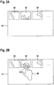
- Fig. 2A three exemplary digital microscopic tissue images 30, 32, 34 are depicted, arranged next to each other on the touch screen 14.
- the first digital microscopic tissue image 30, the second digital microscopic tissue image 32, and the third digital microscopic tissue image 34 belong to the same patient-specific image record. In other words, they are all digitized images of cuts of one or more tissue samples taken from one specific patient.
- the dots on the right of the touch screen 14 indicate that the patient-specific image record may contain additional digital microscopic tissue images.
- the arrangement of the previews of the digital microscopic tissue images 30, 32, 34 on the touch screen 14 allows the user to select a particular one of the digital microscopic tissue images, in order to start the analysis of the patient-specific image record.
- a user selection of the second digital microscopic tissue image 32 as a starting point for the analysis is illustrated.
- a user tap on the preview of the second digital microscopic tissue image 32 is indicated via a hand 50, whose index finger taps on the touch screen 14.
- the tap on the touch screen 14 is illustrated by two concentric circles 52.
- the tap on the touch screen 14 is a touch-based image selection command for selecting the second digital microscopic tissue image 32.
- the full view of the second digital microscopic tissue image 32 is depicted on the primary screen 12.
- the full view of the second digital microscopic tissue image 32 forms the initial area of interest 80 of the particular selected digital microscopic tissue image, which is displayed on the primary screen 12, as illustrated in Fig. 2C .
- Fig. 3 illustrates exemplary steps of a method of determining and displaying an area of interest of a digital microscopic tissue image in accordance with an exemplary embodiment of the invention.
- Fig. 3 illustrates an example of navigating within the particular one of the plurality of digital microscopic tissue images.
- the method steps, illustrated in Fig. 3 start from the display of the initial area of interest 80 on the primary screen 12, as illustrated in Fig. 2C .
- Fig. 3A illustrates that the touch screen 14 receives a two-finger stretch touch gesture 54 from a user. This is illustrated in Fig. 3A by the thumb and the index finger of hand 50 performing a spreading / expanding motion.
- the two-finger stretch gesture 54 is indicated with two arrows pointing away from each other.
- the two-finger stretch touch gesture 54 is part of a first class of touch gestures, which are commands for navigating within the particular one of the plurality of digital microscopic tissue images displayed on the primary screen 12.
- the two-finger stretch touch gesture 54 is in particular interpreted as a zoom in command with respect to the area of interest 80, depicted on the primary screen 12.
- Fig. 3B illustrates an updated area of interest 82, which is the result of the zoom in command with respect to the initial area of interest 80, illustrated in Fig. 2C .
- Said updated area of interest 82 is then displayed on the primary screen 12, as illustrated in Fig. 3B .
- the updated area of interest 82 is a smaller portion of the second digital microscopic image 32, as compared to the initial area of interest 80.
- a two-finger pinch touch gesture is another example of a touch gesture from the first class of touch gestures.
- the two-finger pinch touch gesture may be carried out by bringing the thumb and the index finger of the hand closer together, i.e. by carrying out a compressing / collapsing motion with the thumb and the index finger.
- the two-finger pinch touch gesture is interpreted as a zoom out command, resulting in the updated area of interest being a larger portion of the particular digital microscopic image, as compared to the initial area of interest.
- the two-finger pinch touch gesture is not shown in the Figs., but may be easily envisioned by having the arrows of Fig. 3A pointing towards each other.
- Fig. 4 illustrates exemplary steps of a method of determining and displaying an area of interest of a digital microscopic tissue image in accordance with an exemplary embodiment of the invention.
- Fig. 4 illustrates an example of navigating within the particular one of the plurality of digital microscopic tissue images.
- the method steps, illustrated in Fig. 4 start from the display of the area of interest 82 on the primary screen 12, as illustrated in Fig. 3B . While the area of interest 82 has been referred to as updated area of interest 82 in the context of Fig. 3B , because it is the result of the zoom in touch gesture of Fig. 3A , the area of interest 82 is the initial area of interest for the iteration of the method illustrated in Fig. 4 .
- Fig. 4A illustrates that the touch screen 14 receives a one-finger pan gesture 56 from a user. This is illustrated in Fig. 4A by the index finger of the hand 50 performing a lateral motion on the touch screen 14.
- the one-finger pan gesture 56 is indicated with multiple arrows pointing away from the index finger in different directions, the multiple arrows illustrating that the one-finger pan may be carried out in different directions.
- a one-finger pan in the left direction is assumed as an example of the one-finger pan gesture in the context of Fig. 4 .
- the one-finger pan touch gesture 56 is also part of the first class of touch gestures, which are commands for navigating within the particular one of the plurality of digital microscopic tissue images displayed on the primary screen 12.
- the one-finger pan touch gesture 56 is in particular interpreted as a lateral motion command with respect to the area of interest 82, which is the initial area of interest in the context of Fig. 4 , depicted on the primary screen 12.
- Fig. 4B illustrates an updated area of interest 84, which is the result of the lateral motion command with respect to the initial area of interest 82, illustrated in Fig. 3B .
- Said updated area of interest 84 is then displayed on the primary screen 12, as illustrated in Fig. 4B . It can be seen from the comparison of Fig. 3B and Fig. 4B that the image features of the depicted portion of the second digital microscopic tissue image 32 have been moved to the left.
- Fig. 5 illustrates exemplary steps of a method of determining and displaying an area of interest of a digital microscopic tissue image in accordance with an exemplary embodiment of the invention.
- Fig. 5 illustrates an example of navigating within the particular one of the plurality of digital microscopic tissue images.
- the method steps, illustrated in Fig. 5 start from the display of the area of interest 82 on the primary screen 12, as illustrated in Fig. 3B . While the area of interest 82 has been referred to as updated area of interest 82 in the context of Fig. 3B , because it is the result of the zoom in touch gesture of Fig. 3A , the area of interest 82 is the initial area of interest for the iteration of the method illustrated in Fig. 5 .
- Fig. 5A illustrates that the touch screen 14 receives a two-finger rotational motion gesture 58 from a user. This is illustrated in Fig. 5A by the thumb and the index finger of the hand 50 jointly rotating on the touch screen 14.
- the two-finger rotational motion gesture 58 is indicated with two semi-circular arrows.
- a two-finger rotational motion gesture 58 in the clockwise direction is assumed as an example of the two-finger rotational motion gesture in the context of Fig. 5 .
- the two-finger rotational motion gesture 58 is also part of the first class of touch gestures, which are commands for navigating within the particular one of the plurality of digital microscopic tissue images displayed on the primary screen 12.
- the two-finger rotational motion gesture 58 is in particular interpreted as a rotation command with respect to the area of interest 82, which is the initial area of interest in the context of Fig. 5 , depicted on the primary screen 12.
- Fig. 5B illustrates an updated area of interest 86, which is the result of the rotation command with respect to the initial area of interest 82, illustrated in Fig. 3B .
- Said updated area of interest 86 is then displayed on the primary screen 12, as illustrated in Fig. 5B . It can be seen from the comparison of Fig. 3B and Fig. 5B that the image features of the depicted portion of the second digital microscopic tissue image 32 have been rotated clockwise.
- a two-finger rotational motion gesture in the counter-clockwise direction may work in an analogous manner.
- Such two-finger rotational motion gesture in the counter-clockwise direction is also part of the first class of touch gestures.
- Fig. 6 illustrates exemplary steps of a method of determining and displaying an area of interest of a digital microscopic tissue image in accordance with an exemplary embodiment of the invention.
- Fig. 6 illustrates an example of switching from a particular one of the plurality of digital microscopic tissue images to a different one of the plurality of digital microscopic tissue images.
- the method steps, illustrated in Fig. 6 may start from any area of interest of the second digital microscopic tissue image 32, such as from the display of any of the areas of interest 80, 82, 84, 86 on the primary screen 12, as depicted in Figs. 2C , 3B , 4B , and 5B .
- Fig. 6A illustrates that the touch screen 14 receives a sideways two-finger pan gesture 60 towards the right from a user. This is illustrated in Fig. 6A by the index finger and the middle finger of the hand 50 performing a lateral motion on the touch screen 14 towards the right.
- the two-finger pan gesture 60 is indicated with an arrow pointing towards the right from the index and middle fingers of the hand 50.
- the two-finger pan gesture 60 is part of a second class of touch gestures, which are commands for switching from a particular one of the plurality of digital microscopic tissue images to a different one of the plurality of digital microscopic tissue images.
- the two-finger pan touch gesture 60 towards the right is in particular interpreted as a command to switch to a previous one of the plurality of digital microscopic tissue images.
- the two-finger pan touch gesture 60 towards the right is interpreted as a command to switch to the first digital microscopic tissue image 30.
- Fig. 6B illustrates an initial area of interest 90 of the first digital microscopic tissue image 30.
- the initial area of interest 90 of the first digital microscopic tissue image 30 is a full view of the first digital microscopic tissue image 30. It is also possible that a portion of the first digital microscopic tissue image 30 is displayed as the initial area of interest of the first digital microscopic tissue image 30.
- the last area of interest of the previous inspection of the first digital microscopic tissue image 30 may be displayed as the initial area of interest for the current inspection of the first digital microscopic tissue image 30.
- the initial area of interest 90 is then displayed on the primary screen 12, as illustrated in Fig. 6B .
- Fig. 7 illustrates exemplary steps of a method of determining and displaying an area of interest of a digital microscopic tissue image in accordance with an exemplary embodiment of the invention.
- Fig. 7 illustrates an example of switching from a particular one of the plurality of digital microscopic tissue images to a different one of the plurality of digital microscopic tissue images.
- the method steps, illustrated in Fig. 7 may start from any area of interest of the second digital microscopic tissue image 32, such as from the display of any of the areas of interest 80, 82, 84, 86 on the primary screen 12, as depicted in Figs. 2C , 3B , 4B , and 5B .
- Fig. 7A illustrates that the touch screen 14 receives a sideways two-finger pan gesture 62 towards the left from a user. This is illustrated in Fig. 7A by the index finger and the middle finger of the hand 50 performing a lateral motion on the touch screen 14 towards the left.
- the two-finger pan gesture 62 is indicated with an arrow pointing towards the left from the index and middle fingers of the hand 50.
- the two-finger pan gesture 62 is part of the second class of touch gestures, which are commands for switching from a particular one of the plurality of digital microscopic tissue images to a different one of the plurality of the digital microscopic tissue images.
- the two-finger pan touch gesture 62 towards the left is in particular interpreted as a command to switch to a next one of the plurality of digital microscopic tissue images.
- the two-finger pan touch gesture 62 towards the left is interpreted as a command to switch to the third digital microscopic tissue image 34.
- Fig. 7B illustrates an initial area of interest 92 of the third digital microscopic tissue image 34.
- the initial area of interest 92 of the third digital microscopic tissue image 34 is a full view of the third digital microscopic tissue image 34. It is also possible that a portion of the third digital microscopic tissue image 34 is displayed as the initial area of interest of the third digital microscopic tissue image 34.
- the last area of interest of the previous inspection of the third digital microscopic tissue image 34 may be displayed as the initial area of interest for the current inspection of the third digital microscopic tissue image 34.
- the initial area of interest 92 is then displayed on the primary screen 12, as illustrated in Fig. 7B .
- the meaning of the two-finger pan to the right and the two-finger pan to the left may be reversed, i.e. that the two-finger pan to the right is interpreted as a command to switch to a next one of the plurality of digital microscopic tissue images and that the two-finger pan to the left is interpreted as a command to switch to a previous one of the plurality of digital microscopic tissue images.
- the convention may be set in accordance with the particulars of the input / output system in question, in accordance with user preferences, etc.
- the second class of touch gestures may also comprise two-finger pan gestures towards the top and/or towards the bottom and/or other multi-finger pan gestures and/or other touch gestures suitable for intuitively communicating an image switching command.
- Some or all of the exemplary method steps, illustrated with respect to Figs. 2-7 may be combined in a method of determining and displaying an area of interest of a digital microscopic tissue image in accordance with an exemplary embodiment of the invention.
- the particular method steps carried out in a particular run / iteration of the method may depend on the particular touch gestures received at the touch-based input device.
- the method may behave differently depending on which particular touch gestures out of the first and second classes of touch gestures are received.
- Figs. 2-7 While the touch gestures of Figs. 2-7 are depicted to be performed with the right hand, it is understood that they may also be performed with the left hand, e.g. in the set-up of the work place of Fig. 1 .
- the hand to be used may depend on the positions of the touch screen 14 and the user.
- the touch screen 14 is depicted in Figs. 3-7 as a substantially empty screen, accepting touch gestures from the user. It is understood that the touch screen 14 may also display an image and accept touch gestures from the user. For example, the touch screen 14 may mirror the image output of the primary screen 12 and accept touch gestures on this mirrored image output. Further, the method may allow for the user to switch between the mode of accepting touch gestures, as illustrated in Figs. 3-7 , and the preview mode for selecting a particular one of the plurality of digital microscopic tissue images, as illustrated in Figs. 2A and 2B , at any point in the analysis of the patient-specific image record.
- Fig. 8 shows a work place 2 in accordance with another exemplary embodiment of the invention in a perspective view.
- the work place 2 of Fig. 8 is similar to the work place 2 of Fig. 1 .
- Like elements are denoted with the same reference numerals, and reference is made to the description thereof above.
- the work place 2 of Fig. 8 differs from the work place 2 of Fig. 1 in that the touch screen 14 is replaced with a motion sensor 22.
- the motion sensor 22 is configured to detect touchless motion gestures from a user.
- the word touchless indicates that no physical contact between the motion sensor 22 and the user, in particular between the motion sensor 22 and the user's hand, is necessary for conveying input data to the motion sensor 22.
- the motion sensor 22 comprises an upper motion sensor bar and a lower portion, which is a stand-like structure for the upper motion sensor bar and which may house circuitry for processing signals from the upper motion sensor bar.
- the motion sensor 22 has an overall cuboid outer shape and is positioned upright on the desk 4.
- the motion sensor may have any other shape and set-up that is suitable for detecting touchless motion gestures from a user.
- the motion sensor 22 is directed towards a region at or above a tabletop of the desk 4.
- the motion sensor may be directed to a region at the tabletop of the desk 4 similar to the position of the touch screen 14 in the exemplary embodiment of Fig. 1 .
- the user may conveniently and ergonomically perform touchless motion gestures that are detected by the motion sensor 22.
- the touchless motion gestures are an alternative way of inputting commands for navigating through the patient-specific image record.
- the touchless motion gestures may correspond to the touch gestures, discussed above with respect to the touch screen 14. It is also possible that additional and/or alternative gestures are accepted by the motion sensor 22. Again, the motion sensor accepts a first class of touchless motion gestures, which result in an updated area of interest of the particular one of the plurality of digital microscopic tissue images being determined and displayed, and a second class of touchless motion gestures, which result in a switching from the particular one of the plurality of digital microscopic tissue images to a different one of the plurality of digital microscopic tissue images and an initial area of interest of the different one of the plurality of digital microscopic tissue images being displayed.
- a first class of touchless motion gestures which result in an updated area of interest of the particular one of the plurality of digital microscopic tissue images being determined and displayed
- a second class of touchless motion gestures which result in a switching from the particular one of the plurality of digital microscopic tissue images to a different one of the plurality of digital microscopic tissue images and an initial area
- Fig. 9 shows an exemplary excerpt of a patient-specific text-based record 94, as may be displayed on the tertiary screen 16 in the work place 2 of Fig. 1 or Fig. 8 .
- the five input fields “Name”, “Date of Birth”, “Previous Illnesses”, “Diagnosis”, and “Medication” are shown. While the input field “Date of Birth” has three subfields for day, month, and year, which may be populated via drop down menus, the other four input fields are text fields.
- Fig. 9 further shows a cursor 96, which indicates which input field currently has the input focus, i.e. to which input field a data input would be written.
- the cursor 96 may be a blinking cursor bar, as illustrated in Fig. 9 , or a frame around a particular input field or a color highlighting of a particular input field or any other suitable marker for indicating the input field accepting data input.
- the patient-specific text-based record 94 is depicted on the tertiary screen 16 and can be accessed / populated via the keyboard 18 and the mouse 20.
- Both the keyboard 18 and the mouse 20 may be used for issuing cursor placement commands, i.e. for moving the cursor 96 to a desired position.
- both the keyboard 18 and the mouse 20 may be used for data input into the patient-specific text-based record 94.
- the user may provide data input via the drop down menus.
- the user may provide data input into text fields, drop down menus, etc.
- the keyboard 18 and the mouse 20 are used for manipulating the patient-specific text-based record 94
- the touch screen 14 / motion sensor 22 is used for manipulating the tissue image output on the primary screen 12, as discussed above.
- the accepting of touch gestures on the touch screen 14 and the ensuing modification of the tissue image output on the primary screen 12 does not interfere with the position of the cursor 96 in the patient-specific text-based record 94 and does not interfere with the readiness of the patient-specific text-based record 94 to accept data input from the keyboard 18 and the mouse 20.
- a user may input text into the patient-specific text-based record 94 via the keyboard 18.
- the user may (re)evaluate one or more of the digital microscopic tissue images by navigating through them via the touch screen 14 / motion sensor 22.
- the method / system in accordance with an exemplary embodiment of the invention may provide for an updated tissue image output on the primary screen 12 and may return the input focus to the tertiary screen right away, with the cursor 96 being at the same position as before the touch gesture on the touch screen 14 / touchless motion gesture.
- the user may continue inputting data into the patient-specific text-based record 94 in a seamless manner.
- no time-consuming re-placement of the cursor 96 and no time-consuming re-activation of the window containing the patient-specific text-based patient record 94 has to take place.
- Fig. 10 illustrates various method steps of a method of determining and displaying an area of interest of a digital microscopic tissue image in accordance with another exemplary embodiment of the invention.
- the method displays a single digital microscopic tissue image or a portion of a single digital microscopic tissue image on the primary screen 12.
- the method illustrated with respect to Fig. 10 allows for the display of multiple digital microscopic tissue images / the display of portions of multiple digital microscopic tissue images on the primary screen 12.
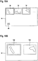
- Fig. 10A is similar to Fig. 2A . It also shows an exemplary situation at the beginning of the analysis of the patient-specific image record, when previews of some or all of the plurality of digital microscopic tissue images are displayed on the touch screen 14. Again, the first digital microscopic tissue image 30, the second digital microscopic tissue image 32, the third digital microscopic tissue image 34, and dots indicating potential further digital microscopic tissue images are depicted in Fig. 10A .
- Fig. 10A shows a multi-select mode. This multi-select mode is indicated by the cross ticked in a box 40, displayed in the left bottom corner of the touch screen 14. By ticking said box 40, the user may enter or leave the multi-select mode. It is pointed out that the election of the multi-select mode may also be accepted in other ways, such as via a user input via the mouse 20 or the keyboard 18.
- Fig. 10A depicts the exemplary situation that a user has selected both the first digital microscopic tissue image 30 and the third digital microscopic tissue image 34. This selection is indicated with respective frames around the previews of the first digital microscopic tissue image 30 and the third digital microscopic tissue image 34 on the touch screen 14. Instead of with frames, the selected digital microscopic tissue images may be indicated with other markers, such as arrows, color codes, check symbols, etc.
- the method Upon receiving the selection of the first digital microscopic tissue image 30 and the third digital microscopic tissue image 34, the method displays a first initial area of interest 190 of the first digital microscopic tissue image 30 and a second initial area of interest 192 of the third digital microscopic tissue image 34 on the primary screen 12.
- the first initial area of interest 190 is a full view of the first digital microscopic tissue image 30
- the second initial area of interest 192 is a full view of the third digital microscopic tissue image 34. This is illustrated in Fig. 10B .
- the method further assigns the input focus to one of the selected digital microscopic tissue images.
- the method assigns the input focus to the first initial area of interest 190.
- the input focus is indicated with a frame in Fig. 10B .
- the input focus may be indicated with other markers, such as an arrow, a color code, a check symbol, etc., allowing the user to know which of the displayed areas of interest has the input focus, i.e. which of the displayed areas of interest is the subject of commands given in the form of touch gestures. It is pointed out that the method may initially assign the input focus in an algorithmic manner or in a random manner.
- the first initial area of interest 190 has the input focus.
- the method determines an updated area of interest of the first digital microscopic tissue image 30 and displays said updated area of interest of the first digital microscopic tissue image 30 on the primary screen 12 at the position and with the extension of the first initial area of interest 190.
- the touch gestures of the first class of touch gestures result in updated areas of interest being displayed on that portion of the primary screen 12 that has the input focus.
- Fig. 10C illustrates the reception of a touch gesture from the second class of touch gestures.
- Fig. 10C illustrates the reception of a two-finger pan 60 towards the right.
- the method switches the input focus from the first initial area of interest 190 to the second initial area of interest 192. This is illustrated in Fig. 10D , where the first initial area of interest 190 is no longer depicted with a frame, but where the second initial area of interest 192 is depicted with a frame.
- the method determines an updated area of interest of the third digital microscopic tissue image 34 and displays said updated area of interest of the third digital microscopic tissue image 34 on the primary screen 12 at the position and with the extension of the second initial area of interest 192.
- the method illustrated with respect to Fig. 10 allows for navigating through a particular one of the plurality of digital microscopic tissue images of the patient-specific image record via the first class of touch gestures and allows for switching the input focus between different digital microscopic tissue images, displayed simultaneously on the primary screen, via the second class of touch gestures.
- the patient-specific image record may be navigated in a quick and ergonomic manner, while allowing the user to view different ones of the plurality of digital microscopic tissue images concurrently.
- the method illustrated with respect to Fig. 10 has been described in the context of the input / output system of Fig. 1 . It is also applicable to the input / output system of Fig. 8 .
- the first class of touchless motion gestures and the second class of touchless motion gestures may be used for updating the area of interest of that digital microscopic tissue image that has the input focus and for switching the input focus between displayed areas of interest of different digital microscopic tissue images, respectively.
- the initial selection of multiple digital microscopic tissue images may be carried out in another suitable way, such as by displaying an arrangement of previews on the primary screen and selecting multiple digital microscopic tissue images via the mouse 20 or the keyboard 18.
Landscapes
- Engineering & Computer Science (AREA)
- Theoretical Computer Science (AREA)
- General Engineering & Computer Science (AREA)
- Physics & Mathematics (AREA)
- General Physics & Mathematics (AREA)
- Human Computer Interaction (AREA)
- Health & Medical Sciences (AREA)
- Nuclear Medicine, Radiotherapy & Molecular Imaging (AREA)
- Radiology & Medical Imaging (AREA)
- Epidemiology (AREA)
- General Health & Medical Sciences (AREA)
- Medical Informatics (AREA)
- Primary Health Care (AREA)
- Public Health (AREA)
- Multimedia (AREA)
- User Interface Of Digital Computer (AREA)
Priority Applications (4)
| Application Number | Priority Date | Filing Date | Title |
|---|---|---|---|
| EP19209611.3A EP3822982A1 (de) | 2019-11-17 | 2019-11-17 | Verfahren zur bestimmung und anzeige eines interessierenden bereichs eines digitalen mikroskopischen gewebebildes, ein-/ausgabesystem zur navigation eines patientenspezifischen bilddatensatzes und arbeitsplatz mit einem solchen ein-/ausgabesystem |
| CN202080029372.0A CN114730240A (zh) | 2019-11-17 | 2020-11-13 | 确定和显示数字显微组织图像的感兴趣区域的方法、用于导航患者特异性图像记录的输入/输出系统和包括这种输入/输出系统的工作场所 |
| PCT/EP2020/082065 WO2021094540A1 (en) | 2019-11-17 | 2020-11-13 | Method of determining and displaying an area of interest of a digital microscopic tissue image, input / output system for navigating a patient-specific image record, and work place comprising such input / output system |
| US17/777,487 US12237070B2 (en) | 2019-11-17 | 2020-11-13 | Method of determining and displaying an area of interest of a digital microscope tissue image, input/output system for navigating a patient-specific image record, and work place comprising such input/output system |
Applications Claiming Priority (1)
| Application Number | Priority Date | Filing Date | Title |
|---|---|---|---|
| EP19209611.3A EP3822982A1 (de) | 2019-11-17 | 2019-11-17 | Verfahren zur bestimmung und anzeige eines interessierenden bereichs eines digitalen mikroskopischen gewebebildes, ein-/ausgabesystem zur navigation eines patientenspezifischen bilddatensatzes und arbeitsplatz mit einem solchen ein-/ausgabesystem |
Publications (1)
| Publication Number | Publication Date |
|---|---|
| EP3822982A1 true EP3822982A1 (de) | 2021-05-19 |
Family
ID=68609901
Family Applications (1)
| Application Number | Title | Priority Date | Filing Date |
|---|---|---|---|
| EP19209611.3A Pending EP3822982A1 (de) | 2019-11-17 | 2019-11-17 | Verfahren zur bestimmung und anzeige eines interessierenden bereichs eines digitalen mikroskopischen gewebebildes, ein-/ausgabesystem zur navigation eines patientenspezifischen bilddatensatzes und arbeitsplatz mit einem solchen ein-/ausgabesystem |
Country Status (4)
| Country | Link |
|---|---|
| US (1) | US12237070B2 (de) |
| EP (1) | EP3822982A1 (de) |
| CN (1) | CN114730240A (de) |
| WO (1) | WO2021094540A1 (de) |
Citations (5)
| Publication number | Priority date | Publication date | Assignee | Title |
|---|---|---|---|---|
| WO1999038149A1 (en) * | 1998-01-26 | 1999-07-29 | Wayne Westerman | Method and apparatus for integrating manual input |
| WO2005114369A2 (en) * | 2004-05-06 | 2005-12-01 | Apple Computer, Inc. | Multipoint touchscreen |
| US20090138800A1 (en) * | 2007-11-23 | 2009-05-28 | Mckesson Financial Holdings Limited | Apparatus, method and computer-readable storage medium for directing operation of a software application via a touch-sensitive surface |
| US20140139541A1 (en) * | 2012-10-18 | 2014-05-22 | Barco N.V. | Display with optical microscope emulation functionality |
| WO2018002776A1 (en) * | 2016-06-28 | 2018-01-04 | Koninklijke Philips N.V. | System and architecture for seamless workflow integration and orchestration of clinical intelligence |
Family Cites Families (8)
| Publication number | Priority date | Publication date | Assignee | Title |
|---|---|---|---|---|
| KR100825773B1 (ko) * | 2005-08-23 | 2008-04-28 | 삼성전자주식회사 | 방향 추정 방법 및 장치 |
| JP2008306799A (ja) * | 2007-06-05 | 2008-12-18 | Canon Inc | 電源装置、電源装置の制御方法、プログラム、及び記憶媒体 |
| US8463741B2 (en) * | 2009-09-04 | 2013-06-11 | Omnyx, LLC | Digital pathology system |
| DE102012220195B4 (de) | 2012-11-06 | 2021-03-04 | Leica Microsystems Cms Gmbh | Widefield-Mikroskop oder Konfokalmikroskop |
| KR101529033B1 (ko) * | 2014-02-14 | 2015-06-18 | 크루셜텍 (주) | 극소 센싱 영역을 포함하는 전자 장치 및 이의 지문 정보 프로세싱 방법 |
| DE102014102425B4 (de) | 2014-02-25 | 2018-06-28 | Carl Zeiss Meditec Ag | Mikroskopsystem und Mikroskopieverfahren unter Verwendung digitaler Marker |
| DE102018104704A1 (de) | 2018-03-01 | 2019-09-05 | Carl Zeiss Microscopy Gmbh | Digitales Mikroskop und Verfahren zum Verändern einer Vergrößerung eines digitalen Mikroskops |
| DE102018107033A1 (de) * | 2018-03-23 | 2019-09-26 | Leica Microsystems Cms Gmbh | Mikroskopsystem und Verfahren zur Steuerung eines solchen Mikroskopsystems |
-
2019
- 2019-11-17 EP EP19209611.3A patent/EP3822982A1/de active Pending
-
2020
- 2020-11-13 US US17/777,487 patent/US12237070B2/en active Active
- 2020-11-13 WO PCT/EP2020/082065 patent/WO2021094540A1/en not_active Ceased
- 2020-11-13 CN CN202080029372.0A patent/CN114730240A/zh active Pending
Patent Citations (5)
| Publication number | Priority date | Publication date | Assignee | Title |
|---|---|---|---|---|
| WO1999038149A1 (en) * | 1998-01-26 | 1999-07-29 | Wayne Westerman | Method and apparatus for integrating manual input |
| WO2005114369A2 (en) * | 2004-05-06 | 2005-12-01 | Apple Computer, Inc. | Multipoint touchscreen |
| US20090138800A1 (en) * | 2007-11-23 | 2009-05-28 | Mckesson Financial Holdings Limited | Apparatus, method and computer-readable storage medium for directing operation of a software application via a touch-sensitive surface |
| US20140139541A1 (en) * | 2012-10-18 | 2014-05-22 | Barco N.V. | Display with optical microscope emulation functionality |
| WO2018002776A1 (en) * | 2016-06-28 | 2018-01-04 | Koninklijke Philips N.V. | System and architecture for seamless workflow integration and orchestration of clinical intelligence |
Non-Patent Citations (1)
| Title |
|---|
| ANONYMOUS: "Touchpad - Wikipedia", 2 October 2019 (2019-10-02), pages 1 - 5, XP093153018, Retrieved from the Internet <URL:https://en.wikipedia.org/w/index.php?title=Touchpad&oldid=919278408> * |
Also Published As
| Publication number | Publication date |
|---|---|
| US12237070B2 (en) | 2025-02-25 |
| CN114730240A (zh) | 2022-07-08 |
| US20230207103A1 (en) | 2023-06-29 |
| WO2021094540A1 (en) | 2021-05-20 |
Similar Documents
| Publication | Publication Date | Title |
|---|---|---|
| EP2249708B1 (de) | Elektronisches anzeigesystem mit doppelmonitor | |
| US10444960B2 (en) | User interface for medical image review workstation | |
| US10324602B2 (en) | Display of 3D images | |
| US20090021475A1 (en) | Method for displaying and/or processing image data of medical origin using gesture recognition | |
| Vogel et al. | Hand occlusion on a multi-touch tabletop | |
| Uddin et al. | HandMark Menus: Rapid command selection and large command sets on multi-touch displays | |
| US20100235786A1 (en) | Enhanced 3d interfacing for remote devices | |
| US20080114614A1 (en) | Methods and systems for healthcare application interaction using gesture-based interaction enhanced with pressure sensitivity | |
| Vogel et al. | Hand occlusion with tablet-sized direct pen input | |
| RU2672624C2 (ru) | Графический интерфейс пользователя, обеспечивающий функциональные возможности виртуального супермасштабирования | |
| US20250328002A1 (en) | Systems and methods for processing electronic images of pathology data and reviewing the pathology data | |
| Wagner et al. | Gaze, wall, and racket: Combining gaze and hand-controlled plane for 3D selection in virtual reality | |
| Weiss et al. | Radiologist assessment of PACS user interface devices | |
| US12237070B2 (en) | Method of determining and displaying an area of interest of a digital microscope tissue image, input/output system for navigating a patient-specific image record, and work place comprising such input/output system | |
| US11994665B2 (en) | Systems and methods for processing electronic images of pathology data and reviewing the pathology data | |
| Reibert et al. | Multitouch interaction with parallel coordinates on large vertical displays | |
| WO2025117406A1 (en) | Input device with rotatable control knobs | |
| Stuij | Usability evaluation of the kinect in aiding surgeon computer interaction | |
| Motti et al. | Touchscreen interaction of older adults: a literature review | |
| Kim et al. | ShifTable: A natural remote target-selection technique on large displays | |
| Nigay | Bimanual input for multiscale navigation with pressure and touch gestures | |
| HK1201709B (en) | Twin-monitor electronic display system comprising slide potentiometers |
Legal Events
| Date | Code | Title | Description |
|---|---|---|---|
| PUAI | Public reference made under article 153(3) epc to a published international application that has entered the european phase |
Free format text: ORIGINAL CODE: 0009012 |
|
| STAA | Information on the status of an ep patent application or granted ep patent |
Free format text: STATUS: THE APPLICATION HAS BEEN PUBLISHED |
|
| AK | Designated contracting states |
Kind code of ref document: A1 Designated state(s): AL AT BE BG CH CY CZ DE DK EE ES FI FR GB GR HR HU IE IS IT LI LT LU LV MC MK MT NL NO PL PT RO RS SE SI SK SM TR |
|
| STAA | Information on the status of an ep patent application or granted ep patent |
Free format text: STATUS: REQUEST FOR EXAMINATION WAS MADE |
|
| 17P | Request for examination filed |
Effective date: 20211015 |
|
| RBV | Designated contracting states (corrected) |
Designated state(s): AL AT BE BG CH CY CZ DE DK EE ES FI FR GB GR HR HU IE IS IT LI LT LU LV MC MK MT NL NO PL PT RO RS SE SI SK SM TR |
|
| STAA | Information on the status of an ep patent application or granted ep patent |
Free format text: STATUS: EXAMINATION IS IN PROGRESS |
|
| 17Q | First examination report despatched |
Effective date: 20240423 |